Colour ~ la te s - Springer978-94-011-0743-3/1.pdf · cover-uncover test 22 ocular motility 33...
24
Colour _____ _ 179
Transcript of Colour ~ la te s - Springer978-94-011-0743-3/1.pdf · cover-uncover test 22 ocular motility 33...

Colour ~_la_te_s _____ _
179

WEAKENING OF THE MEOIAL RbCTUS MUSCLE
Plate I. Recession
I. The mosquito forceps is applied at the limbus.
J. Teno" s capsule is opened making a first buttonhole.
5. The squim hook is film ed and bulges OL/t beloll" the 11111 cle.
2. The conjllnctiva is Cllt perpendicular to the plica alld at the IIpper border of the mil cle.
4. ITltroductioll of the sqlliTlt hook ill the woulld with the tip directed IIpwards.
6. The second bl/ttoTlhole is made.
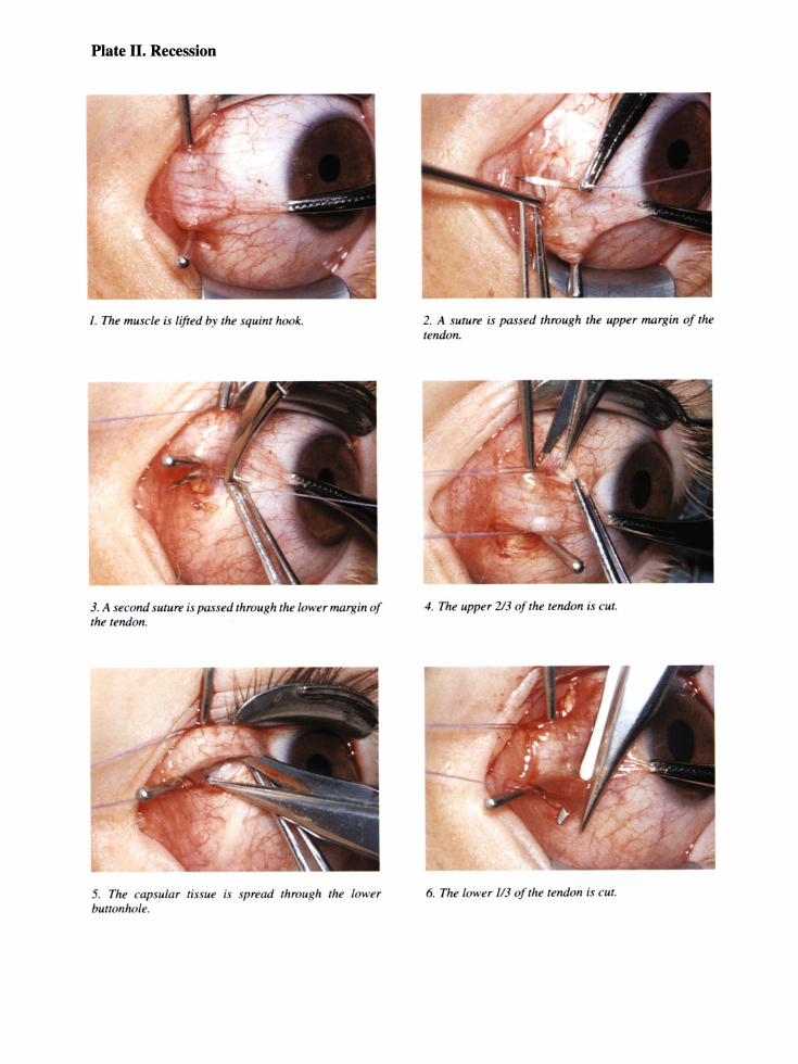
Plate II. Recession
I. The muscle is lifted by the squint hook.
3. A second suture is passed through the lower margin of the tendon.
5. The capslliar tissue is spread through the lower billion hole.
2. A sUlure is passed through the upper margin of the tendon.
4. The upper 213 of the tendon is cUI.
6. The lower 1/3 of the tendon is CUI.

Plate III. Recession
I. The tendon is free from the sclera.
3. The SUTllre is knoTTed over the 5 mm spawla.
5. A 5 mm hang back loop connects the tendon with the original insertion.
2. The needle is pllt in the sclera at fhe lower end offhe original insertion.
4. The suWre is cuI close 10 Ihe knol.
6. The cross handledforceps.

Plate IV. Recession with a loop
I. The cross handled forceps opens the 1V0und.
3. The swure is knotted over a probe.
5. The loop allows further retraction of the muscle.
2. The needle is put into the sclera at 5 mmfrom the original insertion.
4. The suture is cw near the knot.
6. The conjunctiva is not sl/tured: the bwtOnholes close spontaneously.

Plate V. Recession with a secondary loop
I. Schematic view of a ecol/dary loop placed on a pseudotendon.
3. Tile inner surface of tile pseudotendon.
5. The sutures are knotted over a probe.
2. The squint hook underneath the pseudotendon.
4. Tire outer urface of the pseudotendon.
6. The secondary loop allows the muscle to retract further back.
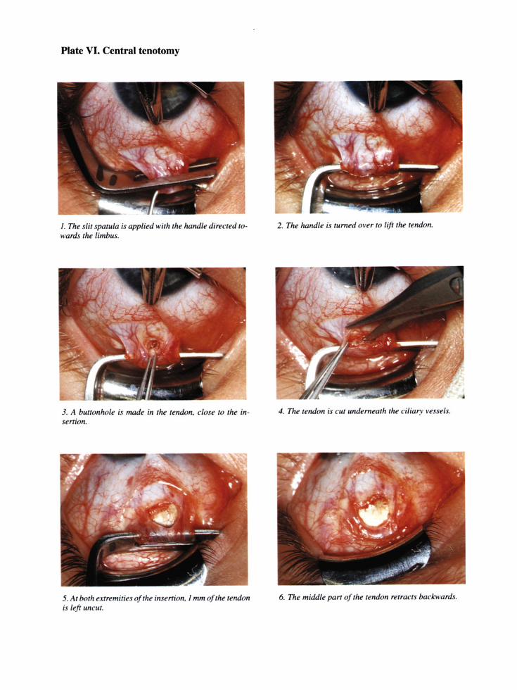
Plate VI. Central tenotomy
I . The slit spatula is applied with the handle directed towards the limbus.
3. A buttonhole is made in the tendon. close to the insertion.
5. At both extremities o/the insertion. I mm o/the tendon is left LlnCIlf.
2. Tile handle is filmed over to lift the tendon.
4. Tile tendon is CIIf underneath the ciliary vessels.
6. The middle parr 0/ the tendon retracts backwards.

DESAGITTALISATION OF THE INFERIOR OBLIQUE MUSCLE
Plate VII. Anteroposition
J. The mosquito forceps is applied at the lower-temporal side of the limbu .
3. The conjunctiva is cut at I emf rom the limbus. belWeen the inferior and lateral rectus muse/e.
5. The capsule is lifted and the squint hook is introduced into the wOllnd.
2. The weight of the forceps rums the eye up and nasally.
4. Tenon s capsule is opened.
6. The hook is pressed again t the tarsus. keeping the cap IIle lifted and the inferior obliqlle with il.

Plate VIII. Anteroposition
I . The muscle is laken near Ihe infe rior reClIIs muscle.
3. A Jameson squinl hook i s put undenreath Ihe inferior reClus mllscle.
5. Th e allIerior pari of lite muscle is CUI. all' lite narrow lendinol/ edge.
2. Tlte squiTII hook pulls the muscle downwards. exposing Ihe while Iriangle.
4. The Slllure is placed al the eqlwlor of the globe.
6. The poslerior p art is CIII wilh the scissor direcled t em· poral/y.

Plate IX. Anteroposition
I. Inspection 0/ the external perimysium thar has to be illfact.
3. Schematic view o/the ameropositioned mllscle.
5. A econd Sl/tllre is not necessary as the mllscle lies against the sclera.
2. The Slitllre is passed throllgh the allferior tip of the mllscle.
4. The sllwre is clit.
6. The conjllnctiva is lIot slitllred.

Plate X. Posterior tenotomy and disinsertion
I. This series i a mirror image from the previous one. The muscle is cut just behind the forcep .
3. The disinsened inferior oblique muscle protrude through Tenon s capsule.
5. A buried knot i applied.
2. The posterior pan of the muscle.
4. A suture is passed through the cap ule at both sides of the muscle stump.
6. The capsule covers the muscle stump.
DESAGITTALISATION OF THE SUPERIOR OBLIQUE MUSCLE
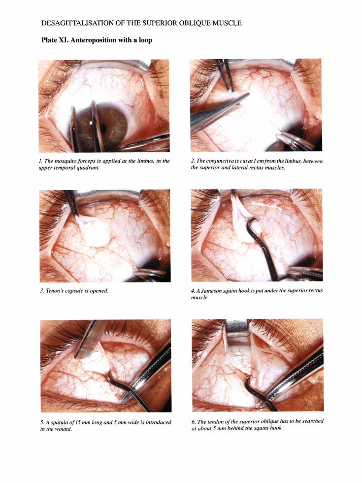
Plate XI. Anteroposition with a loop
I. The mosquito /orcep is applied at the limbus. in the upper temporal quadrant.
3. Tenon s capsule is opened.
5. A spatula 0/15 mm Ion and 5 mm wide is introduced in the wound.
2. The conjunctiva is cut at I cm/rom the limbus. between the superior and lateral rectus muscles.
4. A Jameson squint hook is put under the superior rectus muscle.
6. The tendon o/the superior oblique has to be searched at about 5 mm behind the squint hook.

Plate XII. Anteroposition with a loop
I. The tendon can be taken by grasping backwards over the sclera with theJorceps.
3. A small squim hook is pili benveen the tendon and the sclera.
5. The poim oj the hook i moved posteriorly.
2. The anterior tip oJthe tendon is caught.
4. The hook is pushed underneath the superior recllls until it touches the na al orbital wall.
6. The tendon oj the superior oblique muscle is hooked.

Plate XIII. Anteroposition with a loop
I. Tire Ileedle is put ill tlte clera at tire alllerior tip of tire insertion of the uperior oblique muscle.
3. The posterior part i sti/l attached to tlte clera.
5. The tendon is cut and 1I0mlO/ly does 1101 retract.
2. The alllerior part of the telldon is cut.
4. Thi posterior part is cm with the scissors directed temporally.
6. The Slllllrt! is plll at tire allterior tip of tile telldoll.

Plate XIV. Anteroposition with a loop
I. The suture is turned around the squint hook under the superior reclll muscle and knotted.
3. A squint hook is slid along the original insertion 10
check whether the whole tendon is cut.
5. The tendo" is pu hed backwards to avoid reattachment anterior 10 the equator of the g lobe.
2. The Slllure is CIII near the knot.
4. The tendon is caught between the superior rectus and the g lobe.
6. The eye is pushed up and I/a ally 10 stretch the tendon of the superior oblique muscle.

Plate XV. Posterior tenotomy
I. This series is presented mirrorwise to the previou ones.
3. Nibbling the remaining parr of the tendon.
5. The hook is slid along the illsertion 10 make sure that the whole tendon is CI4I.
2. The points of the scissors are directed temporally.
4. The posterior parr of the tendon is held by a forceps showing the severed part of the tendon.
6. The globe is tllmed up and nasally to stretch the tendon of the silperior obliqlle nm cle.

WEAKbNINti UF THb INl<bl<.lUl<.l<.bLTUS MUSLLb
Plate XVI. Recession
I. The ciliary ve el are taken into the ullire .
3. CUlling this junction.
5. CUlling tire ciliary vessels of a vertical rectus lead 10
abundalll bleeding.
2. Tire inferor oblique adheres in the deptlr to the inferior reCIliS. Note the illlenr/U cular membranes.
4. The two muscle are eparatedfrom each other.
6. This bleeding i slOpped by pre sing tire muscle slIimp against the globe.
A-eso/exotropia 54 A-esotropia 52 A-pattern 4,42,46-51,140
treatment 134, 137 horizontal effect of vertical
muscle surgery 125 torticollis 20
Abnormal retinal correspondence 33
residual angle 89, 146 AC/ A ratio 78
tonic accommodation 84 Accommodation overdrive 78 Accommodation, amplitude 77
convergence excess, retinos-copy 63
fast phasic 77-81 slow tonic 77-81 AC/ A ratio 84 spasm of 75
Accommodative convergence 5,7, 47,78-81,83-86
Accommodative esodevia-tion 83-86
Accommodative miosis 84 Accommodative squint 76-81,83 Adduction excess 130
head tum 17 Maddox cross 32
Adhesions 122, 127, 129 diplopia 167 limitation of duction 128, 149,
155, 163 traction on the eyelid 176
Alphabetical incomitances 46-51, 77
binocular vision Alphabetical syndrome 5, 33 Alternate cover test 26
ocular motility 33 vertical deviation 136
Alternating squint 51
Index
postoperati ve treatment 71-72 Amblyopia 127, 140, 167
anamnesis 59-60 anisometropia 70 assessment 59-62 bilateral 85 corneal reflex 26 cover-uncover test 26,61 deprivation 67,75 hypoaccommodation 84 diplopia 167 fixation 62 management 69-73 microtropia 60 occlusion, age 70 postoperative treatment 71-73 relapse 72 reverse 69 surgery 131
Ametropia 63,167 hypoaccommodation 84 management 73-75
Anaesthesia 91-97 adults 96 anxiolytic 94 children 95 perioperative management 92 postoperative period 97 preoperative management 93
Anaesthetic system 94 Anisometropia 70,74 Antero-posterior position of the
eye 171-173 aetiology 171-172 treatment 172-173
Anteroposition of the inferior oblique muscle 113-114,133
Anteroposition with a loop of the superior oblique muscle 118-119, 134
Anteroposition 1 effect 125
197
Anti-Trendelenburg position 92 Arterial blood pressure 92 Asthenopic complaints 73,81
exophoria 146 phasic system 84
Astigmatism 63,74-75 Atropine, anaesthesia 94 Atropine-sulphate, retinoscopy 63 Atropinisation 71, 170
Barraquer eye speculum 99 Barraquer needle holder 100 Bielschowsky's head tilt
test 42-45, 77 Bifoveal fixation, photoscreen
ing 67 Binocular comfort 146 Binocular vision 5,7-9,76, 146
exodeviation 90 examination 26-30
Bleeding anaesthetic 91 surgical 91
Blepharostat 99 surgery of the superior
oblique 116 Botulinum toxin 171 Brown's syndrome, torticollis 17 Bruch's membrane 65 Bruckner test, photoscreening 68 Buttonhole incision 100, 108
Calliper, fork 101 Canthus 102,107,161,168
limitation of duction 153 Catgut 101 Central tenotomy 109-11 0, 131 Changes of the antero-posterior
position of the eye 171-173 Check ligaments 49, 124, 149
limitation of duction 128, 160 progressive brake 122

198
recession 104 recurrence 138 vertical rectus muscle 124
Ciliary vessels 107, 109 Comitant RVD 53 Complications 149-177 Concomitance 4 Conjunctiva 162
shrunken 150, 164 Consecutive esotropia 5
treatment 145-146 Consecutive exotropia 5
treatment 146-147 Convergence excess 63, 73
exercises 73, 90 Convergence
accommodation 77-81 amplitude 77 fast phasic 77-81 insufficiency 81 retinal disparity 78 slow tonic 77-81
Corneal reflex 28,31,61 cover-uncover test 22 ocular motility 33
Cover test 25-26,61 alternate 26
Cover-uncover test 25,61 Cyclodeviation 4 Cycloplegics,
atropine 63 short acting 63
Cyclovertical imbalance 2 accommodative squint 83
Decussation 5-6, 124, 166-167 Depression in adduction 34-39
treatment 134 Desagittalisation 8,110--115,125,
136 four obliques 134-135 inferior oblique muscle 51-57,
133, 136 superior oblique muscle 52-57,
134,136 Diagonal deviations, primary
surgery 133-135 Diagonal incomitances, examina
tion 34-49 Diagonal overcorrections, treat
ment 147 Diagonal undercorrections,
treatment Diplopia
143-144
fixation switch 167,170 overcorrection 5,167 persistent 167 postoperative 166-171 transient 166-167 treatment 169-171
Disinsertion of the inferior oblique muscle 114, 133
effect 126 Disinsertion of the superior oblique
muscle 119, 134 effect 126
Disparity, peripheral 79 Dissociated vertical deviation
(DVD) 32,36,41-42 treatment 138
Dominant eye 24-25,69,142-146, 167
Double interaction accommodation -convergence 77-81
Duane's retraction syndrome, head tum 16-17
Elevation in adduction examination 34-39 treatment 133-134
Emmetropia 78,81 tonic vergence 78
Emmetropisation 85 Endotracheal intubation 94 Enophthalmos 103, 156, 171-172
resection 103 Epicanthus 103, 141 Equator of the globe 113,157,160,
171 Eso-/exotropia, treatment 132-133 Esophoria, hypermetropia 78 Esotropia 5,7
consecuti ve, indications for surgery 145-146,170
indications for surgery 130-133 management 83-86 miotics 86, 89 residual, treatment 142 role of hypermetropia 76-81
Examination, methods 13-68 Excyclodeviation 4
head tilt 15 Excyclotropia 4-5,21 Exodeviation
management 90 postoperative diplopia 90
Exodisparity, vergence mechanism 78
Exophoria emmetropia 79 relative 79
Exophthalmos 171-172 Exotropia,
Index
hemiretinal suppression 7 tendon of the superior oblique
muscle 131 Exotropia, consecutive 146, 169
surgery of the accommodatif component 86
tonic convergence 124 Exotropia, indications for sur
gery 131-132 residual 143
Eyelids, traction upon 174-177
Fixation disparity 78-79,81,84 Fixation in adduction 130 Fixation switch diplopia 167, 170 Fixation,
amblyopia 62 bifoveal, photoscreening 67 bifoveal, amblyopia 73 central 62
Fixation eccentric 26, 62 surgery 70
Fixation, passive 62 Fixation, pattern 62 Fiurbiprofen 158 Footplate insertion 109 Forced duction test 129, 154 Forceps,
crossed handle 100, 109 Halstead mosquito 99, 106, 115 straight tissue 100 suture-fixation 100
Fork 101 Fundal reflex, photoscreen
ing 64-68 Fusion 1,26-27,78
head posture 20--21 Fusion motor 4, 24, 27
DVD 138 horizontal muscle surgery 124 orthophorisation 49,128,140 overcorrection 124
Fusion

Index
sensory 2, 27 vergence 77
Glasses 73-75 anisometropia 74 astigmatism 74-75 deprivation amblyopia 75 esodeviations, manage-
ment 76-81 myopia 75 negative, exodeviation 90 orthophorisation 75 prismatic imbalance 74 psychological impact 85
Haematomas 92 Halotane 96 Halstead mosquito forceps 98-99,
106, 115 Head inclination 19 Head tilt 14-15 Head turn 16-19 Hemidecussation line, diplopia 5,
166-167 Hemiretina,
nasal 28 temporal 28
Hemiretinal suppression 5-7, 166-167
overcorrection 124 Hering's law 50, 123-124, 163
desagittalisation 138 DVD 138 limitation of duction 142
Hess-Lancaster test 9,33 Heterophoria 72 Heterotropia 7 Hirschberg's test 22 Horizontal deviations, treat-
ment 130-133 Horizontal incomitances,
examination 46-51 Hirschberg 15
Horizontal overcorrec-tions 145-146
Horizontal undercorrec-tions 141-143
Hypercarbia 92 Hypermetropia 74,83 167
corrected 78 latent 75,84 manifest 84
role in estropia 76-81 uncorrected, tonic vergence 78
Hypertropia 129, 136, 147 amblyopia 138
Hypo-accommodation 67,73,84 deprivation amblyopia 84 tonic 79
Hypotropia 128, 137, 147 Hypoventilation 92, 96
Incision buttonhole 100, 108 conjunctival 110 limbal 102
Incomitances 1 examination 34-51 latent 9,33 lateral 49-51 paralytic 77
Incyclotropia 2-4,21 Indications for primary sur
gery 127-138 Indications for secondary sur
gery 138-141 Indomethacine 158 Induction
gaseous 96 intravenous 96
Infantile esotropia, torticollis 15 Inferior oblique muscle 2-5
anteroposition 113-114 desagittalisation 51-57 disinsertion 114 hyperfunction 34-36 hypofunction 36-39 overcorrection 57 paresis, postoperative 155 posterior myotomy 114 pseudoparesis 15 surgery 11 0-115 undercorrection 56
Inferior rectus muscle, overcorrec-tion 58
Insertion, footplate 109 Instruments 97-101 Intermittent exotropia,
reversed prism test 60 Intermittent exotphoria, reversed
prism test 60 Intermittent squint, occlusion 61 Intraocular infection 101 Intraorbital tissue 110
Intrathoracic pressure 92 Intubation 92
Javal 81
Keratometer, Javal's 63
Lang two-pencil test 29 Latent deviation, cover-uncover
test 26 Lateral incomitances 49-51 Leash effect 121 Leiden school 83-86 Limbal incision 102
199
Limbal margin, ocular motility 33 Limitation of duction,
adhesions 126, 128 aetiology 149-152 check ligaments 128,149 diagnosis 140,153-157 inferior oblique,
pseudoparesis 126 inferior oblique, reversed
action 126 postoperati ve 149-165 resection 103, 158 recession 104, 124 surgically induced 131 torticollis 16, 18 treatment 157-165 vertical overcorrection 148
Lockwood's ligament surgery of inferior oblique 110,
113 surgery of vertical muscle 124 Loop 101 Loop probe 10 1
Maddox cross 30, 130, 141 Maddox rod, ocular motility 33, 39 Medial rectus muscle, spasm 47 Microtropia 60,70, 141
diplopia 166 Miotics 73,85,88-89, 141
postoperative treatment 89 preoperative treatment 88
Mobilisation of the eyes 106 Mosaic perception 27 Motor fusion reflexes 4, 24, 27
horizontal muscle surgery 124 DVD 138 orthophorisation 49, 128, 140
200
overcorrection 124 Muscle hook,
handle 101 Jameson 100 von Graefe 100
Muscle, slipped 149, 160, 173 Myopia 75, 167
amblyopia 60 exotropia 90
Needle holder 100 Needles, spatulated 101 Non-dominant eye 23-25,142-146 Non-steroidal anti-inflammatory
drops 158 Nystagmus,
latent 62 torticollis 17, 22
Objective angle of deviation 28 examination 30-32
Occlusion 61,75, 166 6/1 69 7/0 69 anisometropia 70 alternate 69-73 anti suppressive 72 diagnostic 133 ortho-or microtropia 70 part-time 69-73 postoperative 71-73 torticollis 14 total 69-73
Ocular dominance 22-25,128 Ocular motility schemes 51-59
V-esotropia 51 A-esotropia 52 X-esotropia 52 V -orthotropia 52 comitant RVD 53 V -exotropia 53 A-eso/exotropia 54 Aeso 54 exo X-A esotropia 55 X-V exotropia 56 undercorrection of the inferior
oblique muscle 56 undercorrection of the superior
oblique muscle 57 overcorrection of the inferior
oblique muscle 57 overcorrection of the superior
oblique muscle 58 overcorrection of the inferior
rectus muscle 58 paresis of the superior oblique
muscle + V -pattern 58 paresis of the superior oblique
muscle + A-pattern 59 stamp 51
Ocular motility, examina-tion 33-59
Oedema 106 Ophthalmoscope 62 Optic disc 62 Optomotor reaction 6, 7 Orbital wall 116 Ortho-microtropia 70 Orthophoria 78 Orthophorisation 29, 75 Orthoptic exercises 166 Orthotropia 7,70, 131 Otago-photoscreener 64 Overcorrection,
diplopia 167 progressive brake 140
Overcorrections, treat-ment 145-148,169-171
Palpebral fissure 133,174--175 Partially accommodative squint 81 Penalisation 170 Perimysium 105, 149 Peripheral disparity 79 Persistent diplopia,
causes 167 treatment 169
Phasic system 77-81 Phasic
accommodation 77-81 convergence 77-81
Phospholine iodide 88-89 Photorefraction 64, 84 Photophobia, resection 103 Photo screening 64--68
abnormal 66 borderline 66 clinical applications 67-68 normal 66 optical principles 65-66
Plica semilunaris, resection 103 Posterior fixation suture 128 Posterior foot 144
surgery of inferior oblique 110
Index
Posterior myotomy of the inferior oblique muscle 114, 133
effect 125 Posterior tenotomy of the superior
oblique muscle 119,134 effect 125
Postoperative diplopia 166-171 aetiology 166-167 diagnosis 167-169 treatment 169-171
Prism cover test 33 Prism test
4 dioptre 22 15 dioptre 22-25 15 dioptre, amblyopia 60
Prismatic imbalance 74, 170 Prisms 170 Progressive brake 8, 103, 123
resection, recession 121-122 check ligaments 122
Protractor of the eye 103, 171 Pseudoparalysis both elevators,
torticollis 19,21 Pseudoparalysis oblique muscle,
postoperative 147 Pseudoptosis 176 Pseudostrabismus, resection 103 Pseudo tendon 109, 131, 144 Ptosis 177 Pupillary apertures, photoscreen
ing 64 Purely accommodative and refractive
squint 81
Real vertical deviation (RVD) 32, 38,41
primary surgery undercorrection
135-138 145
overcorrection 147 Recession with a loop 108-109 Recession,
rectus muscles 104--110 horizontal recti, amount 130 progressive brake 122, 124 vertical recti, amount 138
Redisinsertion 144 Refraction 63-68 Resection 102-104
progressive brake 122 surgical effect 121
Residual esotropia, treatment 142 Residual exotropia, treatment 143

Index
Retina, decussation 6 temporal half 6, 90
Retinal disparity 78-81, 83-86, 90 Retinal haemorrhages in
neonates 77 Retinal rivalry 26 Retinoscopy 63, 75 Retractor of the eye 103, 171 Retractor of the lower eyelid 105 Reversed prism test 24-25, 60
Sagittalisation of the oblique muscles 2-5, 135
Sagittalisation 2-9 accommodative squint 83
Scar tissue 102, 164 spring 100 straight Stevens-Sevrin
tenotomy 100 Sclera 101,108
Secondary insertion 142 Secondary loop 142 Secondary surgery 138-148 Secondary surgery 140 Self-adhering drape 97 Semicircular canals 157 Sensory cyclofusion 2 Slipped muscle 140, 149, 173
treatment 160 Sodiumhyaluronate 159, 177
Spatula 100,116 handle 101
Split spatula 109 Squint
accommodative component I, 76-81,83-86
alternating 51 intermittent 61
Squint, partially accommodative 81 glasses 86
Squint, phasic accommodative 81 Squint, purely accommodative and
refracti ve 81 Stenopeic effect 84 Sterno-cleido-mastoid muscle,
torticollis 21 Steroid drops 158 Strabismus, unilateral, postoperative
treatment 72-73 Streak-retinoscope 63 Subcapsular recession 108, 142 Succinylcholine 96
Superior oblique muscle + A-pattern, palsy 59
Superior oblique muscle + V -pattern, palsy 58
Superior oblique muscle, anteroposition with a
loop 118-119 desagittalisation 52-57 disinsertion 119 head turn 17 hyperfunction 35-39 hypofunction 34-39 overcorrection 58 palsy 39,42-43,49,77,83 palsy, accommodative squint 83 posterior tenotomy 119 sagittalisation 4 tendon 118 undercorrection 57
Suppression 8, 90, 131 amblyopia 72 central 22, 27, 127 central, fixation disparity 79 central, postoperative
diplopia 166-167 central, tonic system 84 hemiretinal 5-7,169 evolution 29 limits 29 retinal disparity 73, 83
Surgery age 127 anteroposition of the inferior
oblique muscle 113-114 anteroposition of the superior
oblique muscle 118-119 atraumatic 105 central tenotomy 109-110 complications 149-177 desagittalisation 110-115 diagonalovercorrections 147 diagonal undercorrec-
tions 143-144 disinsertion of the inferior oblique
muscle 114 disinsertion of the superior
oblique muscle 119 effects of horizontal sur-
gery 121-124,131 effects of oblique muscle
surgery 125-127, 133 effects of vertical muscle
surgery 124-125 horizontal deviations, specific
guide-lines 130-133 horizontal overcorrec-
tions 145-146 horizontal undercorrec-
201
tions 141-143 indications for diagonal devia
tions 133-135 indications for horizontal
deviations 130-133 indications for primary sur
gery 127-138 indications for secondary
surgery 138-141 inferior oblique muscle 110-115 limitation of duction 149-165 loop recession 108-109,163 oblique muscles 110-120 overcorrections 145-148 posterior myotomy of the inferior
oblique muscle 114 posterior tenotomy of the superior
oblique muscle 119 recession with a loop 108-109,
124 rectus muscles 106-11 0 redisinsertion 144 secondary insertion 142 secondary loop 142 slipped muscle 149 smooth 106 superior oblique
muscle 115-120 timing, visual acuity 70 traction on the eyelids 174-177 traumatic 158 undercorrections 141-145 vertical overcorrec-
tions 147-148 vertical undercorrections 145
Surgical techniques 102-148 treatment 91-148
Sutures 101, 106 Synoptophore 27,76
RVD 136 surgery 141-142
Tarsus, surgery of inferior oblique 111,113
Tenon's capsule 108, 110-111

202
Tight lateral rectus 173 Tonic accommodative squint 81 Tonic system 77-81,90 Tonic,
accommodation 77-81 convergence 77-81
Torsional imbalance 2 Torsional incomitances, examina
tion 42-45 Torticollis 13-22
aetiology 14-22 analysis 13 limitation of abduction 145
Traction on the eyelids, aetiology 174 treatment 175-177
Traction suture 99, 106 Transient diplopia
causes 166-167 treatment 169
Trial frame 63
Undercorrections, treatment 141-145
Unilateral strabismus, postoperative
treatment 72-73
V-esotropia 51 V-exotropia 53 V -orthotropia 52 V-pattern 3,44-51,137,140
horizontal effect of vertical muscle surgery 125
V -pattern, surgical treat-ment 133-134
Venous congestion 92 Ventilation 96 Vertical deviation
dissociated (DVD) 32,36,41, 138
real (RVD) 32,38,41, 135-138, 145, 147
Vertical deviation, treatment 135-138, 145
indications 136 Vertical incomitances
accommodative squint 83 examination 39-42 role 76-77,83
Verticalovercorrections,
Index
treatment 147-148 Vertical undercorrections, treat
ment 145 Vicryl 101 Visual acuity 69-75
anisometropia 74 assessment 60 astigmatism 74-75 hypermetropia 74
Visual axis 2, 125 Vitreo-retinal interface 65 Vortex vein
surgery of the inferior oblique 110, 112
surgery of the superior oblique 117
White triangle, surgery of the inferior oblique 112, 114
X-A esotropia 55 X-esotropia 52 X-pattern 46-48, 134-135, 137

Monographs in Ophthalmology
1. P.C. Maudgal and L. Missotten (eds.): Superficial Keratitis. 1981 ISBN 90-6193-801-5 2. P.F.J. Hoyng: Pharmacological Denervation and Glaucoma. A Clinical Trial Report with Guanethidine
and Adrenaline in One Eyedrop. 1981 ISBN 90-6193-802-3 3. N.W.H.M. Dekkers: The Cornea in Measles. 1981
4. P. Leonard and J. Rommel: Lens Implantation. 30 Years of Progress. 1982 ISBN 90-6193-803-1
ISBN 90-6193-804-X 5. C.E. van Nouhuys: Dominant Exudative Vitreoretinopathy and Other Vascular Developmental Disorders
of the Peripheral Retina. 1982 ISBN 90-6193-805-8
6. L. Evens (ed.): Convergent Strabismus. 1982 ISBN 90-6193-806-6
7. A. Neetens, A. Lowenthal and J.J. Martin (eds.): The Visual System in Myelin Disorders. 1984 ISBN 90-6193-807-4
8. H.J.M. VOlker-Dieben: The Effect of Immunological and Non-Immunological Factors on Corneal Graft Survival. A Single Centre Study. 1984 ISBN 90-6193-808-2
9. J.A. Oosterhuis (ed.): Ophthalmic Tumours. 1985 ISBN 90-6193-528-8 10. O. van Nieuwenhuizen: Cerebral Visual Disturbance in Infantile Encephalopathy. 1987
ISBN 0-89838-860-0 11. E.A.C.M. Sanders, R.J.W. de Keizer and D.S. Zee (eds.): Eye Movement Disorders. 1987
ISBN 0-89838-874-0
12. R. Zivojnovic: Silicone Oil in Vitreoretinal Surgery. 1987 ISBN 0-89838-879-1
13. A. Brini, P. Dherrny and J. Sahel: Oncology of the Eye and Adnexa. Atlas of Clinical Pathology I Oncologie de NEil et des Annexes. Atlas Anatomo-Clinique I Onkologische Diagnostik in der Ophthalmologie. Vergleichender Klinisch-Pathologischer Atlas. 1990 ISBN 0-7923-0409-8
14. J.J. De Laey and M. Hanssens: Vascular Tumors and Malformations of the Ocular Fundus. 1990 ISBN 0-7923-0750-X
15. M.H. Gobin and J.J.M. Bierlaagh: Simultaneous Horizontal and Cyclovertical Strabismus Surgery. 1994 ISBN 0-7923-2246-0
KLUWER ACADEMIC PUBLISHERS - DORDRECHT I BOSTON I LONDON