April 17 Tufts Presentation PDF Version
51
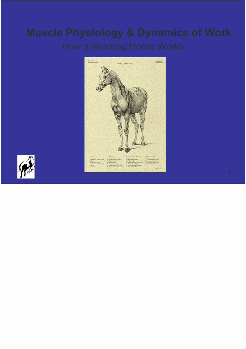
1 Muscle Physiology & Dynamics of Work How a Working Horse Works
-
Upload
leeannswenson -
Category
Documents
-
view
219 -
download
0
Transcript of April 17 Tufts Presentation PDF Version
8/8/2019 April 17 Tufts Presentation PDF Version
http://slidepdf.com/reader/full/april-17-tufts-presentation-pdf-version 1/51
1
Muscle Physiology & Dynamics of WorkHow a Working Horse Works

8/8/2019 April 17 Tufts Presentation PDF Version
http://slidepdf.com/reader/full/april-17-tufts-presentation-pdf-version 2/51
2
Equine Muscle Physiology & Mechanics
Muscle Tissue Intro
Structure & Function Muscle Microanatomy & Physiology
Dynamics of Work
Specific Muscle Fibers & Energy Substrates
Exercise & Effects on Muscle
8/8/2019 April 17 Tufts Presentation PDF Version
http://slidepdf.com/reader/full/april-17-tufts-presentation-pdf-version 3/51
3
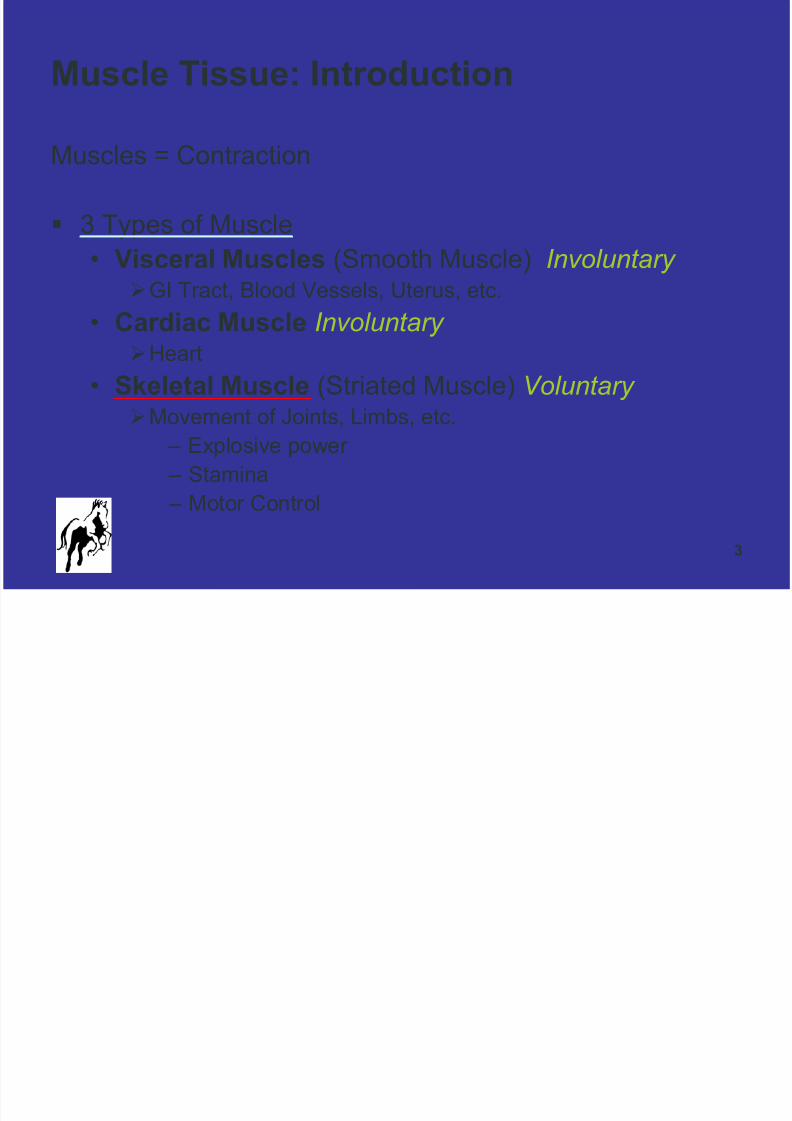
Muscle Tissue: Introduction
Muscles = Contraction
3 Types of Muscle
• Visceral Muscles (Smooth Muscle) Involuntary GI Tract, Blood Vessels, Uterus, etc.
• Cardiac Muscle Involuntary Heart
• Skeletal Muscle (Striated Muscle) Voluntary
Movement of Joints, Limbs, etc. – Explosive power
– Stamina
– Motor Control

8/8/2019 April 17 Tufts Presentation PDF Version
http://slidepdf.com/reader/full/april-17-tufts-presentation-pdf-version 4/51
4
Skeletal Muscle: Structure & Function
Large part of body weight (up to 40% including H20)
Closely associated with the skeletal, nervous, andcirculatory systems
• Manipulation impacts a range of tissues & systems
Generates heat Each muscle is a collection of fibers & associated tissues
Attached to bone via tendons & connective tissue
• Least moveable attachment = origin• Most moveable attachment = insertion

8/8/2019 April 17 Tufts Presentation PDF Version
http://slidepdf.com/reader/full/april-17-tufts-presentation-pdf-version 5/51
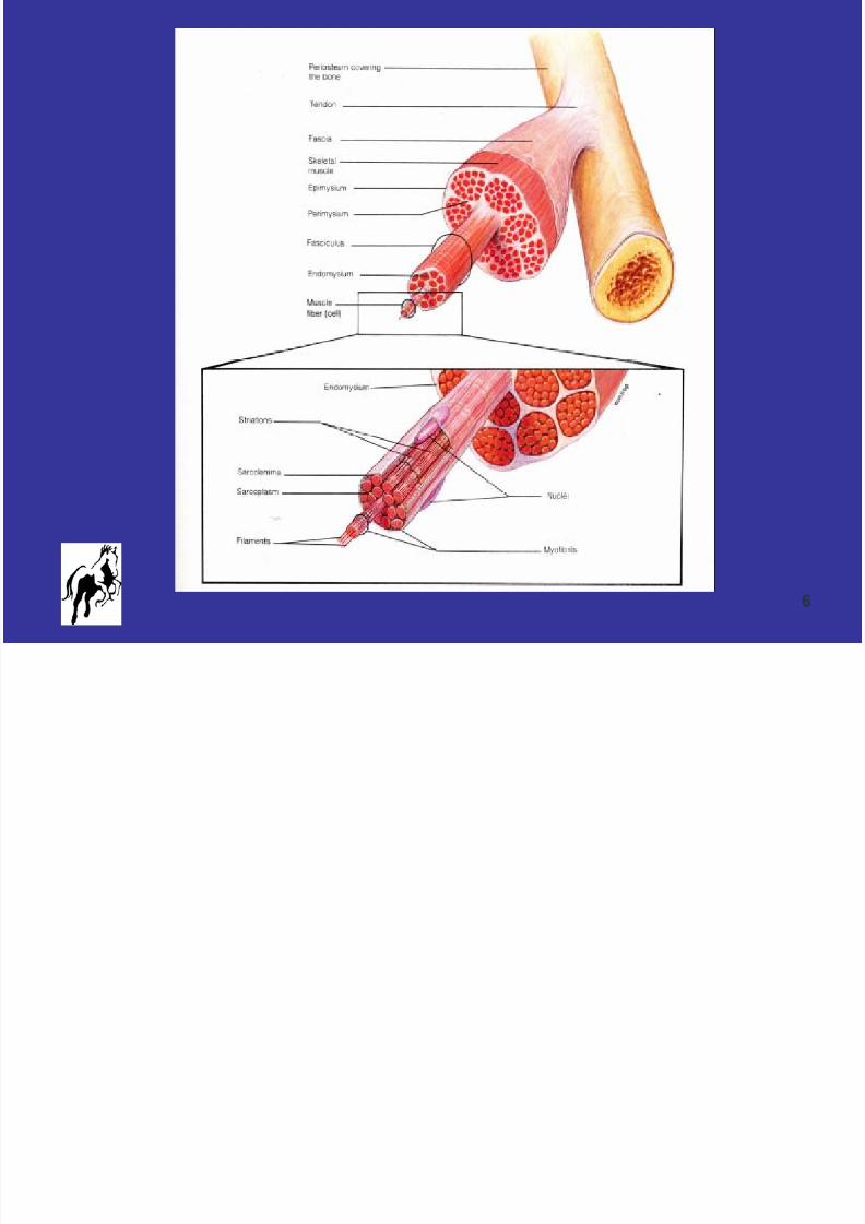
5
Microanatomy
& Physiology
8/8/2019 April 17 Tufts Presentation PDF Version
http://slidepdf.com/reader/full/april-17-tufts-presentation-pdf-version 6/51
6
8/8/2019 April 17 Tufts Presentation PDF Version
http://slidepdf.com/reader/full/april-17-tufts-presentation-pdf-version 7/51
7
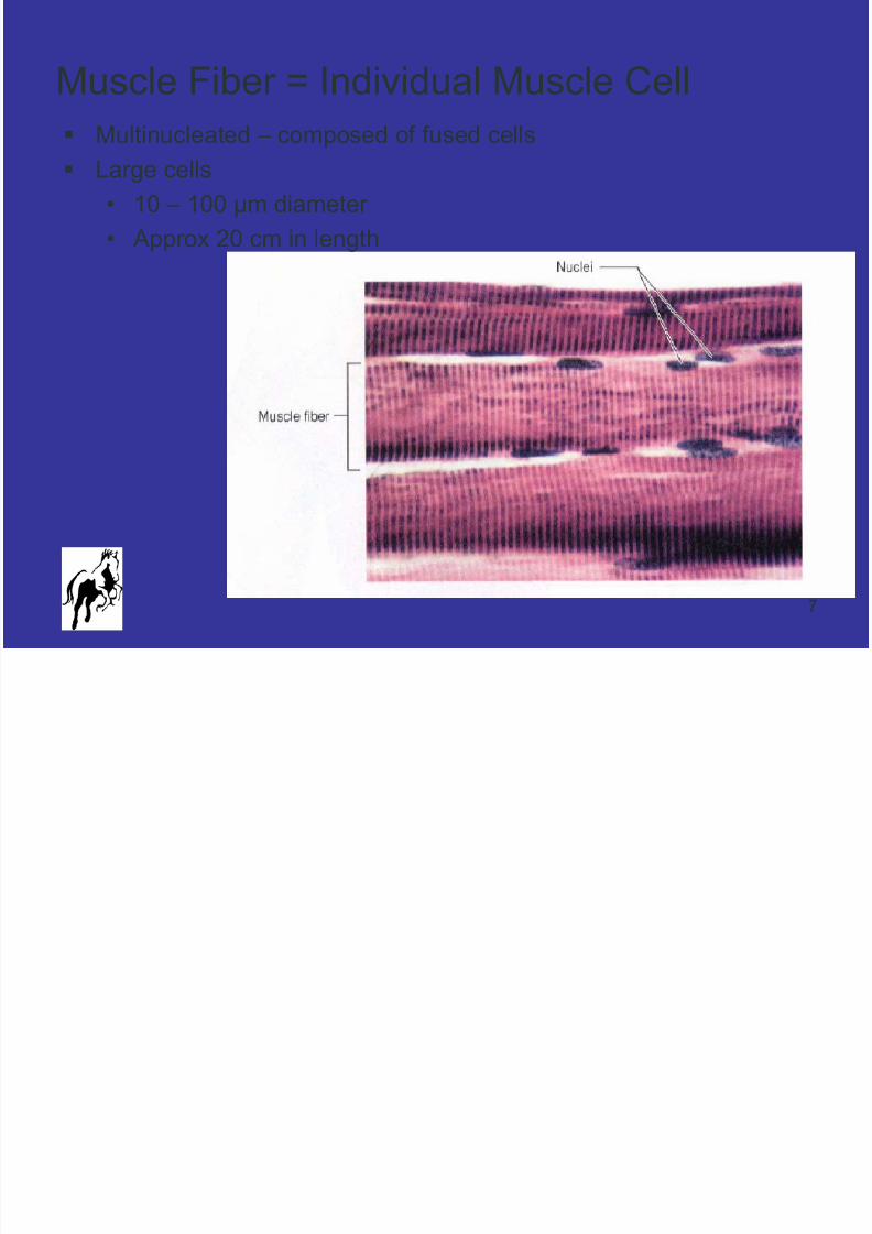
Muscle Fiber = Individual Muscle Cell
Multinucleated – composed of fused cells
Large cells
• 10 – 100 µm diameter
• Approx 20 cm in length
8/8/2019 April 17 Tufts Presentation PDF Version
http://slidepdf.com/reader/full/april-17-tufts-presentation-pdf-version 8/51
8
Muscle Cells
Specialized to contract
• Generate FORCE and MOVEMENT Do not divide
• Increased muscle size is due to Increased cell size
Key Qualities of Muscle Cells
• Excitable
• Conductive• Contractile

8/8/2019 April 17 Tufts Presentation PDF Version
http://slidepdf.com/reader/full/april-17-tufts-presentation-pdf-version 9/51
9
Muscle Cell Key Components
Membrane = Sarcolemma
T-Tubules
• Transmit Messages Mitochondria
• Generate Energy
• Numerous Myofibrils
• 2 Proteins in long strands
• Heart of the contractile function
Sarcoplasmic Reticulum (Endoplasmic Reticulum)

8/8/2019 April 17 Tufts Presentation PDF Version
http://slidepdf.com/reader/full/april-17-tufts-presentation-pdf-version 10/51
10
Muscle Cell & Associated Structures
As visible with a standard light microscope

8/8/2019 April 17 Tufts Presentation PDF Version
http://slidepdf.com/reader/full/april-17-tufts-presentation-pdf-version 11/51
11
Skeletal Muscle
Electron Micrograph
8/8/2019 April 17 Tufts Presentation PDF Version
http://slidepdf.com/reader/full/april-17-tufts-presentation-pdf-version 12/51
12
Dynamics of Work
Mechanism of Contraction
Stimulus of Contraction
Energy for Contraction

8/8/2019 April 17 Tufts Presentation PDF Version
http://slidepdf.com/reader/full/april-17-tufts-presentation-pdf-version 13/51
13
Sarcomere = Smallest Unit of Contraction
Repeating Pattern of Striations
Thick and Thin Filaments
Actin (Thin) & Myosin (Thick)
Myofilaments arranged in a specific pattern
H-Zone
Z-Line
A-Band

8/8/2019 April 17 Tufts Presentation PDF Version
http://slidepdf.com/reader/full/april-17-tufts-presentation-pdf-version 14/51
8/8/2019 April 17 Tufts Presentation PDF Version
http://slidepdf.com/reader/full/april-17-tufts-presentation-pdf-version 15/51
15
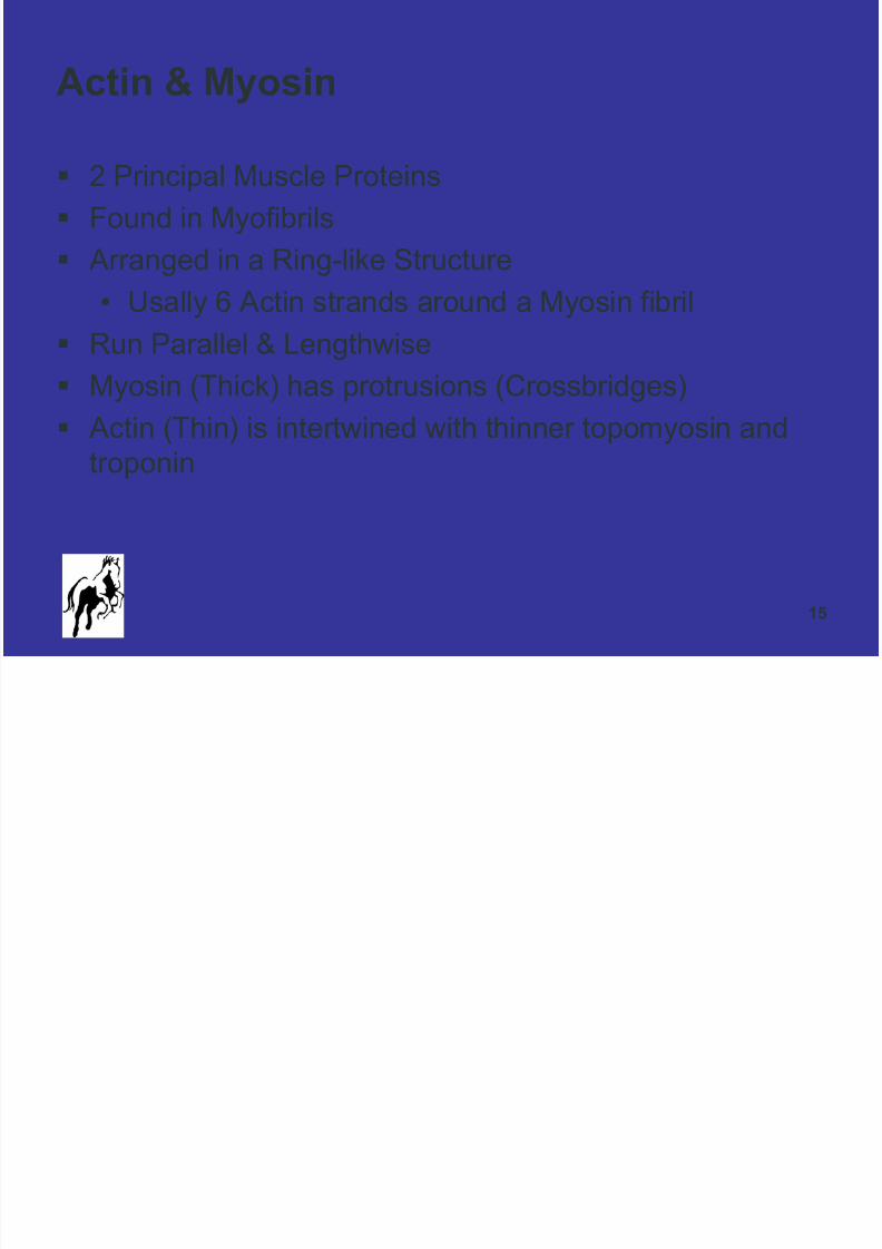
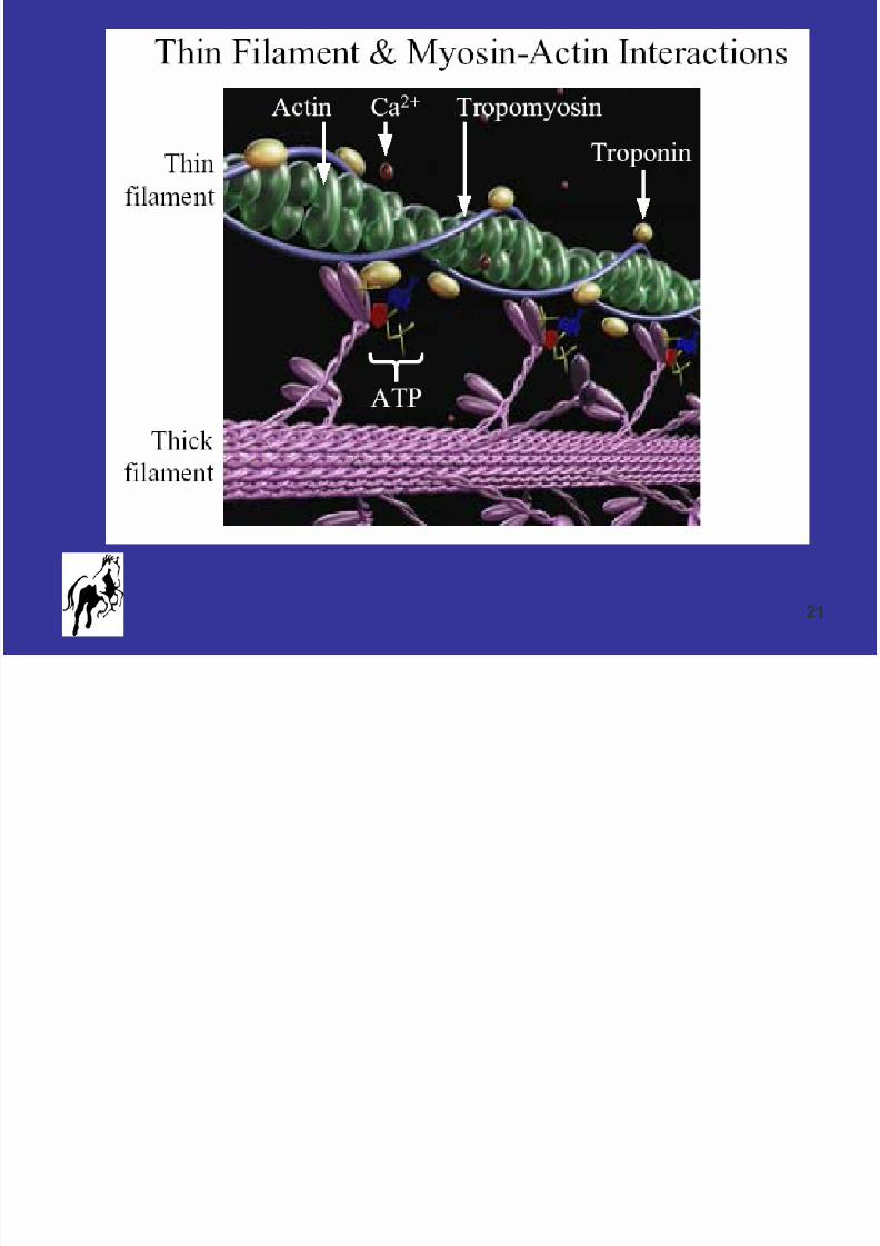
Actin & Myosin
2 Principal Muscle Proteins
Found in Myofibrils
Arranged in a Ring-like Structure
• Usally 6 Actin strands around a Myosin fibril
Run Parallel & Lengthwise Myosin (Thick) has protrusions (Crossbridges)
Actin (Thin) is intertwined with thinner topomyosin and
troponin

8/8/2019 April 17 Tufts Presentation PDF Version
http://slidepdf.com/reader/full/april-17-tufts-presentation-pdf-version 16/51
16
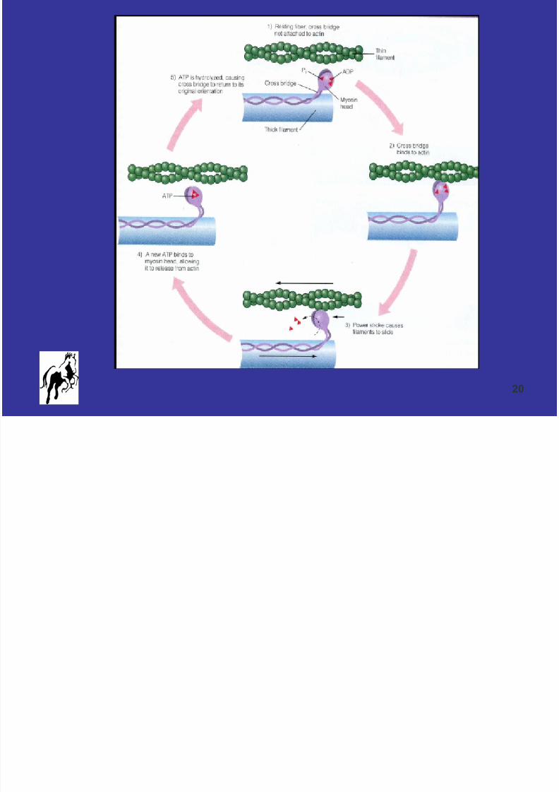
Mechanism of Contraction
1. Nerve Impulse Stimulation
2. CA++ Released into Cytoplasm by Sarcoplasmic
Reticulum
3. CA++ Binds to Troponin, which Rotates
4. Tropomyosin Moves and Actin is Exposed to Myosin
5. Myosin Crossbridge Binds to Actin6. Crossbridge Drags Along Actin (Power Stroke)

8/8/2019 April 17 Tufts Presentation PDF Version
http://slidepdf.com/reader/full/april-17-tufts-presentation-pdf-version 17/51
17
8/8/2019 April 17 Tufts Presentation PDF Version
http://slidepdf.com/reader/full/april-17-tufts-presentation-pdf-version 18/51
18
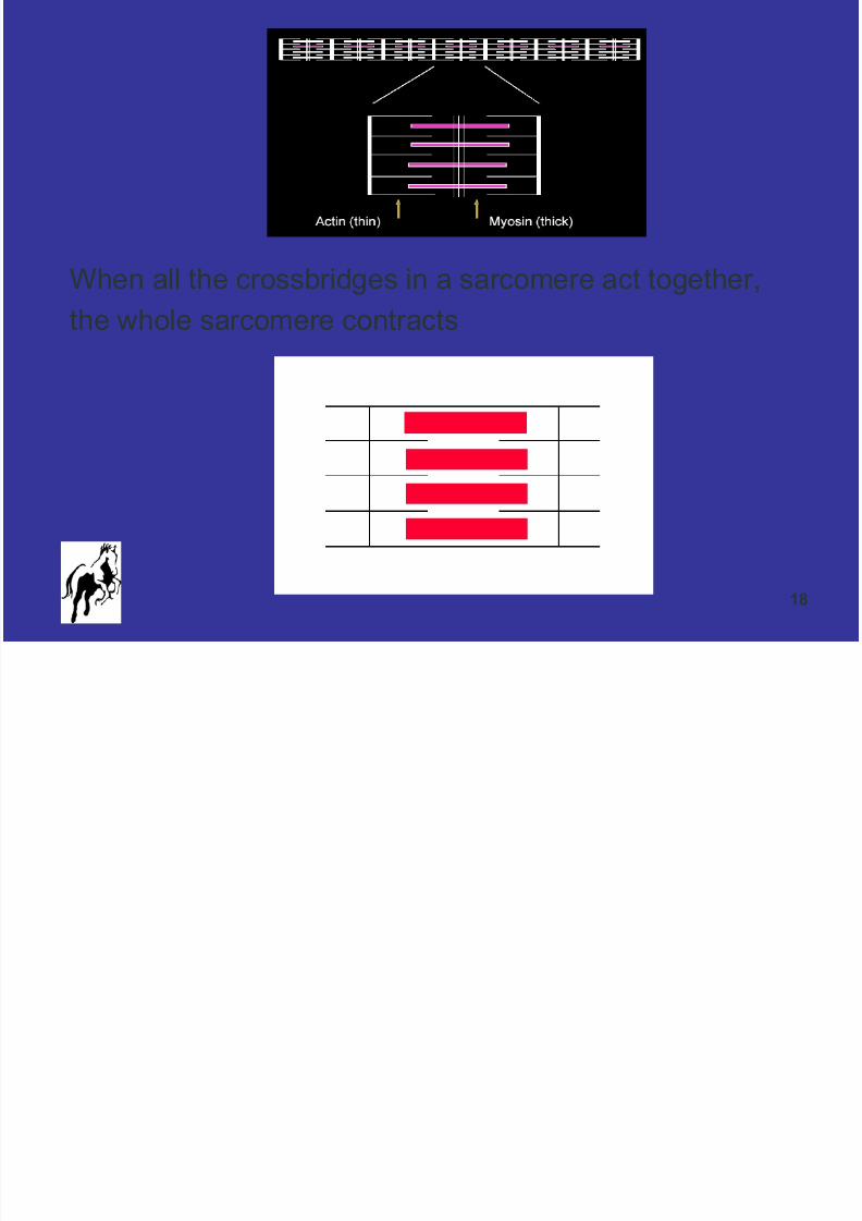
When all the crossbridges in a sarcomere act together,
the whole sarcomere contracts

8/8/2019 April 17 Tufts Presentation PDF Version
http://slidepdf.com/reader/full/april-17-tufts-presentation-pdf-version 19/51
19
Mechanism of Relaxation
7. Nerve Impulse Ends
8. SR Reabsorbs CA++9. CA++ Dissociates from Troponin
10.ATP Binds to the Crossbridge
11.Crossbridge Disconnects from Actin12.Actin Fibers Return to Previous Positions
13.Sarcomere Relaxes
8/8/2019 April 17 Tufts Presentation PDF Version
http://slidepdf.com/reader/full/april-17-tufts-presentation-pdf-version 20/51
20
8/8/2019 April 17 Tufts Presentation PDF Version
http://slidepdf.com/reader/full/april-17-tufts-presentation-pdf-version 21/51
21
8/8/2019 April 17 Tufts Presentation PDF Version
http://slidepdf.com/reader/full/april-17-tufts-presentation-pdf-version 22/51
22
Contraction-Relaxation
A muscle cell may not go back to immediate complete
relaxation
Contraction can continue through a series of stimulations
(Summation)
Summation increases the total force of contraction
If the stimulus is great enough, many sarcomeres inmany fibers are recruited, and the muscle as a whole
contracts.
Allows for varying amounts of work
Muscle failure occurs when the maximum number of
fibers are stressed beyond their limits

8/8/2019 April 17 Tufts Presentation PDF Version
http://slidepdf.com/reader/full/april-17-tufts-presentation-pdf-version 23/51
23
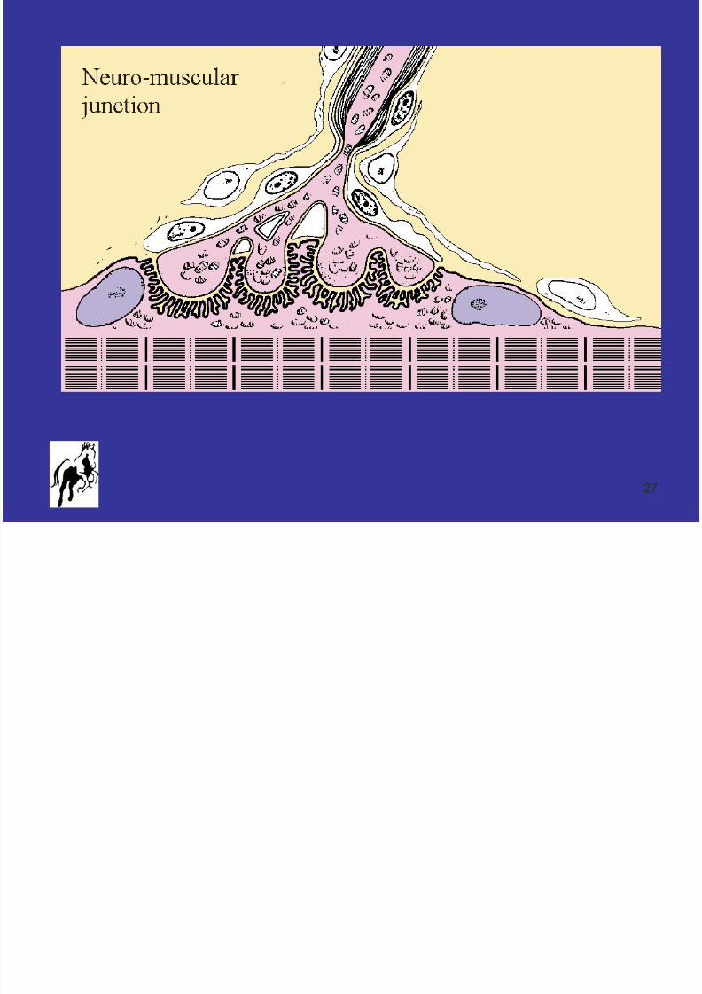
Stimulus of Contraction:
Muscle Contraction is Controlled by Motor Nerves
8/8/2019 April 17 Tufts Presentation PDF Version
http://slidepdf.com/reader/full/april-17-tufts-presentation-pdf-version 24/51
24
Interaction of Motor Nerves and Muscle Fibers
Each muscle is innervated by only one motor nerve
One nerve can innervate a number of muscles Each nerve controls many fibers (motor units), the fewer
the fibers the more delicate the movement
If nerve contact is lost, fibers shrink (atrophy)
The pattern of nerve activity determines the fiber type

8/8/2019 April 17 Tufts Presentation PDF Version
http://slidepdf.com/reader/full/april-17-tufts-presentation-pdf-version 25/51
25
Feedback Loop
Feedback from the tendon and stretch receptors
controls motor nerve activity
Motor nerve activity is also controlled by higher
centers (brain)
8/8/2019 April 17 Tufts Presentation PDF Version
http://slidepdf.com/reader/full/april-17-tufts-presentation-pdf-version 26/51
8/8/2019 April 17 Tufts Presentation PDF Version
http://slidepdf.com/reader/full/april-17-tufts-presentation-pdf-version 27/51
27
8/8/2019 April 17 Tufts Presentation PDF Version
http://slidepdf.com/reader/full/april-17-tufts-presentation-pdf-version 28/51
28
Relaxation
When electrical activity stops, the calcium is removed
and contraction stops
Muscle must relax between each contraction by actively
pumping Ca back to SR
Ion pumps in the cell membrane actively repolarize the
muscle cell membranes
All processes necessary for relaxation are active –
require energy

8/8/2019 April 17 Tufts Presentation PDF Version
http://slidepdf.com/reader/full/april-17-tufts-presentation-pdf-version 29/51
29
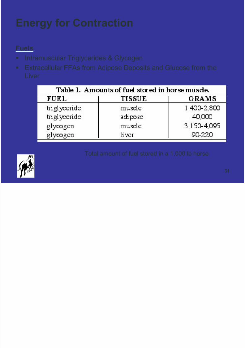
Energy for Contraction
Each crossbridge requires ATP
Each myosin strand has dozens of crossbridges
Each muscle fiber has hundreds of myosin strands
Muscle Contraction Requires Significant Energy
Basic Unit of Energy = ATP
ATP ADP & Pi ENERGY
(ATP + H2O ADP + Pi +H+ + Energy)
ATP= adenosine triphosphate; ADP=adenosine diphosphate;Pi=Inorganic phosphate
8/8/2019 April 17 Tufts Presentation PDF Version
http://slidepdf.com/reader/full/april-17-tufts-presentation-pdf-version 30/51
30
For a horse to maintain exercise for more than a few
seconds, ATP stores in muscle must be replenished at
an appropriate rate.
8/8/2019 April 17 Tufts Presentation PDF Version
http://slidepdf.com/reader/full/april-17-tufts-presentation-pdf-version 31/51
31
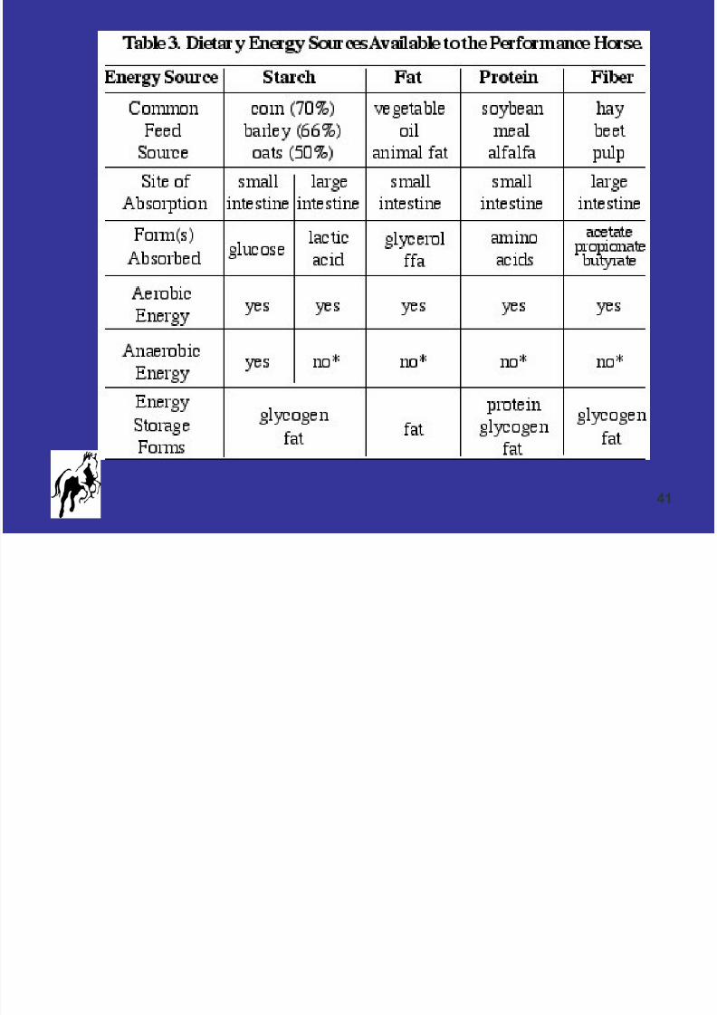
Energy for Contraction
Fuels
Intramuscular Triglycerides & Glycogen
Extracellular FFAs from Adipose Deposits and Glucose from theLiver
Total amount of fuel stored in a 1,000 lb horse

8/8/2019 April 17 Tufts Presentation PDF Version
http://slidepdf.com/reader/full/april-17-tufts-presentation-pdf-version 32/51
32
AEROBIC
ANAEROBIC
Two Main Pathways For Energy Metabolism

8/8/2019 April 17 Tufts Presentation PDF Version
http://slidepdf.com/reader/full/april-17-tufts-presentation-pdf-version 33/51
33
Aerobic Metabolism
Occurs in Mitochondria
For low energy demands of slow speed exercise
Primary pathway for endurance exercise
Gallop speeds < 18sec/200m can usually be met byaerobic metabolism in fit horses
Training can increase capacity to generate energyaerobically
• Enhanced oxygen delivery to muscle• Increased mitochondrial density
• Increased enzyme concentrations
8/8/2019 April 17 Tufts Presentation PDF Version
http://slidepdf.com/reader/full/april-17-tufts-presentation-pdf-version 34/51
34
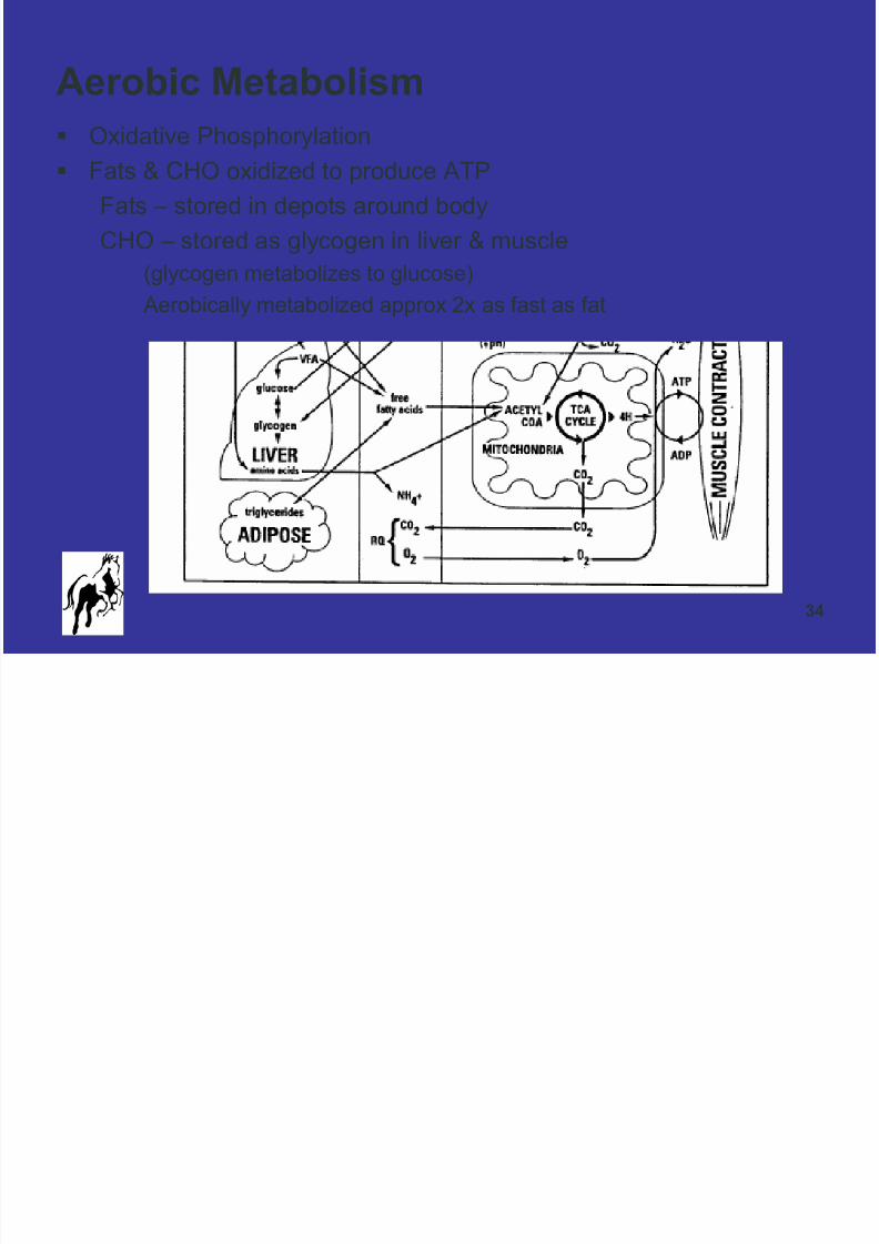
Aerobic Metabolism
Oxidative Phosphorylation
Fats & CHO oxidized to produce ATP
Fats – stored in depots around body
CHO – stored as glycogen in liver & muscle
(glycogen metabolizes to glucose)
Aerobically metabolized approx 2x as fast as fat
8/8/2019 April 17 Tufts Presentation PDF Version
http://slidepdf.com/reader/full/april-17-tufts-presentation-pdf-version 35/51
35
Aerobic Metabolism
Limitations
Primarily limited by availability of oxygen in working
muscles
Upper airway obstructions
Cardiovascular system impairment
Hemoglobin concentration

8/8/2019 April 17 Tufts Presentation PDF Version
http://slidepdf.com/reader/full/april-17-tufts-presentation-pdf-version 36/51
8/8/2019 April 17 Tufts Presentation PDF Version
http://slidepdf.com/reader/full/april-17-tufts-presentation-pdf-version 37/51
37
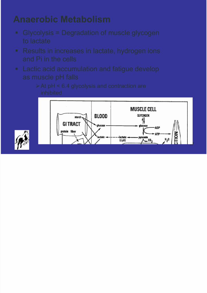
Anaerobic Metabolism
Glycolysis = Degradation of muscle glycogen
to lactate
Results in increases in lactate, hydrogen ions
and Pi in the cells
Lactic acid accumulation and fatigue develop
as muscle pH falls
At pH < 6.4 glycolysis and contraction are
inhibited
8/8/2019 April 17 Tufts Presentation PDF Version
http://slidepdf.com/reader/full/april-17-tufts-presentation-pdf-version 38/51
38
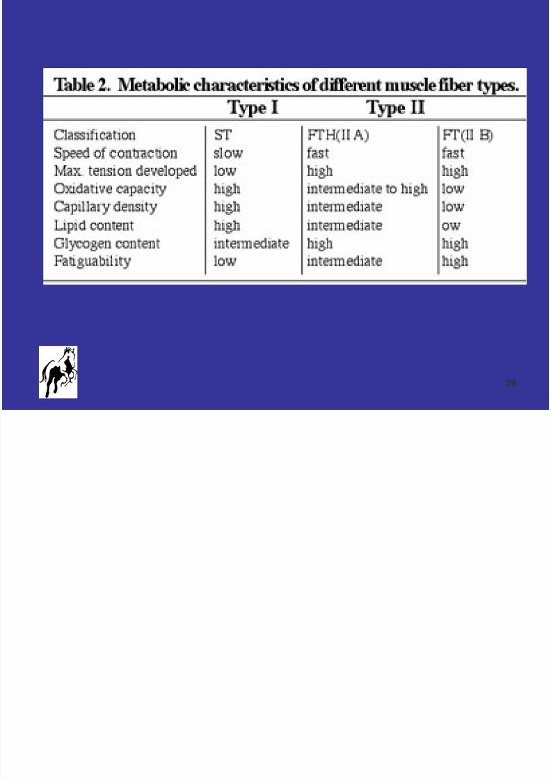
Different Muscles have Fibers with
Different PropertiesType I & Type IIA
High Oxidative Capacity
Store Triglycerides & Glycogen
Standing and posture: Slow contracting fibers that are well suppliedwith oxygen – example stay apparatus
Type I aka “Slow Twitch” Fibers “Red Fibers”
Type IIB
Low Aerobic Capacity
Store Glycogen
Athletic Movements: Muscles that generate rapid movement containfast fibers and can work for short periods without oxygen
Type II aka “White” Fibers, “Fast Twitch” Fibers
8/8/2019 April 17 Tufts Presentation PDF Version
http://slidepdf.com/reader/full/april-17-tufts-presentation-pdf-version 39/51
8/8/2019 April 17 Tufts Presentation PDF Version
http://slidepdf.com/reader/full/april-17-tufts-presentation-pdf-version 40/51
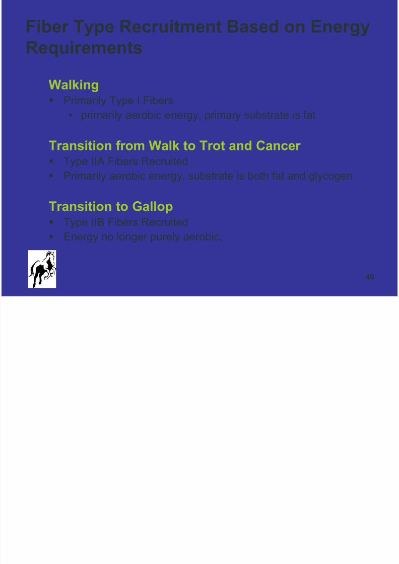
40
Walking Primarily Type I Fibers
• primarily aerobic energy, primary substrate is fat
Transition from Walk to Trot and Cancer
Type IIA Fibers Recruited Primarily aerobic energy, substrate is both fat and glycogen
Transition to Gallop Type IIB Fibers Recruited
Energy no longer purely aerobic,
Fiber Type Recruitment Based on Energy
Requirements
8/8/2019 April 17 Tufts Presentation PDF Version
http://slidepdf.com/reader/full/april-17-tufts-presentation-pdf-version 41/51
41

8/8/2019 April 17 Tufts Presentation PDF Version
http://slidepdf.com/reader/full/april-17-tufts-presentation-pdf-version 42/51
42
Exercise
Concentric Exercise
• Isometric – constant length
• Isotonic – constant force
• Or a mixture of the two
Eccentric Exercise
• Lengthening contractions

8/8/2019 April 17 Tufts Presentation PDF Version
http://slidepdf.com/reader/full/april-17-tufts-presentation-pdf-version 43/51
43
8/8/2019 April 17 Tufts Presentation PDF Version
http://slidepdf.com/reader/full/april-17-tufts-presentation-pdf-version 44/51
44
8/8/2019 April 17 Tufts Presentation PDF Version
http://slidepdf.com/reader/full/april-17-tufts-presentation-pdf-version 45/51
45
Effects of Exercise on Muscle
Lack of exercise leads to fiber atrophy
Gentle exercise maintains muscle mass & flexibility
Moderate long term activity increases fatigue resistance
High load exercise leads to muscle fiber hypertrophy
8/8/2019 April 17 Tufts Presentation PDF Version
http://slidepdf.com/reader/full/april-17-tufts-presentation-pdf-version 46/51
46
Muscle Fatigue
Prolonged and/or strong contraction Fatigue
• Inability of contractile and metabolic processes to
continue supplying the same work output
Nerve sends electric stimulation, NMJ transmits, action
potentials spread over muscle fibers
However contraction becomes progressively weaker due
to reduced ATP in the muscle fibers
Interruption of blood flow through a contracting muscle
leads to almost complete fatigue in less than a minute
due to loss of nutrient supply
8/8/2019 April 17 Tufts Presentation PDF Version
http://slidepdf.com/reader/full/april-17-tufts-presentation-pdf-version 47/51
47
Muscle Fatigue
Endurance Horses
Most often due to glycogen depletion, as most work is
performed aerobically
Race Horses Most often due to lactic acid accumulation

8/8/2019 April 17 Tufts Presentation PDF Version
http://slidepdf.com/reader/full/april-17-tufts-presentation-pdf-version 48/51
48
Lactic Acid or Lactate
By product of anaerobic glycolysis
A potential cause of late onset muscle soreness 24 – 48
hours after intense exercise
Sent from muscle to blood and removed via liver
Removal requires oxygen and is hastened by light workduring recovery

8/8/2019 April 17 Tufts Presentation PDF Version
http://slidepdf.com/reader/full/april-17-tufts-presentation-pdf-version 49/51
49
Muscle Atrophy
Results anytime a muscle is not used or used only for
weak contractions
Denervated muscle begins immediate atrophy
• Example: Sweeney
Injury to Suprascapular N causing atrophy in supraspinatus &
infraspinatus

8/8/2019 April 17 Tufts Presentation PDF Version
http://slidepdf.com/reader/full/april-17-tufts-presentation-pdf-version 50/51
50
Muscle Hypertrophy
Diameter of individual muscle fibers increase
Sarcoplasm increases
Fibers gain in nutrient and intermediary metabolic
substances (ATP, creatine phosphate, glycogen,
intracellular lipids, additional mitochondria)
Myofibrils may also increase in size
Hypertrophy increases both power of the muscle and the
nutrient mechanisms to maintain that power

8/8/2019 April 17 Tufts Presentation PDF Version
http://slidepdf.com/reader/full/april-17-tufts-presentation-pdf-version 51/51
51
Conclusion
Muscle Microanatomy & Physiology
Dynamics of Work Specific Muscle Fibers & Energy
Substrates
Together IMPACT Exercise &Its Effects on Muscle