Apical periodontitis and related risk factors: Cross ...
7
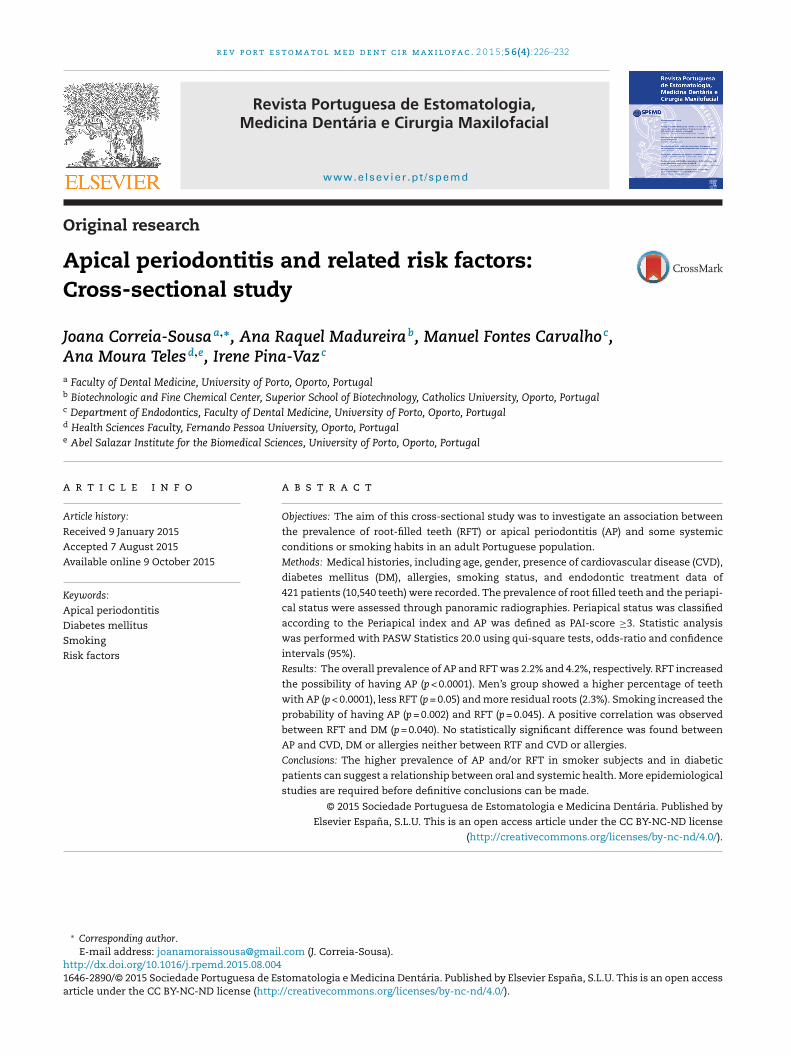
r e v p o r t e s t o m a t o l m e d d e n t c i r m a x i l o f a c . 2 0 1 5; 5 6(4) :226–232 www.elsevier.pt/spemd Revista Portuguesa de Estomatologia, Medicina Dentária e Cirurgia Maxilofacial Original research Apical periodontitis and related risk factors: Cross-sectional study Joana Correia-Sousa a,∗ , Ana Raquel Madureira b , Manuel Fontes Carvalho c , Ana Moura Teles d,e , Irene Pina-Vaz c a Faculty of Dental Medicine, University of Porto, Oporto, Portugal b Biotechnologic and Fine Chemical Center, Superior School of Biotechnology, Catholics University, Oporto, Portugal c Department of Endodontics, Faculty of Dental Medicine, University of Porto, Oporto, Portugal d Health Sciences Faculty, Fernando Pessoa University, Oporto, Portugal e Abel Salazar Institute for the Biomedical Sciences, University of Porto, Oporto, Portugal a r t i c l e i n f o Article history: Received 9 January 2015 Accepted 7 August 2015 Available online 9 October 2015 Keywords: Apical periodontitis Diabetes mellitus Smoking Risk factors a b s t r a c t Objectives: The aim of this cross-sectional study was to investigate an association between the prevalence of root-filled teeth (RFT) or apical periodontitis (AP) and some systemic conditions or smoking habits in an adult Portuguese population. Methods: Medical histories, including age, gender, presence of cardiovascular disease (CVD), diabetes mellitus (DM), allergies, smoking status, and endodontic treatment data of 421 patients (10,540 teeth) were recorded. The prevalence of root filled teeth and the periapi- cal status were assessed through panoramic radiographies. Periapical status was classified according to the Periapical index and AP was defined as PAI-score ≥3. Statistic analysis was performed with PASW Statistics 20.0 using qui-square tests, odds-ratio and confidence intervals (95%). Results: The overall prevalence of AP and RFT was 2.2% and 4.2%, respectively. RFT increased the possibility of having AP (p < 0.0001). Men’s group showed a higher percentage of teeth with AP (p < 0.0001), less RFT (p = 0.05) and more residual roots (2.3%). Smoking increased the probability of having AP (p = 0.002) and RFT (p = 0.045). A positive correlation was observed between RFT and DM (p = 0.040). No statistically significant difference was found between AP and CVD, DM or allergies neither between RTF and CVD or allergies. Conclusions: The higher prevalence of AP and/or RFT in smoker subjects and in diabetic patients can suggest a relationship between oral and systemic health. More epidemiological studies are required before definitive conclusions can be made. © 2015 Sociedade Portuguesa de Estomatologia e Medicina Dentária. Published by Elsevier España, S.L.U. This is an open access article under the CC BY-NC-ND license (http://creativecommons.org/licenses/by-nc-nd/4.0/). ∗ Corresponding author. E-mail address: [email protected] (J. Correia-Sousa). http://dx.doi.org/10.1016/j.rpemd.2015.08.004 1646-2890/© 2015 Sociedade Portuguesa de Estomatologia e Medicina Dentária. Published by Elsevier España, S.L.U. This is an open access article under the CC BY-NC-ND license (http://creativecommons.org/licenses/by-nc-nd/4.0/).
Transcript of Apical periodontitis and related risk factors: Cross ...
r e v p o r t e s t o m a t o l m e d d e n t c i r m a x i l o f a c . 2 0 1 5;5 6(4):226–232
www.elsev ier .p t /spemd
Revista Portuguesa de Estomatologia,Medicina Dentária e Cirurgia Maxilofacial
Original research
Apical periodontitis and related risk factors:Cross-sectional study
Joana Correia-Sousaa,∗, Ana Raquel Madureirab, Manuel Fontes Carvalhoc,Ana Moura Telesd,e, Irene Pina-Vazc
a Faculty of Dental Medicine, University of Porto, Oporto, Portugalb Biotechnologic and Fine Chemical Center, Superior School of Biotechnology, Catholics University, Oporto, Portugalc Department of Endodontics, Faculty of Dental Medicine, University of Porto, Oporto, Portugald Health Sciences Faculty, Fernando Pessoa University, Oporto, Portugale Abel Salazar Institute for the Biomedical Sciences, University of Porto, Oporto, Portugal
a r t i c l e i n f o
Article history:
Received 9 January 2015
Accepted 7 August 2015
Available online 9 October 2015
Keywords:
Apical periodontitis
Diabetes mellitus
Smoking
Risk factors
a b s t r a c t
Objectives: The aim of this cross-sectional study was to investigate an association between
the prevalence of root-filled teeth (RFT) or apical periodontitis (AP) and some systemic
conditions or smoking habits in an adult Portuguese population.
Methods: Medical histories, including age, gender, presence of cardiovascular disease (CVD),
diabetes mellitus (DM), allergies, smoking status, and endodontic treatment data of
421 patients (10,540 teeth) were recorded. The prevalence of root filled teeth and the periapi-
cal status were assessed through panoramic radiographies. Periapical status was classified
according to the Periapical index and AP was defined as PAI-score ≥3. Statistic analysis
was performed with PASW Statistics 20.0 using qui-square tests, odds-ratio and confidence
intervals (95%).
Results: The overall prevalence of AP and RFT was 2.2% and 4.2%, respectively. RFT increased
the possibility of having AP (p < 0.0001). Men’s group showed a higher percentage of teeth
with AP (p < 0.0001), less RFT (p = 0.05) and more residual roots (2.3%). Smoking increased the
probability of having AP (p = 0.002) and RFT (p = 0.045). A positive correlation was observed
between RFT and DM (p = 0.040). No statistically significant difference was found between
AP and CVD, DM or allergies neither between RTF and CVD or allergies.
Conclusions: The higher prevalence of AP and/or RFT in smoker subjects and in diabetic
patients can suggest a relationship between oral and systemic health. More epidemiological
studies are required before definitive conclusions can be made.
© 2015 Sociedade Portuguesa de Estomatologia e Medicina Dentária. Published by
Elsevier España, S.L.U. This is an open access article under the CC BY-NC-ND license
(http://creativecommons.org/licenses/by-nc-nd/4.0/).
∗ Corresponding author.E-mail address: [email protected] (J. Correia-Sousa).
http://dx.doi.org/10.1016/j.rpemd.2015.08.0041646-2890/© 2015 Sociedade Portuguesa de Estomatologia e Medicina Dentária. Published by Elsevier España, S.L.U. This is an open accessarticle under the CC BY-NC-ND license (http://creativecommons.org/licenses/by-nc-nd/4.0/).

r e v p o r t e s t o m a t o l m e d d e n t c i r m a x i l o f a c . 2 0 1 5;5 6(4):226–232 227
Periodontite apical e fatores de risco associados: estudo transversal
Palavras chave:
Periodontite apical
Diabetes mellitus
Fumar
Fatores de risco
r e s u m o
Objetivos: O objetivo deste estudo transversal foi investigar a associacão entre a prevalên-
cia de dentes com tratamento endodôntico (RFT) ou periodontite apical (AP) e algumas
condicões sistémicas ou hábitos tabágicos numa populacão adulta portuguesa.
Métodos: Histórias médicas, incluindo idade, género, presenca de doencas cardiovasculares
(CVD), diabetes mellitus, alergias e hábitos tabágicos, e registos dos tratamentos endodôn-
ticos de 421 pacientes (10.540 dentes) foram recolhidos. A prevalência de dentes com
tratamento endodôntico e status apical foram avaliados através de radiografias panorâmi-
cas. O status apical foi classificado de acordo com o índice periapical e a AP definida para
valores PAI≥3. A análise estatística foi realizada através do PASW Statistics 20.0 utilizando
os testes chi-quadrado, valores odds-ratio e intervalos confianca (95%).
Resultados: A prevalência da AP e RFT foi de 2,2% e 4,2%, respectivamente. RFT aumentou a
possibilidade de ter AP (p<0,0001). Os homens revelaram uma maior percentagem de dentes
com AP (p<0,0001), menos RFT (p=0,05) e mais raízes residuais (2,3%). Fumar aumentou a
probabilidade de ter AP (p=0,002) e RFT (p=0,045). Uma relacão positiva foi observada entre
RFT e DM (p = 0,040). Não se encontraram diferencas estatisticamente significativas entre
AP e CVD, DM ou alergias nem entre RTF e CVD ou alergias.
Conclusões: Uma maior percentagem de AP e/ou RFT nos fumadores e nos pacientes com
diabetes sugere uma relacão entre a saúde oral e sistémica. Mais estudos epidemiológicos
são necessários antes de se fazerem conclusões definitivas.© 2015 Sociedade Portuguesa de Estomatologia e Medicina Dentária. Publicado por
Elsevier España, S.L.U. Este é um artigo Open Access sob a licença de CC BY-NC-ND
I
Atimaiiimtateh
outllT9dn
cdh
ntroduction
pical periodontitis (AP) is “an acute or chronic inflamma-ory lesion around the apex of a tooth caused by bacterialnfection of the pulp and root canal system”.1 The inflam-
atory cells cause, among other effects, resorption of thedjacent supporting bone. The diagnosis is primarily basedn the observation of a periradicular radiolucency, althought can be supported by patient’ symptoms or clinical signsn the acute phases.1,2 AP is highly prevalent, and the esti-
ated percentage of individuals with AP, in at least oneooth, is 34–70%3-7, which can rise in older patients.8–11 Over-ll, the percentage of teeth with AP has been estimatedo range between 1.7% and 6.6%.5,9,12 However, amongstndodontically treated teeth the percentage is significantlyigher.9,11,13–16
Root canal treatment is the most frequent therapeuticption for preserving teeth with AP and to restoring perirradic-lar tissues’ health. Therefore, its prevalence can be linked tohe presence of severe caries lesions or traumatic injuries thatead to pulp necrosis. The prevalence of individuals with, ateast, one root canal treatment is between 41 and 87%.5,7,17,18
he frequency of root-filled teeth varies between 2.2% and.39%.5,6,9,11,15,19 This broad variation can be due to eitherifferent age stratification in the studies or variation withinational health care services.
Several epidemiological studies have found an asso-
iation between chronic dental infection, cardiovascularisease (CVD)20–25, diabetes mellitus (DM)26–29 and smokingabits30,31, most of them relating to periodontal disease.(http://creativecommons.org/licenses/by-nc-nd/4.0/).
AP is, in many instances, very similar to periodontal diseaseregarding the microbial aetiology and the presence of elevatedsystemic cytokines.32,33
Patients with DM, hypertension or coronary heart diseasemight have decreased tissue resistance to bacterial infectionand reduced ability of tissue repair after endodontic treat-ment. Wang et al.34 found an increased risk of tooth extractionafter nonsurgical endodontic treatment in patients with thesediseases. Furthermore, the association of two of those con-ditions was a significant predictor of extraction or pooreroutcome of the endodontic treatment32,35,36. However, limiteddata is available on the long-term prognosis of AP and root-filled teeth, in patients with systemic diseases and smokinghabits.
DM, a syndrome characterized by abnormalities incarbohydrate, lipid and protein metabolism, also affectsmany functions of the immune system. For instance,up-regulation of pro-inflammatory cytokines from mono-cytes/polymorphonuclear leukocytes and down-regulationof growth factors from macrophages, resulting in dysreg-ulated macrophage phagocytosis.37 Consequently, there isdelay in healing process and commitment of the immuneresponse.38,39 These events predispose to chronic inflamma-tion, progressive tissue breakdown and diminished tissuerepair capability.40–42 DM has been considered as a possiblemodulating factor or disease modifier in endodontic infec-tions, in the sense that diabetic individuals, especially whenpoorly controlled, could be more prone to developing AP.43,44
The literature on the pathogenesis, progression and healingof endodontic pathology in diabetic patients is still scarce andshow controversial results.29,43,45–47

t c i r
of RFT (4.2%) were also found in other epidemiological studies,with prevalence’s ranging between 1.3% and 4.8%.9,11,15,19,52,53
By contrast, others authors have reported values ranging
74.8
97.7
7 0.8 8.2
0.7 6.8 0.5 3.2 0.3
PAI 1
PAI 2
PAI 3
PAI 4
PAI 5
228 r e v p o r t e s t o m a t o l m e d d e n
Current evidence indicates that smoking is a signif-icant risk factor for the inflammation of the marginalperiodontium.25,48 Cross-sectional and longitudinal studiesdemonstrated the harmful effects of tobacco smoking onthe supporting structures of the teeth.25,49 Smoking impairsthe body’s responses to infection, exacerbates bone loss,decreases the blood’s oxygen-carrying capacity and causesvascular dysfunction.50 It can be assumed that smoking canact as a risk factor to the development AP, exerting a nega-tive influence on the apical periodontium of endodonticallycompromised teeth, allowing the extension of periapical bonedestruction and/or interfering with the healing and repair pro-cess after root canal treatment.25
In the recent years, there has been a high level of interest inresearch focused on Dentistry, namely Endodontics, related tosystemic health. To date, the role of systemic conditions andhealth-related habits as risk factors for adverse outcome of APhas not been thoroughly explored.
The present study is aimed at exploring an associationbetween endodontic status and systemic conditions, such as,cardiovascular diseases, diabetes mellitus or allergies, andsmoking habits as possible risk factors for AP, in an adultPortuguese population.
Methods
The sample included medical histories and endodontictreatment data of all the patients attending the clinic ofthe Dental Faculty of Oporto University and of the HealthSciences Faculty of Fernando Pessoa University (Oporto) forthe first time in 2012. The following were used as inclusioncriteria: age (at least 18 years old) and number of teeth (noless than 8 remaining teeth). 421 patients were selected, witha total of 10,540 teeth assessed. The institutional scientificcommittee of each of the faculties involved formally approvedthe present study.
Age, gender, aspects of general health (presence of CVD,DM, allergies) and health-related habits (smoking status),were recorded from the medical questionnaire. CVD, DM andallergies were assessed through a dichotomy key (yes/no).Coronary heart disease, stroke, hypertension, atherosclerosis,and myocardial infarction were included in the cardiovasculardisease category. Type 1, type 2 and gestational diabetes wereincluded in the DM group. The following criteria were moni-tored in the allergies’ category: pollen season, asthma, atopicdermatitis, Chron’s disease, rheumatoide arthritis, allergicrhinitis and allergy medication, such as penicillin.
Smoking status was classified as non-smoker, if the patientanswer was never smoker/former smoker for more than5 years ago, or current smoker.
The periapical status and the prevalence of root-filledteeth (RFT) were assessed using panoramic radiography. TheOrthoralix® 9200 DDE (Gendex) was used in all cases. Themethod of viewing the radiographies was standardized: filmswere examined in a darkened room using a computer in which
the ambient light could be controlled for the best possible con-trast. Teeth were categorized as RFT, if they presented anyradiopaque material in the pulpal space. Periapical status ofeach tooth was classified according to the Periapical Indexm a x i l o f a c . 2 0 1 5;5 6(4):226–232
(PAI)51 and the presence of AP was defined as PAI-score ≥3. Incases of multi-rooted teeth, the worst root score was chosen.Three observers performed the PAI assessment, after trainingand calibration. The coefficient Cohen’s kappa was applied.
In order to characterize the oral health status of the sub-jects, additional clinical data such as the number of missingteeth as well as residual roots were also recorded.
Statistical analysis was performed with PASW Statistics20.0 (version 20, SPSS®). A descriptive statistical study of theselected variables was performed. Data were analyzed by esti-mating frequencies in percentages. The association and thelevel of significance between two variables were evaluatedusing the qui-square test. The odds-ratio and the respec-tive confidence intervals (95%) were obtained by associationmeasures in 2 × 2-cross tabs. For statistic analysis tooth wasadopted as sampling unit.
Results
The sample included a total of 421 patients’ records. Fromthese 43% were male and 57% female, with a mean age of41 ± 16 years old (range 18–82 years). Of the 10,540 examinedteeth, 2.2% had AP and 4.2% had RFT. The prevalence of APwas greater in root-filled teeth (Fig. 1 and Table 1) and in themen’s group (p < 0.05) (Table 2). The probability of having AP inmen was almost two times higher than in women (Table 2).
A significant association was observed between smokingstatus, AP and RFT (Table 3). RFT were more prevalent in DMand CVD groups (p < 0.05) (Table 3). No significant associationwas found between AP and CVD, DM or allergies groups, nei-ther between RFT and allergies (Table 3).
Regarding the number of absent teeth, there was no sig-nificant association between the different systemic diseases,habits or gender (p > 0.05) (Table 1). Similarly, no significantassociation was found between the number of residual rootsin the different groups (p > 0.05) (Table 1).
Discussion
The total prevalence of AP (2.2%) recorded in this study is inagreement with other European countries.60,9,11 Similar values
RFT Without RFT
Fig. 1 – Periapical status (PAI) in teeth with RFT and withoutRFT. Values present as %.

r e v p o r t e s t o m a t o l m e d d e n t c i r m a x i l o f a c . 2 0 1 5;5 6(4):226–232 229
Table 1 – Prevalence of apical periodontitis (AP) and root-filled tooth (RFT), residual roots (RR) and missing teeth (MM).
Study’ population AP RFT RR MT AP*RFT2.2 4.2 1.73 87.86 18.2
Gender Male FemaleAP RFT RR MT AP RFT RR MT2.82 3.9 2.28 84.45 1.7 4.7 1.31 90.4
Smoking status Smoking Non-SmokingAP RFT RR MT AP RFT RR MT3 4.9 3.18 83.74 1.87 3.93 1.46 89.86
Diabetes mellitus DM Non-DMAP RFT RR MT AP RFT RR MT2.4 6 0.98 100 2.38 4.27 1.96 86.6
CVD CVD Non-CVDAP RFT RR MT AP RFT RR MT2.4 3.5 1.35 95.01 2.4 4.65 2.06 85.37
Allergies Allergies Non-allergiesAP RFT RR MT AP RFT RR MT1.6 5.5 1.2 88.9 2.5 4.26 2.02 87.1
bscpWmls
vtlttet
me
dacTs
Smoking interferes with the wound healing process byaffecting the fibroblasts growth, the microvasculature andthe immune’s system normal functioning48,68 Nicotine has
Table 3 – Apical periodontitis (AP) and root-filled tooth(RFT) in smoking, diabetes mellitus (DM), cardiovasculardisease (CVD) or allergies’ groups.
OR CI p*
Values are presented as number (%).
etween 34% and 87%. Differences in health care services andocioeconomically related factors34 may account for this dis-repancy (1.3% till 34%)7,9,11,15,19,34,52,54–56 The present resultsoint out that AP is less prevalent in women than in men.omen might be more health conscious and seek dental careore often.9,57 In fact, in the present study women showed
ess residual roots and more RFT, although not statisticallyignificant.
The higher prevalence of AP in RFT confirms pre-ious observations linking AP to endodontic treatedeeth.9,27,49,56,58–61 Paradoxically, in some studies, the preva-ence of AP may appear very low and not so closely associatedo root filled teeth due to a lower number of remainingeeth, suggesting that those affected by AP may have beenxtracted, resulting in an unknown proportion of root canalreated teeth being lost to follow-up.1,58
Other considerations, as the quality of root canal treat-ent, not considered in this study, may also significantly influ-
nce the perirradicular status of endodontic treated teeth.62
Despite some limitations of conventional radiography foretection of periapical bone lesions, panoramic radiographsre still used in epidemiological studies.6,7,9,19,63 Moreover,
ost-effectiveness of high-resolution Cone Beam Computedomography (CBCT) images, in clinical routine and research,hould be weighed.64 Additionally, the PAI index has beenTable 2 – Apical periodontitis (AP) and root-filled tooth(RFT) according to gender.
OR CI p*
AP*RFT 14.81 11.07–19.82 <0.0001AP*Gender 1.68 1.29–2.17 <0.0001RFT*Gender 0.82 0.676–1.00 0.05
Values are presented as odds-ratio (OR) and confidence intervals (CI95%). Testing of group differences by *qui-square test. At bold (P) arethe relationships between the variables that are significant.
widely used in endodontic literature allowing for comparisonwith previous studies.6,7,11,19,35,63,65 The reproducibility of thePAI index between the observers has been found.
Present data revealed a statistically significant associa-tion between smoking habits and the prevalence of AP andRFT, in agreement with others studies.35,36,66,67 Besides, Krallet al.50 reported a significant dose dependent relationshipbetween the number of cigarettes smoked and the risk of hav-ing RFT. In the present study, we couldn’t identify the numberof cigarettes smoked being only considered current/formersmokers and non-smokers subjects.
SmokingSmoking*AP 1.63 1.19–2.24 0.002Smoking*RFT 1.27 1.01–1.61 0.045
DMDM*AP 1.03 0.60–1.75 0.92DM*RFT 1.44 1.02–2.04 0.040
CVDCVD*AP 0.98 0.70–1.37 0.91CVD*RFT 0.75 0.58–0.98 0.035
AllergiesAllergies*AP 0.64 0.40–1.03 0.067Alllergies*RFT 1.31 0.99–1.73 0.054
Values are presented as odds-ratio (OR) and confidence intervals (CI95%). Testing of group differences by *qui-square test. At bold (P) arethe relationships between the variables that are significant.

t c i r
r
230 r e v p o r t e s t o m a t o l m e d d e n
been linked to thicker Streptococcus mutans biofilms, sug-gesting that smoking can increase the development ofcaries.69 Socio-economical factors, prevalence of dental caries,regularity of dental care or even association with other sys-temic conditions35 were not taken into consideration, in thepresent study. Root canal treatment is only one of the pos-sible options for the treatment of teeth with AP, and wedidn’t considerer other therapeutics70–72. However, there wasno association with number of absent teeth or prevalence ofresidual roots with the smoking status.
In the present study a range of cardiac pathologies, likecoronary heart disease, stroke, hypertension, atherosclerosisand myocardial infarction were included in the CVD group.This was due to the non-specific records of the medical cardiacconditions. Hypertension leads to shortened life expectancyand, if persistent, is an important risk factor for coronaryheart disease, stroke or heart failure.73 No statistically signif-icant association was found between AP and CVD (p < 0.05).Segura-Egea35 found a significant association between hyper-tension and AP, but only within smokers. There must beconsidered some confounding factors in the interpretationof these results. Aleksejuniene et al.’s observations65 suggestthat dentists, in some countries, are more radical and preferto extract teeth with AP in patients displaying cardiovascularproblems. Moreover, these individuals may be generally morehealth concerned and seek dental care more often.
Marotta et al.29 found that AP was significantly more preva-lent in teeth of diabetic individuals than in non-diabeticcontrols. Nevertheless, this only occurred in untreated teeth.Another study showed that worse periapical status correlateswith poorer glycemic control levels in diabetic patients.74 Onthe other hand, Wolle et al.46 showed that the extension of theperiapical lesion in type 2 diabetes was similar to that seenin control, non diabetic, animals. Since diabetes is the thirdmost prevalent condition in medically compromised patientsseeking dental treatment75, dentists should be aware of thepossible relationship between endodontic infections and DM.In the Segura-Egea et al.’s review32 DM was associated witha higher prevalence of AP, greater size of osteolityc lesions,greater likelihood of asymptomatic infections and worse prog-nosis of RFT. Our study failed to associate a higher prevalenceof AP in diabetic patients (p = 0.918). Similar to what suc-ceeded with the CVD group, that included a range of cardiacpathologies we were unable to distinguish between type 1, 2or gestational diabetes.
Fouad43 found a poorer treatment outcome of teethwith pre-operative AP, for diabetic as compared with non-diabetic patients, suggesting that some bacterial speciesmay be more prevalent in necrotic pulps of diabetic than innon-diabetic patients.
Our results, although without association between AP andDM, show that RFT were more prevalent in the DM’ groupaccording to previous data published.76 These patients maybe more prone to develop caries77 as wells as severe carieslesions78 with a direct negative effect on dental pulp integrity,specially in non-controlled patients.79
80
Several risk factors have already been identified thatcan affect the severity, prognosis and the outcome of theendodontic treatment of teeth with AP. Allergies, an immuneresponse to foreign substances, might also be consideredm a x i l o f a c . 2 0 1 5;5 6(4):226–232
an AP’ modifier, through the present results. An interestingand borderline link was found between AP and allergies,however not statistically significant (p = 0.067). More studiesare required to confirm this association.
In the present investigation the tooth was the unit of study.Many authors refer the same methodology. Furthermore,Lopez-Lopez76 showed similar results when considering thetooth or the individual respecting some systemic conditions
Results of cross-sectional studies should be interpretedwith caution, preventing the accurate assessment of whichof the endodontically treated teeth recorded as having AP,actually represent a treatment failure or a lesion in a healingprocess. Other uncontrolled variables, namely the presenceof AP pre-operatively and the time elapsed since the respec-tive treatment can introduce bias in the results. Additionalstudies that provide long-term observations and randomizedclinical trials are needed to clarify the prevalence of AP andrelated risk factors.
Conclusion
The influence of related risk factors over the prognosis of api-cal periodontitis is a valid tool for the clinician’ treatmentdecision. The data in the present study suggest an associa-tion between some systemic diseases, as CVD, DM, allergiesand smoking status with RTF and AP, which must be furtherexplored.
Ethical disclosures
Protection of human and animal subjects. The authorsdeclare that no experiments were performed on humans oranimals for this study.
Confidentiality of data. The authors declare that no patientdata appear in this article.
Right to privacy and informed consent. The authors declarethat no patient data appear in this article.
Conflicts of interest
The authors declare no conflicts of interest.
e f e r e n c e s
1. Erikson HM. Epidemiology of apical periodontitis. In: OrstavikD, Pitt Ford TR, editors. Essential endodontology: preventionand treatment for apical periodontitis. Oxford: BlackwellScience d.; 1998. p. 179–91.
2. Caplan D. Epidmiologic issues in studies of associationbetween apical periodontitis and systemic health. EndodonticTopics. 2004;8:15–35.
3. Odesjo B, Hellden L, Salonen L, Langeland K. Prevalence of
previous endodontic treatment, technical standard andoccurrence of periapical lesions in a randomly selected adult,general population. Endod Dent Traumatol. 1990;6:265–72.
i r m
1
1
1
1
1
1
1
1
1
1
2
2
2
2
2
2
2
2
2
2
3
3
3
3
3
3
3
3
3
3
4
r e v p o r t e s t o m a t o l m e d d e n t c
4. Saunders WP, Saunders EM. Prevalence of periradicularperiodontitis associated with crowned teeth in an adultScottish subpopulation. Br Dent J. 1998;185:137–40.
5. Lopez-Lopez J, Jane-Salas E, Estrugo-Devesa A,Castellanos-Cosano L, Martin-Gonzalez J, Velasco-Ortega E,et al. Frequency and distribution of root-filled teeth andapical periodontitis in an adult population of Barcelona,Spain. Int Dent J. 2012;62:40–6.
6. Loftus JJ, Keating AP, McCartan BE. Periapical status andquality of endodontic treatment in an adult Irish population.Int Endod J. 2005;38:81–6.
7. Tsuneishi M, Yamamoto T, Yamanaka R, Tamaki N, SakamotoT, Tsuji K, et al. Radiographic evaluation of periapical statusand prevalence of endodontic treatment in an adult Japanesepopulation. Oral Surg Oral Med Oral Pathol Oral Radiol Endod.2005;100:631–5.
8. Eriksen HM, Bjertness E. Prevalence of apical periodontitisand results of endodontic treatment in middle-aged adultsin Norway. Endod Dent Traumatol. 1991;7:1–4.
9. Rocha JL, Braga AC, Carvalho MFIP-V. Prevalence of apicalperiodontitis and endodontic treatment in an adultPortuguese population. Arch Oral Res. 2012;8:219–27.
0. Figdor D. Apical periodontitis: a very prevalent problem. OralSurg Oral Med Oral Pathol Oral Radiol Endod. 2002;94:651–2.
1. Jimenez-Pinzon A, Segura-Egea JJ, Poyato-Ferrera M,Velasco-Ortega E, Rios-Santos JV. Prevalence of apicalperiodontitis and frequency of root-filled teeth in an adultSpanish population. Int Endod J. 2004;37:167–73.
2. De Moor RJ, Hommez GM, De Boever JG, Delme KI, Martens GE.Periapical health related to the quality of root canal treatmentin a Belgian population. Int Endod J. 2000;33:113–20.
3. Skudutyte-Rysstad R, Eriksen HM. Endodontic status amongst35-year-old Oslo citizens and changes over a 30-year period.Int Endod J. 2006;39:637–42.
4. Boucher Y, Matossian L, Rilliard F, Machtou P. Radiographicevaluation of the prevalence and technical quality of rootcanal treatment in a French subpopulation. Int Endod J.2002;35:229–38.
5. Weiger R, Hitzler S, Hermle G, Lost C. Periapical status, qualityof root canal fillings and estimated endodontic treatmentneeds in an urban German population. Endod DentTraumatol. 1997;13:69–74.
6. Buckley M, Spangberg LS. The prevalence and technicalquality of endodontic treatment in an Americansubpopulation. Oral Surg Oral Med Oral Pathol Oral RadiolEndod. 1995;79:92–100.
7. Chueh LH, Chen SC, Lee CM, Hsu YY, Pai SF, Kuo ML, et al.Technical quality of root canal treatment in Taiwan. IntEndod J. 2003;36:416–22.
8. Georgopoulou MK, Spanaki-Voreadi AP, Pantazis N,Kontakiotis EG. Frequency and distribution of root filled teethand apical periodontitis in a Greek population. Int Endod J.2005;38:105–11.
9. Gencoglu N, Pekiner FN, Gumru B, Helvacioglu D. Periapicalstatus and quality of root fillings and coronal restorations inan adult Turkish subpopulation. Eur J Dermatol. 2010;4:17–22.
0. Janket SJ, Baird AE, Chuang SK, Jones JA. Meta-analysis ofperiodontal disease and risk of coronary heart disease andstroke. Oral Surg Oral Med Oral Pathol Oral Radiol Endod.2003;95:559–69.
1. Grau AJ, Becher H, Ziegler CM, Lichy C, Buggle F, Kaiser C,et al. Periodontal disease as a risk factor for ischemic stroke.Stroke. 2004;35:496–501.
2. Beck J, Garcia R, Heiss G, Vokonas PS, Offenbacher S.
Periodontal disease and cardiovascular disease. J Periodontol.1996;67 10 Suppl:1123–37.4
a x i l o f a c . 2 0 1 5;5 6(4):226–232 231
3. Golebiewska M, Taraszkiewicz-Sulik K, Kuklinska A, MusialWJ. Periodontal condition in patients with cardiovasculardiseases. Adv Med Sci. 2006;51 Suppl. 1:69–72.
4. Khader YS, Albashaireh ZS, Alomari MA. Periodontal diseasesand the risk of coronary heart and cerebrovascular diseases:a meta-analysis. J Periodontol. 2004;75:1046–53.
5. Cotti E, Dessi C, Piras A, Mercuro G. Can a chronic dentalinfection be considered a cause of cardiovascular disease?A review of the literature. Int J Cardiol. 2011;148:4–10.
6. Mealey BL, Oates TW. Diabetes mellitus and periodontaldiseases. J Periodontol. 2006;77:1289–303.
7. Lalla E, Papapanou PN. Diabetes mellitus and periodontitis:a tale of two common interrelated diseases. Nat RevEndocrinol. 2011;7:738–48.
8. Katagiri S, Nitta H, Nagasawa T, Izumi Y, Kanazawa M, MatsuoA, et al. Effect of glycemic control on periodontitis in type 2diabetic patients with periodontal disease. J Diabetes Investig.2013;4:320–5.
9. Marotta PS, Fontes TV, Armada L, Lima KC, Rocas IN, SiqueiraJF Jr. Type 2 diabetes mellitus and the prevalence of apicalperiodontitis and endodontic treatment in an adult Brazilianpopulation. J Endod. 2012;38:297–300.
0. Walter C, Friedmann A. Evidence supports the impact ofsmoking cessation protocols in periodontal therapy. J EvidBased Dent Pract. 2013;13:142–4.
1. Chambrone L, Preshaw PM, Rosa EF, Heasman PA, Romito GA,Pannuti CM, et al. Effects of smoking cessation on theoutcomes of non-surgical periodontal therapy: a systematicreview and individual patient data meta-analysis. J ClinPeriodontol. 2013;40:607–15.
2. Segura-Egea JJ, Castellanos-Cosano L, Machuca G,Lopez-Lopez J, Martin-Gonzalez J, Velasco-Ortega E, et al.Diabetes mellitus, periapical inflammation and endodontictreatment outcome. Med Oral Patol Oral Cir Bucal.2012;17:e356–61.
3. Martin-Gonzalez J, Carmona-Fernandez A, Perez-Perez A,Sanchez-Jimenez F, Sanchez-Margalet V, Segura-Egea JJ.Expression and immunohistochemical localization of leptinin human periapical granulomas. Med Oral Patol Oral CirBucal. 2015;20:e334–9.
4. Wang CH, Chueh LH, Chen SC, Feng YC, Hsiao CK, Chiang CP.Impact of diabetes mellitus, hypertension, and coronaryartery disease on tooth extraction after nonsurgicalendodontic treatment. J Endod. 2011;37:1–5.
5. Segura-Egea JJ, Castellanos-Cosano L, Velasco-Ortega E,Rios-Santos JV, Llamas-Carreras JM, Machuca G, et al.Relationship between smoking and endodontic variables inhypertensive patients. J Endod. 2011;37:764–7.
6. Lopez-Lopez J, Jane-Salas E, Martin-Gonzalez J,Castellanos-Cosano L, Llamas-Carreras JM, Velasco-Ortega E,et al. Tobacco smoking and radiographic periapical status:a retrospective case–control study. J Endod. 2012;38:584–8.
7. Delamaire M, Maugendre D, Moreno M, Le Goff MC, AllannicH, Genetet B. Impaired leucocyte functions in diabeticpatients. Diabet Med. 1997;14:29–34.
8. Vernillo AT. Diabetes mellitus: relevance to dental treatment.Oral Surg Oral Med Oral Pathol Oral Radiol Endod.2001;91:263–70.
9. Graves DT, Liu R, Oates TW. Diabetes-enhanced inflammationand apoptosis: impact on periodontal pathosis. Periodontol2000. 2007;45:128–37.
0. Manfredi M, McCullough MJ, Vescovi P, Al-Kaarawi ZM, PorterSR. Update on diabetes mellitus and related oral diseases.Oral Dis. 2004;10:187–200.
1. Kidambi S, Patel SB. Diabetes mellitus: considerationsfor dentistry. J Am Dent Assoc. 2008;139 Suppl.:8S–18S.

t c i r
4
4
4
4
4
4
4
4
5
5
5
[5
5
5
5
5
5
5
6
6
6
6
6
6
6
6
6
6
7
7
7
7
7
7
7
7
7
7
232 r e v p o r t e s t o m a t o l m e d d e n
2. Lamster IB, Lalla E, Borgnakke WS, Taylor GW. Therelationship between oral health and diabetes mellitus. J AmDent Assoc. 2008;139 Suppl.:19S–24S.
3. Fouad AF. Diabetes mellitus as a modulating factor ofendodontic infections. J Dent Educ. 2003;67:459–67.
4. Siqueira JF Jr. Treatment of Endodontic Infections. London:Quintessence Publishing; 2011.
5. Segura-Egea JJ, Jimenez-Pinzon A, Rios-Santos JV,Velasco-Ortega E, Cisneros-Cabello R, Poyato-Ferrera M. Highprevalence of apical periodontitis amongst type 2 diabeticpatients. Int Endod J. 2005;38:564–9.
6. Wolle CF, Zollmann LA, Bairros PO, Etges A, Leite CE, MorroneFB, et al. Outcome of periapical lesions in a rat model of type2 diabetes: refractoriness to systemic antioxidant therapy.J Endod. 2013;39:643–7.
7. Ferreira MM, Carrilho E, Carrilho F. Diabetes mellitus and itsinfluence on the success of endodontic treatment: aretrospective clinical study. Acta Med Port. 2014;27:15–22.
8. Duncan HF, Pitt Ford TR. The potential association betweensmoking and endodontic disease. Int Endod J. 2006;39:843–54.
9. Bergstrom J, Babcan J, Eliasson S. Tobacco smoking and dentalperiapical condition. Eur J Oral Sci. 2004;112:115–20.
0. Krall EA, Abreu Sosa C, Garcia C, Nunn ME, Caplan DJ, GarciaRI. Cigarette smoking increases the risk of root canaltreatment. J Dent Res. 2006;85:313–7.
1. Orstavik D, Kerekes K, Eriksen HM. The periapical index:a scoring system for radiographic assessment of apicalperiodontitis. Endod Dent Traumatol. 1986;2:20–34.
2. De Cleen MJ, Schuurs AH, Wesselink PR, Wu MK. Periapicalstatus and prevalence of endodontic treatment in an adultDutch population. Int Endod J. 1993;26:112–9.
3]. Diogo P, Palma P, Caramelo F, Marques dos Santos J. Estudo daprevalência de periodontite apical numa populacão adultaportuguesa. Rev Port Estomatol Med Dent Cir Maxilofac.2014;55:36–42.
4. Dugas NN, Lawrence HP, Teplitsky PE, Pharoah MJ, FriedmanS. Periapical health and treatment quality assessment ofroot-filled teeth in two Canadian populations. Int Endod J.2003;36:181–92.
5. Kirkevang LL, Orstavik D, Horsted-Bindslev P, Wenzel A.Periapical status and quality of root fillings and coronalrestorations in a Danish population. Int Endod J.2000;33:509–15.
6. Segura-Egea JJ, Jimenez-Pinzon A, Poyato-Ferrera M,Velasco-Ortega E, Rios-Santos JV. Periapical status and qualityof root fillings and coronal restorations in an adult Spanishpopulation. Int Endod J. 2004;37:525–30.
7. Frisk F, Hakeberg M. Socio-economic risk indicators for apicalperiodontitis. Acta Odontol Scand. 2006;64:123–8.
8. Marques MD, Moreira B, Eriksen HM. Prevalence of apicalperiodontitis and results of endodontic treatment in an adult,Portuguese population. Int Endod J. 1998;31:161–5.
9. Holmlund A, Holm G, Lind L. Severity of periodontal diseaseand number of remaining teeth are related to the prevalenceof myocardial infarction and hypertension in a study basedon 4,254 subjects. J Periodontol. 2006;77:1173–8.
0. Engstrom S, Gahnberg L, Hogberg H, Svardsudd K. Associationbetween high blood pressure and deep periodontal pockets:a nested case-referent study. Ups J Med Sci. 2007;112:95–103.
1. Kirkevang LL, Vaeth M, Horsted-Bindslev P, Wenzel A.
Longitudinal study of periapical and endodontic status in aDanish population. Int Endod J. 2006;39:100–7.2. Moreno JO, Alves FR, Goncalves LS, Martinez AM, Rocas IN,Siqueira JF Jr. Periradicular status and quality of root canal
8
m a x i l o f a c . 2 0 1 5;5 6(4):226–232
fillings and coronal restorations in an urban Colombianpopulation. J Endod. 2013;39:600–4.
3. Kamberi B, Hoxha V, Stavileci M, Dragusha E, Kuci A, Kqiku L.Prevalence of apical periodontitis and endodontic treatmentin a Kosovar adult population. BMC Oral Health. 2011;11:32.
4. Estrela C, Bueno MR, Leles CR, Azevedo B, Azevedo JR.Accuracy of cone beam computed tomography andpanoramic and periapical radiography for detection of apicalperiodontitis. J Endod. 2008;34:273–9.
5. Aleksejuniene J, Eriksen HM, Sidaravicius B, Haapasalo M.Apical periodontitis and related factors in an adultLithuanian population. Oral Surg Oral Med Oral Pathol OralRadiol Endod. 2000;90:95–101.
6. Segura-Egea JJ, Jimenez-Pinzon A, Rios-Santos JV,Velasco-Ortega E, Cisneros-Cabello R, Poyato-Ferrera MM.High prevalence of apical periodontitis amongst smokers in asample of Spanish adults. Int Endod J. 2008;41:310–6.
7. Kirkevang LL, Wenzel A. Risk indicators for apicalperiodontitis. Community Dent Oral Epidemiol. 2003;31:59–67.
8. Mani A, Tejnani A, Pawar B, Marawar P. The relationshipbetween periodontitis and systemic diseases – hype or hope?JCDR. 2013;7:758–63.
9. Huang R, Li M, Gregory RL. Effect of nicotine on growth andmetabolism of Streptococcus mutans. Eur J Oral Sci.2012;120:319–25.
0. Hanioka T, Ojima M, Tanaka K, Matsuo K, Sato F, Tanaka H.Causal assessment of smoking and tooth loss: a systematicreview of observational studies. BMC Public Health.2011;11:221.
1. Toure B, Faye B, Kane AW, Lo CM, Niang B, Boucher Y. Analysisof reasons for extraction of endodontically treated teeth:a prospective study. J Endod. 2011;37:1512–5.
2. Johnson GK, Guthmiller JM. The impact of cigarette smokingon periodontal disease and treatment. Periodontol 2000.2007;44:178–94.
3. Carretero OA, Oparil S. Essential hypertension. Part I:Definition and etiology. Circulation. 2000;101:329–35.
4. Sanchez-Dominguez B, Lopez-Lopez J, Jane-Salas E,Castellanos-Cosano L, Velasco-Ortega E, Segura-Egea JJ.Glycated hemoglobin levels and prevalence of apicalperiodontitis in type 2 diabetic patients. J Endod.2015;41:601–6.
5. Dhanuthai K, Sappayatosok K, Bijaphala P, Kulvitit S, SereeratT. Prevalence of medically compromised conditions in dentalpatients. Med Oral Patol Oral Cir Bucal. 2009;14:E287–91.
6. Lopez-Lopez J, Jane-Salas E, Estrugo-Devesa A, Velasco-OrtegaE, Martin-Gonzalez J, Segura-Egea JJ. Periapical andendodontic status of type 2 diabetic patients in Catalonia,Spain: a cross-sectional study. J Endod. 2011;37:598–601.
7. Hegde MN, Tahiliani D, Shetty S, Devadiga D. Salivary alkalinephosphatase and calcium in caries-active type II diabetesmellitus patients: an in vivo study. Contemp Clin Dent.2014;5:440–4.
8. Moore PA, Weyant RJ, Etzel KR, Guggenheimer J, MongelluzzoMB, Myers DE, et al. Type 1 diabetes mellitus and oral health:assessment of coronal and root caries. Community Dent OralEpidemiol. 2001;29:183–94.
9. Lima SM, Grisi DC, Kogawa EM, Franco OL, Peixoto VC,Goncalves-Junior JF, et al. Diabetes mellitus and inflammatorypulpal and periapical disease: a review. Int Endod J.
2013;46:700–9.0. Siqueira JF Jr. Systemic implications of endodontic Infections.In: Treatment of endodontic infections. London:Quintessence Publishing; 2011.