Anatomia 2
66
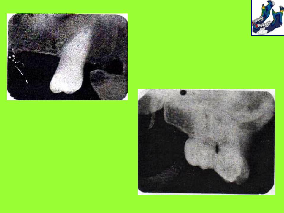
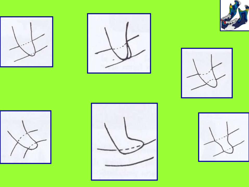
Malar (zygomatic) process. U or j-shaped radiopacity, often superimposed over the roots of the molars, especially when using the bisecting-angle technique. The red arrows define the lower border of the zygomatic bone. facial view
-
Upload
kevin-munoz-h -
Category
Health & Medicine
-
view
616 -
download
4
Transcript of Anatomia 2
Malar (zygomatic) process. U or j-shaped
radiopacity, often superimposed over the roots
of the molars, especially when using the
bisecting-angle technique. The red arrows
define the lower border of the zygomatic bone.
facial view
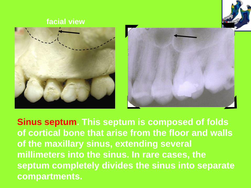
Sinus septum. This septum is composed of folds
of cortical bone that arise from the floor and walls
of the maxillary sinus, extending several
millimeters into the sinus. In rare cases, the
septum completely divides the sinus into separate
compartments.
facial view
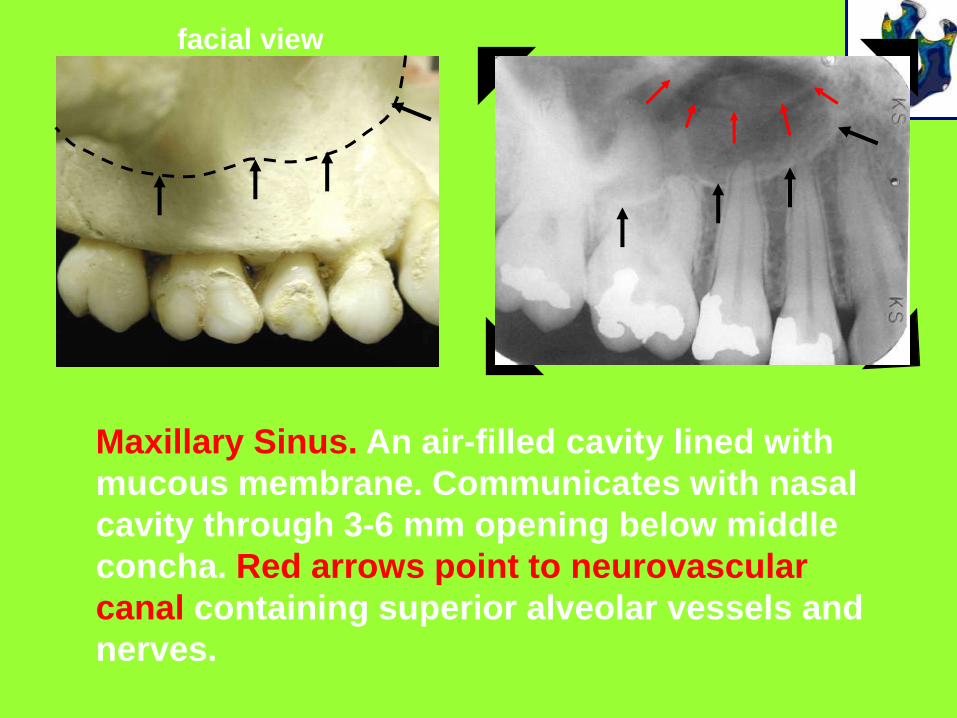
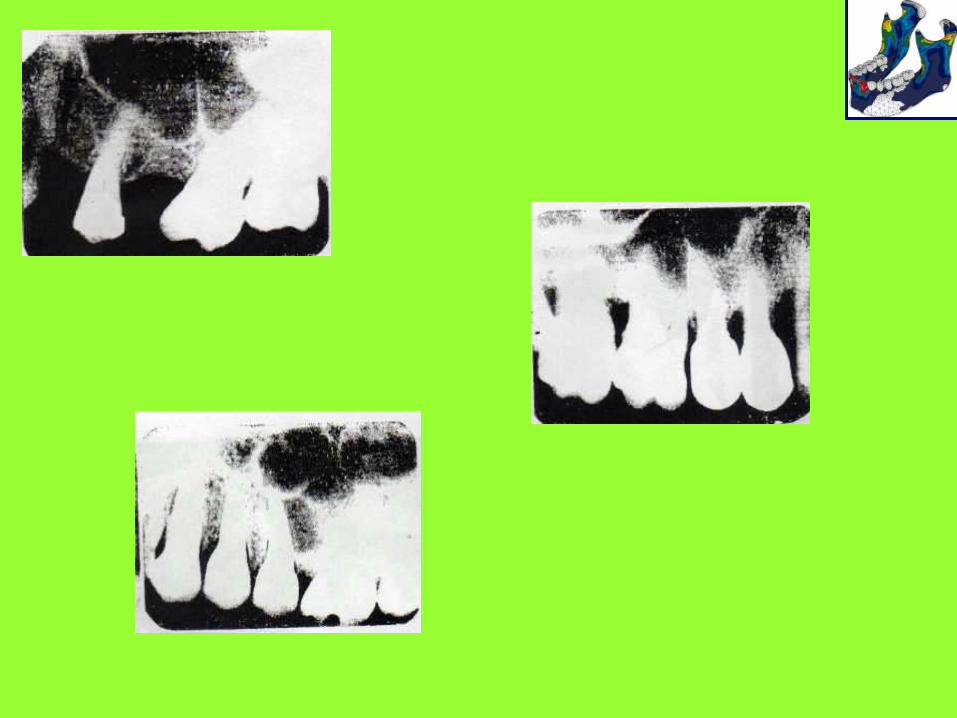
Maxillary Sinus. An air-filled cavity lined with
mucous membrane. Communicates with nasal
cavity through 3-6 mm opening below middle
concha. Red arrows point to neurovascular
canal containing superior alveolar vessels and
nerves.
facial view
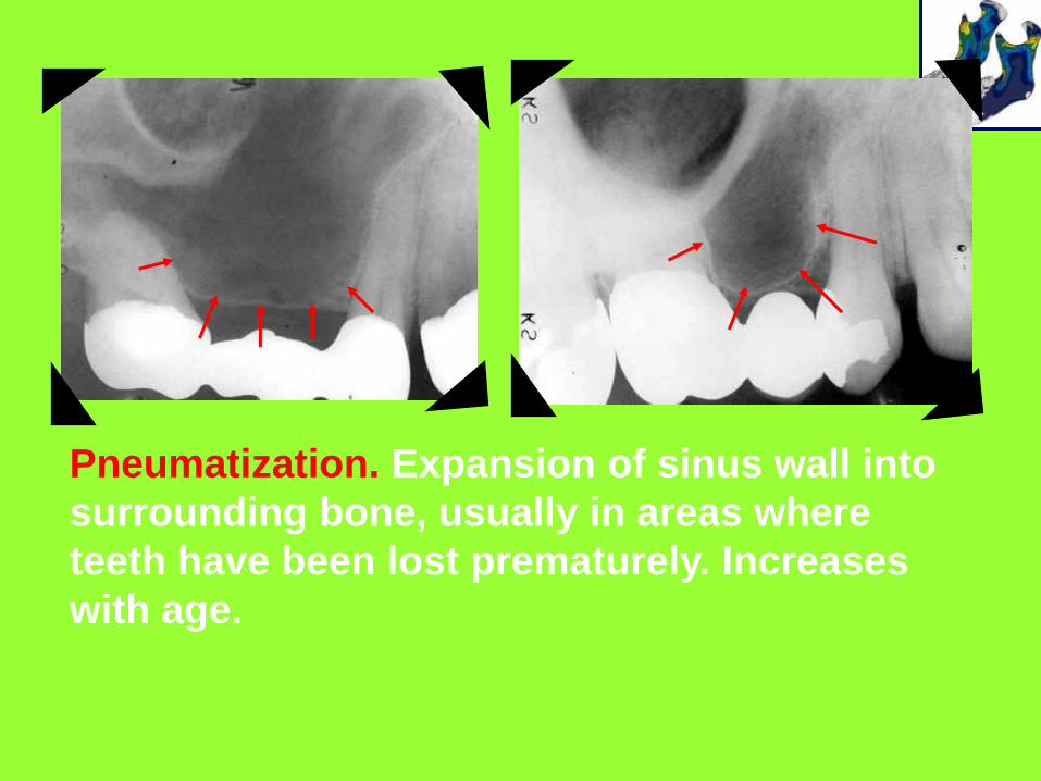
Pneumatization. Expansion of sinus wall into
surrounding bone, usually in areas where
teeth have been lost prematurely. Increases
with age.

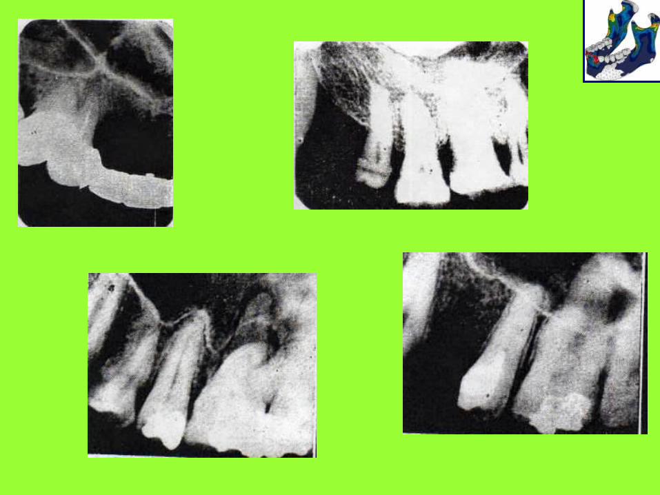
Maxillary Molar
a = maxillary tuberosity
b = coronoid process
c = hamular process
d = pterygoid plates
e = zygoma
f = maxillary sinus
fe
dc
b
a

g
d
a
e
f
a = maxillary tuberosity* e = zygoma (dotted lines)
b = coronoid process f = maxillary sinus
c = hamular process g = sinus recess
d = pterygoid plates
* image of impacted third molar superimposed
c
b
facial view
d
b
a
e
c f
g
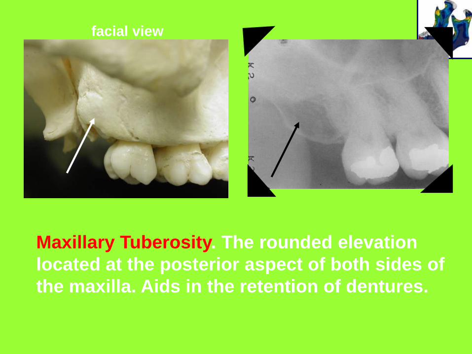
Maxillary Tuberosity. The rounded elevation
located at the posterior aspect of both sides of
the maxilla. Aids in the retention of dentures.
facial view
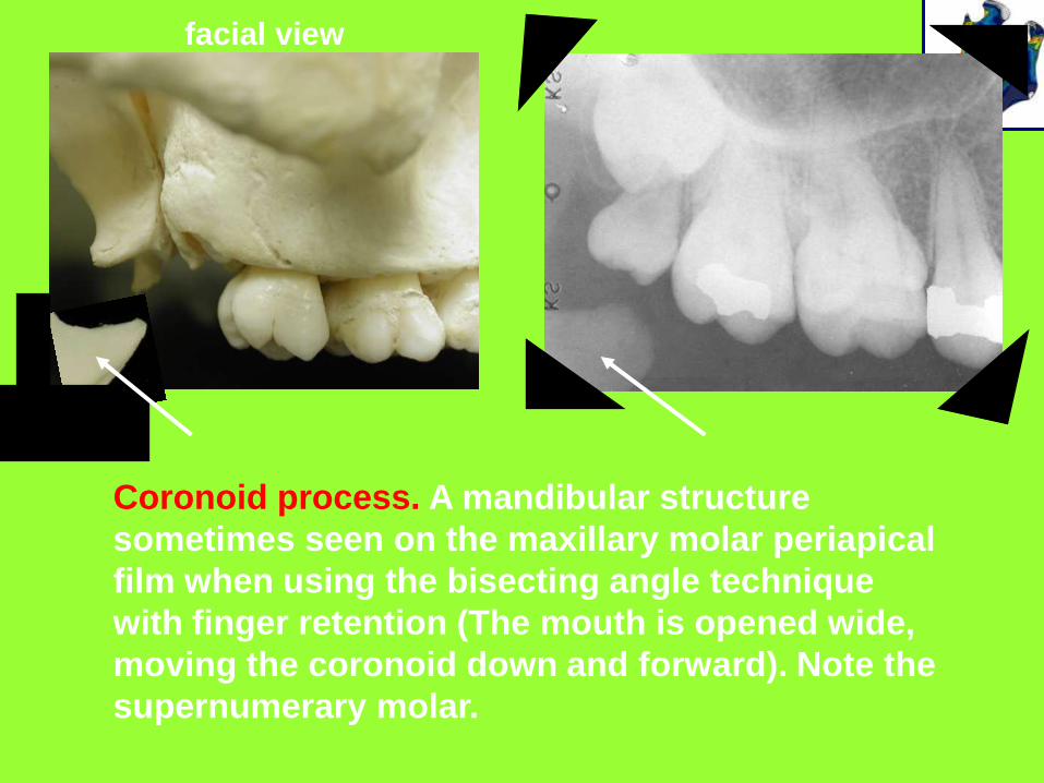
Coronoid process. A mandibular structure
sometimes seen on the maxillary molar periapical
film when using the bisecting angle technique
with finger retention (The mouth is opened wide,
moving the coronoid down and forward). Note the
supernumerary molar.
facial view
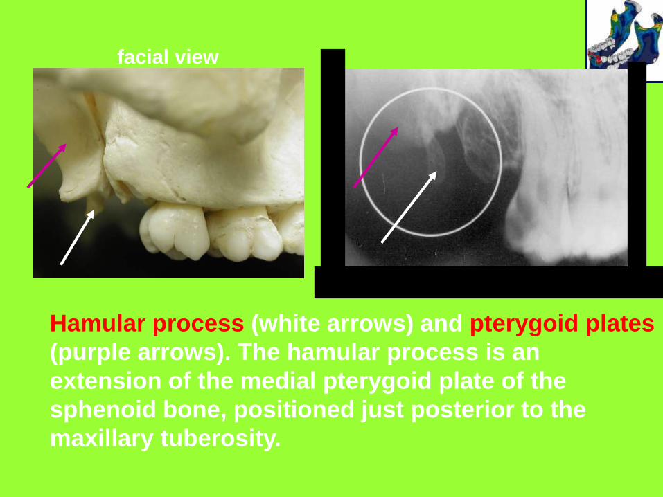
Hamular process (white arrows) and pterygoid plates
(purple arrows). The hamular process is an
extension of the medial pterygoid plate of the
sphenoid bone, positioned just posterior to the
maxillary tuberosity.
facial view
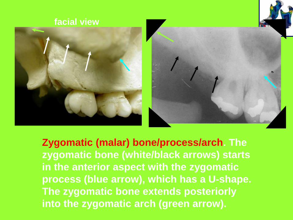
Zygomatic (malar) bone/process/arch. The
zygomatic bone (white/black arrows) starts
in the anterior aspect with the zygomatic
process (blue arrow), which has a U-shape.
The zygomatic bone extends posteriorly
into the zygomatic arch (green arrow).
facial view
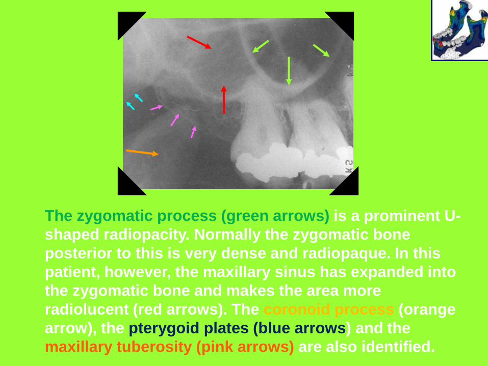
The zygomatic process (green arrows) is a prominent U-
shaped radiopacity. Normally the zygomatic bone
posterior to this is very dense and radiopaque. In this
patient, however, the maxillary sinus has expanded into
the zygomatic bone and makes the area more
radiolucent (red arrows). The coronoid process (orange
arrow), the pterygoid plates (blue arrows) and the
maxillary tuberosity (pink arrows) are also identified.


1.- Bordes orbitario
2.- Seno maxilar
5.- Fosa Pterigo palatina
6.- Hueso cigomatico
7.- Linea Innominada
8.- Arco cigomatico
9.- Condilo
10.- Conducto auditivo interno
11.- Conducto auditivo externo
12.- Radiolucencia causada por la
base del craneo
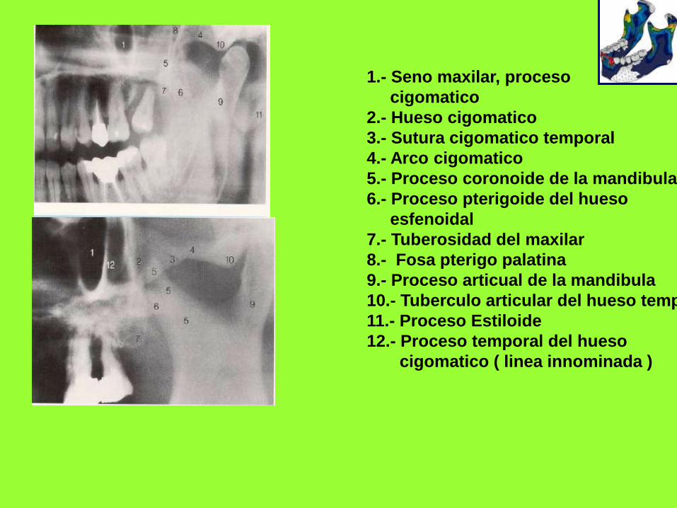
1.- Seno maxilar, proceso
cigomatico
2.- Hueso cigomatico
3.- Sutura cigomatico temporal
4.- Arco cigomatico
5.- Proceso coronoide de la mandibula
6.- Proceso pterigoide del hueso
esfenoidal
7.- Tuberosidad del maxilar
8.- Fosa pterigo palatina
9.- Proceso articual de la mandibula
10.- Tuberculo articular del hueso temp.
11.- Proceso Estiloide
12.- Proceso temporal del hueso
cigomatico ( linea innominada )
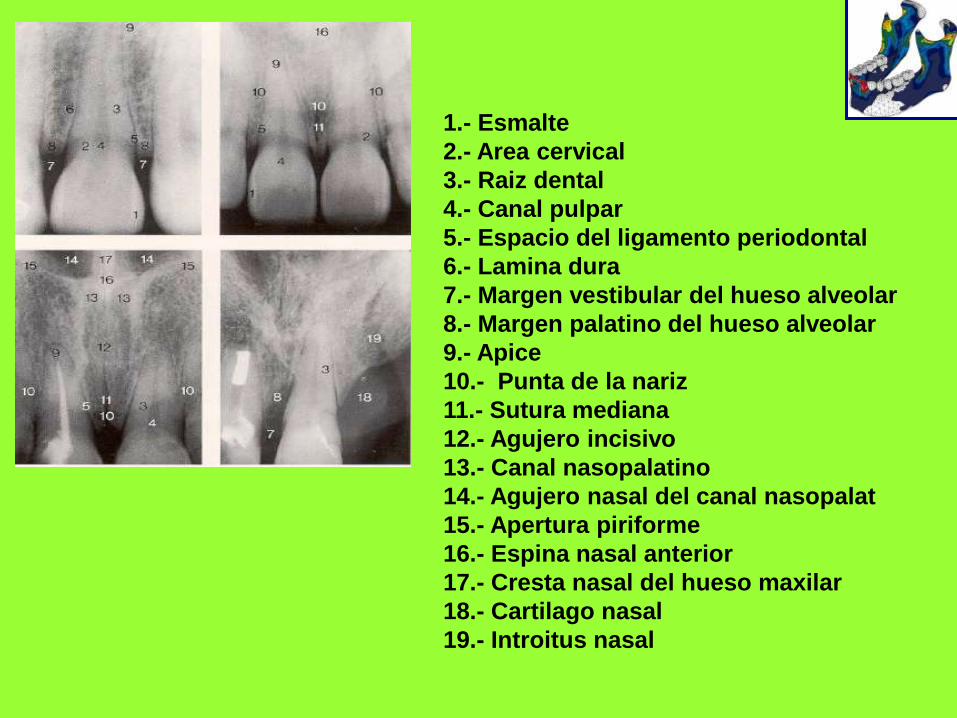
1.- Esmalte
2.- Area cervical
3.- Raiz dental
4.- Canal pulpar
5.- Espacio del ligamento periodontal
6.- Lamina dura
7.- Margen vestibular del hueso alveolar
8.- Margen palatino del hueso alveolar
9.- Apice
10.- Punta de la nariz
11.- Sutura mediana
12.- Agujero incisivo
13.- Canal nasopalatino
14.- Agujero nasal del canal nasopalat
15.- Apertura piriforme
16.- Espina nasal anterior
17.- Cresta nasal del hueso maxilar
18.- Cartilago nasal
19.- Introitus nasal
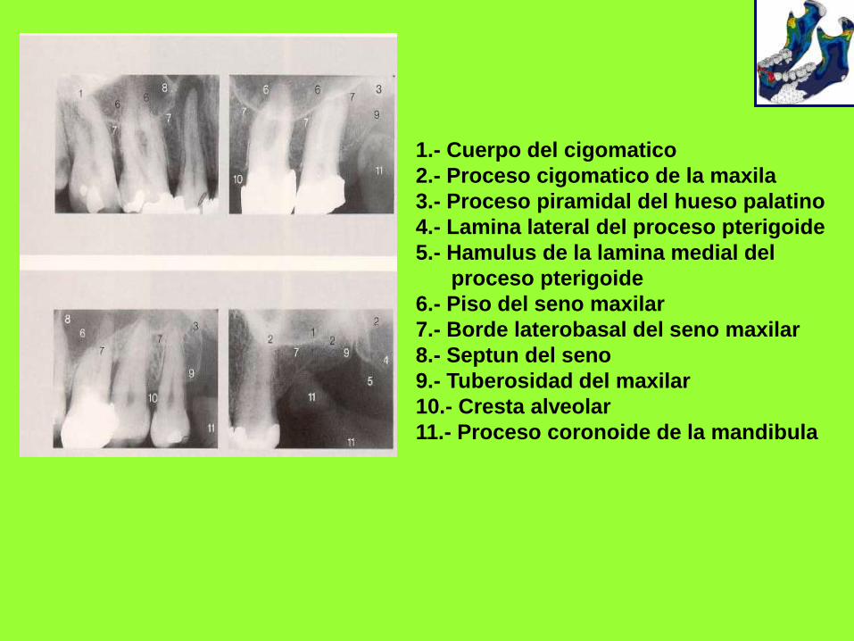
1.- Cuerpo del cigomatico
2.- Proceso cigomatico de la maxila
3.- Proceso piramidal del hueso palatino
4.- Lamina lateral del proceso pterigoide
5.- Hamulus de la lamina medial del
proceso pterigoide
6.- Piso del seno maxilar
7.- Borde laterobasal del seno maxilar
8.- Septun del seno
9.- Tuberosidad del maxilar
10.- Cresta alveolar
11.- Proceso coronoide de la mandibula
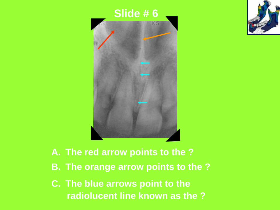
Slide # 6
A. The red arrow points to the ?
B. The orange arrow points to the ?
C. The blue arrows point to the
radiolucent line known as the ?
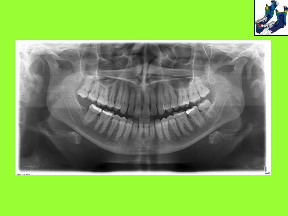
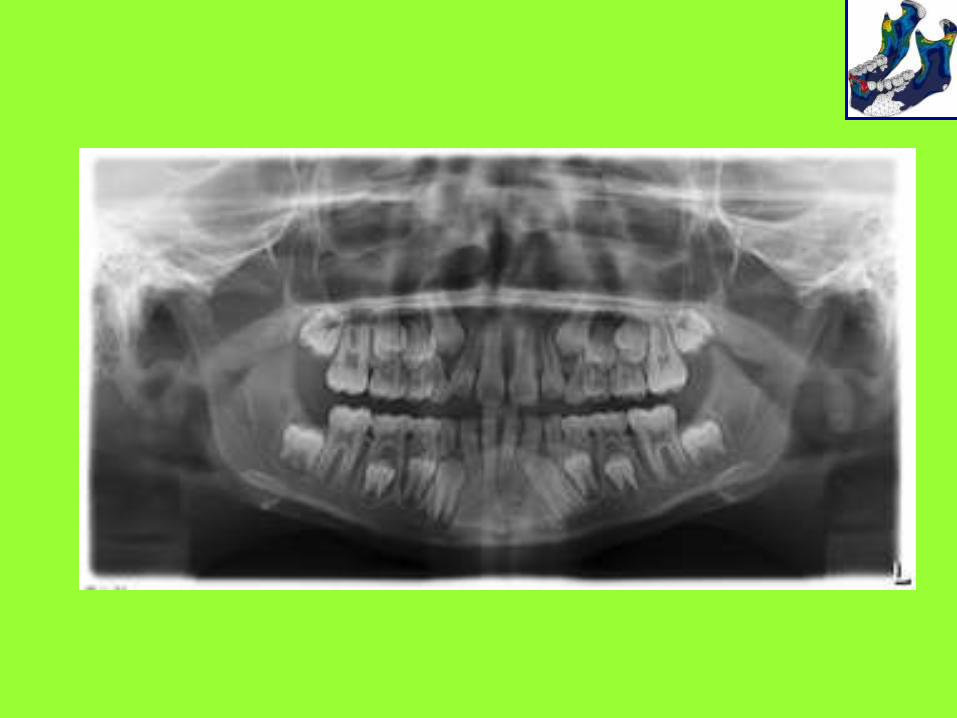
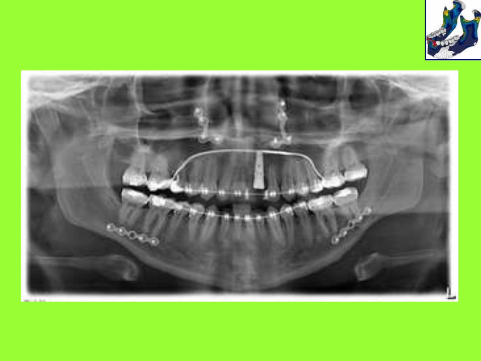
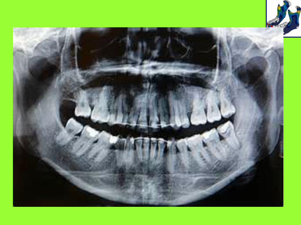
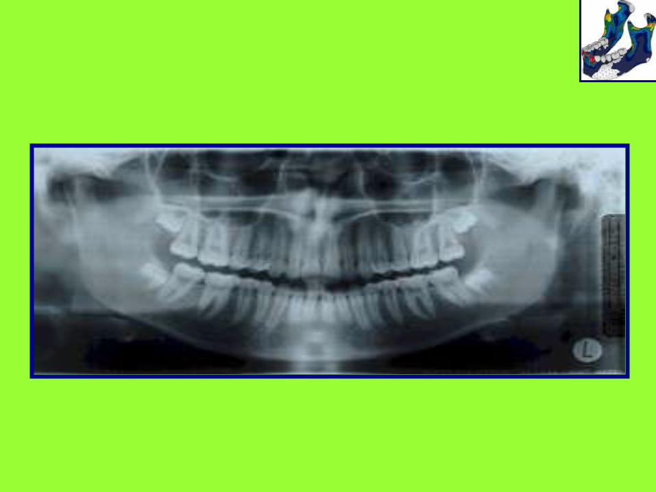
RADIOGRAFIAS
PANORAMICAS
2423
28
27
18
17
1914
1315
20
8
10
97
29
37
38
33
30
39
3
5
11
21
6
1
1216
31
3225
4
26
34
35
36
22
2
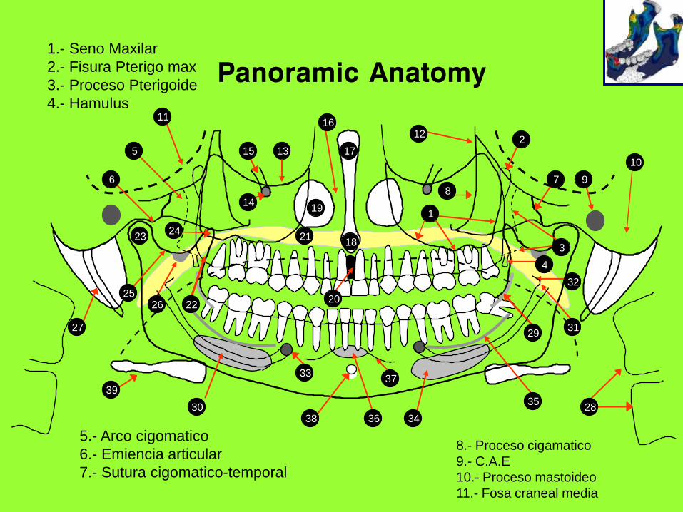
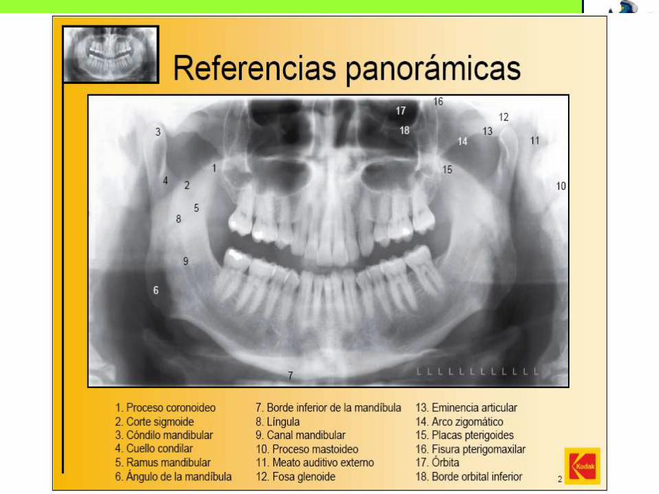
1.- Seno Maxilar
2.- Fisura Pterigo max
3.- Proceso Pterigoide
4.- Hamulus
5.- Arco cigomatico
6.- Emiencia articular
7.- Sutura cigomatico-temporal
8.- Proceso cigamatico
9.- C.A.E
10.- Proceso mastoideo
11.- Fosa craneal media
2423
28
27
18
17
1914
1315
20
8
10
97
29
37
38
33
30
39
3
5
11
21
6
1
1216
31
3225
4
26
34
35
36
22
2
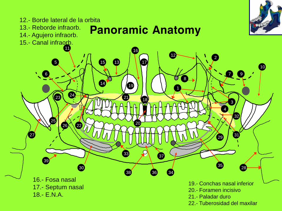
12.- Borde lateral de la orbita
13.- Reborde infraorb.
14.- Agujero infraorb.
15.- Canal infraorb.
16.- Fosa nasal
17.- Septum nasal
18.- E.N.A.
19.- Conchas nasal inferior
20.- Foramen incisivo
21.- Paladar duro
22.- Tuberosidad del maxilar
2423
28
27
18
17
1914
1315
20
8
10
97
29
37
38
33
30
39
3
5
11
21
6
1
1216
31
3225
4
26
34
35
36
22
2
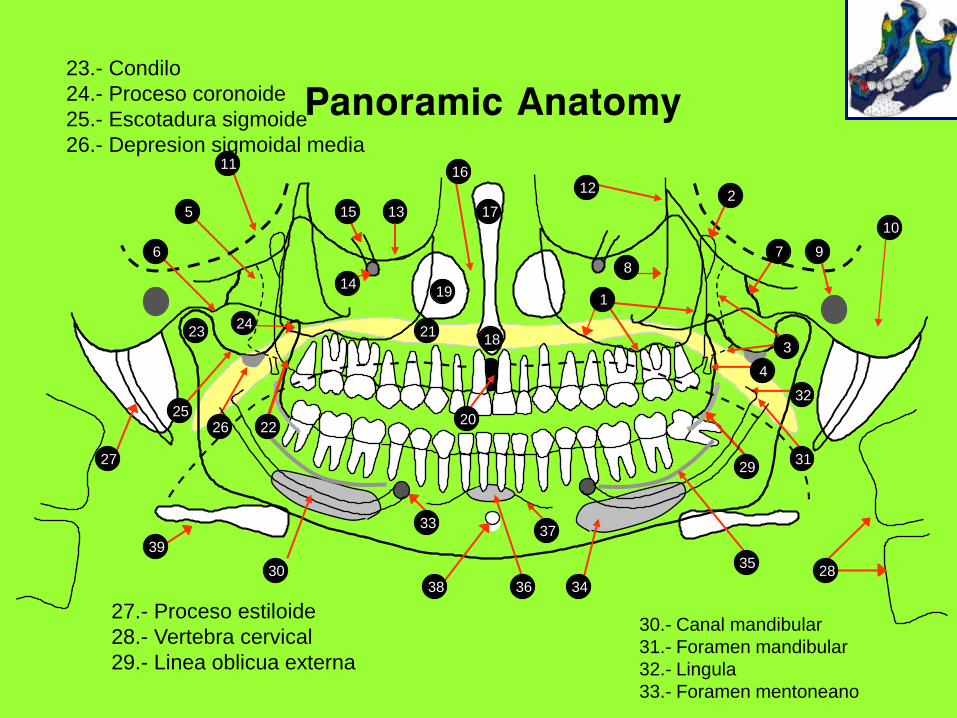
23.- Condilo
24.- Proceso coronoide
25.- Escotadura sigmoide
26.- Depresion sigmoidal media
27.- Proceso estiloide
28.- Vertebra cervical
29.- Linea oblicua externa
30.- Canal mandibular
31.- Foramen mandibular
32.- Lingula
33.- Foramen mentoneano
2423
28
27
18
17
1914
1315
20
8
10
97
29
37
38
33
30
39
3
5
11
21
6
1
1216
31
3225
4
26
34
35
36
22
2
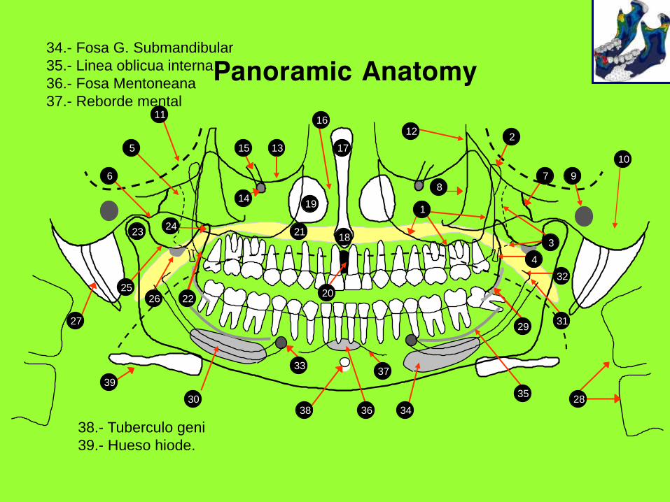
34.- Fosa G. Submandibular
35.- Linea oblicua interna
36.- Fosa Mentoneana
37.- Reborde mental
38.- Tuberculo geni
39.- Hueso hiode.
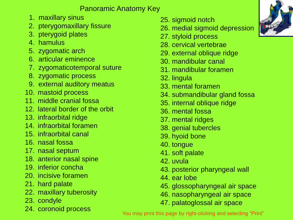
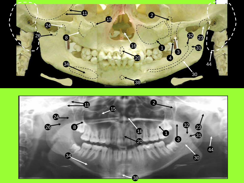
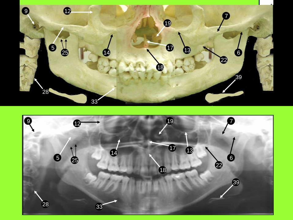
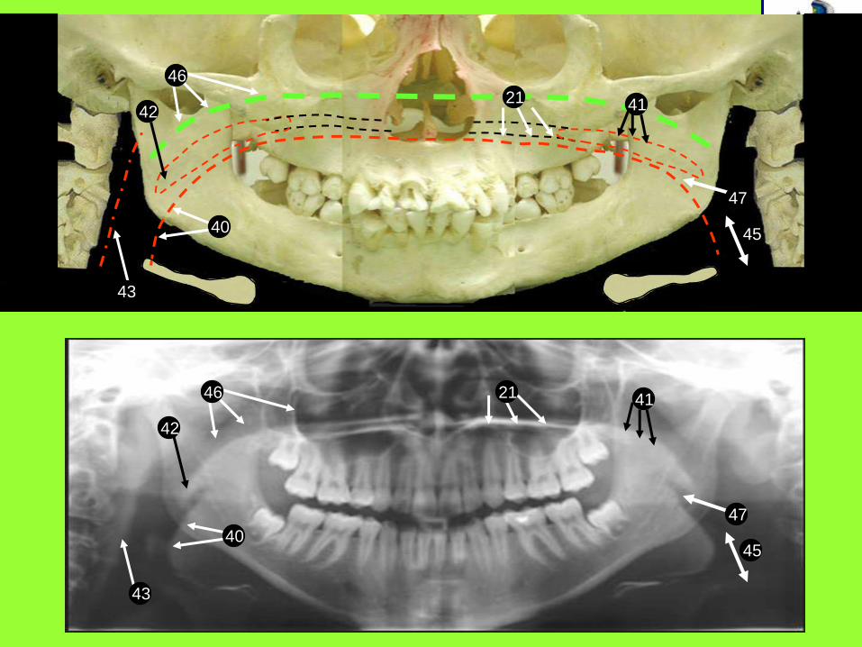
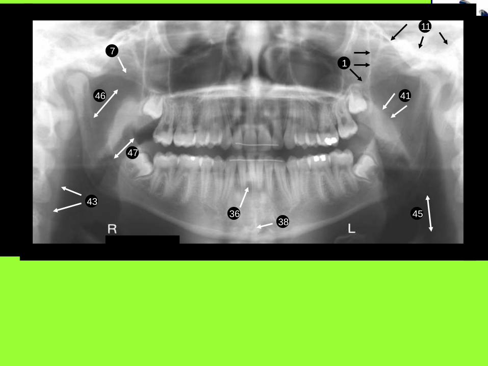
Panoramic Anatomy Key
1. maxillary sinus
2. pterygomaxillary fissure
3. pterygoid plates
4. hamulus
5. zygomatic arch
6. articular eminence
7. zygomaticotemporal suture
8. zygomatic process
9. external auditory meatus
10. mastoid process
11. middle cranial fossa
12. lateral border of the orbit
13. infraorbital ridge
14. infraorbital foramen
15. infraorbital canal
16. nasal fossa
17. nasal septum
18. anterior nasal spine
19. inferior concha
20. incisive foramen
21. hard palate
22. maxillary tuberosity
23. condyle
24. coronoid process
25. sigmoid notch
26. medial sigmoid depression
27. styloid process
28. cervical vertebrae
29. external oblique ridge
30. mandibular canal
31. mandibular foramen
32. lingula
33. mental foramen
34. submandibular gland fossa
35. internal oblique ridge
36. mental fossa
37. mental ridges
38. genial tubercles
39. hyoid bone
40. tongue
41. soft palate
42. uvula
43. posterior pharyngeal wall
44. ear lobe
45. glossopharyngeal air space
46. nasopharyngeal air space
47. palatoglossal air space
You may print this page by right-clicking and selecting “Print”
8
20
1115
116
3
30
44
32 23
2
31
26
38
34
24
8
20
11
15
1
2
3
30
44
32 23
31
38
34
16
24
26
4
9
525
28
14
33
12
18
17
19
13
22
7
39
6
33
255
28
9 12
14
18
17
19
22
13
7
6
39
40
43
43
42
42
4121
40
21
46
46
41
45
45
47
47
36
41
38
7
11
1
43
47
46
45

9
11
320

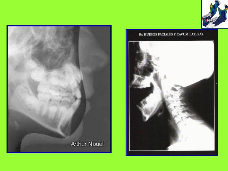
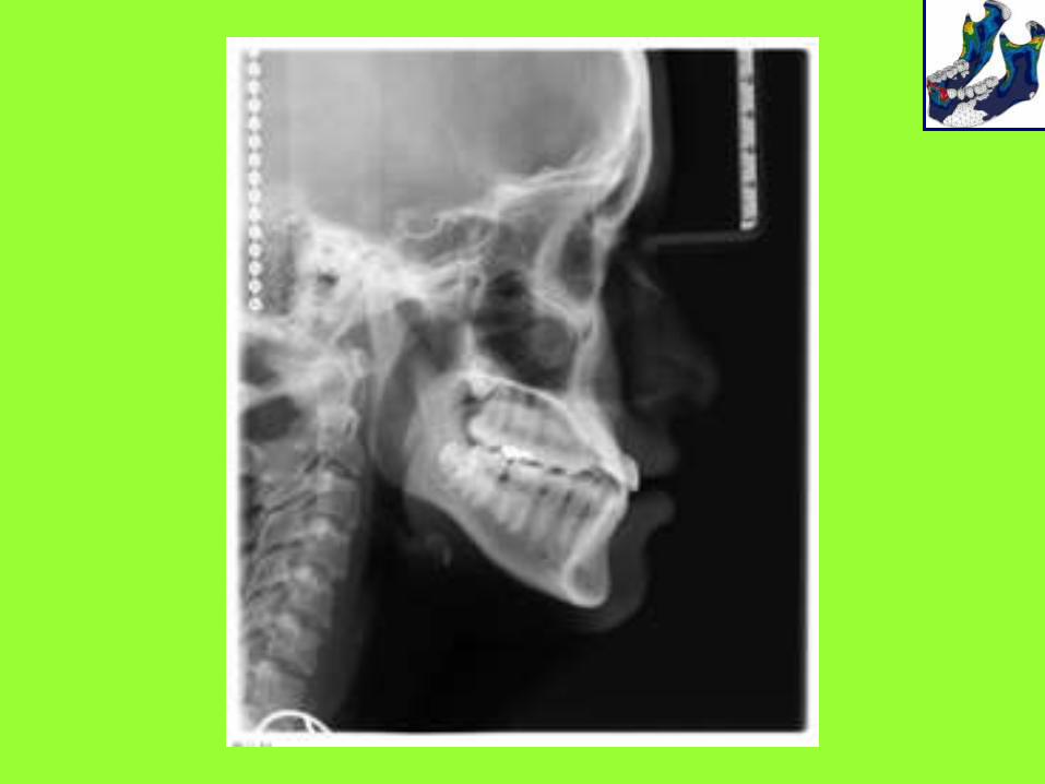
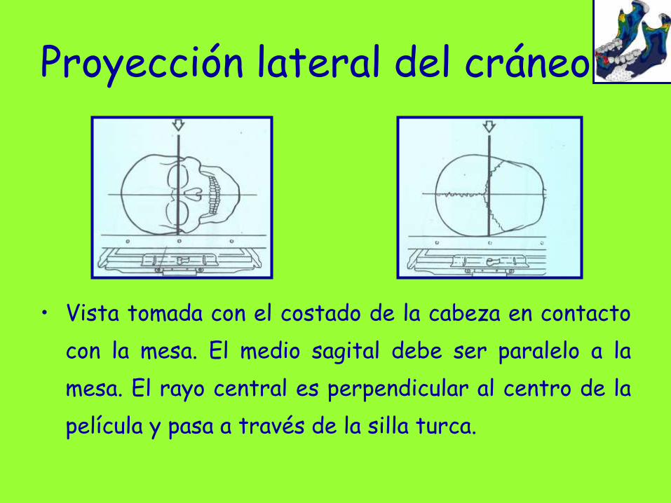
Proyección lateral del cráneo
• Vista tomada con el costado de la cabeza en contacto
con la mesa. El medio sagital debe ser paralelo a la
mesa. El rayo central es perpendicular al centro de la
película y pasa a través de la silla turca.
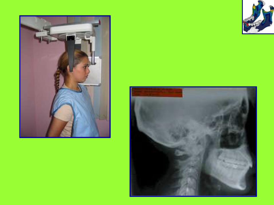
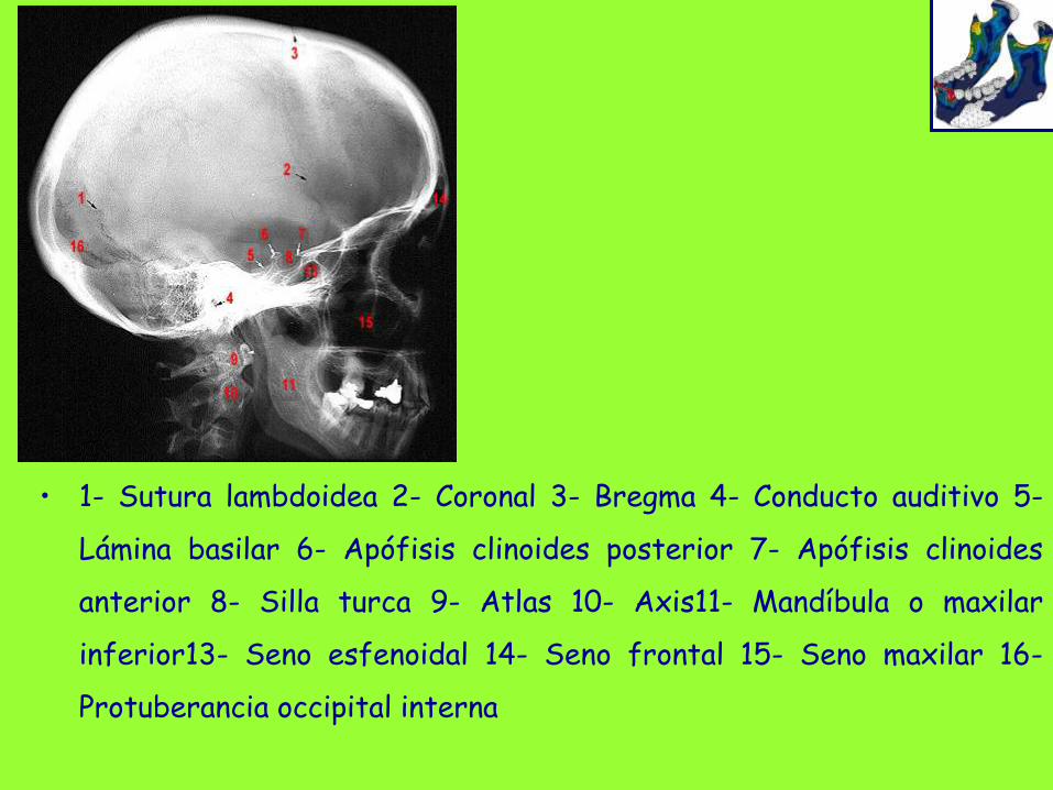
• 1- Sutura lambdoidea 2- Coronal 3- Bregma 4- Conducto auditivo 5-
Lámina basilar 6- Apófisis clinoides posterior 7- Apófisis clinoides
anterior 8- Silla turca 9- Atlas 10- Axis11- Mandíbula o maxilar
inferior13- Seno esfenoidal 14- Seno frontal 15- Seno maxilar 16-
Protuberancia occipital interna
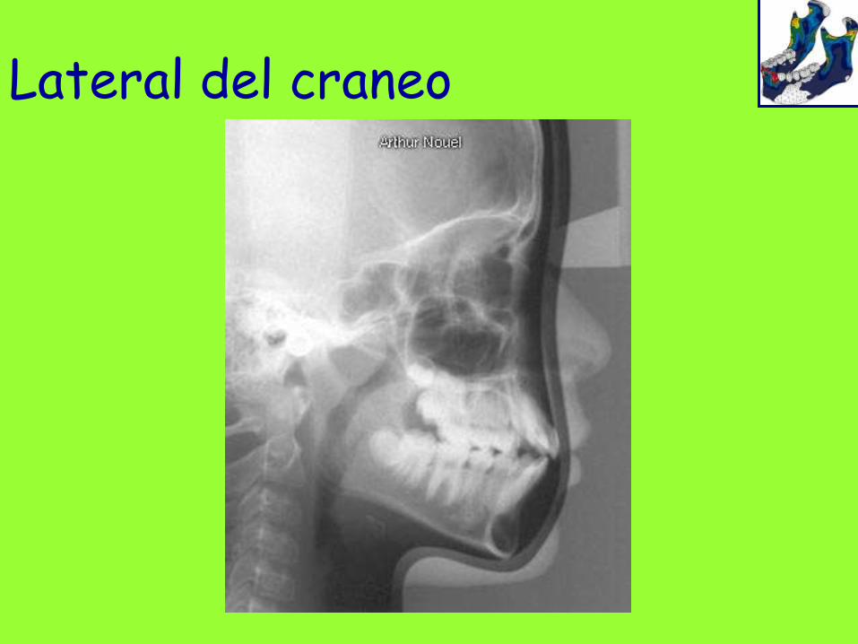
Lateral del craneo
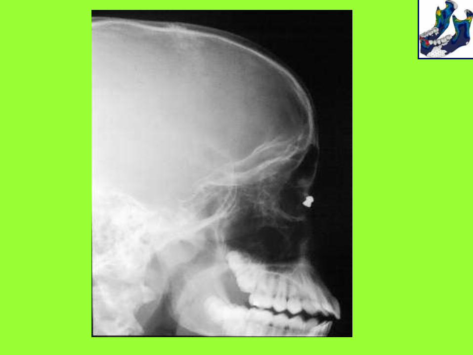
Proyección lateral del cráneo
• Vista tomada con el costado de la cabeza en contacto
con la mesa. El medio sagital debe ser paralelo a la
mesa. El rayo central es perpendicular al centro de la
película y pasa a través de la silla turca.
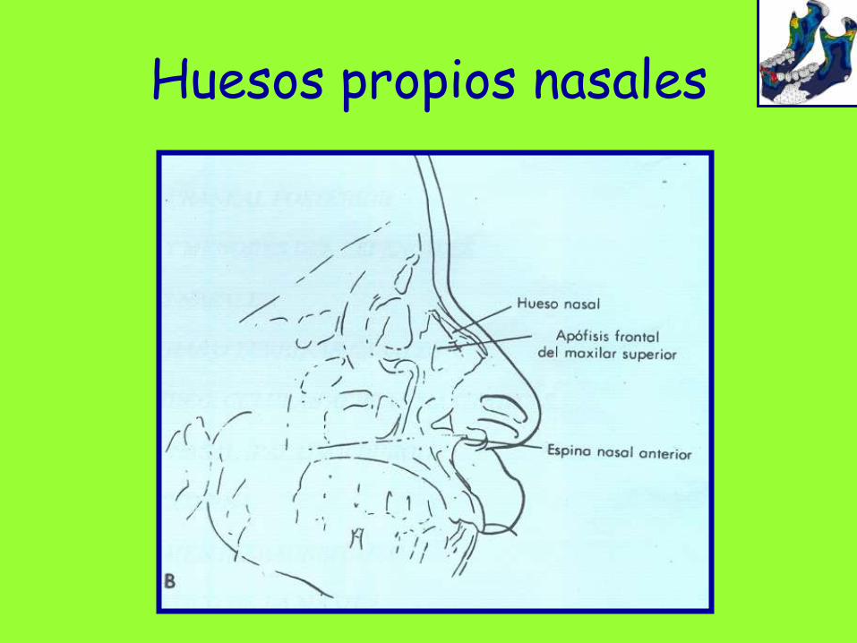
Huesos propios nasales


Legend for Fig. 128:
1 Saggital suture
2 Frontal sinus, with septa
3 Cirsta galli with falciform process
of cerebrum
4 Cribriform lamina of ethmoid bone
5 Nasal bone
6 Orbit
7 Innominate line (of the major ala)
8 Minor ala of the sphenoid bone
9 Median cranial fossa (borders)
10 Optical canal
11 Superior orbital fissure
12 Infraorbital canal
13 Foramen rotundum (round
foramen of
sphenoid bone)
14 Ethmoid labyrinth
15 Osseous nasal septum
16 Nasal conchae

17 Maxillary sinus
18 Zygomatic bone
19 Frontal process of the
zygomatic bone
20 Frontal zygomatic suture
21 Zygomatic arch
22 Zygomaticoalveolar crest
23 Condylar process of the
mandible
24 Coronoid process of the
mandible
25 Mastoid sinuses
26 Petrosal portion of the
temporal bone
27 Anterior nasal spine
28 Posterior nasal spine
29 Sphenoidal spine
30 Basilar apophysis
31 Dorsum of the tongue
32 Lateral mass of atlas
33 Odontoid bone (dentoid
process of axis)
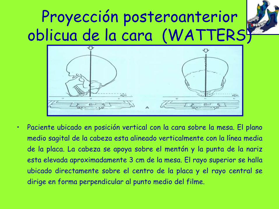
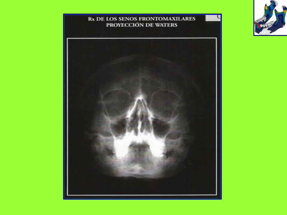
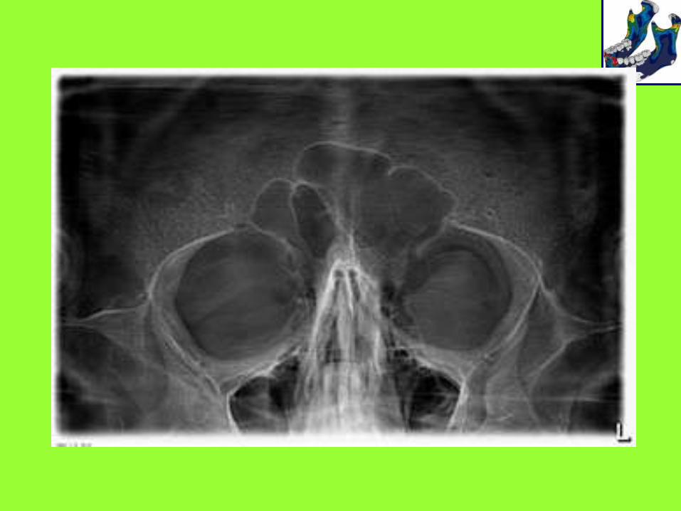
Proyección posteroanterior oblicua de la cara (WATTERS)
• Paciente ubicado en posición vertical con la cara sobre la mesa. El plano
medio sagital de la cabeza esta alineado verticalmente con la línea media
de la placa. La cabeza se apoya sobre el mentón y la punta de la nariz
esta elevada aproximadamente 3 cm de la mesa. El rayo superior se halla
ubicado directamente sobre el centro de la placa y el rayo central se
dirige en forma perpendicular al punto medio del filme.
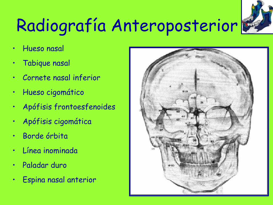
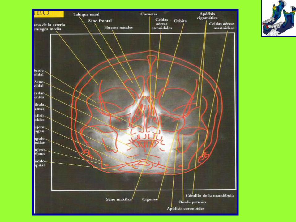
Radiografía Anteroposterior• Hueso nasal
• Tabique nasal
• Cornete nasal inferior
• Hueso cigomático
• Apófisis frontoesfenoides
• Apófisis cigomática
• Borde órbita
• Línea inominada
• Paladar duro
• Espina nasal anterior
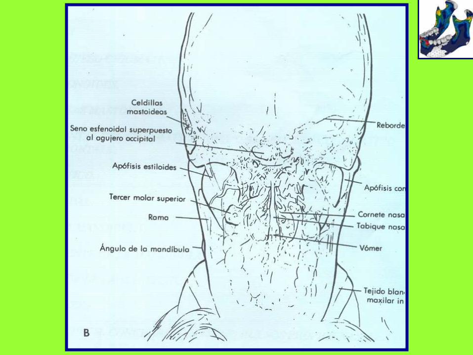
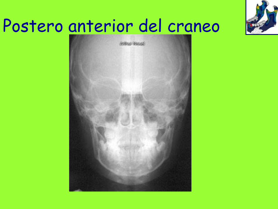
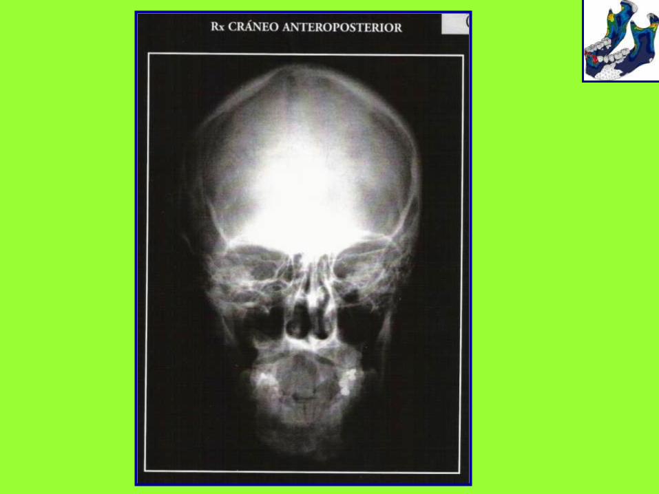
Postero anterior del craneo
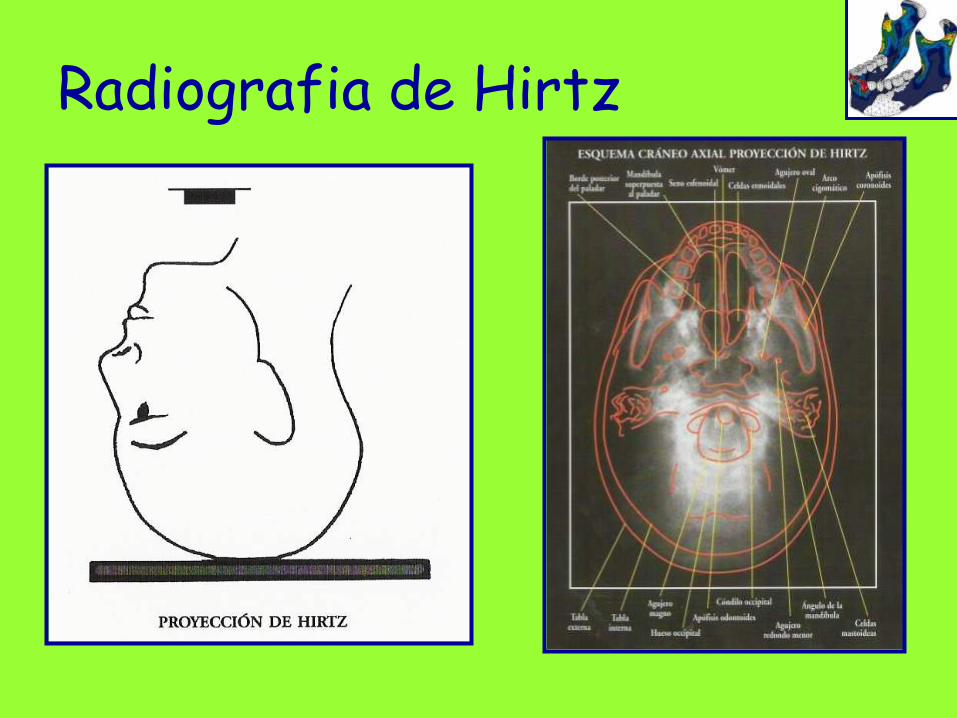
Radiografia de Hirtz