American Board of Dental Examiners (ADEX) Dental Examination · The ADEX Dental Examination Series...
164
CANDIDATE MANUAL American Board of Dental Examiners (ADEX) Dental Examination Curriculum Integrated Format Class of 2014 Approved by American Board of Dental Examiners, Inc. Administered by: Council of Interstate Testing Agencies, Inc. 1003 High House Road, Suite 101 Cary, NC 27513 www.citaexam.com North East Regional Board of Dental Examiners, Inc. 1304 Concourse Drive, Suite 100 Linthicum, MD 21090 www.nerb.org AND Southern Regional Testing Agency, Inc. 4698 Honeygrove Road, Suite 2 Virginia Beach, VA 23455 www.srta.org Please read this manual in detail prior to attending the examination and bring it with you to the orientation and examination. This manual should also be retained for future reference. Copyright © 2013 American Board of Dental Examiners, Inc. Copyright © 2013 North East Regional Board of Dental Examiners, Inc. Copyright © 2013 Southern Regional Testing Agency, Inc.
-
Upload
nguyennhan -
Category
Documents
-
view
219 -
download
0
Transcript of American Board of Dental Examiners (ADEX) Dental Examination · The ADEX Dental Examination Series...
CANDIDATE MANUAL
American Board of Dental Examiners (ADEX)
Dental Examination
Curriculum Integrated FormatClass of 2014
Approved byAmerican Board of Dental Examiners, Inc.
Administered by:
Council of Interstate Testing Agencies, Inc.1003 High House Road, Suite 101
Cary, NC 27513www.citaexam.com
North East Regional Board of Dental Examiners, Inc. 1304 Concourse Drive, Suite 100
Linthicum, MD 21090www.nerb.org
AND
Southern Regional Testing Agency, Inc.4698 Honeygrove Road, Suite 2
Virginia Beach, VA 23455www.srta.org
Please read this manual in detail prior to attending the examinationand bring it with you to the orientation and examination.This manual should also be retained for future reference.
Copyright © 2013 American Board of Dental Examiners, Inc.Copyright © 2013 North East Regional Board of Dental Examiners, Inc.
Copyright © 2013 Southern Regional Testing Agency, Inc.
ATTENTION DENTAL CANDIDATES
The ADEX Dental Examination Series is administered on behalf of a number of state dental boards and in accordance with state licensing requirements. This examination should be valid in any state accepting the ADEX Dental Examination. However, to be certain, candidates should check with the state dental board of any state in which they wish to be licensed to determine whether this examination will qualify them for licensure in that state.
Currently there are three testing agencies which administer the ADEX examination. Although content, basic administration and scoring systems are uniform, each agency may have some administrative elements which are unique to that agency. Therefore candidates should obtain and thoroughly read the manual published by the agency which will be administering the examination at the date and site the candidate selects. This manual is published jointly by the North East Regional Board, Inc. (NERB) and the Southern Regional Testing Agency, Inc. (SRTA).
For information about examination sites, dates and fees, visit the CITA website at www.citaexam.org, NERB website: www.nerb.org. or the SRTA website: www.srta.org The examination sites and dates will be found under the Examination Calendar drop-down list.
Occasionally examinations are interrupted or postponed because of hurricanes, blizzards, other severe weather, power outages, or similar occurrences. CITA, NERB and SRTA reserve the right in its sole discretion, to delay, halt, postpone, or cancel an examination because of unforeseen and serious events.
Table of ContentsThe ADEX Dental Examination Series ..................................................................................... 2 I. Introduction ............................................................................................................................. 2 Introduction ................................................................................................................................ 3
About the ADEX Dental Examination Series – 2014 ............................................................. 3 Purpose of the Examination Series ....................................................................................... 3 About the American Board of Dental Examiners, Inc. (ADEX) ............................................... 3 ADEX Mission Statement ............................................................................................. 4 Obtaining Licensure ............................................................................................................... 4 Eligibility for the ADEX Examination Series ............................................................................ 4 ADEX Status .......................................................................................................................... 5
The ADEX Dental Examination Series ..................................................................................... 6 II. Examination .......................................................................................................................... 6 Examination Overview ............................................................................................................... 7
About the Curriculum Integrated Format ............................................................................... 7 Examination Schedules .......................................................................................................... 7 Dates and Sites ............................................................................................................ 7 Score Release ...................................................................................................................... 7 Examination Scoring System ................................................................................................. 7 Penalties ....................................................................................................................... 8 Professional Conduct ................................................................................................... 9 18-Month Completion Rule .......................................................................................... 9 3-Time Failure Rule ...................................................................................................... 9 Section I: Diagnostic Skills Examination (DSE) – Based on 100 Points ............................ 11 Procedures Specific to the Endodontics Examination Section .................................. 13 Periodontal Scaling Examination Section Procedure and Patient Management Guidelines . 32 Retesting Schedule .............................................................................................................. 34 Restorative Examination Section Requirements .................................................................. 40 Radiographs .............................................................................................................. 40 Treatment Selection and Patient Check-in ................................................................. 41 Treatment Guidelines ................................................................................................ 43 Modification Requests ................................................................................................ 44 Restorative Procedure and Patient Management Guidelines .............................................. 47 Separation of Scoring for the Anterior and Posterior Restorations ....................................... 48 Requirements for the Class III Composite Preparation and Restoration ............................. 49 Requirements for Class II Amalgam Preparation and Restoration ....................................... 50 Requirements for Class II Composite Preparation and Restoration ..................................... 51 Requirements for the Posterior Slot Composite Preparation and Restoration ..................... 51
The ADEX Dental Examination Series .................................................................................... 77 VI. Examination Forms ............................................................................................................ 77 Examination Forms .................................................................................................................. 78
Forms Completed Before the Examination ........................................................................... 78 Medical History Form ................................................................................................. 78 Patient Consent Form (Patient Consent, Disclosure and Assumption of Liability) .... 78 Periodontal Scaling Treatment Selection Worksheet ................................................. 78 Electronic Periodontal Scaling Evaluation Form ........................................................ 79 Forms Completed at the Examination .................................................................................. 79 Progress Forms .......................................................................................................... 79 Modification Request Form ........................................................................................ 79 Instruction to Candidate Form .................................................................................... 80
Follow-Up Form .......................................................................................................... 80 Medical History Form .............................................................................................................. 81 Progress Forms: Periodontal & Pros/Endo ........................................................................... 84 Instruction to Candidate Form ................................................................................................ 87 Follow-up Form ........................................................................................................................ 88 The ADEX Dental Examination Series ................................................................................... 89 IV. Policies and Procedures .................................................................................................... 89
Standards of Conduct .......................................................................................................... 90 Patient’s Medical History ............................................................................................ 94 Patient Selection ........................................................................................................ 96 Procedures ................................................................................................................. 96 General Guidelines for Clinical Exercises ............................................................................ 98 Infection Control Procedures ................................................................................................ 99
The ADEX Dental Examination Series ................................................................................. 101 V. Score Certification and Appeals ........................................................................................................................... 101 Score Certification and Appeals ........................................................................................................................... 102
Score Certification Procedure ............................................................................................ 102 Candidate Appeals Procedure ........................................................................................... 102
APPENDIX A ........................................................................................................................... 103 ................................................................................................................................................ 103
Glossary of Words, Terms and Phrases ............................................................................. 104 APPENDIX B ........................................................................................................................... 113
Certification of Status as a Graduate Student of Record .................................................... 114 SOUTHERN REGIONAL TESTING AGENCY SUPPLEMENT ................................................... 3
About the Southern Regional Testing Agency, Inc. (SRTA) ................................................... 6 SRTA Member States ........................................................................................................... 6 Examination Sites, Dates and Fees ....................................................................................... 6 Progressive Integrated Examination (PIE) Sites and Dates ........................................ 6 Traditional and Sectional Examination Sites and Dates .............................................. 7 Examination Fees .................................................................................................................. 8 PIE Examination Fees .................................................................................................. 8 Traditional Examination Fees ....................................................................................... 8 Reexamination Fees .................................................................................................... 8 Application Procedure ............................................................................................................ 9 Diagnostic Skills Examination (DSE) Section ......................................................................... 9 Online Registration Process ................................................................................................. 10 Test Site Locations ............................................................................................................... 12 Schedule of Examinations .................................................................................................... 13 PIE I Examination Schedule ....................................................................................... 13 PIE II and Traditional Examination Schedule ............................................................. 14 Policies and Procedures ...................................................................................................... 17 Appeals ............................................................................................................................... 17 Assistants ............................................................................................................................. 18 Candidate Accessibility ...................................................................................................... 19 Candidate Guidelines ........................................................................................................... 19 Candidate Qualifications ...................................................................................................... 19 Candidate Responsibilities after the Examination ................................................................ 20 Dismissal for Unethical Conduct .......................................................................................... 20 Dismissal or Failure - Reasons ............................................................................................ 20 Electronic Equipment ........................................................................................................... 22 Evaluation after Designated Cutoff Time .............................................................................. 22 Examination Documents ...................................................................................................... 22
Examination Results ............................................................................................................ 22 Ineligible Candidates ............................................................................................................ 23 Interpreters .......................................................................................................................... 23 Malpractice Insurance .......................................................................................................... 23 Medical History Form ........................................................................................................... 23 Patient Approval ................................................................................................................... 25 Patient Privacy Statement .................................................................................................... 25 Patient Selection and Guidelines ......................................................................................... 25 Professional Standards and Competency ............................................................................ 26 Questions ............................................................................................................................. 26 Radiographs - Standards of Acceptability ............................................................................ 27 Reexamination Requirements ............................................................................................. 29 Refunds ............................................................................................................................... 29 State Boards of Dentistry and Licensure Information ........................................................... 29 Supplies Provided by Test Sites ........................................................................................... 31
Frequently Asked Questions .................................................................................................. 32 Helpful Hints ............................................................................................................................. 34 Checklist ................................................................................................................................... 36
Candidate Criteria Quick Reference ..................................................................................... 37
Policies and Procedures Supplement for the appropriate testing agency (NERB or SRTA) is attached to the end of this manual. This includes:
Exam schedules for specific exam sitesAdmission and orientation informationScore reporting and retake informationSpecial testing requestsRegistration proceduresOnline registrationCheck listFrequently Asked QuestionsHelpful HintsCandidate Criteria Quick Reference
The ADEX Dental Examination Series
Curriculum Integrated Format
I. Introduction
2
IntroductionAbout the ADEX Dental Examination Series – 2014
The ADEX Dental Examination Series is the examination approved by the American Board of Dental Examiners, Inc. (ADEX) and administered by the Council of Interstate Testing Agencies, Inc. (CITA), the North East Regional Board of Dental Examiners, Inc. (NERB) and the Southern Regional Testing Agency, Inc. (SRTA). The ADEX Examination Series consists of computer simulations and clinical examinations performed on patients and manikins. The ADEX Examination Series is utilized to assist licensing jurisdictions in making decisions concerning the licensure of dentists. The ADEX Dental Examination Series for 2014 consists of up to five individual, skill-specific clinical examinations:
Three simulated clinical examinations-Computer-based Diagnostic Skills Examination (DSE) Section-Endodontic Clinical Examination Section (manikin-based)-Fixed Prosthodontic Clinical Examination Section (manikin-based)
Two clinical examinations performed on patients-Restorative Clinical Examination Section-Periodontal Scaling Clinical Examination Section (optional, based on the requirements in the state where the candidate seeks licensure)
Candidates taking this examination do so voluntarily and agree to accept the provisions and to follow the rules established by ADEX, CITA, NERB and SRTA for the examination as detailed in this manual.
Purpose of the Examination Series
This Candidate Manual has been designed to assist in candidate’s preparation for and participation in this examination series. The purpose of the ADEX Examination Series is to provide state dental boards with a uniform, accurate, third party assessment of the clinical skills of candidates who are applying for dental licensure and to identify areas of deficiency or weakness within skill sets so that candidates and dental schools can accomplish remediation. The examination series is based on specific performance criteria used to measure clinical competence.
About the American Board of Dental Examiners, Inc. (ADEX)
The American Board of Dental Examiners, Inc. (ADEX) is a private not-for-profit consortium of state and regional dental boards throughout the United States and its territories. ADEX provides for the ongoing development of a series of common, national dental licensing examinations that are uniformly administered by individual state or regional testing agencies on behalf of their participating and recognizing licensing jurisdictions.CITA, NERB and SRTA are members of ADEX and have adopted the ADEX Dental Examination Series.
3
ADEX Mission Statement
To provide the dental community with test construction and administrative standardization for national uniform dental and dental hygiene clinical licensure examinations. The schedule of these examinations, when delivered in the Curriculum Integrated Format (CIF), allows for early identification of deficiencies or weaknesses within clinical skill sets and provides opportunities for remediation in an educational environment. These examinations will demonstrate integrity and fairness in order to assist state boards of dentistry with their mission to protect the health, safety and welfare of the public by assuring that only competent and qualified individuals are allowed to practice dentistry and dental hygiene.
Obtaining Licensure
Typically, applicants must complete three steps in order to obtain a dental license.
1. The candidate must take and successfully complete Parts I and II of the National Board Dental Examinations, typically offered during dental school.
2. The candidate must take and pass the appropriate state or regional clinical examination. CITA, NERB and SRTA are three of these regional testing agencies. Proof of passing the National Board examinations prior to taking the ADEX Examination Series is not required. The school where the clinical examination takes place may have forms that need to be completed and may require a separate fee for the use of its facilities and/or equipment during the examination.
3. The candidate must apply for state licensure. The state board of dentistry in the state in which the candidate wishes to practice will require proof that the candidate has passed Parts I and II of the National Boards and the appropriate state or regional clinical examination. State boards of dentistry will also require proof of graduation from an accredited dental school and other documentation. Candidates should familiarize themselves with the requirements of the state(s) in which they wish to be licensed as soon as possible and complete an application with that individual jurisdiction. Passing the ADEX Examination Series and obtaining ADEX status does not necessarily mean that all state required clinical examinations have been completed nor does this automatically lead to a state dental license.
Candidates should address questions to the appropriate agency:• The Joint Commission on National Dental Examinations can answer questions about Parts I and II of the
National Boards.• CITA, NERB or SRTA can answer questions about the ADEX Examination Series.• Questions regarding licensure or state requirements should be addressed to the appropriate state board of
dentistry.
Eligibility for the ADEX Examination Series
Students (or graduate students) of record attending a dental school accredited by the American Dental Association Commission on Dental Accreditation or the Commission on Dental Accreditation of Canada are eligible to apply to take the Curriculum Integrated Format of the ADEX Dental Examination Series when the dean (or designated school official) certifies that the candidate is a student (or graduate student) and is sufficiently prepared to participate. Students in schools not participating in the Curriculum Integrated Format examinations or dentists who are graduates of U.S. or international dental schools are not eligible to participate in the Curriculum Integrated Format examinations. They should apply online to register for the Traditional Format of the examination.
4
ADEX Status
“ADEX Status” is achieved when a candidate has successfully complied with all established rules and completed all of the required sections of the ADEX Dental Examination Series with a score of 75 or more in each of the sections.
Individual jurisdictions may require an additional state jurisprudence or other additional examinations. It is the candidate’s responsibility to contact the licensing jurisdiction of interest to determine current eligibility and additional requirements.
Test Development
The examination series is developed and revised by the ADEX Dental Examination Committee. This committee is comprised of representatives from every ADEX member state. The committee has considerable content expertise and also relies on practice surveys, current curricula, standards of competency and the American Association of Dental Boards (AADB)’s Guidance for Clinical Licensure Examinations in Dentistry to ensure that the content and protocol of the examination are current and relevant to practice. Examination content is also determined by such considerations as patient availability, logistical restraints and the potential to ensure that a skill can be evaluated reliably. The examination content and evaluation methodologies are reviewed annually.
5
The ADEX Dental Examination Series
Curriculum Integrated Format
II. Examination
6
Examination OverviewThe ADEX Dental Examination Series consists of five sections, each testing different aspects of the candidate’s professional skill and knowledge.
About the Curriculum Integrated Format The Curriculum Integrated Format (CIF) is the pre-graduation format of the ADEX Dental Examination Series for senior dental students of record. Both the Curriculum Integrated Format and the Traditional Format examinations are identical in content, criteria and scoring. The major difference between the two formats is in the sequencing of examination sections.
In the Traditional Format, the manikin- and patient-based examination sections are administered in their entirety over the course of two consecutive days. In the Curriculum Integrated Format, examination sections are administered in segments over the course of up to 18 months to eligible dental students or students in a graduate program at a dental school where this examination is offered. The spaced sequencing of this format provides the opportunity for candidates to remediate when necessary within the dental school curriculum and provides for the timely issuance of licenses upon graduation.
Examination Schedules
Dates and SitesSpecific examination dates for a participating dental school can be found on the NERB or SRTA websites. Please refer to the supplement to this manual for policies specific to the testing agency you have chosen to administer your examination.
Score Release Because the opportunity for remediation within the dental school is intended to be a significant feature of the Curriculum Integrated Format, the candidate’s individual scores will be released electronically to the candidate’s dental school, in addition to the candidate to facilitate the remediation. Scores are not released to candidates or their representatives by telephone or fax. Scores are not released to anyone other than the candidate, the candidate’s dental school and the participating jurisdictions, unless a request for a Score Report is received. (See the information regarding requesting a Score Report at the website of the appropriate testing agency.)
Beginning with the CIF Class of 2014 examinations, scores will be listed as “Pass, score 75 or above” for a passing score and “Fail, score below 75” for a failing score. Scores are automatically sent for each individual ADEX Dental Examination to the participating licensing jurisdictions.
A critique of clinical performance for all failing candidates is furnished to the candidate and the candidate’s school along with the examination score. In order to maintain the security of the examinations, this critique is issued in lieu of a review of actual examination papers or clinical paper or Electronic Evaluation Forms.
In order to obtain “ADEX status” it is not necessary to take or pass the Periodontal/Scaling exercise. However, some states will require this for licensure. Check with the state dental board in the state where you wish to practice before applying to take the ADEX examination.
Examination Scoring SystemThe scoring system is criterion referenced and is based on an analytical model. The examination is conjunctive, in that its content is divided into five separate sections containing related skill sets, and each section scored independently.
7
Candidates must demonstrate competence in each of the five sections. A compensatory scoring system is used within each examination section to compute the final score for each section as explained below.To pass the ADEX Dental Examination Series and achieve “ADEX Status,” the candidate must score 75 or better on each exam section. While only state boards of dentistry can legally determine the standards of competency for licensure in their states, ADEX has recommended a score of 75 to be a demonstration of sufficient competency, and the participating state dental boards have agreed to accept this standard.
PenaltiesThroughout the examination, the conduct and clinical performance of the candidate will be observed and evaluated. A number of considerations are weighed in determining the final scores. Penalties are assessed for violation of the examination standards for certain procedural errors as described below:
• Any of the following may result in a deduction of points from the score of the entire examination part or dismissal from the examination:
o Violation of universal precautions, infection control or disease barrier technique or failure to dispose of potentially infectious materials and clean the operatory after individual examination sections
o Unprofessional demeanor: unkempt, unclean or unprofessional appearance; inconsiderate or uncooperative behavior with other candidates, examiners or testing site personnel
o Poor patient management, disregard for patient welfare or comfort
o Improper management of significant history or pathosis
o Request or repeated requests to modify/extend the approved treatment plan without clinical justification (i.e., attempting to have the examiner “coach” the candidate)
o Unsatisfactory completion of required modifications
o Improper operator/patient/manikin position
o Improper record keeping
o Improper treatment selection
o Improper liner/base placement
o Inadequate isolation
o Administration of anesthetic before approval of tooth selection or periodontal assignment by examiners
• The following will result in the loss of all points for an individual examination:
o Temporization or failure to complete a finished restoration
o Violation of examination standards, rules or guidelines
o Treatment of teeth other than those approved or assigned by examiners
o Gross damage to adjacent teeth or tissue
o Unrecognized exposure
o Unavoidable mechanical exposure that is poorly managed or irreparable
o Avoidable mechanical pulpal exposure
o Failure to complete treatment within the stated time guidelines
o Critical lack of diagnostic/clinical judgment skills. This penalty may only be assigned by the Restorative Captain and would be applied when the candidate’s lack of clinical judgment or clinical skills seriously jeopardizes the prognosis of the treatment and/or the patient’s well-being. For example,
8
The candidate requests a modification anticipating pulpal exposure, but the preparation is still in enamel.
The candidate requests a modification to extend the preparation, but an unauthorized extension already exists.
The candidate tells the Clinic Floor Examiner (CFE) that an exposure exists. The CFE finds an exposure that is determined to be unjustified, and there has been no prior approved request for modification in anticipation of the exposure.
The candidate tells the CFE that an exposure exits. The CFE finds no exposure, nor do the examiners at the Express Chair.
This listing is not exhaustive, and penalties may be applied for errors not specifically listed, since some procedures will be classified as unsatisfactory for other reasons, or for a combination of several deficiencies.
Professional ConductAll substantiated evidence of falsification or intentional misrepresentation of registration requirements, collusion, dishonesty or use of unwarranted assistance during the course of the examination will result in automatic failure of the entire examination series.
In addition, there will be no refund of examination fees and the candidate will not be allowed to reapply for reexamination for one full year from the time of the infraction. Any of the following infractions will result in failure of the entire examination series:
• Falsification or intentional misrepresentation of registration requirements• Cheating (Candidate will be dismissed immediately)• Demonstrating complete disregard for the oral structures or welfare of the patient • Demonstrating a complete lack of skill and dexterity to perform the required clinical procedures • Misappropriation of equipment (theft)• Receiving unauthorized assistance• Alteration of examination records and/or radiographs
18-Month Completion Rule All required (4 or 5) sections must be completed successfully within 18 months after the first section is initiated. If any section is not successfully completed within 18 months, regardless of reason, all required (4 or 5) sections must be retaken utilizing the Traditional Format. See the Testing Agency Supplement for how to reapply for one or more sections.
3-Time Failure RuleA candidate may apply to retake each failed or incomplete section of the exam during the following available examination period. A candidate may attempt each examination section up to 3 times during the 18 months after the date he/she took the first section. After three failures of any one section, the entire exam must be retaken. See the Testing Agency Supplement for how to reapply for the entire examination.
9

Section 1: Computer-Based Diagnostic Skills Examination (DSE) – 100 points
CONTENT FORMAT
1. Patient Evaluation (PE)
• Anatomical identification• Pathology of bone/teeth/soft tissue • Identification of systemic conditions• Radiology techniques/errors• Physical evaluation/laboratory diagnosis• Therapeutics
2. Comprehensive Treatment Planning (CTP)
• Systemic diseases/medical emergencies/special care
• Oral medicine• Endodontics• Orthodontics• Restorative dentistry• Oral surgery• Pediatric dentistry
3. Periodontics, Prosthodontics and Medical Considerations (PPMC)
• Medical emergencies• Infection control• Medical considerations in treatment planning • Periodontal diagnosis and treatment planning• Periodontal treatment and follow-up• Prosthodontic diagnosis and treatment planning • Prosthodontic treatment and follow-up
Simulated patients presented on a computer. For candidates taking SRTA, please go to psiexams.com for a listing of testing sites.
150 scored questions
• PE: 20%• CTP: 40% • PPMC: 40%
Time: 4 hours*
*Please note- all times listed in this manual are the maximum allowed for each section. Time used may be less at the discretion of the candidate.
DSE Test ConstructionThe test construction maximizes input from across the United States and avoids emphasis on any concept or procedure that may have limited applicability. The ADEX Examination Committee, which is responsible for test development, consists of equal numbers of examiners and educators. In addition, special consultants review the examination before it is finalized. Because of the broad-based approach to test development, no single textbook or publication can be used as a reference. Every effort is made to ensure that the examination is based on concepts taught and accepted by educational institutions accredited by the American Dental Association or Canadian Commissions on Dental Accreditation. Any current textbook relevant to the subject matter of the examination utilized in such institutions should be suitable as a study reference.
10
Section I: Diagnostic Skills Examination (DSE) – Based on 100 Points
The ADEX Diagnostic Skills Examination (DSE) Section is a multiple-choice computerized examination.
It is divided into three subsections, each designed to assess more complex levels of diagnosis and treatment planning knowledge, skills and abilities.
1. The PE Subsection (Patient Evaluation) is designed to assess the candidate’s abilities to recognize critical clinical conditions or situations encountered regularly in the general practice of dentistry.
2. The CTP Subsection (Comprehensive Treatment Planning) is designed to assess the candidate’s abilities to recognize critical clinical conditions or situations encountered regularly in the general practice of dentistry, and also to identify the appropriate treatment options required for the clinical condition or situation depicted in simulations.
3. The PPMC Subsection (Periodontics, Prosthodontics and Medical Considerations) is designed to assess the candidate’s abilities to recognize critical clinical conditions or situations encountered regularly in the general practice of dentistry and to formulate appropriate treatment options in a more integrated fashion than in the CTP Subsection.
Simulations of actual patients are utilized through computer-enhanced photographs, radiographs, optical images of study and working models, laboratory data and other clinical digitized reproductions. The ADEX DSE is a computerized objective simulated clinical examination (OSCE).
There are 30 items in the PE Subsection, 60 items in the CTP Subsection and 60 items in the PPMC Subsection. Pilot items (i.e., questions that are being tested for use in future versions of the examination) may be added but do not affect the score. Appropriate additional time is provided for these items.
In each subsection, the candidate may skip or mark items to be considered later. Once a subsection is completed, the candidate must lock out of the subsection and will not be able to return to that subsection again. The time indicated on the computer screen is the amount of time for that subsection. There is no specific time limitation for each item.
The computer-based ADEX Diagnostic Skills Examination (DSE) Section is administered at a Testing Center upon authorization by the respective testing agency and can be taken six days a week throughout the year. The DSE Section may be taken either before or after the patient-based and manikin-based examination sections.
Additionally, candidates should consider the availability of appointments at Testing Centers when planning to take the DSE. Candidates who wait may encounter difficulties scheduling appointments prior to graduation. Candidates may take the DSE section up to three times during the 18-month exam period. All sections must be completed successfully within the 18 months after the restorative section of the series is initiated.
Information will be provided about the testing centers along with the candidate’s authorization to schedule their appointment for the DSE. This will include information on appointment scheduling, arriving at the center and material required. Candidates must follow the rules for conduct of the examination as established by the testing center. Candidates should consider the availability of appointments at Testing Centers when planning to take the DSE. Candidates who wait may encounter difficulties scheduling appointments prior to graduation. Note: an ID badge is not issued nor required to take the DSE computer-based examination.
11
Scoring System for Diagnostic Skills Examination (DSE) - 100 PointsThe ADEX Diagnostic Skills Examination (DSE) Section consists of three subsections:
• Patient Evaluation (PE)
• Comprehensive Treatment Planning (CTP)
• Periodontics, Fixed Prosthodontic and Medical Considerations (PPMC)
The score for the DSE Section is based on the percentage of items answered correctly and scaled to equate scores from year to year. A scaled score of 75 or higher is required to pass.
Scoring System for Manikin- and Patient-Based Examinations Testing agencies throughout the U.S. have worked together through ADEX to draft and refine the performance criteria for each procedure in this examination. For the majority of those criteria, gradations of competence are described across a 3-level rating scale. Those criteria appear in the manual and are the basis for the scoring system. The three rating levels may be generally described as follows:
• Acceptable: The treatment is of acceptable quality, demonstrating competence in clinical judgment, knowledge and skill; however, slight deviations from the mechanical and physiological principles of the satisfactory level may exist which do not damage the patient nor significantly shorten the expected life of the restoration.
• Marginally Substandard: The treatment is of poor quality, demonstrating less than desirable clinical judgment, knowledge of or skill in the mechanical and physiological principles of restorative dentistry, which if left unmodified, will substantially shorten the life of the restoration.
• Critically Deficient: The treatment is of unacceptable quality, demonstrating critical areas of incompetence in clinical judgment, knowledge or skill of the mechanical and physiological principles of restorative dentistry. The tooth may or may not be temporized, or the treatment plan must be altered and additional care provided in order to sustain the function of the tooth and the patient’s oral health and well-being.
In this manual are the specific grading criteria as well as the ideal treatment goals for each gradable procedure. In Sections II - V, a rating is assigned for each criterion in every procedure by three different examiners evaluating independently. Based on the level at which a criterion is rated by at least two of the three examiners, points will be awarded to the candidate. If none of the three examiners’ ratings are in agreement, the median score is assigned. However, if a criterion is assigned a rating of critically deficient by two or more examiners, no points are awarded for that procedure or for the examination section.
Manikin ProceduresBoth Endodontics and Fixed Prosthodontics are administered together on the same manikin head. All procedures will be performed as if the manikin were a live patient. The manikin head and facial shroud must be maintained in an acceptable operating position, and the candidate must follow all appropriate infection control procedures.
When unpacking the typodont, all packing material should be saved and used in repacking the typodont for shipment following the examination. The box and packing materials must not be discarded.
Manikin heads may be mounted in simulation labs as part of a simulated patient work area, or they may be chair mounted in a clinic setting. In either scenario, the manikin head may not be disassembled or removed from the dental chair for any reason without prior permission of a CFE. In either situation, if any problems with the typodonts arise
12
during the examination, a CFE must be notified immediately for resolution of the problem. ProceduresThe Endodontics Examination Section (limited to up to three hours) is followed by the Prosthodontics Examination Section (limited to four hours). However, if a candidate finishes theEndodontics Section early, he/she may proceed to the Prosthodontics Section without waiting but will only be allowed the standard four hours for this section. In any case, before proceeding to the Prosthodontic Section a CFE must be called to check the completion of the two Endodontic procedures.
Air/Water spray: The Candidate should use only air, but may use both air and water spray when preparing the teeth. If water spray is utilized, a mechanism to collect and remove the water must be in place during the use of the water spray.
Assigned teeth: Only the assigned teeth may be treated. If the candidate begins a procedure on the wrong tooth, he/she must notify the CFE.
Assistants: Auxiliary personnel are not permitted to assist at chairside or in a laboratory during the manikin-based examination sections. Candidates may not assist each other or critique or discuss one another’s work.
Check-out ProcessThe Endodontics Section and the Prosthodontics Section must be completed by the published time(s). Candidates who finish the Endodontics Section early may proceed to the Prosthodontics Section without waiting; however, the four hour time limit for the Prosthodontics Section will still apply, and thus the candidate must be completed by the assigned finish time for the second section.
Upon completion of all sections of the Endodontics and/or Fixed Prosthodontics Clinical Examination Sections, the candidate must notify a CFE for permission to disassemble the manikin head mounting. When the candidate turns in the typodont, the CFE will check to see that the three crown preparations are in place and the two endodontically treated teeth are present. The CFE will affix a candidate identification label, as appropriate, to the typodont and repackage it in its original box.
Security requirements: No written materials may be in the operating area other than a copy of the Candidate Manual or parts thereof, notes written on these copies and examination forms.
Section II: Endodontics Clinical Examination – 100 points
CONTENT FORMAT
1. Access opening on a first molar 2. Access opening, canal instrumentation and obturation (tooth
#8)
Performed on a manikin
Time: approximately 3 hours
Section II: Endodontics Examination Section – 100 PointsProcedures Specific to the Endodontics Examination Section
During the Endodontics Examination Section, the candidate will perform
13
1. An access opening on a posterior tooth (#3 on Columbia typodont or #14 on the Acadental typodont. Candidates must achieve direct access to all three canals.
2. An access opening, canal instrumentation and obturation on an anterior tooth (#8). Tooth #8 is considered to have a normal size pulp chamber for a 21 year old. The access opening must be triangular in shape, in the middle third of the tooth both inciso-gingivally and mesio-distally and otherwise appropriate for a young adult. Canal instrumentation to a size 50-55 on the Columbia typodont and 35-40 on the Acadental typodont will be required when obturating the canal on tooth #8. The Endodontics Examination Section is a manikin-based examination consisting of three procedures:
• Access opening and identification of canals on an artificial posterior tooth
• Access opening, canal instrumentation and obturation of an artificial anterior tooth
Anterior Endodontics 12 Criteria
Posterior Endodontics 6 Criteria
Filling material: No temporary filling material, cotton pellet or restorative material should be placed in the pulp chamber.
Instruments: Other than the instruments and materials provided by the testing site, the candidates are responsible for providing the instruments, files and materials of their choice. Rotary instruments are permissible during the Endodontics Section.
Isolation dam: The use of an isolation dam is required for each endodontic procedure (two isolation dams, one for each tooth treated). An isolation dam clamp should not be placed on tooth #3 and 8 for those Candidates using the Columbia Typodont or #8 and 14 for those Candidates using the Acadental Typodont. Doing so may cause the crown to separate from the root of these manikin teeth. Clamping of adjacent teeth or ligation is acceptable. All treatment must be done with the dam in place.
Prohibited treatments: On the anterior tooth, the use of warm gutta-percha or carrier-based, thermoplasticized gutta-percha techniques should not be used, as they may cause damage to the plastic endodontic tooth.
Radiographs: Since the tooth length is directly measured prior to the procedure, no radiographs are utilized before or after treatment.
Reference point: The cemento-enamel junction (CEJ) on the facial surface should be used as the reference point to determine the fill depth in the pulp chamber.
Tooth Fractures: If the anterior endodontic tooth fractures during filling, the treatment should be continued/completed. If the crown fractures during treatment, contact the CFE immediately.
14
SCORING CRITERIA: ANTERIOR ENDODONTIC PROCEDURECanal Instrumentation
TREATMENT GOALS 1. The cervical portion of the canal is enlarged faciolingually and mesiodistally to allow access to the apical portion
of the canal.2. The mid-root portion of the canal blends with the cervical portion, and no ledges or shoulders are present.3. The apical portion is instrumented to within 0.5 to 1 mm of the anatomical apex.
ACCEPTABLE1. The cervical portion of the canal is too small and makes access to the apical portion of the canal difficult.2. The mid-root portion of the canal does not blend smoothly with the cervical portion, but no ledges or shoulders
exist.3. The apical portion of the canal is prepared to the anatomical apex, or the apical portion of the canal is prepared
more than 1 mm but less than 2 mm short of the anatomical apex.
MARGINALLY SUBSTANDARD1. In the cervical portion, the canal is over- or under-prepared.2. The mid-root portion of the canal does not blend with the cervical region of the canal, and/or ledging or shoulders
are present that will inhibit canal obturation.3. The apical portion of the canal is under-prepared 2 mm to 3 mm short of the anatomical apex.4. The mid-root or apical portion of the canal is transported, but the apical portion still blends with the anatomical
apex.
CRITICAL DEFICIENCY1. The cervical portion of the canal is grossly over-prepared and/or perforated.2. The mid-root portion of the canal is perforated and/or has gross shoulders or ledges that will prevent canal
obturation.3. The apical portion of the canal is over-prepared and instrumented beyond the anatomical apex or is under-
prepared more than 3 mm from the anatomical apex.4. The apical portion of the canal is transported and there is a perforation of the root.5. The root is fractured during root canal instrumentation.
15

SCORING CRITERIA: ANTERIOR ENDODONTIC PROCEDURERoot Canal Obturation
TREATMENT GOALS
1. The root canal is obturated with gutta percha 1 mm or less from the apical foramen. 2. There is less than 1 mm of sealer extruded beyond the apical foramen.3. There are no voids in the gutta percha from the CEJ to the apical foramen.4. There is no gutta percha, restorative material or sealer in the pulp chamber.5. There is no evidence of a separated file.
ACCEPTABLE1. The root canal is obturated with gutta percha 1.5 mm from the apical foramen or up to 0.5 mm beyond the apical
foramen.2. There is more than 1 mm of sealer extruded beyond the apical foramen.3. The apical third of the gutta percha in the root canal is dense and without voids.4. The gutta percha in the root canal is 1 mm to 2 mm short of the CEJ.5. Gutta percha and/or sealer is evident in the pulp chamber extending up to 1 mm above the CEJ.6. A file is separated in the root canal but does not prevent the obturation of the root canal.
MARGINALLY SUBSTANDARD1. The root canal is obturated with gutta percha more than 1.5 mm but no more than 3 mm short of the apical
foramen. The root canal is obturated with gutta percha greater than 0.5 mm but no more than 1.5 mm beyond the apical foramen.
2. There are significant voids throughout the obturation of the root canal.3. The gutta percha in the root canal is more than 2 mm but less than 3 mm short of the CEJ.4. Gutta percha and/or sealer is evident in the pulp chamber extending more than 1 mm but no more than 2 mm
above the CEJ.5. A file is separated in the root canal but allows obturation of the root canal, which is marginally substandard.
CRITICAL DEFICIENCY1. The root canal is obturated with gutta percha more than 3 mm short of the apical foramen. The root canal is
obturated with gutta percha greater than 1.5 mm beyond the apical foramen.2. There are large voids throughout the obturation of the root canal, there is no gutta percha present in the root
canal or a material other than gutta percha was used to obturate the canal.3. The gutta percha in the root canal is more than 3 mm short of the CEJ.4. Gutta percha and/or sealer is evident in the pulp chamber extending more than 2 mm above the CEJ.5. A file is separated in the root canal and prevents the obturation of the root canal, which is critically deficient.6. There is restorative material present in the pulp chamber.7. The root is fractured during root canal obturation.
16
SCORING CRITERIA: POSTERIOR ENDONDONTIC PROCEDUREAccess Opening ONLY
TREATMENT GOALS
1. The placement of the access opening reflects the position of the pulp chamber and allows for complete debridement of the pulp chamber or straight-line access to the root canal system.
2. The access opening is of optimal size (confined to the mesial triangular pit and central fossa of the tooth, up to but not including the mesiobuccal cusp tip so that the marginal ridge, oblique ridge and all other cusps are supported by dentin) and allows for complete debridement of the pulp chamber without ledges remaining.
3. The internal form tapers to the canal opening with no ledges.4. All pulp horns are removed through the access opening.5. There is no reduction of the crown.
ACCEPTABLE1. The placement of the access opening is not directly over the pulp chamber but allows for debridement of the pulp chamber
and straight-line access to the root canal system.2. The access opening is in the mesial triangular pit and central fossa of the tooth but infringes on the mesial marginal ridge,
leaving less than 3 mm but not less than 2 mm; the opening infringes on the oblique ridge, leaving not less than 1 mm thickness. The access opening is over-extended up to 1 mm short of the mesiolingual and/or distobuccal cusp tips. The access opening is over-extended to include the mesiobuccal cusp tip but does not extend beyond the occlusal table. The access opening allows for full debridement of the pulp chamber, and the cusps and/or marginal ridges have dentinal support.
3. The internal form tapers to the canal opening with slight ledges. 4. Pulp horns are not fully removed through the access opening.
MARGINALLY SUBSTANDARD1. The placement of the access opening is not over the pulp chamber and hinders complete debridement of the pulp chamber
or does not allow straight-line access to the root canal system.2. The access opening is in the mesial triangular pit and central fossa of the tooth but infringes on the mesial marginal ridge
leaving less than 2 mm but not less than 1mm. The access opening infringes on the oblique ridge leaving less than 1mm thickness without complete obliteration of the ridge. The access opening is over-extended to include the cusp tips of the mesial lingual and/or distal buccal cusps but does not extend beyond the occlusal table. The access opening is over-extended, including the mesiobuccal cusp tip and extends up to 1 mm beyond the occlusal table. The access is too small, preventing complete debridement of the pulp chamber.
3. The internal form lacks taper to the canal orifice(s); gouges are present that do not affect access to the canal orifices.4. Pulp horns are not entered.
CRITICAL DEFICIENCY 1. The placement of the access opening is not over the pulp chamber and does not allow complete debridement of the pulp
chamber or straight-line access to the root canal system. 2. The access opening extends beyond the mesial triangular pit and central fossa of the tooth and undermines the mesial
marginal ridge leaving less than 1 mm thickness; the opening undermines and/or completely obliterates the oblique ridge. The access opening is over-extended to include the cusp tips of the mesiolingual and/or distobuccal cusps and extends beyond the occlusal table. The access opening is over-extended to include the mesiobuccal cusp tip and extends greater than 1 mm beyond the occlusal table. The access opening is under-extended so that debridement of the pulp chamber is impossible or one or more canal orifices are not accessed.
3. The pulp chamber not entered.4. The internal form exhibits excessive ledging or gouges that do not allow access to the canal orifices and/or perforation. 5. Reduction of the crown has been performed.
17
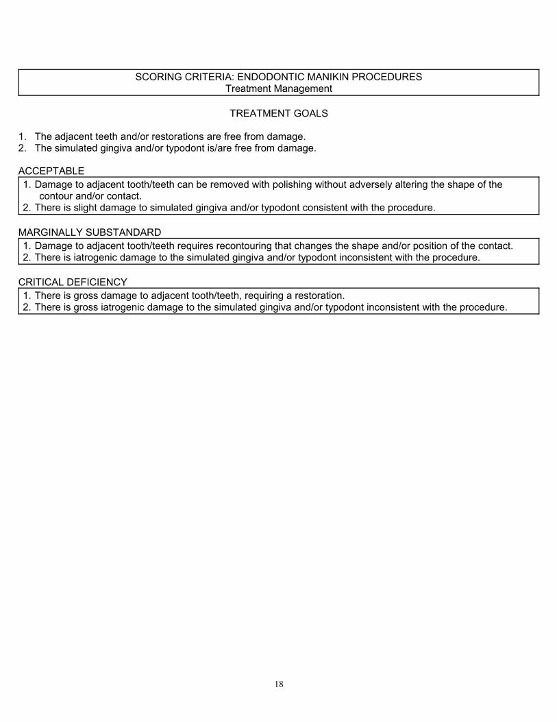
SCORING CRITERIA: ENDODONTIC MANIKIN PROCEDURESTreatment Management
TREATMENT GOALS
1. The adjacent teeth and/or restorations are free from damage.2. The simulated gingiva and/or typodont is/are free from damage.
ACCEPTABLE1. Damage to adjacent tooth/teeth can be removed with polishing without adversely altering the shape of the
contour and/or contact.2. There is slight damage to simulated gingiva and/or typodont consistent with the procedure.
MARGINALLY SUBSTANDARD1. Damage to adjacent tooth/teeth requires recontouring that changes the shape and/or position of the contact.2. There is iatrogenic damage to the simulated gingiva and/or typodont inconsistent with the procedure.
CRITICAL DEFICIENCY1. There is gross damage to adjacent tooth/teeth, requiring a restoration.2. There is gross iatrogenic damage to the simulated gingiva and/or typodont inconsistent with the procedure.
18
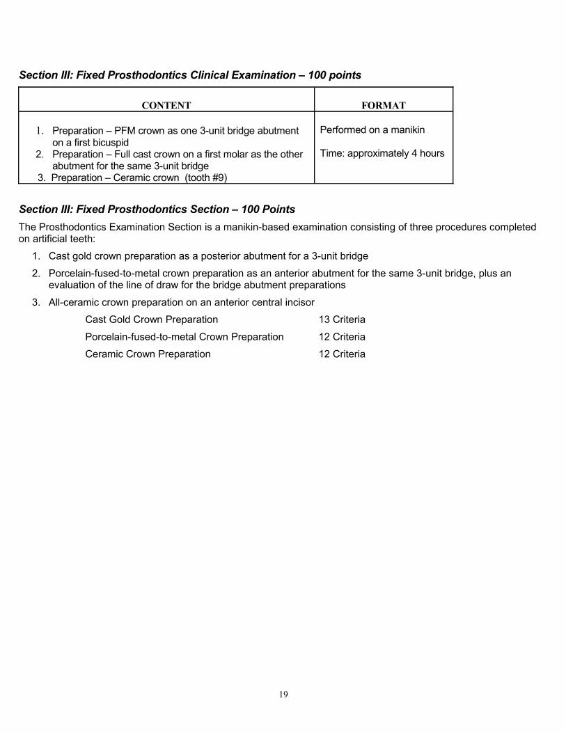
Section III: Fixed Prosthodontics Clinical Examination – 100 points
CONTENT FORMAT
1. Preparation – PFM crown as one 3-unit bridge abutment on a first bicuspid
2. Preparation – Full cast crown on a first molar as the other abutment for the same 3-unit bridge
3. Preparation – Ceramic crown (tooth #9)
Performed on a manikin
Time: approximately 4 hours
Section III: Fixed Prosthodontics Section – 100 PointsThe Prosthodontics Examination Section is a manikin-based examination consisting of three procedures completed on artificial teeth:
1. Cast gold crown preparation as a posterior abutment for a 3-unit bridge
2. Porcelain-fused-to-metal crown preparation as an anterior abutment for the same 3-unit bridge, plus an evaluation of the line of draw for the bridge abutment preparations
3. All-ceramic crown preparation on an anterior central incisor
Cast Gold Crown Preparation 13 Criteria
Porcelain-fused-to-metal Crown Preparation 12 Criteria
Ceramic Crown Preparation 12 Criteria
19
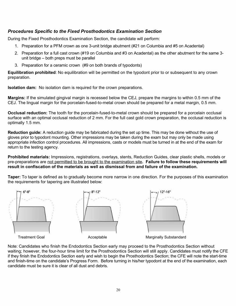
Procedures Specific to the Fixed Prosthodontics Examination SectionDuring the Fixed Prosthodontics Examination Section, the candidate will perform:
1. Preparation for a PFM crown as one 3-unit bridge abutment (#21 on Columbia and #5 on Acadental)
2. Preparation for a full cast crown (#19 on Columbia and #3 on Acadental) as the other abutment for the same 3-unit bridge – both preps must be parallel
3. Preparation for a ceramic crown (#9 on both brands of typodonts)
Equilibration prohibited: No equilibration will be permitted on the typodont prior to or subsequent to any crown preparation.
Isolation dam: No isolation dam is required for the crown preparations.
Margins: If the simulated gingival margin is recessed below the CEJ, prepare the margins to within 0.5 mm of the CEJ. The lingual margin for the porcelain-fused-to-metal crown should be prepared for a metal margin, 0.5 mm.
Occlusal reduction: The tooth for the porcelain-fused-to-metal crown should be prepared for a porcelain occlusal surface with an optimal occlusal reduction of 2 mm. For the full cast gold crown preparation, the occlusal reduction is optimally 1.5 mm.
Reduction guide: A reduction guide may be fabricated during the set up time. This may be done without the use of gloves prior to typodont mounting. Other impressions may be taken during the exam but may only be made using appropriate infection control procedures. All impressions, casts or models must be turned in at the end of the exam for return to the testing agency.
Prohibited materials: Impressions, registrations, overlays, stents, Reduction Guides, clear plastic shells, models or pre-preparations are not permitted to be brought to the examination site. Failure to follow these requirements will result in confiscation of the materials as well as dismissal from and failure of the examination.
Taper: To taper is defined as to gradually become more narrow in one direction. For the purposes of this examination the requirements for tapering are illustrated below:
Treatment Goal Acceptable Marginally Substandard
Note: Candidates who finish the Endodontics Section early may proceed to the Prosthodontics Section without waiting; however, the four-hour time limit for the Prosthodontics Section will still apply. Candidates must notify the CFE if they finish the Endodontics Section early and wish to begin the Prosthodontics Section; the CFE will note the start-time and finish-time on the candidate’s Progress Form. Before turning in his/her typodont at the end of the examination, each candidate must be sure it is clear of all dust and debris.
20
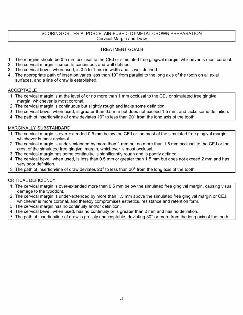
SCORING CRITERIA: PORCELAIN-FUSED-TO-METAL CROWN PREPARATIONCervical Margin and Draw
TREATMENT GOALS
1. The margins should be 0.5 mm occlusal to the CEJ or simulated free gingival margin, whichever is most coronal.2. The cervical margin is smooth, continuous and well defined.3. The cervical bevel, when used, is 0.5 to 1 mm in width and is well defined.4. The appropriate path of insertion varies less than 10° from parallel to the long axis of the tooth on all axial
surfaces, and a line of draw is established.
ACCEPTABLE1. The cervical margin is at the level of or no more than 1 mm occlusal to the CEJ or simulated free gingival
margin, whichever is most coronal.2. The cervical margin is continuous but slightly rough and lacks some definition.3. The cervical bevel, when used, is greater than 0.5 mm but does not exceed 1.5 mm, and lacks some definition.4. The path of insertion/line of draw deviates 10° to less than 20° from the long axis of the tooth.
MARGINALLY SUBSTANDARD1. The cervical margin is over-extended 0.5 mm below the CEJ or the crest of the simulated free gingival margin,
whichever is most occlusal.2. The cervical margin is under-extended by more than 1 mm but no more than 1.5 mm occlusal to the CEJ or the
crest of the simulated free gingival margin, whichever is most occlusal.3. The cervical margin has some continuity, is significantly rough and is poorly defined.4. The cervical bevel, when used, is less than 0.5 mm or greater than 1.5 mm but does not exceed 2 mm and has
very poor definition.5. The path of insertion/line of draw deviates 20° to less than 30° from the long axis of the tooth.
CRITICAL DEFICIENCY1. The cervical margin is over-extended more than 0.5 mm below the simulated free gingival margin, causing visual
damage to the typodont.2. The cervical margin is under-extended by more than 1.5 mm above the simulated free gingival margin or CEJ,
whichever is more coronal, and thereby compromises esthetics, resistance and retention form.3. The cervical margin has no continuity and/or definition.4. The cervical bevel, when used, has no continuity or is greater than 2 mm and has no definition.5. The path of insertion/line of draw is grossly unacceptable, deviating 30° or more from the long axis of the tooth.
21
SCORING CRITERIA: PORCELAIN-FUSED-TO-METAL CROWN PREPARATIONWalls, Taper and Shoulder
TREATMENT GOALS
1. Axial tissue removal is optimally 1.5 mm to be sufficient for convenience, retention and resistance form.2. Walls are smooth and well defined, with no undercuts.3. There is full visual taper (6°– 8°).4. The facial shoulder is optimally 1.5 mm wide.5. Reduction of the occlusal wall is optimally 2 mm.6. Internal line angles and cusp tips are rounded.7. The general occlusal anatomy is maintained.
ACCEPTABLE1. The axial tissue removal deviates no more than + 0.5 mm from optimal.2. The walls are slightly rough and lack some definition.3. Taper is present, but nearly parallel (<6°) or slightly excessive (= 8° - 12° per wall).4. The facial shoulder varies slightly in width but deviates no more than ± 0.5 mm from ideal.5. Occlusal reduction deviates no more than + 0.5 mm from optimal.6. Internal line angles and cusp tip areas are not completely rounded and show a slight tendency of being sharp.
MARGINALLY SUBSTANDARD1. The axial tissue removal is over-reduced or under-reduced but deviates no more than + 1 mm from optimal. 2. The axial walls are rough. 3. There is no taper or excessive taper (= 12° - 16° per wall).4. The facial shoulder varies slightly in width but deviates no more than ± 1 mm from ideal.5. Occlusal reduction deviates no more than + 1 mm from optimal6. The internal line angles and cusp tip areas show only minimal evidence of rounding with a greater tendency of
being sharp.7. The occlusal anatomy is flat.
CRITICAL DEFICIENCY1. The axial tissue removal is grossly over-reduced or under-reduced. The reduction is less than 0.5 mm or greater
than 2.5 mm.2. The taper is grossly over-reduced (>16° per wall).3. There is an undercut.4. The facial shoulder is less than 0.5 mm or more than 2.5 mm in width.5. The occlusal wall is grossly over-reduced (greater than 3 mm, encroaching on the pulp and impacting resistance
and retention form) or grossly under-reduced (less than 0.5 mm, resulting in insufficient occlusal clearance for adequate porcelain restorative material).
6. The internal line angles or cusp tip areas are excessively sharp with no evidence of rounding.
22
SCORING CRITERIA: CAST GOLD CROWN PREPARATIONCervical Margin and Draw
TREATMENT GOALS
1. The margins are 0.5 mm occlusal to the CEJ or simulated free gingival margin, whichever is most coronal.2. The cervical margin is smooth, continuous and well defined.3. The cervical bevel, when used, is 0.5 to 1 mm in width and is well defined. 4. The appropriate path of insertion varies less than 10° from parallel to the long axis of the tooth on all axial
surfaces, and a line of draw is established.
ACCEPTABLE1. The cervical margin is at the level of or no more than 1mm occlusal to the CEJ or simulated free gingival margin,
whichever is most coronal.2. The cervical margin is continuous but slightly rough and lacks some definition.3. The cervical bevel, when used, is greater than 1 mm but does not exceed 1.5 mm and lacks some definition.4. The path of insertion/line of draw deviates 10° to less than 20° from the long axis of the tooth.
MARGINALLY SUBSTANDARD1. The cervical margin is over-extended 0.5 mm below the CEJ or the crest of the simulated free gingival margin,
whichever is most occlusal.2. The cervical margin is under-extended by more than 1 mm but no more than 1.5 mm occlusal to the CEJ or the
crest of the simulated free gingival margin, whichever is most occlusal.3. The cervical margin has some continuity, is significantly rough and is poorly defined.4. The cervical bevel, when used, is less than 0.5 mm or greater than 1.5 mm but does not exceed 2 mm and has
very poor definition.5. The path of insertion/line of draw deviates 20° to less than 30° from the long axis of the tooth.
CRITICAL DEFICIENCY1. The cervical margin is over-extended more than 0.5 mm below the simulated free gingival margin, causing visual
damage to the typodont.2. The cervical margin is under-extended more than 1.5 mm above the simulated free gingival margin or CEJ,
whichever is more coronal, and thereby compromises esthetics, resistance and retention form.3. The cervical margin has no continuity and/or definition.4. The cervical bevel, when used, has no continuity or is greater than 2 mm and has no definition.5. The path of insertion/line of draw is grossly unacceptable, deviating 30° or more from the long axis of the tooth.
23

SCORING CRITERIA: CAST GOLD CROWN PREPARATIONWalls, Taper and Margin
TREATMENT GOALS
1. Axial tissue removal is optimally 1.5 mm to be sufficient for convenience, retention and resistance form.2. Walls are smooth and well defined, with no undercuts.3. There is full visual taper (6°– 8°). 4. The margin (includes knife-edge, chamfer and shoulder with bevel) is optimally 0.5 mm wide.5. Reduction of the occlusal wall is optimally 1.5 mm.6. Internal line angles and cusp tips are rounded.7. The general occlusal anatomy is maintained.
ACCEPTABLE1. The axial tissue removal deviates no more than + 0.5 mm from optimal.2. The walls are slightly rough and lack some definition.3. Taper is present, but nearly parallel (<6°) or slightly excessive (= 8° - 12° per wall).4. The margin varies slightly in width but is no greater than 1 mm.5. Occlusal reduction deviates no more than + 0.5 mm from optimal.6. The walls are slightly rough and lack some definition. 7. Internal line angles and cusp tip areas are not completely rounded and show a slight tendency of being sharp.
MARGINALLY SUBSTANDARD1. The axial tissue removal is over-reduced or under-reduced and deviates more than 0.5 mm but no more than + 1
mm from optimal. 2. The axial walls are rough. 3. There is no taper or excessive taper (= 12° - 16° per wall).4. The margin varies significantly in width and deviates no more than 1 mm from optimal.5. Occlusal reduction deviates no more than + 1 mm from optimal.6. Internal line angles and cusp tip areas show only minimal rounding, with a greater tendency of being sharp.7. The occlusal anatomy is flat.
CRITICAL DEFICIENCY1. The axial tissue removal is grossly over-reduced or under-reduced. The reduction is less than 0.5 mm or greater
than 2.5 mm.2. There is an undercut.3. The taper is grossly over-reduced (>16° per wall). 4. The margin width is less than 0.5 mm or greater than 2.5 mm.5. The occlusal wall is grossly over-reduced by greater than 2.5 mm or grossly under-reduced by less than 0.5 mm,
resulting in insufficient occlusal clearance for adequate restorative material.6. The internal line angles or cusp tip areas are excessively sharp with no evidence of rounding.
24
SCORING CRITERIA: BRIDGE FACTORPath of Insertion/Line of Draw
TREATMENT GOALS
1. The line of draw or path of insertion would allow for the full seating of a fixed prosthesis in a direct vertical plane without rotation, either mesiodistally or buccolingually.
ACCEPTABLE1. The line of draw or path of insertion would, require altering the path of insertion from a direct vertical axis to allow
full seating.
MARGINALLY SUBSTANDARD1. The line of draw or path of insertion would not, due to angulations of the surface of the preparations, allow
seating of a fixed prosthesis, regardless of the rotation through all available planes, without removal of tooth structure from the coronal third of either/both of the preparations.
CRITICAL DEFICIENCY1. No line of draw or path of insertion exits through any plane of rotation without the removal of additional tooth
structure in the apical two-thirds of either/both of the preparations.
25
SCORING CRITERIA: CERAMIC CROWN PREPARATIONCervical Margin and Draw
TREATMENT GOALS
1. The cervical margin is placed 0.5 mm incisal to the CEJ or simulated free gingival margin, whichever is most coronal.
2. The cervical margin is smooth, continuous and well defined on all axial surfaces and exhibits no bevel.3. The appropriate path of insertion varies less than 10° from parallel to the long axis of the tooth on all axial
surfaces, and a line of draw is established.
ACCEPTABLE1. The cervical margin is at the level of or no more than 1 mm incisal to the CEJ or simulated free gingival margin,
whichever is most coronal.2. The cervical margin is continuous but slightly rough and lacks some definition.3. The path of insertion/line of draw deviates 10° to less than 20° from the long axis of the tooth.
MARGINALLY SUBSTANDARD1. The cervical margin is over-extended 0.5 mm below the CEJ or the crest of the simulated free gingival margin,
whichever is most incisal.2. The cervical margin is under-extended by more than 1 mm but no more than 1.5 mm occlusal to the CEJ or the
crest of the simulated free gingival margin, whichever is most incisal.3. The cervical margin has some continuity, is significantly rough and is poorly defined.4. The path of insertion/line of draw deviates 20° to less than 30° from the long axis of the tooth.
CRITICAL DEFICIENCY1. The cervical margin is over-extended by more than 0.5 mm below the simulated free gingival margin, causing
visual damage to the typodont.2. The cervical margin is under-extended by more than 1.5 mm above the simulated free gingival margin or CEJ,
whichever is more coronal, and thereby compromises esthetics, resistance and retention form.3. The cervical margin has no continuity and/or definition.4. The cervical margin is beveled.5. The path of insertion/line of draw is grossly unacceptable, deviating 30° or more from the long axis of the tooth.
26
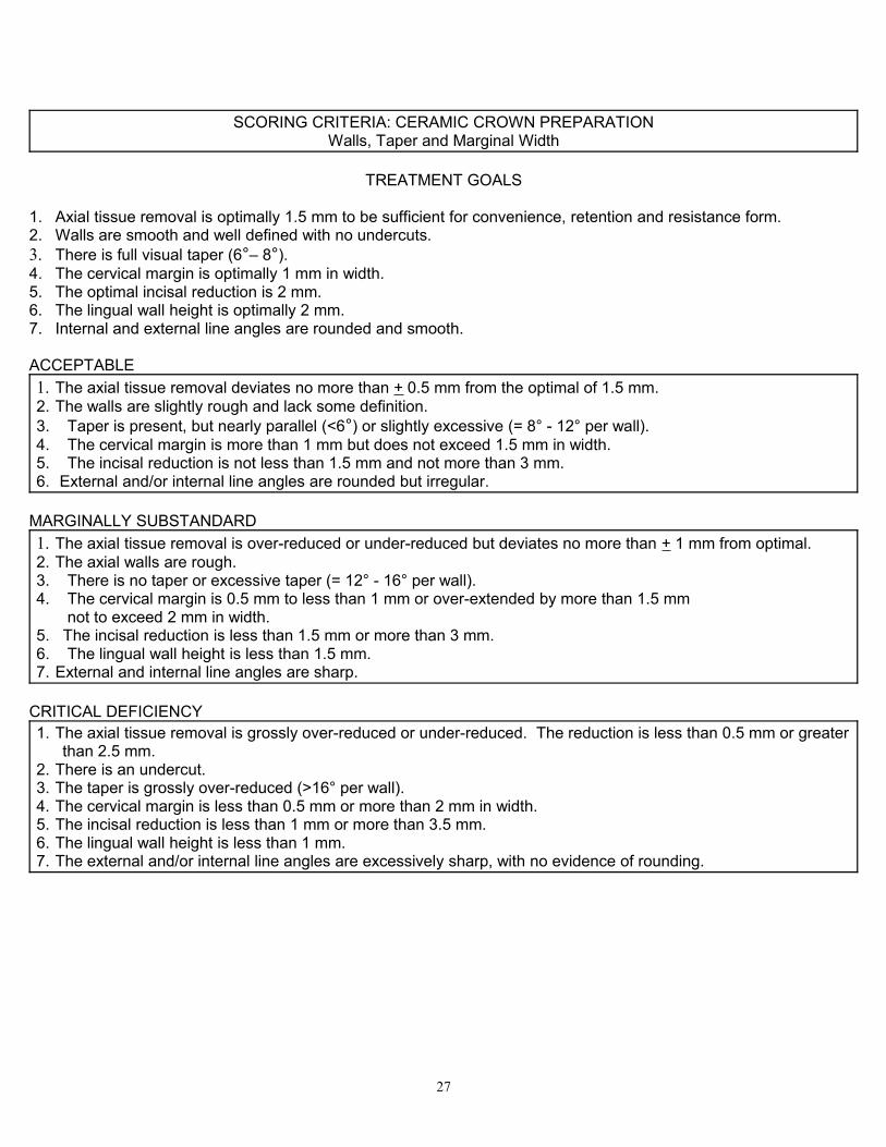
SCORING CRITERIA: CERAMIC CROWN PREPARATIONWalls, Taper and Marginal Width
TREATMENT GOALS
1. Axial tissue removal is optimally 1.5 mm to be sufficient for convenience, retention and resistance form.2. Walls are smooth and well defined with no undercuts.3. There is full visual taper (6°– 8°). 4. The cervical margin is optimally 1 mm in width.5. The optimal incisal reduction is 2 mm. 6. The lingual wall height is optimally 2 mm.7. Internal and external line angles are rounded and smooth.
ACCEPTABLE1. The axial tissue removal deviates no more than + 0.5 mm from the optimal of 1.5 mm.2. The walls are slightly rough and lack some definition.3. Taper is present, but nearly parallel (<6°) or slightly excessive (= 8° - 12° per wall).4. The cervical margin is more than 1 mm but does not exceed 1.5 mm in width.5. The incisal reduction is not less than 1.5 mm and not more than 3 mm.6. External and/or internal line angles are rounded but irregular.
MARGINALLY SUBSTANDARD1. The axial tissue removal is over-reduced or under-reduced but deviates no more than + 1 mm from optimal. 2. The axial walls are rough. 3. There is no taper or excessive taper (= 12° - 16° per wall).4. The cervical margin is 0.5 mm to less than 1 mm or over-extended by more than 1.5 mm not to exceed 2 mm in width. 5. The incisal reduction is less than 1.5 mm or more than 3 mm.6. The lingual wall height is less than 1.5 mm. 7. External and internal line angles are sharp.
CRITICAL DEFICIENCY1. The axial tissue removal is grossly over-reduced or under-reduced. The reduction is less than 0.5 mm or greater
than 2.5 mm.2. There is an undercut.3. The taper is grossly over-reduced (>16° per wall). 4. The cervical margin is less than 0.5 mm or more than 2 mm in width.5. The incisal reduction is less than 1 mm or more than 3.5 mm.6. The lingual wall height is less than 1 mm.7. The external and/or internal line angles are excessively sharp, with no evidence of rounding.
27
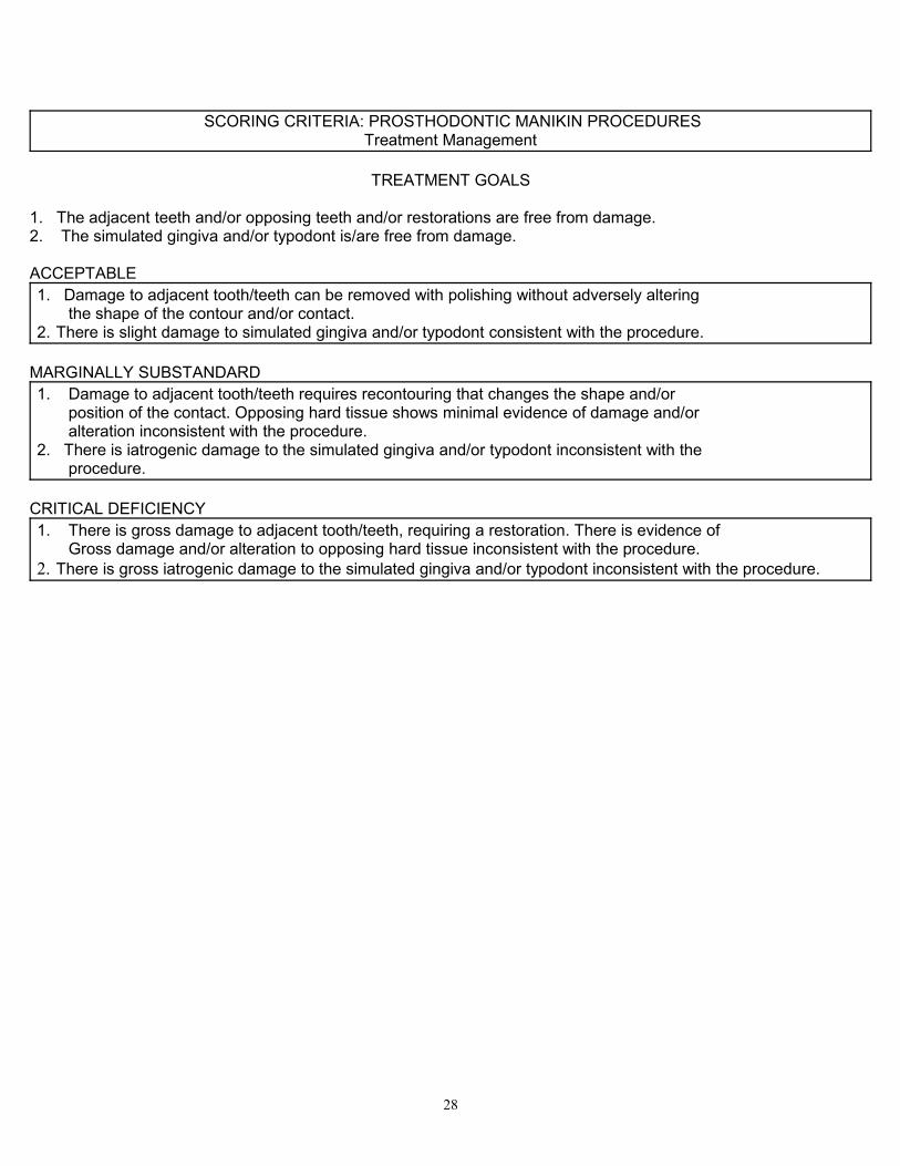
SCORING CRITERIA: PROSTHODONTIC MANIKIN PROCEDURESTreatment Management
TREATMENT GOALS
1. The adjacent teeth and/or opposing teeth and/or restorations are free from damage. 2. The simulated gingiva and/or typodont is/are free from damage.
ACCEPTABLE1. Damage to adjacent tooth/teeth can be removed with polishing without adversely altering the shape of the contour and/or contact.2. There is slight damage to simulated gingiva and/or typodont consistent with the procedure.
MARGINALLY SUBSTANDARD1. Damage to adjacent tooth/teeth requires recontouring that changes the shape and/or position of the contact. Opposing hard tissue shows minimal evidence of damage and/or alteration inconsistent with the procedure.2. There is iatrogenic damage to the simulated gingiva and/or typodont inconsistent with the procedure.
CRITICAL DEFICIENCY1. There is gross damage to adjacent tooth/teeth, requiring a restoration. There is evidence of Gross damage and/or alteration to opposing hard tissue inconsistent with the procedure.2. There is gross iatrogenic damage to the simulated gingiva and/or typodont inconsistent with the procedure.
28

29
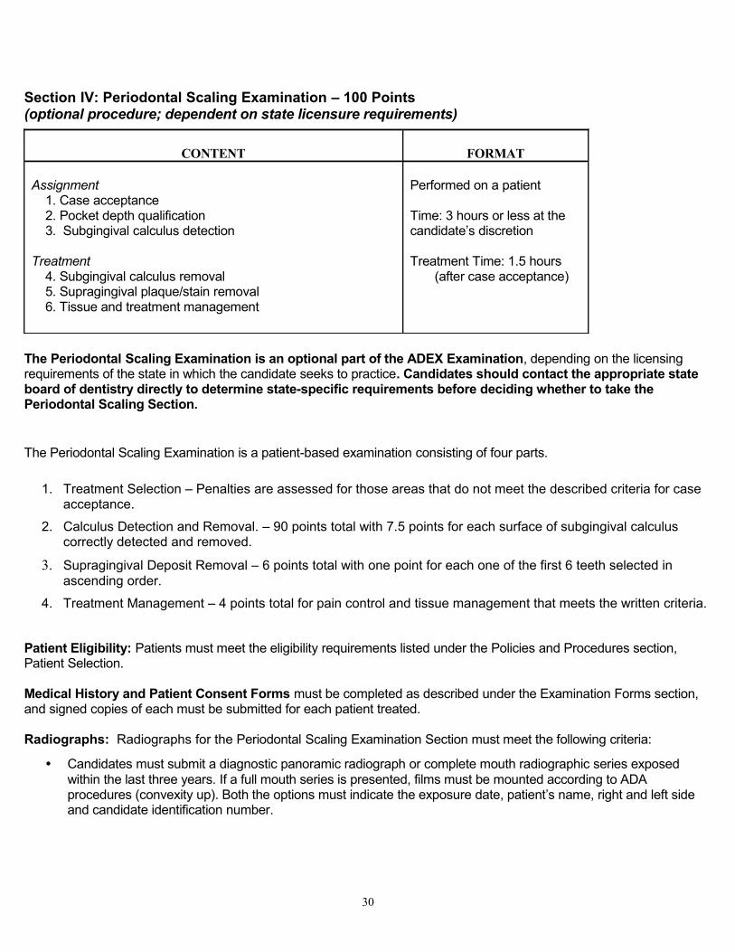
Section IV: Periodontal Scaling Examination – 100 Points (optional procedure; dependent on state licensure requirements)
CONTENT FORMAT Assignment 1. Case acceptance 2. Pocket depth qualification 3. Subgingival calculus detection
Treatment 4. Subgingival calculus removal 5. Supragingival plaque/stain removal 6. Tissue and treatment management
Performed on a patient
Time: 3 hours or less at the candidate’s discretion
Treatment Time: 1.5 hours (after case acceptance)
The Periodontal Scaling Examination is an optional part of the ADEX Examination, depending on the licensing requirements of the state in which the candidate seeks to practice. Candidates should contact the appropriate state board of dentistry directly to determine state-specific requirements before deciding whether to take the Periodontal Scaling Section.
The Periodontal Scaling Examination is a patient-based examination consisting of four parts.
1. Treatment Selection – Penalties are assessed for those areas that do not meet the described criteria for case acceptance.
2. Calculus Detection and Removal. – 90 points total with 7.5 points for each surface of subgingival calculus correctly detected and removed.
3. Supragingival Deposit Removal – 6 points total with one point for each one of the first 6 teeth selected in ascending order.
4. Treatment Management – 4 points total for pain control and tissue management that meets the written criteria.
Patient Eligibility: Patients must meet the eligibility requirements listed under the Policies and Procedures section, Patient Selection.
Medical History and Patient Consent Forms must be completed as described under the Examination Forms section, and signed copies of each must be submitted for each patient treated.
Radiographs: Radiographs for the Periodontal Scaling Examination Section must meet the following criteria:
• Candidates must submit a diagnostic panoramic radiograph or complete mouth radiographic series exposed within the last three years. If a full mouth series is presented, films must be mounted according to ADA procedures (convexity up). Both the options must indicate the exposure date, patient’s name, right and left side and candidate identification number.
30
• Candidates must also submit four bitewing radiographs exposed within the previous year.
• If utilizing a full mouth series, this must be mounted separately from the bitewings, unless the complete mouth series were taken within the previous year.
• Copies are acceptable for the Periodontal/Scaling Examination.
• Digital images or digital prints are permitted. Candidates from outside the host school will need to contact the host school to inquire if they will up-load images from outside the facility. If digital prints are to be used, the radiographs must be printed and submitted on photo quality paper or acetate (preferably blue).
• If the school name is normally incorporated into the digital image, this should be removed or masked, if possible, before printing out the image on photo quality paper or the CFE should be asked to cover such a school identifier on the day of the examination.
• Alternatively, images may be displayed on monitors if they are available from the school’s database. Candidates from outside the host school will need to contact the host school to inquire if they will up-load images from outside the facility.
• Radiographs must not be retaken simply to produce a “perfect” image. Radiographs that have minor errors such as minor cone cutting, not showing all of a third molar or a slightly off center panoramic film, will not result in any loss of points and should not be retaken. Radiographic technique is not being evaluated in this part of the examination.
Periodontal instruments: Please reference the chart at the beginning of this section.
The candidate’s performance will not be evaluated without the proper instruments. Sonic/ultrasonic instruments are permissible for scaling, but they may not be available at the examination site (check with the school coordinator). If the candidate elects to provide his/her own unit, he/she must check with the school about appropriate connection mechanisms. Air-abrasive polishers are not permissible.
Treatment selection: The candidate’s treatment selection must include the proper number of teeth, adequate deposits of calculus and appropriate pocket depths as defined below:
• Teeth. There must be at least six and not more than eight permanent teeth selected, at least three of which are molars or premolars, including at least one molar. All posterior teeth must have at least one approximating tooth surface within 2 mm distance. Each of the selected teeth must have at least one surface of subgingival calculus selected for removal.
• Pocket depths. There must be three pockets of 4 mm or greater in depth, each on a separate tooth from among the six to eight teeth selected for treatment. There is a penalty if one of these pockets is less than 4 mm and a severe penalty if two or more are less than 4 mm. (Examiners allow a ± 1 mm leeway in measurement.) It is recommended that pocket depths greater than 6 mm not be included; however, the patient will not be rejected if pockets are this deep. Although the three pockets of 4 mm or more must be on the teeth within the treatment selection, it is not necessary that those surfaces be selected for calculus removal.
• Calculus. There must be exactly 12 surfaces of explorer-detectable subgingival calculus identified on the selected teeth, and no more than four surfaces may be on the incisors. Three of the 12 identified surfaces of calculus must be on interproximal surfaces of posterior teeth, i.e., on molars and/or premolars.
o Explorer detectable subgingival calculus is defined as a distinct deposit of calculus that can be felt with an #11/12 explorer as it passes over the calculus. Qualified deposits may exhibit such characteristics as:
A definite “jump” or “bump” felt by the explorer, with the rough surface characteristic of calculus
Ledges or ring formations
Spiny or nodular formations
31
o Qualified deposits must be apical to the gingival margin and may occur with or without associated supragingival deposits.
• Exclusions. Patients with full-banded orthodontics are not acceptable. Implants or teeth with any fixed appliance – banded, bonded or splinted, either orthodontically or periodontally – may not be included in the treatment selection. No retained primary teeth may be included in the treatment selection.
Candidates may use the Treatment Selection Worksheet (available in Appendix B) to identify and document a selection of the patient’s teeth that meet these criteria. The candidate should indicate the presence of subgingival calculus on the Treatment Selection Worksheet by marking the appropriate letter for the surface in the box next to the number of the tooth selected for treatment. If subgingival calculus is present on the line angles of the tooth, it must be marked on the interproximal surface, e.g., a deposit on the distofacial line angle would be marked on the distal surface. The numbers of the selected teeth must be listed in ascending order.
Prior to the examination, the candidate who is taking the ADEX exam must transfer the information about his/her treatment selection from the Worksheet to the Electronic Periodontal Scaling Evaluation Form, which will be reviewed by examiners. Visit the NERB or SRTA website to access the Electronic Periodontal Scaling Evaluation Form.
Scaling. After the candidate performs the periodontal procedure, the subgingival surfaces of the assigned teeth must be smooth, with no deposits detectable with an 11/12 explorer. Air may be used to deflect the tissue to locate areas for tactile confirmation. (All subgingival surfaces on an assigned tooth must be scaled, but only the selected surface will be evaluated.)
Supragingival deposits (polishing). All supragingival calculus, plaque and stain must be removed from all coronal surfaces of the assigned teeth so that all surfaces are visually clean when air-dried and tactilely smooth upon examination with an 11/12 explorer. The use of disclosing solution is not permitted.
Periodontal Scaling Examination Section Procedure and Patient Management Guidelines
1. The patient must be informed that he/she will be participating in an examination and that additional treatment may be required to meet his/her oral health needs.
2. Only one patient may be presented for the Periodontal Scaling Clinical Examination Section. Once a patient has been submitted to the Clinic Floor Examiner for patient check-in, a back-up patient may not be presented if that patient is found not to be acceptable due to examination protocols, guidelines or requirements. If the patient is otherwise acceptable but there has been a correctable paperwork error, the candidate may be allowed to correct those errors and re-submit that patient for approval. In all circumstances the candidate must have their patients presented and approved for treatment BEFORE proceeding further with the examination. Treatment on a patient without documented approval by a Clinic Floor Examiner is a violation of examination protocol and may subject the candidate to dismissal from the examination.
3. The Treatment Selection Worksheet, a practice form provided in Appendix B, may be completed prior to the day of the examination to help the candidate identify the selection of teeth he/she will present for evaluation. Candidates are responsible for independently (without the help of faculty and/or colleagues) selecting and documenting teeth and surfaces for treatment that fulfill the published criteria. Prior to the day of the examination the information on the Treatment Selection Worksheet should be accurately transferred to the Electronic Periodontal Scaling Evaluation Form, which is the official form used by examiners.
4. The candidate must accurately transfer the information from the Treatment Selection Worksheet to the Electronic Periodontal Scaling Evaluation Form (available on the NERB website up to 48 hours prior to the exam and on the SRTA website 2 weeks prior to the exam) to indicate his/her treatment selection. The teeth
32
should be listed in ascending order, and the surfaces to be treated should be indicated in the smaller box to the right.
5. The Periodontal Scaling Progress Form will be provided at the examination site. When the candidate receives the Progress Form, he/she should place a candidate identification label on the form and enter his/her cubicle number.
6. The procedures, instruments and materials used are the choice of the candidate, as long as they are currently accepted and taught by accredited dental schools and the candidate has been trained in their use. It is the responsibility of the candidate to provide the instruments used in this examination and listed in this Candidate Manual, unless such instruments are furnished by the school.
7. If the candidate is scheduled to perform the periodontal scaling procedure as the first procedure of the day, he/she may call over a CFE as early as 7:00 a.m. to check the Medical History Form, Patient Consent Form (including the anesthetic record section), radiographs and confirm the patient’s blood pressure was taken that day. At 8:00 a.m. the patient may be sent to the Evaluation Station for patient check-in/case acceptance. If the Periodontal Scaling Examination Section is not the candidate’s first procedure of the day, the candidate may begin the periodontal procedure at any time after the first restorative procedure is completed.
8. If any problems arise during the examination, the candidate should immediately notify a CFE. The CFE is also
present to aid in any emergencies that may occur.
9. Candidates must complete the anesthesia portion on the Progress Form whether or not anesthesia is to be used. If the patient is too sensitive to withstand the use of a periodontal probe or explorer during patient check-in, the candidate may request authorization from a CFE to anesthetize the patient prior to patient check-in.
10. When the patient is sent to the Evaluation Station for patient check-in, he/she will first sign in with the Desk Coordinator. Patients will be evaluated for case acceptance in the order in which they are signed in. Patients must take the required forms and instruments with them to the Evaluation Station. Only the patient may carry the tray to the Evaluation Station.
Periodontal instruments: Please reference the chart at the beginning of this section.
11. The examiners will evaluate the three teeth with 4 mm or deeper pockets and the six to eight teeth with the 12 surfaces of subgingival calculus charted.
12. The Desk Coordinator will indicate a Finish Treatment Time on the Periodontal Scaling Progress Form. The approximate total time for the Periodontal Scaling Examination Section is about 3 hours. The patient treatment time is 1 ½ hours. Candidates must receive a start time 45 minutes prior to the end of the examination day if they are beginning the Periodontal Scaling Section after completing the Restorative Section.
When the patient returns from the Evaluation Station, treatment should begin. Treatment continues until it is completed or until the Finish Time, as noted on the Periodontal Scaling Progress Form. If candidates finish the patient treatment before their assigned Finish Time, they may sign-in the patient with the Desk Coordinator for evaluation. The candidate must scale all subgingival surfaces on the six to eight selected teeth, but only the 12 selected surfaces selected by the candidate will be evaluated. Supragingival calculus, plaque and stain must be removed from all surfaces of the selected teeth. No other teeth may be scaled or polished during the examination, and once the examination is completed, the patient must be dismissed.
33

13. By the stated Finish Time, each candidate should have completed subgingival calculus removal on the 12 selected surfaces and removed all supragingival calculus, plaque and stain from the entire crown of the selected teeth. The patient must be signed in with the Desk Coordinator for evaluation at the Evaluation Station by the recorded Finish Time.
14. For the treatment evaluation, the candidate must send the patient, wearing a clean napkin, to the Evaluation Station..
Periodontal instruments: Please reference the chart at the beginning of this section.
15. The examiners will evaluate tissue management and subgingival calculus removal from the selected tooth surfaces and evaluate supragingival calculus, stain and plaque removal from all surfaces on the selected teeth.
16. When the patient returns from the Evaluation Station, the candidate may dismiss the patient, unless directed to do otherwise. The candidate must clean the clinic area following accepted infection control procedures.
Retesting ScheduleIf a candidate needs to retake only the Periodontal Scaling or the Restorative Examination Section, he/she will have three hours total (90 minutes actual treatment time) for the Periodontal Scaling Section or a total of seven hours to complete the Restorative Section (both required restorative procedures).
34

SCORING CRITERIA: PERIODONTAL SCALING EXAMINATIONPatient Selection
TREATMENT GOALS1. The Patient Consent Form, Medical History, Progress Form and Periodontal Evaluation Form are complete, accurate and
current.2. Both systolic and diastolic blood pressure are less than or equal to 159/94, or systolic and diastolic blood pressure are
between 160/95 and 179/109 with a written medical clearance from a physician authorizing treatment during the examination.
3. Radiographs are of diagnostic quality and reflect the current clinical condition of the mouth. Periapicals have been exposed within the past three years, and bitewings have been exposed within the past six months. Radiographs are properly mounted and labeled with exposure date and patient’s name.
4. The Calculus Detection portion of the Periodontal Evaluation Form is properly completed, indicating - Six to eight teeth selected, each with at least one surface of calculus charted
- At least three posteriors (molars, premolars), including at least one molar, in the selection. All posterior teeth must have at least one approximating tooth within 2 mm distance.
- Exactly 12 surfaces of subgingival calculus charted, including at least three surfaces of interproximal calculus on molars/premolars- At least eight of the surfaces on canines, premolars or molars (no more than four surfaces on incisors)- Three pockets of 4 mm or greater in depth, each on a different tooth within the selection
ACCEPTABLE1. The Patient Consent Form is incorrect or not signed by patient.* 2. The Medical History is incomplete*, is missing candidate initials* or patient signature* or has slight inaccuracies that do not
endanger the patient or change the treatment.3. The Progress Form has inaccuracies or is incomplete or missing.*4. Blood pressure has not been taken or is not recorded* but, upon correction, meets Satisfactory criteria. 5. Radiographs are available but were not submitted with the patient for initial evaluation.*** 6. The Calculus Detection portion of the Evaluation Form has not been filled out or is filled out incorrectly, e.g., the form
demonstrates- Fewer than six or more than eight selected teeth
- Fewer than three molars or premolars and/or no approximating tooth within 2 mm of one or more of the selected posterior teeth- One or more selected teeth without any surfaces of calculus charted- More or fewer than 12 surfaces of subgingival calculus charted- Fewer than three surfaces of interproximal calculus on molars and/or premolars more than four surfaces of subgingival calculus on incisors**- Three separate teeth and/or surfaces are not indicated for Pocket Depth Qualification and/or one or more of the teeth are outside the treatment selection.**
* Records and patient will be sent back to the candidate with an Instruction to Candidate Form requesting correction. (If the Periodontal Evaluation Form is completed correctly, it
will be retained in the Evaluation Station.) ** Records and patient will be sent back to the candidate with an Instruction to Candidate Form requesting correction.*** The candidate will receive an Instruction to Candidate Form requesting radiographs.
35

SCORING CRITERIA: PERIODONTAL SCALING EXAMINATIONPatient Selection Continued
MARGINALLY SUBSTANDARD1. Medical History has inaccuracies that do not endanger the patient but do change the treatment or require further
explanation by candidate. The candidate submits an incomplete or incorrect Periodontal Progress Form or Evaluation Form for the second time.*
2. Radiographs are of poor diagnostic quality and/or do not meet all of the criteria to be considered Satisfactory. 3. Of the three teeth indicated with pocket measurements of 4 mm or more in depth, only two teeth are found to
have measurements of 4 mm or more and/or one or more of these teeth are outside the treatment selection on the second submission.
* Records and patient are sent back to the candidate with an Instruction to Candidate Form Requesting corrections.
CRITICAL DEFICIENCY1. The Medical History has inaccuracies or indicates the presence of conditions that do endanger the patient,
candidate and/or examiners (in this situation, the Periodontal Examination Section will be stopped). The candidate submits an incomplete and/or incorrect Patient Consent Form or Medical History for the second time.
2. The patient’s systolic and/or diastolic blood pressure is between 160/95 and 179/109 without a written medical clearance from a physician authorizing treatment, or blood pressure is 180/110 or greater even with a written medical clearance from a physician authorizing treatment.
3. Radiographs are of unacceptable diagnostic quality and/or are missing and not available on request. (In this situation, the Periodontal Examination Section will be stopped).
4. Of the three teeth indicated with sulcus/pocket measurements of 4 mm or more in depth, fewer than two teeth are found to have pockets of 4 mm or more.
36
SCORING CRITERIA: PERIODONTAL SCALING EXAMINATIONTissue and Treatment Management
TREATMENT GOALS1. The patient has adequate anesthesia for pain control, is comfortable and demonstrates no evidence of distress or
pain.2. Instruments, polishing cups or brushes and dental floss are effectively utilized so that no unwarranted soft or hard
tissue trauma occurs as a result of the scaling and polishing procedures
ACCEPTABLE1. There is slight soft tissue trauma that is consistent with the procedure.
MARGINALLY SUBSTANDARD1. There is inadequate anesthesia for pain control. (The patient is in obvious distress or pain.)2. There is minor soft tissue trauma that is inconsistent with the procedure. Soft tissue trauma may include, but is
not limited to, abrasions, lacerations or ultrasonic burns.3. There is minor hard tissue trauma that is inconsistent with the procedure. Hard tissue trauma may include root
surface abrasions that do not require additional definitive treatment.
CRITICAL DEFICIENCY1. There is major damage to the soft and/or hard tissue that is inconsistent with the procedure and preexisting
condition. This damage may include, but is not limited to, such trauma as- Amputated papillae- Exposure of the alveolar process- A laceration or damage that requires suturing and/or periodontal packing- One or more ultrasonic burns that require follow up treatment- A broken instrument tip in the sulcus or soft tissue- Root surface abrasions that require additional definitive treatment
37
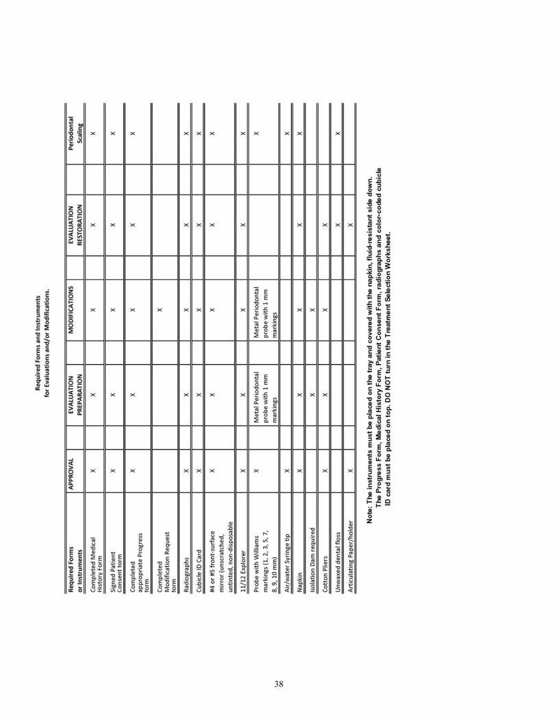
38
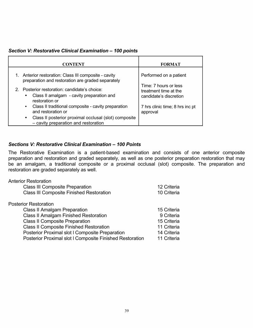
Section V: Restorative Clinical Examination – 100 points
CONTENT FORMAT
1. Anterior restoration: Class III composite - cavity preparation and restoration are graded separately
2. Posterior restoration: candidate’s choice: • Class II amalgam - cavity preparation and
restoration or• Class II traditional composite - cavity preparation
and restoration or• Class II posterior proximal occlusal (slot) composite
– cavity preparation and restoration
Performed on a patient
Time: 7 hours or less treatment time at the candidate’s discretion
7 hrs clinic time; 8 hrs inc pt approval
Sections V: Restorative Clinical Examination – 100 Points The Restorative Examination is a patient-based examination and consists of one anterior composite preparation and restoration and graded separately, as well as one posterior preparation restoration that may be an amalgam, a traditional composite or a proximal occlusal (slot) composite. The preparation and restoration are graded separately as well.
Anterior RestorationClass III Composite Preparation 12 CriteriaClass III Composite Finished Restoration 10 Criteria
Posterior RestorationClass II Amalgam Preparation 15 CriteriaClass II Amalgam Finished Restoration 9 CriteriaClass II Composite Preparation 15 CriteriaClass II Composite Finished Restoration 11 CriteriaPosterior Proximal slot l Composite Preparation 14 CriteriaPosterior Proximal slot l Composite Finished Restoration 11 Criteria
39
Restorative Examination Section Requirements
Patient Eligibility. Patients must meet the eligibility requirements listed under Policies and Procedures section, Patient Selection. Medical History and Patient Consent Forms must be completed as listed under Examination Forms section, and signed copies of each must be submitted for each patient treated.
Radiographs Radiographs for the Restorative Examination Section must meet the following requirements:
• For the posterior tooth to be treated, the candidate must provide periapical and bitewing radiographs or single digital periapical and bitewing images.
• For the anterior tooth, the candidate must provide a periapical radiograph or single digital periapical image.
• Interproximal caries must be shown radiographically to penetrate at least to the dento-enamel junction (or have evidence of equivalent depth clinically).
• The radiographs should not have been exposed more than one year prior to the examination, and must depict the current clinical condition of the tooth to be treated. If a candidate is utilizing a patient treated by another candidate during the same examination series and wishes to treat an adjacent tooth, he/she need not submit a new radiograph unless there is a specific clinical indication.
• If films are being utilized, they should be mounted according to ADA format, in a small plastic mount with transparent tape to the appropriate Restorative Progress Form provided by the testing agency. Digital printouts should be attached with a paper clip to the Progress form.
• Copies are acceptable for the Restorative examination.
• Digital prints on photo quality paper or on acetate (preferably blue) may be utilized. The following information must be on the digital print: patient’s name, date of exposure, candidate’s ID number and right and left sides indicated.
• If a school name is normally incorporated into the digital image, this should be removed or masked, if possible, before printing out the image on photo quality paper, or the CFE should be asked to cover such a school identifier on the day of the examination.
• Alternatively, digital images may be displayed on monitors if they are available from the school’s database. Candidates from outside the school will need to submit digital prints when using digital images, as the school will not up load images from an outside facility. (For SRTA exams, candidates from outside the school will need to contact the host school to inquire if they will up-load images from outside the facility.)
• A radiograph must not be retaken simply to produce a “perfect” image. Radiographs that have minor errors such as minor cone cutting or not angled directly through the interproximal contacts, particularly if unrelated to the proposed treatment, will not result in any loss of points and should not be retaken. Radiographic technique is not being evaluated in this part of the examination.
• Note: Lesions seen on high speed films (such as F-speed film) and with some digital techniques may clinically be considerably larger than they appear.
40
If the candidate submits one or two poor quality or non-diagnostic radiographs (film or digital prints), for either the perio scaling procedure or the restorative procedures, examiners will take the following action:
• First offense – examiners will request new film(s).
• Second offense – examiners will deduct points and request new film(s).
• Third offense – candidate will be dismissed from the examination.
Post-operative radiographs: Post-operative radiographs are not routinely required. However, a post-op radiograph may be requested at any time at the discretion of the examiners to evaluate the clinical condition of the patient. The radiograph should meet the same criteria as specified for pre-op radiographs and should be mounted and returned to the requesting examiner for evaluation.
Time ManagementIn scheduling patients and planning the utilization of time, the candidate should be aware that the time allowed for the examination includes the time during which the patient(s) will be at the Evaluation Station for assignment and evaluation. The minimum time patients will be in the Evaluation Station is 30-45 minutes – possibly longer, depending on the time of day. Times may vary according to the procedure being evaluated, the testing site and the number of candidates.
Additionally, when a candidate submits a Modification Request, his/her patient must be sent to the Express Chair in the Evaluation Station. Candidates are allowed to submit multiple requests for modification simultaneously in order to save time.
Treatment Selection and Patient Check-inPatient selection is very important. If the candidate is unable to complete a procedure due to patient management problems, the procedure cannot be evaluated and no credit will be assigned. No more than two treatment selections may be submitted for a procedure. If a second treatment selection is rejected or a second treatment selection is not presented after the first lesion is rejected, a candidate may not continue with that procedure and will receive a “0” for that portion of the examination.
41
Careful clinical judgments should be used if planning approximating lesions.
Treating all lesions: The tooth selected for the anterior restoration may have more than one lesion present but it is permissible for a candidate to treat only one proximal surface on the exam day. All lesions on this tooth do not need to be treated by the end of the examination day.
The tooth selected for the posterior restoration will need all existing lesions treated by the end of the examination day. If a tooth selected for treatment has other lesions that are not planned for treatment during the examination then the submission will be denied. Alternatively, lesions other than the one required for the examination may be treated prior to the examinations day.
The candidate must enter on the posterior restoration Progress Form, all proposed treatment for the selected tooth. This includes the primary lesion and any other lesions requiring treatment, except Class V lesions. Class V lesions must be treated prior to the examination. The primary lesion is circled at the top left of the Progress Form and any additional lesions that will be treated are written in ink below. The same restorative material must be utilized for all restorations on the same tooth.
All lesions/preparations will be considered together as one submission. The same general criteria as used for the standard preparations will be used for any additional treatments. Any confirmed findings on any of the lesions/preparations will be graded and reported the same as for the required lesion.
Sharing patients: This is not recommended.
For the anterior restoration, one tooth may be shared by two candidates but it is highly recommended that candidates not share treatment of the same tooth.
For the posterior restoration, one tooth may not be shared by two candidates for treatment during the examination. If the tooth has a mesial and distal lesion when presented for evaluation, the candidate must treat both lesions by the end of the examination. Any other carious lesions on the tooth must have been previously treated or the submission will be rejected.
Exclusions: The following will not be accepted for the Restorative Examination Section:
• Non-vital teeth, and/or teeth with pulpal pathology or endodontic treatment
• Teeth with facial veneers
• Mobility of Class III or greater
Other recommendations:
• Lesions on the distal surface of mandibular first premolars are acceptable for Class II amalgam, Class II composite or proximal occlusal box (slot) restoration but they are not recommended due to pulpal anatomy.
• If there are proximal but no occlusal caries on a posterior tooth, the candidate may elect to do a Class II amalgam or Class II composite restoration but a proximal occlusal box (slot) preparation is recommended.
• Lesions on the distal surface of cuspids are allowed for Class III composite only, not Class II amalgam.
• Avoid potential pulpal involvement (too large a lesion) or cuspal replacement.
• Circumferential decalcification contiguous with the lesion or proposed restoration is discouraged.
Patient check-in: If the candidate is scheduled to perform the restorative examination as the first procedure of the day, they may begin setting up at 7:00 AM. Between 7:00 AM and 7:20 AM a CFE may be called over to document that the blood pressure was taken that day and check that the required forms and radiographs are complete and correct. Then, the patient is sent to the Evaluation Station, where all treatment selections are approved or rejected by at least two of three examiners evaluating anonymously. Restorative treatment begins at 8:00AM. If a patient meets
42
the requirements for both the posterior and anterior restorations, both may be approved in the Evaluation Station at the same time, but the first restoration must be completed before the second restoration may be begun. Only one patient may be submitted for a patient check-in at a time. If the candidate is utilizing two patents for the Restorative Examination Section, only one may be submitted to start the examination. The second may not be submitted until the first is finished. The local anesthesia request portion of the Restorative Progress Form must be filled out prior to submitting the patient to the Evaluation Station for patient check-in/case acceptance. Local anesthesia may not be provided until the lesion has been accepted for treatment. In the event that the first lesion submitted is not approved, a second lesion may be submitted to the Evaluation Station.
For patient check-in, please reference the chart at the beginning of this section for forms and instruments that will accompany the patient as they are sent to the evaluation station.
Under no circumstances can anesthetic solution be administered prior to patient check-in.
Treatment Guidelines Restorative instruments and equipment: Candidates must provide the following materials for use during the Restorative Examination Section:
• Isolation dam: During the Restorative Examination Section, cavity preparations may be instrumented with or without an isolation dam. An isolation dam that is intact (not torn or leaking) must be in place when the patient is sent for evaluation of the amalgam and composite preparations, as well as for all requests for modification and for the placement of restorative materials. An isolation dam must be in place if a pulpal exposure is anticipated or occurs. The isolation dam must be removed when the patient is sent for evaluation of the finished amalgam and composite restorations. Bite blocks may be used during treatment, but the patient may not travel to the Evaluation Station with a bite block in place under an isolation dam. The isolation dam must be placed by the candidate and not the assistant.
• Cavity sealers/liners/bases: The candidate must decide if a treatment liner or base is indicated, and if so, check the liner/base request box on the appropriate Progress Form prior to sending the patient in to the Evaluation Station for evaluation of the preparation. Liners will be required only in very deep preparations to cover areas immediately impacting pulpal health and integrity. Failure to request a liner in this circumstance will result in penalties.
• The examiners in the Evaluation Station will either approve or disapprove the placement of the liner or base as part of the evaluation of the preparation. The examiner will indicate his/her decision by placing his/her examiner number in either the Granted or Not Granted box next to the request. If the liner/base is granted, the candidate should place it and then summon the CFE to check its placement before continuing with the final restoration. If the examiners in the Evaluation Station, during grading of the final preparation, make a determination that a liner/base is indicated and has not been requested by the candidate, an Instruction to Candidate Form will be issued to ask that the candidate place the liner/base. In this instance, the liner/base must also be checked by the CFE. In either case, if the CFE finds the insertion of the liner/base to be defective, the patient must be sent to the Express Chair evaluators for assessment along with the Progress Form (with a red dot, provided by a CFE, affixed to the top left of the form) prior to any alteration and before permission is given to insert the restoration.
If a liner or base is placed, it must fulfill the following criteria:
1. The liner must be placed only in those pulpal and/or axial wall areas that deviate from the established ideal depth.
2. The liner must not be placed on enamel or within 1 mm of any cavosurface margin.
3. The liner must not compromise the internal retentive and resistance features of the cavity preparation.
4. The liner must not be subject to dislodgement during placement of the permanent restoration.
43
5. Placement must reflect consideration of limitations of the materials used.
Caries detector: Caries detector liquid (except red) may be used. If used, it must be completely removed prior to the submission of the preparation for evaluation.
Pulpal exposure: If a candidate anticipates a pulpal exposure, a modification request must be completed describing what the candidate intends to do prior to continuing with the preparation, then send the patient and modification form to the Express Chair.
If the candidate actually experiences a pulpal exposure, the candidate should write in the Notes section on the Progress Form that a pulpal exposure has occurred, indicate the time and briefly describe how the situation should be treated. Then call a CFE, who will consult with the Chief Examiner to determine the appropriate management of the exposure. All exposures must be sent to the evaluation station. If an exposure is well documented and appropriately managed, it is not always a failure.
Recontouring: Recontouring of adjacent teeth or restorations is allowed only after the preparation has been evaluated and only with the approval of a CFE. Candidates must enter the request to recontour the adjacent tooth in the Additional Comments section of the Progress Form. A CFE must then review the situation and will place his/her examiner number and the time next to the request. The candidate may then restore the tooth after the CFE checks the adjacent tooth recontouring.
Instructions to candidates: Evaluators may provide written instructions to candidates if they believe a treatment should be modified during the course of the examination. When the patient returns from the Evaluation Station, if the candidate does not receive an Instruction to Candidate Form, the candidate should continue to the next step of the treatment. If the candidate does receive an Instruction to Candidate Form, it should be delivered by a CFE. The CFE will review the instructions with the candidate, and both the candidate and CFE will sign the form to indicate that the candidate understands the instructions. The corrections must be completed as stated on the form and checked by a CFE.
Modification RequestsIf, during the preparation, it is evident that the tooth requires a significant deviation from the criteria outlined for a Satisfactory preparation, the candidate should make a modification request(s) prior to proceeding with the modification or extending the preparation for caries or decalcified enamel removal. Bundling of modification requests is not allowed. Each request must be separate and answer the question where, how much, and why.
(Exception: modification to extend the proximal box because of tooth rotation or position does not require a Modification Request Form. Document this modification in the Additional Comments area on the Progress Form and request a CFE to initial it.)
The tooth must be prepared to ideal dimensions and all preexisting restorative material in the new preparation must be removed prior to submission of a Modification Request Form. If removing preexisting restorative material will result in a preparation that extends beyond Satisfactory-level criteria, a CFE must be called to evaluate the preparation prior to removal of the additional restorative material; the CFE will document and initial the modification on the Progress Form. In addition, if a candidate anticipates a carious (or mechanical) pulpal exposure during the course of treatment, another Modification Request Form must also be submitted.
To request a modification, the candidate must briefly write each modification on the Modification Request Form provided to the candidate at orientation or if this is a SRTA administered exam the candidate must receive a Modification Request Form from a CFE on the clinic floor. The request for each modification should include:
• What is the candidate requesting to do? (Type of modification)
44
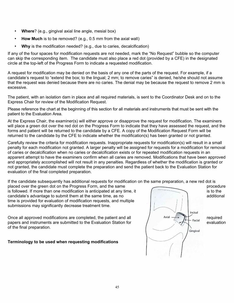
• Where? (e.g., gingival axial line angle, mesial box)
• How Much is to be removed? (e.g., 0.5 mm from the axial wall)
• Why is the modification needed? (e.g., due to caries, decalcification)
If any of the four spaces for modification requests are not needed, mark the “No Request” bubble so the computer can skip the corresponding item. The candidate must also place a red dot (provided by a CFE) in the designated circle at the top-left of the Progress Form to indicate a requested modification.
A request for modification may be denied on the basis of any one of the parts of the request. For example, if a candidate’s request to “extend the box; to the lingual; 2 mm; to remove caries” is denied, he/she should not assume that the request was denied because there are no caries. The denial may be because the request to remove 2 mm is excessive.
The patient, with an isolation dam in place and all required materials, is sent to the Coordinator Desk and on to the Express Chair for review of the Modification Request.
Please reference the chart at the beginning of this section for all materials and instruments that must be sent with the patient to the Evaluation Area.
At the Express Chair, the examiner(s) will either approve or disapprove the request for modification. The examiners will place a green dot over the red dot on the Progress Form to indicate that they have assessed the request, and the forms and patient will be returned to the candidate by a CFE. A copy of the Modification Request Form will be returned to the candidate by the CFE to indicate whether the modification(s) has been granted or not granted.
Carefully review the criteria for modification requests. Inappropriate requests for modification(s) will result in a small penalty for each modification not granted. A larger penalty will be assigned for requests for a modification for removal of caries or decalcification when no caries or decalcification exists or for repeated modification requests in an apparent attempt to have the examiners confirm when all caries are removed. Modifications that have been approved and appropriately accomplished will not result in any penalties. Regardless of whether the modification is granted or not granted, the candidate must complete the preparation and send the patient back to the Evaluation Station for evaluation of the final completed preparation.
If the candidate subsequently has additional requests for modification on the same preparation, a new red dot is placed over the green dot on the Progress Form, and the same procedure is followed. If more than one modification is anticipated at any time, it is to the candidate’s advantage to submit them at the same time, as no additional time is provided for evaluation of modification requests, and multiple submissions may significantly decrease treatment time.
Once all approved modifications are completed, the patient and all required papers and instruments are submitted to the Evaluation Station for evaluation of the final preparation.
Terminology to be used when requesting modifications
45
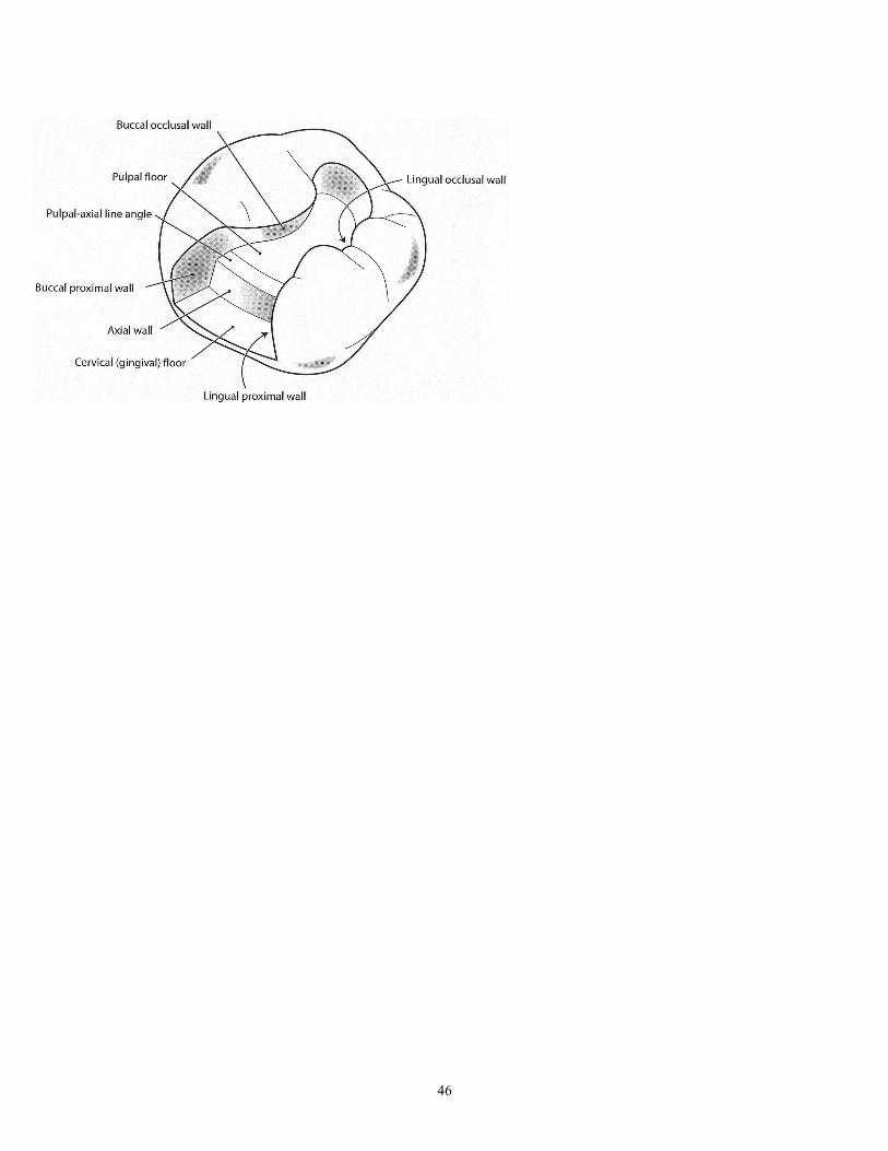
46
Restorative Procedure and Patient Management Guidelines
Final evaluation of the preparation. When a patient is sent to the Evaluation Station, the prepared tooth must be isolated by an isolation dam. (To be properly isolated, at least one tooth on either side of the prepared tooth must be included under the isolation dam unless it is the most posterior tooth.) Candidate requests for a liner or base must be filled out on the appropriate Progress Form.
An instrument tray containing the following items must be sent with the patient:• Completed Medical History Form
• Signed Patient Consent Form
• Amalgam or Composite Progress Form with properly mounted radiographs
• Color-coded cubicle ID card
• Instruments
o Fine and sharp pigtail explorer or 11/12 explorer)
o Metal periodontal probe with Williams Markings (1, 2, 3, 5, 7, 8, 9, 10 mm) markings
o Clear mirror (unscratched, untinted, non-disposable)o Cotton plier
• Napkin
Note: The instruments must be placed on the tray and covered with the napkin, fluid-resistant side down. The Progress Form, Medical History Form, radiographs and color-coded cubicle ID card must be placed on top.
Three independent examiners will evaluate the prepared cavity. If modifications of the preparation are required prior to restoration, the examiners will complete an Instruction to Candidate Form and return it to the candidate with the patient. If a candidate receives an Instruction to Candidate Form, he/she must follow the instructions. The candidate must not request an opinion from CFEs concerning the instructions on the Instruction to Candidate Form. If the instructions are to temporize the tooth, the chief examiner must be notified and a Follow-Up Form completed.
Final evaluation of the restoration. For the Class II amalgam restoration, the amalgam must be sufficiently set to allow a check of the occlusion. Any of the composite restorations must be presented without any surface glaze/sealer on the restoration. After removing the isolation dam and any wedges placed during treatment, please reference the chart at the beginning of this section for all materials and instruments that must be sent with the patient to the Evaluation Area.
If adjustments to the restoration are required, an Instruction to Candidate Form will be issued. Candidates must perform the corrections as instructed. Please note that the second restorative preparation may not be started by the candidate until the first restorative patient is dismissed (that is, after the completed restoration has been evaluated and any required modifications have been completed by the candidate and approved by a CFE).
If the final restoration is unacceptable, the candidate will receive an Instruction to Candidate Form and will be instructed to remove the restoration and temporize the tooth. Before this additional treatment is started, a CFE and the chief examiner must be notified. The patient, the candidate, a CFE and the chief examiner will meet to confirm that the responsibility for further treatment is understood and that the patient will be cared for properly. A Follow-Up Form will be issued to the candidate. When treatment is completed, the CFE will be called to check the provisional restoration before the patient is dismissed. Any restoration left in place at the discretion of the chief examiner does
47
not indicate a Satisfactory restoration. If temporization occurs on the first restorative procedure, the candidate will be dismissed from the examination before attempting the second restorative procedure and will fail the Restorative Section.
Any post-examination treatment required as a result of treatment rendered during the examination process is the responsibility of the candidate and will be completed at the expense of the candidate. A Follow-Up Form must be completed to indicate the follow-up treatment required and clarify responsibility for the treatment. If the candidate receives no communication from the examiners in the Evaluation Station, a CFE should be notified before the patient is dismissed.
Separation of Scoring for the Anterior and Posterior RestorationsBeginning with the CIF Class of 2014 Examinations, the anterior and posterior restorations are graded separately. If the first restorative procedure, which is the choice of the candidate, receives a passing grade, that grade will stand even if the grade on the second restoration is below 75. In that case the candidate may apply and retake the second restorative procedure alone at a subsequent examination, following remediation.
However, if the grade on the first restorative procedure is not passing, the candidate will not be permitted to proceed to the second restoration for reasons of patient safety and the candidate will need to apply to retake both restorative procedures at a later examination.
48
Requirements for the Class III Composite Preparation and Restoration 1. The tooth selected for the Class III composite restoration must be a permanent anterior tooth that meets the
following requirements:
• At least one proximal primary carious lesion that shows no signs of previous excavation and appears, radiographically or clinically, to extend to the DEJ.
OR
• A defective restoration, defined as one that exhibits recurrent caries or a defective cavosurface margin that, even though it may not yet be carious, can be penetrated with an explorer. (A mismatched shade is not an acceptable indication.) Existing defective restorations must be completely removed before submitting the patient to the Evaluation Station for a modification request or evaluation of the completed preparation.
• The proximal contact of the tooth must be visually closed and meet resistance to dental floss passing through the contact with the adjacent tooth on the proximal surface to be restored, although the area to be restored may or may not be in contact.
• The approximating contact of the adjacent tooth must be natural tooth structure or a permanent restoration.
• There may be a lesion on the proximal surface of the adjacent tooth, provided that there is no cavitation of the contact before or during the preparation that would prevent the candidate from restoring an ideal contour or contact of the restoration.
• Occlusion may or may not be present.
2. Lesions that may initially be described as Class IV will not be accepted.
However, Class III lesions that may require modifications resulting in Class IV restorations are acceptable.
3. Lingual dovetails are acceptable when appropriately used.
4. Surface sealants must not be place on the finished composite restoration.
49
Requirements for Class II Amalgam Preparation and Restoration1. The amalgam must be a Class II restoration, and the tooth selected for the amalgam restoration must be a
permanent posterior tooth that meets these requirements:
• At least one proximal surface being restored must have a primary carious lesion that shows no signs of being previously excavated and appears, radiographically or clinically, to extend at least to the DEJ.
• The tooth must be in contact with a sound enamel surface or a permanently restored surface of an adjacent tooth.
• There may be a lesion on the proximal surface of the adjacent tooth, provided that there is no breakdown of the contact before or during the preparation that would jeopardize proximal contour or contact of the finished restoration.
• When in centric occlusion, the selected tooth must be in cusp/fossa occlusion with an opposing tooth or teeth. Those opposing tooth/teeth may be natural dentition, a fixed bridge or any permanent artificial replacement thereof.
2. Other surfaces of the selected tooth may have an existing occlusal or proximal restoration, as long as there is a qualified surface with primary caries. Preexisting restorations and any underlying liner must be entirely removed, and the preparation must demonstrate acceptable principles of cavity preparation. An MOD treatment selection must have at least one proximal contact to be restored. In the event of a defect that would qualify as an acceptable lesion on the proximal surface opposite from the surface with primary caries, the treatment plan must be a MOD unless there is an intact transverse or oblique ridge, in which case the restoration must be treatment planned as a MO – DO.
3. The condensed and carved amalgam surface should not be polished or altered by abrasive rotary instrumentation except for the purpose of adjusting occlusion. Proximal contact is a critical part of the evaluation, and the candidate should be aware that the examiners will be checking the contact with floss. Please note that, for this examination, proximal contacts must be visibly closed. Some resistance to the passage of floss is not sufficient for judging a contact to be closed. Also, contacts must not prevent floss from passing through. Proximal contacts that are not visibly closed or that do not permit the passage of floss are evaluated as Critical Deficiencies. The candidate must be familiar with the properties of the amalgam being used and should be sure to allow sufficient time for the amalgam to set before sending the finished restoration to the Evaluation Station. A developed and mounted post-operative bitewing may be requested at any time at the discretion of the examiners.
50
Requirements for Class II Composite Preparation and Restoration• The tooth selected for the Class II Conventional Composite restoration must be a permanent posterior
tooth that meets the following requirements:
• At least one proximal surface being restored must have a primary carious lesion that shows no signs of being previously excavated and appears, radiographically or clinically, to extend at least to the DEJ.
• The tooth must be in contact with a sound enamel surface or a permanently restored surface of an adjacent tooth.
• There should be evidence of caries and/or an existing occlusal restoration on the occlusal surface of the tooth that warrants extending the preparation across the occlusal surface.
• There may be a lesion on the proximal surface of the adjacent tooth, provided that there is no breakdown of the contact before or during the preparation that would jeopardize the placement of an ideal proximal contour or contact of the finished restoration.
• When in centric occlusion, the selected tooth must be in cusp/fossa occlusion with an opposing tooth or teeth. Those opposing tooth/teeth may be natural dentition, a fixed bridge or any permanent artificial replacement thereof.
Other surfaces of the selected tooth may have an existing occlusal or proximal restoration, as long as there is a qualified surface with primary caries. Preexisting restorations and any underlying liner must be entirely removed, and the preparation must demonstrate acceptable principles of cavity preparation. An MOD treatment selection must have at least one proximal contact to be restored. In the event of a defect that would qualify as an acceptable lesion on the proximal surface opposite from the surface with primary caries, the treatment plan must be an MOD unless there is an intact transverse or oblique ridge. If this exists the tooth must be treatment planned for a MO-DO restoration.
Requirements for the Posterior Slot Composite Preparation and Restoration The tooth selected for the posterior proximal occlusal (slot) composite restoration must be a permanent posterior tooth that meets the following requirements:
• At least one proximal surface being restored must have a primary carious lesion that shows no signs of being previously excavated and appears, radiographically or clinically, to extend at least to the DEJ.
• The tooth must be in contact with a sound enamel surface or a permanently restored surface of an adjacent tooth.
• If there is both occlusal caries and proximal caries, an occlusal restoration and a separate proximal occlusal (slot) restoration is permitted, if there is at least 1 mm of sound tooth structure between the two preparations. Otherwise, a Class II restoration is required.
• There may be a lesion on the proximal surface of the adjacent tooth, provided that there is no breakdown of the contact before or during the preparation that would jeopardize the proximal contour or contact of the finished restoration.
• When in centric occlusion, the selected tooth must be in cusp/fossa occlusion with an opposing tooth or teeth. Those opposing tooth/teeth may be natural dentition, a fixed bridge or any permanent artificial replacement thereof.
51
SCORING CRITERIA: ANTERIOR CLASS III COMPOSITE PREPARATIONExternal Outline Form
TREATMENT GOALS
1. Outline form provides adequate access for complete removal of caries and/or previous restorative material and for insertion of composite resin. Access entry is appropriate to the location of caries and tooth position.
2. The gingival contact must be broken. The incisal contact need not be broken, unless indicated by the location of the caries. If a lingual approach is initiated, facial contact may or may not be broken as long as the margin terminates in sound tooth structure.
3. Cavosurface margins form a smooth, continuous curve with no sharp angles.4. Cavosurface margins terminate in sound natural tooth structure. There is no previous restorative material, including sealants,
at the cavosurface margin. All unsupported enamel is removed unless it compromises facial esthetics. 5. Enamel cavosurface margins may be beveled.
ACCEPTABLE1. The wall opposite the access, if broken, may extend no more than 1 mm beyond the contact area.2. The gingival clearance does not exceed 1.5 mm.3. The outline form may be over-extended mesiodistally 0.5-1 mm beyond what is necessary for complete removal of caries
and/or previous restorative material. 4. The cavosurface margins may be slightly irregular.5. There may be a small area of unsupported enamel, which is not necessary to preserve facial esthetics.6. Enamel cavosurface margin bevels, if present, do not exceed 1 mm in width.
MARGINALLY SUBSTANDARD1. The outline form is under-extended, making caries removal or insertion of restorative material questionable.2. The outline form is over-extended mesiodistally by more than 1 mm but no more than 2 mm beyond what is necessary for
complete removal of caries and/or previous restorative material. 3. The incisal cavosurface margin is over-extended so that the integrity of the incisal angle is compromised.4. The wall opposite the access opening extends more than 1 mm beyond the contact area.5. The gingival clearance is greater than 1.5 mm. 6. Gingival contact is not visually broken. 7. The cavosurface margin is rough and severely irregular.8. The cavosurface margin does not terminate in sound natural tooth structure, or there is explorer-penetrable decalcification
or previous restorative material remaining on the cavosurface margins.9. There are large or multiple areas of unsupported enamel that are not necessary to preserve facial esthetics.10. Enamel cavosurface margin bevels, if present, exceed 1 mm in width, are not uniform or are inappropriate for the size of
the restoration.
CRITICAL DEFICIENCY1. The outline form is under-extended, making it impossible to manipulate and finish the restorative material.2. The outline form is over-extended mesiodistally by more than 2 mm beyond what is necessary for complete removal of
caries and/or previous restorative material. 3. The incisal cavosurface margin is over-extended so that the incisal angle is removed or fractured. A Class IV restoration is
now necessary without prior justification.4. The gingival clearance is greater than 2 mm.5. The wall opposite the access opening extends more than 2.5 mm beyond the contact area.6. There are caries remaining.
52
SCORING CRITERIA: ANTERIOR CLASS III COMPOSITE PREPARATIONInternal Form
TREATMENT GOALS
1. The axial wall follows the external contours of the tooth, and the depth should not exceed 0.5 mm beyond the DEJ.
2. All prepared surfaces are smooth and well defined.3. If used, rounded internal retention is placed in the dentin of the gingival and incisal walls just axial to the DEJ as
dictated by cavity form. Retention is observed tactilely and visually.4. All carious tooth structure and/or previous restorative materials are removed.
ACCEPTABLE1. The depth of the axial wall is no more than 1.5 mm beyond the DEJ.2. The internal walls may be slightly rough and irregular.
MARGINALLY SUBSTANDARD1. The depth of the axial wall is deeper than 1.5 mm beyond the DEJ.2. When used, retention is excessive and undermines enamel, jeopardizes the incisal angle or encroaches on the
pulp.3. The internal walls are rough and irregular.
CRITICAL DEFICIENCY1. Caries or previous restorative material remains.2. The axial wall is more than 2.5 mm beyond the DEJ.
53
SCORING CRITERIA: ANTERIOR CLASS III COMPOSITE PREPARATIONTreatment Management
TREATMENT GOALS
1. The isolation dam is adequate to isolate sufficient teeth for visibility and accessibility with no debris, saliva or hemorrhagic leakage into the preparation. Ideally, the treated tooth and both proximal adjacent teeth should be isolated, if possible.
2. The patient has adequate anesthesia for pain control. 3. The adjacent teeth and/or restorations are free from damage.4. The soft tissue is free from damage, or there is tissue damage that is consistent with the procedure.
ACCEPTABLE1. Any damage to adjacent tooth/teeth can be removed with polishing without adversely altering the shape of the
contour and/or contact.
MARGINALLY SUBSTANDARD1. The isolation dam is inappropriately applied, torn and/or leaking, rendering the preparation unsuitable for
evaluation or the subsequent manipulation of the restorative material. 2. There is inadequate anesthesia for pain control.3. Damage to adjacent tooth/teeth requires recontouring that changes the shape and/or contour and/or contact.4. There is iatrogenic soft tissue damage that is inconsistent with the procedure.
CRITICAL DEFICIENCY1. There is gross damage to adjacent tooth/teeth, requiring a restoration.2. There is gross iatrogenic damage to the soft tissue that is inconsistent with the procedure and preexisting
condition of the soft tissue.
54
SCORING CRITERIA: ANTERIOR CLASS III COMPOSITE FINISHED RESTORATIONMargin Integrity and Surface Finish
TREATMENT GOALS
1. There is no marginal excess (overhang) or deficiency. No marginal excess is detectable, either visually or with the tine of an explorer, at the restoration-tooth interface. There is no evidence of voids or open margins.
2. The surface of the restoration is uniformly smooth and free of pits and voids.3. There is no evidence of unwarranted or unnecessary removal, modification or recontouring of tooth structure
adjacent to the restoration.4. The restoration is bonded to the prepared tooth structure.5. The shade of the restoration blends with the surrounding tooth structure.
ACCEPTABLE1. There may be a marginal excess or deficiency at the restoration-tooth interface, detectable either visually or with
the tine of an explorer, but it is no greater than 0.5 mm. There is no evidence of pits and voids at the cavosurface margin.
2. The surface of the restoration may be slightly grainy or rough, but it is free of significant pits and voids.3. There may be minimal evidence of unwarranted or unnecessary removal, modification or recontouring of tooth
structure adjacent to the restoration.
MARGINALLY SUBSTANDARD1. The restoration-tooth interface is detectable visually or with the tine of an explorer. There is evidence of
marginal excess or deficiency of more than 0.5 mm and up to 1 mm, including pits and voids at the cavosurface margin.
2. The surface of the restoration is rough and exhibits significant surface irregularities, pits or voids. 3. There is evidence of unwarranted or unnecessary removal, modification or recontouring of tooth structure
adjacent to the restoration.4. There is flash with contamination, but the contamination is not internal to the cavosurface margin and could be
removed by polishing or finishing. 5. The shade of the restoration contrasts markedly with the surrounding tooth structure.
CRITICAL DEFICIENCY1. There is evidence of marginal excess or deficiency of more than 1 mm, including pits and voids at the
cavosurface margin, or there is an open margin.2. There is internal contamination at the interface between the restoration and the tooth. 3. The restoration is debonded and/or movable in the preparation. 4. There is gross enameloplasty resulting in the exposure of dentin.5. The restoration is fractured.
55
SCORING CRITERIA: ANTERIOR CLASS III COMPOSITE FINISHED RESTORATIONContour, Contact and Occlusion
TREATMENT GOALS
1. Interproximal contact is present, the contact is visually closed and is properly shaped and positioned and there is definite, but not excessive, resistance to dental floss when passed through the interproximal contact area.
2. When checked with articulating ribbon or paper, all centric and excursive contacts on the restoration are consistent in size, shape and intensity with such contacts on other teeth in that quadrant.
3. The restoration reproduces the normal physiological proximal contours of the tooth, lingual anatomy and marginal ridge anatomy.
ACCEPTABLE1. Interproximal contact is visually closed, and the contact is adequate in size, shape or position but may
demonstrate little resistance to dental floss.2. The restoration may not reproduce the normal lingual anatomy, proximal contours of the tooth or marginal ridge
anatomy but would not be expected to adversely affect the tissue health.
MARGINALLY SUBSTANDARD1. Interproximal contact is visually closed, but the contact is deficient in size, shape or position and demonstrates
little resistance to dental floss or shreds the floss. 2. Interproximal contact is visually open, but the tooth already lacked proximal contact at the time of assignment.
The final restoration demonstrates physiologic contour.3. When checked with articulating ribbon or paper, the restoration is in hyperocclusion inconsistent in size, shape
and intensity with the contacts on surrounding teeth. The restoration requires adjustment.4. The restoration does not reproduce the normal lingual anatomy, proximal contours of the tooth or marginal ridge
anatomy and would be expected to adversely affect the tissue health.
CRITICAL DEFICIENCY1. The interproximal contact is visually open or will not allow floss to pass through the contact area. 2. There is gross hyperocclusion so that the restoration is the only point of occlusion in that quadrant.
56

SCORING CRITERIA: ANTERIOR CLASS III COMPOSITE FINISHED RESTORATIONTreatment Management
TREATMENT GOALS
1. The patient demonstrates no post-operative discomfort that is inconsistent with the procedure.2. The adjacent and/or opposing hard tissue is free from evidence of damage and/or alteration.3. The soft tissue is free from damage, or there is soft tissue damage consistent with the procedure
ACCEPTABLE1. Any damage to adjacent tooth/teeth can be removed with polishing without adversely altering the shape of the
contour and/or contact.
MARGINALLY SUBSTANDARD1. The patient demonstrates discomfort inconsistent with the procedure.2. Adjacent and/or opposing hard tissue shows evidence of damage and/or alteration inconsistent with the
procedure.3. There is iatrogenic damage to the soft tissue inconsistent with the procedure.
CRITICAL DEFICIENCY1. There is evidence of gross damage and/or alteration to adjacent and/or opposing hard tissue inconsistent with
the procedure.2. There is gross iatrogenic trauma to the soft tissue inconsistent with the procedure and preexisting condition of
the soft tissue.
57

SCORING CRITERIA: CLASS II AMALGAM PREPARATIONExternal Outline Form
TREATMENT GOALS
1. Contact is visibly open up to 0.5 mm proximally and gingivally.2. The proximal gingival point angles may be rounded or sharp. 3. The isthmus must be 1-2 mm wide, but not more than one-fourth the intercuspal width of the tooth. 4. The external cavosurface margin meets the enamel at 90°. There are no gingival bevels. The gingival floor is
flat, smooth and perpendicular to the long axis of the tooth.5. The outline form includes all carious and non-coalesced fissures and is smooth, rounded and flowing.6. The cavosurface margin terminates in sound natural tooth structure. There is no previous restorative material,
including sealants, at the cavosurface margin. There is no decalcification on the gingival margin.
ACCEPTABLE1. Contact is visibly open when viewed proximally, and proximal clearance at the height of contour extends not
more than 1.5 mm on either one or both proximal walls. 2. The gingival clearance is up to 0.5 mm but not greater than 2 mm.3. The isthmus is from 1 to 2 mm wide to not more than one-third of the intercuspal width.4. The proximal cavosurface margin may deviate from 90° but is unlikely to jeopardize the longevity of the tooth or
restoration; this includes small areas of unsupported enamel.
MARGINALLY SUBSTANDARD1. The gingival floor and/or proximal contact is not visually open, or proximal clearance at the height of contour
extends beyond 1.5 mm but not more than 2.5 mm on either one or both proximal walls.2. The gingival clearance is greater than 2 mm but not more than 3 mm or is not visually open.3. The outline form is inappropriately over-extended so that it compromises the remaining marginal ridge and/or
cusp(s). The outline form is under-extended, and remaining non-coalesced fissure(s) extend to the DEJ and are contiguous with the outline form.
4. The isthmus is less than 1 mm or greater than ⅓ the intercuspal width.5. The proximal cavosurface margin deviates from 90° and is likely to jeopardize the longevity of the tooth or
restoration. This includes unsupported enamel and/or excessive bevel(s).6. The cavosurface margin does not terminate in sound natural tooth structure, or there is explorer penetrable
decalcification remaining on the cavosurface margin, or the cavosurface margin terminates in previous restorative material.
7. There is explorer-penetrable decalcification remaining on the gingival floor.
CRITICAL DEFICIENCY1. The proximal clearance at the height of contour extends beyond 3 mm on either one or both proximal walls.2. The gingival clearance is greater than 3 mm.3. The isthmus is greater than one-half the intercuspal width.4. The outline form is over-extended so that it compromises, undermines and leaves unsupported the remaining
marginal ridge to the extent that the pulpal-occlusal wall is unsupported by dentin or the width of the marginal ridge is 1 mm or less.
58
SCORING CRITERIA: CLASS II AMALGAM PREPARATIONInternal Form
TREATMENT GOALS
1. The axial wall is 0.5 mm from the DEJ, follows the external contours of the tooth and is entirely in dentin.2. The pulpal floor depth should be 0.5 mm beyond the DEJ in all areas.3. The pulpal-axial line angle is rounded.4. All caries and/or previous restorative material are removed.5. When used, retention is well defined, in dentin and does not undermine enamel.6. The walls of the proximal box should be convergent occlusally and meet the external surface at a 90° angle.
ACCEPTABLE1. The pulpal floor depth is 0.5 mm to 1.5 mm beyond the DEJ. 2. The depth of the axial wall is 0.5 mm to 1.5 mm beyond the DEJ.3. The walls of the proximal box should be convergent but may be parallel, but appropriate internal retention is
present.
MARGINALLY SUBSTANDARD1. Enamel remains on the axial wall.2. The axial wall is more than 1.5mm beyond the DEJ.3. The pulpal floor is not entirely in dentin and island(s) of enamel are evident. The pulpal floor is more than 1.5
mm beyond the DEJ.4. Retention, when used, undermines the enamel or may compromise the tooth or restoration.5. The walls of the proximal box diverge occlusally, which is likely to jeopardize the longevity of the tooth or
restoration.
CRITICAL DEFICIENCY1. The axial wall is more than 2.5 mm beyond the DEJ.2. The pulpal floor is more than 2.5 mm beyond the DEJ or entirely in enamel.3. Caries or previous restorative material remains in the preparation.4. Retention, when used, grossly compromises the tooth or restoration.5. The walls of the proximal box diverge occlusally, offering no retention and jeopardizing the longevity of the tooth
or restoration.
59
SCORING CRITERIA: CLASS II AMALGAM PREPARATIONTreatment Management
TREATMENT GOALS
1. The isolation dam is adequate to isolate sufficient teeth for visibility and accessibility and has no debris, salivary or hemorrhagic leakage into the preparation. Ideally, the treated tooth and both proximal adjacent teeth should be isolated, if possible.
2. The patient has received adequate anesthesia for pain control.3. The adjacent teeth and/or restorations are free from damage.4. The soft tissue is free from damage, or there is tissue damage that is consistent with the procedure.
ACCEPTABLE1. Any damage to adjacent tooth/teeth can be removed with polishing without adversely altering the shape of the
contour and/or contact.
MARGINALLY SUBSTANDARD1. The isolation dam is inappropriately applied, torn and/or leaking, resulting in debris, saliva and/or hemorrhage leakage, rendering the preparation unsuitable for evaluation or the subsequent manipulation of the restorative material.2. There is inadequate anesthesia for pain control.3. Damage to adjacent tooth/teeth requires recontouring that changes the shape and/or contour and/or contact.4. There is iatrogenic soft tissue damage that is inconsistent with the procedure.
CRITICAL DEFICIENCY1. There is gross damage to adjacent tooth/teeth, requiring a restoration.2. There is gross iatrogenic damage to the soft tissue inconsistent with the procedure and preexisting condition of
the soft tissue.
60
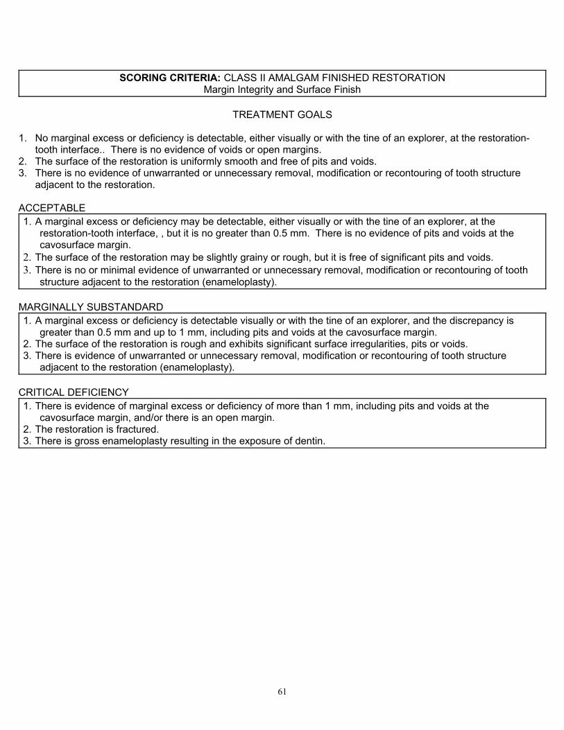
SCORING CRITERIA: CLASS II AMALGAM FINISHED RESTORATIONMargin Integrity and Surface Finish
TREATMENT GOALS
1. No marginal excess or deficiency is detectable, either visually or with the tine of an explorer, at the restoration-tooth interface.. There is no evidence of voids or open margins.
2. The surface of the restoration is uniformly smooth and free of pits and voids.3. There is no evidence of unwarranted or unnecessary removal, modification or recontouring of tooth structure
adjacent to the restoration.
ACCEPTABLE1. A marginal excess or deficiency may be detectable, either visually or with the tine of an explorer, at the
restoration-tooth interface, , but it is no greater than 0.5 mm. There is no evidence of pits and voids at the cavosurface margin.
2. The surface of the restoration may be slightly grainy or rough, but it is free of significant pits and voids.3. There is no or minimal evidence of unwarranted or unnecessary removal, modification or recontouring of tooth
structure adjacent to the restoration (enameloplasty).
MARGINALLY SUBSTANDARD1. A marginal excess or deficiency is detectable visually or with the tine of an explorer, and the discrepancy is
greater than 0.5 mm and up to 1 mm, including pits and voids at the cavosurface margin. 2. The surface of the restoration is rough and exhibits significant surface irregularities, pits or voids. 3. There is evidence of unwarranted or unnecessary removal, modification or recontouring of tooth structure
adjacent to the restoration (enameloplasty).
CRITICAL DEFICIENCY1. There is evidence of marginal excess or deficiency of more than 1 mm, including pits and voids at the
cavosurface margin, and/or there is an open margin. 2. The restoration is fractured. 3. There is gross enameloplasty resulting in the exposure of dentin.
61
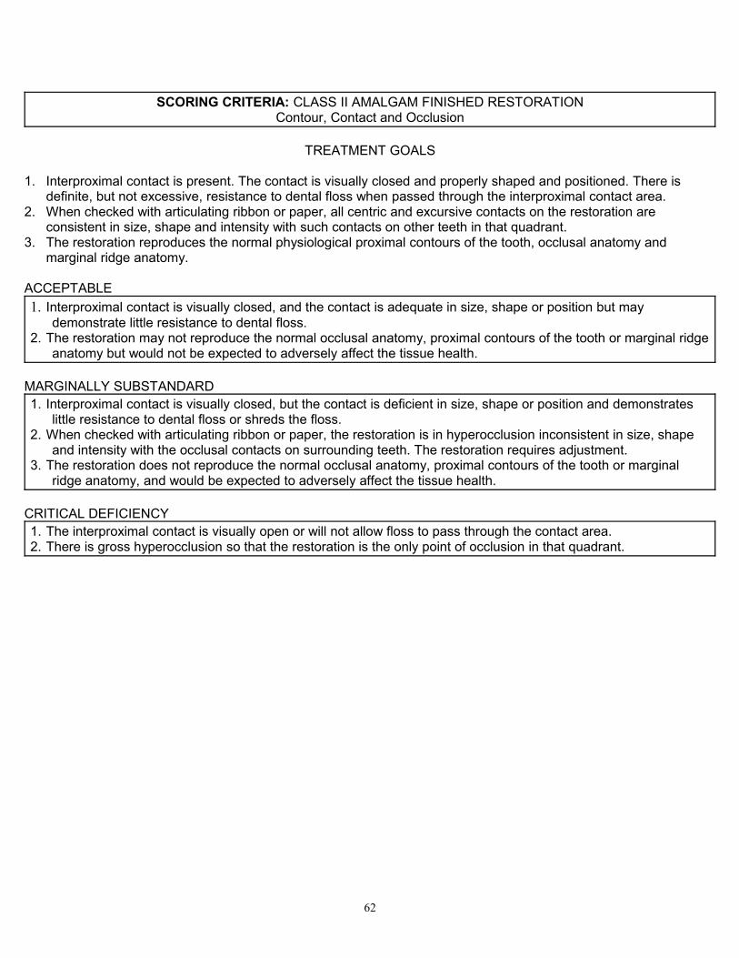
SCORING CRITERIA: CLASS II AMALGAM FINISHED RESTORATIONContour, Contact and Occlusion
TREATMENT GOALS
1. Interproximal contact is present. The contact is visually closed and properly shaped and positioned. There is definite, but not excessive, resistance to dental floss when passed through the interproximal contact area.
2. When checked with articulating ribbon or paper, all centric and excursive contacts on the restoration are consistent in size, shape and intensity with such contacts on other teeth in that quadrant.
3. The restoration reproduces the normal physiological proximal contours of the tooth, occlusal anatomy and marginal ridge anatomy.
ACCEPTABLE1. Interproximal contact is visually closed, and the contact is adequate in size, shape or position but may
demonstrate little resistance to dental floss.2. The restoration may not reproduce the normal occlusal anatomy, proximal contours of the tooth or marginal ridge
anatomy but would not be expected to adversely affect the tissue health.
MARGINALLY SUBSTANDARD1. Interproximal contact is visually closed, but the contact is deficient in size, shape or position and demonstrates
little resistance to dental floss or shreds the floss. 2. When checked with articulating ribbon or paper, the restoration is in hyperocclusion inconsistent in size, shape
and intensity with the occlusal contacts on surrounding teeth. The restoration requires adjustment.3. The restoration does not reproduce the normal occlusal anatomy, proximal contours of the tooth or marginal
ridge anatomy, and would be expected to adversely affect the tissue health.
CRITICAL DEFICIENCY1. The interproximal contact is visually open or will not allow floss to pass through the contact area. 2. There is gross hyperocclusion so that the restoration is the only point of occlusion in that quadrant.
62
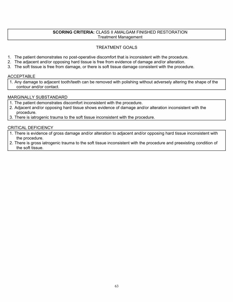
SCORING CRITERIA: CLASS II AMALGAM FINISHED RESTORATIONTreatment Management
TREATMENT GOALS
1. The patient demonstrates no post-operative discomfort that is inconsistent with the procedure.2. The adjacent and/or opposing hard tissue is free from evidence of damage and/or alteration.3. The soft tissue is free from damage, or there is soft tissue damage consistent with the procedure.
ACCEPTABLE1. Any damage to adjacent tooth/teeth can be removed with polishing without adversely altering the shape of the
contour and/or contact.
MARGINALLY SUBSTANDARD1. The patient demonstrates discomfort inconsistent with the procedure.2. Adjacent and/or opposing hard tissue shows evidence of damage and/or alteration inconsistent with the
procedure.3. There is iatrogenic trauma to the soft tissue inconsistent with the procedure.
CRITICAL DEFICIENCY1. There is evidence of gross damage and/or alteration to adjacent and/or opposing hard tissue inconsistent with
the procedure.2. There is gross iatrogenic trauma to the soft tissue inconsistent with the procedure and preexisting condition of
the soft tissue.
63
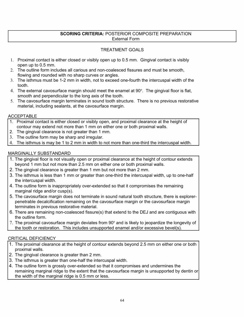
SCORING CRITERIA: POSTERIOR COMPOSITE PREPARATIONExternal Form
TREATMENT GOALS
1. Proximal contact is either closed or visibly open up to 0.5 mm. Gingival contact is visibly open up to 0.5 mm.
2. The outline form includes all carious and non-coalesced fissures and must be smooth, flowing and rounded with no sharp curves or angles.
3. The isthmus must be 1-2 mm in width, not to exceed one-fourth the intercuspal width of the tooth.
4. The external cavosurface margin should meet the enamel at 90o. The gingival floor is flat, smooth and perpendicular to the long axis of the tooth.
5. The cavosurface margin terminates in sound tooth structure. There is no previous restorative material, including sealants, at the cavosurface margin.
ACCEPTABLE1. Proximal contact is either closed or visibly open, and proximal clearance at the height of
contour may extend not more than 1 mm on either one or both proximal walls. 2. The gingival clearance is not greater than 1 mm.3. The outline form may be sharp and irregular. 4. The isthmus is may be 1 to 2 mm in width to not more than one-third the intercuspal width.
MARGINALLY SUBSTANDARD1. The gingival floor is not visually open or proximal clearance at the height of contour extends
beyond 1 mm but not more than 2.5 mm on either one or both proximal walls.2. The gingival clearance is greater than 1 mm but not more than 2 mm. 3. The isthmus is less than 1 mm or greater than one-third the intercuspal width, up to one-half
the intercuspal width.4. The outline form is inappropriately over-extended so that it compromises the remaining
marginal ridge and/or cusp(s).5. The cavosurface margin does not terminate in sound natural tooth structure, there is explorer-
penetrable decalcification remaining on the cavosurface margin or the cavosurface margin terminates in previous restorative material.
6. There are remaining non-coalesced fissure(s) that extend to the DEJ and are contiguous with the outline form.
7. The proximal cavosurface margin deviates from 90o and is likely to jeopardize the longevity of the tooth or restoration. This includes unsupported enamel and/or excessive bevel(s).
CRITICAL DEFICIENCY1. The proximal clearance at the height of contour extends beyond 2.5 mm on either one or both
proximal walls.2. The gingival clearance is greater than 2 mm.3. The isthmus is greater than one-half the intercuspal width.4. The outline form is grossly over-extended so that it compromises and undermines the
remaining marginal ridge to the extent that the cavosurface margin is unsupported by dentin or the width of the marginal ridge is 0.5 mm or less.
64
SCORING CRITERIA: POSTERIOR COMPOSITE PREPARATIONInternal Form
TREATMENT GOALS
1. The axial wall follows the external contours of the tooth and includes the DEJ but does not exceed 0.5 mm beyond the DEJ.
2. The pulpal floor depth must be at least 1.5-2 mm in all areas; there may be remaining enamel.
3. All caries and/or previous restorative material are removed.4. All prepared surfaces are smooth, rounded and well defined.5. When used, retention is well defined, placed in dentin and does not undermine enamel.6. The walls of the proximal box should be parallel or convergent occlusally.
ACCEPTABLE3. The pulpal floor depth is between 1.5 and 3 mm in all areas; there may be remaining
enamel. 4. The depth of the axial wall is no more than 1.5 mm beyond the DEJ.5. The walls of the proximal box may be slightly divergent, but not likely to jeopardize the
longevity of the tooth or restoration.
MARGINALLY SUBSTANDARD1. The pulpal floor depth is less than 1.5 mm or greater than 3 mm, up to 4 mm.2. The axial wall is more than 1.5 mm, but no more than 2.5 mm beyond the DEJ.3. The walls of the proximal box are too divergent or too convergent (resulting in excessively
undermined enamel). 4. Prepared surfaces are rough, sharp and irregular.5. Retention, when used, undermines the enamel.
CRITICAL DEFICIENCY1.The pulpal floor depth is 4 mm or greater from the cavosurface margin or is less than 0.5 mm.2.The axial wall is more than 2.5 mm beyond the DEJ or is still in enamel and does not include
the DEJ.3.Caries or previous restorative material remains in the preparation.
65
SCORING CRITERIA: POSTERIOR COMPOSITE PREPARATIONTreatment Management
TREATMENT GOALS
1. The preparation is adequately isolated with no debris, salivary or hemorrhagic leakage in the preparation.
2. The patient has received adequate anesthesia for pain control.3. The isolation dam is adequate to isolate sufficient teeth for visibility, accessibility and a dry
preparation. Ideally, the treated tooth and both proximal adjacent teeth should be isolated, if possible.
4. The adjacent teeth and/or restorations are free from damage.5. The soft tissue is free from damage, or there is tissue damage that is consistent with the
procedure.
ACCEPTABLE1. Any damage to adjacent tooth/teeth can be removed with polishing without adversely
altering the shape of the contour and/or contact.
MARGINALLY SUBSTANDARD1.Damage to adjacent tooth/teeth requires recontouring that changes the shape and/or contour
and/or contact.2.The isolation dam is inappropriately applied, torn and/or leaking, rendering the preparation
unsuitable for evaluation or the subsequent manipulation of the restorative material.3.There is inadequate anesthesia for pain control.4.There is iatrogenic soft tissue damage that is inconsistent with the procedure.
CRITICAL DEFICIENCY1.There is gross damage to adjacent tooth/teeth, requiring a restoration.2.There is gross iatrogenic damage to the soft tissue inconsistent with the procedure and
preexisting condition of the soft tissue.
66
SCORING CRITERIA: POSTERIOR COMPOSITE FINISHED RESTORATIONMargin Integrity and Surface Finish
TREATMENT GOALS
1.There is no marginal excess (overhang) or deficiency. No marginal excess is detectable, either visually or with the tine of an explorer, at the restoration-tooth interface. There is no evidence of voids or open margins.
2.The surface of the restoration is uniformly smooth and free of pits and voids.3.There is no evidence of unwarranted or unnecessary removal, modification or recontouring
of tooth structure adjacent to the restoration. 4.The restoration is bonded to the prepared tooth structure.5.Shade selection matches surrounding tooth structure.
ACCEPTABLE1. Any marginal excess or deficiency is detectable, either visually or with the tine of an explorer,
at the restoration-tooth interface, but it is no greater than 0.5 mm. There is no evidence of pits and voids at the cavosurface margin.
2. The surface of the restoration may be slightly grainy or rough, but it is free of significant pits and voids.
3. There is no or minimal evidence of unwarranted or unnecessary removal, modification or recontouring of tooth structure adjacent to the restoration.
MARGINALLY SUBSTANDARD1.The restoration-tooth interface is detectable visually or with the tine of an explorer. There is
evidence of marginal excess or deficiency of greater than 0.5 mm and up to 1 mm, including pits and voids at the cavosurface margin.
2.The surface of the restoration is rough and exhibits significant surface irregularities, pits or voids.
3.There is significant evidence of unwarranted or unnecessary removal, modification or recontouring of tooth structure adjacent to the restoration.
4.Shade selection does not match surrounding tooth structure.
CRITICAL DEFICIENCY1.There is evidence of marginal excess or deficiency of more than 1 mm, including pits and
voids at the cavosurface margin, and/or there is an open margin.2.There is gross enameloplasty resulting in the exposure of dentin.3.The restoration is debonded and/or movable in the preparation. 4.The restoration is fractured.
67
SCORING CRITERIA: POSTERIOR COMPOSITE FINISHED RESTORATIONContour, Contact and Occlusion
TREATMENT GOALS
1. Interproximal contact is present. The contact is visually closed and is properly shaped and positioned. There is definite, but not excessive, resistance to dental floss when passed through the interproximal contact area.
2. When checked with articulating paper, all centric and excursive contacts on the restoration are consistent in size, shape and intensity with such contacts on other teeth in that quadrant.
3. The restoration reproduces the normal physiological proximal contours of the tooth, occlusal anatomy and marginal ridge anatomy.
ACCEPTABLE1. Interproximal contact is visually closed, and the contact appears adequate in size, shape or
position but may demonstrate little resistance to dental floss.2. The restoration may not reproduce the normal occlusal anatomy, proximal contours of the
tooth or marginal ridge anatomy but would not be expected to adversely affect the tissue health.
MARGINALLY SUBSTANDARD1. Interproximal contact is visually closed, but the contact is deficient in size, shape or position
and demonstrates little resistance to dental floss or shreds the floss. 2. When checked with articulating paper, the restoration is in hyperocclusion inconsistent in
size, shape and intensity with the occlusal contacts on surrounding teeth. The restoration requires adjustment.
3. The restoration does not reproduce the normal occlusal anatomy, proximal contours of the tooth or marginal ridge anatomy and would be expected to adversely affect the tissue health.
CRITICAL DEFICIENCY1. The interproximal contact is visually open or will not allow floss to pass through the contact
area. 2. There is gross hyperocclusion, such that the restoration is the only point of occlusion in that
quadrant.
68

SCORING CRITERIA: POSTERIOR COMPOSITE FINISHED RESTORATIONTreatment Management
TREATMENT GOALS
1. The adjacent and/or opposing hard tissue is free from evidence of damage and/or alteration.2. The patient demonstrates no post-operative discomfort that is inconsistent with the
procedure.3. The soft tissue is free from damage, or there is soft tissue damage consistent with the
procedure.
ACCEPTABLE1. Any minimal damage to adjacent tooth/teeth can be removed with polishing without adversely
altering the shape of the contour and/or contact.
MARGINALLY SUBSTANDARD1. There is iatrogenic damage to the soft tissue inconsistent with the procedure.2. Adjacent and/or opposing hard tissue shows evidence of damage and/or alteration
inconsistent with the procedure.3. The patient demonstrates discomfort inconsistent with the procedure.
CRITICAL DEFICIENCY1. There is gross iatrogenic damage to the soft tissue inconsistent with the procedure.2. There is evidence of gross damage and/or alteration to adjacent and/or opposing hard tissue
inconsistent with the procedure.
69
SCORING CRITERIA: POSTERIOR PROXIMAL OCCLUSAL COMPOSITE PREPARATIONExternal Form
TREATMENT GOALS
1. Proximal contact is either closed or visibly open up to 0.5 mm. Gingival contact is visibly open up to 0.5mm.
2. The outline form must be smooth, flowing and rounded with no sharp curves or angles.3. The external cavosurface margin meets the enamel at 90o. The gingival floor is flat,
smooth and perpendicular to the long axis of the tooth.4. The cavosurface margin terminates in sound tooth structure. There is no previous
restorative material, including sealants, at the cavosurface margin.
ACCEPTABLE1. Proximal clearance at the height of contour is closed or visually open not more than 1
mm on either one or both proximal walls. 2. The gingival clearance is not greater than 1 mm. 3. Outline form may be irregular and sharp.
MARGINALLY SUBSTANDARD1. The gingival floor is not visually open or proximal clearance at the height of contour extends
beyond 1 mm but not more than 2.5 mm on either one or both proximal walls.2. The gingival clearance is greater than 1 mm but not more than 2 mm. 3. The outline form is inappropriately over-extended so that it compromises the cusp(s).4. The cavosurface margin does not terminate in sound natural tooth structure, or there is
explorer-penetrable decalcification remaining on the cavosurface margin, or the cavosurface margin terminates in previous restorative material.
5. The proximal cavosurface margin deviates from 90o and is likely to jeopardize the longevity of the tooth or restoration. This includes unsupported enamel and/or excessive bevel(s).
CRITICAL DEFICIENCY1. The proximal clearance at the height of contour extends beyond 2.5 mm on either one or
both proximal walls.2. The gingival clearance is greater than 2 mm. The outline form is grossly over-extended so
that it compromises and undermines the remaining cusp(s) to the extent that the cavosurface margin is unsupported by dentin.
70
SCORING CRITERIA: POSTERIOR PROXIMAL OCCLUSAL COMPOSITE PREPARATIONInternal Form
TREATMENT GOALS
1.The axial wall follows the external contours of the tooth and includes the DEJ but should not exceed 0.5 mm beyond the DEJ.
2.The proximal walls should be parallel or convergent occlusally.3.All caries and/or previous restorative material are removed.4.All prepared surfaces are smooth, rounded and well defined.5.When used, retention is well defined, placed in dentin and does not undermine enamel.
ACCEPTABLE1.The depth of the axial wall is from 0.5 mm to 1.5 mm from the DEJ.2. The proximal walls are parallel or convergent occlusally but may be slightly divergent and are
not likely to jeopardize the longevity of the tooth or restoration.
MARGINALLY SUBSTANDARD1.The axial wall is more than 1.5 mm up to 2.5 mm beyond the DEJ.2.The proximal walls are too divergent or too convergent (resulting in excessively undermined
enamel). 3.Prepared surfaces are rough, sharp and irregular.4.Retention, when used, undermines the enamel.5.The internal retentive features may be inadequate and may compromise the tooth or
restoration.
CRITICAL DEFICIENCY1. The axial wall is more than 2.5 mm beyond the DEJ or is still in enamel and does not
include the 2. DEJ.3. Caries or previous restorative material remains in the preparation.
71
SCORING CRITERIA: POSTERIOR PROXIMAL OCCLUSAL COMPOSITE PREPARATIONTreatment Management
TREATMENT GOALS
1.The preparation is adequately isolated with no debris, salivary or hemorrhagic leakage in the preparation.
2.The patient has adequate anesthesia for pain control.3.The isolation dam is adequate to isolate sufficient teeth for visibility, accessibility and a dry
preparation. Ideally, the treated tooth and both proximal adjacent teeth should be isolated, if possible.
4.The adjacent teeth and/or restorations are free from damage.5.The soft tissue is free from damage, or there is tissue damage that is consistent with the
procedure.
ACCEPTABLE1.Any damage to adjacent tooth/teeth can be removed with polishing without adversely altering
the shape of the contour and/or contact.
MARGINALLY SUBSTANDARD1.Damage to adjacent tooth/teeth requires recontouring that changes the shape and/or contour
and/or contact.2.The isolation dam is inappropriately applied, torn and/or leaking, rendering the preparation
unsuitable for evaluation or the subsequent manipulation of the restorative material.3.There is inadequate anesthesia for pain control.4.There is iatrogenic soft tissue damage that is inconsistent with the procedure.
CRITICAL DEFICIENCY1.There is gross damage to adjacent tooth/teeth, requiring a restoration.2.There is gross iatrogenic damage to the soft tissue inconsistent with the procedure and
preexisting condition of the soft tissue.
72

SCORING CRITERIA: POSTERIOR PROXIMAL OCCLUSAL COMPOSITE FINISHED RESTORATION
4. Margin Integrity and Surface Finish
TREATMENT GOALS
1. There is no marginal excess (overhang) or deficiency. There is no detectable marginal excess at the restoration-tooth interface, either visually or with the tine of an explorer. There is no evidence of voids or open margins.
2. The surface of the restoration is uniformly smooth and free of pits and voids.3. There is no evidence of unwarranted or unnecessary removal, modification or recontouring of
tooth structure adjacent to the restoration. 4. The restoration is bonded to the prepared tooth structure.5. Shade selection matches surrounding tooth structure.
ACCEPTABLE1. There may be a marginal excess or deficiency at the restoration-tooth interface detectable
either visually or with the tine of an explorer, but it is no greater than 0.5 mm. There is no evidence of pits and voids at the cavosurface margin.
2. The surface of the restoration may be slightly grainy or rough, but it is free of significant pits and voids.
3. There is minimal or no evidence of unwarranted or unnecessary removal, modification or recontouring of tooth structure adjacent to the restoration.
MARGINALLY SUBSTANDARD1. The restoration-tooth interface is detectable visually or with the tine of an explorer. There is
evidence of marginal excess or deficiency of greater than 0.5 mm and up to 1 mm, including pits and voids at the cavosurface margin.
2. The surface of the restoration is rough and exhibits significant surface irregularities, pits or voids.
3. There is evidence of unwarranted or unnecessary removal, modification or recontouring of tooth structure adjacent to the restoration.
4. Shade selection does not match surrounding tooth structure.
CRITICAL DEFICIENCY1. There is evidence of marginal excess or deficiency of more than 1 mm, including pits and
voids at the cavosurface margin and/or there is an open margin.2. There is gross enameloplasty resulting in the exposure of dentin.3. The restoration is debonded and/or movable in the preparation. 4. The restoration is fractured.
73
SCORING CRITERIA: POSTERIOR PROXIMAL OCCLUSAL COMPOSITE FINISHED RESTORATION
Contour, Contact and Occlusion
TREATMENT GOALS
1. Interproximal contact is present. The contact is visually closed and is properly shaped and positioned. There is definite, but not excessive, resistance to dental floss when passed through the interproximal contact area.
2. When checked with articulating paper, all centric and excursive contacts on the restoration are consistent in size, shape and intensity with such contacts on other teeth in that quadrant.
3. The restoration reproduces the normal physiological proximal contours of the tooth, occlusal anatomy and marginal ridge anatomy.
ACCEPTABLE1. Interproximal contact is visually closed, and the contact is adequate in size, shape or position
but may demonstrate little resistance to dental floss.2. The restoration may not reproduce the normal occlusal anatomy, proximal contours of the
tooth or marginal ridge anatomy but would not be expected to adversely affect the tissue health.
MARGINALLY SUBSTANDARD1. Interproximal contact is visually closed, but the contact is deficient in size, shape or position
and demonstrates little resistance to dental floss or shreds the floss. 2. The restoration does not reproduce the normal occlusal anatomy, proximal contours of the
tooth or marginal ridge anatomy and would be expected to adversely affect the tissue health.3. When checked with articulating paper, the restoration is in hyperocclusion inconsistent in
size, shape and intensity with the occlusal contacts on surrounding teeth. The restoration requires adjustment.
CRITICAL DEFICIENCY1. The interproximal contact is visually open or will not allow floss to pass through the contact
area. 2. There is gross hyperocclusion, such that the restoration is the only point of occlusion in that
quadrant.
74
SCORING CRITERIA: POSTERIOR PROXIMAL OCCLUSAL COMPOSITE FINISHED RESTORATION
5. Treatment Management
TREATMENT GOALS
1. The adjacent and/or opposing hard tissue is free from evidence of damage and/or alteration.2. The patient demonstrates no post-operative discomfort that is inconsistent with the
procedure.3. The soft tissue is free from damage, or there is soft tissue damage consistent with the
procedure.
ACCEPTABLE1. Any damage to adjacent tooth/teeth can be removed with polishing without adversely altering
the shape of the contour and/or contact.
MARGINALLY SUBSTANDARD1. There is iatrogenic damage to the soft tissue inconsistent with the procedure.2. Adjacent and/or opposing hard tissue shows evidence of damage and/or alteration
inconsistent with the procedure.3. The patient demonstrates discomfort inconsistent with the procedure.
CRITICAL DEFICIENCY1. There is gross iatrogenic damage to the soft tissue inconsistent with the procedure.2. There is evidence of gross damage and/or alteration to adjacent and/or opposing hard tissue
inconsistent with the procedure.
75
Check-Out Procedure for ALL Examination SectionsUpon completion of all examinations, candidates must personally submit all examination packets and typodonts to a central location determined by the chief examiner. The following items must be submitted in the provided white envelope and accounted for prior to dismissal from the examination site:
• Pre-operative and post-operative (if requested during the examination) radiographs of teeth restored during the examination must be submitted and clearly marked for identification. The Full mouth series of radiographs for the Periodontal Scaling Clinical Examination Section need not be submitted unless requested by an examiner. (If the testing site requires that radiographs be retained in the patient record, the candidate may submit duplicates of restorative or periodontal radiographs.) At sites where digital images are displayed on a monitor, an electric copy of the digital images used must be submitted on disk to the testing agency.
• Completed Progress Forms. The Fixed Prosthodontic and Endodontic Clinical Examination Progress Forms are submitted with the typodont to the CFE.
• Photo ID card for candidate and chairside assistant
• Patient Consent Form(s)
• Medical History Form(s)
• Color-coded cubicle ID cards (2)
Check-Out Procedure for Retakes Check-out for those retaking a single examination section is on the day the clinical examination is completed. Those taking examination sections on both days will check out the second day.
76
The ADEX Dental Examination Series
Curriculum Integrated Format
VI. Examination Forms
77
Examination FormsForms Completed Before the Examination
Please note: samples of all forms referenced may be found at the end of this section.
Medical History FormThe candidate must complete a Medical History Form for each patient participating in the examination. This form is available on the NERB website www.nerb.org under “Forms and Manuals” for the examinations administered by NERB and on the SRTA website www.srta.org for the examinations administered by SRTA. The Medical History Form may be completed prior to the examination and will be reviewed by the CFE prior to patient check-in. If the patient will be treated by more than one candidate, each candidate must submit a separate Medical History Form.
The patient’s blood pressure must be taken on the day of the examination and documented by a clinic floor examiner (CFE). The CFE does not witness the candidate taking the patients’ blood pressure and heart rate, but must verify and sign-off that it was taken the day of the exam and is on the appropriate form before sending the patient to the Scoring Area for examiner approval.
If the patient has a medical condition that could affect his/her suitability for treatment, the candidate must obtain a written medical clearance from the patient’s physician to indicate that the patient is healthy enough to participate in the examination. The medical clearance must also be submitted on the day of the examination and should meet the following criteria:
• Clearly legible statement from a licensed physician
• Written within 30 days prior to the examination on official letterhead
• Containing a positive statement of how the patient should be medically managed
• Containing the physician’s clearly legible name, address and phone number
• Containing a telephone number where the physician may be reached on the day of the examination if a question arises regarding the patient’s health
Patient Consent Form (Patient Consent, Disclosure and Assumption of Liability) Candidates must review the Patient Consent Form with their patients and submit a signed copy on the day of the examination. This form is available on the NERB website www.nerb.org under “Forms and Manuals” for the examination administered by NERB and on the SRTA website www.srta.org for SRTA administered examination
Because this form will be reviewed by examiners during the procedure, candidates should initial – but not sign – the form before beginning treatment, in order to preserve anonymity. (Patients should sign with their full signature.) After the examination is completed and before submitting all records during check-out, candidates should complete the form with their full signature.
Periodontal Scaling Treatment Selection WorksheetThe Periodontal Treatment Selection Worksheet is a practice form candidates may use to identify the teeth they will treat during the Periodontal Clinical Examination Section. This form is available on the NERB website www.nerb.org under “Forms and Manuals” for the examination administered by NERB and on the SRTA website www.srta.org for SRTA administered examination.
To earn a Satisfactory rating for patient selection on the Periodontal Scaling Examination Section, the candidate must identify a selection of teeth that meet these criteria:
78
• Six to eight teeth selected, each with at least one surface of calculus charted
• At least three posteriors (molars, premolars), including at least one molar, in the selection
• All posterior teeth must have at least one approximating tooth within 2 mm distance
• Exactly 12 surfaces of subgingival calculus charted, including at least three surfaces of interproximal calculus on molars/premolars
• At least eight of the surfaces on canines, premolars or molars (no more than four surfaces on incisors)
• Three pockets of 4 mm or greater in depth, each on a different tooth within the selection
Electronic Periodontal Scaling Evaluation FormElectronic Evaluation Forms are used by examiners to score the candidate’s performance. In most cases, candidates will not have access to these forms, with one exception: prior to the Periodontal Scaling Clinical Examination Section, candidates must enter their treatment selection into the Electronic Periodontal Scaling Evaluation Form to indicate to examiners which teeth are to be evaluated.
Typically, candidates use the Periodontal Scaling Treatment Selection Worksheet to identify and chart the selected teeth, and then transfer their responses from the Worksheet onto the Electronic Periodontal Scaling Evaluation Form.
To enter the treatment selection on the Electronic Periodontal scaling Evaluation Form, candidates can log-in to their profile on the administering agency website and follow the online instructions. Access to the Electronic Periodontal Evaluation Form is closed beginning 48 hours prior to the first exam day for NERB and 2 weeks prior to the exam date for SRTA, at a given exam site, to allow uploading of the information prior to the examination. However, a computer will be available at the Coordinator’s Desk to enter the periodontal treatment selection on the day of the examination. In order to reduce lost time on the day of the examination, it is highly recommended that this step is completed prior to the day of the examination.
Forms Completed at the ExaminationOnce the examination begins, examination materials distributed by the testing agency may not be removed from the examining area. Forms may not be reviewed by unauthorized personnel.
Progress FormsColor-coded Progress Forms are utilized to track the candidate’s progress through each procedure, document anesthesia administered and treatment provided, collect examiner signatures for all completed portions of the examination and provide appropriate progress notes from the candidate to examiners during the course of treatment.
Candidates will be provided with identification labels to place on each procedure’s Progress Form, as indicated on the form.
The appropriate Progress Forms must be presented to the examiners at the time of patient check-in. Original, pre-operative radiographs must be mounted with transparent tape on the appropriate Progress Forms for the Restorative Clinical Examination Section, or be available on the monitors located in the scoring area.
The Fixed Prosthodontic and Endodontic Examination Section Progress Form will remain with the candidate throughout the examination. It must be filled out at the beginning of the examination and turned in as directed on the day of the examination. Modification Request FormModification Request Forms are utilized to request permission to deviate from a Satisfactory-level restorative preparation. The form requires the candidate to provide the following information:
• What is the candidate requesting to do? (Type of modification)79
• Where? (e.g., gingival axial line angle, mesial box)
• How Much is to be removed? (e.g., 0.5 mm from the axial wall)
• Why is the modification needed? (e.g., due to caries, decalcification)
Candidates who need to request a modification should place an identification label on the Modification Request Form and indicate their cubicle number, procedure, day and time.
Instruction to Candidate FormCandidates may receive written instructions from examiners on an Instruction to Candidate Form if the examiners believe the treatment should be modified. The Instruction to Candidate Form is generated electronically by the examiners in the Evaluation Station, printed out at the Coordinator’s Desk and delivered to the candidate by a clinic floor examiner, in order to preserve anonymity. The candidate must initial on the Instruction to Candidate Form that he/she understands the instructions.
Follow-Up FormThe Follow-Up Form is utilized to advise the patient and candidate of additional treatment needs or whenever the treatment started by the candidate is incomplete or the final treatment is unacceptable. Like the Instruction to Candidate Form, the Follow-Up Form is sent electronically to the Coordinator Desk for delivery to the candidate. The Follow-Up Form identifies the problem and establishes responsibility for further treatment. The patient is informed that follow-up care is necessary, financial responsibility is clarified and the candidate and Chief Examiner sign the form.
80
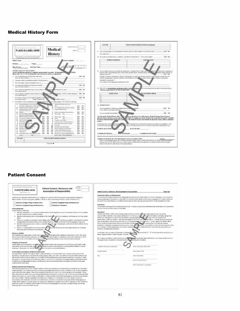
Medical History Form
Patient Consent
81
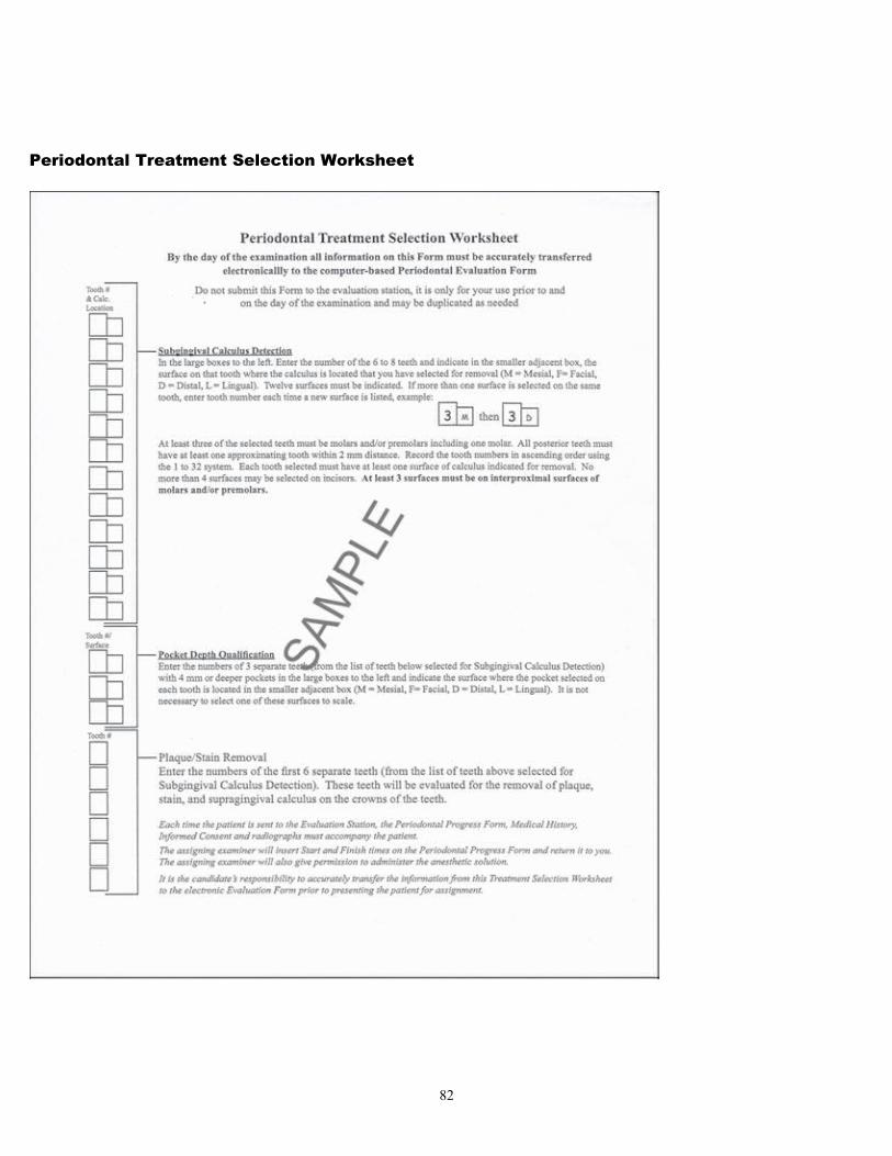
Periodontal Treatment Selection Worksheet
82
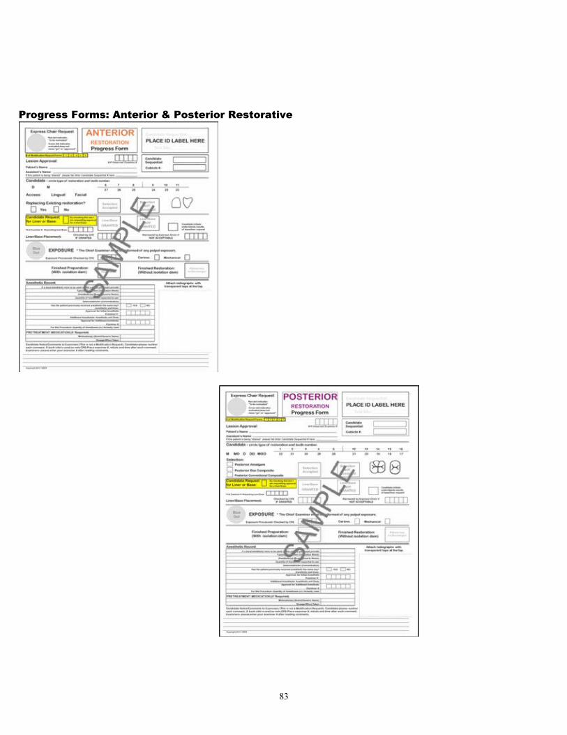
Progress Forms: Anterior & Posterior Restorative
83
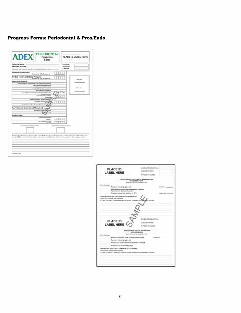
Progress Forms: Periodontal & Pros/Endo
84

85
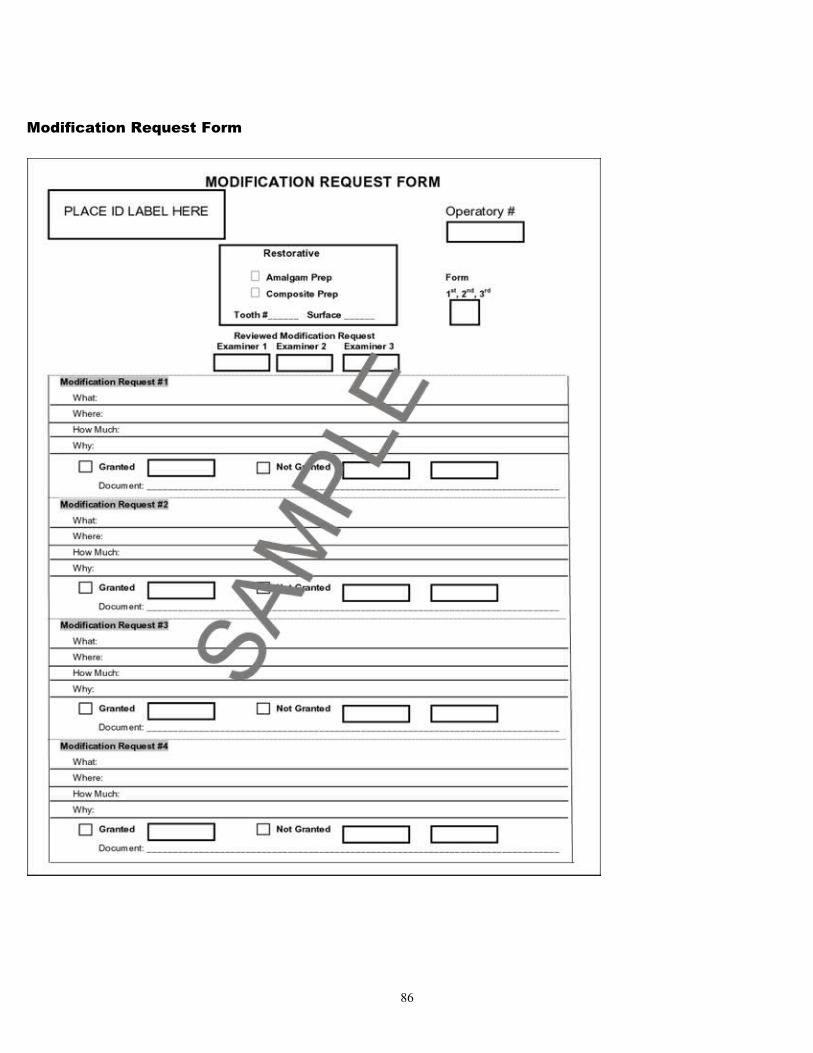
Modification Request Form
86
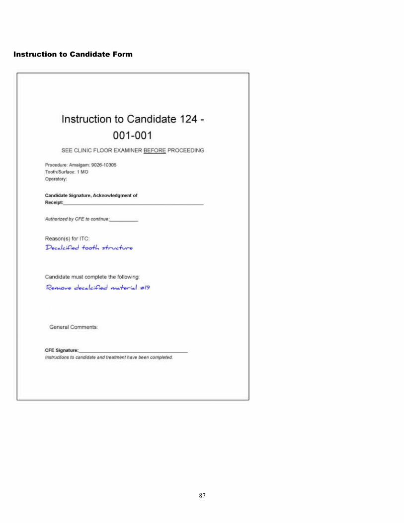
Instruction to Candidate Form
87
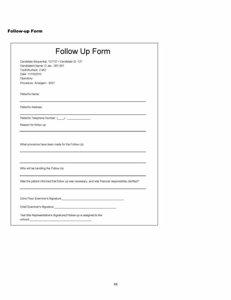
Follow-up Form
88
The ADEX Dental Examination Series
Curriculum Integrated Format
IV. Policies and Procedures
89
Standards of ConductThe ADEX examination strives to evaluate the candidate’s clinical judgment and skills in a fair manner. In addition, conduct, decorum and professional demeanor are evaluated. The candidate is required to adhere to the rules, regulations and standards of conduct for the ADEX Dental Examination Series.
1. Assigned procedures: Only the treatment and/or procedures assigned may be performed. (In the Periodontal Scaling Clinical Examination Section, all surfaces of the selected teeth may be scaled and polished at the discretion of the candidate, but only the selected surfaces will be evaluated.) Performing other treatment or procedures may result in failure of the examination.
2. Completion of the examinations: All five examination sections (or four, if the candidate is not taking the Periodontal Scaling Section) of the ADEX Dental Examination Series must be completed within the specified time frame in order to be considered for “ADEX Status.” Examination procedures performed outside the assigned time will be considered incomplete, and the candidate will fail the examination section. If all specified materials and required documentation are not turned in at the end of an examination section that section will be considered incomplete, and the candidate will fail the section.
3. Electronic equipment: The use of pagers, cell phones, computers, DVDs, CDs, PDAs, Blackberries, radios (including walkie-talkies with or without earphones) and any other electronic equipment is not permitted on the clinic floor by candidates, auxiliaries or patients during the examination. Any such use will be considered unprofessional conduct and may result in dismissal from the examination.
4. Electronic recording devices and cameras: The use of electronic recording devices or cameras by the candidate, an auxiliary or a patient during any part of the examination is a violation of examination guidelines and may result in failure of the entire ADEX Dental Examination Series. However, intra-oral photographs may be taken by authorized examiners or school personnel during the course of the examination for the purpose of future examiner standardization and calibration.
5. Misappropriation and/or damage of equipment: No equipment, instruments or materials shall be removed from the examination site without written permission of the owner. Willful or careless damage of dental equipment, typodonts, manikins or shrouds may result in failure. All resulting repair or replacement costs will be charged to the candidate and must be paid to the host site before the candidate’s examination results will be released.
6. Personal/professional conduct: Any substantiated evidence of collusion, dishonesty, use of unauthorized assistance or intentional misrepresentation during registration or during the course of the examinations or failure of the candidate to carry out a directive of the chief examiner shall automatically result in failure of all five examination sections. The candidate and assisting auxiliary must behave in an ethical and proper manner. Patients shall be treated with proper concern for their safety and comfort. Improper behavior is cause for dismissal from the examination at the discretion of the chief examiner and will result in failure of the examination. Additionally, the candidate shall be denied reexamination for one full year from the time of the infraction.
7. Submission of examination records: All required records and radiographs must be turned in before the examination is considered complete. If all required documentation is not turned in at the end of the examination, the examination will be considered incomplete, and the candidate will fail all exam sections involved.
8. Termination of the examinations: The right is reserved to terminate or delay the examinations at any time if 1) that action becomes necessary to safeguard the health, safety or comfort of the patient, 2) the candidate or examiners are threatened in any manner or 3) other interfering events occur that are not under the control of the administering testing agency.
90
Standards for Section I: Diagnostic Skills Examination (DSE) 1. Behavior at the Testing Center: Unseemly behavior of the candidate or improper behavior toward personnel at
the Testing Center will result in failure of the DSE and forfeiture of the examination fee.
2. Examination security: Security measures established by the testing centers must be followed. Failure to do so may result in failure of the examination series.
3. Extraneous materials: Only materials distributed or authorized by the Testing Centers may be brought to the DSE Section. Use of unauthorized materials will result in failure of the entire examination series. No textbooks or study materials are permitted at the Testing Center at any time.
4. Timely arrival: Candidates must adhere to the appointment time as established by the candidate. Failure to arrive on time will result in forfeiture of the examination fee.
5. Time limits: A specified total amount of time is allowed for each subsection (PE, CTP, and PPMC) of the Diagnostic Skills Examination Section. Once a candidate has locked out of a designated subsection of the DSE, he/she may not re-enter that section.
Standards for Sections II and III: Endodontics and Fixed Prosthodontics Examinations Assigned operatories: The candidate shall work only in the clinic, operatory or laboratory spaces as authorized. Violation of this standard will result in failure of the examination section.
Assigned teeth: Once a procedure has been started, the procedure must be carried to completion on the assigned tooth/teeth with no substitutions permitted. Substitution of teeth or preparation of the wrong tooth/teeth will result in failure of the Fixed Prosthodontics or Endodontics Clinical Examination Sections. If a candidate discovers that the wrong tooth has been prepared, he/she must immediately contact the CFE.
Assistants: Auxiliary personnel and/or laboratory technicians are not permitted to assist a candidate during the Fixed Prosthodontics or Endodontics Clinical Examination Sections. Violation may result in failure of these examination sections.
Equipment failure: A CFE must be notified immediately in case of equipment failure; he/she will contact school maintenance personnel. The malfunction must be corrected or the candidate relocated. Extension of examination time is not granted because of equipment malfunction or failure.
Examination start time: Treatment procedures may not be initiated prior to the established starting time. Violation of this standard will result in failure of the examination section.
Infection control: Candidates must follow the infection control procedures recommended by the Centers for Disease Control and Prevention (as with a live patient), including setting up prior to the examination and cleaning up after the examination has ended. Violation of this standard may result in failure of the examination section.
Isolation: Adequate and proper isolation must be used for the Endodontics Examination Section. Vioation will result in a penalty.
Manikins: Only manikins, typodonts and teeth approved by ADEX may be used for the Fixed Prosthodontic and Endodontic Clinical Examination Sections. Violation of this standard will result in failure of the examination section.
Operating position: The manikin must be mounted and maintained in a physiologically acceptable operating position while performing the Fixed Prosthodontics and Endodontics Clinical Examination Sections. The facial shroud must be maintained in the same position as the normal facial tissue. Violations will result in a reduction in the score.
91
Patient management: The manikin must be considered as a patient and treated in the same manner and with the same consideration. Violation of this standard will result in failure of the examination section.
Removal of typodont or manikin: During the examination, the teeth, typodont or manikin may not be removed or dismantled without specific permission from a CFE. Violations will result in penalties.
Standards for Sections IV and V: Restorative and Periodontal Scaling Clinical Examinations
Anesthetic/Analgesic: For both the Restorative and Periodontal Scaling Examination Sections, the anesthetic section on the Progress Form must be completed whether or not local anesthesia will actually be administered. Candidates must receive permission from the CFE before administering anesthesia at any point during the examination. Candidates may provide local anesthesia for periodontal patients prior to initial assessment in the Evaluation Station but restorative patients may only receive local anesthesia after the lesion has been accepted in the Evaluation Station. If the patient has previously been given an anesthetic on the same day, the candidate must note the previous anesthesia on the Progress Form and obtain signed permission (by a CFE for the Restorative Clinical Examination and by a CFE for the Periodontal Scaling Clinical Examination) before administering more anesthetic solution. The candidate is responsible for ensuring that the amount of anesthetic used is correctly recorded on the Progress Form before sending the patient to the scoring area for final examiner evaluation. Inhalation or intravenous analgesia or anesthetics are not permitted for the examination. Violation of this standard will result in failure of the examination section.
Anesthetic record: At the time of the examination and prior to the start-check for each restorative or periodontal scaling clinical procedure, the following anesthetic information must be indicated on the appropriate Progress Form:
• Type(s) of Injection (specific block or infiltration to be administered)
• Anesthetic(s) (generic or brand name and percent used)
• Vasoconstrictor (type and concentration)
• Quantity (volume)
If more than two dental anesthetic carpules (approximately 3.6 cc) of local anesthetic are needed during any clinical procedure, the candidate must request approval from a CFE, who will document and initial the request. This protocol must be followed for each subsequent carpule. Additional anesthetic solution may be administered only with approval by the CFE. The total quantity of anesthetic solution used must also be documented on the Progress Form prior to sending the patient to the evaluation station for the final restorative scoring.
An aspirating syringe and proper aspirating technique must be used for the administration of local anesthetic solutions. The administration of inhalation or parenteral analgesia or sedation is not permitted for any clinical procedures.
If the patient has already received anesthesia earlier on the same day, the candidate must present the record of the previous anesthesia to the CFE before administering additional anesthesia.
Anonymity: The anonymous testing procedure prevents the possibility that any person involved with the evaluation of the examination may know, learn of or establish the identity of a candidate or relate or connect the patient or work-product to a particular candidate. The candidate’s name and school information should not appear on any examination forms, materials or instruments. Grading examiners will be physically isolated from the candidates in a separate area of the clinic, and the movement of patients from the clinical areas to the grading area shall be controlled by the use of testing agency messengers/assistants. All examination forms and materials are identified by the candidate’s identification number, which is assigned prior to the examination.
92
Assigned operatories: Candidates must perform all procedures in their assigned operatories. Working in areas not authorized will result in failure of the examination section.
Auxiliary personnel: Candidates may use chairside assistants during the Restorative and Periodontal Examination Sections (patient-based sections). Individuals who meet the following criteria may not serve as assistants:
Individuals under 18 years of ageDentists and dental hygienists (licensed or unlicensed)Third- or fourth-year dental studentsDental hygiene students in the final year of studyDental technicians
Please refer to the Agency specific Supplement for chairside assistant registration information.
Clinic attire: Candidates must wear clinic attire that meets CDC and OSHA standards. No bare arms or legs or open-toed shoes are allowed in the clinic areas. Lab coats, lab jackets and/or long-sleeved protective garments are all acceptable. Color and style are not restricted. There must be no personal or school identification on clinic attire other than the candidate identification badge.
Equipment failure: In case of equipment failure, the clinic floor captain must be notified immediately; he/she will request that maintenance personnel assess and correct the specific situation. Extension of examination time will not be granted. Maintenance and repair of equipment (chair, light and dental unit) is the responsibility of and is provided by the school.
Failure to follow directions: Failure to follow directions and instructions from examiners will be considered unprofessional conduct. Unprofessional conduct and improper behavior are cause for dismissal from the examination and will result in failure of the examination series.
Identification badges: During the examination, candidate ID badges must be worn at all times during the clinical examinations.
Infection control standards: Candidates and auxiliary personnel must follow the infection control procedures recommended by the Centers for Disease Control and Prevention, including setting up prior to the examination and cleaning up after the examination has ended. Violation of the infection control standards may result in failure of the examination.
Interpreters: Candidates should employ the services of an interpreter when their patient does not speak English or has a hearing impairment that cannot be corrected. (The use of an interpreter is particularly important when the patient has a history of medical problems or is on medications.)
Individuals who meet the following criteria are not permitted to serve as interpreters:Individuals under 18 years of ageFaculty membersDentists and dental hygienists (licensed or unlicensed)Third- or fourth-year dental studentsDental hygiene students in the final year of studyIndividuals serving as chairside assistants
Please refer to the Agency specific Supplement for Interpreter registration information.
Isolation of the restorative field: Adequate and proper isolation must be provided as necessary to avoid contamination and as stipulated by examination requirements. Violation will result in penalties.
Management of significant history and pathosis: The candidate shall accurately complete the appropriate Medical History Form and establish a diagnosis and treatment plan as required for each selected patient. Misinformation or missing information that would endanger the patient, candidate, auxiliary personnel or examiners is considered cause for dismissal from the examination.
93
New technology: New technologies are constantly being developed and marketed in dentistry. However, until such time as these innovations become the standard of care and are readily available to all candidates at all testing sites, the use of such innovative technologies are discouraged for use in this examination. If there is a question about such new technologies, contact the appropriate testing agency.
Patient management: The candidate and assisting auxiliary must behave in an ethical and proper manner. Patients shall be treated with proper concern for their safety and comfort. Either the candidate or the assistant should be with the patient whenever the patient is in the treatment clinic, especially whenever an isolation dam is in place. Chairside assistants must register with the chief examiner and display the proper identification. Failure to follow directions and instructions from examiners will be considered unprofessional conduct. Improper behavior is cause for dismissal from the examination and will result in failure of the examination. Additionally, the candidate shall be denied reexamination for one full year from the time of the infraction.
Patient’s Medical HistoryA Medical History Form must be completed independently by the candidate (without help of faculty or colleagues) for each clinical patient prior to the examination. This form may be completed prior to the examination date; however, the form must reflect the patient’s current health at the time of the examination.
The Medical History Form includes questions pertaining to medical conditions that might affect the patient’s suitability for treatment. If the patient gives a positive response to one of these questions, the candidate must explore the nature of the condition and provide an adequate explanation on the Medical History Form.
Blood pressure: A screening blood pressure reading should be taken when the patient is selected and must be retaken on the day of the examination and recorded on the Medical History Form. The examination-day reading must be documented by a CFE. If the patient is sitting for more than one examination section on the same day, his/her blood pressure must be taken and recorded prior to each section.
Medications: On the day of the examination, the candidate must document on the Medical History Form all medications or supplements taken by the patient within the last 24 hours. Candidates should document antibiotic premedication on the appropriate Progress Form, as well as on the Medical History Form.
Health qualifications: In order to participate in the examination, patients must meet the following criteria:
1. Patients must have a blood pressure reading of 159/94 or below to proceed without medical clearance. Patients with a blood pressure reading between 160/95 and 179/109 are accepted only with a written medical clearance from the patient’s physician. Patients with a blood pressure reading 180/110 or greater will not be accepted for this examination, even if a physician authorizes treatment.
2. Candidates who are sharing a patient requiring antibiotic prophylaxis must treat the patient the same day. Treatment of the same patient on subsequent clinical days will not be permitted.
3. Patients must have no history of heart attack (myocardial infarction), stroke or cardiac surgery within the last six months.
4. Patients may not have active tuberculosis. A patient who has tested positive for tuberculosis or who is being treated for tuberculosis but does not have the clinical symptoms is acceptable.
5. Patients may not have undergone chemotherapy treatment for cancer within the last six months.
6. Patients participating in the Periodontal Examination Section may not have a history of taking IV or orally-administered bisphosphonate medications. Patients participating in the Restorative Examination Section may not have a history of taking IV-administered bisphosphonate medications (except an annual IV dosage for osteoporosis). However, patients who have taken oral bisphosphonates may participate in the Restorative Section.
94
7. Patients may not have active incidence of bisphosphonate osteonecrosis of the jaw (BON), also known as osteochemonecrosis or, osteonecrosis of the jaw (ONJ).
8. Patients may not have any condition or medication/drug history that might be adversely affected by the length or nature of the examination procedures.
9. Patients with latex allergies may not participate. (SRTA allows patients with latex allergies at latex free exam sites.)
10. If the patient answers “yes” to any of the questions on the Medical History Form, the candidate must explore the item further and determine whether a medical clearance from a licensed physician would be appropriate. A medical clearance is required if the finding could affect the patient’s suitability for elective dental treatment during the examination.
Candidates must follow the current American Heart Association antibiotic premedication recommendations when treating patients at potential risk of infective endocarditis following dental treatment. A medical clearance may be indicated to determine the patient’s potential risk of infective endocarditis.
Patient Medical clearance: If the patient indicates a medical history that could affect his/her suitability for treatment, the candidate must receive written medical clearance from a licensed physician indicating that the patient may participate in the examination. The medical clearance, if necessary, must include
• A clearly legible statement from a licensed physician written within 30 days prior to the examination on official letterhead
• A positive statement of how the patient should be medically managed
• The physician’s clearly legible name, address and phone number
• A telephone number where the physician may be reached on the day of the examination if a question arises regarding the patient’s health
The Medical History Form and medical clearance will be reviewed by a CFE for the Restorative and Periodontal Clinical Examinations and must accompany the patient when the treatment selection is submitted for evaluation (patient check-in/case acceptance). If the patient sits for more than one candidate, a separate Medical History Form and Patient Consent Form must be completed for each examination.
Patient Consent Form: A Patient Consent Form must be completed and signed by each patient prior to any treatment being rendered. Initially, only the candidate’s initials and date should be recorded on the Consent Form; the candidate’s name must be added after the examination is completed and before the records are turned in.
Premedication record: A record must be kept for each patient who requires premedication prior to or during the course of the examination. For each procedure, there is a place on the Progress Form to record the type(s) and dosage(s) of medication(s) administered. Candidates who are sharing a patient requiring antibiotic prophylaxis must treat the patient the same clinical day. Treatment of the same patient on subsequent days will not be permitted.
Anesthetic record: At the time of the examination and prior to the start-check for each restorative or periodontal clinical procedure, the following anesthetic information must be indicated on the appropriate Progress Form:
• Type(s) of Injection (specific block or infiltration to be administered)
• Anesthetic(s) (generic or brand name and percent used)
• Vasoconstrictor (type and concentration)
• Quantity (volume)
If more than two dental anesthetic carpules (approximately 3.6 cc) of local anesthetic are needed during any clinical procedure, the candidate must request approval from a CFE, who will document and initial the request. This protocol
95
must be followed for each subsequent carpule. Additional anesthetic solution may be administered only with approval by the CFE. The total quantity of anesthetic solution used must also be documented on the Progress Form.
An aspirating syringe and proper aspirating technique must be used for the administration of local anesthetic solutions. The administration of inhalation or parenteral analgesia or sedation is not permitted for any clinical procedures.
If the patient has already received anesthesia earlier on the same day, the candidate must present the record of the previous anesthesia to the CFE before administering additional anesthesia.
Patient Selection Candidates must furnish their own patients for the Periodontal Scaling and restorative Sections. Patient selection and management is an important part of the examination and should be completed independently, without the help or assistance of faculty or colleagues.
For the Restorative Examination Section, candidates may present a backup patient if their first-choice patient is not accepted by the examiners. However, only one patient may be submitted for the Periodontal Examination. Due to the natural stress of an examination, candidates should avoid selecting patients who are apprehensive, hypersensitive, have physical limitations that could hinder the examination process or aren’t able to stay for the duration of the examination. However, at the candidate’s discretion, an individual who has a physical disability may, in most cases, be a patient in the examination. Candidates must contact the testing agency a minimum of 60 days prior to the examination for authorization for patients with special requirements.
Patients who fall into these categories will not be accepted :
• Patients who are under 18 years of age or who are unable to give legal consent (SRTA allows patients 14 years of age with guardian consent)
• Dentists (licensed or unlicensed) and third- or fourth-year (final year) dental students
• Dental hygienists (licensed or unlicensed) and final-year dental hygiene students (Periodontal Section only)
ProceduresSequence of treatment: Candidates will be assigned to start with either the periodontal procedure or one of the two restorative procedures. Once the initial procedure is completed, the candidate may begin the remaining two procedures in whichever order he/she desires.
Instruments submitted with the patient to the Evaluation Station must be fully functional. Mirrors that are clouded, tinted or unclear and explorers that are not fine and sharp will be rejected, and the candidate will be required to submit new instruments.
Communication from examiners: Candidates may receive written instructions (Instruction to Candidate Form) from the examiners in the Evaluation Station to resubmit a treatment selection or to modify their treatment. A CFE should deliver this instruction and will check to see that the candidate understands its contents.
Candidates who receive an Instruction to Candidate Form should not assume that they have failed. It is possible to pass the examination after being instructed to modify a procedure. Conversely, candidates who receive no instructions to modify procedures should not necessarily assume that their performance is totally satisfactory or will result in a passing grade. In every instance, each procedure is evaluated as it is presented rather than as it may be modified. The examiner ratings are not converted to scores until after the examination is completed and all records are processed by computer. Examiners at the examination site do not know and cannot provide information on whether each candidate has passed or failed a specific examination.
96

Infection control: Candidates must follow all infection control guidelines required by the state where the examination is taking place and must follow the current CDC guidelines for infection control in dental healthcare settings. It is the candidate’s responsibility to ensure that both the candidate and his/her auxiliary fully comply with these protocols. Failure to comply will result in loss of points, and any violation that could lead to direct patient harm will result in termination of the examination and loss of all points.
Radiographs: The radiographs, which are appropriate for each part of the examination, must demonstrate sufficient contrast to reveal clearly the extent of caries and other pathoses. If the candidate submits poor quality radiographs (film or digital prints), examiners will take the following action:
• First offense – examiners will request new film(s).
• Second offense – examiners will deduct points and request new film(s).
• Third offense – candidate will be dismissed from the examination.
Additional radiographs may be required by the examiner during the course of the examination. The radiographic films or digital prints or images used in the examination may be collected at the end of the examination and become the property of the testing agency. Post-operative radiographs or digital prints or images are not routinely required. However, a post-operative radiograph may be requested at any time at the discretion of an examiner. Altering or failing to provide radiographs or digital prints or images will result in failure of the examination.
Test site fees: Schools may charge a rental fee for use of instruments, clinic facilities, supplies and disposables. This fee is independent of the examination fee and is not collected by testing agency. Testing site fees vary from school to school. If fees are not paid in advance, candidates should be prepared to pay the fee on the day of the examination. School-specific information about fees and forms of payment will be included in the information sent electronically to the candidate or may be obtained by contacting the testing coordinator at the school. . (SRTA exam sites charge facility fees that are collected by SRTA at the time of making payment for the SRTA exam. See the SRTA Supplement for a list of fees.)
Tissue management: There shall be no unwarranted damage to either hard or soft tissue. Incompetent or careless management of tissue will result in penalties.
Tooth identification: The tooth numbering system 1-32 will be used throughout the examination. In this system, the maxillary right third molar is #1 and the mandibular left third molar is #17.
Treatment consent: The candidate must complete a Patient Consent Form for each patient treated during the patient-based examination sections. This form can be found in the back of this manual. Copies may be made and may be completed and signed by the patient prior to the examination date. However, signed copies must be presented to the examiners at the time of patient check-in.
Treatment selection: Candidates must present treatment selections that fulfill examination requirements published for each procedure. Candidates who fail to meet the treatment standards will not be allowed to continue with the examination section. Candidates must make treatment selection decisions independently (without the help of faculty and/or colleagues).
97
General Guidelines for Clinical Exercises
PenaltiesThroughout the examination, the candidate’s professional conduct and clinical performance will be evaluated. A number of considerations will weigh in determining the candidate’s final score, and penalties may be assessed for violation of examination standards and/or for certain procedural errors, as defined and described within this manual.
Reasons for DismissalIn addition to the standards of conduct listed in the previous section, the following list is provided as a quick reference for candidates. While the following is not an all-inclusive listing, it does provide examples of behaviors that may result in dismissal/failure of the examination:
• Using unauthorized equipment at any time during the examination time
• Altering patient records or radiographs
• Performing required examination procedures outside the allotted examination time
• Failure to follow the published time limits and/or complete the examination within the allotted time
• Receiving assistance from another practitioner, including another candidate, dentist, school representative(s), etc.
• Exhibiting dishonesty
• Failure to recognize or respond to systemic conditions that potentially jeopardize the health of the patient, and/or total disregard for patient welfare, comfort and safety
• Unprofessional, rude, abusive, uncooperative or disruptive behavior to other candidates, patient and/or exam personnel
• Misappropriation or thievery during the examination
• Noncompliance with anonymity requirements
• Noncompliance with established guidelines for asepsis and/or infection control
• Charging patients for services performed
• Use of cellular telephones, pagers or other electronic equipment in patient care areas
• Use of electronic recording devices or cameras by the candidate, auxiliary or patient during any part of the examination
98
Infection Control ProceduresCandidates must follow the current recommended infection control procedures as published by the Centers for Disease Control and Prevention for the Restorative, Periodontal Scaling, Fixed Prosthodontics and Endodontics Examination Sections. These infection control procedures must begin with the initial set-up of the unit, continue throughout the Restorative, Periodontal Scaling, Fixed Prosthodontics and Endodontics Clinical Examination Sections and include the final clean-up of the operatory. It is the candidate’s responsibility to ensure that both the candidate and his/her auxiliary fully comply with these procedures. Failure to comply will result in loss of points, and any violation that could lead to direct patient harm will result in failure of the examination.
As much as possible, dental professionals must help prevent the spread of infectious diseases. Because many infectious patients are asymptomatic, all patients shall be treated as if they are, in fact, contagious. Use of barrier techniques, disposables whenever possible, and proper disinfection and sterilization are essential. Candidates must adhere to the following infection control procedures:
1. Barrier protection
• Gloves must be worn when setting up or performing any intra-oral procedures and when cleaning up after any treatment. If rips or tears occur, don new gloves. Do not wear gloves outside the operatory. Patients with known allergies to latex will not be allowed to sit for the examination at those sites where latex may still be used or present.
• Wash and dry hands between patients and whenever gloves are changed. Do not wear hand jewelry that can tear or puncture gloves.
• Wear clean, long-sleeved uniforms, gowns or laboratory coats, and change them if they become visibly soiled. Remove gowns or laboratory coats before leaving the clinic area. Wear facemasks and protective eyewear during all procedures in which splashing of any body fluids is likely to occur. Discard masks after each patient, or sooner if the masks become damp or soiled.
• Do not wear sandals or open-toed shoes.
• Cover surfaces that may become contaminated with impervious-backed paper, aluminum foil or plastic wrap. Remove these coverings (while gloved), discarded them and replace them between patients (after removing gloves).
• The patient must wear a clean patient napkin when he/she goes to the Evaluation Station.
• Patients must wear protective eyewear during all clinical procedures and are required to bring protective eyewear with them to the Evaluation Station for use during the evaluation of clinical procedures.
2. Sterilization and Disinfection• Instruments that become contaminated must be placed in an appropriate receptacle and identified
as contaminated.
• Any instrument that penetrates soft or hard tissue shall be disposed of or sterilized before and after each use. Instruments that do not penetrate hard or soft tissues but do come in contact with oral tissues shall be single-use disposable items and must be properly discarded.
• If not barrier wrapped, surfaces and counter tops shall be pre-cleaned and disinfected with a site-approved tuberculocidal hospital-level disinfectant.
• Handpieces, prophy angles and air/water syringes shall be sterilized before and after use or properly disposed of after use.
99
• Used sharps are to be placed in a spill-proof, puncture-resistant container. Needles are to be recapped with a one-handed method or with special devices designed to prevent needle-stick injuries and disposed of properly.
• All waste and disposable items shall be considered potentially infectious and shall be disposed of in accordance with federal, state and local regulations.
• Resuscitation equipment (sterilizable or disposable), pocket masks, resuscitation bags or other ventilation devices will be provided by the school in strategic locations to minimize the need for any emergency mouth-to-mouth contact. Candidates should be familiar with their use.
3. Exposure to bloodborne pathogensAn exposure incident is defined as contact with blood or other potentially infectious materials (PIMS) through
• Needle stick, sharp or other percutaneous exposure
• Non-intact skin exposure, such as an open cut, burn or abrasion
• Contact with a mucous membrane (e.g., inside nose, eye or mouth)
Since maximum benefit of therapy is most likely to occur with prompt treatment, the following policy has been established:
• Immediately following the exposure incident, puncture wounds or other percutaneous exposures should be cleaned with soap and water. Mucous membrane exposed to blood or other PIMS should be extensively rinsed with water or sterile saline.
• All percutaneous exposures and other exposures to blood and PIMS should be reported immediately to the Chief Examiner and the person in authority at the examination site so that appropriate measures can be initiated and the exposure incident documented.
• If possible, post-exposure prophylactic treatment should be initiated at the examination site if appropriate, as determined by the U.S. Department of Health and Human Services recommendations, or an appropriate referral should be made.
At the completion of all clinical examinations performed in operatories, it is the responsibility of candidates to clean the operatory thoroughly utilizing accepted infection control procedures.
100
The ADEX Dental Examination Series
Curriculum Integrated Format
V. Score Certification and Appeals
101

Score Certification and Appeals
Score Certification ProcedureScore Certification is a procedure whereby the documents from which the examination score was generated are re-checked for any irregularities or errors that may have occurred in establishing the score. Irregularities or errors in scoring include any evidence of incorrect entries on an Evaluation Form or a mathematical error. Score Certification is not a review of the examination process or candidate performance, and a listing of specific candidate errors is not included.
Candidate Appeals ProcedureA candidate may appeal the results of his/her examination if he/she believes the results were adversely affected by extraordinary conditions during the examination that affected the final outcome of the candidate’s examination. Appeals are reviewed on the basis of facts surrounding the decision during the examination. Appeals based on patient behavior, tardiness or failure to appear will not be considered. The appeals process is the final review authority, and if the appeal is denied there is no further review process within NERB or SRTA..
All reviews of candidate appeals include the score certification procedure described above and are based on a reassessment of the documentation of the candidate’s performance on the examination. The review is limited to a determination of whether there exists substantial evidence to support the judgment of the examiners at the time of the examination.
The review will not take into consideration other documentation that is not part of the examination process. Opinions of the candidate, auxiliaries, faculty members, patients, colleagues, examiners acting outside of the area of their assignment and records of academic achievement are not considered in determining the results of the examination and do not constitute a factual basis for an appeal. Consideration can only be given to documents, radiographs or other materials that were submitted during the examination and remain in the possession of the testing agency. Any other information such as radiographs, photographs or models of a patient taken after the completion of the examination will not be considered in the appeals process.
Any candidate who receives a failing score on an ADEX examination may, on his/her own behalf, submit a candidate appeal of that failing score.
For information on how to submit a request for Score Certification or an Appeal to the appropriate Testing Agency, see the Policy and Procedure Supplement for that Agency.
102
APPENDIX A
103

Glossary of Words, Terms and Phrases
Abfraction The deep V-shaped groove, usually noted at the CEJ that is caused by bruxism. Abfraction may be visible or below the gingival margin.
Abrasion Abnormal wearing of tooth substance or restoration by mechanical factors other than tooth contact.
Abutment A tooth used to provide support or anchorage for a fixed or removable prosthesis.
Acrylic resin Synthetic resin derived from acrylic acid used to manufacture dentures/denture teeth and provisional restorations.
Adjustment Selective grinding of teeth or restorations to alter shape or contour and establish stable occlusion.
Angle
A corner.
• Cavosurface angle: An angle formed between the cavity wall and surface of the tooth.
• Line angle: The angle formed between two cavity walls or tooth surfaces.
Apical The tip or apex of a root of a tooth and its immediate surroundings.
Attached gingiva The portion of the gingiva that extends apically from the base of the sulcus to the mucogingival junction.
Attrition Loss of tooth substance or restoration caused by mastication or tooth contact.
Axial wall An internal cavity surface parallel to the long axis of the tooth.
BaseA replacement material for missing dentinal tooth structure, used for bulk build-up and/or for blocking out undercuts. Examples include ZOIB&T, IRM and zinc-phosphate cement.
Bevel A plane sloping from the horizontal or vertical wall that creates a cavosurface angle greater than 90°.
Bonding agentA component of a bonded resin restorative system, which is applied to an etched tooth surface and to which, after it is cured, the restorative material is applied and cured. A bonding agent may also be used to seal the surface of a cured composite resin restoration.
Bridge A permanent restoration that replaces one or more missing natural teeth.
Build-upA restoration associated with a cast restoration that replaces some, but not all, of the missing tooth structure coronal to the cementoenamel junction. The buildup provides resistance and retention form for the subsequent cast restoration. Also called Pin Amalgam Build Up (PABU) or Foundation.
Calculus A hard deposit attached to the teeth, usually consisting of mineralized bacterial plaque.
104
Caries
An infectious microbiological disease that results in localized dissolution and destruction of the calcified tissues of the teeth. The diagnosis of dentinal caries is made by tactile sensation with light pressure on an explorer, described as 1) a defect with a soft, sticky base or 2) a defect that can be penetrated and exhibits definite resistance upon withdrawal of the explorer.
Cavity preparation
Removal and shaping of diseased or weakened tooth tissue to allow placement of a restoration.
Cavosurface margin
The line angle formed by the prepared cavity wall with the unprepared tooth surface. The margin is a continuous entity enclosing the entire external outline of the prepared cavity. Also called the cavosurface line angle.
Cementoenamel junction
Line formed by the junction of the enamel and cementum of a tooth.
Contact area The area where two adjacent teeth approximate.
Convenience form
The shape or form of a cavity preparation that allows adequate observation, accessibility and ease of operation in preparing and restoring the cavity.
ConvergenceThe angle of opposing cavity walls that, when projected in a gingival to occlusal direction, would meet at a point some distance occlusal to the occlusal or incisal surface.
CoreA restoration, associated with a full coverage restoration that replaces a major portion of the coronal tooth structure and is usually associated with a post of one type or another. The core provides resistance and retention form for the subsequent full coverage restoration.
Crown Cast-metal restoration or porcelain restoration covering most of the surfaces of an anatomical crown.
Cusp, functional Cusps of teeth that provide vertical stops that interdigitate with fossae or marginal ridges of an opposing tooth/teeth when the teeth are occluded.
Cusp, non-functional
Cusps of teeth, which by their present occlusion, do not provide a centric stop that interdigitates with a fossa or marginal ridge of an opposing tooth/teeth.
Debondedrestoration
A restoration that exhibits immediate marginal leakage as a result of adhesive failure, which may include, but is not limited to, marginal discoloration, movement of the restoration or foreign substance between the restoration and tooth interface.
DebrisScattered or fragmented remains of the cavity preparation procedure. All debris should be thoroughly removed from the preparation before the restoration is placed.
Defective restoration
Any dental restoration that is judged to be causing or is likely to cause damage to the remaining tooth structure if not modified or replaced.
Dentin Calcified tissue surrounding the pulp and forming the bulk of the tooth.
105
Deposits, subgingival
Deposits that are apical to the gingival margin.
Deposits, supragingival
Deposits that are coronal to the gingival margin.
DivergenceThe angle of opposing cavity walls that, when projected in an occlusal to gingival direction, would meet at a point some distance gingival to the crown of the tooth.
EmbrasureA “V” shaped space continuous with an interproximal space formed by the point of contact and the subsequent divergence of these contacting surfaces in an occlusal (incisal), gingival, facial or lingual direction.
Enameloplasty The selective reshaping of the enamel surfaces of teeth to improve their form.
Erosion Abnormal dissolution of tooth material by chemical substances. Typically, erosion involves exposed cementum at the CEJ.
FissureA developmental linear fault in the occlusal, buccal or lingual surface of a tooth, commonly the result of the imperfect fusion of adjoining enamel lobes.
Flash Excess restorative material extruded from the cavity preparation extending onto the unprepared surface of the tooth.
FoundationSee build-up.
Gingival recession
The visible apical migration of the gingival margin, which exposes the CEJ and root surface.
Gingival wallAn internal cavity surface perpendicular to the long axis of the tooth near the apical or cervical end of the crown of the tooth or cavity preparation, which in a Class II preparation, is the floor of the proximal box.
Gingivitis Inflammation of the gingiva.
Glass ionomer Material containing polyacrylic acid and aluminosilicate glass that that can be used as restorative, lining or luting material.
Grainy The rough, perhaps porous, poorly detailed surface of a material.
Ill-defined A cavity preparation that, while demonstrating the fundamentals of proper design, lacks detail and refinement in that design.
Infra-occlusion A tooth or restoration that lacks opposing tooth contact in centric when such contact should be present.
Interproximal contact
The area of contact between two adjacent teeth. Also called proximal contact.
Isthmus A narrow connection between two areas or parts of a cavity preparation.
106
Keratinized gingiva
In healthy mouths, keratinized gingiva includes both the free marginal and attached gingiva, which are covered with a protective layer of keratin. It is the masticatory oral mucosa, which withstands the frictional stresses of mastication and tooth brushing and provides a solid base for the movable alveolar mucosa for the action of the cheeks, lips and tongue.
Line angleThe angle formed by the junction of two surfaces. In cavity preparations there can be internal and external line angles, which are formed at the junction of two cavity walls.
Line of draw The path or direction of withdrawal or seating of a removable or cast restoration.
LinerResin or cement coating of minimal thickness (usually less than 0.5 mm) to achieve a physical barrier and/or therapeutic effect (a chemical effect that in some way benefits the health of the tooth pulp). Examples include Dycal, Life, Cavitec, Hydroxyline, Vitrebond and Fuji Lining LC.
Liner, treatmentAn appropriate dental material placed in deep portions of a cavity preparation to produce desired effects on the pulp, such as insulation, sedation, stimulation of odontoblasts, bacterial reduction, etc. Also called therapeutic liner.
Long axis An imaginary straight line passing through the center of the whole tooth occlusoapically.
Marginal deficiencies
Failure of the restorative material to meet the cut surface of the cavity preparation properly and completely; the marginal discrepancy does not exceed 0.5 mm, and the margin is sealed. Marginal deficiencies may include voids or under-contour.
Marginal excessRestorative material that extends beyond the cavosurface margin of the cavity walls. Marginal excess may or may not extend onto the unprepared surface(s) of the tooth. See also: over-contoured, flash, over-extension.
Mobility The degree of looseness of a tooth.
Occlusion As used in this manual, occlusion refers to the closed bite relationship of the teeth in which the cusps are maximally interdigitated, i.e., “centric occlusion,” also known as CO, maximal intercuspal position (MI/MIP), habitual occlusion or acquired occlusion).
Occlusoaxial line angle
In a casting preparation, the angle formed by the junction of the prepared occlusal and axial (lingual, facial, mesial, distal) surfaces.
Open marginA cavity margin or section of margin at which the restorative material is not tightly adapted to the cavity preparation wall(s). Margins are generally determined to be open when they can be penetrated by the tine of a sharp dental explorer.
Outline form, external
The external boundary or perimeter of the finished cavity preparation.
Outline form, internal
The internal details and dimensions of the finished cavity preparation.
107
Over-contouring Excessive shaping of the surface of a restoration so as to cause it to extend beyond the normal physiologic contours of the tooth when in health.
Over-extension of preparation
The placement of final cavity preparation walls beyond the position required to restore the tooth properly as determined by the factors that necessitated the treatment.
Over-extension of restoration
Restorative material that extends beyond the cavosurface margin of the cavity walls. Marginal excess may or may not extend onto the unprepared surface(s) of the tooth. See also over-contoured, flash, and marginal excess.
Overhang, restoration
The projection of restorative material beyond the cavosurface margin of the cavity preparation but not extending onto the unprepared surface of the tooth. Also refers to the projection of a restoration outward from the nominal tooth surface. See also flash.
Path of insertion The path or direction of withdrawal or seating of a removable or cast restoration. See also line of draw.
Periapical Area around the root end of a tooth.
PeriodontitisInflammation of the supporting tissues of the teeth. Usually a progressively destructive change leading to loss of bone and periodontal ligament. An extension of inflammation from gingiva into the adjacent bone and ligament.
Pits, surface Small voids on the polished surface (but not at the margins) of a restoration.
Polishing, restoration
The act or procedure of imparting a smooth, lustrous and shiny character to the surface of the restoration.
Pontic The suspended portion of a bridge that replaces the lost tooth or teeth.
Porous, restoration
Describes the surface of a restoration with minute orifices or openings that allow fluids or light to pass through.
Previous restorative material
Any preexisting restorative material present on a tooth, including pit and fissure sealants, liners, bases, composites, resin-based materials, alloys or cements.
Provisional restoration
Any restoration that, by intent, is placed for a limited period of time or until some event occurs. Any restorative material can be placed as a provisional restoration. The intent in placing the restoration and not the material determines the provisional status.
Pulp cap, direct
The technique of placing a liner (composed of an appropriate protective material) over the exposed pulp to promote reparative dentin formation and the formation of a dentinal bridge across the exposure. Usually a base is placed over the liner to provide structural support. The decision to perform a pulp cap or endodontics and the success of the procedure is determined by the conditions under which the pulp was exposed.
108
Pulp cap, indirect
The technique of deliberate incomplete caries removal in deep excavation to prevent frank pulp exposure, followed by basing of the area with an appropriate pulpal protection material to promote reparative dentin formation. The tooth may or may not be re-entered in six to eight weeks to remove the remaining dentinal caries.
Pulp exposure, carious
The frank exposure of the pulp through clinically carious dentin.
Pulp exposure, general
The exposure of the pulp chamber or former pulp chamber of a tooth with or without evidence of pulp hemorrhage.
Pulp exposure, irreparable
Generally, a pulp exposure in which most or all of the following conditions apply:
• The exposure is greater than 0.5 mm.
• The tooth had been symptomatic.
• The hemorrhage is not easily controlled.
• The exposure occurred in a contaminated field.
• The exposure was relatively traumatic.
Pulp exposure, mechanical/ unwarranted
The frank exposure of the pulp through non-carious dentin caused by operator error, misjudgment, pulp chamber aberration, etc.
Pulp exposure, reparable
Generally, a pulp exposure in which most or all of the following conditions apply:
• The exposure is less than 0.5 mm.
• The tooth had been asymptomatic.
• The pulp hemorrhage is easily controlled.
• The exposure occurred in a clean, uncontaminated field.
• The exposure was relatively atraumatic.
Pulpal wallAn internal cavity surface perpendicular to the long axis of the tooth, which is the floor of the occlusal portion of the cavity preparation. Also referred to as the pulpal floor.
Pulpoaxial line angle
The line angle formed by the junction of the pulpal wall and axial wall of a prepared cavity.
PulpotomyThe surgical amputation of the vital dental pulp coronal to the cementoenamel junction in an effort to retain the radicular pulp in a healthy, vital state.
Reduction of the crown, in endodontics
Reduction of the occlusal surface of a posterior tooth or lingual and/or incisal surfaces of an anterior tooth to take the tooth out of occlusion purposely.
109
Resistance form The feature of a tooth preparation that resists dislodgment of a restoration in a vertical direction or along the path of placement.
Retention form The feature of a tooth preparation that resists dislodgment of a crown in a vertical direction or along the path of placement.
Root planingA definitive treatment procedure designed to remove cementum or surface dentin that is rough, impregnated with calculus or contaminated with toxins or microoganisms.
Scaling Instrumentation of the crown and root surfaces of the teeth to remove plaque, calculus and stains from these surfaces.
Surface sealant, composite resin restoration coating
The application and curing of an unfilled resin to the surface of a composite restoration to fill porosities or voids or to provide a smooth surface after polishing the restoration.
Sealers
Cavity sealers provide a protective coating for freshly cut tooth structure of the prepared cavity.
• Varnish: A natural gum, such as copal rosin or a synthetic resin dissolved in an organic solvent, such as acetone, chloroform or ether. Examples include Copalite, Plastodent, Varnish, and Barrier.
• Resin bonding agents: Include the primers and adhesives of dentinal and all-purpose bonding agents. Examples include All-Bond 2, Scotchbond MP+, Optibond, ProBond, Amalgambond, etc.
Shade, restoration
The color of a restoration as defined by hue, value and chroma, which is selected to match as closely as possible the natural color of the tooth being restored.
Shoulder preparation
A shelf cut around the tooth as for a porcelain jacket crown.
Sound tooth structure
Enamel that has not been demineralized or eroded; it may include proximal decalcification that does not exceed one-half the thickness of the enamel and cannot be penetrated by an explorer. Previous restorative material or calcareous deposits do not qualify as sound tooth structure.
Stain, extrinsicStain that forms on and can become incorporated into the surface of a tooth after development and eruption. These stains can be caused by a number of developmental and environmental factors.
Stain, intrinsicStain that becomes incorporated into the internal surfaces of the developing tooth. These stains can be caused by a number of developmental and environmental factors.
Sonic scalerAn instrument tip attached to a transducer through which high frequency current causes sonic vibrations (approximately 6,000 cps). These vibrations, usually accompanied by the use of a stream of water, produce a turbulence, which in turn removes adherent deposits from the teeth.
110
Sterilization A heat or chemical process to destroy microorganisms.
Supra-occlusionA tooth or restoration that has excessive or singular opposing tooth contact in centric occlusion or in excursions from centric occlusion when such contact should not be present or should be balanced with the other contacts in the quadrant or arch.
Taper To gradually become more narrow in one direction.
Temporary restoration
See provisional restoration.
Tissue traumaUnwarranted iatrogenic damage to extra/intraoral tissues resulting in significant injury to the patient, such as lacerations greater than 3 mm, burns, amputated papillae or large tissue tags.
Ultrasonic scalerAn instrument tip attached to a transducer through which high frequency current causes ultrasonic vibrations (approximately 30,000 cps). These vibrations, usually accompanied by the use of a stream of water, produce a turbulence, which in turn removes adherent deposits from the teeth.
Uncoalesced The failure of surfaces to fuse or blend together, such as the lobes of enamel, resulting in a tooth fissure.
Under-contouring
Excessive removal of the surface of a restoration so as to cause it to be reduced beyond the normal physiologic contours of the tooth when in health.
Undercuta. Feature of tooth preparation that retains the intracoronal restorative
material. An undesirable feature of tooth preparation for an extracoronal restoration.
Under-extension of preparation
Failure to place the final cavity preparation walls at the position required to restore the tooth properly, as determined by the factors that necessitated the treatment.
Under-extension of restoration
Restorative material that fails to extend to the cavosurface margin of the cavity walls thereby causing exposure of the prepared cavity wall.
Undermined enamel
During cavity preparation procedures, an enamel tooth surface (particularly enamel rods) that lacks dentinal support. Also called unsupported enamel.
Unsound marginal enamel
Loose or fragile cavosurface enamel that is usually discolored or demineralized, which can be removed easily with hand instruments when mild to moderate pressure is applied.
Varnish See sealers.
Void(s)An unfilled space within the body of a restoration or at the restoration margin, which may or may not be present at the external surface and therefore may or may not be visible to the naked eye.
111
INFORMATION TO BE SUPPLIED TO ALL PATIENTS WHO SIT FORTHE EXAMINATION IN DENTISTRY
You are sitting as a patient for a qualifying examination for licensure in dentistry. This is a most important day for the dentist who is a candidate for licensure in the states, which participate in this examination. Everything you can do to cooperate with him/her is greatly appreciated. Your promptness and understanding are most important. A successful result of this examination for your dentist means he/she will be able to enter practice and render a valuable service of oral health care to many people.
As a patient of this licensure candidate, any continuing care which you may require as a result of the procedures performed on this examination is the responsibility of the candidate who performed the service for you. Please be sure that your name, address and telephone number are supplied to the candidate and are recorded on the Progress Form. Conversely, be sure you receive the same information concerning your dentist.
Qualified examiners are always present during this examination to evaluate the performance of the candidate. The examiners are unbiased and professional. Their behavior should not seem to be unfriendly, but to ensure fairness; they are instructed to not fraternize with patients or candidates at any time. Patients, candidates and auxiliary personnel will be treated with respect and understanding according to the rules of the examination.
Thank you for your cooperation.
112

APPENDIX B
113
Certification of Status as a Graduate Student of Record
If you are a graduate student, faculty member, etc. this form must be signed by the Dean or other designated official at the school you are currently attending/serving granting you permission to take the CIF examination at that school. Once signed, please scan the form and submit it with your online application, instructions will be provided online. You MUST also submit a scanned copy of your diploma online in the appropriate area.
Candidate Name:
Last Name First Name Middle Name
Candidate SS#: - -
School:
This letter certifies that the candidate listed above is a graduate student, faculty member, etc. of record and is granted permission to take the CIF examination at our examination site.
Signature of Dean or designated school official
Date
114

2
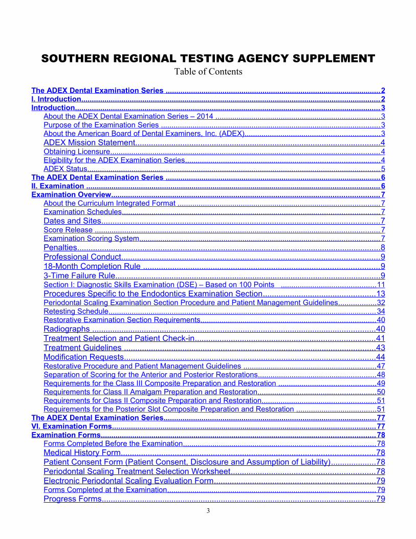
SOUTHERN REGIONAL TESTING AGENCY SUPPLEMENTTable of Contents
The ADEX Dental Examination Series ...................................................................................................... 2 I. Introduction .............................................................................................................................................. 2 Introduction ................................................................................................................................................. 3
About the ADEX Dental Examination Series – 2014 .............................................................................. 3 Purpose of the Examination Series ........................................................................................................ 3 About the American Board of Dental Examiners, Inc. (ADEX) ................................................................ 3 ADEX Mission Statement ............................................................................................................. 4 Obtaining Licensure ................................................................................................................................ 4 Eligibility for the ADEX Examination Series ............................................................................................ 4 ADEX Status ........................................................................................................................................... 5
The ADEX Dental Examination Series ...................................................................................................... 6 II. Examination ........................................................................................................................................... 6 Examination Overview ............................................................................................................................... 7
About the Curriculum Integrated Format ................................................................................................ 7 Examination Schedules .......................................................................................................................... 7 Dates and Sites ............................................................................................................................ 7 Score Release ....................................................................................................................................... 7 Examination Scoring System .................................................................................................................. 7 Penalties ....................................................................................................................................... 8 Professional Conduct ................................................................................................................... 9 18-Month Completion Rule ......................................................................................................... 9 3-Time Failure Rule ...................................................................................................................... 9 Section I: Diagnostic Skills Examination (DSE) – Based on 100 Points ............................................. 11 Procedures Specific to the Endodontics Examination Section .................................................. 13 Periodontal Scaling Examination Section Procedure and Patient Management Guidelines .................. 32 Retesting Schedule ............................................................................................................................... 34 Restorative Examination Section Requirements ................................................................................... 40 Radiographs .............................................................................................................................. 40 Treatment Selection and Patient Check-in ................................................................................ 41 Treatment Guidelines ................................................................................................................ 43 Modification Requests ................................................................................................................ 44 Restorative Procedure and Patient Management Guidelines ............................................................... 47 Separation of Scoring for the Anterior and Posterior Restorations ........................................................ 48 Requirements for the Class III Composite Preparation and Restoration .............................................. 49 Requirements for Class II Amalgam Preparation and Restoration ........................................................ 50 Requirements for Class II Composite Preparation and Restoration ...................................................... 51 Requirements for the Posterior Slot Composite Preparation and Restoration ...................................... 51
The ADEX Dental Examination Series ..................................................................................................... 77 VI. Examination Forms ............................................................................................................................. 77 Examination Forms .................................................................................................................................. 78
Forms Completed Before the Examination ........................................................................................... 78 Medical History Form ................................................................................................................. 78 Patient Consent Form (Patient Consent, Disclosure and Assumption of Liability) .................... 78 Periodontal Scaling Treatment Selection Worksheet ................................................................ 78 Electronic Periodontal Scaling Evaluation Form ........................................................................ 79 Forms Completed at the Examination ................................................................................................... 79 Progress Forms .......................................................................................................................... 79
3
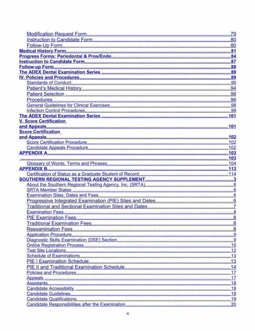
Modification Request Form ........................................................................................................ 79 Instruction to Candidate Form .................................................................................................... 80 Follow-Up Form .......................................................................................................................... 80
Medical History Form ............................................................................................................................... 81 Progress Forms: Periodontal & Pros/Endo ............................................................................................ 84 Instruction to Candidate Form ................................................................................................................. 87 Follow-up Form ......................................................................................................................................... 88 The ADEX Dental Examination Series .................................................................................................... 89 IV. Policies and Procedures ..................................................................................................................... 89
Standards of Conduct ........................................................................................................................... 90 Patient’s Medical History ............................................................................................................ 94 Patient Selection ....................................................................................................................... 96 Procedures ................................................................................................................................. 96 General Guidelines for Clinical Exercises ............................................................................................. 98 Infection Control Procedures ................................................................................................................. 99
The ADEX Dental Examination Series .................................................................................................. 101 V. Score Certification and Appeals ............................................................................................................................................ 101 Score Certification and Appeals ............................................................................................................................................ 102
Score Certification Procedure ............................................................................................................. 102 Candidate Appeals Procedure ............................................................................................................ 102
APPENDIX A ............................................................................................................................................ 103 ................................................................................................................................................................ 103
Glossary of Words, Terms and Phrases ............................................................................................. 104 APPENDIX B ............................................................................................................................................ 113
Certification of Status as a Graduate Student of Record ..................................................................... 114 SOUTHERN REGIONAL TESTING AGENCY SUPPLEMENT .................................................................... 3
About the Southern Regional Testing Agency, Inc. (SRTA) .................................................................... 6 SRTA Member States ............................................................................................................................ 6 Examination Sites, Dates and Fees ........................................................................................................ 6 Progressive Integrated Examination (PIE) Sites and Dates ........................................................ 6 Traditional and Sectional Examination Sites and Dates .............................................................. 7 Examination Fees ................................................................................................................................... 8 PIE Examination Fees .................................................................................................................. 8 Traditional Examination Fees ...................................................................................................... 8 Reexamination Fees .................................................................................................................... 8 Application Procedure ............................................................................................................................. 9 Diagnostic Skills Examination (DSE) Section .......................................................................................... 9 Online Registration Process .................................................................................................................. 10 Test Site Locations ............................................................................................................................... 12 Schedule of Examinations .................................................................................................................... 13 PIE I Examination Schedule ....................................................................................................... 13 PIE II and Traditional Examination Schedule ............................................................................ 14 Policies and Procedures ....................................................................................................................... 17 Appeals ................................................................................................................................................ 17 Assistants ............................................................................................................................................. 18 Candidate Accessibility ....................................................................................................................... 19 Candidate Guidelines ............................................................................................................................ 19 Candidate Qualifications ....................................................................................................................... 19 Candidate Responsibilities after the Examination ................................................................................. 20
4
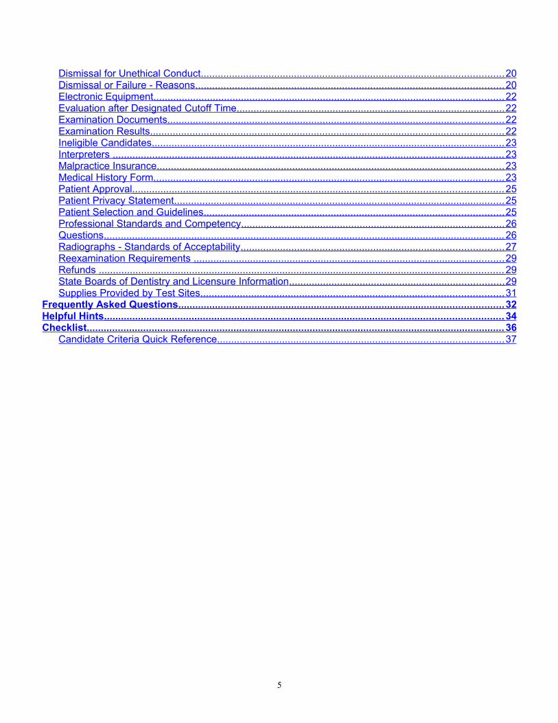
Dismissal for Unethical Conduct ........................................................................................................... 20 Dismissal or Failure - Reasons ............................................................................................................. 20 Electronic Equipment ............................................................................................................................ 22 Evaluation after Designated Cutoff Time ............................................................................................... 22 Examination Documents ....................................................................................................................... 22 Examination Results ............................................................................................................................. 22 Ineligible Candidates ............................................................................................................................. 23 Interpreters .......................................................................................................................................... 23 Malpractice Insurance ........................................................................................................................... 23 Medical History Form ............................................................................................................................ 23 Patient Approval ................................................................................................................................... 25 Patient Privacy Statement ..................................................................................................................... 25 Patient Selection and Guidelines .......................................................................................................... 25 Professional Standards and Competency ............................................................................................. 26 Questions .............................................................................................................................................. 26 Radiographs - Standards of Acceptability ............................................................................................. 27 Reexamination Requirements .............................................................................................................. 29 Refunds ............................................................................................................................................... 29 State Boards of Dentistry and Licensure Information ............................................................................ 29 Supplies Provided by Test Sites ........................................................................................................... 31
Frequently Asked Questions ................................................................................................................... 32 Helpful Hints ............................................................................................................................................. 34 Checklist .................................................................................................................................................... 36
Candidate Criteria Quick Reference ..................................................................................................... 37
5
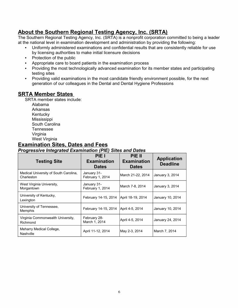
About the Southern Regional Testing Agency, Inc. (SRTA)The Southern Regional Testing Agency, Inc. (SRTA) is a nonprofit corporation committed to being a leader at the national level in examination development and administration by providing the following:
• Uniformly administered examinations and confidential results that are consistently reliable for use by licensing authorities to make initial licensure decisions
• Protection of the public• Appropriate care to board patients in the examination process• Providing the most technologically advanced examination for its member states and participating
testing sites• Providing valid examinations in the most candidate friendly environment possible, for the next
generation of our colleagues in the Dental and Dental Hygiene Professions
SRTA Member States SRTA member states include:
AlabamaArkansasKentuckyMississippiSouth CarolinaTennesseeVirginiaWest Virginia
Examination Sites, Dates and FeesProgressive Integrated Examination (PIE) Sites and Dates
Testing SitePIE I
Examination Dates
PIE II Examination
DatesApplication
Deadline
Medical University of South Carolina, Charleston
January 31-February 1, 2014 March 21-22, 2014 January 3, 2014
West Virginia University, Morgantown
January 31-February 1, 2014 March 7-8, 2014 January 3, 2014
University of Kentucky, Lexington
February 14-15, 2014 April 18-19, 2014 January 10, 2014
University of Tennessee, Memphis February 14-15, 2014 April 4-5, 2014 January 10, 2014
Virginia Commonwealth University, Richmond
February 28-March 1, 2014 April 4-5, 2014 January 24, 2014
Meharry Medical College, Nashville
April 11-12, 2014 May 2-3, 2014 March 7, 2014
6
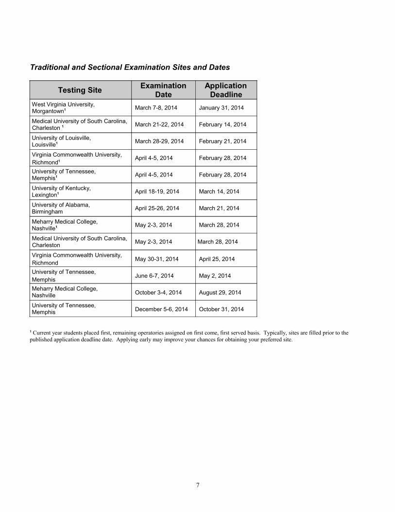
Traditional and Sectional Examination Sites and Dates
Testing Site Examination Date
Application Deadline
West Virginia University, Morgantown1 March 7-8, 2014 January 31, 2014
Medical University of South Carolina, Charleston 1 March 21-22, 2014 February 14, 2014
University of Louisville, Louisville1 March 28-29, 2014 February 21, 2014
Virginia Commonwealth University, Richmond1 April 4-5, 2014 February 28, 2014
University of Tennessee, Memphis1 April 4-5, 2014 February 28, 2014
University of Kentucky, Lexington1 April 18-19, 2014 March 14, 2014
University of Alabama, Birmingham April 25-26, 2014 March 21, 2014
Meharry Medical College, Nashville1 May 2-3, 2014 March 28, 2014
Medical University of South Carolina, Charleston May 2-3, 2014 March 28, 2014
Virginia Commonwealth University, Richmond
May 30-31, 2014 April 25, 2014
University of Tennessee, Memphis
June 6-7, 2014 May 2, 2014
Meharry Medical College, Nashville October 3-4, 2014 August 29, 2014
University of Tennessee, Memphis December 5-6, 2014 October 31, 2014
1 Current year students placed first, remaining operatories assigned on first come, first served basis. Typically, sites are filled prior to the published application deadline date. Applying early may improve your chances for obtaining your preferred site.
7
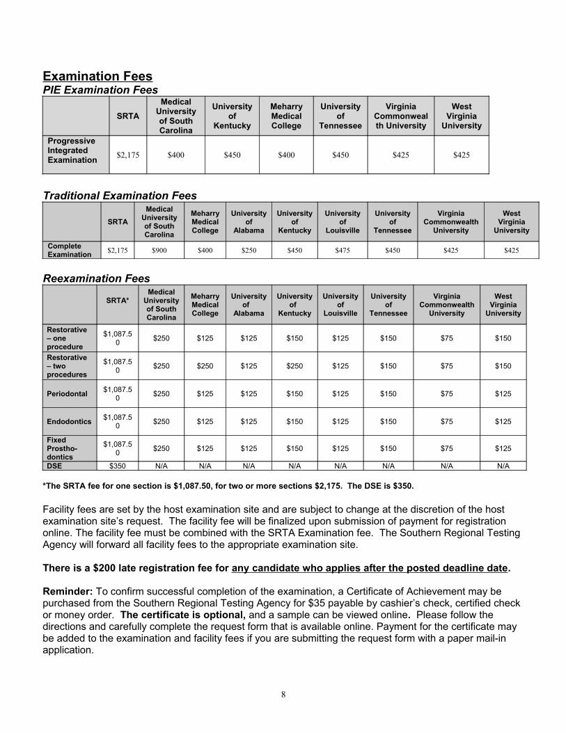
Examination FeesPIE Examination Fees
SRTAMedical
University of South Carolina
University of
Kentucky
Meharry Medical College
University of
Tennessee
Virginia Commonwealth University
West Virginia
University
Progressive Integrated Examination $2,175 $400 $450 $400 $450 $425 $425
Traditional Examination Fees
SRTAMedical
University of South Carolina
Meharry Medical College
University of
Alabama
University of
Kentucky
University of
Louisville
University of
Tennessee
Virginia Commonwealth
University
West Virginia
University
Complete Examination $2,175 $900 $400 $250 $450 $475 $450 $425 $425
Reexamination Fees
SRTA*Medical
University of South Carolina
Meharry Medical College
University of
Alabama
University of
Kentucky
University of
Louisville
University of
Tennessee
Virginia Commonwealth
University
WestVirginia
University
Restorative – one procedure
$1,087.50 $250 $125 $125 $150 $125 $150 $75 $150
Restorative – two procedures
$1,087.50 $250 $250 $125 $250 $125 $150 $75 $150
Periodontal $1,087.50 $250 $125 $125 $150 $125 $150 $75 $125
Endodontics $1,087.50 $250 $125 $125 $150 $125 $150 $75 $125
Fixed Prostho-dontics
$1,087.50 $250 $125 $125 $150 $125 $150 $75 $125
DSE $350 N/A N/A N/A N/A N/A N/A N/A N/A
*The SRTA fee for one section is $1,087.50, for two or more sections $2,175. The DSE is $350.
Facility fees are set by the host examination site and are subject to change at the discretion of the host examination site’s request. The facility fee will be finalized upon submission of payment for registration online. The facility fee must be combined with the SRTA Examination fee. The Southern Regional Testing Agency will forward all facility fees to the appropriate examination site.
There is a $200 late registration fee for any candidate who applies after the posted deadline date.
Reminder: To confirm successful completion of the examination, a Certificate of Achievement may be purchased from the Southern Regional Testing Agency for $35 payable by cashier’s check, certified check or money order. The certificate is optional, and a sample can be viewed online. Please follow the directions and carefully complete the request form that is available online. Payment for the certificate may be added to the examination and facility fees if you are submitting the request form with a paper mail-in application.
8
Application ProcedureCandidates have the following options for testing:
Progressive Integrated Examination (PIE)The PIE format allows candidates to take the manikin-based sections and patient-based sections on separate weekends. Candidates take both parts of the PIE examination at their home university. This option is available to current students, graduates or residents deemed eligible by their participating universities. All others must get permission from the examination site. An operatory is reserved for the candidate for the first testing date at his/her respective school for the manikin-based sections (PIE I) and for the second testing date at his/her school for the patient-based sections (PIE II).
Traditional Dental ExaminationFor the Traditional Dental Examination, candidates take all four (or five, as required by state regulations) skills sections (manikin-based procedures and patient-based procedures) in the same weekend. Upon applying for the Traditional Dental Examination, the patient will be assigned operatory space for both days of the examination (Friday and Saturday).
Sectional ExaminationsCandidates are allowed to retake individual sections of the examination up to three times per section in an 18 month period. Candidates who fail one or two sections of the PIE I format may apply to retake those sections at the same site where they are scheduled to take the PIE II.
Candidates may apply online to take the examination or may submit an application by mail.
Questions concerning examination procedures, content, applications and test dates should be directed to the Southern Regional Testing Agency. (757) 318-9082. If you prefer to email your questions, use [email protected] for questions relating to the dental examination and [email protected] for general questions.
Diagnostic Skills Examination (DSE) Section The DSE is administered at PSI Testing Centers by appointment. Once SRTA authorizes a candidate to take the examination, he/she will be sent instructions on how to make an appointment for the examination by phone or online.
Candidates who fail the DSE must complete a registration for reexamination before receiving authorization to schedule an appointment with the Testing Center to retake the exam. All SRTA rules for registration procedures apply.
9
Online Registration ProcessApplying online is a multi-stage process:
1. Go to http://www.srta.org/apply. Click the fill out a basic profile link and complete the form.
The email address you enter will become your username to login to your profile and will be used to communicate your site assignment and notify you when results are available for release. Double check your email address and choose a secure password. After completing the online form, click the Apply button.
2. The next page that is displayed is the one you will see each time you login to your profile. The Dashboard tab is displayed by default. Here you will find a list of current items and their status:
Check Mark = completed item
Exclamation Mark = item requires attention3. On your Dashboard page, you will be prompted to upload a photo. Click the Upload link and follow
the instructions. A photo is required. All photos will be reviewed by SRTA and may be rejected if they are not found to be acceptable for identification purposes. Submitting an unacceptable photo will delay your registration.
• Photos must be in one of the following formats: JPG/JPEG, GIF, or PNG.
• Photos must be square and have a minimum resolution of 200 x 200 and a maximum resolution of 500 x 500.
• Your photo must be a front facing headshot in the format that would be used for a passport.
4. A digital copy of a current and valid CPR certification, which includes the renewal date, is required. Valid certification is defined as a hands-on training program that provides an assessment of cognitive skills and skills acquired via classroom or web-based training. A minimum of Basic CPR skills certification is required. Classes provided by Internet-only instructions are not acceptable. Web-based didactic training must also include a hands-on component.
5. After you submit all required profile information and a valid photo and CPR card, SRTA staff will validate your profile, clearing you to register for the examination series. Staff members cannot validate any profile without a photo or CPR card. Please allow two to three days for validation.
6. Your graduation status must also be validated before you can register for exams. Verification can take several weeks depending on the method used:
• Graduating senior at a school in a SRTA State:
o Your school must provide SRTA with a list of its graduating class.
• Graduating senior NOT in a SRTA State:
o A candidate who has not formally graduated from his/her university is required to secure certification from the dean of his/her program stating that The candidate is eligible and qualifies for the DDS or DMD degree requirements. The candidate will complete the DDS or DMD degree requirements within 18 months of
the examination date. This certification must be in the form of a letter from the dean submitted with the
application or provided to SRTA by the dean prior to the receipt of the candidate’s application.
10
• Graduate:
o Upload a scan of your diploma. To upload a scan, login to your profile, then click the Profile tab followed by the Proof of Graduation link at the top of the screen.
7. After your profile and graduation status are validated, you will be able to click the link on your Dashboard to register for examinations.
8. Submit payment for examination fees. SRTA accepts VISA and MasterCard only. Debit cards may be used if allowable by the issuing bank and if they bear the VISA or MasterCard logo. No international credit/debit cards are accepted. All payments are drawn immediately and must be paid in full. Failure to pay the registration fee at the time of registration may forfeit your ability to sit for the examination. Registrations that are not paid within 72 hours are automatically cancelled.
SRTA Online Profile TabsDashboard. Under this tab you will find a list of items you must submit for your SRTA profile and the status of each item.
Check Mark = completed item
Exclamation Mark = item requires attention
Apply. Once all profile information has been uploaded and your profile has been verified you may use this tab to apply for examinations. Detailed instructions will be presented based on the available examinations. This tab is also where your clinical assignment will be listed once the site schedule is finalized.Documents. Candidates must visit this tab prior to the examination to download and possibly fill out any required forms and documents. Instructions about each document will be given.Profile. Under this tab you can view and edit your personal information and upload your photo, proof of graduation, etc.Results. Your results will be posted under this tab once they are finalized and released.
11
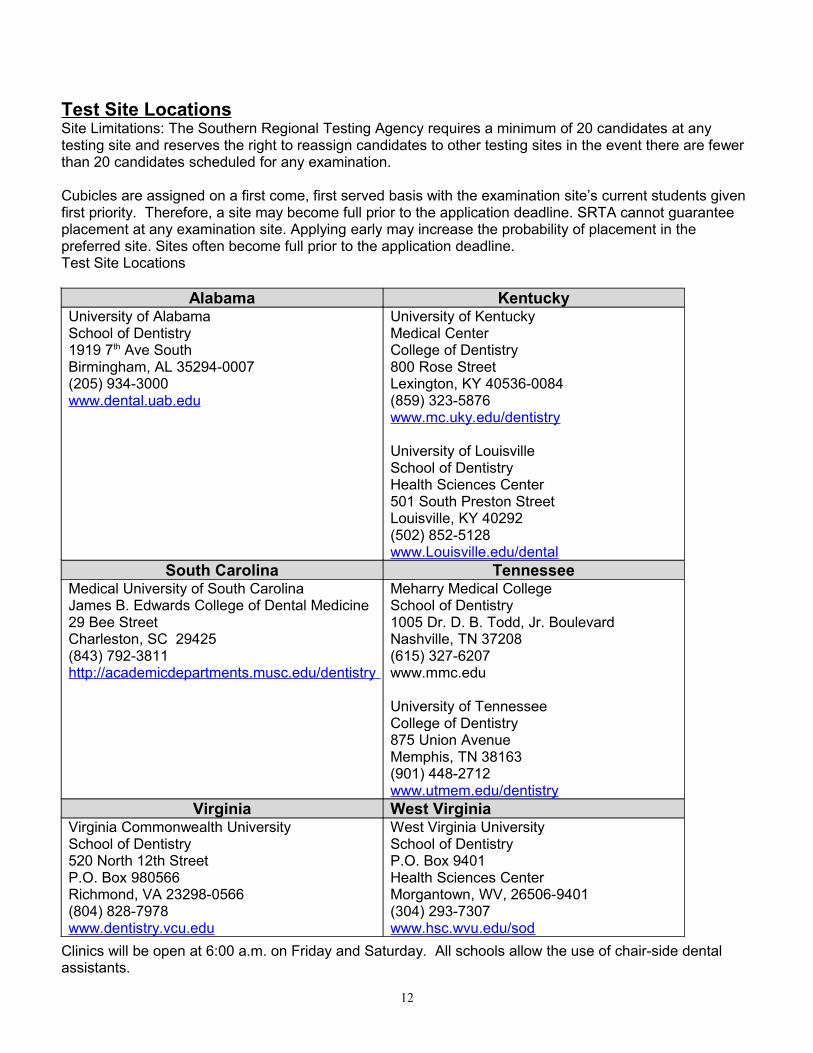
Test Site LocationsSite Limitations: The Southern Regional Testing Agency requires a minimum of 20 candidates at any testing site and reserves the right to reassign candidates to other testing sites in the event there are fewer than 20 candidates scheduled for any examination.
Cubicles are assigned on a first come, first served basis with the examination site’s current students given first priority. Therefore, a site may become full prior to the application deadline. SRTA cannot guarantee placement at any examination site. Applying early may increase the probability of placement in the preferred site. Sites often become full prior to the application deadline. Test Site Locations
Alabama KentuckyUniversity of AlabamaSchool of Dentistry1919 7th Ave SouthBirmingham, AL 35294-0007(205) 934-3000www.dental.uab.edu
University of KentuckyMedical CenterCollege of Dentistry800 Rose StreetLexington, KY 40536-0084(859) 323-5876www.mc.uky.edu/dentistry
University of LouisvilleSchool of DentistryHealth Sciences Center501 South Preston StreetLouisville, KY 40292(502) 852-5128www.Louisville.edu/dental
South Carolina TennesseeMedical University of South CarolinaJames B. Edwards College of Dental Medicine29 Bee StreetCharleston, SC 29425(843) 792-3811http://academicdepartments.musc.edu/dentistry
Meharry Medical CollegeSchool of Dentistry1005 Dr. D. B. Todd, Jr. BoulevardNashville, TN 37208(615) 327-6207www.mmc.edu
University of TennesseeCollege of Dentistry875 Union AvenueMemphis, TN 38163(901) 448-2712www.utmem.edu/dentistry
Virginia West VirginiaVirginia Commonwealth UniversitySchool of Dentistry520 North 12th StreetP.O. Box 980566Richmond, VA 23298-0566(804) 828-7978www.dentistry.vcu.edu
West Virginia UniversitySchool of DentistryP.O. Box 9401Health Sciences CenterMorgantown, WV, 26506-9401(304) 293-7307www.hsc.wvu.edu/sod
Clinics will be open at 6:00 a.m. on Friday and Saturday. All schools allow the use of chair-side dental assistants.
12
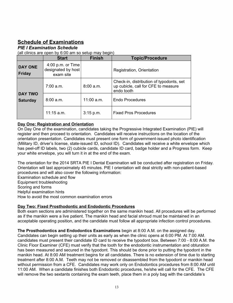
Schedule of ExaminationsPIE I Examination Schedule(all clinics are open by 6:00 am so setup may begin)
Start Finish Topic/Procedure
DAY ONEFriday
4:00 p.m. or Time designated by host
exam siteRegistration, Orientation
DAY TWOSaturday
7:00 a.m. 8:00 a.m.Check-in, distribution of typodonts, set up cubicle, call for CFE to measure endo tooth
8:00 a.m. 11:00 a.m. Endo Procedures
11:15 a.m. 3:15 p.m. Fixed Pros Procedures
Day One: Registration and OrientationOn Day One of the examination, candidates taking the Progressive Integrated Examination (PIE) will register and then proceed to orientation. Candidates will receive instructions on the location of the orientation presentation. Candidates must present one form of government-issued photo identification (Military ID, driver’s license, state-issued ID, school ID). Candidates will receive a white envelope which has peel-off ID labels, two (2) cubicle cards, candidate ID card, badge holder and a Progress form. Keep your white envelope, you will turn it in at the end of the exam.
The orientation for the 2014 SRTA PIE I Dental Examination will be conducted after registration on Friday. Orientation will last approximately 45 minutes. PIE I orientation will deal strictly with non-patient-based procedures and will also cover the following information:Examination schedule and flowEquipment troubleshootingScoring and formsHelpful examination hintsHow to avoid the most common examination errors
Day Two: Fixed Prosthodontic and Endodontic ProceduresBoth exam sections are administered together on the same manikin head. All procedures will be performed as if the manikin were a live patient. The manikin head and facial shroud must be maintained in an acceptable operating position, and the candidate must follow all appropriate infection control procedures.
The Prosthodontics and Endodontics Examinations begin at 8:00 A.M. on the assigned day. Candidates can begin setting up their units as early as when the clinic opens at 6:00 PM. At 7:00 AM. candidates must present their candidate ID card to receive the typodont box. Between 7:00 - 8:00 A.M. the Clinic Floor Examiner (CFE) must verify that the tooth for the endodontic instrumentation and obturation has been measured and secured in the typodont. This should be done prior to putting the typodont in the manikin head. At 8:00 AM treatment begins for all candidates. There is no extension of time due to starting treatment after 8:00 A.M. Teeth may not be removed or disassembled from the typodont or manikin head without permission from a CFE. Candidates may work only on Endodontics procedures from 8:00 AM until 11:00 AM. When a candidate finishes both Endodontic procedures, he/she will call for the CFE. The CFE will remove the two sextants containing the exam teeth, place them in a poly bag with the candidate’s
13
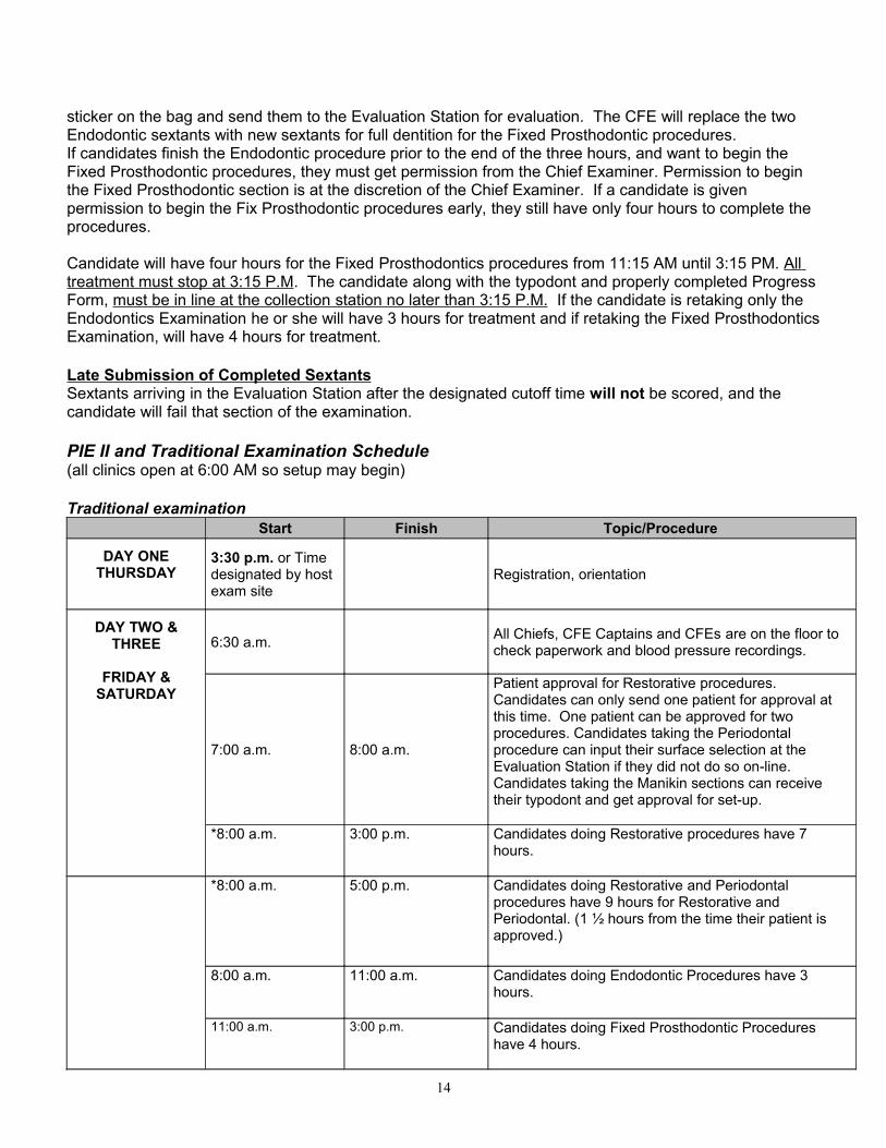
sticker on the bag and send them to the Evaluation Station for evaluation. The CFE will replace the two Endodontic sextants with new sextants for full dentition for the Fixed Prosthodontic procedures.If candidates finish the Endodontic procedure prior to the end of the three hours, and want to begin the Fixed Prosthodontic procedures, they must get permission from the Chief Examiner. Permission to begin the Fixed Prosthodontic section is at the discretion of the Chief Examiner. If a candidate is given permission to begin the Fix Prosthodontic procedures early, they still have only four hours to complete the procedures.
Candidate will have four hours for the Fixed Prosthodontics procedures from 11:15 AM until 3:15 PM. All treatment must stop at 3:15 P.M. The candidate along with the typodont and properly completed Progress Form, must be in line at the collection station no later than 3:15 P.M. If the candidate is retaking only the Endodontics Examination he or she will have 3 hours for treatment and if retaking the Fixed Prosthodontics Examination, will have 4 hours for treatment.
Late Submission of Completed SextantsSextants arriving in the Evaluation Station after the designated cutoff time will not be scored, and the candidate will fail that section of the examination.
PIE II and Traditional Examination Schedule(all clinics open at 6:00 AM so setup may begin)
Traditional examinationStart Finish Topic/Procedure
DAY ONETHURSDAY
3:30 p.m. or Time designated by host exam site
Registration, orientation
DAY TWO & THREE
FRIDAY & SATURDAY
6:30 a.m. All Chiefs, CFE Captains and CFEs are on the floor to check paperwork and blood pressure recordings.
7:00 a.m. 8:00 a.m.
Patient approval for Restorative procedures. Candidates can only send one patient for approval at this time. One patient can be approved for two procedures. Candidates taking the Periodontal procedure can input their surface selection at the Evaluation Station if they did not do so on-line. Candidates taking the Manikin sections can receive their typodont and get approval for set-up.
*8:00 a.m. 3:00 p.m. Candidates doing Restorative procedures have 7 hours.
*8:00 a.m. 5:00 p.m. Candidates doing Restorative and Periodontal procedures have 9 hours for Restorative and Periodontal. (1 ½ hours from the time their patient is approved.)
8:00 a.m. 11:00 a.m. Candidates doing Endodontic Procedures have 3 hours.
11:00 a.m. 3:00 p.m. Candidates doing Fixed Prosthodontic Procedures have 4 hours.
14
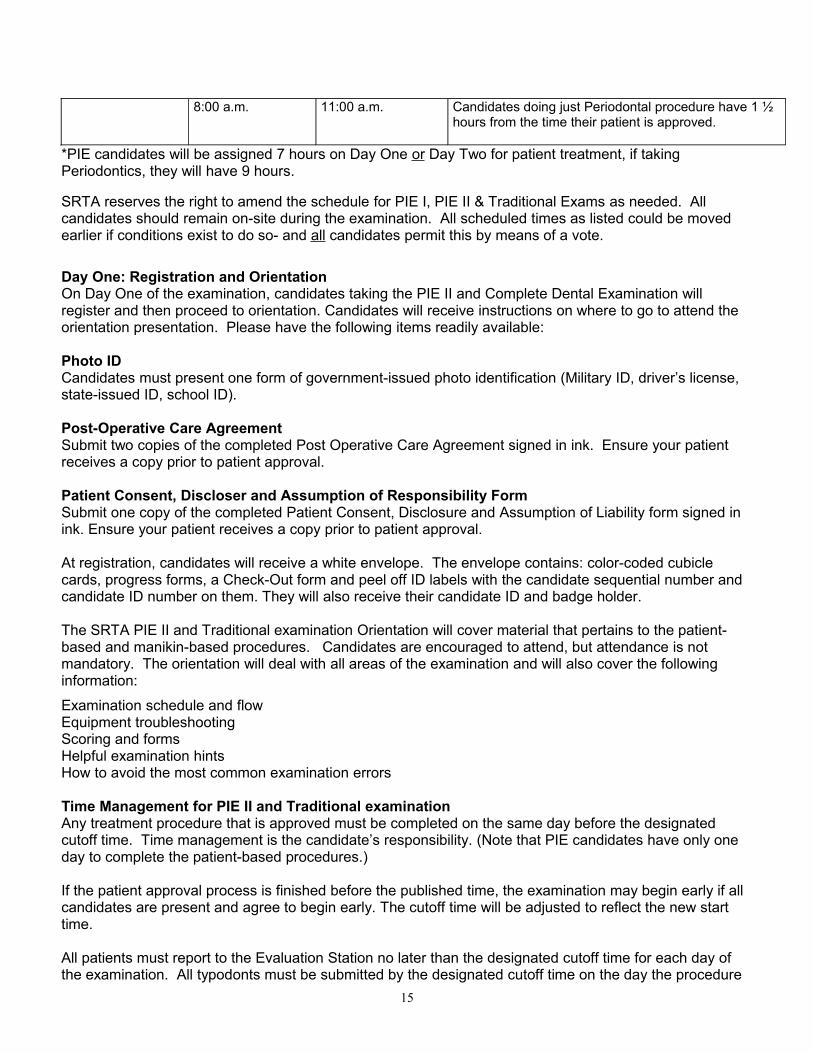
8:00 a.m. 11:00 a.m. Candidates doing just Periodontal procedure have 1 ½ hours from the time their patient is approved.
*PIE candidates will be assigned 7 hours on Day One or Day Two for patient treatment, if taking Periodontics, they will have 9 hours.
SRTA reserves the right to amend the schedule for PIE I, PIE II & Traditional Exams as needed. All candidates should remain on-site during the examination. All scheduled times as listed could be moved earlier if conditions exist to do so- and all candidates permit this by means of a vote.
Day One: Registration and OrientationOn Day One of the examination, candidates taking the PIE II and Complete Dental Examination will register and then proceed to orientation. Candidates will receive instructions on where to go to attend the orientation presentation. Please have the following items readily available:
Photo IDCandidates must present one form of government-issued photo identification (Military ID, driver’s license, state-issued ID, school ID).
Post-Operative Care AgreementSubmit two copies of the completed Post Operative Care Agreement signed in ink. Ensure your patient receives a copy prior to patient approval.
Patient Consent, Discloser and Assumption of Responsibility FormSubmit one copy of the completed Patient Consent, Disclosure and Assumption of Liability form signed in ink. Ensure your patient receives a copy prior to patient approval.
At registration, candidates will receive a white envelope. The envelope contains: color-coded cubicle cards, progress forms, a Check-Out form and peel off ID labels with the candidate sequential number and candidate ID number on them. They will also receive their candidate ID and badge holder.
The SRTA PIE II and Traditional examination Orientation will cover material that pertains to the patient-based and manikin-based procedures. Candidates are encouraged to attend, but attendance is not mandatory. The orientation will deal with all areas of the examination and will also cover the following information:
Examination schedule and flowEquipment troubleshootingScoring and formsHelpful examination hintsHow to avoid the most common examination errors
Time Management for PIE II and Traditional examinationAny treatment procedure that is approved must be completed on the same day before the designated cutoff time. Time management is the candidate’s responsibility. (Note that PIE candidates have only one day to complete the patient-based procedures.)
If the patient approval process is finished before the published time, the examination may begin early if all candidates are present and agree to begin early. The cutoff time will be adjusted to reflect the new start time.
All patients must report to the Evaluation Station no later than the designated cutoff time for each day of the examination. All typodonts must be submitted by the designated cutoff time on the day the procedure
15
was initiated. Any procedure submitted to the Evaluation Station after the designated cutoff time will not be scored, and the candidate will fail that section of the examination. One examiner will evaluate the procedure to determine that proper treatment has been provided.
Candidates found treating patients or with patients in the clinic after the designated cutoff time will fail that section of the examination.
The allocated period for patient treatment includes the time the patient(s) may spend at the Evaluation Station for approval or evaluation of a procedure. On average, patients are away for scoring for 15 to 60 minutes, depending on the procedure to be evaluated and the backlog of patients. Candidates should be aware of conditions and plan accordingly when scheduling patients.
Only one patient may be approved during the designated patient approval period. Multiple procedures may be approved on a single patient. If a patient is sent to the evaluation station for two procedures and only one of them is approved, the candidate must begin with the approved procedure and have the second procedure approved after the first procedure is scored.
Sectional Examination ScheduleNote to candidates taking PIE: If a candidate has previously taken the PIE I (manikin-based) sections and needs to retake one or both sections, he/she can apply for a sectional examination at the site where he/she plans to take PIE II (patient-based procedures). He/she will be assigned an additional 3 hours if he/she is retaking only the Endodontic section, 4 hours for Fixed Prosthodontic section and an additional 7 hours for both sections.
Candidates will be scheduled on Friday (Day One) and/or Saturday (Day Two) according to which sections they are taking, the number of sections and the availability of operatory space.
Candidates scheduled for re-examination(s) must register with the Clinic Floor Examiner the day of their exam if they did not attend orientation.
16
Policies and Procedures
Appeals Appeals are reviewed based on facts surrounding the decision made during the examination. Any other information such as past experience, school performance, character references, testimonials, radiographs, photographs or models taken by the candidate of a patient after completion of the examination cannot be considered in the appeals process. Consideration can only be given to documents, radiographs, etc., that were submitted to the examiners during the examination.
The Appeals Committee is obligated to base its judgment of technical errors upon its knowledge of the examination, the validation and standardization process used by SRTA and evidence presented in the candidate's appeal in a systematic, consistent, reliable and rational manner. It is neither consistent nor reasonable to suppose that grading examiners can judge matters more accurately and objectively after an examination has ended than they did during the examination. Scores cannot be reversed by any member of the Appeals Committee unless the disputed score is determined to be a technical error rather than a perceived judgment error.
Group, second-party and verbal appeals will not be honored. The Southern Regional Testing Agency is not responsible for any expense incurred by any party making an appeal.
Appeals based upon patient behavior, tardiness or failure to appear will not be considered.
SRTA does not provide patients and is not responsible for this aspect of the examination.
Situations requiring follow-up care are the responsibility of the candidate, per the Post Operative Care Agreement.
The host institution publishes an examination site instruction letter (also called the University Instruction Letter) that outlines the provision of certain minimal materials, support personnel to dispense these materials and support personnel to repair and maintain equipment in working condition within a reasonable amount of time. Appeals based upon failure of the host institution will be considered only in instances in which the Clinic Floor Examiner or SRTA Administrator was made aware of the problem with reasonable time for resolution at the examination site, rather than after the fact upon completion of the examination.
Candidates are not notified of all errors. An appeal based on non-notification of an error or failing score during the examination will not be considered.
The appellate process will include review of all documentation of examination results and candidate performance during the examination. It may include such additional investigation as deemed warranted by the circumstance of the appeal. The process will not include records or external opinions obtained by the candidate after he/she is dismissed at the completion of the examination.
The Appeals Committee will make every effort possible to complete the appellate review process within 90 days of the receipt of the appeal in the SRTA administrative office. When an extended investigation becomes necessary, the Agency will inform the candidate by letter of this extended investigation.
In many cases, SRTA cannot process, evaluate and make final decisions on appeals prior to the next examination. In cases in which a candidate successfully completes another examination while his/her appeal is being evaluated, that appeal will be dropped automatically by the chair of the Appeals Committee upon notification by the executive director. If the candidate fails a subsequent examination, the appeal
17
process will continue to completion, and a decision will be rendered without the committee's knowledge of the candidate's performance on that examination.
Application fees shall be refunded in full in the event a candidate has made payment for a subsequent examination and an appeal is granted before that examination is taken. If a favorable decision is made on an appeal, a full refund of the appeal fee will be mailed to the candidate. All member state boards of dentistry will be notified of the results.
The executive director will maintain a log of all appeals and take appropriate action to bring them to a timely completion.
The appeal will become a permanent part of the candidate's file maintained in the SRTA administrative office. The decision of the Appeals Committee is final.
The candidate’s written notification/request for an appeal form must be received within 20 calendar days following the release of scores, not the date the scores are received by the candidate. The Southern Regional Testing Agency cannot be responsible for items lost or delayed by the Postal Service.
Appeals must be filed in writing on a form provided by the Southern Regional Testing Agency and must be sent by certified mail, along with a certified check or money order for $300 for the appeal fee. The formal written appeal must be received within 60 days of the date of the examination.
Please send requests for an appeal form and final appeals to the following address:
Executive DirectorSouthern Regional Testing Agency, Inc.
4698 Honeygrove Road, Suite 2Virginia Beach, VA 23455-5934
AssistantsAssistant(s) cannot function as both a chair-side assistant and a patient.The assistant used for the Restorative Procedures may be a dental assistant, dental hygienist or first or second-year dental student. The assistant may not be a dentist (licensed or unlicensed), junior/senior dental student or dental laboratory technician.
The assistant used for the Periodontal Procedure may be a dental assistant, or first or second-year dental student. The assistant may not be a dentist (licensed or unlicensed), junior/senior dental student, dental hygienist (licensed or unlicensed), final year dental hygiene student or dental laboratory technician.
The assistant must register with the Desk Coordinators between 6:30 a.m. and 7:30 a.m. the morning of the exam. They must have the completed Assistant Certification form, two photographs of themselves and valid photo identification. The assistant may not attend orientation.
18
Candidate Accessibility
Any candidate with a documented physical and/or learning disability that impairs sensory, manual or speaking skills and that requires a reasonable deviation from the normal administration of the examination may be accommodated. A written statement from a qualified physician must be provided at the time of application. The limitation(s) must be clearly defined, and the assistance required to ensure appropriate accommodations must be detailed. Requests will be evaluated on a case-by-case basis. Accommodations/deviations will not be allowed for components and skills the examination must measure.
Information received regarding the physical/learning challenges of a candidate will remain confidential except in the case of disabilities that may require emergency treatment. In this case, onsite safety personnel will be advised.
Candidate GuidelinesCandidates may choose the technical procedures and specific materials used in manikin-based and patient-based procedures. Each candidate must furnish all patients, necessary materials and instruments, including high- and slow-speed hand-pieces. Satisfactory patient treatment is the criterion for acceptance or rejection of any method, procedure or material used. The Southern Regional Testing Agency examines candidates with varying educational backgrounds. Because universities teach different preparations, SRTA does not look for one type of standard preparation; however, we do provide criteria that will be evaluated as described in the clinical sections of this manual. The candidate must demonstrate clinically acceptable cavity preparations and finishes. Examiners are standardized prior to each examination to ensure scoring is to the level of clinical acceptability.All written and oral communication must be in English for purposes of this examination. Candidates may communicate with their patients in another language.
Candidates are strongly advised to visit their testing site at their earliest convenience to familiarize themselves with the facilities and available equipment. The testing site will provide the operating chair and unit. The candidate should ensure that his/her hand-piece can be adapted to the unit available at the university/testing site. Some additional equipment may be available from certain universities if arrangements are made in advance with the university. The Southern Regional Testing Agency is not responsible for the malfunction of any equipment and will not allot additional time due to equipment malfunction. Refer to the University Instruction Letter from the test site, which can be downloaded from the SRTA website. Each testing site charges a fee for the use of facilities and incidental materials where a clinical examination is conducted. All fees are listed under Examination Fees.
Candidate QualificationsCandidates for the examination must be graduates of an American or Canadian dental college accredited by the American Dental Association Commission on Dental Accreditation. A candidate who has not formally graduated from his/her university is required to secure certification from the dean of his/her program stating that:
1. The candidate is eligible and qualifies for the DDS or DMD degree requirements.2. The candidate will complete the DDS or DMD degree requirements within 10 months of the
examination date.
19
This certification must be in the form of a letter from the dean submitted with the application or provided to SRTA by the dean prior to the receipt of the candidate’s application. For candidates who graduated from a school outside of the United States and Canada may apply and be considered for the “state --only” status, pending receipt of the appropriate state authorization. The candidate must furnish a letter from the board of dentistry of a state that accepts the results of this examination indicating that the candidate is eligible for licensure in that state upon successful completion of the examination. In addition, a copy of the candidate’s diploma with an English translation must be provided.
Candidate Responsibilities after the ExaminationThe candidate is required to provide a copy of his/her diploma within 90 days after his/her graduation date, and/or the dean of the candidate’s program is required to submit a list of students who have graduated within that time frame. Failure to complete the degree requirements within 18 months of the examination shall invalidate the examination results. If the Southern Regional Testing Agency does not receive the required diploma or letter from the dean, the Agency will notify each of the individual state boards that scores are invalid. The individual state boards of dentistry determine the acceptance of invalid results. SRTA cannot grant extensions to the graduation requirements as mandated in our corporate documents.
Dismissal for Unethical ConductProfessional behavior is a critical quality in the practice of dentistry. If a candidate is suspected of unethical conduct as defined by SRTA guidelines, he/she will fail the examination.Examples of unethical conduct include, but are not limited to:
• Using unauthorized equipment at any time during the exam
• Using unauthorized assistants
• Using unauthorized patients
• Altering patient records or radiographs
• Treating patients outside clinic hours or receiving assistance from another practitioner
• Altering teeth used in the Manikin Procedure
• Engaging in dishonesty
• Altering candidate Progress Forms
• Any other behavior which compromises the standards of professional behavior
When a candidate is charged with unethical conduct, it is SRTA’s policy to notify all member state boards of the situation. Many state statutes have criteria which include “good moral character” as a requirement for licensure. If a state board finds a candidate guilty of the alleged unethical conduct, the candidate may be ineligible for licensure in that state at any time in the future. While candidates are allowed to retake the SRTA Examination, they may be unable to obtain licensure in any member state. Candidates are encouraged to address these matters with the board of dentistry of the state in which they desire licensure prior to retaking the examination.
Dismissal or Failure - ReasonsThis listing is not an all-inclusive listing of reasons for which a candidate may receive a failing evaluation or be dismissed. Some procedures may be deemed unsatisfactory for other reasons. Additionally, a combination of several unsatisfactory evaluations may result in failure. Reexamination may be denied for
20
one year (12 months) from the date of dismissal from the examination. Infractions that may lead to dismissal or failure include:
• Lack of protection and concern for tooth structure and supporting tissue during patient treatment
• Lack of professional judgment and/or proposing excessive treatment
• Evidence of dishonesty or misrepresentation during the application process, including false or misleading statements or false documentation presented by the candidate or on the candidate’s behalf
• Evidence of dishonesty or misrepresentation during registration of candidates or during the course of the examination
• Rude, abusive or uncooperative behavior exhibited by the candidate and/or those accompanying the candidate to the examination
• Continuing to work after published cutoff time
• Working on a manikin model in a manner that does not simulate actual patient conditions
• Working on Fixed Prosthodontic or Endodontic sextants not provided by SRTA. Any evidence of tampering with or attempting to remove the screws from the sextants will result in failure of the entire examination and will be grounds for dismissal from the exam.
• Failure to complete the examination within the allotted time. (No make-up time, grace period or second effort will be allowed for any part of this examination.)
• Altering or enhancing of radiographs
• Receiving assistance from a dentist, another candidate, university representative(s), etc., during the course of the examination
• Preparing a tooth other than the one approved by the examiners. This is considered major hard tissue damage.
• Failure to submit preoperative radiographs for all patient-based sections of the examination
• Administering or authorizing any form of patient sedation. Candidates who fail for this reason may reapply to take the next available examination.
• Thievery during the course of the examination
• Performance of any unauthorized work outside of the test site designated areas
• Noncompliance with anonymity requirements for patient approval and/or examiner scoring. Candidates are not allowed in or near the area designated for scoring. Candidates must instruct their patients not to handle any paperwork during the course of the examination.
• Noncompliance with established guidelines for asepsis and infectious disease control
• Use of a patient who has previously been rejected for a specific procedure. If the candidate is using another candidate's backup patient or is sharing a patient, it is the candidate's responsibility to determine whether the patient has previously been rejected for a specific procedure.
• Use of a patient who is a dentist or junior/senior dental student
• Charging patients for services performed
• Failure to complete or refusing to provide a Post Operative Care Agreement with verifiable contact name of the practitioner who will provide postoperative care to the patient, or the patient’s statement that he/she will seek care from a practitioner of his/her own choice
• Use of cellular telephones, pagers, cameras or other electronic equipment by candidate, assistant and/or patient(s) within the clinic and Evaluation Stations
21
Electronic EquipmentThe use of cellular telephones, pagers, cameras or other electronic equipment by candidates, patients and assistants is prohibited within the clinic and Evaluation Stations. Violation of this policy is grounds for dismissal from the examination.
Evaluation after Designated Cutoff TimeCandidates must send their patients and/or typodont to the Evaluation Station for evaluation no later than the designated cutoff time. Patients and/or typodonts arriving in the Evaluation Station after the designated cutoff time will not be scored, and the candidate will fail that section of the examination.
Examination DocumentsCandidates must instruct their patients not to handle any paperwork during the course of the examination. Candidates may be dismissed from or fail the examination if their patient(s) handle examination documents. Candidates should advise their patients of this prohibited behavior and the consequences.
Examination ResultsNotification will be sent to the candidate via email when scores are available for viewing online. Examination results will be available online in the afternoon of the next business day after the examination. Scores can be viewed by logging into the SRTA website using the password and username that were created during the online registration process. All candidates can view the details of their evaluation online by going to https://srta.brighttrac.com.
To maintain confidentiality, SRTA staff and examiners will not discuss candidate concerns and questions with a spouse, parent, friend, faculty member or family member.
Upon successful completion of the SRTA Examination, candidates will receive a final report, mailed within four weeks of the conclusion of the examination. The final report reflects the candidate’s most current scores within a five-year period. After the five-year period, the board of dentistry where licensure is being sought determines acceptance of scores.
The examination record of each candidate will automatically be sent to the secretaries of the State Boards of Dentistry of Alabama, Arkansas, Kentucky, Mississippi, South Carolina, Tennessee, Virginia and West Virginia.
Some state boards of dentistry may require a notarized copy of the final report, which the Southern Regional Testing Agency will provide for a nominal fee. Please contact our office to request this additional service. The examination record may also be sent to each current graduate’s university.
Other states accept Southern Regional Testing Agency's results for licensure. Candidates should contact the state board of dentistry where they are applying for licensure to verify acceptance of the SRTA scores and to learn of other state-specific requirements.
After results for the PIE I (manikin-based sections) are posted online and released to the university where the examination was held, any candidate who is not successful in these sections may apply to retake the failed sections at the same site where he/she has applied to take the PIE II (patient-based sections).
22
Results for the computer-simulated sections (DSE) will be posted when received by the Testing Center. Scores on the ADEX DSE are generally reported at the end of the first full week of the month following the month in which the DSE was taken. When the candidate has taken all sections of the examination (PIE I, PIE II and DSE), the results will be mailed to the candidate and automatically sent to the secretaries of the State Boards of Dentistry of Alabama, Arkansas, Kentucky, Mississippi, South Carolina, Tennessee, Virginia and West Virginia.
The Southern Regional Testing Agency supplies the examination results to the member state boards but does not analyze or interpret the records and makes no recommendations on the way the scores are used by the state. Acceptance of the regional scores is determined by the individual state boards.
To confirm successful completion of the examination, a certificate of achievement may be purchased from Southern Regional Testing Agency for $35 payable by cashier’s check, certified check or money order. The certificate is optional, and a sample can be viewed online. Please follow the directions and carefully complete the request form that is available online.
Ineligible CandidatesShould a candidate become ineligible to take the examination, he/she must notify the SRTA office in writing two weeks prior to the scheduled examination. A letter from the dean of the candidate’s institution will be required as proof of ineligibility. The Agency will retain the complete application fee for any candidate declared ineligible by his/her dean. Candidates must contact the university/test site directly for a refund of facility fees. Candidates declared ineligible will be allowed to examine at a future site within a 12-month period upon payment of facility fees and a $200 processing/administration fee. A diploma or letter from the dean stating the candidate’s eligibility is required for a rescheduled exam.
Interpreters Candidates may employ the services of an interpreter when their patient does not speak English or is hearing impaired and the hearing loss cannot be corrected. (This is particularly important when the patient has a history of medical problems or is on medications.) Faculty members, dentists and dental hygienists (licensed or unlicensed), third or fourth year dental students and final year dental hygiene students may not act as interpreters during the examination. Candidates are responsible for the conduct of their interpreter during the examination. Candidates must contact the SRTA office prior to the examination to inform us that an interpreter will be needed at the examination site. The interpreter must register with an SRTA Administrator and receive a badge that must be worn throughout the examination.
Malpractice InsuranceMalpractice insurance is provided for all candidates through SRTA's professional liability insurance company at no additional charge. CNA Insurance Company extends SRTA's professional liability coverage to candidates with the limit of $1,000,000/$3,000,000 for the patient-based portion of the 2014 SRTA clinical examination in dentistry.
Medical History FormA completed medical health history current within 30 days of the examination is required for each patient treated.
The patient’s blood pressure must be taken on the day of the examination and documented by a clinic floor examiner (CFE). The CFE does not witness the candidate taking the patients’ blood, but must verify and sign-off that it was taken the day of the exam and is on the appropriate form before sending the patient to
23
the Evaluation Station for approval. . If the patient is sitting for more than one examination section on the same day, his/her blood pressure must be taken and recorded prior to each section.
The patient’s health status must be acceptable and will be assessed during the patient approval section of the examination before each candidate’s procedure. Therefore, patient selection is essential for successful completion of this examination.
Medications: On the day of the examination, the candidate must document on the Medical History Form all medications or supplements taken by the patient within the last 24 hours. Candidates should document antibiotic premedication on the appropriate Progress Form, as well as on the Medical History Form.
Health qualifications: In order to participate in the examination, patients must meet the following criteria:
11. Patients must have a blood pressure reading of 159/94 or below to proceed without medical clearance. Patients with a blood pressure reading between 160/95 and 179/109 are accepted only with a written medical clearance from the patient’s physician. Patients with a blood pressure reading 180/110 or greater will not be accepted for this examination, even if a physician authorizes treatment.
12. Candidates who are sharing a patient requiring antibiotic prophylaxis must treat the patient the same day. Treatment of the same patient on subsequent clinical days will not be permitted.
13. Patients must have no history of heart attack (myocardial infarction), stroke or cardiac surgery within the last six months.
14. Patients may not have active tuberculosis. A patient who has tested positive for tuberculosis or who is being treated for tuberculosis but does not have the clinical symptoms is acceptable.
15. Patients may not have undergone chemotherapy treatment for cancer within the last six months.
16. Patients participating in the Periodontal Examination Section may not have a history of taking IV or orally-administered bisphosphonate medications. Patients participating in the Restorative Examination Section may not have a history of taking IV-administered bisphosphonate medications (except an annual IV dosage for osteoporosis). However, patients who have taken oral bisphosphonates may participate in the Restorative Section.
17. Patients may not have active incidence of bisphosphonate osteonecrosis of the jaw (BON), also known as osteochemonecrosis or, osteonecrosis of the jaw (ONJ).
18. Patients may not have any condition or medication/drug history that might be adversely affected by the length or nature of the examination procedures.
19. Patients with known allergies to latex will not be allowed to sit for the examination at those sites where latex may still be used or present.
20. If the patient answers “yes” to any of the questions on the Medical History Form, the candidate must explore the item further and determine whether a medical clearance from a licensed physician would be appropriate. A medical clearance is required if the finding could affect the patient’s suitability for elective dental treatment during the examination.
Candidates must follow the current American Heart Association antibiotic premedication recommendations when treating patients at potential risk of infective endocarditis following dental treatment. A medical clearance may be indicated to determine the patient’s potential risk of infective endocarditis. If the patient has a medical condition that could affect his/her suitability for treatment, the candidate must obtain a written medical clearance from the patient’s physician to indicate that the patient is healthy enough to participate in the examination. The medical clearance must also be submitted on the day of the examination and should meet the following criteria:
24
• Clearly legible statement from a licensed physician
• Written within 30 days prior to the examination on official letterhead
• Containing a positive statement of how the patient should be medically managed
• Containing the physician’s clearly legible name, address and phone number
• Containing a telephone number where the physician may be reached on the day of the examination if a question arises regarding the patient’s health
Patient ApprovalIf the candidate is scheduled to perform the restorative examination as the first procedure of the day, they may begin setting up as soon as the clinic is open at 6:00AM. Between 7:00 AM and 7:20 AM a CFE may be called over to document that the blood pressure was taken that day and check that the required forms and radiographs are complete and correct. Then, the patient is sent to the Evaluation Station, where all treatment selections are approved or rejected by at least two of three examiners evaluating anonymously. Restorative treatment begins at 8:00AM. If a patient meets the requirements for both the posterior and anterior restorations, both may be approved in the Evaluation Station at the same time, but the first restoration must be completed before the second restoration may be begun. Only one patient may be submitted for patient check-in at a time. If the candidate is utilizing two patents for the Restorative Examination Section, only one may be submitted to start the examination. The second may not be submitted until the first is finished. The local anesthesia request portion of the Restorative Progress Form must be filled out prior to submitting the patient to the Evaluation Station for patient check-in/case acceptance. Local anesthesia may not be provided until the lesion has been accepted for treatment. In the event that the first lesion submitted is not approved, a second lesion may be submitted to the Evaluation Station.
Patient Privacy StatementUpon conclusion of the SRTA Examination, all patient health information will remain in a sealed envelope submitted by the candidate upon conclusion of the examination. After a 12-month holding period, the contents of the envelope will be mechanically shredded. Patient data is not stored electronically or by any other means and is used only during the course of the clinical examination.
Patient Selection and GuidelinesCandidates must obtain their own patient(s) and are responsible for their arrival and return. SRTA is not responsible for procuring patients used in the examination. Patient selection and management is one of the most important facets of the examination and should be completed independently, without the help or assistance of faculty, colleagues, etc.
Candidates are strongly advised to secure a backup patient/tooth selection for each procedure. Unacceptable patients will be dismissed. Candidates must then procure an acceptable patient in order to continue the examination. Candidates must advise their patients of the time required to participate in this examination; evaluation of a patient can take an average of 45 minutes, in addition to the time required to complete the procedure.
Patients must be at least 14 years of age. A parent or legal guardian must be available in the waiting area during treatment and provide written consent for minors under the age of 18.
A patient may not be:
A dentist (licensed or unlicensed)
A junior/senior dental student25
A dental hygienist (licensed or unlicensed) (Periodontal Section only)
A dental hygiene student in their final year (Periodontal Section only)
A person with a history of taking IV or orally-administered bisphosphonate medications. (Periodontal Section only) A person with a history of taking IV-administered bisphosphonate medications (except an annual IV dosage for osteoporosis). (Restorative Section only)Latex sensitive at those sites where latex may still be used or present.
A female in her first or third trimester of pregnancy
Each patient to be treated will be required to sign a Patient Consent, Disclosure and Assumption of Responsibility Form, and both the candidate and patient must complete a Post-Operative Care Agreement before any clinical procedure may commence. Two copies of the Post-Operative Care Agreement must be submitted during registration. The patient must receive a copy of these forms. Candidates may not administer or authorize any form of patient sedation. The use of sedation is grounds for failure of and dismissal from the entire exam. Candidates may reapply to take the next available examination.
All written and oral communication must be in English for purposes of this examination. Candidates may communicate with their patient in another language. Patients may be photographed during the examination. The images will be used only to revise the Southern Regional Testing Agency’s examiner standardization.
Professional Standards and CompetencyThe purpose of this examination is to assess professional competency. The candidate is expected to maintain professional standards in the following areas:
Suitable operating attire, inclusive of the full barrier technique. Patients must wear protective eyewear. Candidates must follow OSHA and CDC Guidelines.Consideration for patients and cooperation with examiners, test site personnel and other candidates.Aseptic techniques and general cleanliness of the operatory during all procedures. Candidates must maintain proper infection control throughout the entire examination.Protection of and concern for tooth structure and supporting tissue during patient treatment.
Violation of these standards and guidelines is grounds for immediate dismissal (failure) from the examination, and the candidate may be denied reexamination for 12 months.
QuestionsQuestions concerning jurisprudence, licensing, reciprocity and licensure by credentials should be directed to the appropriate state board of dentistry where licensure is sought.
Questions concerning testing facilities and equipment should be directed to the appropriate test site. A University Instruction Letter with detailed site information can be downloaded from the SRTA website. If necessary, please contact the university/testing site after you have thoroughly read this letter. The addresses and telephone numbers for each testing site are listed under Examination Sites, Dates and Fees in this supplement.
26
All questions concerning examination procedures, content, applications and test dates should be directed to the Southern Regional Testing Agency. See the front cover of this manual for address and telephone information. If you prefer to email your questions, use [email protected] for questions relating to the dental examination and [email protected] for general questions. Be sure to include your contact information. Once an application has been processed for a particular site, any questions must be initiated by the candidate only. To preserve confidentiality, we will not discuss candidate concerns and questions related to any aspect of the examination (pre- or post-examination) with a spouse, parent, family member or friend, etc.
Radiographs - Standards of AcceptabilityPreoperative radiographs are required for the Restorative Section and the Periodontal Section. Because schools differ in their radiographic facilities, the candidate must refer to the University Instruction Letter published by the testing site determine what is available. Some examination sites will have only conventional (film) facilities available, some will have only digital and others will have both. The University Instruction Letter will identify what is available at the examination site. Radiographs for the Periodontal Examination Section must meet the following criteria:
• Candidates must submit a diagnostic panoramic radiograph or complete mouth radiographic series exposed within the last three years. If a full mouth series is presented, films must be mounted according to ADA procedures (convexity up). Both the options must indicate the exposure date, patient’s name, right and left side and candidate identification number.
• Candidates must also submit four bitewing radiographs exposed within the previous year.
• If utilizing a full mouth series, this must be mounted separately from the bitewings, unless the complete mouth series were taken within the previous year.
• Copies are acceptable for the Periodontal/Scaling Examination.
• Digital images or digital prints are permitted. Candidates from outside the host school will need to contact the host exam site to inquire if they allow digital images from outside the facility. If digital prints are to be used, the radiographs must be printed and submitted on photo quality paper or acetate (preferably blue).
• If the school name is normally incorporated into the digital image, this should be removed or masked, if possible, before printing out the image on photo quality paper or the CFE should be asked to cover such a school identifier on the day of the examination.
• Alternatively, images may be displayed on monitors if they are available from the school’s database. Candidates from outside the host school will need to contact the host exam site to inquire if they allow digital images from outside the facility.
• Radiographs must not be retaken simply to produce a “perfect” image. Radiographs that have minor errors such as minor cone cutting, not showing all of a third molar or a slightly off center panoramic film, will not result in any loss of points and should not be retaken. Radiographic technique is not being evaluated in this part of the examination.
Radiographs for the Restorative Examination Section must meet the following criteria:
• For the posterior tooth to be treated, the candidate must provide periapical and bitewing radiographs or single digital periapical and bitewing images.
• For the anterior tooth, the candidate must provide a periapical radiograph or single digital periapical image.
27
• Interproximal caries must be shown radiographically to penetrate at least to the dento-enamel junction (or have equivalent depth clinically).
• The radiographs should not have been exposed more than one year prior to the examination, and must depict the current clinical condition of the tooth to be treated. If a candidate is utilizing a patient treated by another candidate during the same examination series and wishes to treat an adjacent tooth, he/she need not submit a new radiograph unless there is a specific clinical indication.
• If films are being utilized, they should be mounted according to ADA format, in a small plastic mount with transparent tape to the appropriate Restorative Progress Form provided by the testing agency. Digital printouts should be attached with a paper clip to the Progress form.
• Copies are acceptable for the Restorative examination.
• Digital prints on photo quality paper or on acetate (preferably blue) may be utilized. The following information must be on the digital print: patient’s name, date of exposure and right and left sides indicated.
• If a school name is normally incorporated into the digital image, this should be removed or masked, if possible, before printing out the image on photo quality paper or the CFE should be asked to cover such a school identifier on the day of the examination.
• Alternatively, digital images may be displayed on monitors if they are available from the school’s database. Candidates from outside the host school will need to contact the host exam site to inquire if they allow digital images from outside the facility.
• A radiograph must not be retaken simply to produce a “perfect” image. Radiographs that have minor errors such as minor cone cutting or not angled directly through the interproximal contacts, particularly if unrelated to the proposed treatment, will not result in any loss of points and should not be retaken. Radiographic technique is not being evaluated in this part of the examination.
• Note: Lesions seen on high speed films (such as F-speed film) and with some digital techniques may clinically be considerably larger than they appear.
If the candidate submits one or two poor quality or non-diagnostic radiographs (film or digital prints), examiners will take the following action:
• First offense – examiners will request new film(s).
• Second offense – examiners will deduct points and request new film(s).
• Third offense – candidate will be dismissed from the examination.
Post-operative radiographsPost-operative radiographs are not routinely required. However, a post-op radiograph may be requested at any time at the discretion of the examiners to evaluate the clinical condition of the patient. The radiograph should meet the same criteria as specified for pre-op radiographs and should be mounted and returned to the requesting examiner for evaluation.
Refer to the University Instruction Letter to determine the availability of digital radiographs at your test site. It is the responsibility of the candidate to ensure that his/her radiographs will conform to SRTA policy for the examination.
28
Reexamination Requirements A first-time candidate is required to complete the entire examination in order to qualify for reexamination. All four (or five, as required by state regulations) sections of the ADEX Examination Series must be completed successfully within 18 months after the first section of the series is initiated. Candidates may retake each section up to three times within the 18-month exam period.
If a candidate needs to retake one or more sections of the exam, all sections must be taken at the same examination site.
Time allowed for the Endodontic section is three (3) hours and Fixed Prosthodontics is four (4) hours. For the Periodontic/Scaling and Restorative sections, one procedure is four (4) hours, two procedures are seven (7) hours and three (3) procedures are 9 hours.
Candidates will be assigned a day and time for sectional reexaminations. This information will be emailed to the candidate.
Your section and day(s) are assigned by SRTA and will be emailed to you. Candidates who do not attend registration and orientation on Thursday must register with the Clinic Floor Examiner between 7:00 and 8:00 a.m. in the appropriate clinic.
Refunds Candidates who fail to appear for a scheduled examination will lose their examination fees unless SRTA has received written notification 15 days prior to the application deadline. In such cases a 50 percent refund may be given. Refunds will not be given for a patient’s failure to appear, non-acceptability of any patient or a candidate’s inability to secure patients for the examination. For candidates with a medical deferment, SRTA will retain the original fee and permit examination within 12 months. A physician’s statement must substantiate the deferment.
State Boards of Dentistry and Licensure InformationEach candidate’s SRTA scores are automatically sent to the state boards of dentistry of SRTA’s member states immediately following each examination. Candidates taking the SRTA Examination must also file applications with those states in which they desire licensure, in addition to meeting the states’ individual licensure requirements. Candidates should apply directly to the boards of the states in which licensure is sought.
Note: Some states require a certified or notarized copy of scores, which SRTA will provide for a nominal fee. Please contact our office to request this additional service.
Licensure application forms for the participating boards of dentistry are not available through SRTA and must be obtained from the various boards of dentistry.
Individual state laws regarding remedial training may vary. Candidates should contact the states in which licensure is sought for their requirements on remedial education.
The Southern Regional Testing Agency’s policy allows score certification of the most recent examination attempt for a period of five years. The individual state boards of dentistry determine acceptance of scores.
29
6. Alabama 7. ArkansasSusan Wilhelm, Executive DirectorAlabama Board of Dental Examiners5346 Stadium Trace Pkwy., Ste. 112Hoover, AL 35244(205) 985-7267www.dentalboard.org
Donna Cobb, Executive DirectorArkansas State Board of Dental Examiners101 East Capitol Avenue, Suite 111Little Rock, AR 72201(501) 682-2085www.asbde.org
8. Kentucky 9. MississippiDavid Beyer, Executive DirectorKentucky Board of Dentistry312 Whittington Parkway, Suite 101Louisville, KY 40222(502) 429-7280http://dentistry.ky.gov
Leah Diane Howell, Executive DirectorMississippi State Board of Dental Examiners600 East Amite Street, Suite 100Jackson, MS 39201-2801(601) 944-9622Dentalboard.ms.gov
10. South Carolina 11. TennesseeKate K. Cox, AdministratorSouth Carolina Board of DentistryDepartment of Labor, Licensing and RegulationSynergy Business Park, Kingstree Building110 Centerview DriveColumbia, SC 29210(803) 896-4599www.llr.state.sc.us
Dea Smith, Executive DirectorTennessee Board of DentistryBureau of Health, Licensure and RegulationDivision of Health Related Boards227 French Landing, Suite 300Heritage Place Metro CenterNashville, TN 37243(800) 778-4123 or (615) 532-3202http://health.state.tn.us/Boards/Dentistry/index.htm
12. Virginia 13. West VirginiaSandra Reen, Executive DirectorVirginia Board of Dentistry9960 Mayland Drive, Suite 300Henrico, VA 23233-1463(804) 367-4538www.dhp.virginia.gov/dentistry
Richard Duff Smith, DDS, Executive Secretary West Virginia Board of Dental Examiners1319 Robert C. Byrd DriveP.O. Box 1447Crab Orchard, WV 25827(877) 914-8266 or (304) 252-8266www.wvdentalboard.org
30
Supplies Provided by Test Sites
Alcohol torches Local anestheticAmalgam capsules MaskArticulating paper MatchesAutoclave tape Mouth washCement Needles, short and longChair covers Operator/patient eyewearCotton pellets Operator gownsCotton rolls Paper towels2” x 2” cotton squares Patient bibsCotton swabs Polishing materialsDeck paper Prophy pasteDisinfectant Red rope waxDisposable irrigation syringe for sodium hypochlorite RC prep (EDTA or other appropriate material)Drinking cups Rubber damEvacuator tips Rubber dam napkinsFacemasks Saliva ejectorsFacial tissue SoapFilm mounts Sodium hypochloriteFloss Topical anestheticGloves Trash bagsHemodent Tray coversImpression material X-ray developer and fixerInstrument trays (disposable or metal) X-ray filmIsopropyl alcohol X-ray film clips
Candidates are responsible for supplying all materials, equipment and supplies not listed above for whichever techniques they choose to use. Candidates should download the University Instruction Letter published by the testing site for any exceptions to this list.
31
Frequently Asked Questions1. How long do I have before patient approval on the morning of the examination?
Clinics open at 6:00 a.m. Restorative Approval is from 7:00 – 8:00 a.m.2. Can I use a dental assistant?
Yes, for the Restorative and Periodontal procedures only.3. Why must my dental assistant have photo identification and 2 photos?
SRTA Administrators will verify that the assistant is indeed the person described/defined on the Assistant Certification Form. Assistants must check-in with the Administrators in the morning with their photo identification, 2 photos and the Dental Assistant certification form. The Administrator will make an ID badge that the assistant will wear while in the clinic.
4. What is the cutoff time to have a patient approved?
Restorative is one hour 45 minutes prior to the end of the day and Pretreatment evaluation for Periodontics is 45 minutes prior to the end of the day.
5. When a patient is approved for two procedures at the same time, can I immediately begin the second procedure following completion of the first, or do I need permission to begin the second procedure?
If you are performing two procedures on the same patient, you can have both procedures approved at the same time. You must fully complete one procedure and have the preparation and restoration scored before the other can be started. You can start the second procedure immediately upon completion of the first.
6. What is the cutoff time for my patient to be sent to the scoring area at the end of the day?
If a candidate is doing the Restorative and Periodontics sections, 5:00 p.m. is the cutoff time. Candidates doing Restorative only, 3:00 p.m. is the cutoff time. (This applies to all candidates who are taking the complete examination. Reexamination candidates should refer to the schedule of times emailed to them.)
7. For the Class II Amalgam Procedure, does there have to be contact on the amalgam?
Yes, the tooth must be in contact with a sound enamel surface or a permanently restored surface of an adjacent tooth.
8. Do I have to use an isolation dam?
During the Restorative Examination Section, cavity preparations may be instrumented with or without an isolation dam. An isolation dam is required when the patient is sent to the evaluation station for the following:
• Amalgam and Composite preparations
• Modification Requests
• Pulpal Exposures, anticipated or actual
• Endodontics Section
9. Where do I obtain new Progress Forms in the event I need to submit a different patient?
Additional copies of these forms are available through the Clinic Floor Examiner and his/her assistant.10. If a patient is rejected, what do I do with the forms? Will I need a new Health History Form,
Procedure Form, etc.? Will they be available?
Submit the forms for the rejected patient to the Clinic Floor Examiner. The CFE will provide you with new forms.
11. When do I administer anesthesia—before or after patient approval?
For the Restorative procedures, anesthesia is administered after patient approval. For the Periodontal procedure, if the patient is too sensitive to withstand the use of a periodontal probe or explorer during patient check-in, the candidate may request authorization from a CFE to anesthetize the patient prior to patient check-in.
32
12. For the Amalgam Procedure, I have cut an ideal preparation but caries is still present. Do I need to send my patient to the scoring area for observation of the condition and then remove the caries and send the patient for a second check?
Yes, you would obtain a Modification Form from the Clinic Floor Examiner and complete the form stating the reason for deviating from ideal. You will then send your patient to the evaluation station. When you have finished the prep, send the patient to the evaluation station for the prep to be scored.
13. I have an exposure. How do I proceed?
Write in the Notes section on the Progress Form that a pulp exposure has occurred, indicate the time and briefly describe how the situation should be treated. Then call a CFE, who will consult with other examiners to determine the appropriate course of treatment. The CFE may instruct you to send the patient to the scoring area.
14. What do I do if my manikin head is damaged?
Notify the Clinic Floor Examiner to observe the condition prior to beginning any work.15. What is the purpose of the Instruction To Candidate Form?
An Instruction To Candidate Form is used by the scoring examiners to convey information to the candidate and/or patient. This form is used only for certain errors that can be adjusted/corrected/removed at the examination site or to inform the patient that postoperative care is required.
16. If the equipment provided by the test site malfunctions, what do I do?
Notify the Clinic Floor Examiner immediately so repairs or appropriate arrangements can be made.17. How do I know that all of the scoring examiners are grading to the same set of standards?
All of the scoring examiners participate in a very detailed standardization program prior to each examination. This training ensures that all examiners are grading reliably to the same criteria.
18. If I think my attempt at the examination was unsuccessful and apply for reexamination, and then receive my scores indicating that I passed, how do I obtain a refund?
You will not be eligible to receive a refund. We strongly recommend that candidates not apply for reexamination until they check their scores online or receive their final report.
19. I sent my application four days prior to the deadline, but I wasn’t placed in my preferred site. Why?
Typically, sites are filled before the published application deadline. There is no guarantee of placement at any site even though the application is submitted prior to the published date. Plan ahead, collect all required items and submit your application as soon as you determine the need for the examination.
33
Helpful Hints• Time management is the candidate’s responsibility. Be familiar with the time schedule each day
and plan accordingly. Be sure your patients know the day and time of their appointments and are aware of the time commitment involved, not only for the procedure, but also for the examiner evaluation.
• When you send your patient to the evaluation station for any procedure, anticipate 15 to 45 minutes for patient evaluation.
• All procedures must be completed/scored in sufficient time to submit the patient to the designated scoring area no later than the published cutoff time.
• Proper tooth selection is a key to success in this examination. Ensure that your patient is cooperative and well informed, and select a tooth/teeth that meets the criteria. Do not put yourself in jeopardy with a difficult patient and/or tooth/teeth selection. It is always recommended to have an alternate selection and/or backup patient. Inform your patients of the examination protocol, procedures and time involved.
• Be familiar with your patient's mouth. Use study models to practice before the examination, and cut preparations in the teeth you are planning to use. If there is something unusual you want the examiners to know, write it on the Progress Form in the "Candidate’s Notes and Comments to Examiners" section at the time of patient approval.
• Following patient approval, ensure your patient has adequate anesthesia for the restorative portions of the examination.
• Verify that all required instruments, forms and supplies are on your tray when you send your patient to the evaluation station to be checked or scored.
• Do not send Post-Operative Care Agreements and Candidate/Patient Incident Disclaimers with the patient for initial approval. Submit these forms at registration or to the Clinic Floor Examiner.
• You must have a rubber dam in place for the following procedures:
o Endodontic Procedures
o Modification requests
o Grading of the Restorative Preparations
o Pulp Exposures – anticipated and actual
• Do not use a rubber dam for:
o Grading of the Restorative Restorations
• The isolation dam must be placed by the candidate and not the assistant. If the patient’s rubber dam is torn or leaking, replace it before sending your patient to the evaluation station.
• If a pulp exposure occurs during the preparation, Write in the Notes section on the Progress Form that a pulp exposure has occurred, indicate the time and briefly describe how the situation should be treated. Then call a CFE, who will consult with other examiners to determine the appropriate course of treatment.
• Exercise caution if using new burs when preparing natural teeth.
• To avoid adjacent damage, use an interproximal wedge and/or shim.
• Avoid teeth with facial and lingual decalcification, especially interproximal cervical decalcification. The preparation must end in sound tooth structure. This decalcification is seen most often on
34

patients who have had orthodontic treatment. The teeth most often affected are the maxillary and mandibular first molars.
• Allow time for the amalgam to set before sending your patient to the scoring area in order to prevent open contact created by flossing, either by the candidate or examiners.
• When you have finished your preparation, get up and stretch or get a drink of water; then, return and take a fresh look at your finished product.
• Be sure to look at your preparations and finished restorations from more than one direction: facial, lingual and occlusal.
• Seat patients in the upright position prior to evaluating occlusion.
• Review your Progress Form to assure your tooth/teeth selection has been approved.
• Each time your patient returns from the scoring area, review the Progress Form to confirm that it has been stamped. If it is missing, contact the CFE.
• When sending your patient for scoring, instruct him/her to report directly to the evaluation station.
• The Anesthetic Record will be reviewed when the patient is sent to the scoring area for initial evaluation after approval. The quantity must be updated prior to sending your patient to the scoring area for potential pulp exposure, preparation and restoration evaluations.
• Work on your Endodontic and Fixed Prosthodontic models as if they were a patient, using the proper position in the operatory chair, etc.
• Use a marker to make an X on the teeth to be treated for the Endodontic and Fixed Prosthodontic procedures.
• Read this manual, keep it in your operatory and refer to it throughout the examination.
35
Checklist Read the entire Candidate Manual for the ADEX Dental Examination Series.
REGISTRATION Complete the online registration by following the instructions in Registration Procedures Section of
this manual.
DIAGNOSTIC SKILLS EXAMINATION SECTION Select the Testing Center where you will take the Diagnostic Skills Examination Section. After your
registration has been processed and SRTA has sent you an authorization letter, schedule your appointment with the Testing Center by phone or online.
TAKE TO THE CLINICAL EXAMINATION SITE AND TO THE ORIENTATION One form of identification, with your signature and photograph.
Acceptable forms of ID include: valid current driver’s license, passport, military ID, school and employee ID. An out-of-date driver’s license is not considered a valid ID for this purpose.
Assigned testing site, time and candidate number, available for printing from your SRTA online profile under the Apply tab.
A ballpoint pen to be used on the Progress Forms
Two #2 lead pencils
All necessary materials and instruments
ADEX Candidate Manual
PATIENTS
36
Complete appropriate SRTA forms for each patient.
Ensure that the patient meets the ADEX requirements as published in this Candidate Manual.
Bring all necessary radiographs to the testing site.
Review all the criteria that are to be evaluated in the clinical sections of the examination series.
Inform the patient that this exercise is not a complete oral care treatment.
Ensure that a back-up patient(s) is/are available if needed.
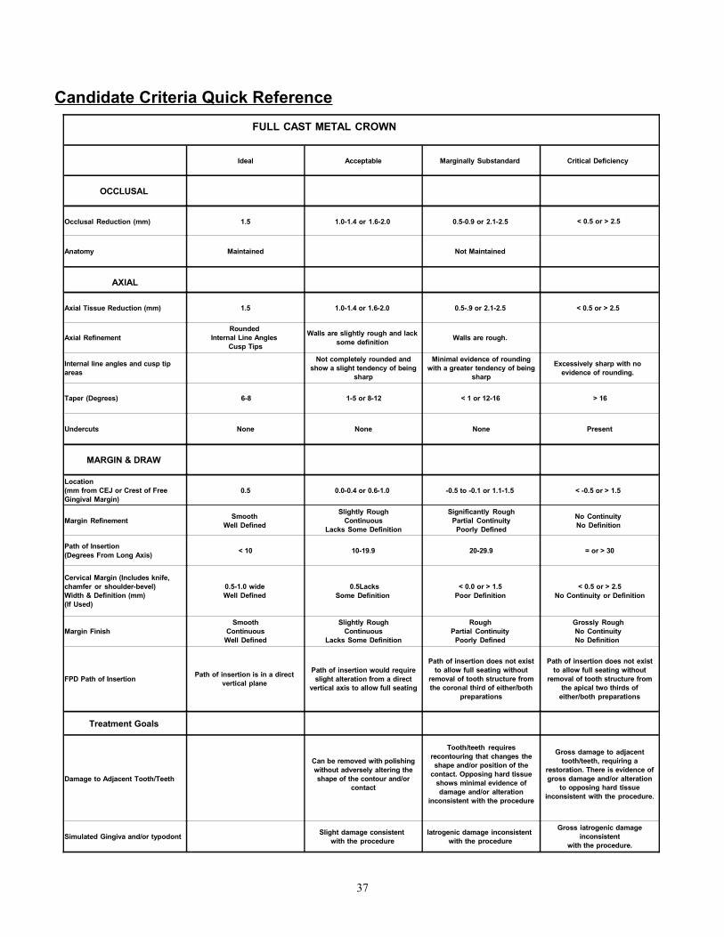
Candidate Criteria Quick ReferenceFULL CAST METAL CROWN
Ideal Acceptable Marginally Substandard Critical Deficiency
OCCLUSAL
Occlusal Reduction (mm) 1.5 1.0-1.4 or 1.6-2.0 0.5-0.9 or 2.1-2.5 < 0.5 or > 2.5
Anatomy Maintained Not Maintained
AXIAL
Axial Tissue Reduction (mm) 1.5 1.0-1.4 or 1.6-2.0 0.5-.9 or 2.1-2.5 < 0.5 or > 2.5
Axial RefinementRounded
Internal Line AnglesCusp Tips
Walls are slightly rough and lack some definition Walls are rough.
Internal line angles and cusp tip areas
Not completely rounded and show a slight tendency of being
sharp
Minimal evidence of rounding with a greater tendency of being
sharp
Excessively sharp with no evidence of rounding.
Taper (Degrees) 6-8 1-5 or 8-12 < 1 or 12-16 > 16
Undercuts None None None Present
MARGIN & DRAW
Location(mm from CEJ or Crest of Free Gingival Margin)
0.5 0.0-0.4 or 0.6-1.0 -0.5 to -0.1 or 1.1-1.5 < -0.5 or > 1.5
Margin Refinement Smooth Well Defined
Slightly Rough Continuous
Lacks Some Definition
Significantly Rough Partial Continuity
Poorly Defined
No Continuity No Definition
Path of Insertion(Degrees From Long Axis) < 10 10-19.9 20-29.9 = or > 30
Cervical Margin (Includes knife, chamfer or shoulder-bevel) Width & Definition (mm)(If Used)
0.5-1.0 wide Well Defined
0.5LacksSome Definition
< 0.0 or > 1.5Poor Definition
< 0.5 or > 2.5No Continuity or Definition
Margin FinishSmooth
Continuous Well Defined
Slightly Rough Continuous
Lacks Some Definition
Rough Partial Continuity
Poorly Defined
Grossly Rough No Continuity No Definition
FPD Path of Insertion Path of insertion is in a direct vertical plane
Path of insertion would require slight alteration from a direct
vertical axis to allow full seating
Path of insertion does not exist to allow full seating without
removal of tooth structure from the coronal third of either/both
preparations
Path of insertion does not exist to allow full seating without
removal of tooth structure from the apical two thirds of
either/both preparations
Treatment Goals
Damage to Adjacent Tooth/Teeth
Can be removed with polishing without adversely altering the shape of the contour and/or
contact
Tooth/teeth requires recontouring that changes the shape and/or position of the
contact. Opposing hard tissue shows minimal evidence of damage and/or alteration
inconsistent with the procedure
Gross damage to adjacent tooth/teeth, requiring a
restoration. There is evidence of gross damage and/or alteration
to opposing hard tissue inconsistent with the procedure.
Simulated Gingiva and/or typodont Slight damage consistent with the procedure
Iatrogenic damage inconsistent with the procedure
Gross iatrogenic damage inconsistent
with the procedure.
37
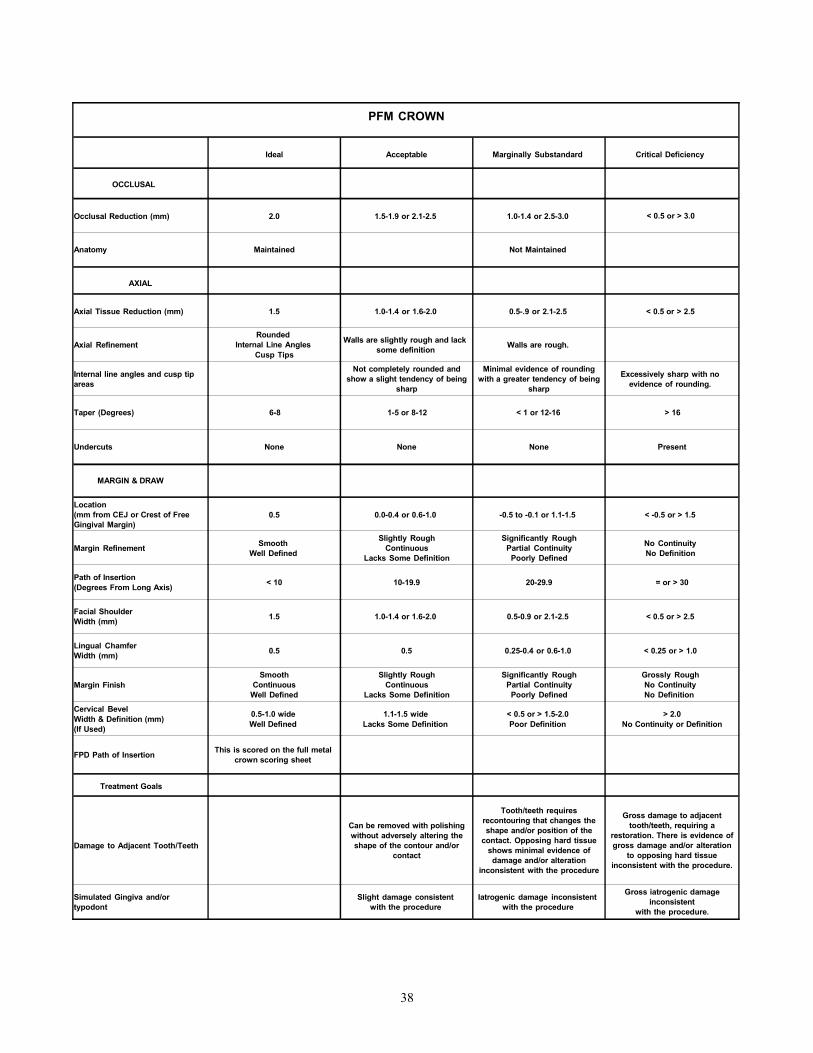
PFM CROWN
Ideal Acceptable Marginally Substandard Critical Deficiency
OCCLUSAL
Occlusal Reduction (mm) 2.0 1.5-1.9 or 2.1-2.5 1.0-1.4 or 2.5-3.0 < 0.5 or > 3.0
Anatomy Maintained Not Maintained
AXIAL
Axial Tissue Reduction (mm) 1.5 1.0-1.4 or 1.6-2.0 0.5-.9 or 2.1-2.5 < 0.5 or > 2.5
Axial RefinementRounded
Internal Line AnglesCusp Tips
Walls are slightly rough and lack some definition Walls are rough.
Internal line angles and cusp tip areas
Not completely rounded and show a slight tendency of being
sharp
Minimal evidence of rounding with a greater tendency of being
sharp
Excessively sharp with no evidence of rounding.
Taper (Degrees) 6-8 1-5 or 8-12 < 1 or 12-16 > 16
Undercuts None None None Present
MARGIN & DRAW
Location(mm from CEJ or Crest of Free Gingival Margin)
0.5 0.0-0.4 or 0.6-1.0 -0.5 to -0.1 or 1.1-1.5 < -0.5 or > 1.5
Margin Refinement Smooth Well Defined
Slightly Rough Continuous
Lacks Some Definition
Significantly Rough Partial Continuity
Poorly Defined
No Continuity No Definition
Path of Insertion(Degrees From Long Axis) < 10 10-19.9 20-29.9 = or > 30
Facial Shoulder Width (mm) 1.5 1.0-1.4 or 1.6-2.0 0.5-0.9 or 2.1-2.5 < 0.5 or > 2.5
Lingual Chamfer Width (mm) 0.5 0.5 0.25-0.4 or 0.6-1.0 < 0.25 or > 1.0
Margin FinishSmooth
Continuous Well Defined
Slightly Rough Continuous
Lacks Some Definition
Significantly Rough Partial Continuity
Poorly Defined
Grossly Rough No Continuity No Definition
Cervical BevelWidth & Definition (mm) (If Used)
0.5-1.0 wide Well Defined
1.1-1.5 wide Lacks Some Definition
< 0.5 or > 1.5-2.0Poor Definition
> 2.0No Continuity or Definition
FPD Path of Insertion This is scored on the full metal crown scoring sheet
Treatment Goals
Damage to Adjacent Tooth/Teeth
Can be removed with polishing without adversely altering the shape of the contour and/or
contact
Tooth/teeth requires recontouring that changes the shape and/or position of the
contact. Opposing hard tissue shows minimal evidence of damage and/or alteration
inconsistent with the procedure
Gross damage to adjacent tooth/teeth, requiring a
restoration. There is evidence of gross damage and/or alteration
to opposing hard tissue inconsistent with the procedure.
Simulated Gingiva and/or typodont
Slight damage consistent with the procedure
Iatrogenic damage inconsistent with the procedure
Gross iatrogenic damage inconsistent
with the procedure.
38
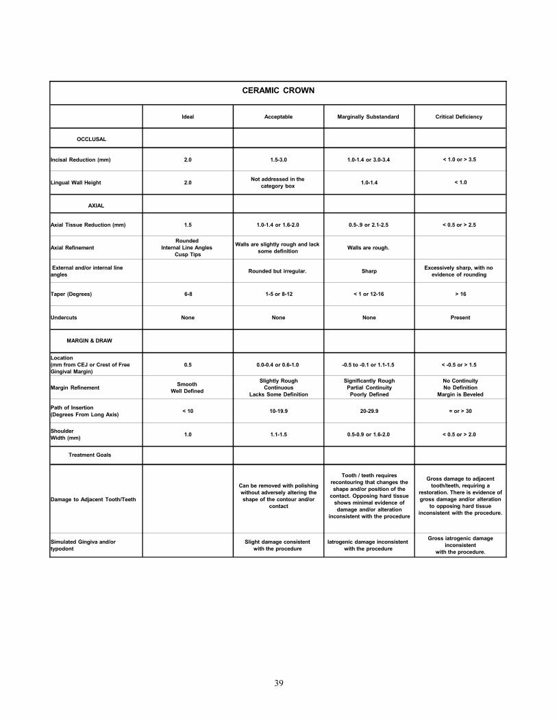
CERAMIC CROWN
Ideal Acceptable Marginally Substandard Critical Deficiency
OCCLUSAL
Incisal Reduction (mm) 2.0 1.5-3.0 1.0-1.4 or 3.0-3.4 < 1.0 or > 3.5
Lingual Wall Height 2.0 Not addressed in the category box 1.0-1.4 < 1.0
AXIAL
Axial Tissue Reduction (mm) 1.5 1.0-1.4 or 1.6-2.0 0.5-.9 or 2.1-2.5 < 0.5 or > 2.5
Axial RefinementRounded
Internal Line AnglesCusp Tips
Walls are slightly rough and lack some definition Walls are rough.
External and/or internal line angles Rounded but irregular. Sharp Excessively sharp, with no
evidence of rounding
Taper (Degrees) 6-8 1-5 or 8-12 < 1 or 12-16 > 16
Undercuts None None None Present
MARGIN & DRAW
Location(mm from CEJ or Crest of Free Gingival Margin)
0.5 0.0-0.4 or 0.6-1.0 -0.5 to -0.1 or 1.1-1.5 < -0.5 or > 1.5
Margin Refinement Smooth Well Defined
Slightly Rough Continuous
Lacks Some Definition
Significantly Rough Partial Continuity
Poorly Defined
No Continuity No Definition
Margin is Beveled
Path of Insertion(Degrees From Long Axis) < 10 10-19.9 20-29.9 = or > 30
Shoulder Width (mm) 1.0 1.1-1.5 0.5-0.9 or 1.6-2.0 < 0.5 or > 2.0
Treatment Goals
Damage to Adjacent Tooth/Teeth
Can be removed with polishing without adversely altering the shape of the contour and/or
contact
Tooth / teeth requires recontouring that changes the shape and/or position of the
contact. Opposing hard tissue shows minimal evidence of damage and/or alteration
inconsistent with the procedure
Gross damage to adjacent tooth/teeth, requiring a
restoration. There is evidence of gross damage and/or alteration
to opposing hard tissue inconsistent with the procedure.
Simulated Gingiva and/or typodont
Slight damage consistent with the procedure
Iatrogenic damage inconsistent with the procedure
Gross iatrogenic damage inconsistent
with the procedure.
39
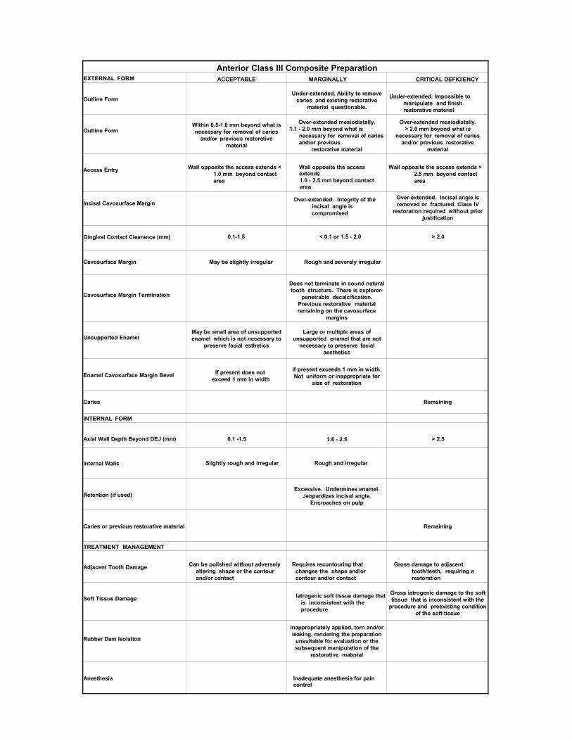
Anterior Class III Composite PreparationEXTERNAL FORM ACCEPTABLE MARGINALLY
SUBSTANDARDCRITICAL DEFICIENCY
Outline FormUnder-extended. Ability to remove
caries and existing restorative material questionable.
Under-extended. Impossible to manipulate and finish restorative material
Outline FormWithin 0.5-1.0 mm beyond what is necessary for removal of caries
and/or previous restorative material
Over-extended mesiodistally.1.1 - 2.0 mm beyond what is
necessary for removal of caries and/or previous
restorative material
Over-extended mesiodistally.> 2.0 mm beyond what is
necessary for removal of caries and/or previous restorative
material
Access Entry Wall opposite the access extends < 1.0 mm beyond contact area
Wall opposite the access extends1.0 - 2.5 mm beyond contact area
Wall opposite the access extends > 2.5 mm beyond contact area
Incisal Cavosurface Margin Over-extended. Integrity of the incisal angle is compromised
Over-extended. Incisal angle is removed or fractured. Class IV
restoration required without prior justification
Gingival Contact Clearance (mm) 0.1-1.5 < 0.1 or 1.5 - 2.0 > 2.0
Cavosurface Margin May be slightly irregular Rough and severely irregular
Cavosurface Margin Termination
Does not terminate in sound natural tooth structure. There is explorer-
penetrable decalcification. Previous restorative material remaining on the cavosurface
margins
Unsupported EnamelMay be small area of unsupported enamel which is not necessary to
preserve facial esthetics
Large or multiple areas of unsupported enamel that are not
necessary to preserve facial aesthetics
Enamel Cavosurface Margin Bevel If present does not exceed 1 mm in width
If present exceeds 1 mm in width. Not uniform or inappropriate for
size of restoration
Caries Remaining
INTERNAL FORM
Axial Wall Depth Beyond DEJ (mm) 0.1 -1.5 1.6 - 2.5 > 2.5
Internal Walls Slightly rough and irregular Rough and irregular
Retention (if used)Excessive. Undermines enamel.
Jeopardizes incisal angle.Encroaches on pulp
Caries or previous restorative material Remaining
TREATMENT MANAGEMENT
Adjacent Tooth Damage Can be polished without adversely altering shape or the contour and/or contact
Requires recontouring that changes the shape and/or contour and/or contact
Gross damage to adjacent tooth/teeth, requiring a restoration
Soft Tissue Damage Iatrogenic soft tissue damage that is inconsistent with the procedure
Gross iatrogenic damage to the soft tissue that is inconsistent with the
procedure and preexisting condition of the soft tissue
Rubber Dam Isolation
Inappropriately applied, torn and/or leaking, rendering the preparation
unsuitable for evaluation or the subsequent manipulation of the
restorative material
Anesthesia Inadequate anesthesia for pain control
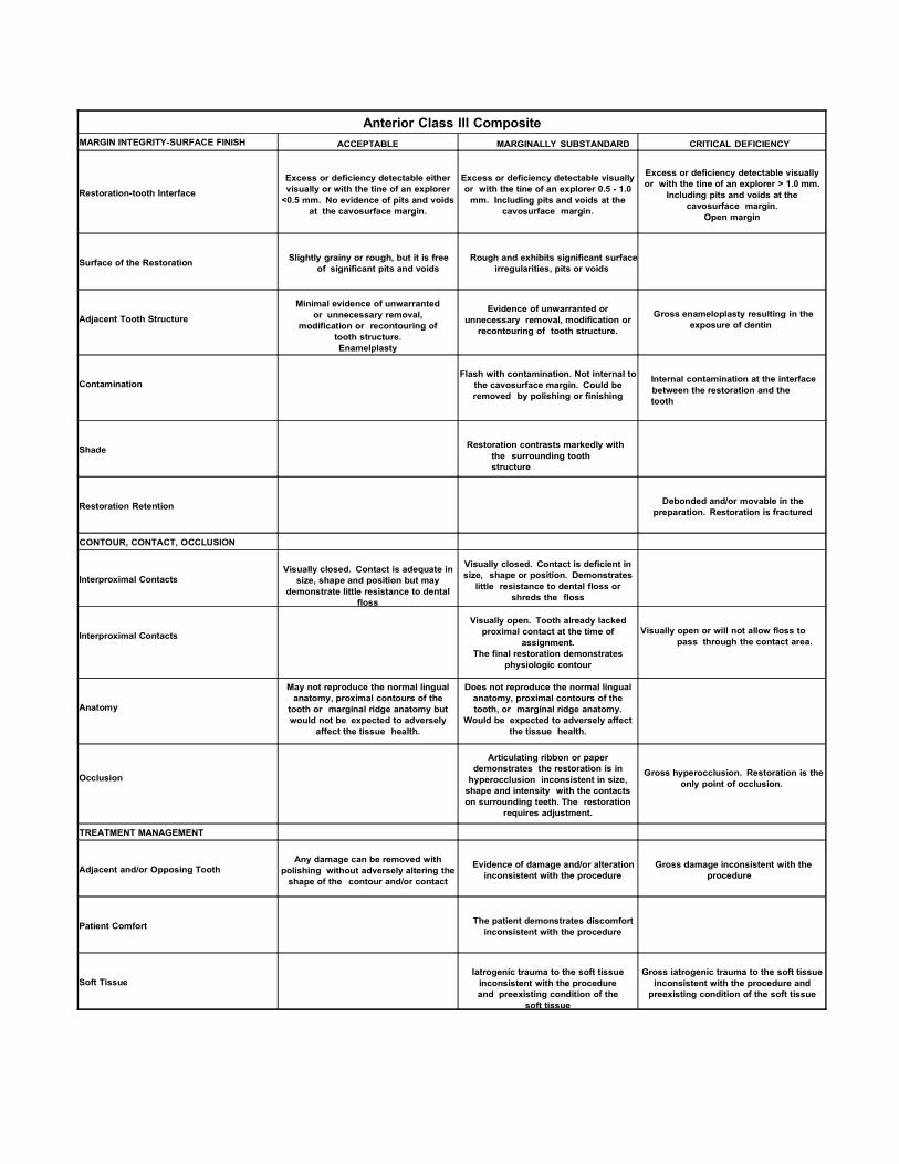
Anterior Class III Composite RestorationMARGIN INTEGRITY-SURFACE FINISH ACCEPTABLE MARGINALLY SUBSTANDARD CRITICAL DEFICIENCY
Restoration-tooth InterfaceExcess or deficiency detectable either visually or with the tine of an explorer
<0.5 mm. No evidence of pits and voids at the cavosurface margin.
Excess or deficiency detectable visually or with the tine of an explorer 0.5 - 1.0
mm. Including pits and voids at the cavosurface margin.
Excess or deficiency detectable visually or with the tine of an explorer > 1.0 mm.
Including pits and voids at the cavosurface margin.
Open margin
Surface of the Restoration Slightly grainy or rough, but it is free of significant pits and voids
Rough and exhibits significant surface irregularities, pits or voids
Adjacent Tooth Structure
Minimal evidence of unwarranted or unnecessary removal,
modification or recontouring of tooth structure.Enamelplasty
Evidence of unwarranted or unnecessary removal, modification or
recontouring of tooth structure.
Gross enameloplasty resulting in the exposure of dentin
ContaminationFlash with contamination. Not internal to
the cavosurface margin. Could be removed by polishing or finishing
Internal contamination at the interface between the restoration and the tooth
Shade Restoration contrasts markedly with the surrounding tooth structure
Restoration Retention Debonded and/or movable in the preparation. Restoration is fractured
CONTOUR, CONTACT, OCCLUSION
Interproximal ContactsVisually closed. Contact is adequate in
size, shape and position but may demonstrate little resistance to dental
floss
Visually closed. Contact is deficient in size, shape or position. Demonstrates
little resistance to dental floss or shreds the floss
Interproximal ContactsVisually open. Tooth already lacked
proximal contact at the time of assignment.
The final restoration demonstrates physiologic contour
Visually open or will not allow floss to pass through the contact area.
Anatomy
May not reproduce the normal lingual anatomy, proximal contours of the
tooth or marginal ridge anatomy but would not be expected to adversely
affect the tissue health.
Does not reproduce the normal lingual anatomy, proximal contours of the tooth, or marginal ridge anatomy.
Would be expected to adversely affect the tissue health.
Occlusion
Articulating ribbon or paper demonstrates the restoration is in
hyperocclusion inconsistent in size, shape and intensity with the contacts on surrounding teeth. The restoration
requires adjustment.
Gross hyperocclusion. Restoration is the only point of occlusion.
TREATMENT MANAGEMENT
Adjacent and/or Opposing ToothAny damage can be removed with
polishing without adversely altering the shape of the contour and/or contact
Evidence of damage and/or alteration inconsistent with the procedure
Gross damage inconsistent with the procedure
Patient Comfort The patient demonstrates discomfort inconsistent with the procedure
Soft TissueIatrogenic trauma to the soft tissue
inconsistent with the procedure and preexisting condition of the
soft tissue
Gross iatrogenic trauma to the soft tissue inconsistent with the procedure and
preexisting condition of the soft tissue
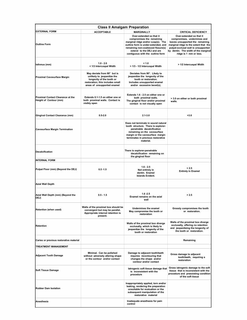
Class II Amalgam PreparationEXTERNAL FORM ACCEPTABLE MARGINALLY
SUBSTANDARDCRITICAL DEFICIENCY
Outline Form
Over-extended so that it compromises the remaining
marginal ridge and/or cusp(s). The outline form is under-extended, and remaining non-coalesced fissure(s)
extend to the DEJ and are contiguous with the outline form
Over-extended so that it compromises, undermines and
leaves unsupported the remaining marginal ridge to the extent that the pulpal-occlusal wall is unsupported
by dentin. The width of the marginal ridge is 1 mm or less.
Isthmus (mm) 1.0 - 2.0< 1/3 Intercuspal Width
< 1.0> 1/3 - 1/2 Intercuspal Width
> 1/2 Intercuspal Width
Proximal Cavosurface MarginMay deviate from 90° but is unlikely to jeopardize the longevity of the tooth or
restoration; this includes small areas of unsupported enamel
Deviates from 90°. Likely to jeopardize the longevity of the
tooth or restoration.Includes unsupported enamel
and/or excessive bevel(s).
Proximal Contact Clearance at the Height of Contour (mm)
Extends 0.1-1.5 on either one or both proximal walls. Contact is visibly open
Extends 1.6 - 2.5 on either one or both proximal walls.
The gingival floor and/or proximal contact is not visually open
> 3.0 on either or both proximal walls.
Gingival Contact Clearance (mm) 0.5-2.0 2.1-3.0 >3.0
Cavosurface Margin Termination
Does not terminate in sound natural tooth structure. There is explorer-
penetrable decalcification remaining on the cavosurface
margin or the cavosurface margin terminates in previous restorative
material.
Decalcification There is explorer-penetrable decalcification remaining on the gingival floor
INTERNAL FORM
Pulpal Floor (mm) (Beyond the DEJ) 0.5 -1.51.6 - 2.5
Not entirely in dentin. Enamel Islands Evident.
> 2.5Entirely in Enamel
Axial Wall Depth
Axial Wall Depth (mm) (Beyond the DEJ)
0.5 - 1.5 1.6 -2.5Enamel remains on the axial
wall
> 2.5
Retention (when used)Walls of the proximal box should be
convergent but may be parallel.Appropriate internal retention is
present.
Undermines the enamelMay compromise the tooth or
restoration
Grossly compromises the tooth or restoration.
RetentionWalls of the proximal box diverge
occlusally, which is likely to jeopardize the longevity of the
tooth or restoration
Walls of the proximal box diverge occlusally, offering no retention
and jeopardizing the longevity of the tooth or restoration.
Caries or previous restorative material Remaining
TREATMENT MANAGEMENT
Adjacent Tooth DamageMinimal. Can be polished
without adversely altering shape or the contour and/or contact
Damage to adjacent tooth/teeth requires recontouring that changes the shape and/or
contour and/or contact
Gross damage to adjacent tooth/teeth, requiring a restoration
Soft Tissue Damage Iatrogenic soft tissue damage that is inconsistent with the procedure
Gross iatrogenic damage to the soft tissue that is inconsistent with the
procedure and preexisting condition of the soft tissue
Rubber Dam Isolation
Inappropriately applied, torn and/or leaking, rendering the preparation
unsuitable for evaluation or the subsequent manipulation of the
restorative material
Anesthesia Inadequate anesthesia for pain control

Class II Amalgam RestorationMARGIN INTEGRITY-SURFACE FINISH ACCEPTABLE MARGINALLY SUBSTANDARD CRITICAL DEFICIENCY
Restoration-tooth Interface
Excess or deficiency detectable either visually or with the tine of an explorer
<0.5 mm. No evidence of pits and voids at the cavosurface margin.
Excess or deficiency detectable visually or with the tine of an explorer 0.5 - 1.0
mm. Including pits and voids at the cavosurface margin.
Excess or deficiency detectable visually or with the tine of an explorer > 1.0
mm.Including pits and voids at the
cavosurface margin.Open margin
Surface of the Restoration Slightly grainy or rough, but it is free of significant pits and voids
Rough and exhibits significant surface irregularities, pits or voids
Adjacent Tooth Structure
Minimal evidence of unwarranted or unnecessary removal,
modification or recontouring of tooth structure.Enamelplasty
Evidence of unwarranted or unnecessary removal, modification or
recontouring of tooth structure.
Gross enameloplasty resulting in the exposure of dentin
Restoration Retention Restoration is fractured
CONTOUR, CONTACT, OCCLUSION
Interproximal ContactsVisually closed. Contact is adequate in
size, shape and position but may demonstrate little resistance to dental
floss
Visually closed. Contact is deficient in size, shape or position and
demonstrates little resistance to dental floss or shreds the floss
Visually open or will not allow floss to pass through the contact area.
Anatomy
May not reproduce the normal occlusal anatomy, proximal contours of the
tooth or marginal ridge anatomy but would not be expected to adversely
affect the tissue health.
Does not reproduce the normal occlusal anatomy, proximal contours
of the tooth or marginal ridge anatomy and would be expected to adversely
affect the tissue health.
Occlusion
When checked with articulating ribbon or paper, the restoration is in
hyperocclusion inconsistent in size, shape and intensity with the contacts on surrounding teeth. The restoration
requires adjustment
Gross hyperocclusion so that the restoration is the only point of occlusion
TREATMENT MANAGEMENT
Adjacent and/or Opposing ToothAny damage can be removed with
polishing without adversely altering the shape of the contour and/or contact
Evidence of damage and/or alteration inconsistent with the procedure
Gross damage inconsistent with the procedure
Patient Comfort Patient demonstrates discomfort inconsistent with the procedure.
Soft TissueIatrogenic trauma to the soft tissue
inconsistent with the procedure and preexisting condition of the
soft tissue
Gross iatrogenic trauma to the soft tissue inconsistent with the procedure and preexisting condition of the soft
tissue
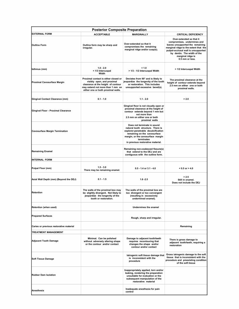
Posterior Composite PreparationEXTERNAL FORM ACCEPTABLE MARGINALLY
SUBSTANDARDCRITICAL DEFICIENCY
Outline Form Outline form may be sharp and irregular.
Over-extended so that it compromises the remaining marginal ridge and/or cusp(s).
Over-extended so that it compromises, undermines and
leaves unsupported the remaining marginal ridge to the extent that the pulpal-occlusal wall is unsupported
by dentin. The width of the marginal ridge is
0.5 mm or less.
Isthmus (mm) 1.0 - 2.0< 1/3 Intercuspal
Width
< 1.0> 1/3 - 1/2 Intercuspal Width
> 1/2 Intercuspal Width
Proximal Cavosurface MarginProximal contact is either closed or
visibly open, and proximal clearance at the height of contour
may extend not more than 1 mm on either one or both proximal walls.
Deviates from 90° and is likely to jeopardize the longevity of the tooth
or restoration. This includes unsupported excessive bevel(s).
The proximal clearance at the height of contour extends beyond
2.5 mm on either one or both proximal walls.
Gingival Contact Clearance (mm) 0.1 - 1.0 1.1 - 2.0 > 2.0
Gingival Floor - Proximal Clearance
Gingival floor is not visually open or proximal clearance at the height of contour extends beyond 1 mm but
not more than2.5 mm on either one or both
proximal walls
Cavosurface Margin Termination
Does not terminate in sound natural tooth structure. There is
explorer-penetrable decalcification remaining on the cavosurface
margin, or the cavosurface margin terminates
in previous restorative material.
Remaining EnamelRemaining non-coalesced fissure(s)
that extend to the DEJ and are contiguous with the outline form.
INTERNAL FORM
Pulpal Floor (mm) 1.5 - 3.0There may be remaining enamel.
0.5 - 1.4 or 3.1 - 4.0 < 0.5 or > 4.0
Axial Wall Depth (mm) (Beyond the DEJ) 0.1 - 1.5 1.6 -2.5> 2.5
Still in enamel.Does not include the DEJ
RetentionThe walls of the proximal box may be slightly divergent. Not likely to
jeopardize the longevity of the tooth or restoration.
The walls of the proximal box are too divergent or too convergent
(resulting in excessively undermined enamel).
Retention (when used) Undermines the enamel
Prepared SurfacesRough, sharp and irregular.
Caries or previous restorative material Remaining
TREATMENT MANAGEMENT
Adjacent Tooth DamageMinimal. Can be polished
without adversely altering shape or the contour and/or contact
Damage to adjacent tooth/teeth requires recontouring that changes the shape and/or
contour and/or contact
There is gross damage to adjacent tooth/teeth, requiring a restoration
Soft Tissue DamageIatrogenic soft tissue damage that
is inconsistent with the procedure
Gross iatrogenic damage to the soft tissue that is inconsistent with the
procedure and preexisting condition of the soft tissue
Rubber Dam Isolation
Inappropriately applied, torn and/or leaking, rendering the preparation
unsuitable for evaluation or the subsequent manipulation of the
restorative material
Anesthesia Inadequate anesthesia for pain control
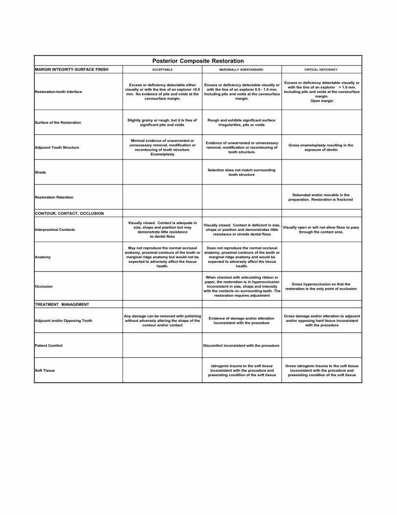
Posterior Composite RestorationMARGIN INTEGRITY-SURFACE FINISH ACCEPTABLE MARGINALLY SUBSTANDARD CRITICAL DEFICIENCY
Restoration-tooth Interface
Excess or deficiency detectable either visually or with the tine of an explorer <0.5 mm. No evidence of pits and voids at the
cavosurface margin.
Excess or deficiency detectable visually or with the tine of an explorer 0.5 - 1.0 mm.
Including pits and voids at the cavosurface margin.
Excess or deficiency detectable visually or with the tine of an explorer > 1.0 mm.
Including pits and voids at the cavosurface margin.
Open margin
Surface of the Restoration Slightly grainy or rough, but it is free of significant pits and voids
Rough and exhibits significant surface irregularities, pits or voids
Adjacent Tooth Structure
Minimal evidence of unwarranted or unnecessary removal, modification or
recontouring of tooth structure.Enamelplasty
Evidence of unwarranted or unnecessary removal, modification or recontouring of
tooth structure.
Gross enameloplasty resulting in the exposure of dentin
Shade Selection does not match surrounding tooth structure
Restoration Retention Debonded and/or movable in the preparation. Restoration is fractured
CONTOUR, CONTACT, OCCLUSION
Interproximal Contacts
Visually closed. Contact is adequate in size, shape and position but may
demonstrate little resistanceto dental floss
Visually closed. Contact is deficient in size, shape or position and demonstrates little
resistance or shreds dental floss
Visually open or will not allow floss to pass through the contact area.
Anatomy
May not reproduce the normal occlusal anatomy, proximal contours of the tooth or marginal ridge anatomy but would not be
expected to adversely affect the tissue health.
Does not reproduce the normal occlusal anatomy, proximal contours of the tooth or
marginal ridge anatomy and would be expected to adversely affect the tissue
health.
Occlusion
When checked with articulating ribbon or paper, the restoration is in hyperocclusion inconsistent in size, shape and intensity
with the contacts on surrounding teeth. The restoration requires adjustment
Gross hyperocclusion so that the restoration is the only point of occlusion
TREATMENT MANAGEMENT
Adjacent and/or Opposing ToothAny damage can be removed with polishing without adversely altering the shape of the
contour and/or contact
Evidence of damage and/or alteration inconsistent with the procedure
Gross damage and/or alteration to adjacent and/or opposing hard tissue inconsistent
with the procedure
Patient Comfort Discomfort inconsistent with the procedure
Soft TissueIatrogenic trauma to the soft tissue inconsistent with the procedure and
preexisting condition of the soft tissue
Gross iatrogenic trauma to the soft tissue inconsistent with the procedure and
preexisting condition of the soft tissue
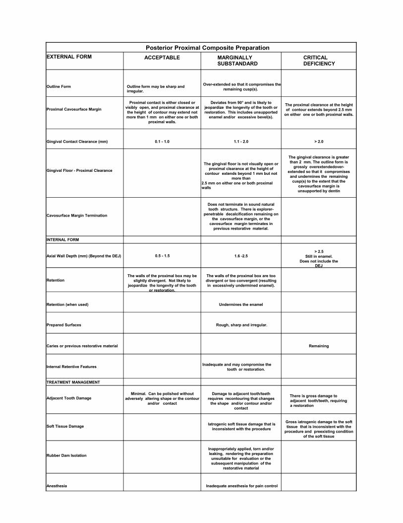
Posterior Proximal Composite PreparationEXTERNAL FORM ACCEPTABLE MARGINALLY
SUBSTANDARDCRITICAL DEFICIENCY
Outline Form Outline form may be sharp and irregular.
Over-extended so that it compromises the remaining cusp(s).
Proximal Cavosurface MarginProximal contact is either closed or
visibly open, and proximal clearance at the height of contour may extend not more than 1 mm on either one or both
proximal walls.
Deviates from 90° and is likely to jeopardize the longevity of the tooth or restoration. This includes unsupported
enamel and/or excessive bevel(s).
The proximal clearance at the height of contour extends beyond 2.5 mm
on either one or both proximal walls.
Gingival Contact Clearance (mm) 0.1 - 1.0 1.1 - 2.0 > 2.0
Gingival Floor - Proximal ClearanceThe gingival floor is not visually open or
proximal clearance at the height of contour extends beyond 1 mm but not
more than2.5 mm on either one or both proximal walls
The gingival clearance is greater than 2 mm. The outline form is
grossly overextendedover-extended so that it compromises and undermines the remaining
cusp(s) to the extent that the cavosurface margin isunsupported by dentin
Cavosurface Margin Termination
Does not terminate in sound natural tooth structure. There is explorer-
penetrable decalcification remaining on the cavosurface margin, or the
cavosurface margin terminates in previous restorative material.
INTERNAL FORM
Axial Wall Depth (mm) (Beyond the DEJ) 0.5 - 1.5 1.6 -2.5> 2.5
Still in enamel.Does not include the
DEJ
RetentionThe walls of the proximal box may be
slightly divergent. Not likely to jeopardize the longevity of the tooth
or restoration.
The walls of the proximal box are too divergent or too convergent (resulting in excessively undermined enamel).
Retention (when used) Undermines the enamel
Prepared Surfaces Rough, sharp and irregular.
Caries or previous restorative material Remaining
Internal Retentive Features Inadequate and may compromise the tooth or restoration.
TREATMENT MANAGEMENT
Adjacent Tooth DamageMinimal. Can be polished without
adversely altering shape or the contour and/or contact
Damage to adjacent tooth/teeth requires recontouring that changes
the shape and/or contour and/or contact
There is gross damage to adjacent tooth/teeth, requiring a restoration
Soft Tissue Damage Iatrogenic soft tissue damage that is inconsistent with the procedure
Gross iatrogenic damage to the soft tissue that is inconsistent with the
procedure and preexisting condition of the soft tissue
Rubber Dam IsolationInappropriately applied, torn and/or leaking, rendering the preparation
unsuitable for evaluation or the subsequent manipulation of the
restorative material
Anesthesia Inadequate anesthesia for pain control
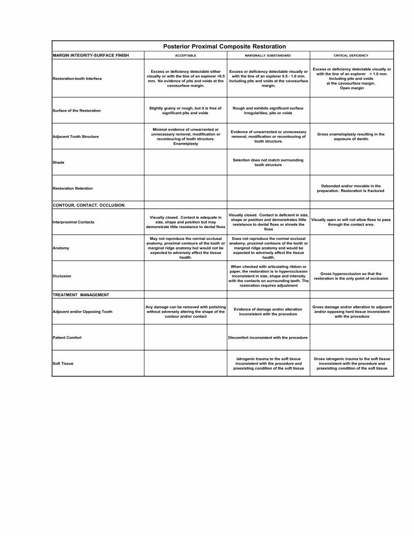
Posterior Proximal Composite RestorationMARGIN INTEGRITY-SURFACE FINISH ACCEPTABLE MARGINALLY SUBSTANDARD CRITICAL DEFICIENCY
Restoration-tooth Interface
Excess or deficiency detectable either visually or with the tine of an explorer <0.5 mm. No evidence of pits and voids at the
cavosurface margin.
Excess or deficiency detectable visually or with the tine of an explorer 0.5 - 1.0 mm.
Including pits and voids at the cavosurface margin.
Excess or deficiency detectable visually or with the tine of an explorer > 1.0 mm.
Including pits and voids at the cavosurface margin.
Open margin
Surface of the Restoration Slightly grainy or rough, but it is free of significant pits and voids
Rough and exhibits significant surface irregularities, pits or voids
Adjacent Tooth Structure
Minimal evidence of unwarranted or unnecessary removal, modification or
recontouring of tooth structure.Enamelplasty
Evidence of unwarranted or unnecessary removal, modification or recontouring of
tooth structure.
Gross enameloplasty resulting in the exposure of dentin
Shade Selection does not match surrounding tooth structure
Restoration Retention Debonded and/or movable in the preparation. Restoration is fractured
CONTOUR, CONTACT, OCCLUSION
Interproximal ContactsVisually closed. Contact is adequate in
size, shape and position but may demonstrate little resistance to dental floss
Visually closed. Contact is deficient in size, shape or position and demonstrates little resistance to dental floss or shreds the
floss
Visually open or will not allow floss to pass through the contact area.
Anatomy
May not reproduce the normal occlusal anatomy, proximal contours of the tooth or marginal ridge anatomy but would not be
expected to adversely affect the tissue health.
Does not reproduce the normal occlusal anatomy, proximal contours of the tooth or
marginal ridge anatomy and would be expected to adversely affect the tissue
health.
Occlusion
When checked with articulating ribbon or paper, the restoration is in hyperocclusion inconsistent in size, shape and intensity
with the contacts on surrounding teeth. The restoration requires adjustment
Gross hyperocclusion so that the restoration is the only point of occlusion
TREATMENT MANAGEMENT
Adjacent and/or Opposing ToothAny damage can be removed with polishing without adversely altering the shape of the
contour and/or contact
Evidence of damage and/or alteration inconsistent with the procedure
Gross damage and/or alteration to adjacent and/or opposing hard tissue inconsistent
with the procedure
Patient Comfort Discomfort inconsistent with the procedure
Soft TissueIatrogenic trauma to the soft tissue inconsistent with the procedure and
preexisting condition of the soft tissue
Gross iatrogenic trauma to the soft tissue inconsistent with the procedure and
preexisting condition of the soft tissue