How to Document a Dental Examination and Procedure Using … · How to Document a Dental...
12
How to Document a Dental Examination and Procedure Using a Dental Chart Stephen S. Galloway, DVM Author’s address: Animal Care Hospital, 8565 Highway 64, Somerville, Tennessee 38068; e-mail: [email protected]. © 2010 AAEP. 1. Introduction A dental chart is a permanent record of a patient’s dental care, and completion of a dental chart is the minimum standard of care for documenting any pro- fessional dental procedure. Dental charting is the process of recording the state of health or disease of the teeth and oral cavity, and it is an integral part of the examination, diagnosis, treatment planning, and monitoring of dental cases. 1 The dental chart provides legal documentation of the procedure per- formed and facilitates communication with colleagues. The scope of this paper is limited to documenting routine equine dental care (occlusal adjustment, floating, periodontal therapy, and simple extrac- tions). Although the purpose of this paper is not to describe how to perform a dental examination, a thorough oral examination is prerequisite to com- pleting an accurate dental chart. Additionally, to properly document any dental procedure and com- municate with colleagues, practitioners must have a working knowledge of dental terminology. Standardized Terminology and Abbreviations To facilitate communication between colleagues, the Nomenclature Committee of the American Veteri- nary Dental College (AVDC) reviews, clarifies, and recommends standardized terminology for dental and oral anatomical locations, pathologies, diag- noses, treatments, procedures, and dental materi- als. Terminology and abbreviations specific to equine dentistry have also been accepted by the Academy of Veterinary Dentistry (AVD). An exten- sive glossary of veterinary dental terminology can be found in veterinary dental texts. 2,3 Extensive lists of the abbreviations accepted by the AVDC and the AVD are available online within the application packets for these organizations. Diagnostic and treatment abbreviations commonly used by the au- thor are listed in Appendix A. Although various systems for describing and num- bering teeth are recognized, the Modified Triadan Tooth Numbering System is the tooth-identification system of choice in veterinary dentistry. 4 This sys- tem is applicable to most domestic animal species and provides accurate tooth identification in both written and oral communication. Each tooth is as- signed a unique three-digit number. The first digit designates the tooth’s quadrant and dentition, and the second and third digits designate the specific tooth. Teeth in each quadrant are numbered se- quentially from the first (central) incisor (X01) dis- tally to the third molar (X11), assuming a complete phenotypic equine dentition ([I 3/3 C 1/1 P 4/4 M 3/3] 2 44). AAEP PROCEEDINGS Vol. 56 2010 429 RESPIRATORY/DENTISTRY NOTES
Transcript of How to Document a Dental Examination and Procedure Using … · How to Document a Dental...

How to Document a Dental Examination andProcedure Using a Dental Chart
Stephen S. Galloway, DVM
Author’s address: Animal Care Hospital, 8565 Highway 64, Somerville, Tennessee 38068;e-mail: [email protected]. © 2010 AAEP.
1. Introduction
A dental chart is a permanent record of a patient’sdental care, and completion of a dental chart is theminimum standard of care for documenting any pro-fessional dental procedure. Dental charting is theprocess of recording the state of health or disease ofthe teeth and oral cavity, and it is an integral part ofthe examination, diagnosis, treatment planning,and monitoring of dental cases.1 The dental chartprovides legal documentation of the procedure per-formed and facilitates communication with colleagues.
The scope of this paper is limited to documentingroutine equine dental care (occlusal adjustment,floating, periodontal therapy, and simple extrac-tions). Although the purpose of this paper is not todescribe how to perform a dental examination, athorough oral examination is prerequisite to com-pleting an accurate dental chart. Additionally, toproperly document any dental procedure and com-municate with colleagues, practitioners must have aworking knowledge of dental terminology.
Standardized Terminology and Abbreviations
To facilitate communication between colleagues, theNomenclature Committee of the American Veteri-nary Dental College (AVDC) reviews, clarifies, andrecommends standardized terminology for dental
and oral anatomical locations, pathologies, diag-noses, treatments, procedures, and dental materi-als. Terminology and abbreviations specific toequine dentistry have also been accepted by theAcademy of Veterinary Dentistry (AVD). An exten-sive glossary of veterinary dental terminology can befound in veterinary dental texts.2,3 Extensive listsof the abbreviations accepted by the AVDC and theAVD are available online within the applicationpackets for these organizations. Diagnostic andtreatment abbreviations commonly used by the au-thor are listed in Appendix A.
Although various systems for describing and num-bering teeth are recognized, the Modified TriadanTooth Numbering System is the tooth-identificationsystem of choice in veterinary dentistry.4 This sys-tem is applicable to most domestic animal speciesand provides accurate tooth identification in bothwritten and oral communication. Each tooth is as-signed a unique three-digit number. The first digitdesignates the tooth’s quadrant and dentition, andthe second and third digits designate the specifictooth. Teeth in each quadrant are numbered se-quentially from the first (central) incisor (X01) dis-tally to the third molar (X11), assuming a completephenotypic equine dentition ([I 3/3 C 1/1 P 4/4 M 3/3]� 2 � 44).
AAEP PROCEEDINGS � Vol. 56 � 2010 429
RESPIRATORY/DENTISTRY
NOTES

101–111: Maxillary right quadrant, permanentdentition.
201–211: Maxillary left quadrant, permanentdentition.
301–311: Mandibular left quadrant, permanentdentition.
401–411: Mandibular right quadrant, perma-nent dentition.
501–508, 601–608, 701–708, and 801–808: de-ciduous 100, 200, 300, 400 dentition, respec-tively.
The typical domestic male horse is missing his man-dibular wolf teeth, and many domestic mares areadditionally missing all canine teeth; therefore, thedental formulae for male and female equids are ([I3/3 C 1/1 P 4/3 M 3/3] � 2 � 42) and ([I 3/3 C 0/0 P4/4 M 3/3] � 2 � 38), respectively. In the ModifiedTriadan System, “The Rule of Four and Nine” isused to simplify annotation among various speciesand variations within a species. Tooth X04 is al-ways the canine tooth (104, 204, 304, 404), and toothX09 is always the first molar (109, 209, 309, 409).Applying this rule, the first molarized cheek tooth(the second premolar) in domestic horses is toothX06 (106, 206, 306, 406).
The Dental ChartThe dental chart is a record of the condition of thepatient’s dentition and oral cavity. It should in-clude a dental history, oral-examination findings,proposed and completed dental procedures, pro-posed future dental care, and home-care instruc-tions.5 Although many small animal and humandentists prefer a two-chart system (one chart forrecording examination findings, diagnoses, and pro-posed treatment planning and a second chart forrecording the treatment performed), most equinedental practitioners use a combined report for boththe examination and treatments. The most com-monly accepted chart format is an anatomical dentaldiagram supplemented by brief descriptions to clar-ify the examination findings, diagnoses, and proce-dure performed. Most dental charts are designedwith a fill-in-the-blank and check-off format to en-sure consistent documentation. The dental chartshould include a legend for non-standardized sym-bols and abbreviations; however, the use of approvedAVDC/AVD abbreviations should minimize this re-quirement. To meet the legal requirements of med-ical documentation, most state veterinary-practiceacts require that the following information be in-cluded in the medical record6:
1. Date2. Primary complaint3. History4. Physical examination findings5. Preliminary diagnosis with rule-outs6. Tests performed and results7. Diagnosis
8. Treatment plan, implementation, drugs ad-ministered, and procedures performed
9. Prognosis10. Patient progress11. Client communication
2. Materials and Methods
The following outline describes the steps in docu-menting a dental procedure using the author’s com-bined format (examination and treatment) equinedental chart (Appendix B):
I. Documentation of all veterinary cases be-gins with recording the owner information,patient’s signalment, and primary com-plaint for the visit.
II. The patient’s history is taken with particu-lar emphasis on the horse’s use, bit and bri-dle, diet, and masticatory and performanceproblems.
III. A thorough physical examination is per-formed and documented. The clinicianmust first rule out sources of systemic dis-ease before any elective dental procedure isperformed. Because sedative restraint isrequired for a thorough dental examination,emphasis during the physical examinationshould be placed on the horse’s body condi-tion and cardiovascular system.
IV. After diseases of other body systems areruled out, the horse’s head is examined, andabnormalities are recorded.
V. On completion of the external examination,the horse is sedated for oral examina-tion. Sedative and other medications arerecorded on the dental chart as they aregiven during the procedure.
VI. Oral examination includes the examinationof all tissues in the mouth. The soft-tissuefindings are documented in the appropriatefill-in-the-blank section of the chart (e.g., acheek laceration caused by a hard enamelpoint on the maxillary right first molar isabbreviated LAC/B 110).
VII. Dental abnormalities are documented on thedental diagram and explained in the exam-findings section of the chart using the appro-priate diagnostic abbreviation followed bythe affected tooth’s Triadan number and theaspect of the tooth, when appropriate. Thetooth aspects are apical, coronal, occlusal,mesial (M), distal (D), palatal (P), lingual(L), and vestibular (V)7. A forward slash (/)or a space is often used between abbrevia-tions for clarity. For example, a hook onthe maxillary right first cheek tooth is ab-breviated HK 106.A. Clinically missing teeth are circled on
the diagram and annotated by the toothnumber and abbreviation O (e.g., an ab-sent maxillary left second incisor is ab-
430 2010 � Vol. 56 � AAEP PROCEEDINGS
RESPIRATORY/DENTISTRY

breviated 0/202). During the mixeddentition period, unerupted molars arerecorded by circling the adult molar onthe dental diagram.
B. The presence of deciduous dentition isannotated on the dental diagram by plac-ing a single line through the adult toothnumber and writing in the appropriatedeciduous tooth number (e.g., 508).
C. Supernumerary teeth and retained de-ciduous teeth are drawn on the diagramand appropriately annotated (e.g., SN111, not 112, and RD 503).
D. An unerupted or partially erupted toothis usually impacted; therefore, a blindmaxillary right wolf tooth is abbreviatedTI 105.
E. Dental malocclusions, fractures, cavities,and periodontal pockets are drawn onthe chart to approximate the outline ofactual finding and annotated in the ex-am-findings section.
VIII. Malocclusions and other abnormal dentalfindings commonly effecting the incisors in-clude the following:A. Diagonal bites are defined with respect
to the mandibular incisors. DGL/3 is adiagonal bite in which the mandibularleft incisors are longer than the mandib-ular right incisors (Fig. 1). DGL/4 is adiagonal bite in which mandibular rightincisors are longer.
B. Ventral curvature (CV) and dorsal cur-vature bites (CD) are the dental termsfor a smile and frown bite, respectively.
C. Although overbites and underbites usu-ally affect the entire dentition of a pa-tient, these malocclusions are typicallyrecorded in the incisor part of the exam-findings section as MAL2 or MAL3, re-spectively.
D. Hooks on the maxillary third incisors area common finding (HK 103/203) (Fig. 2).
E. Abnormal wear patterns or attritionsuch as that seen in cribbers is recorded
by describing the affected aspect of thetooth (e.g., cribbing attrition on the ves-tibular aspect of the maxillary first inci-sors is abbreviated AT 101V/201V).
F. Crown fractures of the incisors should bedrawn on the dental chart and described(e.g., a crown fracture of the maxillaryright third incisor is abbreviated T/FX403CR). The extent of the fracture canbe further described using the tooth-frac-ture abbreviations (T/FX/) in AppendixA.
G. Iatrogenic pulp damage secondary tooverreduction of the incisors with powerinstrumentation is a common find-ing. Exposed pulp is differentiatedbased on its vitality and recorded (e.g., aliving, bleeding pulp in the mandibularright third incisor is abbreviated T/PE/V203, whereas a necrotic, non-vital pulp inthe same tooth is abbreviated T/PE/NV203).
H. Cavities (CA) should be staged accordingto severity.1. Stage 1: cavities in the cementum
only (CA1).2. Stage 2: cavities through the cemen-
tum and into the enamel (CA2).3. Stage 3: cavities involving the ce-
mentum, enamel, and dentin (CA3).4. Stage 4: cavities exposing pulp
(CA4).I. Tooth resorption (TR; equine odontoclas-
tic tooth resorption and hypercementosis[EOTRH]) should be classified using theAVDC classification (see TR in AppendixA).
IX. Dental findings commonly affecting the ca-nine teeth include:A. Tartar (calculus [CAL]) that may be as-
sociated with periodontal disease (dis-cussed below).
B. Blind canines in young males and mares(TI).
Fig. 1. Dental diagram charting a diagonal bite 4 (DGL/4). Fig. 2. Dental diagram charting hook malocclusions on the max-illary third incisors (HK 103/203).
AAEP PROCEEDINGS � Vol. 56 � 2010 431
RESPIRATORY/DENTISTRY
C. Vestigial canines commonly seen inmares. No dental abbreviation is recog-nized for this finding; therefore, the au-thor uses a check-the-box format in theexam-findings section of the dental chartto record this finding.
D. Cavities and tooth resorption are anno-tated as described for incisors.
X. Dental findings commonly involving the wolfteeth include missing (0) and blind teeth(TI).
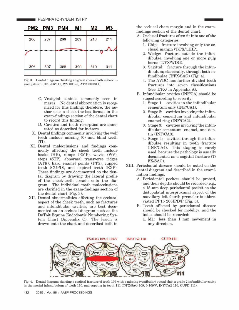
XI. Dental malocclusions and findings com-monly affecting the cheek teeth includehooks (HK), ramps (RMP), waves (WV),steps (STP), abnormal transverse ridges(ATR), hard enamel points (PTS), cuppedteeth (CUPD), and expired teeth (EXP).These findings are documented on the den-tal diagram by drawing the lateral profileof the cheek-tooth arcade onto the dia-gram. The individual tooth malocclusionsare clarified in the exam-findings section ofthe dental chart (Fig. 3).
XII. Dental abnormalities affecting the occlusalaspect of the cheek teeth, such as fracturesand infundibular cavities, are best docu-mented on an occlusal diagram such as theDuToit Equine Endodontic Numbering Sys-tem Chart (Appendix C). The lesion isdrawn onto the chart and described both in
the occlusal chart margin and in the exam-findings section of the dental chart.A. Occlusal fractures often fit into one of the
following categories:1. Chip: fracture involving only the oc-
clusal margin (T/FX/CHIP).2. Wedge: fracture outside the infun-
dibulae, involving one or more pulphorns (T/FX/WDG).
3. Sagittal: fracture through the infun-dibulum; classically, through both in-fundibulae (T/FX/SAG) (Fig. 4).
4. The AVDC has further divided toothfractures into seven classifications(See T/FX/ in Appendix A).
B. Infundibular cavities (INF/CA) should bestaged according to severity:1. Stage 1: cavities in the infundibular
cementum only (INF/CA1).2. Stage 2: cavities involving the infun-
dibular cementum and infundibularenamel ring (INF/CA2).
3. Stage 3: cavities involving the infun-dibular cementum, enamel, and den-tin (INF/CA3).
4. Stage 4: cavities through the infun-dibulae resulting in tooth fracture(INF/CA4). This staging is rarelyused, because the pathology is usuallydocumented as a sagittal fracture (T/FX/SAG).
XIII. Periodontal disease should be noted on thedental diagram and described in the exami-nation findings.A. Periodontal pockets should be probed,
and their depths should be recorded (e.g.,a 15-mm deep periodontal pocket on thedistopalatal interproximal aspect of themaxillary left fourth premolar is abbre-viated PP15 208IPD/P (Fig. 5).
B. Teeth affected by periodontal diseaseshould be checked for mobility, and theindex should be recorded:1. M1: less than 1 mm movement in
any direction.
Fig. 3. Dental diagram charting a typical cheek-tooth malocclu-sion pattern (HK 206/311, WV 308–9, ATR 210/311).
Fig. 4. Dental diagram charting a sagittal fracture of tooth 109 with a missing (vestibular) buccal slab, a grade 2 infundibular cavityin the mesial infundibulum of tooth 110, and cupping in tooth 111 (T/FX/SAG 109, 0 109/V, INF/CA2 110, CUPD 111).
432 2010 � Vol. 56 � AAEP PROCEEDINGS
RESPIRATORY/DENTISTRY

2. M2: less than 2 mm movement inany direction.
3. M3: movement of 3 mm or more inany direction.
C. After radiographic evaluation, the peri-odontal index stage can be classified:1. PD1: gingivitis only, and no bony at-
tachment loss.2. PD2: less than 25% attachment loss.3. PD3: 25–50% attachment loss.4. PD4: greater than 50% attachment
loss.XIV. Many dental and oral pathologies can only
be diagnosed with radiography. Radio-graphic findings should be recorded on thedental chart (preferably) or on a separateradiology report.
XV. After a complete oral examination and an-cillary diagnostics have been completed, atentative treatment plan and fee estimateare formulated. On approval, treatmentprocedures are performed and annotated onthe dental chart (Figs. 6–8).A. Occlusal adjustment reductions are re-
corded on the dental diagram by shadingin the portion of each tooth that has beenremoved and describing the procedure in
the treatment section of the chart. Theappropriate dental term for the adjust-ment of the contour of a tooth crown isodontoplasty (OD).
B. Floating (FLT), the reduction of sharpenamel points (PTS), is recorded in thetreatment section but is not usuallydrawn on the dental diagram.
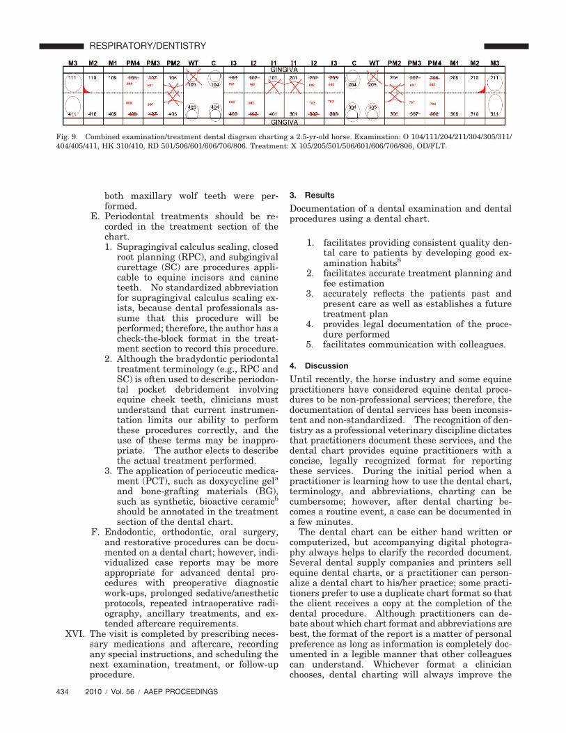
C. Simple extractions of retained deciduousand wolf teeth are common proceduresand are recorded by drawing an Xthrough the extracted tooth on the den-tal diagram and annotating the proce-dure in the treatment section (e.g.,simple extraction of the maxillary rightwolf tooth is abbreviated X105, and sim-ple extraction of the mandibular left sec-ond cheek-tooth cap is abbreviated X707)(Fig. 9).
D. Many commonly used nerve blocks haverecognized abbreviations. Practitionerswho perform infiltration nerve blocks be-fore extracting wolf teeth can abbreviatethe procedure as BUC/LIP/X 105/205 toindicate that a buccal local infiltrationanesthesia, local infiltration anesthesiaof the palate, and simple extraction of
Fig. 6. Dental diagram charting the correction of the DGL/4presented in Figure 1 (I/OD).
Fig. 5. Dental diagram charting a diastema and periodontalpocketing between the maxillary left third and fourth cheek teeth(DIA/PP15 208IPD).
Fig. 7. Dental diagram charting the correction of the HK103/203presented in Figure 2 (OD 103/203).
Fig. 8. Dental diagram charting the correction of the cheek-tooth malocclusion pattern presented in Figure 3 (OD 206/210/308/309/311).
AAEP PROCEEDINGS � Vol. 56 � 2010 433
RESPIRATORY/DENTISTRY
both maxillary wolf teeth were per-formed.
E. Periodontal treatments should be re-corded in the treatment section of thechart.1. Supragingival calculus scaling, closed
root planning (RPC), and subgingivalcurettage (SC) are procedures appli-cable to equine incisors and canineteeth. No standardized abbreviationfor supragingival calculus scaling ex-ists, because dental professionals as-sume that this procedure will beperformed; therefore, the author has acheck-the-block format in the treat-ment section to record this procedure.
2. Although the bradydontic periodontaltreatment terminology (e.g., RPC andSC) is often used to describe periodon-tal pocket debridement involvingequine cheek teeth, clinicians mustunderstand that current instrumen-tation limits our ability to performthese procedures correctly, and theuse of these terms may be inappro-priate. The author elects to describethe actual treatment performed.
3. The application of perioceutic medica-ment (PCT), such as doxycycline gela
and bone-grafting materials (BG),such as synthetic, bioactive ceramicb
should be annotated in the treatmentsection of the dental chart.
F. Endodontic, orthodontic, oral surgery,and restorative procedures can be docu-mented on a dental chart; however, indi-vidualized case reports may be moreappropriate for advanced dental pro-cedures with preoperative diagnosticwork-ups, prolonged sedative/anestheticprotocols, repeated intraoperative radi-ography, ancillary treatments, and ex-tended aftercare requirements.
XVI. The visit is completed by prescribing neces-sary medications and aftercare, recordingany special instructions, and scheduling thenext examination, treatment, or follow-upprocedure.
3. Results
Documentation of a dental examination and dentalprocedures using a dental chart.
1. facilitates providing consistent quality den-tal care to patients by developing good ex-amination habits8
2. facilitates accurate treatment planning andfee estimation
3. accurately reflects the patients past andpresent care as well as establishes a futuretreatment plan
4. provides legal documentation of the proce-dure performed
5. facilitates communication with colleagues.
4. Discussion
Until recently, the horse industry and some equinepractitioners have considered equine dental proce-dures to be non-professional services; therefore, thedocumentation of dental services has been inconsis-tent and non-standardized. The recognition of den-tistry as a professional veterinary discipline dictatesthat practitioners document these services, and thedental chart provides equine practitioners with aconcise, legally recognized format for reportingthese services. During the initial period when apractitioner is learning how to use the dental chart,terminology, and abbreviations, charting can becumbersome; however, after dental charting be-comes a routine event, a case can be documented ina few minutes.
The dental chart can be either hand written orcomputerized, but accompanying digital photogra-phy always helps to clarify the recorded document.Several dental supply companies and printers sellequine dental charts, or a practitioner can person-alize a dental chart to his/her practice; some practi-tioners prefer to use a duplicate chart format so thatthe client receives a copy at the completion of thedental procedure. Although practitioners can de-bate about which chart format and abbreviations arebest, the format of the report is a matter of personalpreference as long as information is completely doc-umented in a legible manner that other colleaguescan understand. Whichever format a clinicianchooses, dental charting will always improve the
Fig. 9. Combined examination/treatment dental diagram charting a 2.5-yr-old horse. Examination: O 104/111/204/211/304/305/311/404/405/411, HK 310/410, RD 501/506/601/606/706/806. Treatment: X 105/205/501/506/601/606/706/806, OD/FLT.
434 2010 � Vol. 56 � AAEP PROCEEDINGS
RESPIRATORY/DENTISTRY

quality of care that the practitioner provides to theequine patient.
Sample dental charts are available online atwww.aaep.org
Suggested Reading
Holmstrom SE, Frost P, Eisner ER. Dentalrecords. In: Veterinary dental techniques for thesmall animal practitioner, 2nd ed. Philadelphia,PA: W.B. Saunders Company, 1998;1–30.
Wiggs RB, Lobprise HB. Abbreviations, dentaland oral indices. In: Veterinary dentistry princi-ples and practice. Philadelphia, PA: Lippincott-Raven Publishers, 1997;677–690.
References and Footnotes1. Wiggs RB, Lobprise HB. Oral examination and diagnosis.
In: Veterinary dentistry principles and practice. Philadel-phia, PA: Lippincott-Raven Publishers, 1997;96.
2. Baker GJ, Easley J, eds. A glossary of equine dental ter-minology. In: Equine dentistry, 2nd ed. Edinburgh, Scot-land: Elsevier Saunders, 2005;329–346.
3. Wiggs RB, Lobprise HB. Glossary of terms. In: Veteri-nary dentistry principles and practice. Philadelphia, PA:Lippincott-Raven Publishers, 1997;628–676.
4. Bellows JE, et al. Clarification of veterinary dental nomen-clature. J Vet Dent 2005;22:276.
5. Bellows JE. Smile book IV, small animal dental anatomy,pathology, and charting. New York, NY: Pfizer AnimalHealth, 2004;4.
6. Scoggins GA. Legal considerations concerning patient med-ical records, in Proceedings. 51st Annual American Associ-ations of Equine Practitioners Convention 2005;516.
7. Bellows JE, et al. Vestibular is preferred to buccal or labial.Clarification of veterinary dental nomenclature. J Vet Dent2005;22:272.
8. Easley J. Dental and oral examination. In: Baker GJ,Easley J, eds. Equine dentistry, 2nd ed. Edinburgh, Scot-land: Elsevier Saunders, 2005;151.
aDoxyrobe Gel, Pfizer Animal Health, Exton, PA 19341.bConsil, Nutramax Laboratories, Edgewood, MD 21040.
AAEP PROCEEDINGS � Vol. 56 � 2010 435
RESPIRATORY/DENTISTRY

Appendix A: Equine Dental Abbreviations
Diagnostic AbbreviationsAbbreviations in RED are recognized by the American Veterinary Dental College (AVDC). Abbreviations in BLUE are recognized by the Academy of Veterinary Dentistry (AVD).
Tooth Aspects:V Vestibular (AVDC Preferred) B Buccal L Lingual P Palatal IPM or D Interproximal: Between teeth. Mesial or distal.
AB Abrasion (Tooth or soft tissue). Pathological wear.AT Attrition. Physiologic wear. ATR Abnormal Transverse Ridge.CA Caries
INF/CA Infundibular Cavity CAL Calculus. CV Ventral Curvature: Maxillary central incisors extend beyond the level of the maxillary intermediate and
corner incisors, “smile”. CD Dorsal Curvature: Mandibular central incisors extend beyond the level of the mandibular intermediate and
corner incisors, “frown”. CUPD Cupped: Crown worn past infundibulum. Still has crown above gingival margin. CWD Crowded Tooth. DGL Diagonal: Mandibular incisors longer on either the left side or right side. Defined with respect to
mandibular incisors longer on arcade number 300 or 400. DGL/4 400 arcade longer DGL/3 300 arcade longer
DIA Diastema between proximal incisor or proximal cheek teeth. E Enamel.
E/D Enamel Defect. EXP Expired: Attrition to gingival margin with crown connecting all roots. EXT Extrution. FB Foreign Body. FX Fracture. Tooth or Bone, Also see Tooth Fracture ( T/FX). HK Hook: Excess crown longer than wide. GH Gingival Hyperplasia/Hypertropy. GR Gingival Recession. LAC Laceration. LAC/B Laceration Cheek (Buccal)
LAC/L Laceration Lip.LAC/T Laceration Tongue.
M Mobile Tooth. M1 Mobile Tooth Index Stage 1. First distinguishable sign of movement. M2 Mobile Tooth Index Satge 2. <3 mm of movement in any direction. M3 Mobile Tooth Index Satge 3. >3 mm of movement in any direction.
MAL2 Class II malocclusion, overbite, brachygnathism, mandibular brachygnathism: Extension of maxillary teeth vertically beyond mandibular teeth.1 Defined by the term "distoclusion", where some or all of the mandibular teeth are distal in relationship to their maxillary counterparts.
MAL3 Class III malocclusion, underbite, prognathism, mandibular prognathism: Defined by the term "mesioclusion", where some or all of the mandibular teeth are mesial in their relationship to their maxillary counterparts.
MN Mandible. MX Maxilla. O Missing/Absent. OAF Oroantral Fistula. ONF Oronasal Fistula.
436 2010 � Vol. 56 � AAEP PROCEEDINGS
RESPIRATORY/DENTISTRY

OM Oral Mass. PDI Periodontal Disease Index
PD1 PD Stage 1: Gingivitis only. PD2 PD Stage 2: < 25% attachment loss. PD3 PD Stage 3: 25%- 50% attachment loss. PD4 PD Stage 4: >50% attachment loss. PE Pulp Exposure PP Periodontal Pocket PTS Sharp Enamel Points: Buccal cusps on maxillary cheek teeth and lingual cusps on mandibular cheek teeth
sharpened from wear (attrition). RAD Radiograph RD Retained Deciduous Tooth RMP Ramp: Excess tooth wider than long. RRT Retained Root Tip: Portion of root or tip retained. RTR Retained Tooth Root. STP Step: One tooth only with excess crown.T Tooth T/A Avulsed Tooth. T/FX Tooth Fracture
T/FX/EI Enamel Infraction. T/FX/EF Enamel Fracture. T/FX/UCF Uncomplicated Crown Fracture. T/FX/CCF Complicated Crown Fracture. T/FX/UCRF Uncomplicated Crown-Root Fracture. T/FX/CCRF Complicated Crown-Root Fracture. T/FX/RF Root Fracture.
T/FX/SAG Sagittal: Below gum line (subgingival) through infundibulum. T/FX/WDG Wedge: Outside infundibulum. T/FX/CHIP Chip: Occlusal margin only. Not fractured down to gingiva. T/I "Tooth impacted”, "Blind": Not completely erupted. Partially or fully covered by bone or soft tissue.
Commonly seen with wolf teeth. T/NE Near Pulp Exposure T/NV Non-vital Tooth T/PE Pulp Exposure T/V Vital Tooth TR Tooth Resorption TR1 TR Stage 1: Mild. Cementum +/- enamel. TR2 TR Stage 2: Moderate. Lesion extends into dentin, but not into pulp cavity. TR3 TR Stage 3: Deep. Lesion extends through dentin into pulp cavity. TR4 TR Stage 4: Extensive. Compromised integrity. TR4a Crown and Root Equally affected. TR4b Crown more severely affected than Root. TR4c Root more severely affected than Crown. TR5 Tooth remnants radiographically. Gingival covering complete. TO Tooth Overlong. WV Wave: More than one tooth with excess crown.
AAEP PROCEEDINGS � Vol. 56 � 2010 437
RESPIRATORY/DENTISTRY

Treatment AbbreviationsB Biopsy B/E Biopsy Excisional. B/I Biopsy Incisional. BG Bone Graft. DB Dentin Bonding FLT Float: Reduction of lingual and buccal enamel points. GV Gingivectomy/ Gingivoplasty. OC Orthodontic Consultation. OD Odontoplasty: Reduction of excessive crown of occlusal surface. PCT Perioceutic Therapy R Restoration
R/C Restoration with Composite R/I Restoration with Glass Ionomer.
SC Subgingival Curettage TP Treatment Planning VP Vital Pulpotomy X Extraction, simple
XS Extraction, Tooth sectioned XSS Surgical extraction
Nerve Blocks:IFA Inferior Alveolar NB (Mandibular Nerve). IFO Infraorbital NB. MAX Maxillary NB. MEN Mental NB.
BUC Buccal Local Nerve Block LIP Local Infiltration of Palate
438 2010 � Vol. 56 � AAEP PROCEEDINGS
RESPIRATORY/DENTISTRY

Appendix B: A Completed Dental Chart
AAEP PROCEEDINGS � Vol. 56 � 2010 439
RESPIRATORY/DENTISTRY

ANIMAL CARE HOSPITAL, 8565 Hwy 64, Somerville, TN 38068, (901) 466-9224 Date: Owner: Patient:
DuToit Endodontic Numbering System
Appendix C: DuToit Endodontic Numbering System
440 2010 � Vol. 56 � AAEP PROCEEDINGS
RESPIRATORY/DENTISTRY