Clinical manifestations and diagnosis and treatment of rhabdomyolysis

CASE REPORT Open Access
Acute recurrent rhabdomyolysis in aChinese boy associated with a novelcompound heterozygous LPIN1 variant: acase reportKe Tong1,2,3,4,5 and Geng-Sheng Yu1,2,3,4,5*
Abstract
Background: LPIN1-related acute recurrent rhabdomyolysis (RM), first reported in 2008, is an autosomal recessiveinherited metabolic disease. In recent years, LPIN1 gene variants have been identified as one of the main causes ofsevere RM in children in Western countries. The disease is extremely rare in China, and we report a case of acuterecurrent RM caused by a novel compound heterozygous LPIN1 variant.
Case presentation: A 15-year-old Chinese boy presented with myalgia after strenuous exercise, accompanied bytransient increases in serum creatine kinase and myoglobin and persistent hyperuricaemia and hyperbilirubinaemia.Genetic analysis using high-throughput genomic sequencing and Sanger sequencing revealed that there was acompound heterozygous variant in the LPIN1 gene of the proband: the paternal c.2047A > G(p.I683V) was anunreported missense variant, and the maternal c.2107_2108 insAGG(p.Q703delin sQE) was an unreported in-framevariant.
Conclusions: In children with RM, LPIN1 variants should always be considered in the differential diagnosis. Theclinical features of our case are atypical, which highlights the importance of an accurate diagnosis by genetictesting. If detected early, the condition may be controlled, and the prognosis may be improved.
Keywords: Acute recurrent rhabdomyolysis, LPIN1 deficiency, LPIN1 gene, Novel variants, Child
BackgroundRhabdomyolysis (RM) is a clinical entity characterizedby the destruction of skeletal muscle with the resultantrelease of intracellular content into the bloodstream thatleads to systemic complications [1]. The classic presenta-tion of this condition is myalgia, transient muscle weak-ness, pigmenturia, and a marked elevation of serum
creatine kinase (CK) five to ten times above the upperlimit of normal serum levels [2]. The incidence of RM islow, and recurrence is even rarer. As far as we know, therecurrent RM in children is predominantly due to gen-etic metabolic muscle diseases [3]. Michot et al. [4] de-termined that variants in the LPIN1 gene encodingLPIN1 are the second most common cause of earlychildhood recurrence and severe RM, second only tomitochondrial fatty acid ß-oxidation defects (FAOs).LPIN1 is an intracellular protein that controls metabol-ism by acting at multiple regulatory levels. This solubleprotein acts at the endoplasmic reticulum to dephos-phorylate phosphatidic acid (PA) to form diacylglycerol
© The Author(s). 2021 Open Access This article is licensed under a Creative Commons Attribution 4.0 International License,which permits use, sharing, adaptation, distribution and reproduction in any medium or format, as long as you giveappropriate credit to the original author(s) and the source, provide a link to the Creative Commons licence, and indicate ifchanges were made. The images or other third party material in this article are included in the article's Creative Commonslicence, unless indicated otherwise in a credit line to the material. If material is not included in the article's Creative Commonslicence and your intended use is not permitted by statutory regulation or exceeds the permitted use, you will need to obtainpermission directly from the copyright holder. To view a copy of this licence, visit http://creativecommons.org/licenses/by/4.0/.The Creative Commons Public Domain Dedication waiver (http://creativecommons.org/publicdomain/zero/1.0/) applies to thedata made available in this article, unless otherwise stated in a credit line to the data.
* Correspondence: [email protected] of Cardiovascular Disease, Children’s Hospital of ChongqingMedical University, 136 Zhongshan 2nd Road, Yuzhong District, Chongqing400014, China2Ministry of Education Key Laboratory of Child Development and Disorders,136 Zhongshan 2nd Road, Yuzhong District, Chongqing 400014, ChinaFull list of author information is available at the end of the article
Tong and Yu BMC Neurology (2021) 21:42 https://doi.org/10.1186/s12883-021-02050-w
(DAG), penultimate steps in Kennedy pathway of triacyl-glycerol (TAG) synthesis. LPIN1 also acts in the nucleusto directly interact with DNA-bound transcription fac-tors to regulate gene expression [5]. LPIN1-related acuterecurrent RM was first reported by Zeharia et al. in2008. It is an autosomal recessive inherited metabolicdisease that is most commonly triggered by fever, exer-cise, fasting, and anaesthesia. Severe RM can cause acutekidney failure and may lead to death [6]. To the best ofour knowledge, we report a case of a Chinese boy withacute recurrent RM caused by a novel compound het-erozygous variant in LPIN1 whose gene variant pointsare different from previous cases.
Case presentationA 14-year-old boy went to the hospital for a healthexamination, which detected a slight increase in bilirubinthat was followed up regularly. Then, the boy sufferedfrom an intermittent fever, mainly a moderate-to-highfever, lasting for 1 week. After a health examination, wefound that the boy’s levels of CK, myoglobin, bilirubinand uric acid were all significantly increased. Serum bio-chemical test results are shown in Table 1. Physicalexamination showed that his body mass index was 22.73kg/m2 (reference: 17.1–23 kg/m2), and muscular toneand power in both lower extremities were normal. Wesuspected this was as characteristic of RM. Then, theboy was advised to take oral sodium bicarbonate toalkalize urine, fructose and vitamin C to improve metab-olism and promote cell repair until another health
examination 1 month later. We found that the boy’slevels of CK, myoglobin, bilirubin and uric acid CK werehigher than the previous levels. The results of theseserum biochemical test are also shown in Table 1. Thiswas the highest CK level and myoglobin level we had de-tected so far. His vital signs were stable, and although hehad no discomfort at rest, discomfort manifested as calfmyalgia after strenuous exercise. Immune function, ab-dominal and cardiac ultrasound, dynamic electrocardio-gram, electromyography, and calf muscle MRI were allunremarkable. At the age of 12, he joined the school’sathletics team, and at the age of 13, he joined theschool’s tennis team. Appropriate exercise can be toler-ated by the boy. When experiencing high-intensity exer-cise, the boy had myalgia throughout the body. To copewith the physical education test, the boy performedhigh-intensity long jump training every day. However,after 5–6 long jumps, the boy began to experience fa-tigue, low back pain, calf pain and dark urine, while thiskind of signs did not manifest among his companions.After he sat down and rested for a while, his symptomswere relieved. The parents claimed that when their childwas 13 years old, he suffered from leg pain during thenight-time. At that time, there were protein and redblood cells in the urinalysis, and the urine was light red.Unfortunately, no further examination was performed atthat time. The boy had hyperbilirubinaemia and hyper-uricaemia since the age of 14 because of high-intensitylong jump training, while CK and myoglobin levels werenormal in between episodes. Sometimes, there was asmall amount of red blood cells and protein appearingin the urinalysis, and a slight increase in serum creatin-ine. Strangely, the boy underwent a bronchoscopy undergeneral anesthesia at the age of 12 due to mycoplasmapneumonia, prior to diagnosis and this did not triggeran episode of RM. He had no chronic diseases and wasnot on any long-term oral medications. There was nofamily history of RM or any other musculoskeletal dis-ease. His parents were both Chinese and non-consanguineous. His father was obese and had fatty liver,hyperbilirubinaemia and hyperuricaemia, but he hadnever experienced myalgia. His mother suffered fromacute nephritis when she was young. Considering therisk of anaesthesia and invasive nature of the procedure,the parents of the boy did not give consent for themuscle biopsy.Peripheral blood samples from the proband and the
parents were collected and high-throughput genomicsequencing (next-generation sequencing) of 233 genesrelated to neuromuscular diseases (Table 2) was per-formed by Beijing Mygenostics Co. Ltd. Informedconsent was obtained from the proband and hisguardian for gene detection. The results showed thatthe LPIN1 gene variants in this proband included the
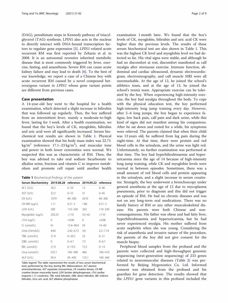
Table 1 Biochemical findings of the patient
Serum Biochemistry 2019.08.28 reference 2019.09.22 reference
ALT (U/L) 26.3 0–50 13 0–40
AST (U/L) 32.3 0–55 31 0–45
CK (U/L) 1079 40–300 2610 40–300
CK-MB (ug/L) 1.11 0.21–5 1.86 0.21–5
LDH (U/L) 200 110–330 263 110–330
Myoglobin (ug/L) 203.23 <110 551.42 <110
cTnI (ug/L) 0 <0.06 0 <0.06
Cr (umol/L) 61 15.4–90.4 59 14–60
Urea (mmol/L) 4.64 2.42–6.72 3.6 2.2–7.14
TBIL (umol/L) 21.9 0–20.5 23 0–21
DBIL (umol/L) 0 0–6.7 7.5 0–6.7
IBIL (umol/L) 21.9 0–19.5 15.5 0–14
Urca (umol/L) 532.1 140–390 466 100–410
ALP (U/L) 95.4 95–405 125.1 100–360
Table legend: The table represented the results of two serum biochemicaltests performed by the boy during RM. Abbreviations: ALT alanineaminotransferase, AST aspartate transaminas, CK creatine kinase, CK-MBcreatine kinase myocardia band, LDH lactate dehydrogenase, cTnI cardiactroponin I, Cr creatinine, TBIL total bilirubin, DBIL direct bilirubin, IBIL indirectbilirubin, Urca uric acid, ALP alkaline phosphatase
Tong and Yu BMC Neurology (2021) 21:42 Page 2 of 9

missense variant c.2047A > G(p.I683V) and in-framevariant c.2107_2108 insAGG(p.Q703delin sQE). Thec.2047A > G variant of the 2047th nucleotide in thecoding region from adenine to guanine results in the683rd amino acid changing from isoleucine to valine;c.2107_2108 insAGG changed by way of indels, andthe 703rd amino acid changed from glutamine (Q) toglutamine (Q) and glutamate (E), which caused theprotein length to change (Fig. 1). Sanger sequencingconfirmed that the proband had compound heterozy-gous LPIN1 variants; the former was from his father,while the latter was from his mother (Fig. 2). Accord-ing to the standards and guidelines of the AmericanSociety for Medical Genetics and Genomics in 2015[7], both variants were classified as variants of uncer-tain significance. The frequency of the missense vari-ant and the in-frame variant detected in our case in
the normal population database was 0.00002, indicat-ing that both were low-frequency variants (criteria:PM2), and each bioinformatics protein function pre-diction software predicted the missense variant to beharmful (criteria: PP3) and the other to be unknown.The in-frame variant resulted in a change in proteinlength caused by a small insertion in the nonrepeti-tive sequence region (criteria: PM4).Although the result of genetic testing was uncertain
regarding pathogenicity, we communicated the possibil-ity of LPIN1-related RM to the parents and recom-mended that the boy restrict strenuous exercise, haveproper rest, drink adequate water, supplement enoughenergy and take oral sodium bicarbonate (0.5 g/time, 3times/day) for urine alkalization when myalgia and darkurine occur. Under our guidance, his daily life and stud-ies have not been affected.
Table 2 Summary of all genes covered by the panel
ABHD5 ACAD8 ACADL ACADM ACADS ACADVL ACTA1 ACVR1
AGK AGL AGRN ALDOA ALG13 ALG14 ALG2 ANO5
ATP2A1 ATP5F1A B3GALNT2 B4GAT1 BAG3 BIN1 CACNA1A CACNA1S
CAPN3 CAV3 CAVIN1 CCDC78 CFL2 CHAT CHKB CHRNA1
CHRNB1 CHRND CHRNE CHRNG CHST14 CLCN1 CNTN1 COL12A1
COL13A1 COL6A1 COL6A2 COL6A3 COLQ COQ2 COQ4 COQ6
COQ7 COQ8A COQ9 CPT1A CPT2 CRYAB DAG1 DES
DGUOK DMD DNA2 DNAJB6 DNM2 DOK7 DOLK DPAGT1
DPM1 DPM2 DYSF ECEL1 ELP1 EMD ENO3 ETFA
ETFB ETFDH ETHE1 FBXL4 FHL1 FKBP14 FKRP FKTN
FLNA FLNC G6PC GAA GBE1 GFPT1 GMPPB GNE
GYG1 GYS1 HACD1 HADH HADHA HADHB HNRNPA1 HNRNPA2B1
HNRNPDL HSPG2 IGHMBP2 ISCU ISPD ITGA7 ITGA9 KBTBD13
KCNA1 KCNE3 KCNE4 KCNH2 KCNJ16 KCNJ2 KCNQ1 KIF21A
KLHL40 KLHL41 KLHL9 LAMA2 LAMB2 LAMP2 LARGE1 LDB3
LDHA LMNA LMOD3 LPIN1 LRP4 MAMLD1 MATR3 MEGF10
MGME1 MLYCD MPV17 MSTN MTM1 MTMR14 MTTP MUSK
MYBPC1 MYBPC3 MYF6 MYH14 MYH2 MYH3 MYH7 MYH8
MYLK2 MYO9A MYOT NDUFB3 NEB OPA1 PABPN1 PDSS1
PDSS2 PFKM PGAM2 PGK1 PGM1 PHKA1 PHOX2A PIEZO2
PLEC PLOD1 PLOD2 PLOD3 PMM2 PNPLA2 POLG POLG2
POMGNT1 POMGNT2 POMK POMT1 POMT2 PREPL PRKAG2 PUS1
PYGM RAPSN RBCK1 RRM2B RXYLT1 RYR1 SCN4A SCN5A
SELENON SGCA SGCB SGCD SGCE SGCG SIL1 SLC16A2
SLC22A5 SLC25A1 SLC25A20 SLC25A4 SLC37A4 SLC3A1 SLC5A7 SNAP25
SPAST SPEG STIM1 SUCLA2 SUCLG1 SYNE1 SYNE2 SYT2
TCAP TIA1 TK2 TMEM43 TNNI2 TNNT1 TNNT3 TNPO3
TOR1A TOR1AIP1 TPM2 TPM3 TRAPPC11 TRIM32 TTN TTR
TUBB3 TWNK TYMP VCP VPS13A XK YARS2 ZBTB42
ZC4H2
Tong and Yu BMC Neurology (2021) 21:42 Page 3 of 9

Discussion and conclusionsLPIN1-related acute recurrent RM is a life-threateningdisease that is characterized by the age of the first onsetbeing younger than 6 years old and the peak CK level atthe onset exceeding 10,000 IU/L [8]. We discovered anovel compound heterozygous variant in the LPIN1 gene(p.I683V and p.Q703delin sQE) in a Chinese boy withacute recurrent RM, and his clinical characteristics arequite different from those reported in previous studies.Our proband is a 15-year-old adolescent with a later ageof onset and only one episode of mild RM. His serumCK and myoglobin were normal in between episodes,but there was long-term hyperuricaemia and hyperbiliru-binaemia. To the best of our knowledge, we report thethird case of LPIN1-related acute recurrent RM in a pa-tient of Chinese ethnicity, while the case with hyperuri-caemia and hyperbilirubinaemia is shared for the firsttime (MIM#268200).We searched the PubMed database using “acute recur-
rent rhabdomyolysis” and “LPIN1” as keywords. Therewere 57 patients (including our case) with complete clin-ical data who were genetically diagnosed with acute recur-rent RM caused by the LPIN1 gene variant [4, 6, 9–22].These patients were reported from 18 different countries,of which only 3 cases were of Chinese ethnicity. The find-ings are presented in Table 3. We found that LPIN1-re-lated acute recurrent RM was more common in malesthan in females (28/57 49.1% vs. 24/57 42.1%). The firstonset was before the age of 6 for the most part, and thenumber of episodes was usually 3 or fewer. The peak CK
level was often greater than 10*104U/L. In 40 cases, poten-tial trigger factors had been reported, among which feverwas the most common (26/40 65.0%) ([4, 6, 11, 13, 15, 16,20, 22], our case), followed by infection (12/40 30.0%) [11,13–16, 19–21], fasting (7/40 17.5%) [4, 16], anaesthesia (5/40 12.5%) [4, 14], and exercise or physical activity (4/4010.0%) [11, 14, 16, 17]. Only 9 cases (9/57 15.8%) [6, 9, 11,13, 17–19, 22] underwent muscle biopsy, which is an inva-sive procedure that seems to be unpopular. There were 8deaths (8/57 14.0%) [4, 9, 13], suggesting a high mortalityrate of this disease. We also summarized the variantpoints of 56 cases whose genetic results were reported andfinally found 38 different variant points [4, 6, 9–23], in-cluding missense variants, frameshift variants, in-framevariants, nonsense variants, and exon deletions. The vari-ant points are summarized in Table 4. There were 32cases with homozygous variants (32/56 57.1%) and 24cases with compound heterozygous variants (24/5642.9%). There were 6 variant points in Chinese patients,all of which were compound heterozygous variants;c.2047A >G(p.I683V), c.2107_2108 insAGG(p.Q703delinsQE), c.2428C > T(p.Arg810Cys) and c.1949_1967dupGTGTCACCACGCAGTACCA (p.Gly657-CysfsX12) were only detected in individuals of Chineseethnicity ([15, 16], our case), and these might be Chineseethnicity-specific variants.The LPIN protein family is composed of three mem-
bers, LPIN1, LPIN2, and LPIN3, encoded by the LPIN1gene, LPIN2 gene, and LPIN3 gene, each of which is 100kDa in size and has 44–48% amino acid similarity. They
Fig. 1 Predicted three-dimensional structure of protein at two variant points of LPIN1 gene. a: Wild-type protein of the 683rd position isisoleucine; b: c.2047A > G variant protein becomes valine; c: Three-dimensional structure of wild-type protein; d: c.2107_2108 insAGG variantprotein structure. WT wild type
Tong and Yu BMC Neurology (2021) 21:42 Page 4 of 9

are Mg2+-dependent phosphatidic acid phosphatase(PAP) enzymes that catalyse a key reaction in glyceroli-pid biosynthesis [24]. Only LPIN1 is expressed inskeletal muscle and plays a key role in human musclefunction [25]. The LPIN1 gene is located on chromo-some 2p25.1 with two highly conserved domains: the N-terminal LPIN (N-LIP) region spans the first 108residues, and the C-terminal LPIN (C-LIP) domainconsists of 624–830 amino acids. The C-LIP domaincontains two key domains: the PAP catalytic motif Asp-Xaa-Asp-Xaa-Thr (DXDXT (689–693)) and the tran-scriptional coactivator motif Leu-Xaa-Xaa-Ile-Leu(LXXIL (678–682)) [16, 26]. They both play an import-ant role in the activity of PAP. We discovered a novelmissense variant c.2047A > G(p.I683V) and a novel in-frame variant c.2107_2108 insAGG(p.Q703delin sQE).The onset of our case did not occur until adolescence,and the cases of delayed disease onset have been re-ported before [17–19, 22]. The peak CK level was only2610 U/L, which was significantly different from previ-ously reported cases. The reason for the delayed diseaseonset and mild phenotype may be due to the existenceof novel missense variants and in-frame variants ratherthan loss-of-function truncating variants. Schweitzeret al. [11] found that single amino acid substitutions inhuman LPIN1 disrupt PAP activity but may not alwaysaffect nuclear transcriptional regulator function, and
truncating variants (such as frameshift, nonsense and de-letion) will cause loss of protein function. Therefore, thenovel variants in the C-LIP domain we discovered mayhave a much smaller impact than the truncating variantswith direct loss of function. In addition, LPIN2 andLPIN3 share a similar structure with LPIN1, and wespeculate their presence can compensate for the lack ofLPIN1. However, the relationship between expressionpatterns and the compensatory roles of LPIN2 andLPIN3 in skeletal muscle with LPIN1 deficiency is un-clear, and further study is necessary. Similarly, environ-mental exposure may also have a regulatory effect onthe occurrence of RM [18]. However, a clear limitationof the present report is that definitive pathogenicity ofthe missense variant and the in-frame variant could notbe established due to lack of experimental evidence atthe protein level. Future studies are therefore warrantedto clarify the significance of c.2047A > G and c.2107_2108 insAGG in LPIN1-related RM.LPIN1 has transcriptional coregulatory activity, which,
through association with peroxisome proliferator-activated receptor alpha (PPARα) and peroxisomeproliferator-activated receptor gamma coactivator 1-alpha (PGC-1α), regulates lipid metabolism and themitochondrial respiratory chain (mRC) [27]. Despite thewell-known physiological roles of LPIN1 in lipid biosyn-thesis and transcriptional regulation in adipocytes and
Fig. 2 Direct sequencing showing two alleles of the proband and his parents, respectively. The proband is a compound heterozygote ofc.2047A > G and p.Q703delin sQE. The first variant is from the father, and the latter is from the mother. The arrow shows the variant point. Pproband, F father, M mother, WT wild type
Tong and Yu BMC Neurology (2021) 21:42 Page 5 of 9

Table
3Summaryof
clinicalinform
ationof
57patientswith
LPIN1-relatedacuterecurren
trhabdo
myolysis
Ethn
icorigin
Num
ber
ofcases
Sex
Ageat
onset
Num
ber
ofep
isod
esCKpea
kUI/L
Num
ber
ofmusclebiopsies
Num
ber
ofdea
ths
MF
NM
<6Y
6~
16Y
>16
YNM
≤3
>3
< 1*10
41*10
4~10
*104
>10
*104
NM
France
[4,6,9,10]
199
82
101
08
109
05
140
23
Jordan
[13]
85
30
71
00
71
05
30
11
Austria[14]
52
30
41
00
41
00
50
00
China
([15,16],our
case)
33
00
21
00
30
10
20
00
UnitedKing
dom
[4,17,18]
31
20
11
10
21
00
30
21
Belgium
[4]
32
10
30
00
30
02
10
02
North
Africa
[4]
21
10
20
00
20
01
10
00
Germany[4]
20
20
20
00
11
00
20
00
Mauritania[4,6]
21
01
20
00
20
00
20
00
America[11,12]
21
10
20
00
20
00
20
10
Arab[6]
11
00
10
00
10
00
10
10
Egypt[4]
11
00
10
00
10
01
00
01
Ireland
[19]
10
10
10
00
00
00
10
10
Palestine[7]
10
01
10
00
10
01
00
00
Canada[20]
11
00
10
00
01
00
10
00
Portug
al[21]
10
01
10
00
10
00
01
00
SriLanka
[22]
10
10
10
00
01
01
00
10
Italy[18]
10
10
00
10
10
00
10
00
Total(%)
5728/
57(49.1)
24/
57(42.1)
5/ 57(8.8)
42/
57(73.7)
5/ 57(8.8)
2/ 57(3.5)
8/ 57(14.0)
41/
57(71.9)
15/
57(26.3)
1/ 57(1.8)
16/57(28.1)
39/
57(68.4)
1/ 57(1.8)
9/57(15.8)
8/57(14.0)
Mmale,
Ffemale,
CKcreatin
ekina
se,Y
year,N
Mno
tmen
tione
d
Tong and Yu BMC Neurology (2021) 21:42 Page 6 of 9

the liver, the pathophysiological mechanisms leading tomuscle dissolution remain to be elucidated. Fever andintense effort lead to high temperatures and high circu-lating levels of proinflammatory mediators such as cyto-kines and chemokines [24]. In addition, theproinflammatory stress induced by the combination oftumor necrosis factor alpha and Interleukin-1 beta(TNFα+IL-1β) intensifies the accumulation of lipiddroplets (LDs), decreases the activity of carnitine palmi-toyltransferase 1 (CPT1) and increases the content of tri-acylglycerol, highlighting the crucial role ofinflammation in the pathogenesis of LPIN1 deficiency[27]. Zhang et al. [5] found that in the case of LPIN1 de-ficiency, a blockade in PAP activity leads to reducedDAG levels and impaired activation of the protein kinaseD (PKD)-Vps34 signalling cascade in autophagy clear-ance, preventing normal maturation of autolysosomes.These defects may lead to the RM observed in LPIN1deficiency. Interestingly, our case showed normal musclefunction between episodes of RM, which indicated that
mitochondrial function was sufficient to meet the energyrequirements of daily activities. Rashid et al. [28] re-vealed that LPIN1 deficiency caused severe sarcoplasmicreticulum (SR) stress, leading to the activation of lipo-genic sterol regulatory element binding protein 1c(SREBP1c)/SREBP2 factors, the accumulation of fibro-blast growth factor 21 (Fgf21) cytokines, and alterationsin SR mitochondrial morphology, which were contribut-ing factors to the myopathy associated with LPIN1deficiency.Pelosi et al. [29] found that the adipose tissue of
patients with LPIN1 gene variants showed a signifi-cant decrease in LPIN1 levels and PAP activity, butthe adipose tissue appeared to develop normally andhad normal lipid reserves. The levels of metaboliccomponents such as triglycerides and cholesterolremained unremarkable in the serum in our case,consistent with the results of previous studies. Ourpatient underwent muscle-related examinations, suchas electromyography and calf muscle MRI, and the
Table 4 Summary of gene variant points
Variant Amino AcidChange
Ethnic origin Variant Amino AcidChange
Ethnic origin
c.2047A > G p.I683V China[our case] c.944C > G p.Ser315X France [9],France-Asia [4]
c.2107_2108 insAGG p.Q703delinsQE China[our case] c.643G > T p.E215X Arab [6]
c.2295-866_2410-30del
p.Glu766_Ser838del
France [9], Belgium [4],United Kingdom [4, 18],Germany [4], Ireland [19],Austria [14], Italy [18]
c.2398C > T p.R800X France [6]
c.1162C > T p.Arg388X France [9], France-Asia [4],North Africa [4], Palestine [6],China [16], Jordan [13]
c.297 + 2 T > C NM Mauritania、France [6]
NM p.Asn417LysfsX22 France [10] c.1949_1967dupGTGTCACCACGCAGTACCA
p.Gly657CysfsX12 China [15]
NM p.Cys30LeufsX3 France [10] c.2410G > C p.Asp804His China [15]、UnitedKingdom [17]
c.192 + 2 T > C p.Cys30LeufsX3 Mauritania [4] C.942C > A p.Cys314X Ireland [19]
c.2401C > T p.Arg801X France [4] c.1904 T > C p.Leu635Pro America [11]
c.1441 + 2 T > C p.Asn417LysfsX22 France [4] NM E766-S838_del America [11]
c.377_380dup p.Met128GlnfsX45 France [4] c.2428C > T p.Arg810Cys China [16]
c.2513 + 1G > A p.Asp804ValfsX6 Egypt [4] c.1255_1256dupGA p.Asp419fs Austria [14]
c.921delT p.Gln308ArgfsX36 United Kingdom [4] c.394_409del p.Asp132fs Austria [14]
c.946_952del p.Asp316LeufsX26 Germany [4] c.2174G > A p.Arg725His Jordan [13]
c.1259delC p.Pro420LeufsX39 France [4] c.2395G > C p.Gly799Arg Jordan [13]
c.2513 + 1G > A p.Asp804ValfsX6 France [4] c.1684G > T p.Glu562X Sri Lanka [22]
c.2253_2254del p.Leu752AlafsX17 Germany [4] NM p.Arg61X NM [23]
c.57C > A p.Tyr19X Germany [4] NM p.Asp170X NM [23]
c.2142-2A > G NM Portugal [21] NM p.Ala227GlyfsX2 NM [23]
c.1381delC p.Leu461SerfsX47 Canada [20] NM p.Glu477X NM [23]
NM not mentioned
Tong and Yu BMC Neurology (2021) 21:42 Page 7 of 9
results were not significant, consistent with the reportof Minton et al. [17], which showed that the skeletalmuscle morphology of patients with LPIN1 deficiencycan be insignificant. It is worth noting that our pa-tient had persistent hyperuricaemia since the first epi-sode of RM. We believe that a large number ofdamaged muscle cells may release endogenous pu-rines, which are metabolized into uric acid, therebyincreasing uric acid production and leading to hyper-uricaemia. Too much uric acid may be further con-centrated and deposited in the renal tubules, whichmay aggravate the obstruction caused by myoglobinand even cause renal dysfunction [30]. We speculatethat hyperuricaemia may increase the risk of renalfailure in RM. If detected early, the condition may becontrolled, and the prognosis may be improved. Inaddition, our patient also had a continuous mild in-crease in bilirubin. Serum bilirubin has antioxidantand anti-inflammatory effects, which can inhibit kid-ney damage to a certain extent [31]. High serum totalbilirubin levels are independently associated with ahigh glomerular filtration rate, and those with highserum total bilirubin levels have a lower risk of renaldamage [32]. Because of normal kidney function inour patient, we speculate that the mild increase inbilirubin is protective to delay the appearance of kid-ney damage, although it is difficult to know in ourpatient whether it is associated with the onset ofLPIN1-associated RM. Moreover, it should be notedthat our case is easily confused with phosphoglyceratekinase (PGK) deficiency, which is a rare cause of con-genital hemolytic anemia accompanied by hyperbiliru-binaemia, muscle weakness, myalgia andmyoglobinuria [33]. Although our case has similarclinical features with PGK deficiency, it has beenruled out by genetic testing.Our patient has been taking supportive treatments at
home, such as rest, rehydration, energy supplementation,a low-purine diet, and supplements to alkalize urine, andhe is living well at present. Symptomatic treatment ofRM with LPIN1 deficiency mainly includes early detec-tion, active hydration, high-energy carbohydrate intake,and monitoring of hyperkalaemia and arrhythmia [19].To avoid long-term complications, it is essential to pre-vent fever, restrict strenuous exercise, and seek medicalattention in cases of myalgia. Moreover, Pichler et al.[14] found that five patients with LPIN1-related RMwere advised to use high-concentration glucose solutionsearly to provide calories, which reduced the duration ofRM from the typical 7–10 days to 5 days. Meijer et al.[20] successfully used dexamethasone, which can stimu-late LPIN1 expression and PAP activity as an inducer ofPGC-1α, to treat a patient diagnosed with LPIN1 vari-ants. However, whether high-concentration glucose
solutions and dexamethasone can effectively treat RMremains to be further explored in the future.In summary, we diagnosed a Chinese boy with
acute recurrent RM and discovered the novel com-pound heterozygous variants c.2047A > G(p.I683V)and c.2107_2108 insAGG (p.Q703delin sQE). Accord-ing to a systematic literature review, LPIN1-relatedacute recurrent RM is more common in males. Thefirst onset is before the age of 6, and the number ofepisodes is usually within 3. The peak CK level isoften greater than 10*104U/L. Fever, infection andfasting are the most common trigger events, and genevariants are common as homozygous variants, whilethe Chinese cases being reported all involve com-pound heterozygous variants. As the mortality ratedue to LPIN1 deficiency is as high as 14.0% and somepatients have atypical symptoms, as in our case, ithighlights that paediatric clinicians in the Depart-ments of Cardiology, Neurology, Nephrology, and Im-munology should promptly detect the disease andtreat it symptomatically. Genetic testing, a method toclearly diagnose and analyse the source of pathogenicgenes, is increasingly recognized. Our research is ofreference value for an in-depth understanding of theclinical and laboratory characteristics of LPIN1-relatedacute recurrent RM and for improving the early diag-nosis and clinical management of patients with thisdisease.
AbbreviationsRM: Rhabdomyolysis; CK: Creatine kinase; DAG: Diacylglycerol;TAG: Triacylglycerol; PA: Phosphatidic acid; HGMD: The Human GeneMutation Database; PAP: Phosphatidic acid phosphatase; N-LIP: The N-terminal LPIN; C-LIP: The C-terminal LPIN; PKD: The protein kinase D;SR: Sarcoplasmic reticulum; PGC-1α: Peroxisome proliferator activatedreceptor gamma coactivator 1-alpha; PGK: Phosphoglycerate kinase;TNFα: Tumor necrosis factor alpha; IL-1β: Interleukin-1 beta
AcknowledgmentsWe thank the patient and his family for participating in this case study.
Authors’ contributionsKT designed the study, drafted the manuscript, and carried out themolecular genetic studies and analyzed the molecular and the clinical data,GSY critically revised the manuscript for important intellectual content. Bothauthors read and approved the final manuscript.
FundingThis work was supported by the Key Project of Medical Research Program ofChongqing Health Bureau (2012-1-047).
Availability of data and materialsThe data used to support the findings of this study are available from thecorresponding author upon request, without breaching participantconfidentiality.
Ethics approval and consent to participateThis study was approved by Ethics Committee of Children’s Hospital ofChongqing Medical University, and written consent was obtained from thepatient’s parents. Our study is in accordance with the Declaration of Helsinkiand the CARE guidelines/methodology. Both authors agree to the PublishingStatements of BMC Neurology.
Tong and Yu BMC Neurology (2021) 21:42 Page 8 of 9

Consent for publicationWritten consent for publication was obtained from each of the familymembers. Written consent was obtained from the patient’s parents topublish his clinical and genetic data.
Competing interestsNeither of the authors has any competing interests or conflicts of interest.
Author details1Department of Cardiovascular Disease, Children’s Hospital of ChongqingMedical University, 136 Zhongshan 2nd Road, Yuzhong District, Chongqing400014, China. 2Ministry of Education Key Laboratory of Child Developmentand Disorders, 136 Zhongshan 2nd Road, Yuzhong District, Chongqing400014, China. 3National Clinical Research Center for Child Health andDisorders (Chongqing), 136 Zhongshan 2nd Road, Yuzhong District,Chongqing 400014, China. 4China International Science and TechnologyCooperation Base of Child Development and Critical Disorders, 136Zhongshan 2nd Road, Yuzhong District, Chongqing 400014, China.5Chongqing Key Laboratory of Pediatrics, 136 Zhongshan 2nd Road,Yuzhong District, Chongqing 400014, China.
Received: 21 August 2020 Accepted: 7 January 2021
References1. Chavez LO, Leon M, Einav S, et al. Beyond muscle destruction: a systematic
review of rhabdomyolysis for clinical practice. Crit Care. 2016;20:135. https://doi.org/10.1186/s13054-016-1314-5.
2. Shao L, Liu C, Xu L, et al. Repeated and progressive rhabdomyolysis due toa novel carnitine palmitoyltransferase II gene variant in an adult male: Acase report. Medicine (Baltimore). 2019;98:e18143. https://doi.org/10.1097/MD.0000000000018143.
3. Alaygut D, Torun Bayram M, Kasap B, et al. Rhabdomyolysis with differentetiologies in childhood. World J Clin Pediatr. 2017;6:161–8. https://doi.org/10.5409/wjcp.v6.i4.161.
4. Michot C, Hubert L, Brivet M, et al. LPIN1 gene mutations: a major cause ofsevere rhabdomyolysis in early childhood. Hum Mutat. 2010;31:E1564–73.https://doi.org/10.1002/humu.21282.
5. Zhang P, Verity MA, Reue K. Lipin-1 regulates autophagy clearance andintersects with statin drug effects in skeletal muscle. Cell Metab. 2014;20:267–79. https://doi.org/10.1016/j.cmet.2014.05.003.
6. Zeharia A, Shaag A, Houtkooper RH, et al. Mutations in LPIN1 causerecurrent acute myoglobinuria in childhood. Am J Hum Genet. 2008;83:489–94. https://doi.org/10.1016/j.ajhg.2008.09.002.
7. Richards S, Aziz N, Bale S, et al. Standards and guidelines for theinterpretation of sequence variants: a joint consensus recommendation ofthe American College of Medical Genetics and Genomics and theAssociation for Molecular Pathology. Genet Med. 2015;17:405–24. https://doi.org/10.1038/gim.2015.30.
8. Hamel Y, Mamoune A, Mauvais FX, et al. Acute rhabdomyolysis andinflammation. J Inherit Metab Dis. 2015;38:621–8. https://doi.org/10.1007/s10545-015-9827-7.
9. Bergounioux J, Brassier A, Rambaud C, et al. Fatal rhabdomyolysis in 2children with LPIN1 mutations. J Pediatr. 2012;160:1052–4. https://doi.org/10.1016/j.jpeds.2012.02.033.
10. Legendre A, Khraiche D, Ou P, et al. Cardiac function and exerciseadaptation in 8 children with LPIN1 mutations. Mol Genet Metab. 2018;123:375–81. https://doi.org/10.1016/j.ymgme.2017.12.429.
11. Schweitzer GG, Collier SL, Chen Z, et al. Rhabdomyolysis-associatedmutations in human LPIN1 lead to loss of phosphatidic acidphosphohydrolase activity. JIMD Rep. 2015;23:113–22. https://doi.org/10.1007/8904_2015_440.
12. Suri NA, Meehan CW, Melwani A. A healthy toddler with fever and lethargy.Pediatrics. 2019;143:e20180412. https://doi.org/10.1542/peds.2018-0412.
13. Jaradat SA, Amayreh W, Al-Qa’qa’ K, et al. Molecular analysis of LPIN1 inJordanian patients with rhabdomyolysis. Meta Gene. 2015;7:90–4. https://doi.org/10.1016/j.mgene.2015.12.003.
14. Pichler K, Scholl-Buergi S, Birnbacher R, et al. A novel therapeutic approachfor LPIN1 mutation-associated rhabdomyolysis--The Austrian experience.Muscle Nerve. 2015;52:437–9. https://doi.org/10.1002/mus.24749.
15. Yim SW, Chan TYC, Belaramani KM, et al. Case Report: The first probableHong Kong Chinese case of LPIN1-related acute recurrent rhabdomyolysis ina boy with two novel variants. F1000Res. 2019;8:1566. https://doi.org/10.12688/f1000research.20343.1.
16. Che R, Wang C, Zheng B, et al. A rare case of pediatric recurrentrhabdomyolysis with compound heterogenous variants in the LPIN1. BMCPediatr. 2020;20:218. https://doi.org/10.1186/s12887-020-02134-5.
17. Minton T, Forrester N, Baba SA, et al. A rare case of adult onset LPIN1associated rhabdomyolysis. Neuromuscul Disord. 2020;30:241–5. https://doi.org/. https://doi.org/10.1016/j.nmd.2020.01.004.
18. Pizzamiglio C, Lahiri N, Nirmalananthan N, et al. First presentation of LPIN1acute rhabdomyolysis in adolescence and adulthood. Neuromuscul Disord.2020;30:566–71. https://doi.org/10.1016/j.nmd.2020.05.004.
19. Stepien KM, Schmidt WM, Bittner RE, et al. Long-term outcomes in a 25-year-old female affected with lipin-1 deficiency. JIMD Rep. 2019;46:4–10.https://doi.org/10.1002/jmd2.12016.
20. Meijer IA, Sasarman F, Maftei C, et al. LPIN1 deficiency with severe recurrentrhabdomyolysis and persistent elevation of creatine kinase levels due tochromosome 2 maternal isodisomy. Mol Genet Metab Rep. 2015;5:85–8.https://doi.org/10.1016/j.ymgmr.2015.10.010.
21. Nunes D, Nogueira C, Lopes A, et al. LPIN1 deficiency: A novel mutationassociated with different phenotypes in the same family. Mol Genet MetabRep. 2016;9:29–30. https://doi.org/10.1016/j.ymgmr.2016.09.004.
22. Indika NLR, Vidanapathirana DM, Jasinge E, et al. Lipin-1 deficiency-associated recurrent rhabdomyolysis and exercise-induced myalgiapersisting into adulthood: a case report and review of literature. Case RepMed. 2020;2020:7904190. https://doi.org/10.1155/2020/7904190.
23. Michot C, Hubert L, Romero NB, et al. Study of LPIN1, LPIN2 and LPIN3 inrhabdomyolysis and exercise-induced myalgia. J Inherit Metab Dis. 2012;35:1119–28. https://doi.org/10.1007/s10545-012-9461-6.
24. Donkor J, Zhang P, Wong S, et al. A conserved serine residue is required forthe phosphatidate phosphatase activity but not the transcriptionalcoactivator functions of lipin-1 and lipin-2. J Biol Chem. 2009;284:29968–78.https://doi.org/10.1074/jbc.M109.023663.
25. Chen Y, Rui BB, Tang LY, et al. Lipin family proteins--key regulators in lipidmetabolism. Ann Nutr Metab. 2015;66:10–8. https://doi.org/10.1159/000368661.
26. Kok BP, Skene-Arnold TD, Ling J, et al. Conserved residues in the N terminusof lipin-1 are required for binding to protein phosphatase-1c, nucleartranslocation, and phosphatidate phosphatase activity. J Biol Chem. 2014;289:10876–86. https://doi.org/10.1074/jbc.M114.552612.
27. Michot C, Mamoune A, Vamecq J, et al. Combination of lipid metabolismalterations and their sensitivity to inflammatory cytokines in human lipin-1deficient myoblasts. Biochim Biophys Acta. 2013;1832:2103–14. https://doi.org/10.1016/j.bbadis.2013.07.021.
28. Rashid T, Nemazanyy I, Paolini C, et al. Lipin1 deficiency causes sarcoplasmicreticulum stress and chaperone-responsive myopathy. EMBO J. 2019;38:e99576. https://doi.org/10.15252/embj.201899576.
29. Pelosi M, Testet E, Le Lay S, et al. Normal human adipose tissue functionsand differentiation in patients with biallelic LPIN1 inactivating mutations. JLipid Res. 2017;58:2348–64. https://doi.org/. https://doi.org/10.1194/jlr.P075440.
30. Wang L, Hong S, Huang H, et al. Rhabdomyolysis following statusepilepticus with hyperuricemia: a case report and literature review. Medicine(Baltimore). 2018;97:e11281. https://doi.org/10.1097/MD.0000000000011281.
31. Boon AC, Bulmer AC, Coombes JS, et al. Circulating bilirubin and defenseagainst kidney disease and cardiovascular mortality: mechanismscontributing to protection in clinical investigations. Am J Physiol RenalPhysiol. 2014;307:F123–36. https://doi.org/10.1152/ajprenal.00039.2014.
32. Lee AT, Wang YY, Lin SY, et al. Higher serum total bilirubin concentration isassociated with lower risk of renal insufficiency in an adult population. Int JClin Exp Med. 2015;8:19212–22.
33. Matsumaru S, Oguni H, Ogura H, et al. A novel PGK1 mutation associatedwith neurological dysfunction and the absence of episodes of hemolyticanemia or myoglobinuria. Intractable Rare Dis Res. 2017;6:132–6. https://doi.org/10.5582/irdr.2017.01020.
Publisher’s NoteSpringer Nature remains neutral with regard to jurisdictional claims inpublished maps and institutional affiliations.
Tong and Yu BMC Neurology (2021) 21:42 Page 9 of 9