Review Article Exercise-Induced Rhabdomyolysis and...
7
Hindawi Publishing Corporation e Scientific World Journal Volume 2013, Article ID 531465, 6 pages http://dx.doi.org/10.1155/2013/531465 Review Article Exercise-Induced Rhabdomyolysis and Stress-Induced Malignant Hyperthermia Events, Association with Malignant Hyperthermia Susceptibility, and RYR1 Gene Sequence Variations Antonella Carsana 1,2 1 Department of Molecular Medicine and Medical Biotechnology, University of Naples Federico II, 80131 Naples, Italy 2 CEINGE-Biotecnologie Avanzate, 80145 Naples, Italy Correspondence should be addressed to Antonella Carsana; [email protected] Received 29 November 2012; Accepted 16 January 2013 Academic Editors: L. Guimar˜ aes-Ferreira, H. Nicastro, J. Wilson, and N. E. Zanchi Copyright © 2013 Antonella Carsana. is is an open access article distributed under the Creative Commons Attribution License, which permits unrestricted use, distribution, and reproduction in any medium, provided the original work is properly cited. Exertional rhabdomyolysis (ER) and stress-induced malignant hyperthermia (MH) events are syndromes that primarily afflict military recruits in basic training and athletes. Events similar to those occurring in ER and in stress-induced MH events are triggered aſter exposure to anesthetic agents in MH-susceptible (MHS) patients. MH is an autosomal dominant hypermetabolic condition that occurs in genetically predisposed subjects during general anesthesia, induced by commonly used volatile anesthetics and/or the neuromuscular blocking agent succinylcholine. Triggering agents cause an altered intracellular calcium regulation. Mutations in RYR1 gene have been found in about 70% of MH families. e RYR1 gene encodes the skeletal muscle calcium release channel of the sarcoplasmic reticulum, commonly known as ryanodine receptor type 1 (RYR1). e present work reviews the documented cases of ER or of stress-induced MH events in which RYR1 sequence variations, associated or possibly associated to MHS status, have been identified. 1. Introduction Rhabdomyolysis is an acute syndrome determined by a direct or indirect muscle injury. It results from skeletal muscle breakdown and massive release of the intracellular content into blood circulation, which can lead to potentially fatal events, such as acute renal failure, hyperkalemia, and other metabolic complications [1, 2]. e etiology of rhabdomyol- ysis is broad and includes inherited diseases, drugs, toxins, muscle compression, overexertion, and infections. Regardless of the mechanism, these muscle injuries ultimately lead to a leakage of Ca 2+ ions into the intracellular space, and the excess of Ca 2+ ions gives rise to a persistent muscle contrac- tion that ends in energy depletion and cell death (Figure 1) [1]. Rhabdomyolysis syndrome may also occur as a result of a strenuous or not strenuous physical exercise (exertional rhabdomyolysis or ER) oſten in hot and humid climates. Although anyone may develop ER under extreme physi- cal and environmental conditions, some individuals seem to be more predisposed than others, suggesting a genetic link. e most commonly identified predisposing conditions of ER are deficiencies of carnitine palmitoyltransferase II (CPT2 gene, OMIM ∗ 600650), myophosphorylase (McArdle disease, PYGM gene, OMIM ∗ 608455), and myoadenylate deaminase (AMPD1 gene, OMIM +102770). Events similar to those occurring in ER are triggered aſter exposure to anes- thetic agents in malignant hyperthermia susceptible (MHS) patients. erefore, an association between ER and malignant hyperthermia (MH) has been investigated and reported [3–10]. However, two studies on the effect of exercise on thermoregulatory and metabolic responses in MHS subjects gave controversial results [11, 12]. Moreover, cases of MH- like events in the absence of anesthetic agents, and caused by high environmental or core body temperature, or even by emotional stress, have been reported [13–16]. Malignant hyperthermia (OMIM #145600) is an auto- somal dominant hypermetabolic condition that occurs in genetically predisposed subjects during general anesthesia, induced by commonly used volatile anesthetics and/or the neuromuscular blocking agent succinylcholine. Triggering agents cause an altered intracellular calcium regulation. An MH attack, unless immediately recognized and treated, is
-
Upload
duonghuong -
Category
Documents
-
view
218 -
download
0
Transcript of Review Article Exercise-Induced Rhabdomyolysis and...

Hindawi Publishing CorporationThe Scientific World JournalVolume 2013, Article ID 531465, 6 pageshttp://dx.doi.org/10.1155/2013/531465
Review ArticleExercise-Induced Rhabdomyolysis and Stress-Induced MalignantHyperthermia Events, Association with Malignant HyperthermiaSusceptibility, and RYR1 Gene Sequence Variations
Antonella Carsana1,2
1 Department of Molecular Medicine and Medical Biotechnology, University of Naples Federico II, 80131 Naples, Italy2 CEINGE-Biotecnologie Avanzate, 80145 Naples, Italy
Correspondence should be addressed to Antonella Carsana; [email protected]
Received 29 November 2012; Accepted 16 January 2013
Academic Editors: L. Guimaraes-Ferreira, H. Nicastro, J. Wilson, and N. E. Zanchi
Copyright © 2013 Antonella Carsana. This is an open access article distributed under the Creative Commons Attribution License,which permits unrestricted use, distribution, and reproduction in any medium, provided the original work is properly cited.
Exertional rhabdomyolysis (ER) and stress-induced malignant hyperthermia (MH) events are syndromes that primarily afflictmilitary recruits in basic training and athletes. Events similar to those occurring in ER and in stress-inducedMHevents are triggeredafter exposure to anesthetic agents in MH-susceptible (MHS) patients. MH is an autosomal dominant hypermetabolic conditionthat occurs in genetically predisposed subjects during general anesthesia, induced by commonly used volatile anesthetics and/orthe neuromuscular blocking agent succinylcholine. Triggering agents cause an altered intracellular calcium regulation. Mutationsin RYR1 gene have been found in about 70% of MH families. The RYR1 gene encodes the skeletal muscle calcium release channelof the sarcoplasmic reticulum, commonly known as ryanodine receptor type 1 (RYR1). The present work reviews the documentedcases of ER or of stress-induced MH events in which RYR1 sequence variations, associated or possibly associated to MHS status,have been identified.
1. Introduction
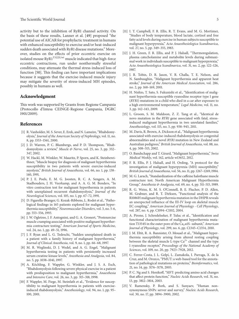
Rhabdomyolysis is an acute syndrome determined by a director indirect muscle injury. It results from skeletal musclebreakdown and massive release of the intracellular contentinto blood circulation, which can lead to potentially fatalevents, such as acute renal failure, hyperkalemia, and othermetabolic complications [1, 2]. The etiology of rhabdomyol-ysis is broad and includes inherited diseases, drugs, toxins,muscle compression, overexertion, and infections. Regardlessof the mechanism, these muscle injuries ultimately lead toa leakage of Ca2+ ions into the intracellular space, and theexcess of Ca2+ ions gives rise to a persistent muscle contrac-tion that ends in energy depletion and cell death (Figure 1)[1]. Rhabdomyolysis syndrome may also occur as a resultof a strenuous or not strenuous physical exercise (exertionalrhabdomyolysis or ER) often in hot and humid climates.Although anyone may develop ER under extreme physi-cal and environmental conditions, some individuals seemto be more predisposed than others, suggesting a geneticlink. The most commonly identified predisposing conditions
of ER are deficiencies of carnitine palmitoyltransferase II(CPT2 gene, OMIM ∗600650), myophosphorylase (McArdledisease, PYGM gene, OMIM ∗608455), and myoadenylatedeaminase (AMPD1 gene, OMIM +102770). Events similar tothose occurring in ER are triggered after exposure to anes-thetic agents in malignant hyperthermia susceptible (MHS)patients.Therefore, an association between ER andmalignanthyperthermia (MH) has been investigated and reported[3–10]. However, two studies on the effect of exercise onthermoregulatory and metabolic responses in MHS subjectsgave controversial results [11, 12]. Moreover, cases of MH-like events in the absence of anesthetic agents, and causedby high environmental or core body temperature, or even byemotional stress, have been reported [13–16].
Malignant hyperthermia (OMIM #145600) is an auto-somal dominant hypermetabolic condition that occurs ingenetically predisposed subjects during general anesthesia,induced by commonly used volatile anesthetics and/or theneuromuscular blocking agent succinylcholine. Triggeringagents cause an altered intracellular calcium regulation. AnMH attack, unless immediately recognized and treated, is

2 The Scientific World Journal
Mitochondria
SR
RyR1Cav1.1
ATP
SERCASarcomere
T-tubule
Ca2+Ca2+Ca2+
Ca2+
Ca+2-Na+exchanger
Na+ K+ Na+-K+-ATPase
Figure 1: Schematic representation of a skeletal muscle cell and of Ca2+ andNa+ ion fluxes across the sarcolemma and sarcoplasmic reticulum(SR). Activation of Cav1.1 by membrane depolarization causes the RyR1 channel to open and to release Ca2+ from SR, thus triggering musclecontraction. Ca2+ concentration is regulated by the Ca2+-ATPase membrane pump (SERCA) that sequesters Ca2+ in the SR and by the Na+-K+-ATPasemembrane pump and the Ca2+-Na+ antiport that exchange Ca2+ forNa+ across the sarcolemma. Regulation of calciumfluxmay bedisrupted at any of these sites. ATP depletion, by consumption duringmuscle contraction, or reduced ATP production, results in intracellularCa2+ increasing, muscle contraction, and continued energy consumption, leading to rhabdomyolysis.
often fatal. Clinical symptoms of a classicMHattack are accel-erated muscle metabolism, muscle contractions, metabolicacidosis, tachycardia, and hyperthermia.These symptoms arecorrelated with some altered biochemical parameters, such asmetabolic acidosis with increased pCO
2and lactate produc-
tion and release of potassium andmuscle proteins, as creatinekinase andmyoglobin, into the blood. Frequent late events aredamage of kidney function due to massive myoglobin releaseand/or a diffuse intravascular coagulation, which is often themain cause of death [17]. The prevalence of MH episodes isestimated to range from 1 : 10,000 to 1 : 220,000 [17]. Malig-nant hyperthermia susceptibility can be diagnosed by an invitro test, based on the differential contractile response ofnormal (MHN) and MHS muscles to caffeine and halothane.Protocols for MH contracture testing of human skeletalmuscle have been developed by the European [18] and NorthAmerican [19] MH Groups, namely, in vitro contracturetest (IVCT) and caffeine halothane contracture test (CHCT),respectively. A considerable genetic heterogeneity has beenreported for MH. Six genetic loci (MHS1, OMIM #180901;MHS2, OMIM #154275; MHS3, OMIM #154276; MHS4,OMIM #600467; MHS5, OMIM #601887; MHS6, OMIM#601888-6), associated with MH, have been identified. About70% of affected families are linked to the MHS1 locus, wherethe RYR1 gene encoding the skeletal muscle calcium releasechannel of the sarcoplasmic reticulum, commonly knownas ryanodine receptor type 1 (RyR1), maps. Dantrolene isan RyR1 antagonist that blocks calcium release from thesarcoplasmic reticulum stores and is the only specific agentavailable for the treatment of an MH attack. Less than 1% ofMHS cases can be attributed to mutations in the CACNA1Sgene (locus MHS5) encoding the 𝛼1S subunit of the voltage-dependent L-type calcium channel of the skeletal muscle,
Cav1.1. Only three MH-causing mutations identified in theCACNA1S gene were hitherto functionally characterized [20–22]. RyR1 and Cav1.1 are the two major proteins involved inthe excitation-contraction coupling in skeletal muscle.
The aim of this paper is to review the documented casesof ER or of stress-induced MH events in which sequencevariations (SVs) of the RYR1 gene, associated or possiblyassociated to MHS, have been identified.
2. Methods
The PubMed and Web of Science databases were consultedto search for studies on documented cases of ER or of stress-induced MH events in which RYR1 SVs, associated or pos-sibly associated to MHS, have been identified. Search termsincluded “RYR1,” “mutation,” “malignant hyperthermia,”“exercise,” “heat stress,” “stress-inducedmalignant hyperther-mia,” and “nonanesthetic malignant hyperthermia.” Single-nucleotide polymorphism (SNP) databases (http://www.ncbi.nlm.nih.gov/snp, http://www.dmd.nl/nmdb2/variants.php?select db=RYR1) were also searched. Three different pro-grams, namely, PMut (http://mmb.pcb.ub.es/PMut/), SIFT(http://sift.jcvi.org/), and PolyPhen-2 (http://genetics.bwh.harvard.edu/pph2/), were used to predict the pathologicalcharacter of RYR1 SVs which have not been functionallycharacterized. PMut is based on the use of neural net-works trained with a very large database of human disease-associated mutations and neutral SVs [23] and combinessequence alignment/position-specific scoring matrix withstructural factors; score >0.5 predicts a pathological effect.SIFT is based on the degree of conservation of amino acidresidues in sequence alignments derived from closely relatedsequences [24]. The SIFT scores range from 0 to 1; the amino

The Scientific World Journal 3
acid substitution is predicted as damaging if the score is≤0.05and as tolerated if the score is >0.05. PolyPhen-2 predicts theeffects of an amino acid substitution using both structure andsequence information [25] and classifies variants as “probablydamaging,” “possibly damaging,” or “benign,” based on pairsof false positive rate thresholds.
3. Results
3.1. RYR1 Gene Sequence Variations (SVs) in ER and Stress-Induced MH Patients. Thus far, more than 300 missense SVshave been identified in the RYR1 gene (http://www.ncbi.nlm.nih.gov/snp, http://www.dmd.nl/nmdb2/variants.php?selectdb=RYR). Some RYR1 SVs have been characterized by invitro functional studies. The demonstration that a SV altersthe kinetic properties of the RyR1 channel allows to defineits role in the pathogenesis of MHS. Various methods havebeen developed to characterize the function of RyR1 variants:analysis of calcium release in human primary myotubes [26–28] and in immortalized B lymphocytes from patients orafter expression by transfection in various cell types [29–31], determination of the channel openings in a ryanodinebinding assay [32], and a metabolic test in vitro based on themeasurements of proton release rate in immortalized B lym-phocytes frompatients [33].MHS-associatedRYR1mutationscause the channels to become hypersensitive to activationby electrical and pharmacolog-i-cal (caffeine, halothane, 4-chloro-m-cresol) stimuli. Identification of causative RYR1mutations is an aid to the diagnosis of MHS. In fact, althoughthe IVCT/CHCT are the gold standard to establish the riskof MHS, an individual harboring an MH causative mutationcan be considered MHS even without an IVCT/CHCTresult (http://www.emhg.org). Furthermore, genetic analysisis crucial to identify and evaluate the few cases of discor-dance between genotype, characterized by the presence ofa causative mutation, and MHN-typed phenotype [34, 35].A retrospective study reported these discordant cases inapproximately 2.6% of RYR1 mutation-positive families [35].Such discordant subjects are regarded as MHS for clinicalpurposes on the basis of genetic data alone, since they beara causative mutation [34, 35].
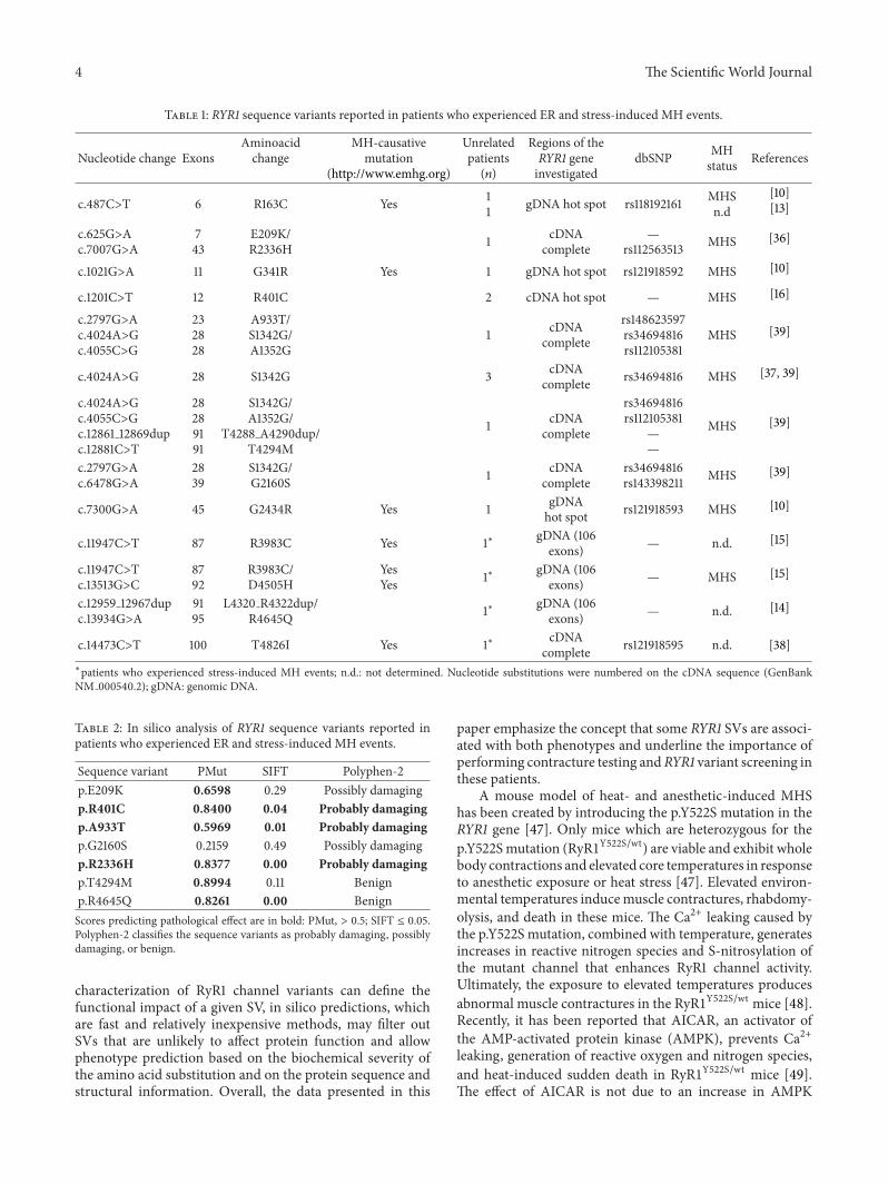
Table 1 shows a list of RYR1 gene missense SVs andthe corresponding amino acid substitutions, identified inpatients who experienced ER or stress-induced MH events[10, 13–16, 36–38]. Four RYR1 SVs, corresponding to theamino acid substitutions p.R163C, p.G341R, p.G2434R, andp.T4826I, have already been demonstrated to be causativeof MHS (http://www.emhg.org). The p.R3983C substitutionwas identified in two unrelated children who had fatal,nonanesthetic awake episodes associated with febrile illnessand heat stress [15]. One of the children also had thevariant p.D4505H. Interestingly, the child who only hadthe p.R3983 variant also had an MH attack during generalanesthesia with halothane. These two SVs were function-ally characterized by evaluating the caffeine sensitivity ofCa2+ release in transfected myotubes. Both p.R3983C andp.D4505H RyR1 channel variants exhibit an increase in thesensitivity to activation by caffeine, although the effect of thep.R3983C substitution alone is quite modest [15]. The SVs
p.R401C, p.A933T, p.G2160S, p.R2336H, p.T4288 A4290dup,p.T4294M, p.L4320 R4322dup, and p.R4645Qwere reportedto be absent in at least 100 control chromosomes. Instead,the p.S1342G and the p.S1352G variants are present amongthe African American population with a frequency of4% and 2.7%, respectively [39], indicating that they areneutral polymorphic changes in RyR1. The p.R2336H,p.T4288 A4290dup, p.L4320 R4322dup, and p.R4645Q SVshave already been reported in MHS families [40–42].
3.2. In Silico Analysis of RYR1 Variants Reported in PatientsWho Experienced ER and Stress-Induced MH Events. Topredict the pathological character of p.E209K, p.R401C,p.A933T, p.G2160S, p.R2336H, p.T4294M, and p.R4645QSVs, I tested them with 3 different prediction programs,namely, PMut (http://mmb.pcb.ub.es/PMut/) [23], SIFT(http://sift.jcvi.org/) [24], and PolyPhen-2 (http://genetics.bwh.harvard.edu/pph2/) [25]. Table 2 shows the resultsobtained by this analysis. The p.R401C, p.A933T, andp.R2336H variants were predicted to have a pathologicalcharacter, while the predictions generated for p.E209K,p.G2160S, p.T4294M, and p.R4645Q variants were divergent.The p.E209K variant, that has been predicted to be neutralby two programs and only possibly damaging by PolyPhen-2,has been found in association with p.R2336H in one patientswho experienced stress-induced MH events and was typedMHS by CHCT (see Table 1) [36]. All the programs testedpredict a pathological effect for the p.R2336H variant, thatcould be the molecular basis of both phenotypes. However,functional studies are needed to conclusively define the exactpathogenic effects of this amino acid substitution and toassess if it is the cause of stress-induced MH events in thepatient.
Wappler et al. [10] found causative mutations (p.R163C,p.G341R, and p.G2434R) in only three out of ten MHSpatients who experienced ER. They screened only eightRYR1 exons located in the hotspot region; therefore, thislimited analysis can explain the low mutation detectionrate. Moreover, Sambuughin et al. [39], by sequencing theRYR1 cDNA, found putative causative SVs (p.A933T andp.T4294M) in only two out of six ER/MHS patients studied.In the remaining cases, the ER/MHS phenotype could becaused by RYR1 SVs which may escape the RYR1 cDNAscreening because they determine unbalanced allelic expres-sion [43–46] or, alternatively, could be caused by mutationsin other candidate MHS loci genes.
4. Conclusions and Perspectives
ER and stress-induced MH events are syndromes withdiverse etiologies that afflict particularly military recruits inbasic training and athletes. This paper reports an overviewof the literature on cases associated with MHS and withRYR1 causative mutations or putative causative SVs. Thepossible disease-causing role of SVs, identified in patientswho experienced ER and stress-induced MH events and thathave not been functionally characterized, was investigated bycomputational analysis by using three different approaches, toincrease the predictive power. Although only the molecular
4 The Scientific World Journal
Table 1: RYR1 sequence variants reported in patients who experienced ER and stress-induced MH events.
Nucleotide change ExonsAminoacidchange
MH-causativemutation
(http://www.emhg.org)
Unrelatedpatients(𝑛)
Regions of theRYR1 geneinvestigated
dbSNP MHstatus References
c.487C>T 6 R163C Yes 11 gDNA hot spot rs118192161 MHS
n.d[10][13]
c.625G>Ac.7007G>A
743
E209K/R2336H 1 cDNA
complete—
rs112563513 MHS [36]
c.1021G>A 11 G341R Yes 1 gDNA hot spot rs121918592 MHS [10]
c.1201C>T 12 R401C 2 cDNA hot spot — MHS [16]
c.2797G>Ac.4024A>Gc.4055C>G
232828
A933T/S1342G/A1352G
1 cDNAcomplete
rs148623597rs34694816rs112105381
MHS [39]
c.4024A>G 28 S1342G 3 cDNAcomplete rs34694816 MHS [37, 39]
c.4024A>Gc.4055C>Gc.12861 12869dupc.12881C>T
28289191
S1342G/A1352G/
T4288 A4290dup/T4294M
1 cDNAcomplete
rs34694816rs112105381
——
MHS [39]
c.2797G>Ac.6478G>A
2839
S1342G/G2160S 1 cDNA
completers34694816rs143398211 MHS [39]
c.7300G>A 45 G2434R Yes 1 gDNAhot spot rs121918593 MHS [10]
c.11947C>T 87 R3983C Yes 1∗ gDNA (106exons) — n.d. [15]
c.11947C>Tc.13513G>C
8792
R3983C/D4505H
YesYes 1∗ gDNA (106
exons) — MHS [15]
c.12959 12967dupc.13934G>A
9195
L4320 R4322dup/R4645Q 1∗ gDNA (106
exons) — n.d. [14]
c.14473C>T 100 T4826I Yes 1∗ cDNAcomplete rs121918595 n.d. [38]
∗patients who experienced stress-induced MH events; n.d.: not determined. Nucleotide substitutions were numbered on the cDNA sequence (GenBankNM 000540.2); gDNA: genomic DNA.
Table 2: In silico analysis of RYR1 sequence variants reported inpatients who experienced ER and stress-induced MH events.
Sequence variant PMut SIFT Polyphen-2p.E209K 0.6598 0.29 Possibly damagingp.R401C 0.8400 0.04 Probably damagingp.A933T 0.5969 0.01 Probably damagingp.G2160S 0.2159 0.49 Possibly damagingp.R2336H 0.8377 0.00 Probably damagingp.T4294M 0.8994 0.11 Benignp.R4645Q 0.8261 0.00 BenignScores predicting pathological effect are in bold: PMut, > 0.5; SIFT ≤ 0.05.Polyphen-2 classifies the sequence variants as probably damaging, possiblydamaging, or benign.
characterization of RyR1 channel variants can define thefunctional impact of a given SV, in silico predictions, whichare fast and relatively inexpensive methods, may filter outSVs that are unlikely to affect protein function and allowphenotype prediction based on the biochemical severity ofthe amino acid substitution and on the protein sequence andstructural information. Overall, the data presented in this
paper emphasize the concept that some RYR1 SVs are associ-ated with both phenotypes and underline the importance ofperforming contracture testing andRYR1 variant screening inthese patients.
A mouse model of heat- and anesthetic-induced MHShas been created by introducing the p.Y522S mutation in theRYR1 gene [47]. Only mice which are heterozygous for thep.Y522Smutation (RyR1Y522S/wt) are viable and exhibit wholebody contractions and elevated core temperatures in responseto anesthetic exposure or heat stress [47]. Elevated environ-mental temperatures inducemuscle contractures, rhabdomy-olysis, and death in these mice. The Ca2+ leaking caused bythe p.Y522S mutation, combined with temperature, generatesincreases in reactive nitrogen species and S-nitrosylation ofthe mutant channel that enhances RyR1 channel activity.Ultimately, the exposure to elevated temperatures producesabnormal muscle contractures in the RyR1Y522S/wt mice [48].Recently, it has been reported that AICAR, an activator ofthe AMP-activated protein kinase (AMPK), prevents Ca2+leaking, generation of reactive oxygen and nitrogen species,and heat-induced sudden death in RyR1Y522S/wt mice [49].The effect of AICAR is not due to an increase in AMPK
The Scientific World Journal 5
activity but to the inhibition of RyR1 channel activity. Onthe basis of these results, Lanner et al. [49] proposed “thepotential use of AICAR for prophylactic treatment in humanswith enhanced susceptibility to exercise and/or heat-inducedsudden death associated with RyR1 diseasemutations.”More-over, studies on the effects of prior eccentric exercise onisolatedmouse RyR1Y522S/wt muscle indicated that high-forceeccentric contractions, run under nonthermally stressfulconditions, may attenuate the thermal stress-induced loss offunction [50]. This finding can have important implicationsbecause it suggests that the exercise-induced muscle injurymay mitigate the severity of stress-induced MH episodes,possibly in humans as well.
Acknowledgment
This work was supported by Grants from Regione Campania(Protocollo d’Intesa CEINGE-Regione Campania, DGRC1901/2009).
References
[1] R. Vanholder,M. S. Sever, E. Erek, andN. Lameire, “Rhabdomy-olysis,” Journal of the American Society of Nephrology, vol. 11, no.8, pp. 1553–1561, 2000.
[2] J. D. Warren, P. C. Blumbergs, and P. D. Thompson, “Rhab-domyolysis: a review,” Muscle & Nerve, vol. 25, no. 3, pp. 332–347, 2002.
[3] W. Hackl, M. Winkler, W. Mauritz, P. Sporn, and K. Steinberei-thner, “Muscle biopsy for diagnosis of malignant hyperthermiasusceptibility in two patients with severe exercise-inducedmyolysis,” British Journal of Anaesthesia, vol. 66, no. 1, pp. 138–140, 1991.
[4] P. J. E. Poels, E. M. G. Joosten, R. C. A. Sengers, A. M.Stadhouders, J. H. Veerkamp, and A. A. G. M. Benders, “Invitro contraction test for malignant hyperthermia in patientswith unexplained recurrent rhabdomyolysis,” Journal of theNeurological Sciences, vol. 105, no. 1, pp. 67–72, 1991.
[5] D. Figarella-Branger, G. Kozak-Ribbens, L. Rodet et al., “Patho-logical findings in 165 patients explored for malignant hyper-thermia susceptibility,”Neuromuscular Disorders, vol. 3, no. 5-6,pp. 553–556, 1993.
[6] J. W. Ogletree, J. F. Antognini, and G. A. Gronert, “Postexercisemuscle cramping associated with positivemalignant hyperther-mia contracture testing,” American Journal of Sports Medicine,vol. 24, no. 1, pp. 49–51, 1996.
[7] J. F. Ryan and L. G. Tedeschi, “Sudden unexplained death ina patient with a family history of malignant hyperthermia,”Journal of Clinical Anesthesia, vol. 9, no. 1, pp. 66–68, 1997.
[8] M. R. Weglinski, D. J. Wedel, and A. G. Engel, “Malignanthyperthermia testing in patients with persistently increasedserum creatine kinase levels,” Anesthesia and Analgesia, vol. 84,no. 5, pp. 1038–1041, 1997.
[9] A. Kochling, F. Wappler, G. Winkler, and J. S. A. Esch,“Rhabdomyolysis following severe physical exercise in a patientwith predisposition to malignant hyperthermia,” Anaesthesiaand Intensive Care, vol. 26, no. 3, pp. 315–318, 1998.
[10] F. Wappler, M. Fiege, M. Steinfath et al., “Evidence for suscep-tibility to malignant hyperthermia in patients with exercise-induced rhabdomyolysis,” Anesthesiology, vol. 94, no. 1, pp. 95–100, 2001.
[11] I. T. Campbell, F. R. Ellis, R. T. Evans, and M. G. Mortimer,“Studies of body temperature, blood lactate, cortisol and freefatty acid levels during exercise in human subjects susceptible tomalignant hyperpyrexia,” Acta Anaesthesiologica Scandinavica,vol. 27, no. 5, pp. 349–355, 1983.
[12] J. H. Green, F. R. Ellis, and P. J. Halsall, “Thermoregulation,plasma catecholamine and metabolite levels during submaxi-mal work in individuals susceptible tomalignant hyperpyrexia,”Acta Anaesthesiologica Scandinavica, vol. 31, no. 2, pp. 122–126,1987.
[13] J. R. Tobin, D. R. Jason, V. R. Challa, T. E. Nelson, andN. Sambuughin, “Malignant hyperthermia and apparent heatstroke,” Journal of the American Medical Association, vol. 286,no. 2, pp. 168–169, 2001.
[14] H. Nishio, T. Sato, S. Fukunishi et al., “Identification of malig-nant hyperthermia-susceptible ryanodine receptor type 1 gene(RYR1) mutations in a child who died in a car after exposure toa high environmental temperature,” Legal Medicine, vol. 11, no.3, pp. 142–143, 2009.
[15] L. Groom, S. M. Muldoon, Z. Z. Tang et al., “Identical denovo mutation in the RYR1 gene associated with fatal, stress-induced malignant hyperthermia in two unrelated families,”Anesthesiology, vol. 115, no. 5, pp. 938–945, 2011.
[16] M.Davis, R. Brown, A. Dickson et al., “Malignant hyperthermiaassociated with exercise-induced rhabdomyolysis or congenitalabnormalities and a novel RYR1 mutation in New Zealand andAustralian pedigrees,” British Journal of Anaesthesia, vol. 88, no.4, pp. 508–515, 2002.
[17] O. Bandschapp and T. Girard, “Malignant hyperthermia,” SwissMedical Weekly, vol. 142, article w13652, 2012.
[18] F. R. Ellis, P. J. Halsall, and H. Ording, “A protocol for theinvestigation of malignant hyperpyrexia (MH) susceptibility,”British Journal of Anaesthesia, vol. 56, no. 11, pp. 1267–1269, 1984.
[19] M. G. Larach, “Standardization of the caffeine halothanemusclecontracture test. North American Malignant HyperthermiaGroup,” Anesthesia & Analgesia, vol. 69, no. 4, pp. 511–515, 1989.
[20] R. G. Weiss, K. M. S. O’Connell, B. E. Flucher, P. D. Allen,M. Grabner, and R. T. Dirksen, “Functional analysis of theR1086Hmalignant hyperthermiamutation in theDHPR revealsan unexpected influence of the III-IV loop on skeletal muscleEC coupling,” American Journal of Physiology - Cell Physiology,vol. 287, no. 4, pp. C1094–C1102, 2004.
[21] A. Pirone, J. Schredelseker, P. Tuluc et al., “Identification andfunctional characterization of malignant hyperthermia muta-tion T1354S in the outer pore of the Cav𝛼1S- subunit,”AmericanJournal of Physiology, vol. 299, no. 6, pp. C1345–C1354, 2010.
[22] J. M. Eltit, R. A. Bannister, O. Mouad et al., “Malignant hyper-thermia susceptibility arising from altered resting couplingbetween the skeletal muscle L-type Ca2+ channel and the type1 ryanodine receptor,” Proceedings of the National Academy ofSciences, vol. 109, no. 20, pp. 7923–7928, 2012.
[23] C. Ferrer-Costa, J. L. Gelpı, L. Zamakola, I. Parraga, X. de laCruz, andM.Orozco, “PMUT: a web-based tool for the annota-tion of pathological mutations on proteins,” Bioinformatics, vol.21, no. 14, pp. 3176–3178, 2005.
[24] P. C. Ng and S. Henikoff, “SIFT: predicting amino acid changesthat affect protein function,” Nucleic Acids Research, vol. 31, no.13, pp. 3812–3814, 2003.
[25] V. Ramensky, P. Bork, and S. Sunyaev, “Human non-synonymous SNPs: server and survey,” Nucleic Acids Research,vol. 30, no. 17, pp. 3894–3900, 2002.

6 The Scientific World Journal
[26] M. Wehner, H. Rueffert, F. Koenig, J. Neuhaus, and D. Olthoff,“Increased sensitivity to 4-chloro-m-cresol and caffeine inprimary myotubes from malignant hyperthermia susceptibleindividuals carrying the ryanodine receptor 1 Thr2206Met(C6617T) mutation,” Clinical Genetics, vol. 62, no. 2, pp. 135–146, 2002.
[27] T. Girard, S. Treves, K. Censier, C. R. Mueller, F. Zorzato, and A.Urwyler, “Phenotyping malignant hyperthermia susceptibilityby measuring halothane-induced changes in myoplasmic cal-cium concentration in cultured human skeletal muscle cells,”British Journal of Anaesthesia, vol. 89, no. 4, pp. 571–579, 2002.
[28] H. Brinkmeier, J. Kramer, R. Kramer et al., “Malignant hyper-thermia causing Gly2435Arg mutation of the ryanodine recep-tor facilitates ryanodine-induced calcium release in myotubes,”British Journal of Anaesthesia, vol. 83, no. 6, pp. 855–861, 1999.
[29] T. Yang, T. A. Ta, I. N. Pessah, and P. D. Allen, “Functionaldefects in six ryanodine receptor isoform-1 (RYR1) mutationsassociated with malignant hyperthermia and their impact onskeletal excitation-contraction coupling,” Journal of BiologicalChemistry, vol. 278, no. 28, pp. 25722–25730, 2003.
[30] T. Girard, D. Cavagna, E. Padovan et al., “B-lymphocytes frommalignant hyperthermia-susceptible patients have an increasedsensitivity to skeletal muscle ryanodine receptor activators,”Journal of Biological Chemistry, vol. 276, no. 51, pp. 48077–48082, 2001.
[31] J. Tong, H. Oyamada, N. Demaurex, S. Grinstein, T. V.McCarthy, and D. H. MacLennan, “Caffeine and halothanesensitivity of intracellular Ca2+ release is altered by 15 calciumrelease channel (ryanodine receptor) mutations associated withmalignant hyperthermia and/or central core disease,” Journal ofBiological Chemistry, vol. 272, no. 42, pp. 26332–26339, 1997.
[32] N. Sambuughin, T. E. Nelson, J. Jankovic et al., “Identificationand functional characterization of a novel ryanodine receptormutation causing malignant hyperthermia in North Americanand SouthAmerican families,”Neuromuscular Disorders, vol. 11,no. 6-7, pp. 530–537, 2001.
[33] A. Zullo, W. Klingler, C. De Sarno et al., “Functional character-ization of ryanodine receptor (RYR1) sequence variants usinga metabolic assay in immortalized B-lymphocytes,” HumanMutation, vol. 30, no. 4, pp. E575–E590, 2009.
[34] G. Fortunato, A. Carsana, N. Tinto, V. Brancadoro, G. Canfora,and F. Salvatore, “A case of discordance between genotypeand phenotype in a malignant hyperthermia family,” EuropeanJournal of Human Genetics, vol. 7, no. 4, pp. 415–420, 1999.
[35] R. L. Robinson, M. J. Anetseder, V. Brancadoro et al., “Recentadvances in the diagnosis of malignant hyperthermia suscept-ability: how confident can we be of genetic testing?” EuropeanJournal of Human Genetics, vol. 11, no. 4, pp. 342–348, 2003.
[36] J. Loke, N. Kraeva, and D. MacLennan, “Mutations in RYR1gene associated with malignant hyperthermia and a non-anaesthetic phenotype,” Canadian Journal of Anaesthesia, vol.54, Supplement 1, p. 4609, 2007.
[37] J. F. Capacchione, N. Sambuughin, S. Bina, L. P. Mulligan, T. D.Lawson, and S. M. Muldoon, “Exertional rhabdomyolysis andmalignant hyperthermia in a patient with ryanodine receptortype 1 gene, L-type calcium channel 𝛼-1 subunit gene, andcalsequestrin-1 gene polymorphisms,” Anesthesiology, vol. 112,no. 1, pp. 239–244, 2010.
[38] R. L. Brown, A. N. Pollock, K. G. Couchman et al., “Anovel ryanodine receptor mutation and genotype-phenotypecorrelation in a large malignant hyperthermia New Zealand
Maori pedigree,” Human Molecular Genetics, vol. 9, no. 10, pp.1515–1524, 2000.
[39] N. Sambuughin, J. Capacchione, A. Blokhin, M. Bayarsaikhan,S. Bina, and S. Muldoon, “The ryanodine receptor type 1gene variants in African American men with exertional rhab-domyolysis andmalignant hyperthermia susceptibility,”ClinicalGenetics, vol. 76, no. 6, pp. 564–568, 2009.
[40] S. Levano,M.Vukcevic,M. Singer et al., “Increasing the numberof diagnostic mutations in malignant hyperthermia,” HumanMutation, vol. 30, no. 4, pp. 590–598, 2009.
[41] D. Carpenter, R. L. Robinson, R. J. Quinnel et al., “Geneticvariation in RYR1 and malignant hyperthermia phenotypes,”British Journal of Anaesthesia, vol. 103, no. 4, pp. 538–548, 2009.
[42] C. A. Ibarra, S. Wu, K. Murayama et al., “Malignant hyper-thermia in Japan: mutation screening of the entire ryanodinereceptor type 1 gene coding region by direct sequencing,”Anesthesiology, vol. 104, no. 6, pp. 1146–1154, 2006.
[43] H. Grievink and K. M. Stowell, “Allele-specific differences inryanodine receptor 1 mRNA expression levels may contributeto phenotypic variability inmalignant hyperthermia,”OrphanetJournal of Rare Diseases, vol. 5, no. 1, article 10, 2010.
[44] R. L. Robinson,D.Carpenter, P. J.Halsall et al., “Epigenetic allelesilencing and variable penetrance of malignant hyperthermiasusceptibility,” British Journal of Anaesthesia, vol. 103, no. 2, pp.220–225, 2009.
[45] H. Zhou, M. Brockington, H. Jungbluth et al., “Epigenetic allelesilencing unveils recessive RYR1mutations in core myopathies,”American Journal of HumanGenetics, vol. 79, no. 5, pp. 859–868,2006.
[46] H. Zhou, H. Jungbluth, C. A. Sewry et al., “Molecular mech-anisms and phenotypic variation in RYR1-related congenitalmyopathies,” Brain, vol. 130, no. 8, pp. 2024–2036, 2007.
[47] M. G. Chelu, S. A. Goonasekera,W. J. Durham et al., “Heat- andanesthesia-inducedmalignant hyperthermia in anRYR1 knock-in mouse,” FASEB Journal, vol. 20, no. 2, pp. 329–330, 2006.
[48] W. J. Durham, P. Aracena-Parks, C. Long et al., “RYR1 S-nitrosylation underlies environmental heat stroke and suddendeath in Y522S RYR1 knockin mice,” Cell, vol. 133, no. 1, pp. 53–65, 2008.
[49] J. T. Lanner, D. K. Georgiou, A. Dagnino-Acosta et al., “AICARprevents heat induced sudden death in RYR1 mutant miceindependent of AMPK activation,”Nature Medicine, vol. 18, no.2, pp. 244–251, 2012.
[50] B. T. Corona, S. L. Hamilton, and C. P. Ingalls, “Effect of priorexercise on thermal sensitivity of malignant hyperthermia-susceptible muscle,” Muscle and Nerve, vol. 42, no. 2, pp. 270–272, 2010.

Submit your manuscripts athttp://www.hindawi.com
Hindawi Publishing Corporationhttp://www.hindawi.com Volume 2014
Anatomy Research International
PeptidesInternational Journal of
Hindawi Publishing Corporationhttp://www.hindawi.com Volume 2014
Hindawi Publishing Corporation http://www.hindawi.com
International Journal of
Volume 2014
Zoology
Hindawi Publishing Corporationhttp://www.hindawi.com Volume 2014
Molecular Biology International
GenomicsInternational Journal of
Hindawi Publishing Corporationhttp://www.hindawi.com Volume 2014
The Scientific World JournalHindawi Publishing Corporation http://www.hindawi.com Volume 2014
Hindawi Publishing Corporationhttp://www.hindawi.com Volume 2014
BioinformaticsAdvances in
Marine BiologyJournal of
Hindawi Publishing Corporationhttp://www.hindawi.com Volume 2014
Hindawi Publishing Corporationhttp://www.hindawi.com Volume 2014
Signal TransductionJournal of
Hindawi Publishing Corporationhttp://www.hindawi.com Volume 2014
BioMed Research International
Evolutionary BiologyInternational Journal of
Hindawi Publishing Corporationhttp://www.hindawi.com Volume 2014
Hindawi Publishing Corporationhttp://www.hindawi.com Volume 2014
Biochemistry Research International
ArchaeaHindawi Publishing Corporationhttp://www.hindawi.com Volume 2014
Hindawi Publishing Corporationhttp://www.hindawi.com Volume 2014
Genetics Research International
Hindawi Publishing Corporationhttp://www.hindawi.com Volume 2014
Advances in
Virolog y
Hindawi Publishing Corporationhttp://www.hindawi.com
Nucleic AcidsJournal of
Volume 2014
Stem CellsInternational
Hindawi Publishing Corporationhttp://www.hindawi.com Volume 2014
Hindawi Publishing Corporationhttp://www.hindawi.com Volume 2014
Enzyme Research
Hindawi Publishing Corporationhttp://www.hindawi.com Volume 2014
International Journal of
Microbiology