Acute Appendicitis. Epidemiology The incidence of appendectomy appears to be declining due to more...
51
Acute Appendicitis
-
Upload
annabel-miller -
Category
Documents
-
view
218 -
download
0
Transcript of Acute Appendicitis. Epidemiology The incidence of appendectomy appears to be declining due to more...

Acute Appendicitis
Epidemiology
• The incidence of appendectomy appears to be declining due to more accurate preoperative diagnosis.
• Despite newer imaging techniques, acute appendicitis can be very difficult to diagnose.
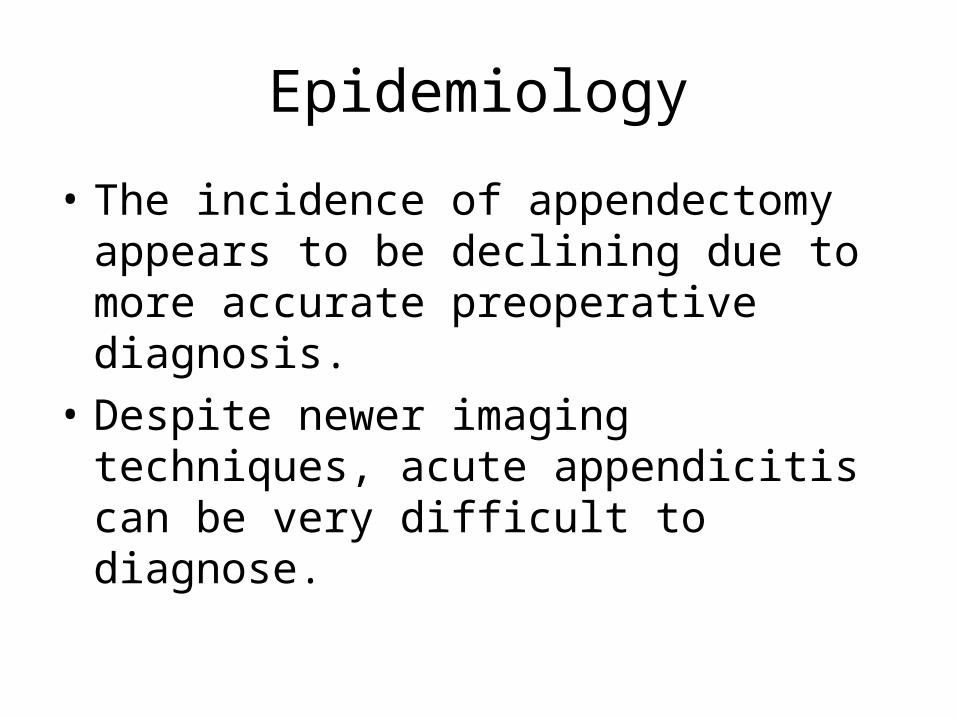
Appendicitis:
• The most common surgical condition of the abdomen
• Lifetime occurrence of 7%
• Peak incidence 10-30y
• The most common nonobstetric surgical intervention during pregnancy
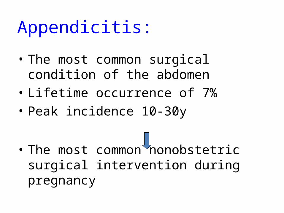
Pathogenesis:• Appendiceal lumen obstruction : lymphoid hyperplasia fecaliths parasites foreign bodies crohn’s disease metastatic cancer carcinoid syndrome
Pathophysiology
• Acute appendicitis is thought to begin with obstruction of the lumen
• Obstruction can result from food matter, adhesions, or lymphoid hyperplasia
• Mucosal secretions continue to increase intraluminal pressure
Pathophysiology
• Eventually the pressure exceeds capillary perfusion pressure and venous and lymphatic drainage are obstructed.
• With vascular compromise, epithelial mucosa breaks down and bacterial invasion by bowel flora occurs.
Pathophysiology
• Increased pressure also leads to arterial stasis and tissue infarction
• End result is perforation and spillage of infected appendiceal contents into the peritoneum
Pathophysiology
• Initial luminal distention triggers visceral afferent pain fibers, which enter at the 10th thoracic vertebral level.
• This pain is generally vague and poorly localized.
• Pain is typically felt in the periumbilical or epigastric area.
Pathophysiology
• As inflammation continues, the serosa and adjacent structures become inflamed
• This triggers somatic pain fibers, innervating the peritoneal structures.
• Typically causing pain in the RLQ
Pathophysiology
• The change in stimulation form visceral to somatic pain fibers explains the classic migration of pain in the periumbilical area to the RLQ seen with acute appendicitis.
Pathophysiology
• Exceptions exist in the classic presentation due to anatomic variability of the appendix
• Appendix can be retrocecal causing the pain to localize to the right flank
• In pregnancy, the appendix ca be shifted and patients can present with RUQ pain
Pathophysiology
• In some males, retroileal appendicitis can irritate the ureter and cause testicular pain.
• Pelvic appendix may irritate the bladder or rectum causing suprapubic pain, pain with urination, or feeling the need to defecate
• Multiple anatomic variations explain the difficulty in diagnosing appendicitis

symptoms :
• Pain – RLQ / RUQ / Flank• Anorexia• Vomiting• Nausea • Pain migration• Fever
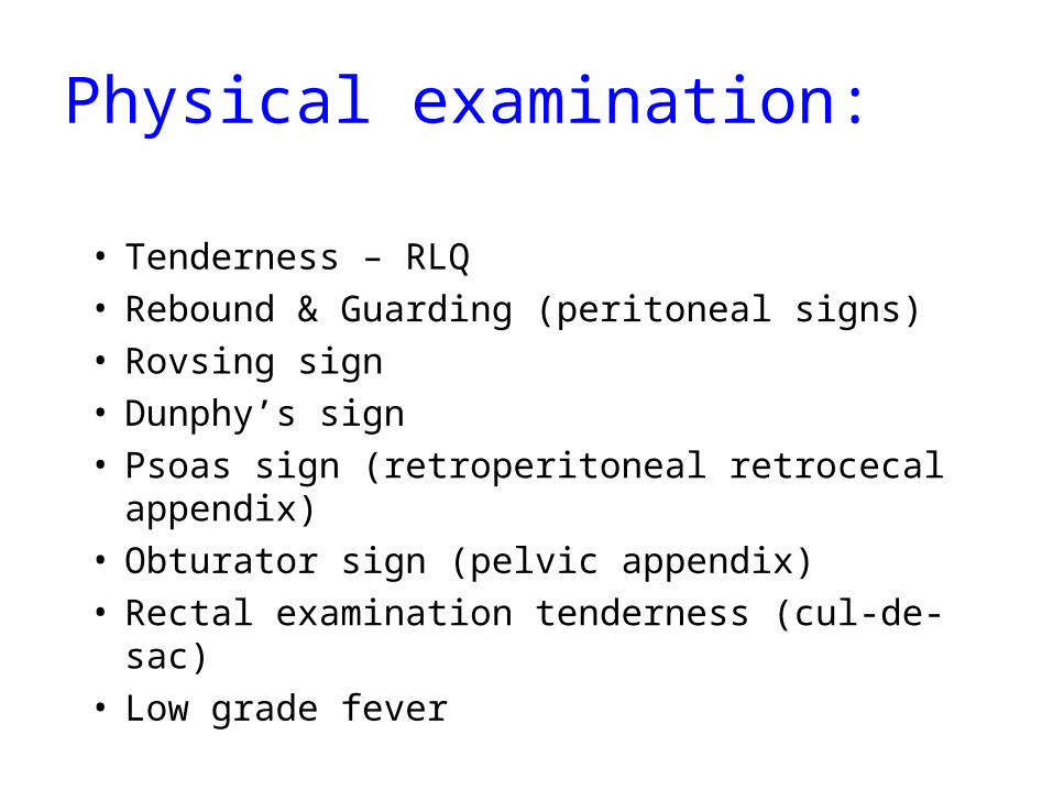
Physical examination:
• Tenderness – RLQ• Rebound & Guarding (peritoneal signs)• Rovsing sign• Dunphy’s sign• Psoas sign (retroperitoneal retrocecal appendix) • Obturator sign (pelvic appendix)• Rectal examination tenderness (cul-de-sac)• Low grade fever
Psoassign
Obturatorsign
History
• Primary symptom: abdominal pain• ½ to 2/3 of patients have the classical
presentation• Pain beginning in epigastrium or periumbilical
area that is vague and hard to localize
History
• Associated symptoms: indigestion, discomfort, flatus, need to defecate, anorexia, nausea, vomiting
• As the illness progresses RLQ localization typically occurs
• RLQ pain was 81 % sensitive and 53% specific for diagnosis
History
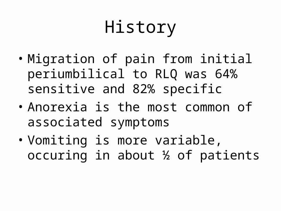
• Migration of pain from initial periumbilical to RLQ was 64% sensitive and 82% specific
• Anorexia is the most common of associated symptoms
• Vomiting is more variable, occuring in about ½ of patients
Physical Exam
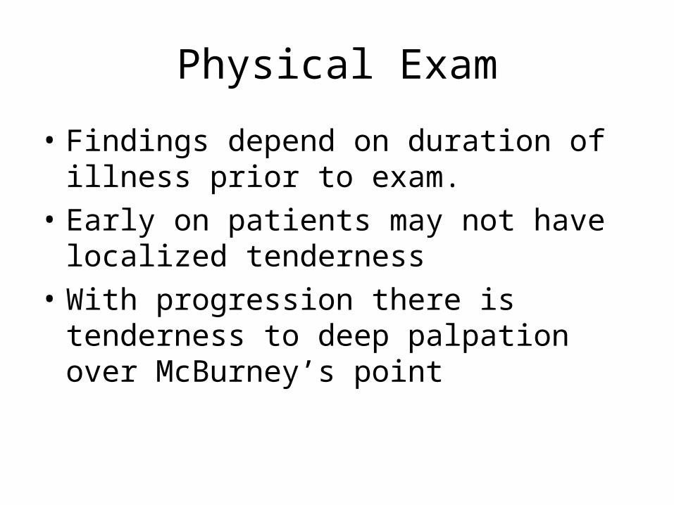
• Findings depend on duration of illness prior to exam.
• Early on patients may not have localized tenderness
• With progression there is tenderness to deep palpation over McBurney’s point
Physical Exam
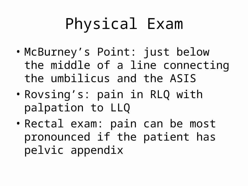
• McBurney’s Point: just below the middle of a line connecting the umbilicus and the ASIS
• Rovsing’s: pain in RLQ with palpation to LLQ• Rectal exam: pain can be most pronounced if
the patient has pelvic appendix
Physical Exam
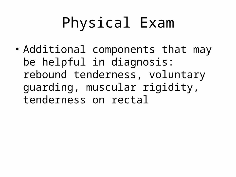
• Additional components that may be helpful in diagnosis: rebound tenderness, voluntary guarding, muscular rigidity, tenderness on rectal
Physical Exam
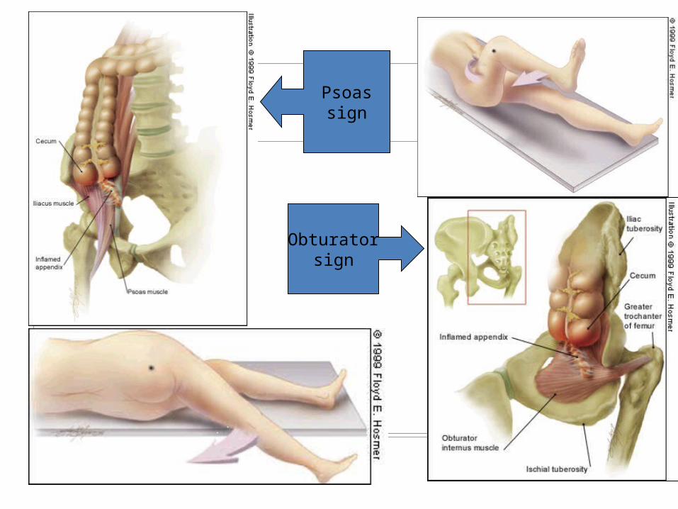
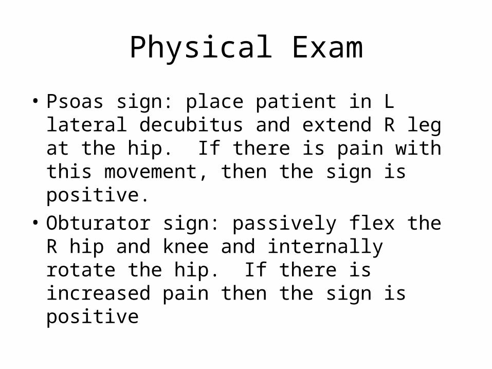
• Psoas sign: place patient in L lateral decubitus and extend R leg at the hip. If there is pain with this movement, then the sign is positive.
• Obturator sign: passively flex the R hip and knee and internally rotate the hip. If there is increased pain then the sign is positive
Physical Exam
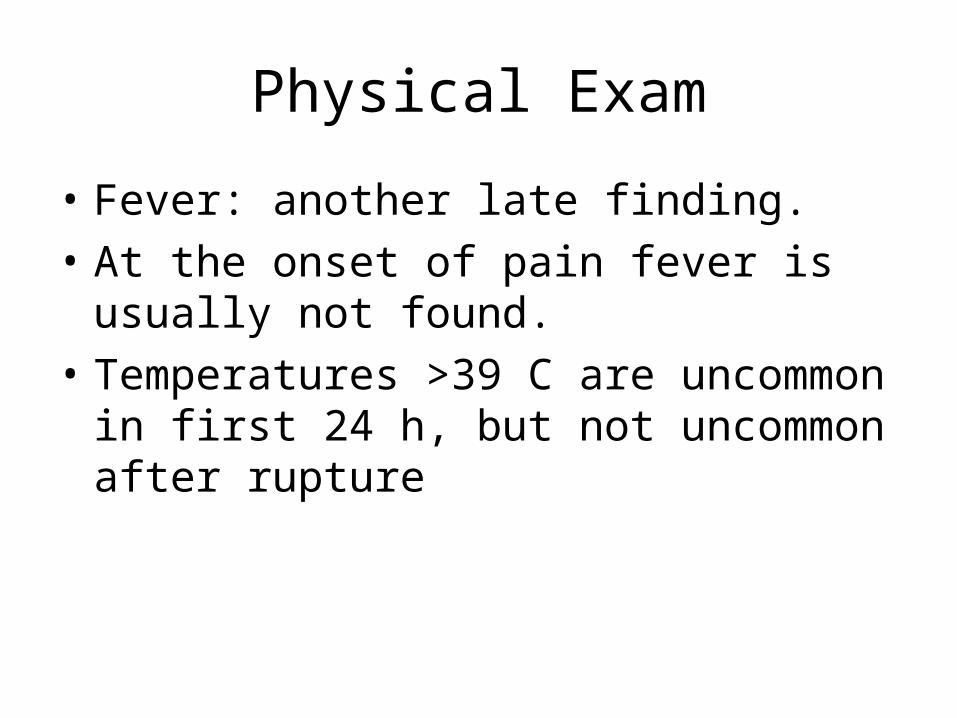
• Fever: another late finding.• At the onset of pain fever is usually not found.
• Temperatures >39 C are uncommon in first 24
h, but not uncommon after rupture
Lab:
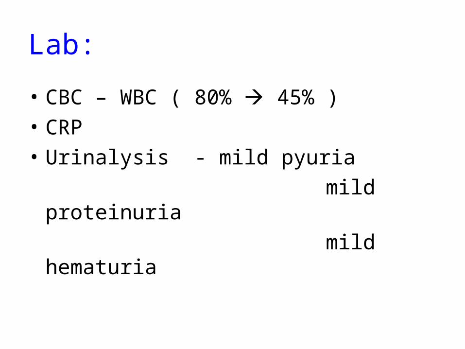
• CBC – WBC ( 80% 45% )• CRP • Urinalysis - mild pyuria mild proteinuria mild hematuria
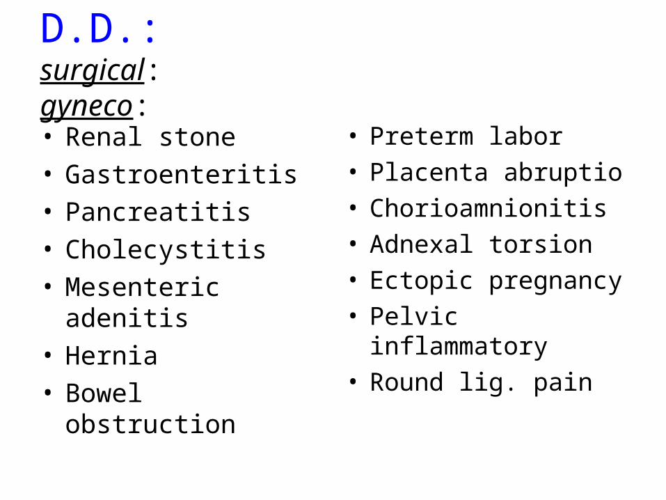
D.D.:surgical: gyneco:
• Renal stone • Gastroenteritis• Pancreatitis• Cholecystitis• Mesenteric adenitis• Hernia• Bowel obstruction
• Preterm labor• Placenta abruptio• Chorioamnionitis• Adnexal torsion • Ectopic pregnancy• Pelvic inflammatory• Round lig. pain
Diagnostic problems:
• Position of appendix: normally 70% intraperitoneal
30% pelvic, retroileal, retrocolic
pregnancy – anatomical changes
gravid uterus displacement upward &
outward flank pain (3rd trimester) (Baer,1932)
increased separation of peritoneum decreased perception of somatic pain and localization
Diagnostic problems:
• Symptoms complex – physical changes anorexia, nausea & vomiting in normal pregnancy• Lab – relative leukocytosis• Imaging techniques
Diagnosis
• Acute appendicitis should be suspected in anyone with epigastric, periumbilical, right flank, or right sided abd pain who has not had an appendectomy
Diagnosis
• Women of child bearing age need a pelvic exam and a pregnancy test.
• Additional studies: CBC, UA, imaging studies
Diagnosis
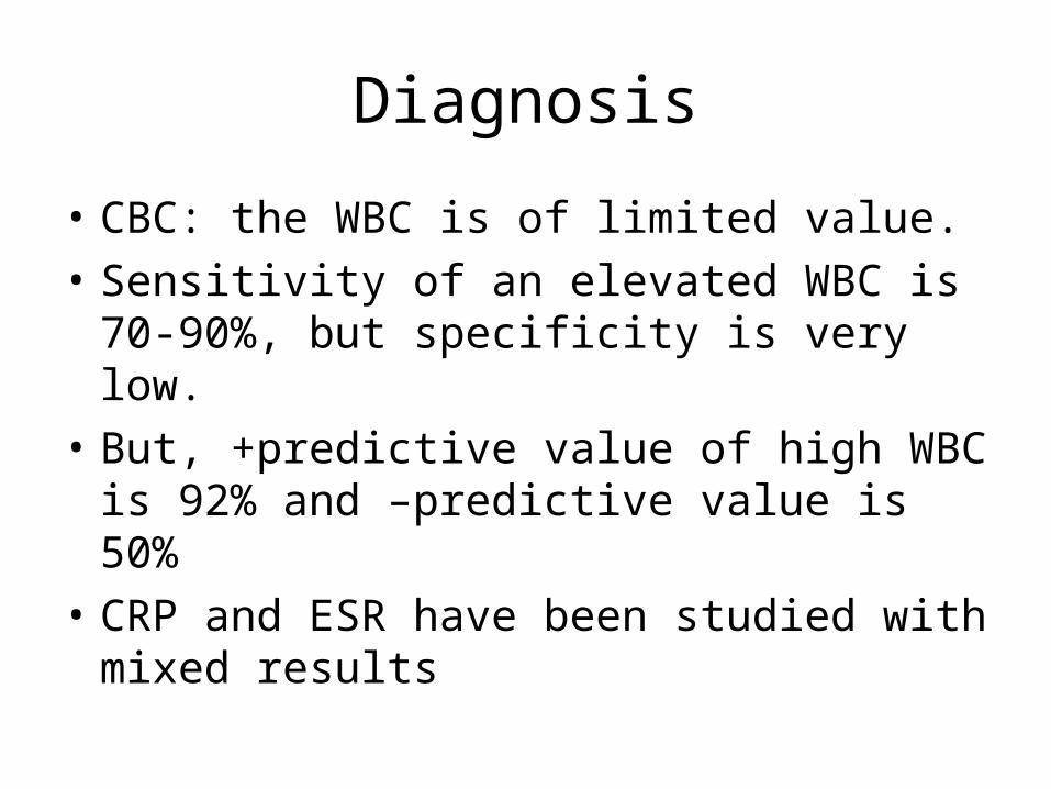
• CBC: the WBC is of limited value. • Sensitivity of an elevated WBC is 70-90%, but
specificity is very low.• But, +predictive value of high WBC is 92% and
–predictive value is 50%• CRP and ESR have been studied with mixed
results
Diagnosis
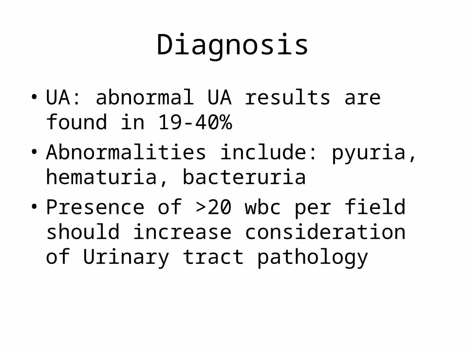
• UA: abnormal UA results are found in 19-40%• Abnormalities include: pyuria, hematuria,
bacteruria• Presence of >20 wbc per field should increase
consideration of Urinary tract pathology
Imaging:
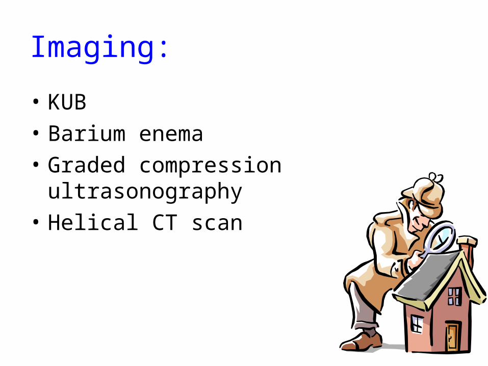
• KUB• Barium enema• Graded compression ultrasonography• Helical CT scan
Diagnosis
• Imaging studies: include X-rays, US, CT• Xrays of abd are abnormal in 24-95%• Abnormal findings include: fecalith,
appendiceal gas, localized paralytic ileus, blurred right psoas, and free air
• Abdominal xrays have limited use b/c the findings are seen in multiple other processes
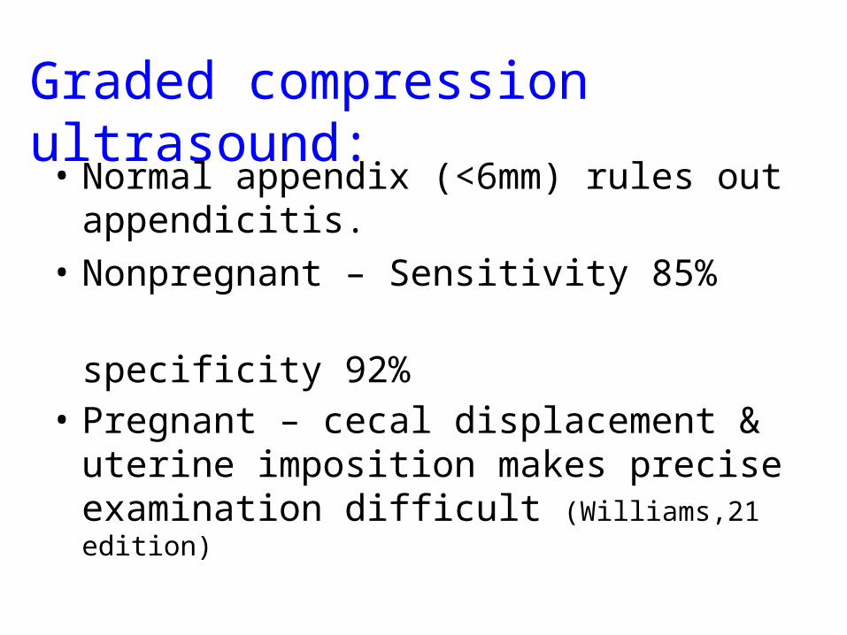
Graded compression ultrasound:• Normal appendix (<6mm) rules out
appendicitis. • Nonpregnant – Sensitivity 85% specificity 92%• Pregnant – cecal displacement & uterine
imposition makes precise examination difficult (Williams,21 edition)
Diagnosis
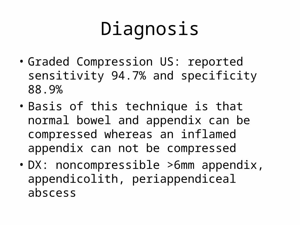
• Graded Compression US: reported sensitivity 94.7% and specificity 88.9%
• Basis of this technique is that normal bowel and appendix can be compressed whereas an inflamed appendix can not be compressed
• DX: noncompressible >6mm appendix, appendicolith, periappendiceal abscess
Diagnosis
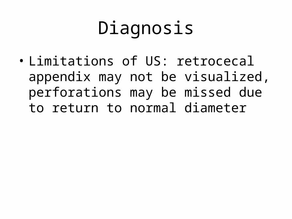
• Limitations of US: retrocecal appendix may not be visualized, perforations may be missed due to return to normal diameter
Diagnosis
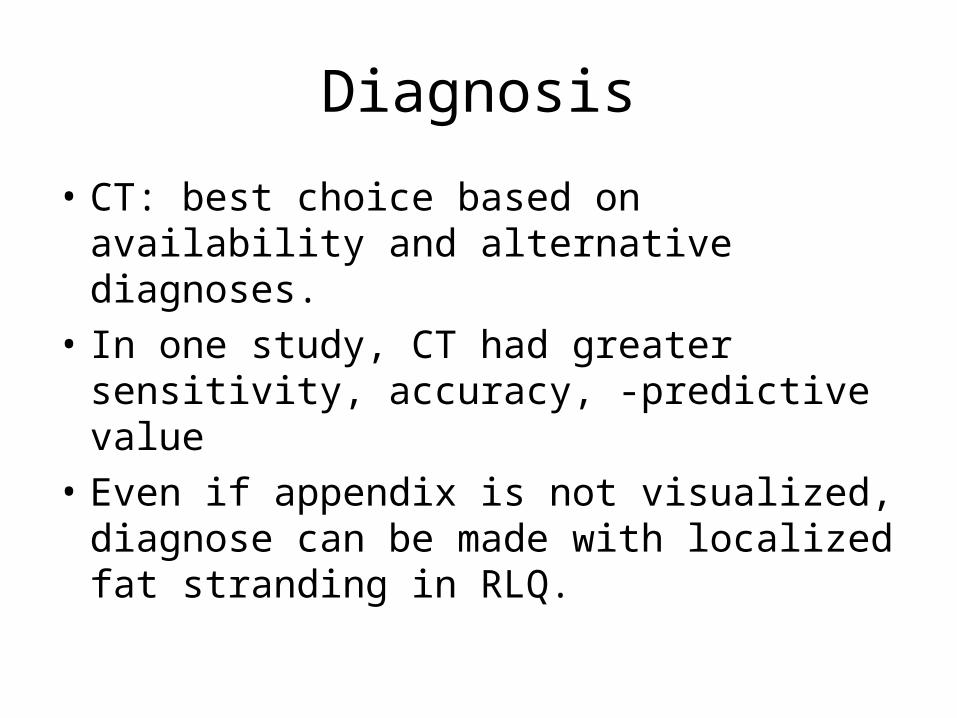
• CT: best choice based on availability and alternative diagnoses.
• In one study, CT had greater sensitivity, accuracy, -predictive value
• Even if appendix is not visualized, diagnose can be made with localized fat stranding in RLQ.
Diagnosis
• CT appears to change management decisions and decreases unnecessary appendectomies in women, but it is not as useful for changing management in men.
Special Populations
• Very young, very old, pregnant, and HIV patients present atypically and often have delayed diagnosis
• High index of suspicion is needed in the these groups to get an accurate diagnosis
Treatment
• Appendectomy is the standard of care• Patients should be NPO, given IVF, and
preoperative antibiotics • Antibiotics are most effective when given
preoperatively and they decrease post-op infections and abscess formation


Treatment
• There are multiple acceptable antibiotics to use as long there is anaerobic flora, enterococci and gram(-) intestinal flora coverage
• One sample monotherapy regimen is Zosyn 3.375g or Unasyn 3g
• Also, short acting narcotics should be used for pain management
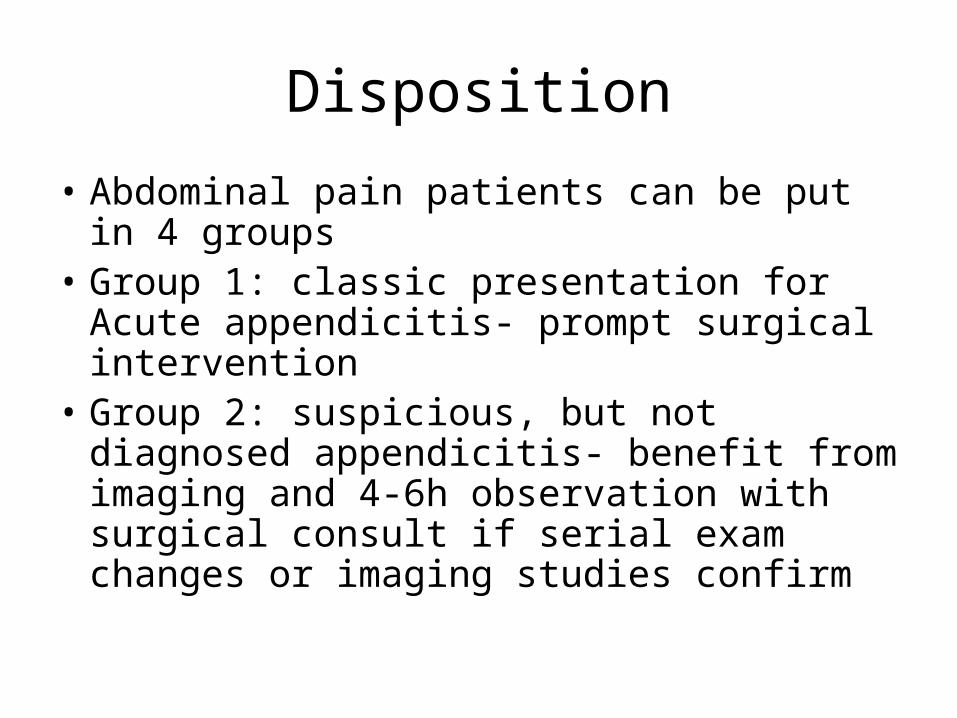
Disposition
• Abdominal pain patients can be put in 4 groups
• Group 1: classic presentation for Acute appendicitis- prompt surgical intervention
• Group 2: suspicious, but not diagnosed appendicitis- benefit from imaging and 4-6h observation with surgical consult if serial exam changes or imaging studies confirm
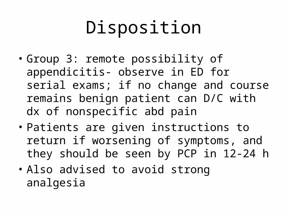
Disposition
• Group 3: remote possibility of appendicitis- observe in ED for serial exams; if no change and course remains benign patient can D/C with dx of nonspecific abd pain
• Patients are given instructions to return if worsening of symptoms, and they should be seen by PCP in 12-24 h
• Also advised to avoid strong analgesia
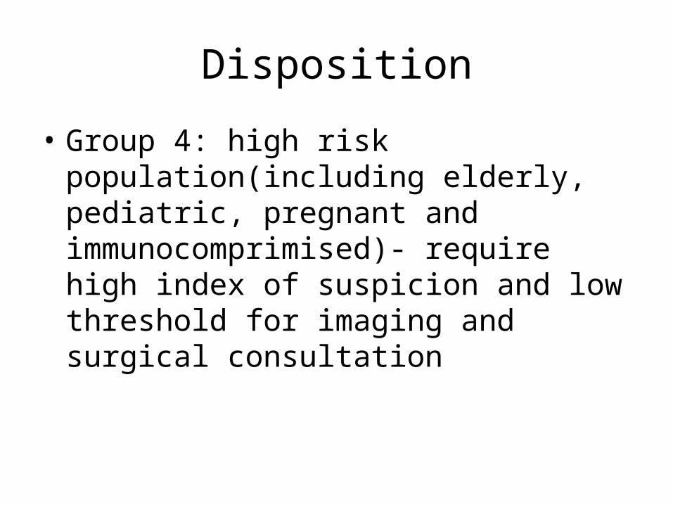
Disposition
• Group 4: high risk population(including elderly, pediatric, pregnant and immunocomprimised)- require high index of suspicion and low threshold for imaging and surgical consultation
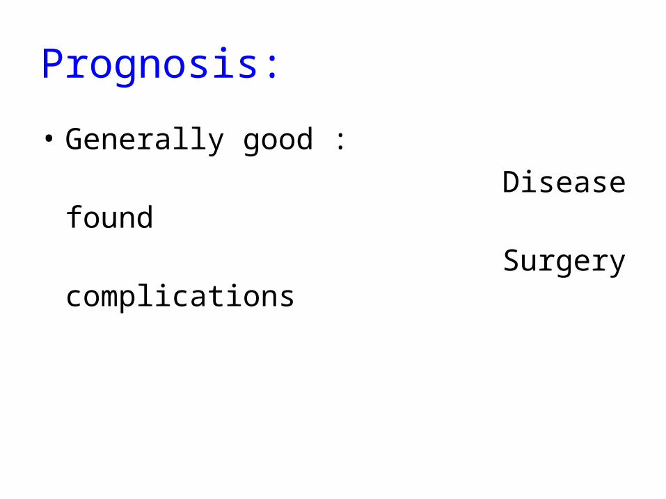
Prognosis:
• Generally good : Disease found Surgery complications

The end


![Case Report Stump Appendicitis: An Uncompleted Surgery, a ... · a er appendectomy, but our case presented only four and half ((/)) months a er laparoscopic appendectomy [, ]. With](https://static.fdocuments.us/doc/165x107/60df3a7ef0e58c30304e41fe/case-report-stump-appendicitis-an-uncompleted-surgery-a-a-er-appendectomy.jpg)


![Laparoscopic or Open Appendectomy for Pediatric …lup.lub.lu.se/search/ws/files/4196774/8611175.pdffor identifying appendicitis [3]. The standard treatment for appendicitis remains](https://static.fdocuments.us/doc/165x107/5feea6f7a3df2365dc7c3e90/laparoscopic-or-open-appendectomy-for-pediatric-luplublusesearchwsfiles4196774.jpg)