AAHFN Heart Failure Review Course - c.ymcdn.com · – Incessant Arrhythmias, tachycardia-induced...
28
6/14/2016 1 Heart Failure Certification Review Course Part 1 Ashley Moore-Gibbs, MSN, AGPCNP-BC, CHFN Objectives ♥ Review the current incidence and prevalence of heart failure (HF) ♥ Discuss the basic pathophysiology of heart failure (HF) ♥ Define HF and discuss various etiologies ♥ Outline the neurohormonal and hemodynamic changes in HF ♥ Discuss how to perform a history and physical exam on a HF patient Heart Failure has Reached Epidemic Proportions Prevalence 5.7 million Americans > 20 years of age have HF - HF will increase 46% from 2012 to 2030 with - Greater than 8 million people > 18 years of age Incidence Between 400,000 – 700,000 new cases/year 75% of HF cases have antecedent HTN Morbidity - > 1 million hospital discharges – - (↑171% from 1979 to 2005) - Most frequent cause of hospitalization in elderly (> 65 y/o) - Accounts for 5% to 10% of all hospital admissions (> 1 million) Mortality One in 9 deaths included HF as contributing cause AHA Heart and Stroke Statistical Update 2015. Circulation.
-
Upload
trinhxuyen -
Category
Documents
-
view
218 -
download
1
Transcript of AAHFN Heart Failure Review Course - c.ymcdn.com · – Incessant Arrhythmias, tachycardia-induced...
6/14/2016
1
Heart Failure Certification
Review CoursePart 1
Ashley Moore-Gibbs, MSN, AGPCNP-BC, CHFN
Objectives
♥ Review the current incidence and prevalence of heart failure (HF)
♥ Discuss the basic pathophysiology of heart failure (HF)
♥ Define HF and discuss various etiologies
♥ Outline the neurohormonal and hemodynamic changes in HF
♥ Discuss how to perform a history and physical exam on a HF patient
Heart Failure has Reached Epidemic Proportions
Prevalence 5.7 million Americans > 20 years of age have HF
- HF will increase 46% from 2012 to 2030 with
- Greater than 8 million people > 18 years of age
Incidence Between 400,000 – 700,000 new cases/year 75% of HF cases have antecedent HTN
Morbidity - > 1 million hospital discharges – - (↑171% from 1979 to 2005)
- Most frequent cause of hospitalization in elderly (> 65 y/o)
- Accounts for 5% to 10% of all hospital admissions (> 1 million)
Mortality One in 9 deaths included HF as contributing cause
AHA Heart and Stroke Statistical Update 2015. Circulation.
6/14/2016
2
Hospitalizations Due to HF Continue to Rise• Progression of disease inevitable
• Incidence of HF rising
– Population of US is aging
• 36 million over age 65 years in 2003, 87 million in 2050
• In 2010, “Baby Boomers” began reaching 65 years
– Survival has improved with acute myocardial infarction and revascularization
• HF is not treated appropriately during hospitalization
• Patients do not adhere to diet and drugs
• By 2030, total Cost of HF will almost 127% ($69.7 billion from the total cost of $30.7 billion in 2012
AHA Heart and Stroke Statistical Update 2015. Circulation.
Implications of a Diagnosis of HF
• Mortality rates at 1 to 2 years estimated at 35%- 50% for advanced heart failure– 4-5 year mortality rate ranges 15% - 40% with mild to moderate symptoms
– 5 year survival following hospitalization is ~ 42.3%; each re-hospitalization increases mortality by 20% - 22%
• Eighty percent of men and 70% of women younger than 65 years of age die within one year
• Re-hospitalization rates 6 months following discharge are as much as 50%
AHA Heart and Stroke Statistical Update 2015. Circulation.
Problems with Conventional Management
• Reactive approach
• Urgency to discharge home – short term management of acute problem that is chronic in nature
• Problems with continuity of care
• Patients do not seek attention in a timely manner - 80% admitted when presenting to ED
• May not receive adequate care
Fonarow, GC. ADHERE Rev Cardiovasc Med. 2003;4 Suppl 7:S21-30.
6/14/2016
3
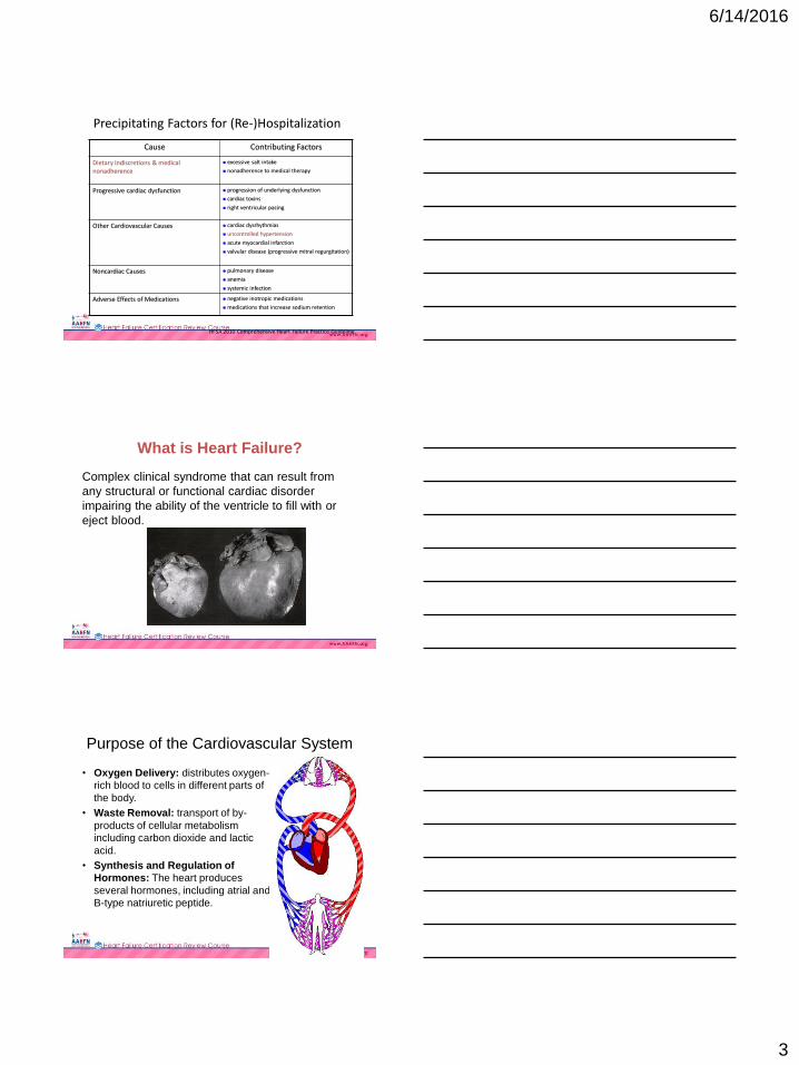
Precipitating Factors for (Re-)Hospitalization
Cause Contributing Factors
Dietary indiscretions & medical nonadherence
excessive salt intake
nonadherence to medical therapy
Progressive cardiac dysfunction progression of underlying dysfunction
cardiac toxins
right ventricular pacing
Other Cardiovascular Causes cardiac dysrhythmias
uncontrolled hypertension
acute myocardial infarction
valvular disease (progressive mitral regurgitation)
Noncardiac Causes pulmonary disease
anemia
systemic infection
Adverse Effects of Medications negative inotropic medications
medications that increase sodium retention
HFSA 2010 Comprehensive Heart Failure Practice Guideline
Complex clinical syndrome that can result from
any structural or functional cardiac disorder
impairing the ability of the ventricle to fill with or
eject blood.
What is Heart Failure?
Purpose of the Cardiovascular System
• Oxygen Delivery: distributes oxygen-
rich blood to cells in different parts of
the body.
• Waste Removal: transport of by-
products of cellular metabolism
including carbon dioxide and lactic
acid.
• Synthesis and Regulation of
Hormones: The heart produces
several hormones, including atrial and
B-type natriuretic peptide.
6/14/2016
4
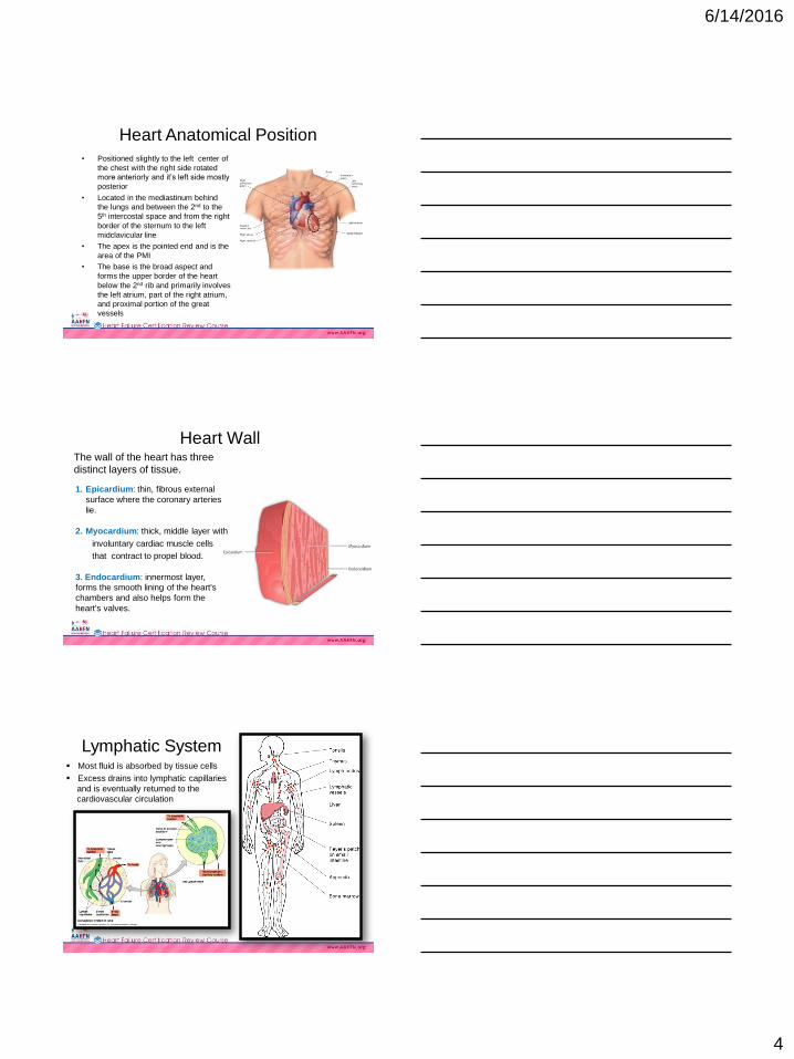
Heart Anatomical Position
• Positioned slightly to the left center of
the chest with the right side rotated
more anteriorly and it’s left side mostly
posterior
• Located in the mediastinum behind
the lungs and between the 2nd to the
5th intercostal space and from the right
border of the sternum to the left
midclavicular line
• The apex is the pointed end and is the
area of the PMI
• The base is the broad aspect and
forms the upper border of the heart
below the 2nd rib and primarily involves
the left atrium, part of the right atrium,
and proximal portion of the great
vessels
Heart WallThe wall of the heart has three
distinct layers of tissue.
1. Epicardium: thin, fibrous external
surface where the coronary arteries
lie.
2. Myocardium: thick, middle layer with
involuntary cardiac muscle cells
that contract to propel blood.
3. Endocardium: innermost layer,
forms the smooth lining of the heart's
chambers and also helps form the
heart's valves.
Lymphatic System Most fluid is absorbed by tissue cells
Excess drains into lymphatic capillaries
and is eventually returned to the
cardiovascular circulation
6/14/2016
5
Defining and Describing Heart Failure
• Ischemic versus Non ischemic Cardiomyopathies
• Heart failure in the setting of Reduced Left Ventricular Ejection Fraction (LVEF) versus Preserved LVEF
• Left-sided, Right-sided and Biventricular Heart Failure
• Remodeling versus Reverse Remodeling
• Chronic versus Acute Decompensated Heart Failure
• Staging & Classifying Heart Failure
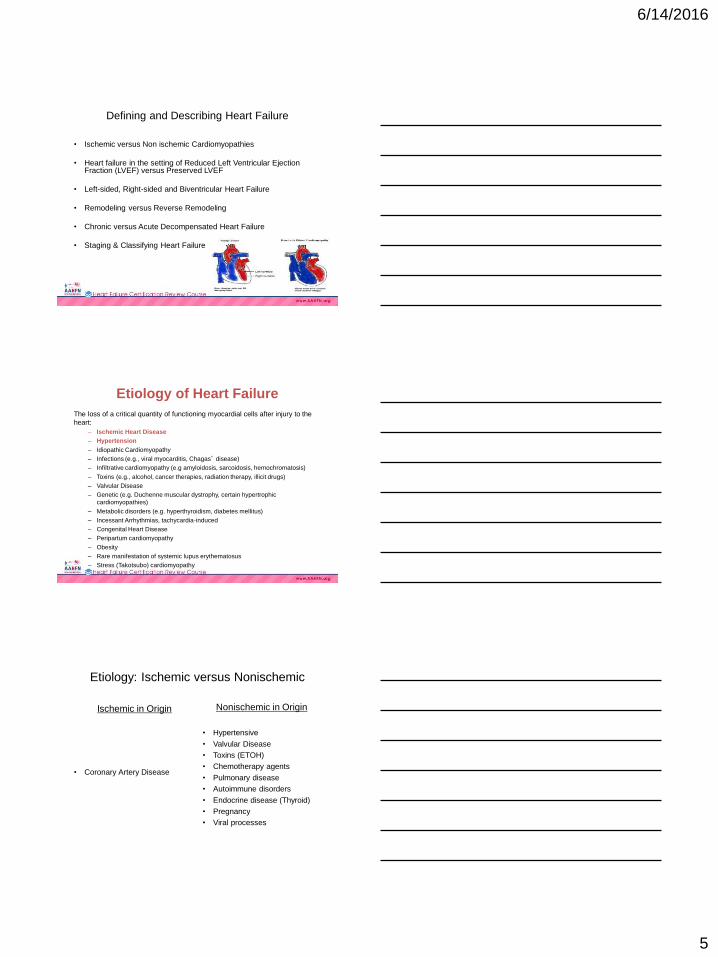
Etiology of Heart Failure
The loss of a critical quantity of functioning myocardial cells after injury to the
heart:
– Ischemic Heart Disease
– Hypertension
– Idiopathic Cardiomyopathy
– Infections (e.g., viral myocarditis, Chagas’ disease)
– Infiltrative cardiomyopathy (e.g amyloidosis, sarcoidosis, hemochromatosis)
– Toxins (e.g., alcohol, cancer therapies, radiation therapy, illicit drugs)
– Valvular Disease
– Genetic (e.g. Duchenne muscular dystrophy, certain hypertrophic
cardiomyopathies)
– Metabolic disorders (e.g. hyperthyroidism, diabetes mellitus)
– Incessant Arrhythmias, tachycardia-induced
– Congenital Heart Disease
– Peripartum cardiomyopathy
– Obesity
– Rare manifestation of systemic lupus erythematosus
– Stress (Takotsubo) cardiomyopathy
Etiology: Ischemic versus Nonischemic
Ischemic in Origin
• Coronary Artery Disease
Nonischemic in Origin
• Hypertensive
• Valvular Disease
• Toxins (ETOH)
• Chemotherapy agents
• Pulmonary disease
• Autoimmune disorders
• Endocrine disease (Thyroid)
• Pregnancy
• Viral processes
6/14/2016
6
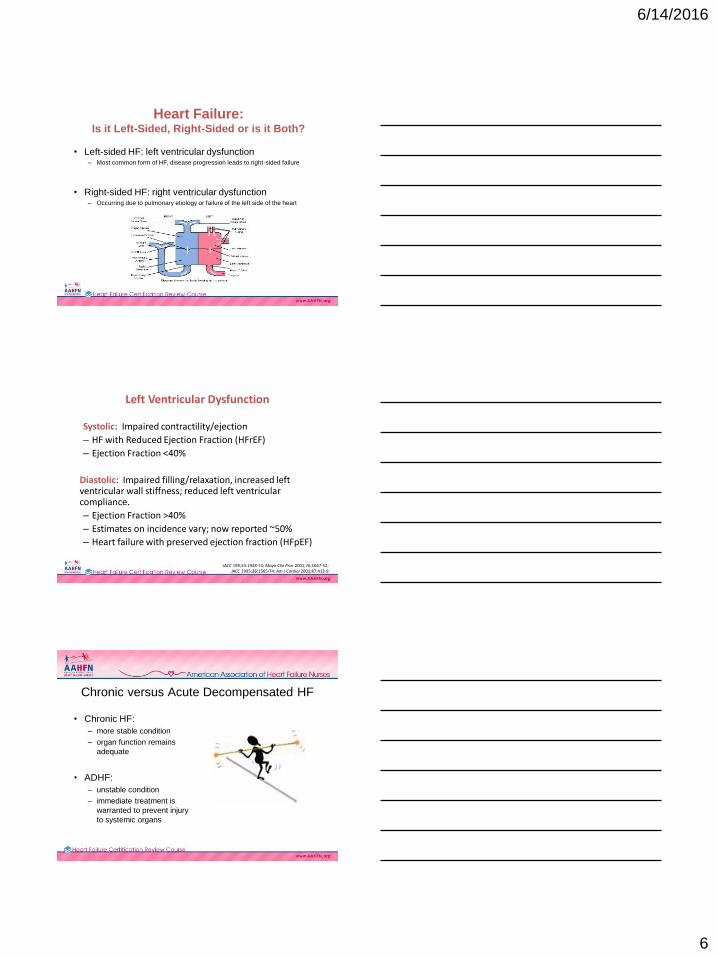
Heart Failure: Is it Left-Sided, Right-Sided or is it Both?
• Left-sided HF: left ventricular dysfunction– Most common form of HF, disease progression leads to right-sided failure
• Right-sided HF: right ventricular dysfunction– Occurring due to pulmonary etiology or failure of the left side of the heart
Left Ventricular Dysfunction
Systolic: Impaired contractility/ejection
– HF with Reduced Ejection Fraction (HFrEF)
– Ejection Fraction <40%
Diastolic: Impaired filling/relaxation, increased left ventricular wall stiffness; reduced left ventricular compliance.
– Ejection Fraction >40%
– Estimates on incidence vary; now reported ~50%
– Heart failure with preserved ejection fraction (HFpEF)
JACC 199;33:1948-55; Mayo Clin Proc 2001;76:1047-52; JACC 1995;26:1565-74; Am J Cardiol 2001;87:413-9.
Chronic versus Acute Decompensated HF
• Chronic HF:
– more stable condition
– organ function remains
adequate
• ADHF:
– unstable condition
– immediate treatment is
warranted to prevent injury
to systemic organs
6/14/2016
7
Case Study 1Defining HF, HF symptoms, History & Physical Exam,
Class/Stage of HF, ECHO, HF Medical Therapy
DF is a 72 year old male with a long-standing history of an ischemic
cardiomyopathy. Prior echocardiogram performed in Jan 2012 showed
LVEF 40%. He has been added on to the office schedule for cardiac
evaluation for complaints of shortness of breath (SOB).
An echocardiogram is obtained the day of the office visit.
19
Heart Failure Pathophysiology
Myocardial injury Fall in LV performance
Morbidity
and mortality
Peripheral vasoconstriction
Sodium & water retentionMyocardial toxicity
Change in gene
expression
Heart failure
symptoms
Remodeling and
progressive worsening
of LV function
Activation of RAAS, SNS, ET,
and others
Shah M, Ali V. Rev Cardiovasc Med. 2001;2(suppl 2):S2–S6.
ANP
BNP
Physiologic Effects of NeurohormonesRAAS (Renin-Angiotensin-Aldosterone System)
Activation of AT1
receptors by angiotensin
VasoconstrictionSodium retentionIncreased aldosterone releaseIncreased cellular growthIncreased sympathetic nervous activity
Natriuretic Peptide System
ANP, BNP
VasodilationSodium excretionDecreased aldosterone levelsInhibition of RAASInhibition of sympathetic nervous activityAntiproliferation of vascular smooth muscle cells
ANP = atrial natriuretic peptide, AT1 = angiotensin I, BNP = endogenous B-type natriuretic peptide
6/14/2016
8
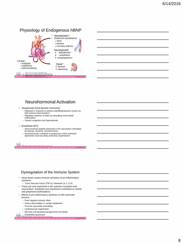
• Hemodynamic1,2
• (balanced vasodilation)
veins
arteries
coronary arteries
Neurohumoral2
aldosterone4
endothelin2
norepinephrine4
Renal1,5
diuresis
natriuresis
DR I
MKRG
S SS
SGLG
FC
CS S
GSGQVM
K V L RR
H
KPS
Cardiac3
lusitropic antifibrotic antiremodeling
1. Marcus LS et al. Circulation. 1996;94:3184–3189.2. Zellner C et al. Am J Physiol. 1999;276(3 pt 2):H1049–H1057.3. Tamura N et al. Proc Natl Acad Sci U S A. 2000;97:4239–4244.4. Abraham WT et al. J Card Fail. 1998;4:37–44.5. Clemens LE et al. J Pharmacol Exp Ther. 1998;287:67–71.
Physiology of Endogenous hBNP
Neurohormonal Activation
• Vasopressin (Anti-diuretic Hormone)– Released in response to arterial underfilling/reduced volume via
high pressure baroreceptors
– Regulates retention of water by stimulating renal tubular reabsorption
– Causes congestion and hyponatremia
• Endothelin (ET)– Vasoconstrictor peptide expressed in the myocardium stimulated
by hypoxia, ischemia, neurohormones
– Increased levels contribute to progression of left ventricular dysfunction and secondary pulmonary hypertension
Dysregulation of the Immune System
• Heart failure causes immune activation of pro-inflammatory
cytokines:
– Tumor Necrosis Factor (TNF-α), Interleukin (IL-1, IL-6)
• These are over-expressed in the systemic circulation and
myocardium. Sustained over-expression contributes to central
and peripheral manifestations
• Effects of pro-inflammatory cytokines on left ventricular
function:
– Exert negative inotropic effect
– Induce abnormalities in cardiac metabolism
– Promote myocardial remodeling
– Cardiomyocyte hypertrophy
– Necrosis and Apoptosis (programmed cell death)
– Endothelial dysfunction
6/14/2016
9
Compensatory Mechanisms in HF
1. Tachycardia and increase contractility
2. Increase preload from the release of renin and
aldosterone
3. Vasoconstriction
4. Ventricular hypertrophy
5. Remodeling
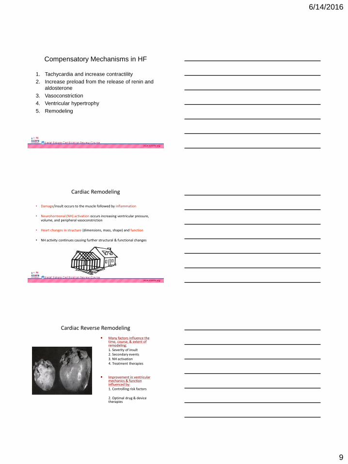
Cardiac Remodeling
• Damage/insult occurs to the muscle followed by inflammation
• Neurohormonal (NH) activation occurs increasing ventricular pressure, volume, and peripheral vasoconstriction
• Heart changes in structure (dimensions, mass, shape) and function
• NH activity continues causing further structural & functional changes
Cardiac Reverse Remodeling
Many factors influence the time, course, & extent of remodeling:1. Severity of insult2. Secondary events3. NH activation4. Treatment therapies
Improvement in ventricular mechanics & function influenced by:1. Controlling risk factors
2. Optimal drug & device therapies
6/14/2016
10
Chronotropy: RATE at which the heart contracts
– Important when looking at cardiac output
Inotropy: STRENGTH at which the heart contracts
Cardiac Vocabulary
Normal Cardiac Function
Cardiac Output= HR x SV
Cardiac Output is the volume of blood pumped per minute, measured by the following equation:
CO = SV x HR
• CO is expressed in L/min (normal ~5L/min )
• CI is the output indexed to body size
• SV is stroke volume ejected per beat
• HR is the number of beats per minute
Cardiac Output
6/14/2016
11
Heart Rate is…
Increased:
• Beta stimulation
• Increase demand
– Fever, exercise, volume depletion
Decreased:
• Beta blockade
• Vagal stimulation
• Decrease metabolic demand
– Cooling, paralyze, sedation
Impacting Hemodynamic Components
Determined by 3 factors:
Preload, Afterload, and Contractility.
–Preload: the VOLUME of blood that the ventricle has available to pump
–Contractility: the FORCE that the muscle can create at it’s given length
–Afterload is the arterial pressure or RESISTANCE against which the muscle will contract.
These factors establish the volume of blood pumped with each heart beat.
Stroke Volume (SV)
Preload: the muscle length prior to contractility, and it is
dependent on diastole (or end diastolic volume…EDV)
– Ventricle fills during this time
•The most important determining factor for preload is
venous return (in other words your JVP or CVP).
– Think of it in terms of volume but don’t forget the ability of the heart
to eject it!
Cardiac Vocabulary
6/14/2016
12
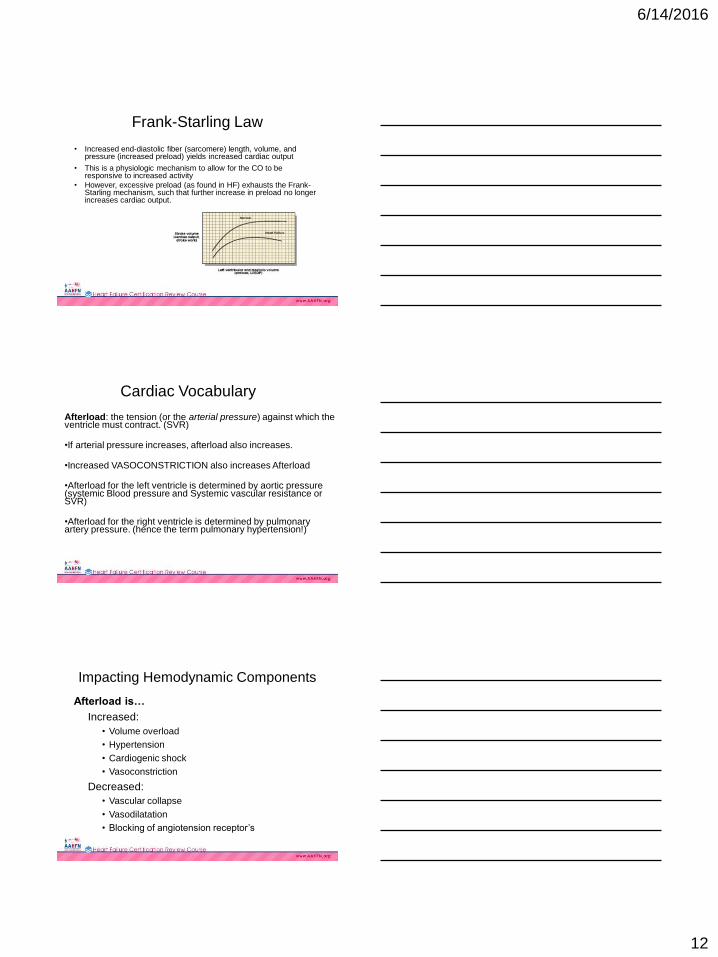
Frank-Starling Law
• Increased end-diastolic fiber (sarcomere) length, volume, and pressure (increased preload) yields increased cardiac output
• This is a physiologic mechanism to allow for the CO to be responsive to increased activity
• However, excessive preload (as found in HF) exhausts the Frank-Starling mechanism, such that further increase in preload no longer increases cardiac output.
Afterload: the tension (or the arterial pressure) against which the ventricle must contract. (SVR)
•If arterial pressure increases, afterload also increases.
•Increased VASOCONSTRICTION also increases Afterload
•Afterload for the left ventricle is determined by aortic pressure (systemic Blood pressure and Systemic vascular resistance or SVR)
•Afterload for the right ventricle is determined by pulmonary artery pressure. (hence the term pulmonary hypertension!)
Cardiac Vocabulary
Afterload is…
Increased:
• Volume overload
• Hypertension
• Cardiogenic shock
• Vasoconstriction
Decreased:
• Vascular collapse
• Vasodilatation
• Blocking of angiotension receptor’s
Impacting Hemodynamic Components
6/14/2016
13
Contractility is…
Increased:
• Inotropy
• Decrease SVR and systemic BP
Decreased:
• Increase SVR along with systemic BP
• Hypovolemia
Impacting Hemodynamic Components
Cardiac Output
Determined by:
1. Stroke volume:
• preload: force used to stretch heart muscle and directly
related to contraction
• afterload: resistance the heart must overcome
• contractility: amount of myofibril shortening
2. Heart Rate: controlled by autonomic nervous system
Clinical Presentation
• Symptoms vary widely
• Symptoms may be vague to severely acute
• Symptoms of HF can be similar to those of other conditions, i.e. COPD exacerbation
6/14/2016
14
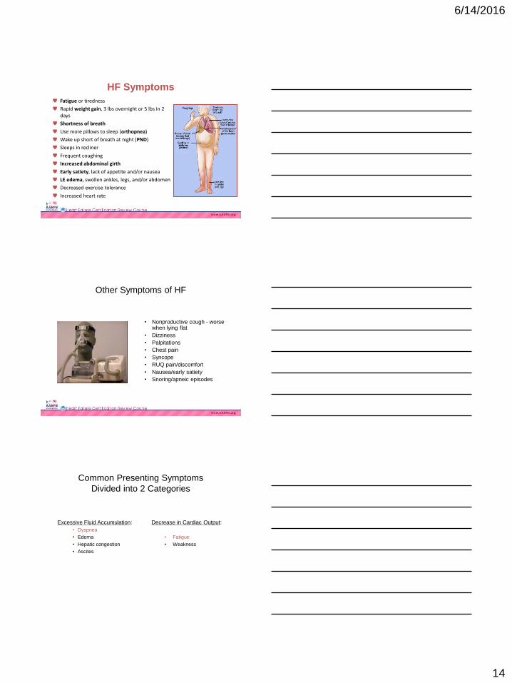
HF Symptoms
♥ Fatigue or tiredness
♥ Rapid weight gain, 3 lbs overnight or 5 lbs in 2 days
♥ Shortness of breath
♥ Use more pillows to sleep (orthopnea)
♥ Wake up short of breath at night (PND)
♥ Sleeps in recliner
♥ Frequent coughing
♥ Increased abdominal girth
♥ Early satiety, lack of appetite and/or nausea
♥ LE edema, swollen ankles, legs, and/or abdomen
♥ Decreased exercise tolerance
♥ Increased heart rate
Other Symptoms of HF
• Nonproductive cough - worse when lying flat
• Dizziness
• Palpitations
• Chest pain
• Syncope
• RUQ pain/discomfort
• Nausea/early satiety
• Snoring/apneic episodes
Common Presenting Symptoms
Divided into 2 Categories
Excessive Fluid Accumulation:
• Dyspnea
• Edema
• Hepatic congestion
• Ascites
Decrease in Cardiac Output:
• Fatigue
• Weakness
6/14/2016
15
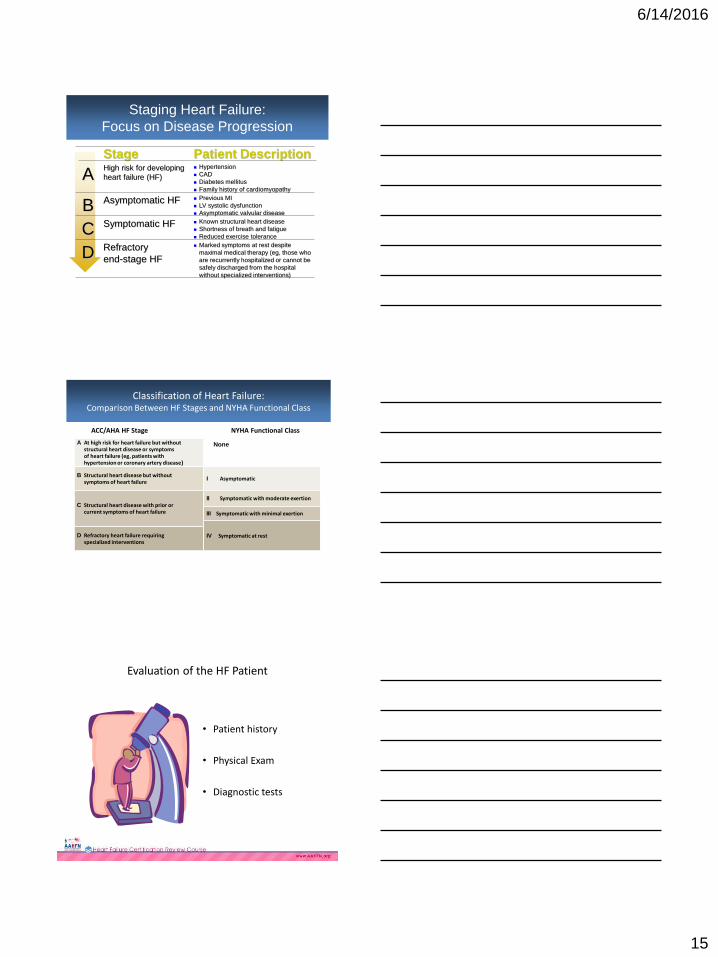
Staging Heart Failure:
Focus on Disease Progression
Marked symptoms at rest despite
maximal medical therapy (eg, those who
are recurrently hospitalized or cannot be
safely discharged from the hospital
without specialized interventions)
Refractory
end-stage HFD
Known structural heart disease
Shortness of breath and fatigue
Reduced exercise tolerance
Symptomatic HFC
Previous MI
LV systolic dysfunction
Asymptomatic valvular disease
Asymptomatic HFB
Hypertension
CAD
Diabetes mellitus
Family history of cardiomyopathy
High risk for developing
heart failure (HF)A
Patient DescriptionStage
Classification of Heart Failure: Comparison Between HF Stages and NYHA Functional Class
ACC/AHA HF Stage NYHA Functional Class
A At high risk for heart failure but withoutstructural heart disease or symptomsof heart failure (eg, patients withhypertension or coronary artery disease)
B Structural heart disease but withoutsymptoms of heart failure
C Structural heart disease with prior orcurrent symptoms of heart failure
D Refractory heart failure requiringspecialized interventions
I Asymptomatic
II Symptomatic with moderate exertion
IV Symptomatic at rest
III Symptomatic with minimal exertion
None
Evaluation of the HF Patient
• Patient history
• Physical Exam
• Diagnostic tests
6/14/2016
16
A thorough history and physical examination should beobtained/performed in patients presenting with HF toidentify cardiac and noncardiac disorders or behaviors that might cause or accelerate the development or progression of HF.
In patients with idiopathic DCM, a 3-generational family history should be obtained to aid in establishing the diagnosis of familial DCM.
Volume status and vital signs should be assessed at each patient encounter. This includes serial assessment of weight, as well as estimates of jugular venous pressure and the presence of peripheral edema or orthopnea.
History and Physical ExaminationI IIa IIb III
I IIa IIb III
I IIa IIb III
2013 ACCF/AHA Heart Failure Guidelines
Components of the Health History
1. Biographical data
2. Reason for seeking care
3. Present health or history
of present illness
4. Past history
5. Review of systems
6. Social history
7. Family history
1. Biographical Data
• Name, address, age, DOB, birthplace, gender, marital
status, race, ethnic origin, occupation
• Language spoken
• Communication needs
• Source of history
– Record who furnishes information
– Record if the patient seems willing and reliable
– Note any special circumstances
6/14/2016
17
2. Reason for Seeking Care
• Brief statement in the person’s own words that describes the reason for the visit
– “Title of the story”
• It states 2 things:
– Symptom
– Sign
• Things to watch for:
– It is not a diagnostic statement
– Watch for self-diagnosing
– Patient’s that list reasons for seeking care – which one is the
most important?
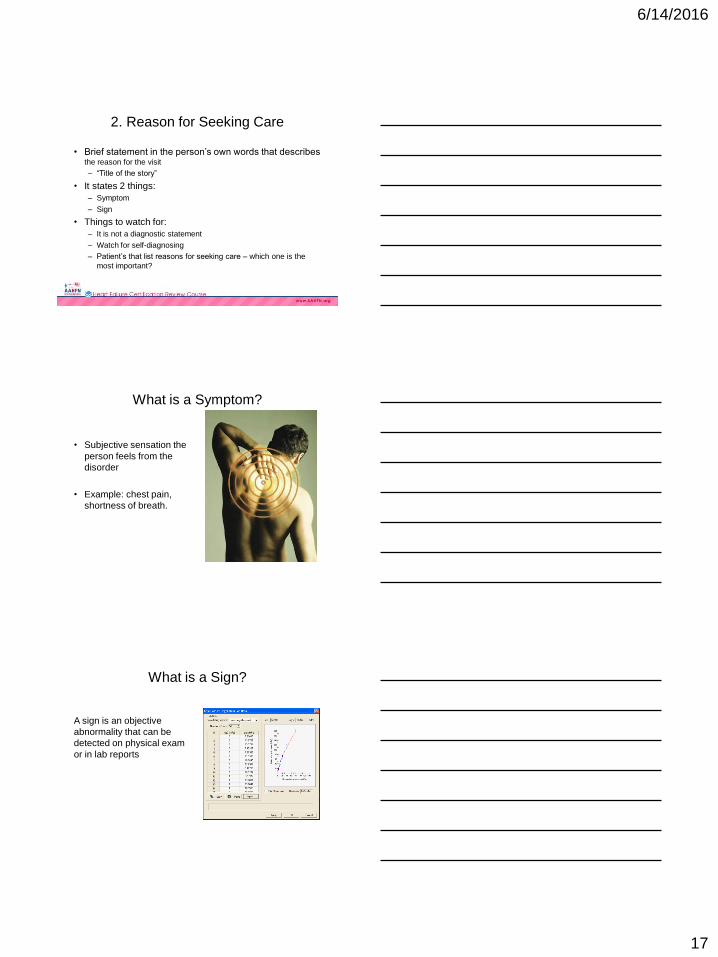
What is a Symptom?
• Subjective sensation the
person feels from the
disorder
• Example: chest pain,
shortness of breath.
What is a Sign?
A sign is an objective
abnormality that can be
detected on physical exam
or in lab reports
6/14/2016
18
3. History of Present Illness
• Includes age, sex, race & occupation
• Well patient’s will have a short HPI
– Short statement about their general health
• Ill patients require further description that reviews:
– Chronologic record of reason for seeking care
– Time symptom first started
– Give details of all symptoms concerned in the illness including
location, character, severity, duration, intermittency, and radiation
of pain. Describe factors making pain worse of better
Characteristics of any Symptom
• Location
– Specific
• Quality
– Requires descriptive terms
• Severity
– Quantifies the sign or
symptom
• Timing
– Onset, duration, frequency
Characteristics of any Symptom Continued
• Setting
– Where was patient
– What brings it on
• Aggravating or relieving factors
– What makes it worse
– What relieves it
• Associated factors
– Are there any other
symptoms
• Patient’s perception
– Effecting ADL
6/14/2016
19
PQRSTU: A Way to Remember how to
Define the Characteristics of Symptoms
• P: Provocative or Palliative
• Q: Quality or Quantity
• R: Region or Radiation
• S: Severity Scale
• T: Timing
• U: Understanding patient perception
4. Past Medical (Health) History
• Childhood illnesses
• Accidents or injuries
• Chronic illnesses
• Hospitalizations
• Operations
• Obstetric history
• Immunizations
• Last Examination date
• Allergies
• Current medications
Relevant Past Medical & Surgical History
• Prior history of HF or myocardial infarction
• CABG/PCIs
• Valvular repairs/replacement
• Device therapy
• HTN
• OSA
• Anemia
• ETOH
6/14/2016
20
5. Review of Systems
• ROS addresses the following:
– Evaluates the past &
present health state of
each body system
– Evaluates health promotion
practices
6. Social History
• Place of birth and residence
• Marital status
• Habits including:
• Self-esteem, self-concept
• Activity/exercise– ability to perform self-care or activities of daily living (ADLs)
– Activities needed for independence, ie grocery shopping, laundry, nutrition, managing finances,
cooking (IADLs)
• Sleep/rest
• Nutrition/elimination
• Interpersonal relationships/resources– Intimate partner violence
• Spiritual resources
• Occupation: past and present, service in military
• Environmental factors:
• Name/contact info of physicians
7. Family History
• Health or age of death & cause
• Close family members
– Contact with communicable
diseases
6/14/2016
21
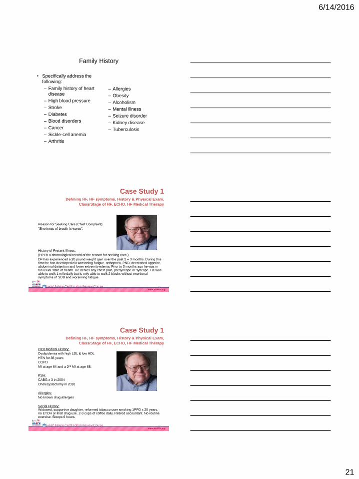
Family History
• Specifically address the
following:
– Family history of heart
disease
– High blood pressure
– Stroke
– Diabetes
– Blood disorders
– Cancer
– Sickle-cell anemia
– Arthritis
– Allergies
– Obesity
– Alcoholism
– Mental illness
– Seizure disorder
– Kidney disease
– Tuberculosis
Case Study 1Defining HF, HF symptoms, History & Physical Exam,
Class/Stage of HF, ECHO, HF Medical Therapy
Reason for Seeking Care (Chief Complaint):
“Shortness of breath is worse”.
History of Present Illness:
(HPI is a chronological record of the reason for seeking care.)
DF has experienced a 20 pound weight gain over the past 2 – 3 months. During this time he has developed c/o worsening fatigue, orthopnea, PND, decreased appetite, abdominal distention and lower extremity edema. Prior to 3 months ago he was in his usual state of health. He denies any chest pain, presyncope or syncope. He was able to walk 1 mile daily but is only able to walk 2 blocks without exertional symptoms of SOB and worsening fatigue.
62
Case Study 1Defining HF, HF symptoms, History & Physical Exam,
Class/Stage of HF, ECHO, HF Medical Therapy
Past Medical History:
Dyslipidemia with high LDL & low HDL
HTN for 35 years
COPD
MI at age 64 and a 2nd MI at age 68.
PSH:
CABG x 3 in 2004
Cholecystectomy in 2010
Allergies:
No known drug allergies
Social History:Widowed, supportive daughter, reformed tobacco user smoking 1PPD x 20 years, no ETOH or illicit drug use. 2-3 cups of coffee daily. Retired accountant. No routine exercise. Sleeps 6 hours.
63

6/14/2016
22
Focused Physical Exam
• Vital Signs
• Neck
• Pulmonary
• Cardiovascular
• Abdominal
• Extremities
Vital Signs
• Blood pressure
• Orthostatic blood pressure
• Heart rate
• Respiratory rate
• Temperature
Vital Signs
• Blood pressure
– Measure of the heart’s ability to pump & indicator of degree of
antihypertensive from medications
– Orthostatic readings helpful in determining dehydration & over-
diuresis
• Heart rate
– Helpful in indicating arrhythmias
– Resting tachycardia may be an indicator of poor prognosis
– Bradycardia may indicate heart block or over medication
6/14/2016
23
Vital Signs
• Blood pressure
– Measure of the heart’s ability to pump & indicator of degree of
antihypertensive from medications
– Orthostatic readings helpful in determining dehydration & over-
diuresis
• Heart rate
– Helpful in indicating arrhythmias
– Resting tachycardia may be an indicator of poor prognosis
– Bradycardia may indicate heart block or over medication
Neck Examination
• Carotid bruits
• Jugular venous distention
• Abdominojugular reflux
Abnormal Lung Sounds
Rales or crackles are lung
sounds that are brief,
discrete, nonmusical
sounds with a popping
quality. Typically heard in
the bases.
6/14/2016
24
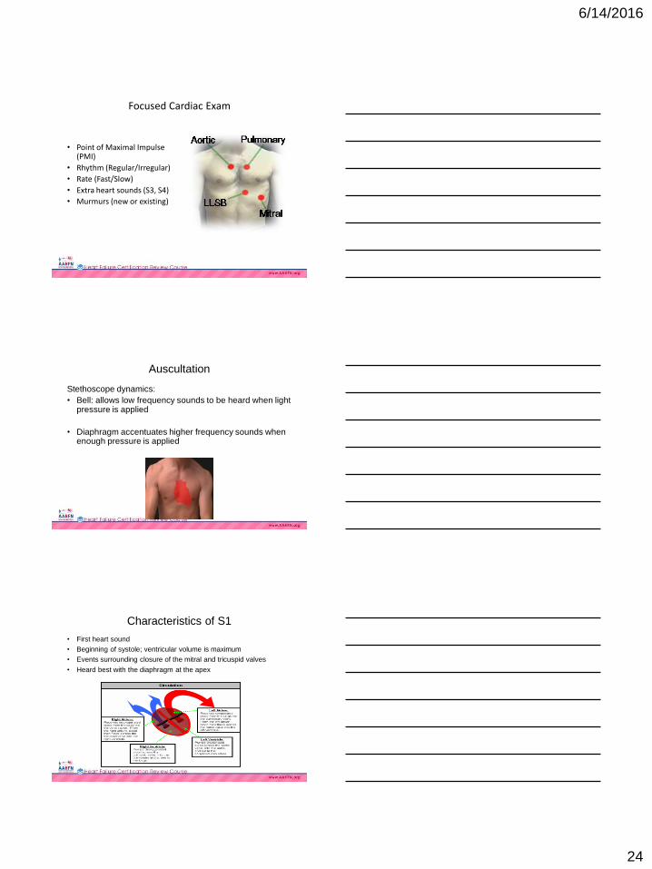
Focused Cardiac Exam
• Point of Maximal Impulse (PMI)
• Rhythm (Regular/Irregular)
• Rate (Fast/Slow)
• Extra heart sounds (S3, S4)
• Murmurs (new or existing)
Auscultation
Stethoscope dynamics:
• Bell: allows low frequency sounds to be heard when light pressure is applied
• Diaphragm accentuates higher frequency sounds when enough pressure is applied
Characteristics of S1
• First heart sound
• Beginning of systole; ventricular volume is maximum
• Events surrounding closure of the mitral and tricuspid valves
• Heard best with the diaphragm at the apex
6/14/2016
25
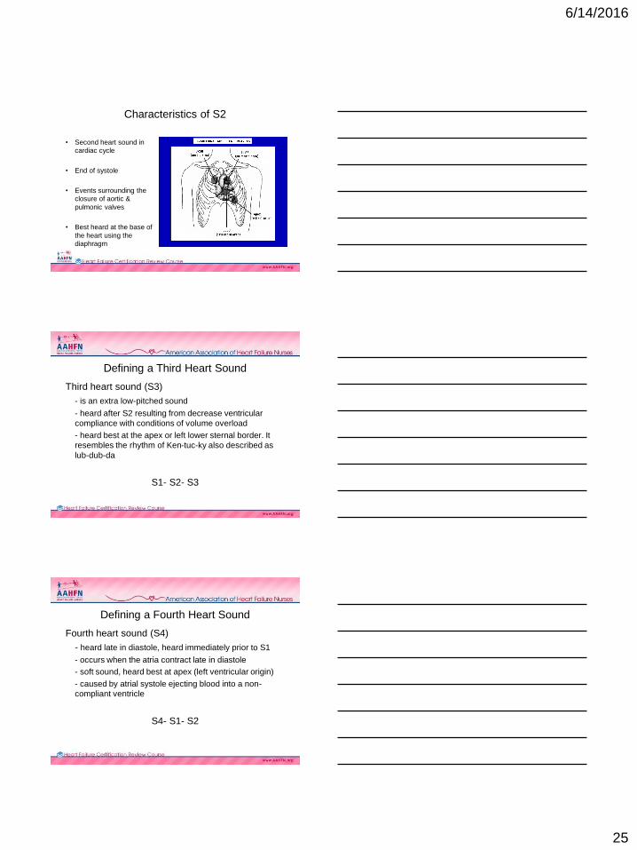
Characteristics of S2
• Second heart sound in
cardiac cycle
• End of systole
• Events surrounding the
closure of aortic &
pulmonic valves
• Best heard at the base of
the heart using the
diaphragm
Defining a Third Heart Sound
Third heart sound (S3)
- is an extra low-pitched sound
- heard after S2 resulting from decrease ventricular
compliance with conditions of volume overload
- heard best at the apex or left lower sternal border. It
resembles the rhythm of Ken-tuc-ky also described as
lub-dub-da
S1- S2- S3
Defining a Fourth Heart Sound
Fourth heart sound (S4)
- heard late in diastole, heard immediately prior to S1
- occurs when the atria contract late in diastole
- soft sound, heard best at apex (left ventricular origin)
- caused by atrial systole ejecting blood into a non-
compliant ventricle
S4- S1- S2
6/14/2016
26
Heart Murmurs
• Originate from failure of heart valves to open
adequately (stenosis) or failure to close
(incompetence causing regurgitation)
• Divided into benign or pathologic
• Sounds that are heard as harsh (AS), rumble
(MS), blowing (AR)
Grading Murmurs
• Grade I: very, very faint
• Grade II: quiet but immediately heard
• Grade III: moderately loud without a thrill
• Grade IV: loud with a thrill
• Grade V: very loud with a thrill
• Grade VI: audible without a stethoscope
Abdominal Exam
• Right upper quadrant
tenderness
• Enlarged, palpable liver
border > 3 fingerbreadths
below the costal margin
• Pulsatile liver
• Ascites
6/14/2016
27
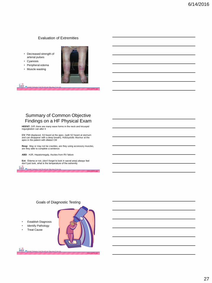
Evaluation of Extremities
• Decreased strength of
arterial pulses
• Cyanosis
• Peripheral edema
• Muscle wasting
Summary of Common Objective
Findings on a HF Physical ExamHEENT: JVP, there are many wave forms in the neck and tricuspid regurgitation can alter it
CV: PMI displaced, S3 heard at the apex, (split S2 heard at sternum and can disappear with a deep breath), Holosystolic Murmur at the apex in the patient with dilated CM
Resp: May or may not be crackles, are they using accessory muscles, are they able to complete a sentence
ABD: HJR, Hepatomegaly, Ascites from RV failure
Ext: Edema or not, (don’t forget to look in sacral area) always feel don’t just look, what is the temperature of the extremity
Goals of Diagnostic Testing
• Establish Diagnosis
• Identify Pathology
• Treat Cause
6/14/2016
28
Laboratory Data Evaluation
Lab Test Assessing for Possible
Precipitating Factors
Cardiac Troponin Ongoing ischemia/ infarction
Complete Blood
Count
Anemia or infection
Liver Function Tests Poor hepatic function
D-dimer Pulmonary embolus
Thyroid Profile Hyper- or hypothyroid disorders
Renal Function
Studies
Renal dysfunction/ failure
Urinalysis Urinary tract infection,
proteinuria
Arterial Blood Gases Hypoxia, comorbid lung
problems
Cardiac BNP Levels
• 32-amino acid peptide secreted primarily from the ventricles of the heart in response to stretch and increased volume in the ventricles
• BNP < 100pg/ml negative
• BNP > 500pg/ml positive
• BNP levels correlate with:– LV end-diastolic pressure and volume
– NYHA classification
– Trends in BNP more meaningful
Maisel et al. N Engl J Med 2002;347:161-7