A Rare Case of Unilateral Pleural Effusion in a Pediatric ...
5
Case report Child Kidney Dis 2018;22:86-90 DOI: https://doi.org/10.3339/jkspn.2018.22.2.86 ISSN 2384-0242 (print) ISSN 2384-0250 (online) A Rare Case of Unilateral Pleural Effusion in a Pediatric Patient on Chronic Peritoneal Dialysis: Is it a Pleuroperi - toneal Leakage? Non-infectious complications of peritoneal dialysis (PD) are relatively less common than infectious complications but are a potentially serious problem in patients on chronic PD. Here, we present a case of a non-infectious complication of PD in a 13- year- old boy on chronic PD who presented with symptoms such as hypertension, edema, dyspnea, and decreased ultrafiltration. Chest and abdominal radiography showed pleural effusion and migration of the PD catheter tip. Laparoscopic PD ca- theter reposition was performed because PD catheter malfunction was suspected. However, pleural effusion relapsed whenever the dialysate volume increased. To identify peritoneal leakage, computed tomography (CT) peritoneography was per- formed, and a defect of the peritoneum in the left lower abdomen with contrast leakage to the left rectus and abdominis muscles was observed. He was treated conservatively by transiently decreasing the volume of night intermittent PD and gradually increasing the volume. At the 2-year follow-up visit, the patient had not experienced similar symptoms. Patients on PD who present with refractory or re- current pleural effusion that does not respond to therapy should be assessed for the presence of infection, catheter malfunction, and pleuroperitoneal communi- cation. Thoracentesis and CT peritoneography are useful for evaluating pleural effusion, and timely examination is important for identifying the defect or fistula. Key words: Peritoneal dialysis, Pleuroperitoneal communication, Peritoneography, Pleural effusion, PD catheter Sukdong Yoo, M.D. 1 Jae-Yeon Hwang, M.D., Ph.D. 2 Ji Yeon Song, M.D. 1 Taek Jin Lim, M.D. 1 Narae Lee, M.D. 1 Su Young Kim, M.D., Ph.D. 1 Seong Heon Kim, M.D 1 Department of Pediatrics 1 , Pusan National University Children’s Hospital, Korea, Department of Radiology 2 , Pusan National University Children’s Hospital, Korea Corresponding author: Seong Heon Kim, M.D. Department of Pediatrics, Pusan National University Children’s Hospital, 20 Geumo- ro, Mulgeum-eup, Yangsan, Goungnam 50612, Korea Tel: +82-55-360-3163 Fax: +82-55-360-2181 E-mail: [email protected] Received: 15 September 2018 Revised: 4 October 2018 Accepted: 10 October 2018 This is an open-access article distributed under the terms of the Creative Commons Attribution Non-Commercial License (http:// creativecommons.org/licenses/by-nc/4.0/) which permits unrestricted non-commercial use, distribution, and reproduction in any medium, provided the original work is properly cited. Copyright © 2018 The Korean Society of Pediatric Nephrology Introduction Noninfectious complications of peritoneal dialysis (PD) are relatively less common than infectious complications, but are a potentially serious problem in patients on chronic PD. The well-known complications include catheter- related complications, such as leakage, hernia, hydrothorax, hemoperitoneum, pancreatitis, ischemic colitis, necrotizing enterocolitis, pneumoperitoneum, and metabolic complications 1,2) . Occasionally, such complications may have resulted from a combination of one or more causes, or they can be confused as a different condition. us, a systematic approach is needed when identifying the causes of the PD com- plications. We present herein a patient on chronic PD who presented with unilateral pleural effusion.
Transcript of A Rare Case of Unilateral Pleural Effusion in a Pediatric ...

Case reportChild Kidney Dis 2018;22:86-90DOI: https://doi.org/10.3339/jkspn.2018.22.2.86
ISSN 2384-0242 (print)ISSN 2384-0250 (online)
A Rare Case of Unilateral Pleural Effusion in a Pediatric Patient on Chronic Peritoneal Dialysis: Is it a Pleuroperitoneal Leakage?
Non-infectious complications of peritoneal dialysis (PD) are relatively less common than infectious complications but are a potentially serious problem in patients on chronic PD. Here, we present a case of a non-infectious complication of PD in a 13-year- old boy on chronic PD who presented with symptoms such as hypertension, edema, dyspnea, and decreased ultrafiltration. Chest and abdominal radiography showed pleural effusion and migration of the PD catheter tip. Laparoscopic PD ca-theter reposition was performed because PD catheter malfunction was suspected. However, pleural effusion relapsed whenever the dialysate volume increased. To identify peritoneal leakage, computed tomography (CT) peritoneography was per-formed, and a defect of the peritoneum in the left lower abdomen with contrast leakage to the left rectus and abdominis muscles was observed. He was treated conservatively by transiently decreasing the volume of night intermittent PD and gradually increasing the volume. At the 2-year follow-up visit, the patient had not experienced similar symptoms. Patients on PD who present with refractory or re-current pleural effusion that does not respond to therapy should be assessed for the presence of infection, catheter malfunction, and pleuroperitoneal communi-cation. Thoracentesis and CT peritoneography are useful for evaluating pleural effusion, and timely examination is important for identifying the defect or fistula.
Key words: Peritoneal dialysis, Pleuroperitoneal communication, Peritoneography, Pleural effusion, PD catheter
Sukdong Yoo, M.D.1
Jae-Yeon Hwang, M.D., Ph.D.2
Ji Yeon Song, M.D.1
Taek Jin Lim, M.D.1
Narae Lee, M.D.1
Su Young Kim, M.D., Ph.D.1
Seong Heon Kim, M.D1
Department of Pediatrics1, Pusan National University Children’s Hospital, Korea, Department of Radiology2, Pusan National University Children’s Hospital, Korea
Corresponding author: Seong Heon Kim, M.D.Department of Pediatrics, Pusan National University Children’s Hospital, 20 Geumo-ro, Mulgeum-eup, Yangsan, Goungnam 50612, KoreaTel: +82-55-360-3163Fax: +82-55-360-2181E-mail: [email protected]
Received: 15 September 2018Revised: 4 October 2018Accepted: 10 October 2018
This is an open-access article distributed under the terms of the Creative Commons Attribu tion Non-Commercial License (http:// crea tivecom mons.org/licenses/by-nc/4.0/) which permits unrestricted non-commercial use, distribution, and reproduction in any medium, provided the original work is properly cited.
Copyright © 2018 The Korean Society of Pediatric Nephrology
Introduction
Noninfectious complications of peritoneal dialysis (PD) are relatively less common than infectious complications, but are a potentially serious problem in patients on chronic PD. The well-known complications include catheter-related complications, such as leakage, hernia, hydrothorax, hemoperitoneum, pancreatitis, ischemic colitis, necrotizing enterocolitis, pneumoperitoneum, and metabolic complications1,2).
Occasionally, such complications may have resulted from a combination of one or more causes, or they can be confused as a different condition. Thus, a systematic approach is needed when identifying the causes of the PD com-plications. We present herein a patient on chronic PD who presented with unilateral pleural effusion.
Yoo SD, et al. • A Rare Case of Unilateral Pleural Effusion in a Pediatric Patient on Chronic PD 87www.chikd.org
Case report
A 13-year-old boy on chronic PD was admitted due to weight gain, edema, and hypertension. He was diagnosed with chronic kidney disease (CKD) secondary to focal segmental glomerulosclerosis due to reflux nephropathy 4 years prior to his admission. Although he remained poly-uric, his uremia was getting worse. Thus, he underwent PD 4 months prior. At that time, night intermittent PD (NIPD) consisting of 5 cycles of 1.5 liter exchanges with 1.5% dex-trose-based solution for 10 hours was performed. He does not have a history of heart disease, peritonitis, hernia, and surgery of the thorax or abdomen.
While playing, this very active boy was kicked by his friend on his abdomen 10 days prior to his admission. Fol-lowing the incident, he complained of intermittent left upper quadrant (LUQ) pain. He initially presented with a 5.6 kg (10.8%) body weight gain, hypertension, and gene-ralized edema with decreased ultrafiltration. Antihyper-tensive drugs, such as losartan and amlodipine, and diure-tics, such as furosemide, were prescribed in the outpatient clinic, but symptoms still progressed. Subsequently, he pre-
sented to the emergency room with dyspnea and hyperten-sion, and was then admitted. Initial vital signs were as fol-lows: respiration rate, 25 breaths/min; heart rate, 105 beats/ min; and blood pressure, 180/100 mmHg.
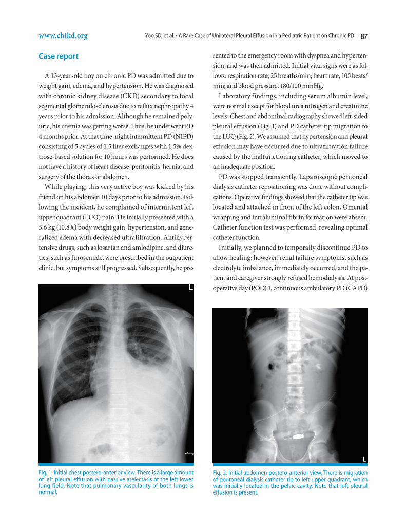
Laboratory findings, including serum albumin level, were normal except for blood urea nitrogen and creatinine levels. Chest and abdominal radiography showed left-sided pleural effusion (Fig. 1) and PD catheter tip migration to the LUQ (Fig. 2). We assumed that hypertension and pleural effusion may have occurred due to ultrafiltration failure caused by the malfunctioning catheter, which moved to an inadequate position.
PD was stopped transiently. Laparoscopic peritoneal dialysis catheter repositioning was done without compli-cations. Operative findings showed that the catheter tip was located and attached in front of the left colon. Omental wrapping and intraluminal fibrin formation were absent. Catheter function test was performed, revealing optimal catheter function.
Initially, we planned to temporally discontinue PD to allow healing; however, renal failure symptoms, such as electrolyte imbalance, immediately occurred, and the pa-tient and caregiver strongly refused hemodialysis. At post-operative day (POD) 1, continuous ambulatory PD (CAPD)
Fig. 1. Initial chest postero-anterior view. There is a large amount of left pleural effusion with passive atelectasis of the left lower lung field. Note that pulmonary vascularity of both lungs is normal.
Fig. 2. Initial abdomen postero-anterior view. There is migration of peritoneal dialysis catheter tip to left upper quadrant, which was initially located in the pelvic cavity. Note that left pleural effusion is present.

88 Child Kidney Dis • 2018;22:86-90 www.chikd.org
was started using 500 mL every 6 hours. At POD 2, the dialysate volume was increased to 750 mL every 6 hours. During dialysis, no leakage occurred, and ultrafiltration was fair. At POD 3, he had a mild cough and decreased lung sound at the left lower lobe. Chest radiography showed mild left-sided pleural effusion. The dialysate volume was the reduced to 500 cc. On the next day, follow-up chest radiography showed improvement of pleural effusion. Although repositioning of the catheter was performed, pleural effusion exacerbated repeatedly whenever the dialysate volume was increased. Because we suspected the presence of peritoneal leakage into the pleural space, CT peritoneography following the method of Cochran et al. was performed. After the peritoneal cavity was completely drained of the dialysate, nonionic contrast material was mixed with the dialysate and infused into the peritoneal cavity, and the patient was encouraged to walk for 1 hour until undergoing CT to achieve good distribution. CT pe-ritoneography showed a defect of the peritoneum in the left lower abdomen with a contrast leakage to the left rectus
and abdominis muscles (Fig. 3)3). The patient was treated conservatively by decreasing the NIPD volume, and then slowly increasing the volume again. The initial regimen of the NIPD was 7 cycles of 500 mL exchanges for 10 hours with 4.25% dextrose solution, and the dialysate volume was gradually increased to 1.5 liter within 2 months. The conservative treatment performed successfully managed the patient’s condition. For the next two years, the patient had not encountered similar problems during his outpa-tient clinic follow-up visits.
Discussion
Pleural effusion in patients with CKD is uncommon but is a well-recognized complication. In children, the incidence of pleural effusion ranges from 0.66% to 3%4,5). It has a di-verse etiology, such as heart failure, tuberculosis, uremic effusion, and volume overload. In patients on PD, pleural effusion is typically managed by increasing the dialysate
B
C
A
Fig. 3. Computed tomography peritoneography after 1 hour of injecting a water-soluble iodinated contrast media in an oblique prone position. Axial images obtained at the L1 (A) and L3 (B) level and a sagittal reconstructed image (C) show focal defect of the peritoneum (arrows). Note the contrast leakage (arrowheads) to the abdominal wall with tiny air bubble (curved arrow).

Yoo SD, et al. • A Rare Case of Unilateral Pleural Effusion in a Pediatric Patient on Chronic PD 89www.chikd.org
volume and ultrafiltration. However, if pleural effusion remained as a refractory problem despite proper medical treatment, the presence of infection, catheter-related com-plications, and pleuroperitoneal communication (PPC) should be considered6,7).
Unfortunately, PPC is a putative diagnosis in this case because the findings during the operation and on CT peri-toneography were not sufficient to reveal a direct clue about the route between PPC and the defect of the peritoneum. However, the relationship between PPC and the defect of the peritoneum is possible for the following reasons. First, the environment, such as increased intraperitoneal pres-sure, can cause a defect of the peritoneum and PPC. Second, both pleural effusion and peritoneal defect occurred ipsi-laterally. Third, PPC is known to be rare on the left, but only left-sided pleural effusion was identified whenever the volume of the dialysate was increased.
European Paediatric Dialysis Working Group reported the following risk factors of PPC: young age, hernia, abdo-minal surgery, and peritonitis5). Suggested risk factors of PD catheter migration from a retrospective review are cons-tipation, greater omentum wrapping, and operation te-chnique, such as catheter insertion site and catheter type 8).
In our case, there was no risk factor of PPC, catheter mi-gration, and peritoneal defect. Location of the defect was at the left lower quadrant; thus, it was not related to PD catheter insertion, which was performed at the right lower quadrant. Left-sided pleural effusion, which was ipsilateral with the peritoneal defect, always recurred whenever in-creasing the dialysate volume. A large multicenter study in Japan reported that 88% of pleural effusion cases was in the right side in PPC. On the other hand, the incidence of left pleural effusion is lower because the pericardium covers the defect and prevents peritoneal fluid leakage into the left pleural space9). These findings suggest that left-sided pleural effusion may be related to left peritoneal defect.
Although we did not perform diagnostic thoracentesis because spontaneous resolution was quickly achieved, tho-racentesis and pleural fluid analysis was useful in revealing transudate and high pleural fluid-to-serum glucose ratio confirming the clinical suspicion of PPC. However, it is im portant to note that the initial pleural fluid can be exu-dates and can have low pleural f luid-to-serum glucose gradient10).
CT peritoneography was performed, which revealed the defect of the peritoneum. However, we could not identify direct evidence of PPC, which could be due to the limitation or timing of the test, because CT was performed after the disappearance of pleural effusion.
We hypothesized that abdominal trauma or increased pressure in abdominal cavity may cause peritoneal defect and PPC, resulting in pleural effusion11). There was minor abdominal trauma history of the patient in our case. How-ever, atraumatic peritoneal damage or intrinsic defect cannot be ruled out. There were some cases of atraumatic damage of the intraabdominal organ, such as spleen rup-ture, bowel erosion, and perforation. Retrospective studies of the serial changes of CT peritoneography in patients with symptomatic ultrafiltration failure have shown that retro-peritoneal leakage, anterior abdominal wall leakage, and inguinal hernia can occur without trauma12-14). Intra-abdo-minal pressure in patients on PD can be changed depen-ding on the patients’ position, dialysate volume, and coug-hing. Moreover, the incidence of hernia and peritoneal leaks are higher in the PD population. However, whether increasing the hydrostatic intraabdominal pressure in-creases the risk of abdominal wall complications still re-mains controversial. In this regard, some authors insisted that intrinsic peritoneal defect may have existed prior to the presentation of complication15).
In conclusion, in patients on PD who present with refrac-tory or recurrent pleural effusion that does not respond to therapy, including increasing the dialysate volume or con-centration for volume overloading, another factor such as infection, catheter malfunction, and PPC should be con-sidered. To evaluate pleural effusion, thoracentesis and CT peritoneography are useful, but timely appropriate exa-mination is important to identify the defect or fistula.
Patient consent
This study was approved by the institutional review board (IRB), and the consent was waived due to the nature of the retrospective study [IRB number 05-2018-155].

90 Child Kidney Dis • 2018;22:86-90 www.chikd.org
Conflicts of interest
No potential conflict of interest relevant to this article was reported.
References
1. Kim JE, Park SJ, Oh JY, Kim JH, Lee JS, Kim PK, et al. Noninfectious Complications of Peritoneal Dialysis in Korean Children: A 26-Year Single-Center Study. Yonsei Med J 2015;56:1359-64.
2. Saha TC, Singh H. Noninfectious complications of peritoneal dia-lysis. South Med J 2007;100:54-8.
3. Cochran ST, Do HM, Ronaghi A, Nissenson AR, Kadell BM. Com-plications of peritoneal dialysis: evaluation with CT peritoneo-graphy. Radiographics 1997;17:869-78.
4. Kawaguchi AL, Dunn JC, Fonkalsrud EW. Management of perito-neal dialysis-induced hydrothorax in children. Am Surg 1996;62: 820-4.
5. Dufek S, Holtta T, Fischbach M, Ariceta G, Jankauskiene A, Cerkau-skiene R, et al. Pleuro-peritoneal or pericardio-peritoneal leak in children on chronic peritoneal dialysis-A survey from the Euro-pean Paediatric Dialysis Working Group. Pediatr Nephrol 2015; 30:2021-7.
6. Lew SQ. Hydrothorax: pleural effusion associated with peritoneal dialysis. Perit Dial Int 2010;30:13-8.
7. Ray S, Mukherjee S, Ganguly J, Abhishek K, Mitras S, Kundu S. A
cross-sectional prospective study of pleural effusion among cases of chronic kidney disease. Indian J Chest Dis Allied Sci 2013; 55:209-13.
8. Lan L, Jiang J, Wang P, Ren W, Hu Z. Peritoneal dialysis catheter placement in the right lower quadrant is associated with a lower risk of catheter tip migration: a retrospective single-center study. Int Urol Nephrol 2015;47:557-62.
9. Nomoto Y, Suga T, Nakajima K, Sakai H, Osawa G, Ota K, et al. Acute hydrothorax in continuous ambulatory peritoneal dialysis--a col-laborative study of 161 centers. Am J Nephrol 1989;9:363-7.
10. Momenin N, Colletti PM, Kaptein EM. Low pleural fluid-to-serum glucose gradient indicates pleuroperitoneal communication in peritoneal dialysis patients: presentation of two cases and a re-view of the literature. Nephrol Dial Transplant 2012;27:1212-9.
11. Mahale AS, Katyal A, Khanna R. Complications of peritoneal di-alysis related to increased intra-abdominal pressure. Adv Perit Dial 2003;19:130-5.
12. Cheung M, Chu FS, Kwan LP. Serial changes of computed tomo-graphic peritoneogram in patients with symptomatic ultrafiltra-tion failure complicating continuous ambulatory peritoneal dia-lysis. J Med Imaging Radiat Oncol 2017;61:321-6.
13. Fujiwara M, Soda T, Okada T, Kanamaru H, Inoue T, Ogawa O. Bowel perforation by a peritoneal dialysis catheter: report of two cases. BMC Nephrol 2017;18:312.
14. Kinoshita C, Quy PN, Honda M. Atraumatic splenic rupture in a peritoneal dialysis patient. CEN case reports. 2018.
15. Del Peso G, Bajo MA, Costero O, Hevia C, Gil F, Diaz C, et al. Risk factors for abdominal wall complications in peritoneal dialysis patients. Perit Dial Int 2003;23:249-54.