A monocarboxylate transporter required for hepatocyte...
13
A monocarboxylate transporter required for hepatocyte secretion of ketone bodies during fasting Sarah E. Hugo, 1,2 Lourdes Cruz-Garcia, 1,2 Santhosh Karanth, 1,2 Ryan M. Anderson, 3,5 Didier Y.R. Stainier, 3 and Amnon Schlegel 1,2,4,6 1 University of Utah Molecular Medicine (U2M2) Program, 2 Department of Internal Medicine, Division of Endocrinology, Metabolism, and Diabetes, University of Utah, Salt Lake City, Utah 84112, USA; 3 Department of Biochemistry and Biophysics, Liver Center, Diabetes Center, Cardiovascular Research Institute, University of California at San Francisco, San Francisco, California 94158, USA; 4 Department of Biochemistry, School of Medicine, University of Utah, Salt Lake City, Utah 84112, USA To find new genes that influence liver lipid mass, we performed a genetic screen for zebrafish mutants with hepatic steatosis, a pathological accumulation of fat. The red moon (rmn) mutant develops hepatic steatosis as maternally deposited yolk is depleted. Conversely, hepatic steatosis is suppressed in rmn mutants by adequate nutrition. Adult rmn mutants show increased liver neutral lipids and induction of hepatic lipid biosynthetic genes when fasted. Positional cloning of the rmn locus reveals a loss-of-function mutation in slc16a6a (solute carrier family 16a, member 6a), a gene that we show encodes a transporter of the major ketone body b-hydroxybutyrate. Restoring wild-type zebrafish slc16a6a expression or introducing human SLC16A6 in rmn mutant livers rescues the mutant phenotype. Radiotracer analysis confirms that loss of Slc16a6a function causes diversion of liver- trapped ketogenic precursors into triacylglycerol. Underscoring the importance of Slc16a6a to normal fasting physiology, previously fed rmn mutants are more sensitive to death by starvation than are wild-type larvae. Our unbiased, forward genetic approach has found a heretofore unrecognized critical step in fasting energy metabolism: hepatic ketone body transport. Since b-hydroxybutyrate is both a major fuel and a signaling molecule in fasting, the discovery of this transporter provides a new direction for modulating circulating levels of ketone bodies in metabolic diseases. [Keywords: hepatic steatosis; ketone body; lipid metabolism; starvation; zebrafish] Supplemental material is available for this article. Received October 13, 2011; revised version accepted December 27, 2011. Hepatic steatosis is present in a large fraction of obese people (Clark 2006). Nonalcoholic fatty liver disease (NAFLD) encompasses hepatic steatosis and several patho- logical states that follow it: inflammation (steatohepatitis), fibrosis (cirrhosis), and cancer (hepatocellular carcinoma) (Liou and Kowdley 2006). There are limited therapeutic options for permanently ameliorating hepatic steatosis (Sanyal et al. 2010), and there are no methods for reversing hepatic fibrosis or preventing hepatocellular carcinoma due to NAFLD (Nugent and Younossi 2007). The first step of NAFLD is the inappropriate accumulation of triacyl- glycerol in hepatocytes (Browning and Horton 2004). This accumulation may be due to excessive de novo hepatic lipid production; decreased hepatic secretion of very low- density lipoprotein (VLDL) particles; diminished b-oxidation of fatty acids in the liver; exaggerated uptake of fatty acids from the circulation; more subtle defects in regulating energy homeostasis, including insulin resistance or CNS nutrient sensing; or a combination of these factors (Hooper et al. 2011). Since each of these processes could be ex- ploited therapeutically, understanding their regulation is paramount. Identifying and characterizing novel genes that modulate hepatic lipid mass are priorities (Cohen et al. 2011). Master transcription factors, metabolic regulators, and multiple enzymes that control lipid homeostasis are pres- ent in zebrafish (Babin and Vernier 1989; Ibabe et al. 2002; Schlombs et al. 2003). This conservation of function and the absence of fasting steatosis in larvae that have never been fed (Schlegel and Stainier 2006) allowed us to screen for mutations causing increased hepatic lipid mass. Per- forming the screen in animals carrying three transgenes that encode fluorescent protein labels of the Islet of Lang- erhans b cells, the liver hepatocytes, and the pancreatic 5 Present address: Department of Pediatrics, Indiana University School of Medicine, 635 Barnhill Drive, MS2053, Indianapolis, IN 46202, USA. 6 Corresponding author. E-mail [email protected]. Article is online at http://www.genesdev.org/cgi/doi/10.1101/gad.180968.111. 282 GENES & DEVELOPMENT 26:282–293 Ó 2012 by Cold Spring Harbor Laboratory Press ISSN 0890-9369/12; www.genesdev.org Cold Spring Harbor Laboratory Press on December 27, 2019 - Published by genesdev.cshlp.org Downloaded from
Transcript of A monocarboxylate transporter required for hepatocyte...

A monocarboxylate transporter requiredfor hepatocyte secretion of ketone bodiesduring fasting
Sarah E. Hugo,1,2 Lourdes Cruz-Garcia,1,2 Santhosh Karanth,1,2 Ryan M. Anderson,3,5
Didier Y.R. Stainier,3 and Amnon Schlegel1,2,4,6
1University of Utah Molecular Medicine (U2M2) Program, 2Department of Internal Medicine, Division of Endocrinology,Metabolism, and Diabetes, University of Utah, Salt Lake City, Utah 84112, USA; 3Department of Biochemistry and Biophysics,Liver Center, Diabetes Center, Cardiovascular Research Institute, University of California at San Francisco, San Francisco,California 94158, USA; 4Department of Biochemistry, School of Medicine, University of Utah, Salt Lake City, Utah 84112, USA
To find new genes that influence liver lipid mass, we performed a genetic screen for zebrafish mutants withhepatic steatosis, a pathological accumulation of fat. The red moon (rmn) mutant develops hepatic steatosis asmaternally deposited yolk is depleted. Conversely, hepatic steatosis is suppressed in rmn mutants by adequatenutrition. Adult rmn mutants show increased liver neutral lipids and induction of hepatic lipid biosynthetic geneswhen fasted. Positional cloning of the rmn locus reveals a loss-of-function mutation in slc16a6a (solute carrierfamily 16a, member 6a), a gene that we show encodes a transporter of the major ketone body b-hydroxybutyrate.Restoring wild-type zebrafish slc16a6a expression or introducing human SLC16A6 in rmn mutant livers rescuesthe mutant phenotype. Radiotracer analysis confirms that loss of Slc16a6a function causes diversion of liver-trapped ketogenic precursors into triacylglycerol. Underscoring the importance of Slc16a6a to normal fastingphysiology, previously fed rmn mutants are more sensitive to death by starvation than are wild-type larvae. Ourunbiased, forward genetic approach has found a heretofore unrecognized critical step in fasting energy metabolism:hepatic ketone body transport. Since b-hydroxybutyrate is both a major fuel and a signaling molecule in fasting,the discovery of this transporter provides a new direction for modulating circulating levels of ketone bodies inmetabolic diseases.
[Keywords: hepatic steatosis; ketone body; lipid metabolism; starvation; zebrafish]
Supplemental material is available for this article.
Received October 13, 2011; revised version accepted December 27, 2011.
Hepatic steatosis is present in a large fraction of obesepeople (Clark 2006). Nonalcoholic fatty liver disease(NAFLD) encompasses hepatic steatosis and several patho-logical states that follow it: inflammation (steatohepatitis),fibrosis (cirrhosis), and cancer (hepatocellular carcinoma)(Liou and Kowdley 2006). There are limited therapeuticoptions for permanently ameliorating hepatic steatosis(Sanyal et al. 2010), and there are no methods for reversinghepatic fibrosis or preventing hepatocellular carcinomadue to NAFLD (Nugent and Younossi 2007). The first stepof NAFLD is the inappropriate accumulation of triacyl-glycerol in hepatocytes (Browning and Horton 2004). Thisaccumulation may be due to excessive de novo hepaticlipid production; decreased hepatic secretion of very low-
density lipoprotein (VLDL) particles; diminished b-oxidationof fatty acids in the liver; exaggerated uptake of fatty acidsfrom the circulation; more subtle defects in regulatingenergy homeostasis, including insulin resistance or CNSnutrient sensing; or a combination of these factors (Hooperet al. 2011). Since each of these processes could be ex-ploited therapeutically, understanding their regulation isparamount. Identifying and characterizing novel genesthat modulate hepatic lipid mass are priorities (Cohenet al. 2011).
Master transcription factors, metabolic regulators, andmultiple enzymes that control lipid homeostasis are pres-ent in zebrafish (Babin and Vernier 1989; Ibabe et al. 2002;Schlombs et al. 2003). This conservation of function andthe absence of fasting steatosis in larvae that have neverbeen fed (Schlegel and Stainier 2006) allowed us to screenfor mutations causing increased hepatic lipid mass. Per-forming the screen in animals carrying three transgenesthat encode fluorescent protein labels of the Islet of Lang-erhans b cells, the liver hepatocytes, and the pancreatic
5Present address: Department of Pediatrics, Indiana University School ofMedicine, 635 Barnhill Drive, MS2053, Indianapolis, IN 46202, USA.6Corresponding author.E-mail [email protected] is online at http://www.genesdev.org/cgi/doi/10.1101/gad.180968.111.
282 GENES & DEVELOPMENT 26:282–293 � 2012 by Cold Spring Harbor Laboratory Press ISSN 0890-9369/12; www.genesdev.org
Cold Spring Harbor Laboratory Press on December 27, 2019 - Published by genesdev.cshlp.orgDownloaded from

acinar cells (Anderson et al. 2009) helped us to avoid mu-tants that displayed changes in liver or pancreas morphol-ogy (Sadler et al. 2005; Akimitsu et al. 2008; Matthewset al. 2009; Thakur et al. 2011). Defects in yolk lipidtransport were avoided by restricting our analysis to onlylarvae that consumed all their yolk by 6 d post-fertilization(dpf) (Schlegel and Stainier 2006; Raldua et al. 2008; Walterset al. 2009). Similarly, animals that showed craniofacialmalformations (Schlombs et al. 2003; Sadler et al. 2005) andovert neuronal defects were excluded because we felt theywould not be amenable to characterization beyond theearly larval period and because they often show residualyolk lipids at 6 dpf.
Here we report the identification, positional cloning,and functional characterization of one mutant that re-veals a heretofore unappreciated bottleneck of hepaticlipid metabolism: ketone body transport.
Results
Isolation and initial characterization of red moon(rmn), a zebrafish mutant with hepatic steatosis
The rmn mutant showed hepatic steatosis just as mater-nally supplied yolk lipids were nearing exhaustion on 5 dpf(Fig. 1A). Aside from the surfactant-lined swim bladder,only the rmn mutant liver showed strong Oil Red O (ORO)staining on 6 and 7 dpf. The accumulation of neutral lipidsin rmn mutant livers was confirmed with both whole-mount and confocal microscopy of animals stained withthe fluorescent neutral lipid dye 4,4-difluoro-1,3,5,7,8-pentamethyl-4-bora-3a,4a-diaza-s-indacene (BODIPY 493/503) (Fig. 1B,C). Numerous cytoplasmic lipid droplets inotherwise normal hepatocytes were observed in rmnmutants examined with transmission electron microscopy(Fig. 1D). Normal macronutrient recycling was appreciatedin rmn livers viewed at higher magnification (Fig. 1D).Similarly, histological analysis demonstrated no evidenceof inflammatory cell infiltration or deposition of excessiveextracellular matrix proteins (Supplemental Fig. S1A,B).Taken together, the histological and ultrastructural char-acterization indicated that our stringent screening criteriawere met: The fulminant hepatic degeneration seen inother zebrafish mutants with hepatic steatosis did notoccur in rmn mutants.
The lack of secondary phenotypes in rmn mutantsenabled us to assess whether the steatosis phenotypewould be modulated by prolonged fasting. Hepatic steato-sis in rmn mutants persisted in the never-fed state to 12dpf in the absence of other, overt morphological defects(Supplemental Fig. S1C). This persistence of hepaticsteatosis prompted us to quantify the major neutral lipidspecies in whole-larval extracts over the course of a pro-longed fast. There were increased levels of triacylglycerol,b-hydroxybutyrate, and free fatty acids in whole-larvalextracts 5 dpf (Fig. 2A). The lipid compositions of wild-typeand rmn mutant larvae changed over the course of the fast.Cholesterol was the most abundant neutral lipid in whole-larval extracts and was more abundant in rmn mutantextracts on 7 and 8 dpf. Similar to b-hydroxybutyrate and
free fatty acids, the triacylglycerol was also higher in 5- and6-dpf rmn mutant extracts. Cholesteryl esters were moreabundant in rmn mutants between 7 and 10 dpf. There-after, the whole-larval levels of all three neutral lipidswere not different. In summary, never-fed rmn mutantsshowed persisting hepatic steatosis marked by accumula-tion of different neutral lipid species until death by 13 dpf.
Since rmn mutant larvae showed no obvious morphol-ogical phenotypes, it was possible that the mutation wasnot lethal. We raised an in-cross of heterozygous carriersunder normal feeding conditions and observed that thermn mutant was viable (recovered in the anticipatedMendelian ratio of a fully penetrant and fully expressiverecessive mutation) and fertile. Thus, we were able toassess the changes in hepatic neutral lipid composition bysubjecting wild-type and rmn mutant adult animals to a
Figure 1. Identification of the fasting hepatic steatosis mutantrmn. (A) Whole-mount ORO staining of larvae. The yolk lipid(arrowhead) is exhausted 5 dpf. Vascular lipid staining (arrows)ceases by the end of 5 dpf. The surfactant-lined swim bladderstained with ORO (asterisk). (B) Whole-mount BODIPY 493/503staining of 6-dpf larvae. This dye also stained the vitreous humorof the eyes (arrowhead) and the ventricles of the CNS (arrow). (C)Confocal stacks of livers fixed and stained as in B. A single sliceof a rmn mutant liver is shown in the inset. (D) Transmissionelectron microscopy of liver sections showing that the rmn
mutant hepatocytes have cytoplasmic lipid droplets (ld). Thenuclear (n) and mitochondrial (m) morphology appears normal inrmn mutants. Higher-magnification micrographs also showmultilamellar structures (asterisks) suggestive of multivesicularbodies and elongated tubular (t) precursors of these structures inboth wild-type (WT) and the rmn mutant livers. Similarly,autophagosomal structures (arrowheads) were observed in bothwild-type and rmn mutant livers. Bar, 1 mm.
Liver ketone body transporter
GENES & DEVELOPMENT 283
Cold Spring Harbor Laboratory Press on December 27, 2019 - Published by genesdev.cshlp.orgDownloaded from
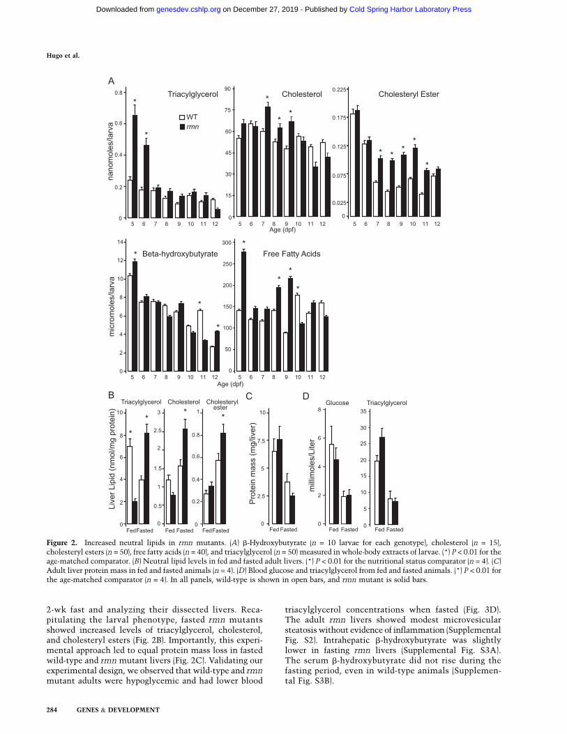
2-wk fast and analyzing their dissected livers. Reca-pitulating the larval phenotype, fasted rmn mutantsshowed increased levels of triacylglycerol, cholesterol,and cholesteryl esters (Fig. 2B). Importantly, this experi-mental approach led to equal protein mass loss in fastedwild-type and rmn mutant livers (Fig. 2C). Validating ourexperimental design, we observed that wild-type and rmnmutant adults were hypoglycemic and had lower blood
triacylglycerol concentrations when fasted (Fig. 3D).The adult rmn livers showed modest microvesicularsteatosis without evidence of inflammation (SupplementalFig. S2). Intrahepatic b-hydroxybutyrate was slightlylower in fasting rmn livers (Supplemental Fig. S3A).The serum b-hydroxybutyrate did not rise during thefasting period, even in wild-type animals (Supplemen-tal Fig. S3B).
Figure 2. Increased neutral lipids in rmn mutants. (A) b-Hydroxybutyrate (n = 10 larvae for each genotype), cholesterol (n = 15),cholesteryl esters (n = 50), free fatty acids (n = 40), and triacylglycerol (n = 50) measured in whole-body extracts of larvae. (*) P < 0.01 for theage-matched comparator. (B) Neutral lipid levels in fed and fasted adult livers. (*) P < 0.01 for the nutritional status comparator (n = 4). (C)Adult liver protein mass in fed and fasted animals (n = 4). (D) Blood glucose and triacylglycerol from fed and fasted animals. (*) P < 0.01 forthe age-matched comparator (n = 4). In all panels, wild-type is shown in open bars, and rmn mutant is solid bars.
Hugo et al.
284 GENES & DEVELOPMENT
Cold Spring Harbor Laboratory Press on December 27, 2019 - Published by genesdev.cshlp.orgDownloaded from

Figure 3. Positional cloning of the rmn locus. (A) The rmn locus was narrowed to a 100-kb critical interval on chromosome 12.Genetic distance of markers is reported on the radiation hybrid map in centiRays (cR) and on the MGH meiotic map in centiMorgans(cM) above the individual bacterial artificial chromosome and fosmid clones used to span this region of the genome. Therecombination events (reported as recombinants/meioses) at the labeled polymorphic markers (poly-CA or ‘‘CA repeat’’) are shownbelow individual clones. Clone acronym definitions and the names of the clones between zK29B5 and zC29A20 are in theSupplemental Material. (B) Fine mapping narrowed the critical interval to the space between two CA repeats within introns ofslc16a6a and calcoco2 on the clone zK106L8 (white region flanked by gray and red portions of the chromosome). Coding exons arefilled red boxes. The 59 and 39 untranslated regions (UTRs) are unfilled boxes. (C) Sanger traces of slc16a6a cDNAs prepared withprimers in the 59 and 39 UTRs indicating that exon 2 is skipped in rmn mutants. The position of leucine codon 86 and methioninecodon 97 are shown. (D) Cartoons of slc16a6a cDNAs prepared as in C. (E) Sanger traces of genomic DNA spanning exon 2 ofslc16a6a reveal retrotransposon sequences in rmn mutants. An in-frame stop codon is shown. (F) Cartoon of the insertion mutationdestroying exon 2 of slc16a6a. (G, left) The sequenced products shown in E differ in size: Amplification of exon 2 and flankingintronic sequences of slc16a6a from homozygous wild-type animals (+/+), heterozygous rmn carriers (+/rmn), and homozygous rmnmutant animals (rmn/rmn) reveals a 450-bp increase in the rmn allele product. (Left panel) Heterozygous carriers proved difficult toscore using this primer pair because the mutant allele amplified less robustly in the presence of the wild-type allele (primers A and B).(Right panel) Thus, we resorted to a three-primer strategy to amplify either the sequences deleted by the insertion or a small portionof the insertion. The latter approach led to confident scoring of heterozygous carriers. The molecular mass standards are shown inkilobase pairs.
GENES & DEVELOPMENT 285
Cold Spring Harbor Laboratory Press on December 27, 2019 - Published by genesdev.cshlp.orgDownloaded from
The rmn mutation disrupts an orphanmonocarboxylate transporter (MCT) gene
Positional cloning methods were used to isolate the genedisrupted by the rmn mutation (Fig. 3A,B). Of the geneswithin the critical interval on chromosome 12, slc16a6a(solute carrier family 16a, member 6a) was a strong can-didate because it encodes an orphan MCT that may berequired in the transport of nutrients as animals transitionfrom fed to fasted states (Price et al. 1998). We cloned andsequenced mature slc16a6a cDNAs from adult liver andobserved aberrant mRNA maturation with skipping of theinitiator ATG codon-containing exon 2 in rmn mutants(Fig. 3C,D). In rmn mutants, the 39 169 base pairs (bp) ofexon 2 and the first 67 bp of intron 2 in the slc16a6a genewere replaced by a 404-bp sequence that is most similar tothe DrNgaro4 retrotransposon (Fig. 3E–G; SupplementalFig. S4; Goodwin and Poulter 2004). Thus, the rmn muta-tion was not induced by N-ethyl-N-nitrosourea. Rather, itwas carried in some of the mutagenized animals we usedfor the screen (Anderson et al. 2009). The rmn mutationwas found in other strains (Supplemental Material), butnot in the one we used in our previous studies (Schlegeland Stainier 2006).
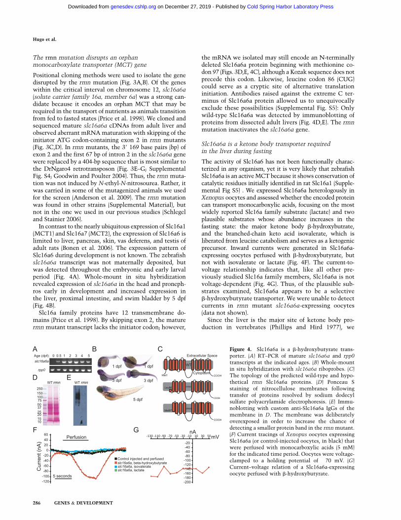
In contrast to the nearly ubiquitous expression of Slc16a1(MCT1) and Slc16a7 (MCT2), the expression of Slc16a6 islimited to liver, pancreas, skin, vas deferens, and testis ofadult rats (Bonen et al. 2006). The expression pattern ofSlc16a6 during development is not known. The zebrafishslc16a6a transcript was not maternally deposited, butwas detected throughout the embryonic and early larvalperiod (Fig. 4A). Whole-mount in situ hybridizationrevealed expression of slc16a6a in the head and proneph-ros early in development and increased expression inthe liver, proximal intestine, and swim bladder by 5 dpf(Fig. 4B).
Slc16a family proteins have 12 transmembrane do-mains (Price et al. 1998). By skipping exon 2, the maturermn mutant transcript lacks the initiator codon; however,
the mRNA we isolated may still encode an N-terminallydeleted Slc16a6a protein beginning with methionine co-don 97 (Figs. 3D,E, 4C), although a Kozak sequence does notprecede this codon. Likewise, leucine codon 86 (CUG)could serve as a cryptic site of alternative translationinitiation. Antibodies raised against the extreme C ter-minus of Slc16a6a protein allowed us to unequivocallyexclude these possibilities (Supplemental Fig. S5): Onlywild-type Slc16a6a was detected by immunoblotting ofproteins from dissected adult livers (Fig. 4D,E). The rmnmutation inactivates the slc16a6a gene.
Slc16a6a is a ketone body transporter requiredin the liver during fasting
The activity of Slc16a6 has not been functionally charac-terized in any organism, yet it is very likely that zebrafishSlc16a6a is an active MCT because it shows conservation ofcatalytic residues initially identified in rat Slc16a1 (Supple-mental Fig S5) . We expressed Slc16a6a heterologously inXenopus oocytes and assessed whether the encoded proteincan transport monocarboxylic acids, focusing on the mostwidely reported Slc16a family substrate (lactate) and twoplausible substrates whose abundance increases in thefasting state: the major ketone body b-hydroxybutrate,and the branched-chain keto acid isovalerate, which isliberated from leucine catabolism and serves as a ketogenicprecursor. Inward currents were generated in Slc16a6a-expressing oocytes perfused with b-hydroxybutyrate, butnot with isovalerate or lactate (Fig. 4F). The current-to-voltage relationship indicates that, like all other pre-viously studied Slc16a family members, Slc16a6a is notvoltage-dependent (Fig. 4G). Thus, of the plausible sub-strates examined, Slc16a6a appears to be a selectiveb-hydroxybutyrate transporter. We were unable to detectcurrents in rmn mutant slc16a6a-expressing oocytes(data not shown).
Since the liver is the major site of ketone body pro-duction in vertebrates (Phillips and Hird 1977), we
Figure 4. Slc16a6a is a b-hydroxybutyrate trans-porter. (A) RT–PCR of mature slc16a6a and rpp0
transcripts at the indicated ages. (B) Whole-mountin situ hybridization with slc16a6a riboprobes. (C)The topology of the predicted wild-type and hypo-thetical rmn Slc16a6a proteins. (D) Ponceau Sstaining of nitrocellulose membranes followingtransfer of proteins resolved by sodium dodecylsulfate polyacrylamide electrophoresis. (E) Immu-noblotting with custom anti-Slc16a6a IgGs of themembrane in D. The membrane was deliberatelyoverexposed in order to increase the chance ofdetecting a smaller protein band in the rmn mutant.(F) Current tracings of Xenopus oocytes expressingSlc16a6a (or control-injected oocytes, in black) thatwere perfused with monocarboxylic acids (5 mM)for the indicated time period. Oocytes were voltage-clamped to a holding potential of �70 mV. (G)Current–voltage relation of a Slc16a6a-expressingoocyte perfused with b-hydroxybutyrate.
Hugo et al.
286 GENES & DEVELOPMENT
Cold Spring Harbor Laboratory Press on December 27, 2019 - Published by genesdev.cshlp.orgDownloaded from

Figure 5. (A) A model of the role of Slc16a6a in hepatic ketone body metabolism. Dietary and stored reduced carbon atoms in the formof fatty acids (intestine and adipose, respectively) can be delivered to the liver, where, after partial b-oxidation, they can be repackagedas ketone bodies for feeding the peripheral organs during fasting. Triacylglycerol can also be packaged into VLDL particles (liver) orchylomicrons (intestine) for delivery to the periphery. The intestine and skeletal muscle also provide ketogenic amino acids, the mostabundant of which is leucine. In birds, reptiles, and mammals, the rate-limiting enzyme of ketogenesis is mitochondrial HMG-CoAsynthase 2 (gray reactions), which arose in birds through gene duplication of the cytosolic HMG-CoA synthase 1 gene (Boukaftane et al.1994). Amphibians, fish, and other lower vertebrates have only the cytosolic, cholesterol-synthetic HMG-CoA synthase 1 and rely onincreased flux through deamination and oxidation of leucine (which also occurs in higher vertebrates) to make mitochondrial HMG-CoA for ketogenesis (Phillips and Hird 1977; Leblanc and Ballantyne 2000). Slc16a6a is a b-hydroxybutyrate-transporting MCT that isexpressed in the liver. In rmn mutants, this transporter is inactivated, and the carbon atoms that would otherwise be secreted as fuelduring fasting are diverted back to de novo lipogenesis and storage in lipid droplets. The peripheral ketone body transporters are notknown (‘‘?’’), but Slc16a1, Slc16a7, and Slc16a3 are expressed in brain endothelial cells, neurons, and astrocytes, respectively (Pierre andPellerin 2005). In zebrafish, there is evidence that Slc5a family members are expressed in muscle and can transport sodiumhydroxybutyrate (Plata et al. 2007). (CoA) Coenzyme A; (Cpt1) Carnitine palmitoyl transferase 1; (Bckdh) Branched chain ketoaciddehydrogenase; (Chyl) chylomicron particle; (FAD) flavine adenine dinucleotide; (FADH2) reduced FAD; (NAD+) nicotinamide adeninedinucleotide; (NADH2) reduced NAD; (VLDL) very low-density lipoprotein particle. (B) Larvae were incubated with L-[14C(U)]-leucine,and then lipids were analyzed as described in the Materials and Methods to calculate the 14C incorporation ratio. Values greater thanunity reflect increased relative incorporation of ketogenic precursor carbon atoms into neutral lipids in rmn mutants.
Liver ketone body transporter
GENES & DEVELOPMENT 287
Cold Spring Harbor Laboratory Press on December 27, 2019 - Published by genesdev.cshlp.orgDownloaded from
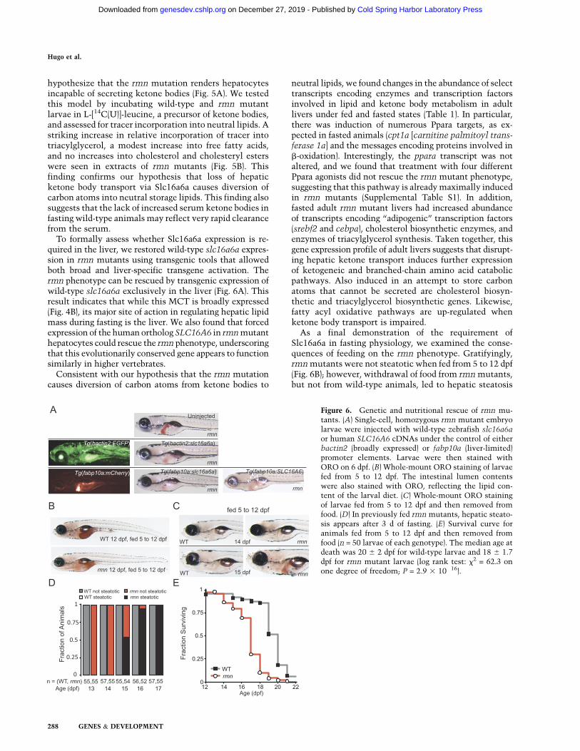
hypothesize that the rmn mutation renders hepatocytesincapable of secreting ketone bodies (Fig. 5A). We testedthis model by incubating wild-type and rmn mutantlarvae in L-[14C(U)]-leucine, a precursor of ketone bodies,and assessed for tracer incorporation into neutral lipids. Astriking increase in relative incorporation of tracer intotriacylglycerol, a modest increase into free fatty acids,and no increases into cholesterol and cholesteryl esterswere seen in extracts of rmn mutants (Fig. 5B). Thisfinding confirms our hypothesis that loss of hepaticketone body transport via Slc16a6a causes diversion ofcarbon atoms into neutral storage lipids. This finding alsosuggests that the lack of increased serum ketone bodies infasting wild-type animals may reflect very rapid clearancefrom the serum.
To formally assess whether Slc16a6a expression is re-quired in the liver, we restored wild-type slc16a6a expres-sion in rmn mutants using transgenic tools that allowedboth broad and liver-specific transgene activation. Thermn phenotype can be rescued by transgenic expression ofwild-type slc16a6a exclusively in the liver (Fig. 6A). Thisresult indicates that while this MCT is broadly expressed(Fig. 4B), its major site of action in regulating hepatic lipidmass during fasting is the liver. We also found that forcedexpression of the human ortholog SLC16A6 in rmn mutanthepatocytes could rescue the rmn phenotype, underscoringthat this evolutionarily conserved gene appears to functionsimilarly in higher vertebrates.
Consistent with our hypothesis that the rmn mutationcauses diversion of carbon atoms from ketone bodies to
neutral lipids, we found changes in the abundance of selecttranscripts encoding enzymes and transcription factorsinvolved in lipid and ketone body metabolism in adultlivers under fed and fasted states (Table 1). In particular,there was induction of numerous Ppara targets, as ex-pected in fasted animals (cpt1a [carnitine palmitoyl trans-ferase 1a] and the messages encoding proteins involved inb-oxidation). Interestingly, the ppara transcript was notaltered, and we found that treatment with four differentPpara agonists did not rescue the rmn mutant phenotype,suggesting that this pathway is already maximally inducedin rmn mutants (Supplemental Table S1). In addition,fasted adult rmn mutant livers had increased abundanceof transcripts encoding ‘‘adipogenic’’ transcription factors(srebf2 and cebpa), cholesterol biosynthetic enzymes, andenzymes of triacylglycerol synthesis. Taken together, thisgene expression profile of adult livers suggests that disrupt-ing hepatic ketone transport induces further expressionof ketogeneic and branched-chain amino acid catabolicpathways. Also induced in an attempt to store carbonatoms that cannot be secreted are cholesterol biosyn-thetic and triacylglycerol biosynthetic genes. Likewise,fatty acyl oxidative pathways are up-regulated whenketone body transport is impaired.
As a final demonstration of the requirement ofSlc16a6a in fasting physiology, we examined the conse-quences of feeding on the rmn phenotype. Gratifyingly,rmn mutants were not steatotic when fed from 5 to 12 dpf(Fig. 6B); however, withdrawal of food from rmn mutants,but not from wild-type animals, led to hepatic steatosis
Figure 6. Genetic and nutritional rescue of rmn mu-tants. (A) Single-cell, homozygous rmn mutant embryolarvae were injected with wild-type zebrafish slc16a6aor human SLC16A6 cDNAs under the control of eitherbactin2 (broadly expressed) or fabp10a (liver-limited)promoter elements. Larvae were then stained withORO on 6 dpf. (B) Whole-mount ORO staining of larvaefed from 5 to 12 dpf. The intestinal lumen contentswere also stained with ORO, reflecting the lipid con-tent of the larval diet. (C) Whole-mount ORO stainingof larvae fed from 5 to 12 dpf and then removed fromfood. (D) In previously fed rmn mutants, hepatic steato-sis appears after 3 d of fasting. (E) Survival curve foranimals fed from 5 to 12 dpf and then removed fromfood (n = 50 larvae of each genotype). The median age atdeath was 20 6 2 dpf for wild-type larvae and 18 6 1.7dpf for rmn mutant larvae (log rank test: x2 = 62.3 onone degree of freedom; P = 2.9 3 10�16).
Hugo et al.
288 GENES & DEVELOPMENT
Cold Spring Harbor Laboratory Press on December 27, 2019 - Published by genesdev.cshlp.orgDownloaded from

(Fig. 6C,D). To assess whether the development of steato-sis in previously fed animals affected survival, wild-typeand rmn mutant larvae were fed from 5 to 12 dpf and thenscored for death after food was withdrawn. Again, asexpected, wild-type animals survived longer than rmnmutant animals (Fig. 6E). Thus, rmn mutant larvae develophepatic steatosis under nutrient-deficient states and arerendered sensitive to starvation.
Discussion
In diverse metazoan species from flies (Gutierrez et al. 2007)to humans (Gibbons et al. 2000), fasting causes mobiliza-tion of energy substrates from the peripheral organs in the
form of fatty acids from adipose and amino acids frommuscle. Upon return to the liver, these metabolites arepartially oxidized into ketone bodies, a fuel source that canbe used by the brain. These molecules and the glucoseliberated from hepatic glycogenolysis and gluconeogenesisare used as fuel in peripheral organs during fasting (Cahill2006). The availability of ketone bodies for use by organsother than the liver reflects the lower hepatic abundance of3-oxoacid coenzyme A (CoA) transferase 1, the enzymerequired to break down ketone bodies (Laffel 1999).
In inbred strains of mice, the influx of fatty acids fromadipose tissue during fasting leads to their re-esterifica-tion and temporary storage in the liver. The extent of thissteatosis varies based on genetic background and reflectsdifferences in muscle fat oxidation and the degree of
Table 1. Quantitative RT–PCR gene expression profiling
Wild type rmn mutant
Gene Protein Fasted Fed Fasted Fed
Transcription factors regulating lipid metabolismcebpa CCAAT/enhancer-binding protein a 1.0 6 0.01 1.8 6 0.01a 2.6 6 0.03a 7.2 6 0.03a,b
pparab Peroxisome proliferator-activated receptor a b 1.0 6 0.02 0.8 6 0.02 0.9 6 0.02 1.0 6 0.03pparg Peroxisome proliferator-activated receptor g 1.0 6 0.004 1.0 6 0.007 1.1 6 0.01 2.7 6 0.02a
nr1h3 Liver x receptor a 1.0 6 0.05 0.7 6 0.01 0.8 6 0.01 0.8 6 0.01srebf1 Sterol regulatory element-binding transcription factor 1 1.0 6 0.03 1.5 6 0.05 1.2 6 0.04 0.9 6 0.03srebf2 Sterol regulatory element-binding transcription factor 2 1.0 6 0.1 3.0 6 0.1a 2.1 6 0.01a 6.6 6 0.4a,b
mlxip MLX-interacting protein (Chrebp) 1.0 6 0.03 1.3 6 0.03 1.7 6 0.05 0.6 6 0.03Cholesterol synthesishmgcra 3-Hydroxy-3-methylglutaryl-coenzyme A reductase a 1.0 6 0.03 25.3 6 1.3a 63 6 1a 3050 6 30a,b
hmgcs1 3-Hydroxy-3-methylglutaryl-coenzyme A synthase 1 1.0 6 0.01 41.3 6 0.6a 5.9 6 0.1a 437 6 6a,b
hmgcl 3-Hydroxy-3-methylglutaryl-coenzyme A lyase 1.0 6 0.06 1.0 6 0.06 1.7 6 0.1a,b 1.0 6 0.06Branched-chain ketoacid and ketone body metabolismbcat2 Branched-chain aminotransferase, mitochondrial 1.0 6 0.02 4.14 6 0.06a 8.1 6 0.1a,b 2.27 6 0.03a
bckdhbl Branched-chain ketoacid dehydrogenase subunit b, like 1.0 6 0.02 1.92 6 0.03a 2.84 6 0.04a,b 1.34 6 0.02a
bdh1 3-Hydroxybutyrate dehydrogenase, mitochondrial 1.0 6 0.06 1.0 6 0.06 2.5 6 0.14a,b 0.5 6 0.03a
oxct1b 3-Oxoacid coenzyme A transferase 1b (liver-specific) 1.0 6 0.02 4.4 6 0.1a 8.5 6 0.2a,b 10.6 6 0.4a
De novo fatty acid and triacylglycerol synthesis andVLDL secretion
acc1 Acetyl-coenzyme A carboxylase 1 1.0 6 0.07 51.9 6 1.3a 0.7 6 0.01 16.8 6 0.2a
fasn Fatty acid synthase 1.0 6 0.05 70.5 6 1.0a 1.2 6 0.04 28.0 6 0.4a
scd Steroyl-CoA desaturase (D-9-desaturase) 1.0 6 0.02 8,520 6 90a 58 6 1a 5080 6 30a
elovl5 Elongation of long chain fatty acids family member 5 1.0 6 0.15 1.0 6 0.23 0.3 6 0.04a,b 0.3 6 0.03a,b
fads2 Fatty acid desaturase 2 (D-5- and D-6-desaturase) 1.0 6 0.01 1.58 6 0.01 0.4 6 0.002 0.7 6 0.004agpat4 1-Acylglycerol-3-phosphate O-acyltransferase 4 1.0 6 0.02 5.0 6 0.1a 16.8 6 0.4a,b 9.9 6 0.3a,b
ppap2ab Phosphatidic acid phosphatase 2a b 1.0 6 0.03 2.8 6 0.1 1.2 6 0.03 0.7 6 0.02dgat2 Diacylglycerol O-acyltransferase 2 1.0 6 0.1 0.9 6 0.1 14.9 6 1.9a,b 22.6 6 2.3a,b
mtp Microsomal triglyceride transfer protein 1.0 6 0.17 2.2 6 0.4a 1.7 6 0.3a 3.4 6 0.6a
Fatty acid oxidationcpt1a Carnitine O-palmitoyltransferase I (liver isoform) 1.0 6 0.03 1.3 6 0.04a 1.4 6 0.04a 1.2 6 0.04a
acadl Acyl-coenzyme A dehydrogenase, long chain 1.0 6 0.03 5.1 6 0.1a 3.2 6 0.04a 3.8 6 0. 04a
acadm Acyl-coenzyme A dehydrogenase, C-4 to C-12straight chain
1.0 6 0.1 2.7 6 0.04a 2.7 6 0.04a 2.3 6 0.2a
ehhadh Enoyl-CoA, hydratase/3-hydroxyacyl CoAdehydrogenase
1.0 6 0.03 2.6 6 0.08a 1.6 6 0.05a 2.3 6 0.08a
mt-nd1 NADH dehydrogenase 1, mitochondrial 1.0 6 0.1 2.1 6 0.1a 2.6 6 0.03a 2.0 6 0.05a
mt-co1 Cytochrome c oxidase 1, mitochondrial 1.0 6 0.02 1.3 6 0.01a 1.3 6 0.02a 1.4 6 0.01a
mt-atp6 ATP synthase 6, mitochondrial 1.0 6 0.1 0.9 6 0.03 0.8 6 0.02 1.2 6 0.03Slc16a6 paralogsslc16a6a Solute carrier family 16a, member 6a 1.0 6 0.02 0.8 6 0.02 1.2 6 0.02 0.1 6 0..002Slc16a6b Solute carrier family 16a, member 6b 1.0 6 0.02 2.0 6 0.04 2.6 6 0.05 0.4 6 0.01a,b
aP < 0.01 versus wild type fasted.bP < 0.01 versus wild type fed.
Liver ketone body transporter
GENES & DEVELOPMENT 289
Cold Spring Harbor Laboratory Press on December 27, 2019 - Published by genesdev.cshlp.orgDownloaded from

adipose lipolysis (Lin et al. 2005; Guan et al. 2009). Fur-thermore, there is an inverse association between hepaticlipid levels and serum ketone body concentrations in fastedmice (Lin et al. 2005). Normal humans subjected to fastingshow an increase in hepatic lipid content in response toa 36-h fast, but there is variation in the extent of lipidaccumulation (Moller et al. 2008).
Here we described the use of a genetic screen to isolatenovel regulators of lipid metabolism. Our approach led tothe identification of a nutritionally suppressible hepaticsteatosis mutant, rmn. In contrast to previously reported,lethal mutants, the steatosis seen in rmn mutants isneither preceded nor followed by the development ofdegenerative changes in hepatocyte architecture or ful-minant inflammation. The rmn mutation causes a com-plete loss of Slc16a6a expression, as revealed by theabsence of mutant protein expression. The mutated geneencodes a MCT that transports b-hydroxybutyrateSlc16a6a. Mutation of this ketone body transporter trig-gers accumulation of neutral lipids in fasted livers andrenders animals sensitive to starvation. The rmn mutantphenotype can be rescued by forced expression of wild-type zebrafish slc16a6a exclusively in the liver. Expres-sion of the human ortholog SLC16A6 in the liver alsorescues the mutant phenotype. Taken together, our loss-of-function genetic data, transgenic rescue observations,transporter characterization, radiotracer study, and nu-tritional manipulations indicate that Slc16a6a is a hepaticketone body transporter required during fasting (Fig. 5).
Placed in a broader context, the rmn mutant phenotypeis reminiscent of that seen when Ppara function is lost.In Ppara�/� mice, there is fasting hepatic steatosis andhypoketonemia (Kersten et al. 1999; Hashimoto et al.2000). Liver-restricted inactivation of Tbl1, which en-codes Transducin b-like 1, a heterodimerization partnerof Ppara, causes phenotypes similar to Ppara inactivation(Kulozik et al. 2011). Several Ppara targets are induced inrmn mutants beyond the anticipated increases seen infasted wild-type animals (Table 1). This induction is prob-ably maximal because treatment with synthetic Pparaligands did not rescue the rmn mutant phenotype (Supple-mental Table S1). This lack of pharmacological rescuecontrasts with the complete amelioration of fasting hepaticsteatosis by administration of a single dose of syntheticPpara ligand to mice bearing liver-specific inactivation ofFasn, the gene encoding Fatty acid synthase, whose prod-uct catalyzes the production of precursors for endogenousPpara ligands (Chakravarthy et al. 2005).
The rmn phenotype is also reminiscent of the phenotypeof mice with liver-specific deletion of Pnpla2, in whichthere is loss of hepatic expression of Adipose triglyceridelipase. These mice develop steatohepatitis, marked by a99% reduction of Ppara transcript levels and a 99% re-duction in expression of the central Ppara target transcriptCpt1a (Wu et al. 2011). Similar to rmn mutant larvae,Srebf1 and Dgat2 transcripts were appropriately reduced inthis model; however, the fasting serum b-hydroxybutyratewas not altered. Thus, nutrient trapping through impairedhepatic lipolysis has some but not all of the hallmarks ofimpaired hepatic ketone body secretion.
In another mouse model with impaired Ppara function,there are further subtle differences in fasting phenotypes.Genetic activation of the Target of rapamycin (Tor) signal-ing pathway by liver-specific deletion of the Tor antagonistTsc1 (LiTsc1KO) causes impaired ketogenesis in fastingadult mice. Furthermore, Tor-dependent suppression ofketogenesis occurs through suppression of Ppara-inducedgene expression. Puzzlingly, LiTsc1KO mice do not developfasting hepatic steatosis, despite impaired induction ofPpara target genes (Sengupta et al. 2010). Thus, alterationsin ketone body production can be divorced from hepaticlipid accumulation, even when Ppara signaling is impaired.
Since intrahepatic lipid accumulation varies in healthyhuman subjects (Moller et al. 2008) and among inbred strainsof mice (Lin et al. 2005), it would be informative to assesswhether there is an inverse association of fasting ketonelevels and intrahepatic lipid accumulation in normalhumans subjected to prolonged fasting. Whether alteredfasting liver metabolism influences NAFLD developmentis also an open question. Likewise, because b-hydroxybuty-rate is both a major nutrient and a signaling molecule thatcoordinates fasting metabolism (Laeger et al. 2010), pharma-cologic manipulation of ketone body transport in such statesas fasting, uncontrolled type 1 diabetes mellitus, dyslipide-mia, and obesity holds therapeutic promise (Veech 2004).
Materials and methods
Fish
The rmns951 allele is carried on a [Tg(ins:dsRed)m1081; Tg(fabp10a:dsRed)gz4; Tg(ela3l:GFP)g2] 3 WIK background. Outcrosses to TLor unrelated AB had no effect on the penetrance or expressivity ofthe mutant phenotype. These studies were approved by theUniversity of Utah Institutional Animal Care and Use Commit-tee and the Radiation Safety Committee.
ORO staining and screening
Forty to 50 6-dpf animals from individual F2 in-crosses werefixed and stained as described previously (Schlegel and Stainier2006). Only fully penetrant (25% of the animals) and fully ex-pressive (unambiguous, uniform increase in liver staining) mu-tants were analyzed further.
Transgenic rescue constructs
Tol2 transposase-mediated transgenic expression of wild-typeslc16a6a cDNA was achieved by placing the slc16a6a cDNAunder the control of either a 5.3-kb bactin2 promoter (for broadexpression) or a 2.8-kb fabp10a promoter (for liver-specificexpression) in a Tol2 transposase destination vector that wascoinjected with Tol2 mRNA into single-cell, fertilized embryos(Her et al. 2003; Kwan et al. 2007). Parallel constructs drivingEGFP under the bactin2 promoter and mCherry under thefabp10a promoter were injected to confirm that broad or liver-limited expression was achieved (respectively) with our experi-mental design. The human SLC16A6 cDNA was placed undercontrol of the fabp10a promoter.
Electrophysiology
Electrophysiological characterization of Slc16a6a was performedat Ecocyte Biosciences using a Robocyte automated two-elec-
Hugo et al.
290 GENES & DEVELOPMENT
Cold Spring Harbor Laboratory Press on December 27, 2019 - Published by genesdev.cshlp.orgDownloaded from
trode voltage clamp recorder (Schnitzler et al. 2003). Xenopusoocytes were harvested and injected with either empty pGEMTvector or pGEMT containing slc16a6a cDNA preceded bya Kozak translation initiation sequence (59-GCCACCACC-39).The cDNA was cloned in the T7 polymerase reading direction.Four days after injection, oocytes were clamped to a holdingpotential of �70 mV, and currents were recorded at a samplingrate of 1000 Hz at room temperature. Substrates (5 mMb-hydroxybutyrate, lactate, or isovalerate) were applied for 10sec, and the inward current was evaluated.
Metabolite analyses
Following thin-layer chromatographic resolution of individualspecies, lipids were visualized by placing the silica plates ina sealed chamber containing iodide crystals (Schlegel and Stainier2006). The resolved lipids were scraped off the plates and analyzedindividually. Cholesteryl oleate, glyceryl triolein, oleic acid, andcholesterol standards were from Sigma. The perchlorate method(Snyder and Stephens 1959) was used for quantifying triacylgly-cerol exactly as we described previously (Schlegel and Stainier2006). Cholesterol and cholesteryl esters were quantified usingthe o-phthalaldehyde method (Rudel and Morris 1973). Free fattyacids were determined using a commercially available (BioVision)assay involving enzymatic activation of coenzyme A derivatives,followed by oxidation in the presence of N-ethylmaleimide(Mizuno et al. 1980). b-Hydroxybutyrate was detected enzymat-ically (Caymen Chemicals). Capillary glucose was measuredwith a Roche glucometer. Serum was diluted in phosphate-buffered saline (PBS) with 1 mM EDTA prior to lipid extractionand thin-layer chromatographic analysis of triacylglycerol.
Radiotracer study
Larvae (5 dpf) were incubated with 10 mM L-[14C(U)]-leucine tracerfor 72 h, and then total lipids were extracted, separated by thin-layerchromatography, and detected by autoradiography using a Molecu-lar Dynamics PhosphorImager. Band intensities (signal) were quan-tified with GelQuantNET. Band intensities were normalized to themasses of lipid present in the wild-type and rmn samples, anda ratio of incorporation relative to mass was calculated as follows:(14C signal/mass lipid)rmn/(14C signal/mass lipid)WT.
Ppara agonist treatment
Forty 5-dpf larvae were treated with clofibrate, fenofibrate,gemfibrozil, or WY 14,643 for 24 h. Animals were then fixedand stained with ORO and scored for the presence of hepaticsteatosis.
In situ hybridization
A 461-bp riboprobe was prepared from the 39 untranslated region(UTR) and final two exons of slc16a6a using the following primersto amplify the sequence: forward, 59-GCAGAAGGAGGAAAAATGGA-39; and reverse, 59-GGAAGAATGGTTAACCCAAA-39.The product was cloned into pGEMT, and an antisense probe wastranscribed using T7 RNA polymerase (Promega) and DIG-labelednucleotides (Roche). Larvae were digested with proteinase K priorto hybridization exactly as described (Thisse and Thisse 2008).Signal was detected with a DIG detection kit (Roche).
Immunoblotting
Six wild-type and rmn mutant livers were sonicated in 300 mL of 50mM Tris (pH 8.0), 150 mM NaCl, 1.0% IGEPAL CA-630, 0.25%
sodium deoxycholate, 0.1% SDS, and 1 mM EDTA supplementedwith protease and phosphatase inhibitor cocktails (Roche Com-plete MINI and PhosSTOP). Debris was pelleted by centrifugation.Twenty micrograms of protein (concentration determined witha BCA kit, Thermo) from each lysate was separated by SDS-PAGE,transferred to nitrocellulose membranes, and detected with anti-zebrafish Slc16a6a (21st Century Biochemicals).
Histology
Hematoxylin and eosin sectioning of adult livers was performedby AML Laboratories.
Confocal microscopy
Larvae (6 dpf) were fixed in PBS containing 4% formaldehyde,washed in PBS containing 0.1% saponin (S-PBS), incubated withS-PBS containing BODIPY 493/503 (10 mg/L), washed withS-PBS, and mounted in glycerol after partial dissection of theliver from the body. Confocal images were taken using a Zeiss510 confocal microscope.
Electron microscopy
Electron microscopy was performed at the University of UtahElectron Microscopy Facility and the J. David Gladstone In-stitute’s Electron Microscopy Core Laboratory. Larvae (6 dpf)were fixed in 2.5% paraformaldehyde and 1% glutaraldehyde in100 mM sodium cacodylate (pH 7.4), counterstained with OsO4,and sectioned at the level of the liver.
Statistical analysis
The two-sided Student’s t-test was used to compare relative geneexpression in fasted and fed animals and to compare lipid andprotein levels in whole-larval extracts and dissected adult livers.For survival analysis, the log rank test was performed usingfreeware (http://bioinf.wehi.edu.au/software/russell/logrank).
Other methods are described in Supplemental Material.
Acknowledgments
We thank Carl S. Thummel, Don L. Gibbons, E. Dale Abel,Elizabeth A. Leibold, Jared Rutter, Ivana De Domenico, JerryKaplan, Philipp Gut, and David J. Grunwald for comments;Takuya Sakaguchi, Leonard Lipovich, and H. Joseph Yost foradvice on positional cloning; Timothy E. Graham for use of hisRT–PCR instrument; and Diana Lim for improving Figure 5A.This work was supported by the U.S. National Institutes of HealthGrant K08-DK078605, a UCSF Diabetes Education and ResearchPilot and Feasibility Award (P30-DK063720), and funds from theUniversity of Utah Molecular Medicine (U2M2) Program to A.S.R.M.A. was supported by a Juvenile Diabetes Research Founda-tion fellowship. D.Y.R.S. was supported by grants from the U.S.National Institutes of Health (R01-DK060322) and the PackardFoundation.
References
Akimitsu M, Shin-ichi H, Daisuke K, Takanori N, Tomoko J,Davin HES, Satoshi O, Nobuaki O, Kenta H, Sayaka K, et al.2008. Mutation in the abcb7 gene causes abnormal iron andfatty acid metabolism in developing medaka fish. Dev
Growth Differ 50: 703–716.
Liver ketone body transporter
GENES & DEVELOPMENT 291
Cold Spring Harbor Laboratory Press on December 27, 2019 - Published by genesdev.cshlp.orgDownloaded from

Anderson RM, Bosch JA, Goll MG, Hesselson D, Dong PD, ShinD, Chi NC, Shin CH, Schlegel A, Halpern M et al. 2009. Lossof Dnmt1 catalytic acitivity reveals multiple roles for DNAmethylation during pancreas development and regeneration.Dev Biol 334: 213–223.
Babin PJ, Vernier JM. 1989. Plasma lipoproteins in fish. J Lipid
Res 30: 467–489.Bonen A, Heynen M, Hatta H. 2006. Distribution of monocar-
boxylate transporters MCT1–MCT8 in rat tissues and hu-man skeletal muscle. Appl Physiol Nutr Metab 31: 31–39.
Boukaftane Y, Duncan A, Wang S, Labuda D, Robert M-F,Sarrazin J, Schappert K, Mitchell GA. 1994. Human mito-chondrial HMG CoA synthase: Liver cDNA and partialgenomic cloning, chromosome mapping to 1p12-p13, andpossible role in vertebrate evolution. Genomics 23: 552–559.
Browning JD, Horton JD. 2004. Molecular mediators of hepaticsteatosis and liver injury. J Clin Invest 114: 147–152.
Cahill GF. 2006. Fuel metabolism in starvation. Annu Rev Nutr
26: 1–22.Chakravarthy MV, Pan Z, Zhu Y, Tordjman K, Schneider JG,
Coleman T, Turk J, Semenkovich CF. 2005. ‘New’ hepatic fatactivates PPARa to maintain glucose, lipid, and cholesterolhomeostasis. Cell Metab 1: 309–322.
Clark JM. 2006. The epidemiology of nonalcoholic fatty liverdisease in adults. J Clin Gastroenterol 40: S5–S10. doi:10.1097/01.mcg.0000168638.84840.ff.
Cohen JC, Horton JD, Hobbs HH. 2011. Human fatty liver disease:Old questions and new insights. Science 332: 1519–1523.
Gibbons GF, Islam K, Pease RJ. 2000. Mobilisation of triacyl-glycerol stores. Biochim Biophys Acta 1483: 37–57.
Goodwin TJD, Poulter RT. 2004. A new group of tyrosinerecombinase-encoding retrotransposons. Mol Biol Evol 21:746–759.
Guan H-P, Goldstein JL, Brown MS, Liang G. 2009. Acceleratedfatty acid oxidation in muscle averts fasting-induced hepaticsteatosis in SJL/J mice. J Biol Chem 284: 24644–24652.
Gutierrez E, Wiggins D, Fielding B, Gould AP. 2007. Specializedhepatocyte-like cells regulate Drosophila lipid metabolism.Nature 445: 275–280.
Hashimoto T, Cook WS, Qi C, Yeldandi AV, Reddy JK, Rao MS.2000. Defect in peroxisome proliferator-activated receptora-inducible fatty acid oxidation determines the severity ofhepatic steatosis in response to fasting. J Biol Chem 275:28918–28928.
Her GM, Chiang C-C, Chen W-Y, Wu J-L. 2003. In vivo studiesof liver-type fatty acid binding protein (L-FABP) gene expres-sion in liver of transgenic zebrafish (Danio rerio). FEBS Lett
538: 125–133.Hooper AJ, Adams LA, Burnett JR. 2011. Genetic determinants
of hepatic steatosis in man. J Lipid Res 52: 593–617.Ibabe A, Grabenbauer M, Baumgart E, Fahimi D, Cajaraville M.
2002. Expression of peroxisome proliferator-activated recep-tors in zebrafish. Histochem Cell Biol 118: 231–239.
Kersten S, Seydoux J, Peters JM, Gonzalez FJ, Desvergne B,Wahli W. 1999. Peroxisome proliferator-activated receptor a
mediates the adaptive response to fasting. J Clin Invest 103:1489–1498.
Kulozik P, Jones A, Mattijssen F, Rose Adam J, Reimann A,Strzoda D, Kleinsorg S, Raupp C, Kleinschmidt J, Muller-Decker K, et al. 2011. Hepatic deficiency in transcriptionalcofactor TBL1 promotes liver steatosis and hypertriglycer-idemia. Cell Metab 13: 389–400.
Kwan KM, Fujimoto E, Grabher C, Mangum BD, Hardy ME,Campbell DS, Parant JM, Yost HJ, Kanki JP, Chien C-B. 2007.The Tol2kit: A multisite gateway-based construction kit for
Tol2 transposon transgenesis constructs. Dev Dyn 236:3088–3099.
Laeger T, Metges CC, Kuhla B. 2010. Role of b-hydroxybutyricacid in the central regulation of energy balance. Appetite 54:450–455.
Laffel L. 1999. Ketone bodies: A review of physiology, patho-physiology and application of monitoring to diabetes. Di-
abetes Metab Res Rev 15: 412–426.Leblanc PJ, Ballantyne JS. 2000. Novel aspects of the activities
and subcellular distribution of enzymes of ketone bodymetabolism in the liver and kidney of the goldfish, Carassius
auratus. J Exp Zool 286: 434–439.Lin X, Yue P, Chen Z, Schonfeld G. 2005. Hepatic triglyceride
contents are genetically determined in mice: Results ofa strain survey. Am J Physiol Gastrointest Liver Physiol
288: G1179–G1189. doi: 10.1152/ajpgi.00411.2004.Liou I, Kowdley KV. 2006. Natural history of nonalcoholic
steatohepatitis. J Clin Gastroenterol 40: S11–S16. doi:10.1097/01.mcg.0000168644.23697.31.
Matthews RP, Lorent K, Manoral-Mobias R, Huang Y, Gong W,Murray IVJ, Blair IA, Pack M. 2009. TNFa-dependent hepaticsteatosis and liver degeneration caused by mutation ofzebrafish s-adenosylhomocysteine hydrolase. Development
136: 865–875.Mizuno K, Toyosato M, Yabumoto S, Tanimizu I, Hirakawa H.
1980. A new enzymatic method for colorimetric determina-tion of free fatty acids. Anal Biochem 108: 6–10.
Moller L, Stodkilde-Jorgensen H, Jensen FT, Jorgensen JOL.2008. Fasting in healthy subjects is associated with intra-hepatic accumulation of lipids as assessed by 1H-magneticresonance spectroscopy. Clin Sci (Lond) 114: 547–552.
Nugent C, Younossi ZM. 2007. Evaluation and management ofobesity-related nonalcoholic fatty liver disease. Nat Clin
Pract Gastroenterol Hepatol 4: 432–441.Phillips JW, Hird FJ. 1977. Ketogenesis in vertebrate livers.
Comp Biochem Physiol B 57: 133–138.Pierre K, Pellerin L. 2005. Monocarboxylate transporters in the
central nervous system: Distribution, regulation and func-tion. J Neurochem 94: 1–14.
Plata C, Sussman CR, Sindic A, Liang JO, Mount DB, JosephsZM, Chang M-H, Romero MF. 2007. Zebrafish Slc5a12encodes an electroneutral sodium monocarboxylate trans-porter (SMCTn). J Biol Chem 282: 11996–12009.
Price NT, Jackson VN, Halestrap AP. 1998. Cloning andsequencing of four new mammalian monocarboxylatetransporter (MCT) homologues confirms the existence ofa transporter family with an ancient past. Biochem J 329:321–328.
Raldua D, Andre M, Babin PJ. 2008. Clofibrate and gemfibrozilinduce an embryonic malabsorption syndrome in zebrafish.Toxicol Appl Pharmacol 228: 301–314.
Rudel LL, Morris MD. 1973. Determination of cholesterol usingo-phthalaldehyde. J Lipid Res 14: 364–366.
Sadler KC, Amsterdam A, Soroka C, Boyer J, Hopkins N. 2005.A genetic screen in zebrafish identifies the mutants vps18,nf2 and foie gras as models of liver disease. Development
132: 3561–3572.Sanyal AJ, Chalasani N, Kowdley KV, McCullough A, Diehl
AM, Bass NM, Neuschwander-Tetri BA, Lavine JE, TonasciaJ, Unalp A, et al. 2010. Pioglitazone, vitamin E, or placebofor nonalcoholic steatohepatitis. N Engl J Med 362: 1675–1685.
Schlegel A, Stainier DY. 2006. Microsomal triglyceride transferprotein is required for yolk lipid utilization and absorption ofdietary lipids in zebrafish larvae. Biochemistry 45: 15179–15187.
Hugo et al.
292 GENES & DEVELOPMENT
Cold Spring Harbor Laboratory Press on December 27, 2019 - Published by genesdev.cshlp.orgDownloaded from

Schlombs K, Wagner T, Scheel J. 2003. Site-1 protease is requiredfor cartilage development in zebrafish. Proc Natl Acad Sci100: 14024–14029.
Schnitzler K, Kuster M, Methfessel C, Fejtl M. 2003. Therobocyte: Automated cDNA/mRNA injection and subse-quent TEVC recording on Xenopus oocytes in 96-well micro-titer plates. Receptors Channels 9: 41–48.
Sengupta S, Peterson TR, Laplante M, Oh S, Sabatini DM. 2010.mTORC1 controls fasting-induced ketogenesis and its mod-ulation by ageing. Nature 468: 1100–1104.
Snyder F, Stephens N. 1959. A simplified spectrophotometricdetermination of ester groups in lipids. Biochim Biophys
Acta 34: 244–245.Thakur PC, Stuckenholz C, Rivera MR, Davison JM, Yao JK,
Amsterdam A, Sadler KC, Bahary N. 2011. Lack of de novophosphatidylinositol synthesis leads to endoplasmic reticu-lum stress and hepatic steatosis in cdipt-deficient zebrafish.Hepatology 54: 452–462.
Thisse C, Thisse B. 2008. High-resolution in situ hybridizationto whole-mount zebrafish embryos. Nat Protoc 3: 59–69.
Veech RL. 2004. The therapeutic implications of ketone bodies:The effects of ketone bodies in pathological conditions:Ketosis, ketogenic diet, redox states, insulin resistance, andmitochondrial metabolism. Prostaglandins Leukot EssentFatty Acids 70: 309–319.
Walters KB, Dodd ME, Mathias JR, Gallagher AJ, Bennin DA,Rhodes J, Kanki JP, Look AT, Grinblat Y, Huttenlocher A.2009. Muscle degeneration and leukocyte infiltration causedby mutation of zebrafish fad24. Dev Dyn 238: 86–99.
Wu JW, Wang SP, Alvarez F, Casavant S, Gauthier N, Abed L,Soni KG, Yang G, Mitchell GA. 2011. Deficiency of liveradipose triglyceride lipase in mice causes progressive hepaticsteatosis. Hepatology 54: 122–132.
Liver ketone body transporter
GENES & DEVELOPMENT 293
Cold Spring Harbor Laboratory Press on December 27, 2019 - Published by genesdev.cshlp.orgDownloaded from
10.1101/gad.180968.111Access the most recent version at doi: 26:2012, Genes Dev.
Sarah E. Hugo, Lourdes Cruz-Garcia, Santhosh Karanth, et al. ketone bodies during fastingA monocarboxylate transporter required for hepatocyte secretion of
Material
Supplemental
http://genesdev.cshlp.org/content/suppl/2012/02/01/26.3.282.DC1
References
http://genesdev.cshlp.org/content/26/3/282.full.html#ref-list-1
This article cites 48 articles, 11 of which can be accessed free at:
License
ServiceEmail Alerting
click here.right corner of the article or
Receive free email alerts when new articles cite this article - sign up in the box at the top
Copyright © 2012 by Cold Spring Harbor Laboratory Press
Cold Spring Harbor Laboratory Press on December 27, 2019 - Published by genesdev.cshlp.orgDownloaded from