8/2/2016amos3.aapm.org/abstracts/pdf/115-34577-397514-125224.pdf8/2/2016 3 Hologic 2016. Clinical...
38
8/2/2016 1 Hologic 2016. 1 Advanced X-ray Breast Imaging Andy Smith, Ph.D. Vice President, Image Research Hologic 2016. Learning Objectives 2 • Tomosynthesis • Theory, QC • C-View™ Synthetic 2D Imaging • Theory, QC • I-View™ Dual Energy Mammography • Theory, QC • Tomo-Guided Biopsy • Theory, QC Hologic 2016. Hologic Breast Tomosynthesis 3 Tube moves in a 15 o arc • 15 low dose images are acquired. Total dose ~ 1 FFDM • 3.7 second continuous motion sweep • X-rays are pulsed on and off Images are reconstructed into 1 mm slices • ~ 100 micron in-plane resolution
Transcript of 8/2/2016amos3.aapm.org/abstracts/pdf/115-34577-397514-125224.pdf8/2/2016 3 Hologic 2016. Clinical...

8/2/2016
1
Hologic 2016. 1
Advanced X-ray Breast Imaging Andy Smith, Ph.D.
Vice President, Image Research
Hologic 2016.
Learning Objectives
2
• Tomosynthesis
• Theory, QC
• C-View™ Synthetic 2D Imaging
• Theory, QC
• I-View™ Dual Energy Mammography
• Theory, QC
• Tomo-Guided Biopsy
• Theory, QC
Hologic 2016.
Hologic Breast Tomosynthesis
3
Tube moves in a 15o arc
• 15 low dose images are acquired. Total dose ~ 1 FFDM
• 3.7 second continuous motion sweep
• X-rays are pulsed on and off
Images are reconstructed into 1 mm slices
• ~ 100 micron in-plane resolution

8/2/2016
2
Hologic 2016.
X-Ray Generation
• Tungsten (W) Anode
• LFS: 0.3 mm; SFS: 0.1 mm X-Ray Tube
• FFDM: 50 μm Rh; 50 μm Ag
• Tomo: 0.7 mm Al
• I-View: 0.3 mm Cu X-Ray Filtration
• 200 mA max for LFS
• 50 mA max for SFS
• Max mA varies with kVp
• mA adjusts to target time range
X-Ray Generator
Hologic 2016.
Selenia Dimensions: Technicals
Conventional 2D Imaging
• a-Se detector, 24×29 cm area
• 70 μm pixel size
• Rh and Ag filters
• 20-39 kVp
• HTC grid in contact mode
• No grid in magnification mode
Tomosynthesis Imaging • a-Se detector, 24×29 cm area
• 140 μm pixel size
• Al filter
• 20-49 kVp
• No anti-scatter grid
• Moving tube, 15° sweep
• 15 projections
• Moving detector
• 3.7 seconds acquisition
• Reconstruction
• ~100 μm pixel size
• 1 mm slice spacing
Hologic 2016.
System Configurations
Selenia Dimensions 2D
AWS 5000/6000/8000/9000 Configuration
Selenia Dimensions 2D/3D
AWS 5000/6000/8000/9000 Configuration
Selenia Dimensions Mobile
2D or 2D/3D Configurations
Selenia Dimensions Screening (Avia)
2D Screening Configuration Upgradable to Stereo/Tomo
8/2/2016
3
Hologic 2016.
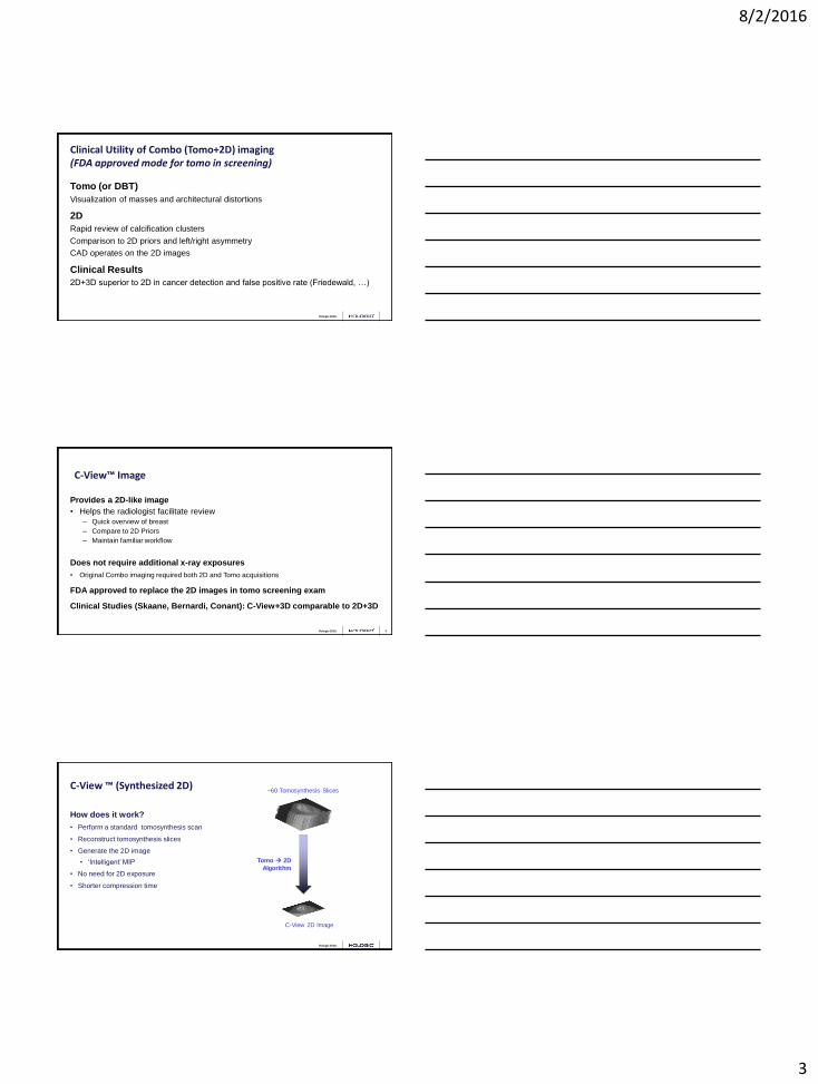
Clinical Utility of Combo (Tomo+2D) imaging (FDA approved mode for tomo in screening)
Tomo (or DBT)
Visualization of masses and architectural distortions
2D
Rapid review of calcification clusters
Comparison to 2D priors and left/right asymmetry
CAD operates on the 2D images
Clinical Results
2D+3D superior to 2D in cancer detection and false positive rate (Friedewald, …)
Hologic 2016. 8
Provides a 2D-like image
• Helps the radiologist facilitate review
– Quick overview of breast
– Compare to 2D Priors
– Maintain familiar workflow
Does not require additional x-ray exposures
• Original Combo imaging required both 2D and Tomo acquisitions
FDA approved to replace the 2D images in tomo screening exam
Clinical Studies (Skaane, Bernardi, Conant): C-View+3D comparable to 2D+3D
C-View™ Image
Hologic 2016.
C-View ™ (Synthesized 2D)
How does it work?
• Perform a standard tomosynthesis scan
• Reconstruct tomosynthesis slices
• Generate the 2D image
• ‘Intelligent’ MIP
• No need for 2D exposure
• Shorter compression time
C-View 2D Image
Tomo 2D
Algorithm
~60 Tomosynthesis Slices
8/2/2016
4
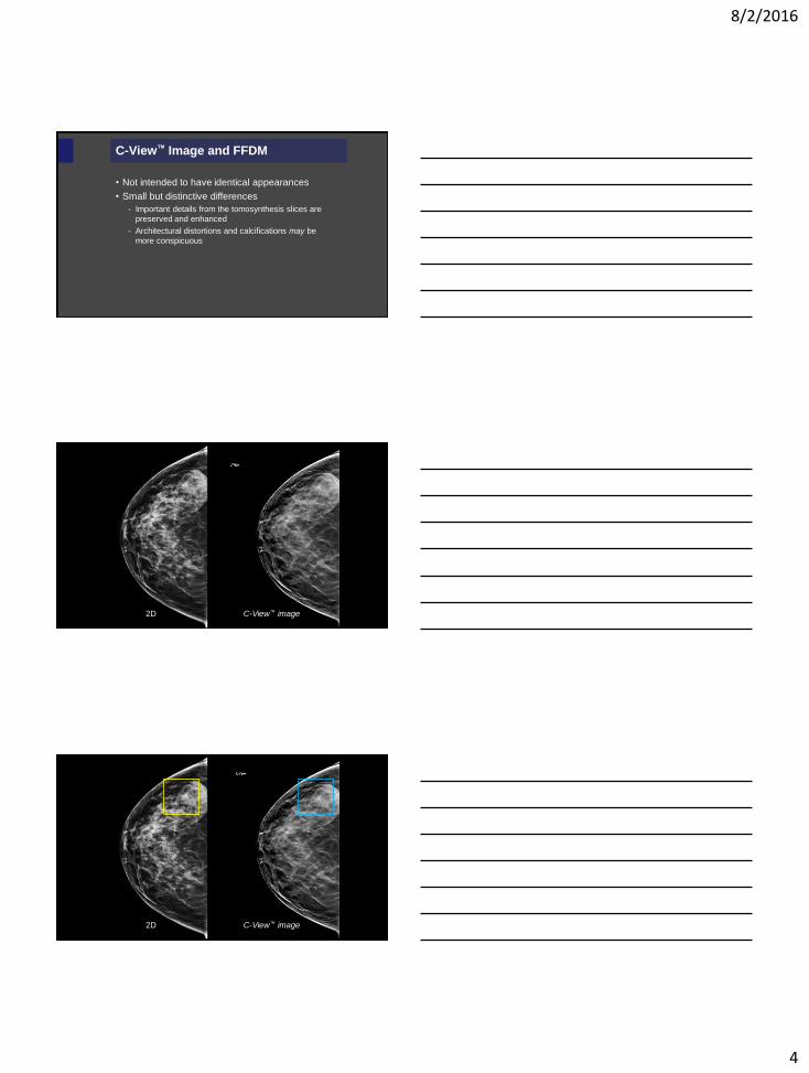
C-View™ Image and FFDM
• Not intended to have identical appearances
• Small but distinctive differences
- Important details from the tomosynthesis slices are
preserved and enhanced
- Architectural distortions and calcifications may be
more conspicuous
2D C-View™ image
2D C-View™ image
8/2/2016
5
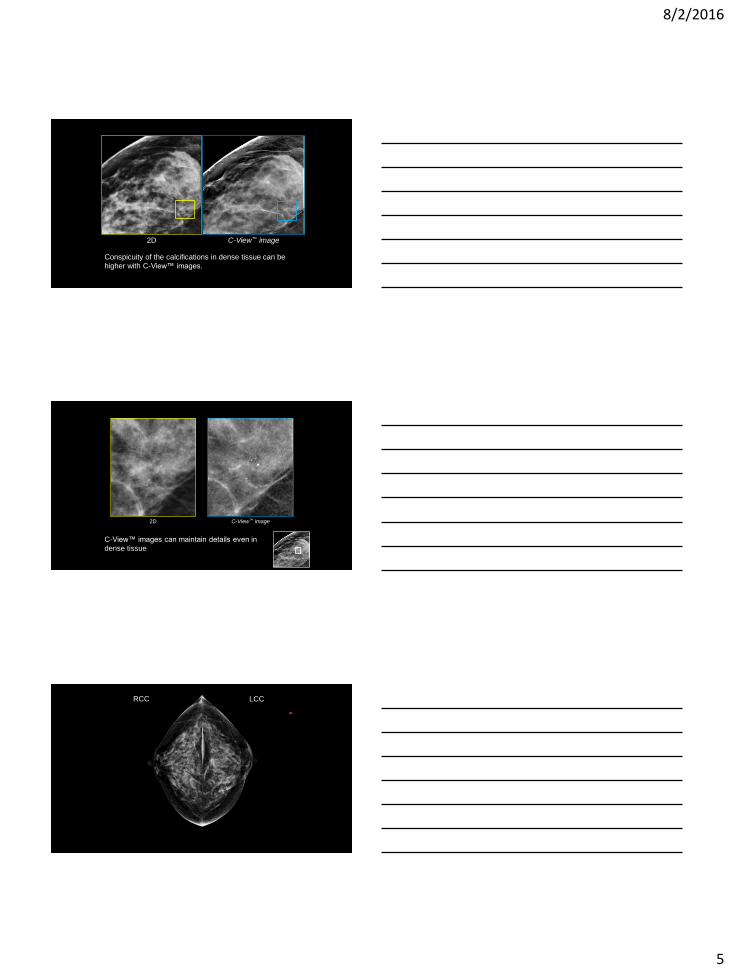
2D C-View™ image
Conspicuity of the calcifications in dense tissue can be
higher with C-View™ images.
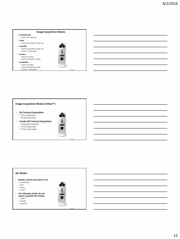
2D C-View™ image
C-View™ images can maintain details even in
dense tissue
*
LCC RCC

8/2/2016
6
LMLO RMLO
LMLO RMLO
2D Tomosynthesis slices

8/2/2016
7
2D Tomosynthesis Slice
Invasive Ductal Carcinoma
2D C-View™ image
Contrast Enhanced 2D Mammography – I-View™
Hologic Proprietary and Confidential

8/2/2016
8
Hologic 2016.
Dual Energy 2D Imaging
Designed to image an iodine contrast agent
Two exposures are made in rapid sequence:
1. Low kV (normal mammogram, ~28-30 kV, Rh/Ag filters). Below Iodine’s k-edge of 33 keV
2. High kV (~45-49 kV, Cu filter). Above Iodine’s k-edge.
Subtraction gives a 2D iodine contrast image
Repeat as desired
Imaging window ends after ~6 minutes due to contrast redistribution
Hologic Proprietary and Confidential
Hologic 2016.
Dual-Energy Subtraction
• You cannot see the iodine contrast drug in high or low kV images
• But you can when you subtract the images:
Subtraction = high kV - low kV
• Only the low kV and subtraction images are viewed
Hologic Proprietary and Confidential
High kV Subtraction Low kV
Hologic 2016.
CE2D – Dual Energy 2D
Low kV, High kV images acquired for each view
Views can be any order
Hologic Proprietary and Confidential
0 s ~ 120 s ~ 180 s
Ipsilateral Breast Contralateral Breast
inject
~ 240 s ~ 300 s ~ 480 s
E n e
r g
y
MLO CC CC MLO
L
H
L
H
L
H
L
H

8/2/2016
9
Hologic 2016.
Dual Energy 2D Combo Imaging
Three exposures are made in rapid sequence:
1. Low kV tomosynthesis scan (~30 kV, Al filter)
2. Low kV (normal mammogram, 28-30 kV, Rh/Ag filters)
3. High kV (~45-49 kV, Cu filter)
Subtraction gives a 2D contrast image
Tomo image is co-registered to the contrast 2D image
Repeat as desired
Imaging window ends after ~6 minutes due to contrast redistribution
Hologic Proprietary and Confidential
Hologic 2016.
Combo CE2D – Dual Energy 2D with Tomosynthesis
Tomo, Low kV, High kV images acquired for each view
Views can be any order
0 s ~120 s ~180 s
Ipsilateral Breast Contralateral Breast
LH
LH
LH
LH
inject
~240 s ~300 s ~480 s
En
erg
y
MLO CC CC MLO
Hologic Proprietary and Confidential
Example Image
Courtesy of Andrea Woodroof, Kentucky Breast Care
Hologic Proprietary and Confidential

8/2/2016
10
CE2D for Discordant Findings
• 52 y.o. female, presented for screening
• 0.8 cm spiculated mass in the left axillary tail noted on both 2D and 3D
• Focal area of possible distortion noted on 3D
Hologic Proprietary and Confidential
CE2D for Discordant Findings
Hologic Proprietary and Confidential
CE2D for Discordant Findings
• Area of distortion couldn’t definitively be correlated on ultrasound for US biopsy
Hologic Proprietary and Confidential

8/2/2016
11
CE2D for Discordant Findings
Hologic Proprietary and Confidential
CE2D for Discordant Findings
• Area of distortion couldn’t definitively be correlated on ultrasound for US biopsy
• The enhancing mass on CE2D was correlated with the tomo distortion
• Tomo-guided biopsy ensued
Hologic Proprietary and Confidential
Hologic 2016.
Quality Control Procedures: Digital Breast Tomosynthesis
8/2/2016
12
Hologic 2016.
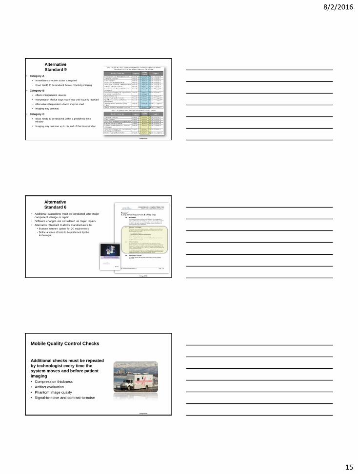
Image Acquisition Modes • Conventional
• Acquires 2D images only
• Tomo
• Acquires tomosynthesis images only
• TomoHD
• Acquires tomosynthesis images only
• Produces C-View images
• Combo
• Acquires 2D images
• Acquires tomosynthesis images
• ComboHD
• Acquires 2D images
• Acquires tomosynthesis images
• Produces C-View images
Hologic 2016.
Image Acquisition Modes (I-View™)
• 2D Contrast Acquisition
• 2D low-energy image
• 2D high-energy image
• Combo 2D Contrast Acquisition
• Tomosynthesis projections
• 2D low-energy images
• 2D high-energy images
Hologic 2016.
QC Modes
• Quality control procedures test
• Conventional
• Tomo
• Combo
• I-View™
• The following modes do not
require separate QC testing
• C-View™
• TomoHD
• ComboHD

8/2/2016
13
Hologic 2016.
DBT
Quality Control
D B T Q U A L I T Y C O N T R O L
Hologic 2016.
5 extra tests that are usually
performed by technologist
12 tests to be performed by
the medical physicist
Medical Physics
Medical Equipment Evaluation (MEE) Testing
Hologic 2016.
7 of them have DBT
components/requirements
12 tests to be performed by
the medical physicist
Medical Physics
QC Tests

8/2/2016
14
Hologic 2016.
3 of them have I-View
components/requirements
Medical Physics
QC Tests
Hologic 2016.
3 of them have DBT
components/requirements
12 tests to be performed by
the technologist
Technologist QC
Tests
Hologic 2016.
2 of them have I-View
components/requirements
Technologist QC
Tests
8/2/2016
15
Hologic 2016.
Alternative
Standard 9
• Category A
• Immediate corrective action is required
• Issue needs to be resolved before resuming imaging
• Category B
• Affects interpretation devices
• Interpretation device stays out of use until issue is resolved
• Alternative interpretation device may be used
• Imaging may continue
• Category C
• Issue needs to be resolved within a predefined time
window
• Imaging may continue up to the end of that time window
Hologic 2016.
Alternative
Standard 6
• Additional evaluations must be conducted after major
component change or repair
• Software changes are considered as major repairs
• Alternative Standard 6 allows manufacturers to:
• Evaluate software update for QC requirements
• Define a series of tests to be performed by the
technologist
Hologic 2016.
Mobile Quality Control Checks
Additional checks must be repeated
by technologist every time the
system moves and before patient
imaging
• Compression thickness
• Artifact evaluation
• Phantom image quality
• Signal-to-noise and contrast-to-noise

8/2/2016
16
Hologic 2016.
Hologic QC Manuals
• http://www.hologic.com/support/dimensions-3d-breast-tomosynthesis-
dimensions-2d-full-field-digital-mammography
• http://www.hologic.com/support/selenia-digital-mammography
MAN-01965
R008
MAN-03706
R002
MAN-01476
R001
Hologic 2016.
Software Version and QC Manuals
MAN-01965
• Applies to software version prior to v1.8.x
MAN-03706
• Applies to software version starting at v1.8.x
Important differences
• Appendix D: CNR Correction Tables
Hologic 2016.
Tomosynthesis Option
Tomosynthesis specific
tests are marked with an
icon
Icon indicates that a
special action is required
under tomosynthesis
NOTE: When testing FFDM only,
these instructions are ignored
8/2/2016
17
Hologic 2016.
Diagnostic Option
Diagnostic specific tests
are marked with an icon
Icon indicates that listed
action is only applicable
to systems licensed for
diagnostic use
NOTE: When testing screening-only
FFDM systems, these instructions
are ignored
Hologic 2016.
Contrast Option
Iodine contrast specific
tests are marked with an
icon
Icon indicates that listed
action is only applicable
to systems licensed for
iodine contrast imaging
NOTE: When testing systems that
are not licensed for iodine contrast,
these instructions are ignored
Hologic 2016.
Tomosynthesis-Specific Quality Control
51
• Tomo Geometry
Calibration
• Usually performed by
technologist
8/2/2016
18
Hologic 2016.
Quality Control
Tests
S Y S T E M D E S C R I P T I O N A N D U S E R I N T E R F A C E
Hologic 2016.
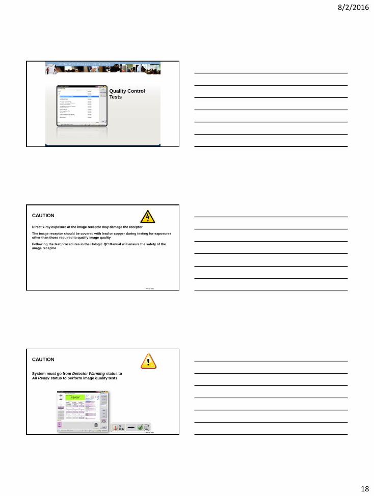
CAUTION
Direct x-ray exposure of the image receptor may damage the receptor
The image receptor should be covered with lead or copper during testing for exposures
other than those required to qualify image quality
Following the test procedures in the Hologic QC Manual will ensure the safety of the
image receptor
Hologic 2016.
CAUTION
System must go from Detector Warming status to
All Ready status to perform image quality tests

8/2/2016
19
Hologic 2016.
1. MAMMOGRAPHIC UNIT ASSEMBLY
EVALUATION
Follow the 1999 ACR Mammography Quality Control Manual
Hologic 2016.
2. COLLIMATOR ASSESSMENT Follow the Hologic Selenia Dimensions Quality Control Manual
Hologic 2016.
2a. X-Ray Field to Light Field
ONLY use the 24x29 cm compression paddle
Cover the image receptor if repeated, high exposures are required (i.e. self-
developing film)

8/2/2016
20
Hologic 2016.
2b. X-Ray Field to Image Receptor
Test with the 24x29 cm compression paddle
Test left, center and right x-ray fields with the 18x24 cm compression paddle
Use the Zero-Degree Tomo view to test under tomosynthesis
Follow the directions in the QC manual
Hologic 2016.
2c. Compression Paddle to Image Receptor
Compression paddles
Manufactured as single pieces
Do not have adjusting parts
Designed to comply with the regulations
Design assumes mild compression (~10lb) to remove play
Hologic 2016.
3. ARTIFACT EVALUATION Follow the Hologic Selenia Dimensions Quality Control Manual

8/2/2016
21
Hologic 2016.
Procedure Highlights
DICOM printer
Send an artificial flat field image to the printer
FFDM testing
Test all focus/filter combinations
(LFS/Rh; LFS/Ag; SFS/Rh; SFS/Ag; LFS Cu)
Preview image in full resolution
DBT testing
Test using middle projection
Preview image in full resolution
Hologic 2016.
4. KVP ACCURACY AND
REPRODUCIBILITY
Follow the 1999 ACR Mammography Quality Control Manual
Hologic 2016.
Procedure Highlights
Cover the image receptor to protect it from radiation exposure
FFDM extends to 39 kVp; DBT extends to 49 kVp
Use the Zero-Degree Tomo mode to test beyond 39 kVp, if needed
Non-invasive meters must be calibrated to the specific filters and energy range used
Hologic Service can assist with equipment
Conventional Tomo Stationary Tomo
8/2/2016
22
Hologic 2016.
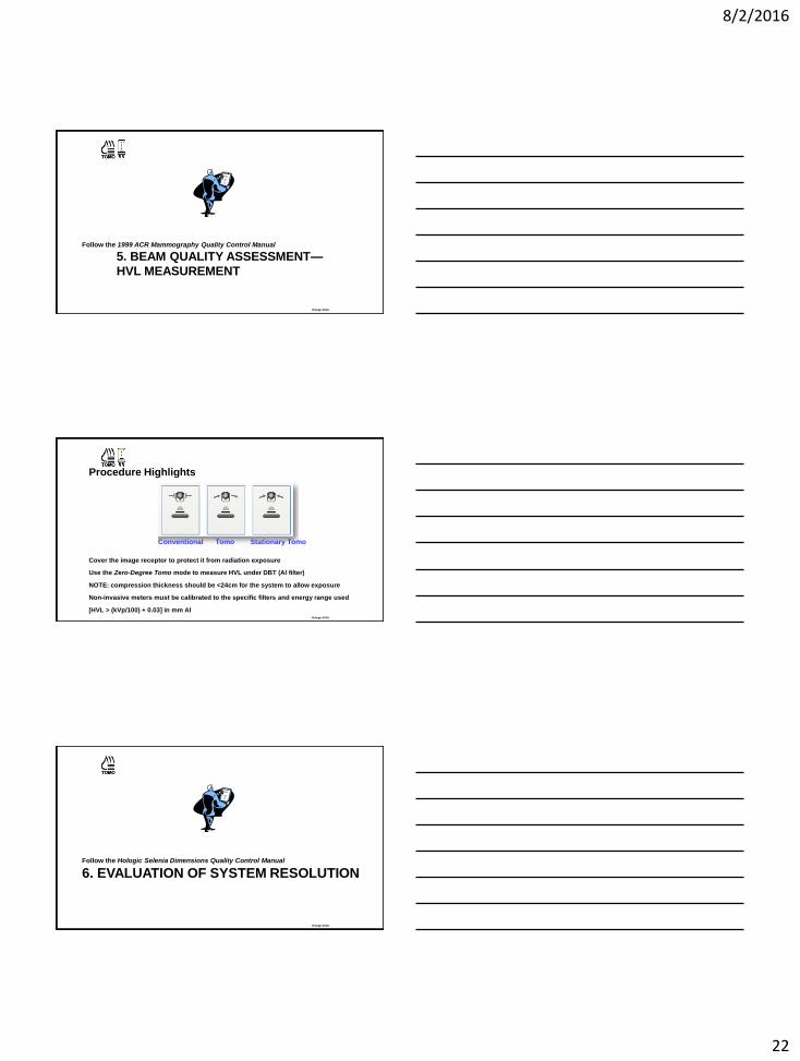
5. BEAM QUALITY ASSESSMENT—
HVL MEASUREMENT
Follow the 1999 ACR Mammography Quality Control Manual
Hologic 2016.
Procedure Highlights
Cover the image receptor to protect it from radiation exposure
Use the Zero-Degree Tomo mode to measure HVL under DBT (Al filter)
NOTE: compression thickness should be <24cm for the system to allow exposure
Non-invasive meters must be calibrated to the specific filters and energy range used
[HVL > (kVp/100) + 0.03] in mm Al
Conventional Tomo Stationary Tomo
Hologic 2016.
6. EVALUATION OF SYSTEM RESOLUTION Follow the Hologic Selenia Dimensions Quality Control Manual

8/2/2016
23
Hologic 2016.
Procedure Highlights
Place the line pair phantom on top of the 4 cm acrylic block
Rotate the line pair phantom 45°
Apply 15-20 lb of compression to avoid vibration during DBT
Use the Flat Field view (no image processing)
Resolution guidelines:
FFDM: > 7 lp/mm @ 45°
DBT: > 3 lp/mm @ 45°
Hologic 2016.
Procedure Highlights
Hologic 2016.
7. AEC FUNCTION PERFORMANCE Follow the Hologic Selenia Dimensions Quality Control Manual

8/2/2016
24
Hologic 2016.
AEC Function Description
AEC modes
• Auto-Filter
• Auto-kV
• Auto-Time
AEC positions
• Auto AEC: two ~1cm2 floating sensors in 5 x 14cm2 area
• One of seven manual positions (marked on compression paddle)
AEC function
• kVp and filter parameters are determined by compression thickness and AEC technique tables
• kVp can be adjusted upwards if the exposure time will be too long
• Starting mAs is determined from short pre-exposure targeting a specific exposure index (EI)
• Final mAs is adjusted by CNR correction factor
Hologic 2016.
Procedure Highlights
Compression thickness must be
set using the compression
display
FFDM testing
• Range of phantom thickness
• Different operating modes (i.e. mag)
• Exposure compensation steps
DBT testing
• Range of phantom thickness
Compression
Thickness
Hologic 2016.
Exposure Index (EI)
EI is defined as the digital value
of a detector element
“Raw” EI values need to be
corrected by
• Subtracting the DC offset (value of 50)
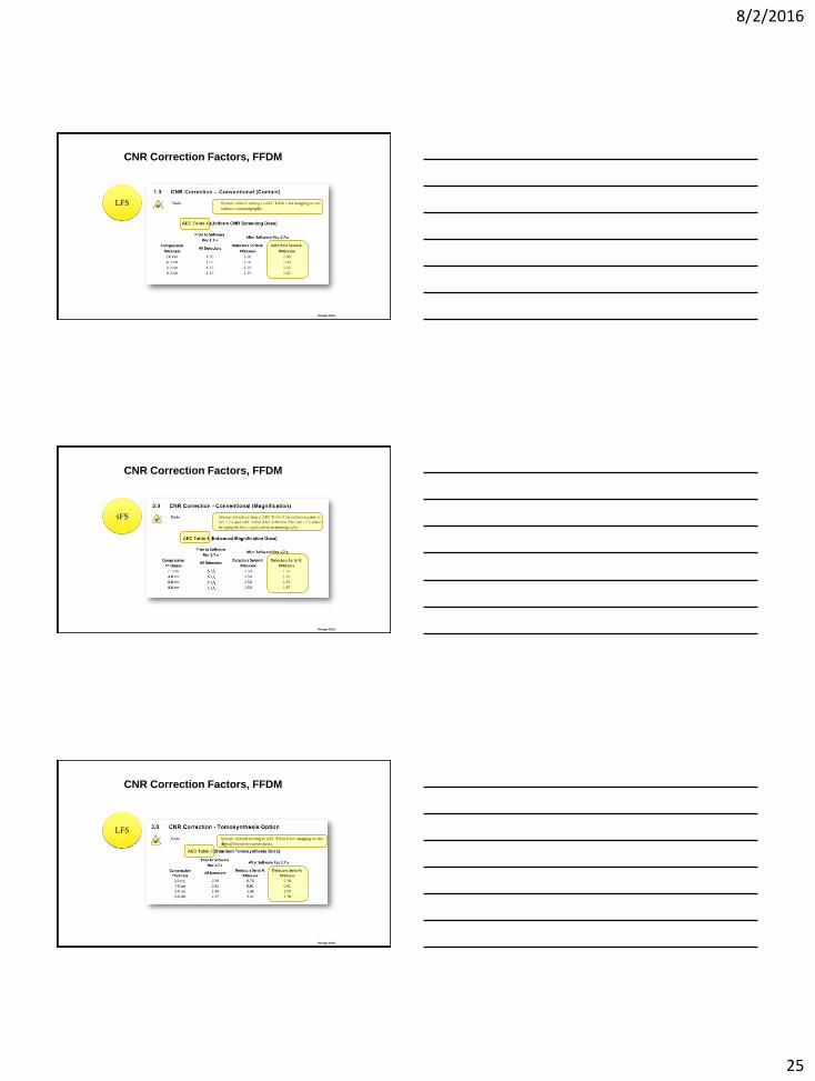
• Normalizing by the CNR correction factor (given in Appendix D of the Hologic QC Manual)
𝑃𝑖𝑥𝑒𝑙 𝑉𝑎𝑙𝑢𝑒 =𝑅𝑂𝐼 𝑚𝑒𝑎𝑛 − 𝐷𝐶 𝑂𝑓𝑓𝑠𝑒𝑡
𝐶𝑁𝑅 𝐶𝑜𝑟𝑟𝑒𝑐𝑡𝑖𝑜𝑛 𝐹𝑎𝑐𝑡𝑜𝑟
8/2/2016
25
Hologic 2016.
CNR Correction Factors, FFDM
LFS
Hologic 2016.
CNR Correction Factors, FFDM
SFS
Hologic 2016.
CNR Correction Factors, FFDM
LFS

8/2/2016
26
Hologic 2016.
Calculation Example
Hologic 2016.
8. BREAST ENTRANCE EXPOSURE,
AEC REPRODUCIBILITY AND AGD
Follow the Hologic Selenia Dimensions Quality Control Manual
Hologic 2016.
Procedure Highlights
Wait until the image receptor
goes from Warming to
Ready status
Use ACR Phantom view to
overwrite compression
thickness to 4.2 cm
8/2/2016
27
Hologic 2016.
Procedure Highlights
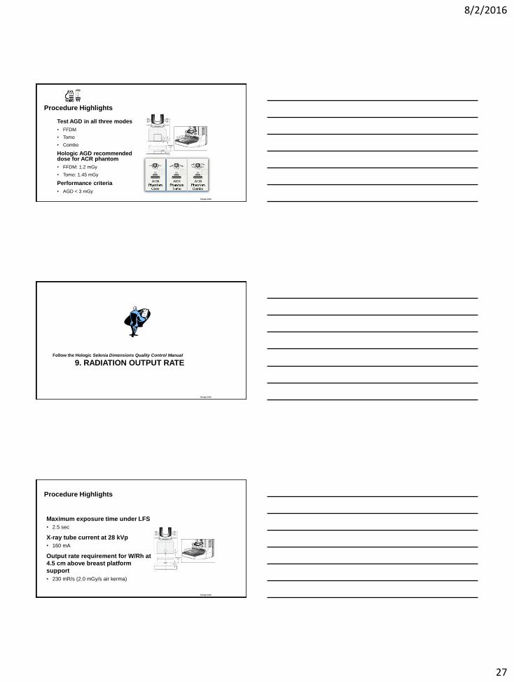
Test AGD in all three modes
• FFDM
• Tomo
• Combo
Hologic AGD recommended dose for ACR phantom
• FFDM: 1.2 mGy
• Tomo: 1.45 mGy
Performance criteria
• AGD < 3 mGy
Hologic 2016.
9. RADIATION OUTPUT RATE Follow the Hologic Selenia Dimensions Quality Control Manual
Hologic 2016.
Procedure Highlights
Maximum exposure time under LFS
• 2.5 sec
X-ray tube current at 28 kVp
• 160 mA
Output rate requirement for W/Rh at
4.5 cm above breast platform
support
• 230 mR/s (2.0 mGy/s air kerma)

8/2/2016
28
Hologic 2016.
10. PHANTOM IMAGE QUALITY
EVALUATION
Follow the Hologic Selenia Dimensions Quality Control Manual
Hologic 2016.
Procedure Highlights
Wait until the image receptor
goes from Warming to
Ready status
Use ACR Phantom view to
overwrite compression
thickness to 4.2 cm
Hologic 2016.
Phantom Scoring
Score phantom on AWS display
Review image in full resolution
FFDM scoring
• 5 fibers, 4 specs, 4 masses
• Due to phantom variations a score of 4.5/4.0/3.5 is
acceptable providing SNR and high contrast
resolution tests pass
DBT scoring
• Scroll to the slice that puts the different elements
in focus
• 4 fibers, 3 specs, 3 masses

8/2/2016
29
Hologic 2016.
11. SNR AND CNR MEASUREMENTS Follow the Hologic Selenia Dimensions Quality Control Manual
Hologic 2016.
Procedure Highlights
Wait until the image receptor goes
from Warming to Ready status
Use ACR Phantom view to overwrite
compression thickness to 4.2 cm
ACR Phantom view allows
automatic SNR/CNR calculations
Test is performed under FFDM mode
only
Hologic 2016.
Automatic Computation

8/2/2016
30
Hologic 2016.
12. DIAGNOSTIC REVIEW
WORKSTATION QUALITY CONTROL
Follow the Hologic Selenia Dimensions Quality Control Manual
Hologic 2016.
Procedure Highlights
The Hologic QC Manual offers an
alternative QC procedure for the
review workstation
Most review workstation offer their
own QC software and QC
procedures
Follow their QC procedures and
performance requirements
Hologic 2016.
13. DETECTOR GHOSTING
(TROUBLESHOOTING USE ONLY)
Follow the Hologic Selenia Dimensions Quality Control Manual

8/2/2016
31
Hologic 2016.
Procedure Highlights
Test to be performed if ghosting is
noticed on clinical images
Is not required under acceptance or
annual evaluation
Wait until the image receptor goes from
Warming to Ready status
Typical reasons for failure
• Erase LED array failure
Hologic 2016.
Procedure Highlights
3 2 1
Hologic 2016.
Computation of Ghosting
𝐺ℎ𝑜𝑠𝑡 =𝑚𝑒𝑎𝑛𝑅3 − 𝑚𝑒𝑎𝑛𝑅2
𝑚𝑒𝑎𝑛𝑅1 − 𝑚𝑒𝑎𝑛𝑅2
8/2/2016
32
Hologic 2016.
Tomo-guided Biopsy
Hologic 2016. 95
2D
Tomo
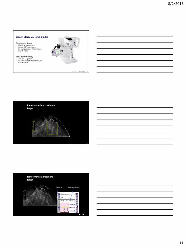
How do you biopsy this lesion, occult in 2D and U/S and
MRI?
Hologic 2016.
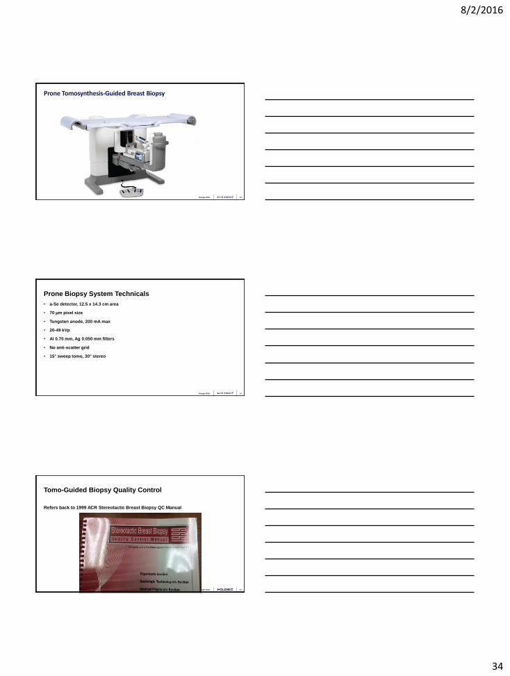
Affirm™ Interventional Add-On Device Utilizing Tomosynthesis Localization
8/2/2016
33
Hologic 2016. 97
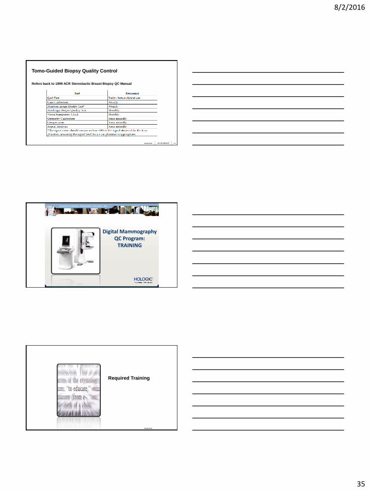
Stereotactic Biopsy • Take 2D scout exposure
• Acquire ±30° stereo pairs
• Use triangulation to determine x,y,z
lesion location
Tomo-guided Biopsy • Take Tomo exposure
• Use tomo slices to determine x,y,z
lesion location
Biopsy: Stereo vs. Tomo-Guided
Hologic 2016. 98
Tomosynthesis procedure – Target
Hologic 2016.
Tomosynthesis procedure - Target
Lesion coordinates Targeting
8/2/2016
34
Hologic 2016.
Prone Tomosynthesis-Guided Breast Biopsy
100
Hologic 2016.
Prone Biopsy System Technicals
101
• a-Se detector, 12.5 x 14.3 cm area
• 70 μm pixel size
• Tungsten anode, 200 mA max
• 20-49 kVp
• Al 0.70 mm, Ag 0.050 mm filters
• No anti-scatter grid
• 15° sweep tomo, 30° stereo
Hologic 2016.
Tomo-Guided Biopsy Quality Control
102
Refers back to 1999 ACR Stereotactic Breast Biopsy QC Manual
8/2/2016
35
Hologic 2016.
Tomo-Guided Biopsy Quality Control
103
Refers back to 1999 ACR Stereotactic Breast Biopsy QC Manual
Hologic 2016.
QUALITY CONTROL PROGRAMS, HOLOGIC, INC.
Digital Mammography QC Program:
TRAINING
Hologic 2016.
Required Training

8/2/2016
36
Hologic 2016.
FDA Training Requirements: FFDM
FFDM training is specific to the type of user
Everybody needs FFDM training
• 8 hours for Medical Physicists
• 8 hours for Technologists
• 8 hours for Radiologists
PRE-00023 PRE-00023
Hologic 2016.
FDA Training Requirements: DBT
DBT is considered a new imaging modality
DBT requires its own training
DBT training is specific to the type of user
Everybody needs additional training
• 8 hours for Medical Physicists
• 8 hours for Technologists
• 8 hours for Radiologists
PRE-00023 PRE-00023
Hologic 2016.
Medical Physicist DBT Training
8 hours of FFDM training is required
8 hours of DBT training is required
Available training sources • This AAPM meeting
• MTMI hands-on workshops
• Hologic – On-line training
– Field training during system installation
PRE-00023 PRE-00023

8/2/2016
37
Hologic 2016.
Facility Certification
2D MQSA certification through ACR or other approved accreditation body
No approved accreditation bodies DBT today
– DBT systems will be accredited under facility’s existing FFDM certification through FDA’s
Certification Extension Program
– Facility must be FFDM MQSA certified before applying for DBT extension certification
Certification Extension Program Division of Mammography Quality and Radiation Programs FDA/CDRH/OCER 10903 New Hampshire Avenue, WO66-4621 Silver Spring, MD 20903-0002 Phone: 301-796-5710 Fax: 301-847-8502
PRE-00023 PRE-00023
Hologic 2016.
Quality Control with new ACR Protocol/Phantom
110
Hologic 2016.
Quality Control with new ACR Protocol/Phantom
111

8/2/2016
38
Hologic 2016.
Quality Control with new ACR Protocol/Phantom
112
• Designed to replace the manufacturer’s FFDM QC protocols
• Does not apply to systems with tomosynthesis options
• Does not apply to systems with dual-energy contrast options
• What about clinics with some systems with tomo and other
systems FFDM only?
Hologic 2016.
Thank you!
Email:
113
Hologic 2016. 114