2008 terni, workshop interattivo, tecniche di impianto dei pacemaker in urgenza
51
Principi di tecnica di impianto Principi di tecnica di impianto del Pacemaker temporaneo del Pacemaker temporaneo in urgenza in urgenza Stefano Nardi, MD, PhD Toracich and Cardio-Vascular Departement Divison of Cardiology Arrhythmia, EP Center and Cardiac Pacing Unit Santa Maria General Hospital, Terni Santa Maria General Hospital, Terni
-
Upload
centro-diagnostico-nardi -
Category
Health & Medicine
-
view
86 -
download
3
Transcript of 2008 terni, workshop interattivo, tecniche di impianto dei pacemaker in urgenza

Principi di tecnica di impianto Principi di tecnica di impianto del Pacemaker temporaneo del Pacemaker temporaneo
in urgenza in urgenzaStefano Nardi, MD, PhD
Toracich and Cardio-Vascular DepartementDivison of Cardiology
Arrhythmia, EP Center and Cardiac Pacing UnitSanta Maria General Hospital, TerniSanta Maria General Hospital, Terni

BRADIARITMIABRADIARITMIA
EMERGENZAEMERGENZA
SINTOMATICA (Sincope, Lipotrimia, Dispnea)SINTOMATICA (Sincope, Lipotrimia, Dispnea)
FC < 40 B\MFC < 40 B\M
GRAVITA’ DELLA CARDIOPATIA PRESENTEGRAVITA’ DELLA CARDIOPATIA PRESENTE
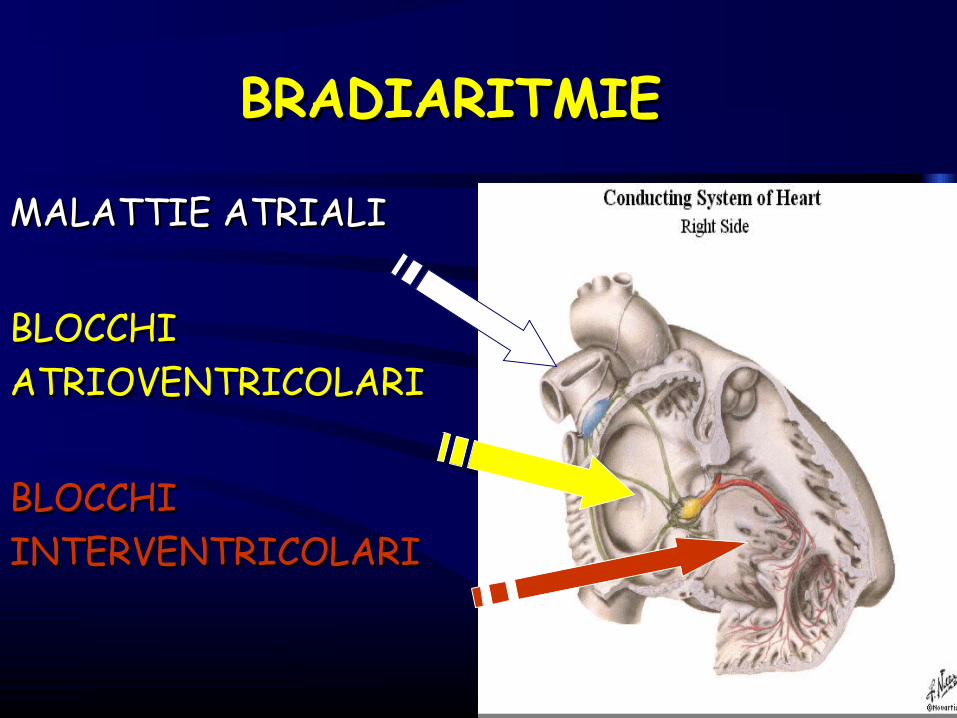
BRADIARITMIEBRADIARITMIE
MALATTIE ATRIALIMALATTIE ATRIALI
BLOCCHIBLOCCHIATRIOVENTRICOLARIATRIOVENTRICOLARI
BLOCCHIBLOCCHIINTERVENTRICOLARIINTERVENTRICOLARI

Malattie AtrialiMalattie AtrialiBradicardia Sinusale Marcata Bradicardia Sinusale Marcata Blocchi Seno-Atriali II° e III° sintomatici Blocchi Seno-Atriali II° e III° sintomatici Sindrome bradi-tachi (Pause lunghe)Sindrome bradi-tachi (Pause lunghe)
EziologiaIMA inferiore IMA inferiore Patologie Extra-cardiachePatologie Extra-cardiache
(neuromuscolari – emorragie digestive)(neuromuscolari – emorragie digestive)Farmaci Farmaci
ß-bloccanti, Digoxina, Verapamil, etcß-bloccanti, Digoxina, Verapamil, etc Ipertono VagaleIpertono Vagale
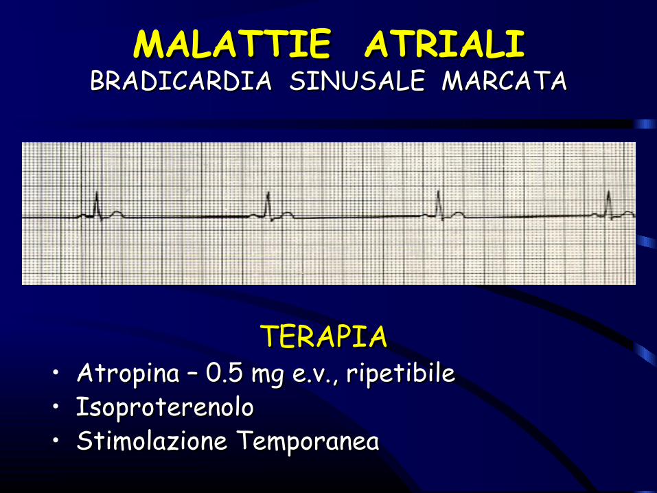
MALATTIE ATRIALIMALATTIE ATRIALIBRADICARDIA SINUSALE MARCATABRADICARDIA SINUSALE MARCATA
TERAPIATERAPIA• Atropina – 0.5 mg e.v., ripetibileAtropina – 0.5 mg e.v., ripetibile• IsoproterenoloIsoproterenolo• Stimolazione TemporaneaStimolazione Temporanea

BLOCCHI ATRIO-VENTRICOLARIBLOCCHI ATRIO-VENTRICOLARIBAV II E III GRADO SINTOMATICIBAV II E III GRADO SINTOMATICI
EZIOLOGIAEZIOLOGIACardiopatia Organica CronicaCardiopatia Organica Cronica
((Ischemica, Reumatica, Congenita, DCM)Ischemica, Reumatica, Congenita, DCM)
IMAIMAFarmaci Farmaci
((ß-bloccanti, Digoxina, Verapamil, etc)ß-bloccanti, Digoxina, Verapamil, etc)
Ipertono VagaleIpertono Vagale
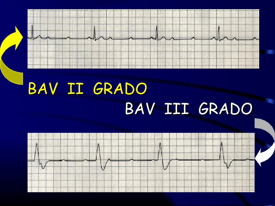
BAV II GRADOBAV II GRADO BAV III GRADOBAV III GRADO

BAV AVANZATOBAV AVANZATOINDICAZIONI AL TRATTAMENTOINDICAZIONI AL TRATTAMENTO
P M T E M P O R A N E O
S I N T O M A T I C OA T R O P I N A E . V . 0 . 5
R I P E T I B I L E
A S I N T O M A T I C OO S S E R V A Z I O N E
Q R S S T R E T T O
P M T E M P O R A N E O
Q R S L A R G O
B A V T O T A L EB A V I I G R A D O A V A N Z A T O

BLOCCHI INTERVENTRICOLARIBLOCCHI INTERVENTRICOLARIACUTIACUTI
EZIOLOGIAEZIOLOGIA
IMAIMA
Indicazioni alla Stimolazione Temporanea Indicazioni alla Stimolazione Temporanea
Blocco Trifascicolare Blocco Trifascicolare Blocco Bifascicolare (BBD+EAS, BBD+EPS) Blocco Bifascicolare (BBD+EAS, BBD+EPS) BAV I° + EAS o EPSBAV I° + EAS o EPS
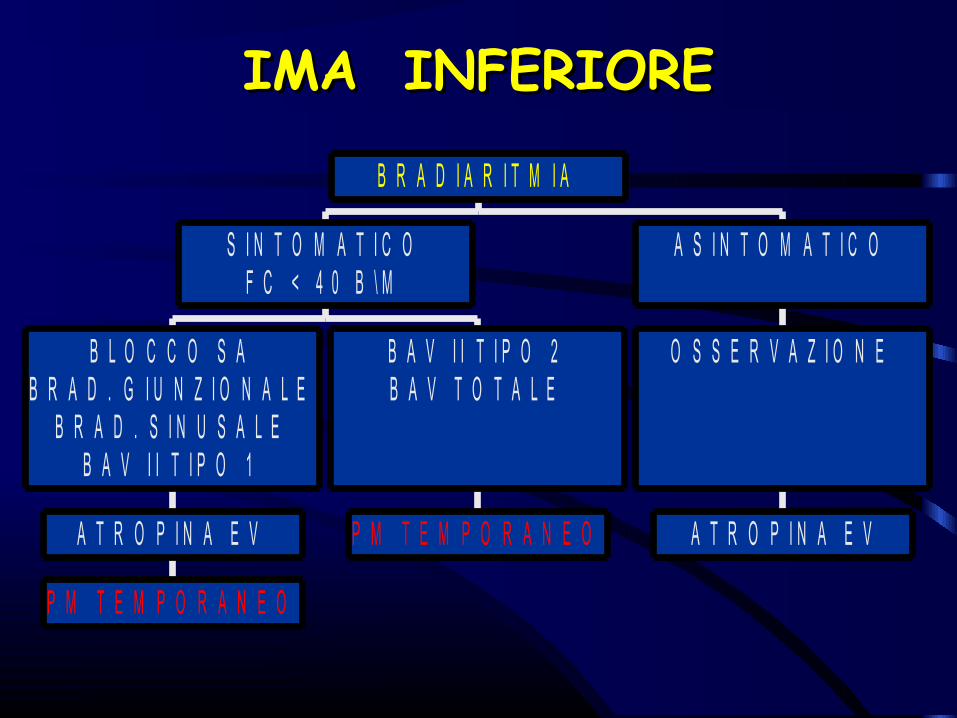
IMA INFERIOREIMA INFERIORE
P M T E M P O R A N E O
A T R O P I N A E V
B L O C C O S AB R A D . G I U N Z I O N A L E
B R A D . S I N U S A L EB A V I I T I P O 1
P M T E M P O R A N E O
B A V I I T I P O 2B A V T O T A L E
S I N T O M A T I C OF C < 4 0 B \ M
A T R O P I N A E V
O S S E R V A Z I O N E
A S I N T O M A T I C O
B R A D I A R I T M I A
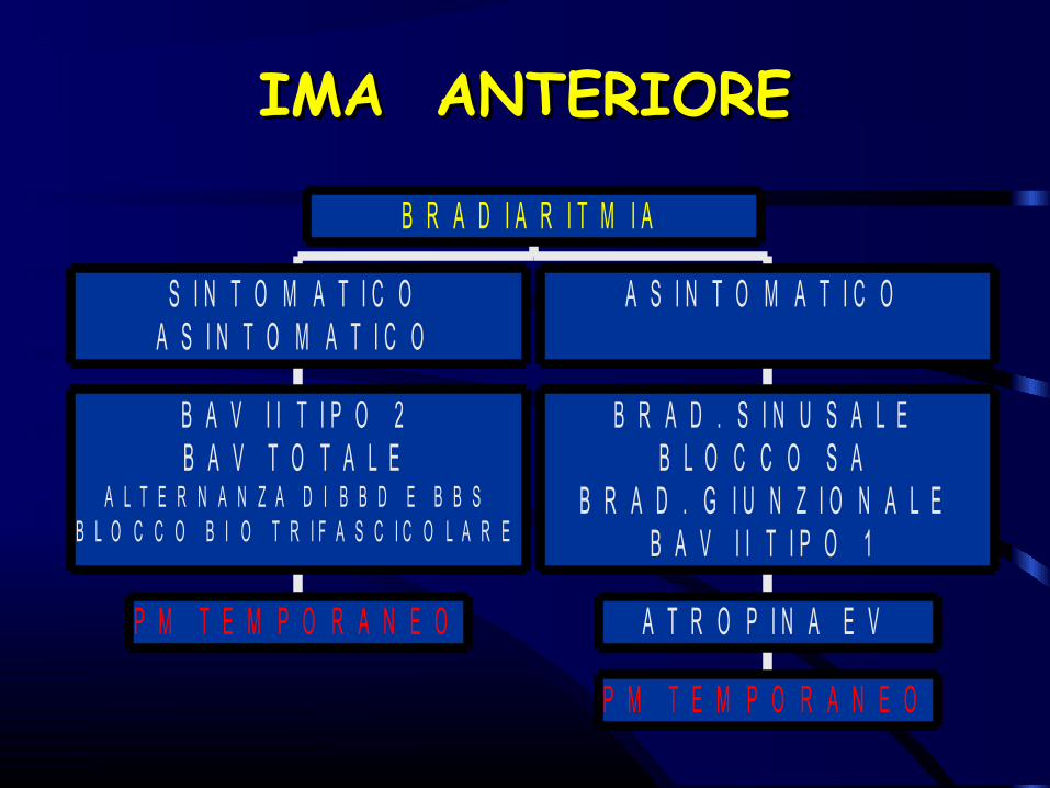
IMA ANTERIOREIMA ANTERIORE
P M T E M P O R A N E O
B A V I I T I P O 2B A V T O T A L E
A L T E R N A N Z A D I B B D E B B SB L O C C O B I O T R I F A S C I C O L A R E
S I N T O M A T I C OA S I N T O M A T I C O
P M T E M P O R A N E O
A T R O P I N A E V
B R A D . S I N U S A L EB L O C C O S A
B R A D . G I U N Z I O N A L EB A V I I T I P O 1
A S I N T O M A T I C O
B R A D I A R I T M I A
Stimolazione temporanea in urgenza anche nelle tachiaritmie?

TORSIONE DI PUNTATORSIONE DI PUNTA
QTc long Syndrome QTc long Syndrome SINDROME di Jerwell-Lange-Nielsen (con Sordità)SINDROME di Jerwell-Lange-Nielsen (con Sordità)SINDROME DI Romano-Ward (senza Sordità)SINDROME DI Romano-Ward (senza Sordità)
FORME ACQUISITEFORME ACQUISITEDisturbi Elettrolitici (IPOK+ IPOMG+)Disturbi Elettrolitici (IPOK+ IPOMG+)Farmaci (Chinidina, Amiodarone, Sotalolo, Ibutilide, etc)Farmaci (Chinidina, Amiodarone, Sotalolo, Ibutilide, etc)Aritmie Ipocinetiche (BAV TOTALE)Aritmie Ipocinetiche (BAV TOTALE)

TORSIONE DI PUNTATORSIONE DI PUNTAALGORITMO TERAPEUTICOALGORITMO TERAPEUTICO
E L E T T R O S T I M O L A Z I O N E T E M P O R A N E A1 0 0 - 1 2 0 / M I N : A T R I A L E O V E N T R I C O L A R E
T d P
I N C R E M E N T O D E L L A F CI S O P R O T E R E N O L O : 0 . 0 1 - 0 . 0 2 u g / k g / m i n
A T R O P I N A E V : B O L O 0 . 5 m g , R I P E T I B I L E O G N I 1 0 '
M E T O P R O L O L O : F O R M A C O N G E N I T A E D A A . T R I C I C L I C I
T d P
E V E N T U A L E C O R R E Z I O N ED E L D I S T U R B O E L E T T R O L I T I C O
M g S O 4 E V : 1 - 2 G . I N 5 - 1 0 ' , I N F U S I O N E D I 1 - 2 G \ H P E R 4 - 6 H
K C L : 1 0 m E q / h F I N O A L L A C O R R E Z I O N E D E L L O S Q U I L I B R I O
T O R S I O N E D I P U N T A ( T d P )

QUALE STIMOLAZIONE TEMPORANEA ?
EPICARDICATRANSESOFAGEA
TRANSTORACICA
TRANSVENOSA
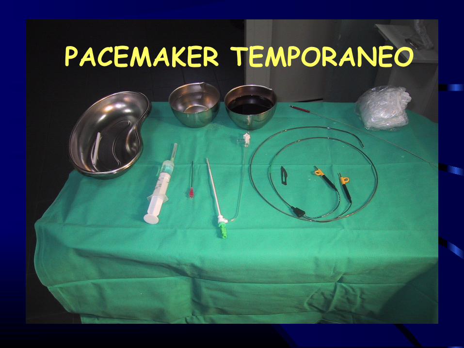
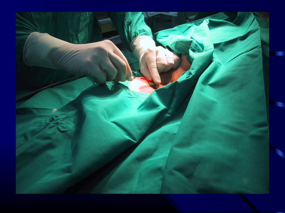
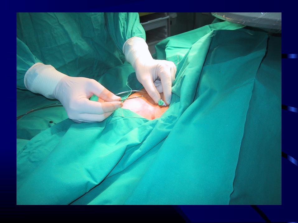
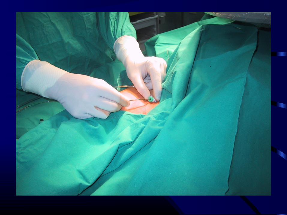
PACEMAKER TEMPORANEO


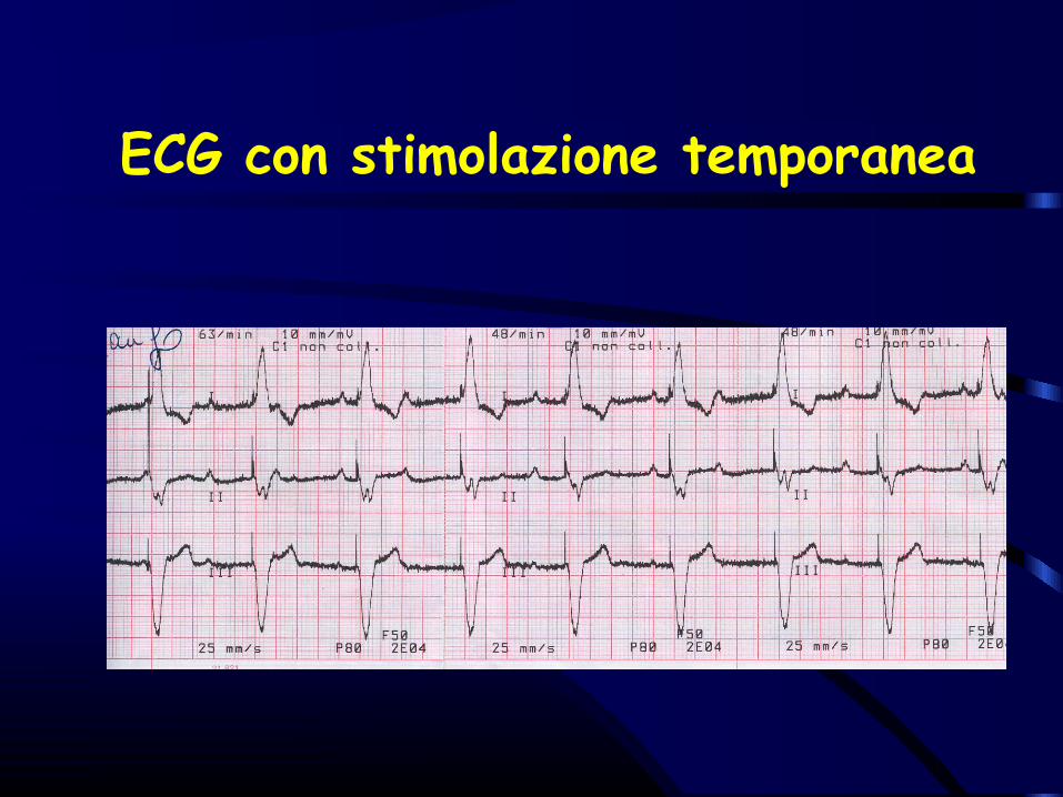
ECG con stimolazione temporanea

Complications• Pacing Failure
– Failure to Output– Failure to Capture
• Pseudomalfunction• Sensing Failure
– Oversensing– Undersensing
• Operative Failure
Transvenous Pacemaker (TVP)

Failure to Output
• Occurs when no PM spike is present • This may be due to battery failure, lead fracture,
a break in lead insulation, oversensing (inhibiting), poor lead connection at the take-off from the PM, and "cross-talk" (when A output is sensed by a V lead in a dual-chamber PM).
Transvenous Pacemaker

Failure to CaptureA spike not followed by either a V complex
- lead fracture- lead dislodgement- elevated PM threshold- MI at the lead tip- certain AADs (eg, flecainide)- metabolic abnormalities (hyper-k+, acidosis, alkalosis)- cardiac perforation- poor lead connection at the take-off from the generator- improper amplitude or pulse width settings.
Transvenous Pacemaker

Sensing• Definizione
- capacità del PM di percepire un segnale elettrico intrinseco, in funzione dell’ampiezza, dello Slew-rate, della frequenza del segnale e della posizione degli elettrodi.
• Sensibilità programmata- Indica il minimo segnale intracardiaco che il PM
percepisce per attivare la risposta del PM (inibito o triggerato).
5 mV
2 mV
1 mV
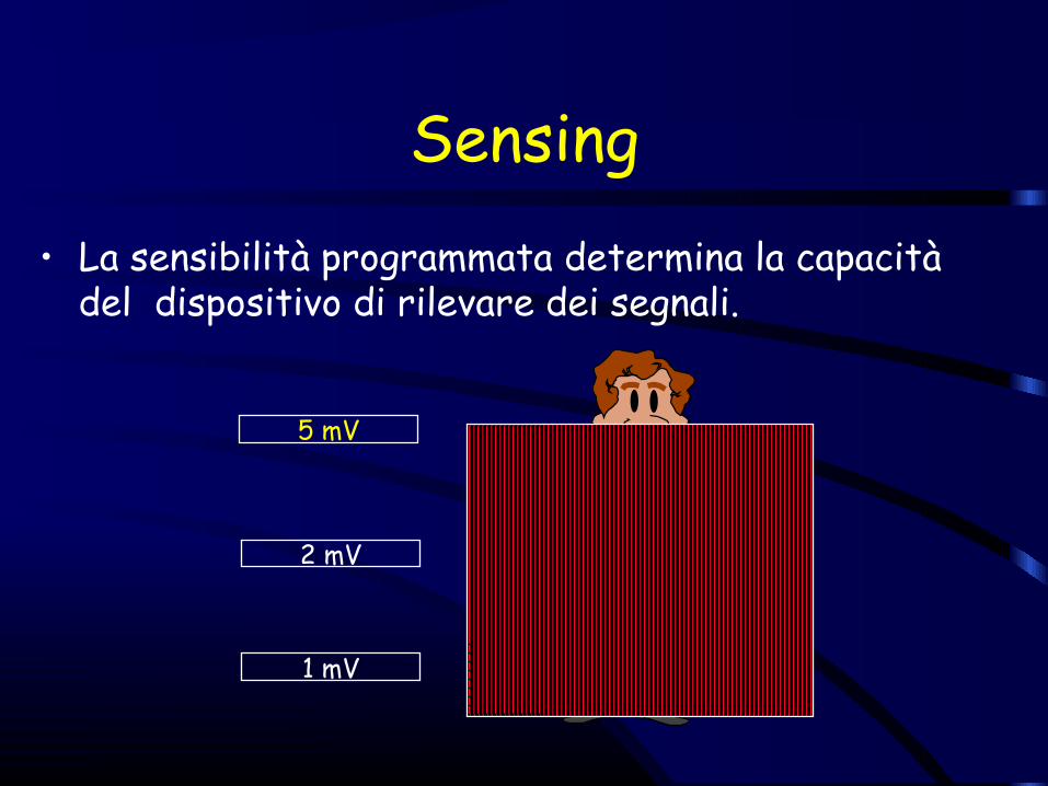
Sensing• La sensibilità programmata determina la capacità
del dispositivo di rilevare dei segnali.

5 mV
2 mV
1 mV
Sensing• Quando si programma la sensibilità, se si
diminuisce il suo valore, si rende il PM più sensibile (“sente” meglio).

5 mV
2 mV
1 mV
Sensing• Se la sensibilità programmata è troppo bassa
il dispositivo “sente” troppo
8 mV
6 mV
4 mV
2 mV
0 mV
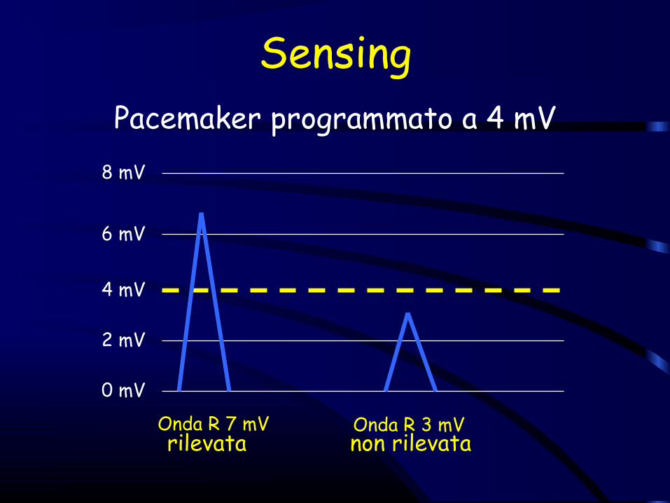
Onda R 7 mV Onda R 3 mV
Pacemaker programmato a 4 mV
Sensing
rilevata non rilevata
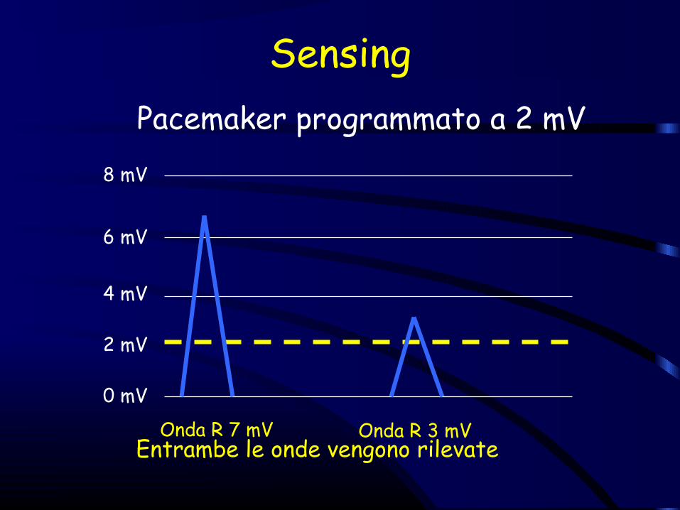
Pacemaker programmato a 2 mV8 mV
6 mV
4 mV
2 mV
0 mV
Entrambe le onde vengono rilevateOnda R 7 mV Onda R 3 mV
Sensing

Oversensing Ventricolare
Frequenza basedi stimolazione
Viene sentito un evento non corrispondente a onda R e viene inibito il pacemaker
Pause prolungate tra uno stimolo e il
successivo

Oversensing
• When a PM incorrectly senses electrical activity and is inhibited from correctly pacing.
• This may be due to muscular activity (particularly oversensing of the diaphragm or pectoralis muscles), electromagnetic (EM) interference, or lead insulation breakage.
Transvenous Pacemaker
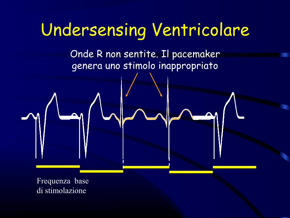
Undersensing Ventricolare
Frequenza basedi stimolazione
Onde R non sentite. Il pacemaker genera uno stimolo inappropriato
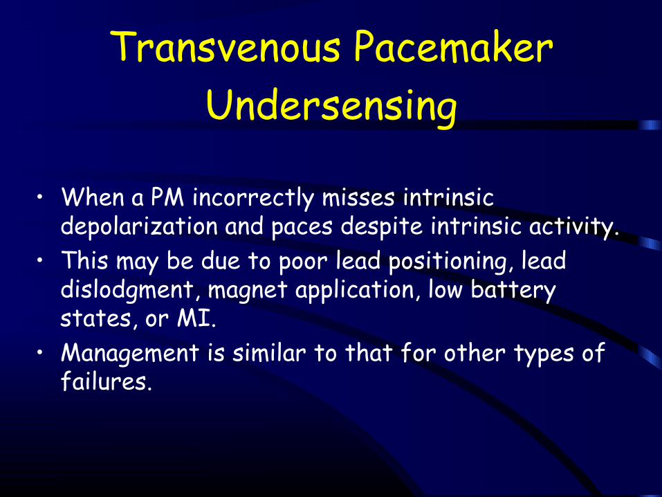
Undersensing
• When a PM incorrectly misses intrinsic depolarization and paces despite intrinsic activity.
• This may be due to poor lead positioning, lead dislodgment, magnet application, low battery states, or MI.
• Management is similar to that for other types of failures.
Transvenous Pacemaker

• A final category of PM failures is termed operative • This includes malfunction due to mechanical
factors, such as PNX, pericarditis, infection, skin erosion, hematoma, lead dislodgment, and venous thrombosis.
• Treatment depends on the etiology.
Transvenous Pacemaker (TVP)

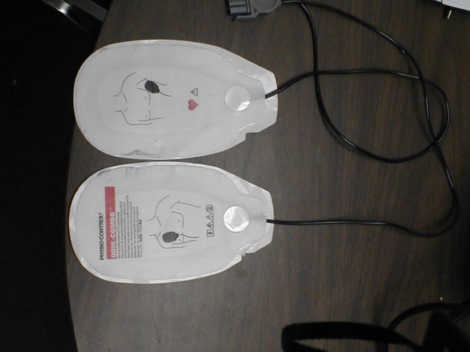
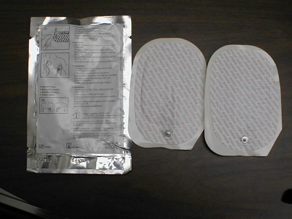
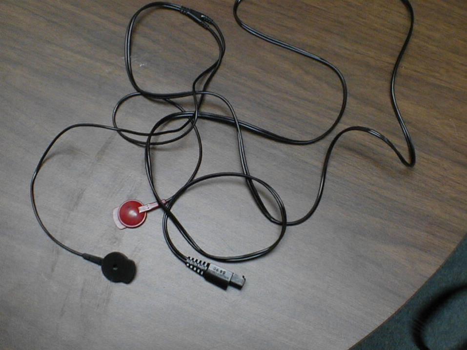

(and electrodes if Demand or Back-up Pacing)
• Stat-Padz application should be Anterior/Posterior
• 3-lead ECG electrodes must be placed also
Think of 2 pieces of white bread and you are making a myocardial sandwich
Transcutaneous PM (TCP)
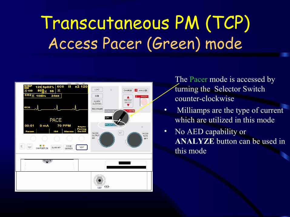
Access Pacer (Green) mode
The Pacer mode is accessed by turning the Selector Switch counter-clockwise
• Milliamps are the type of current which are utilized in this mode
• No AED capability or ANALYZE button can be used in this mode
Transcutaneous PM (TCP)
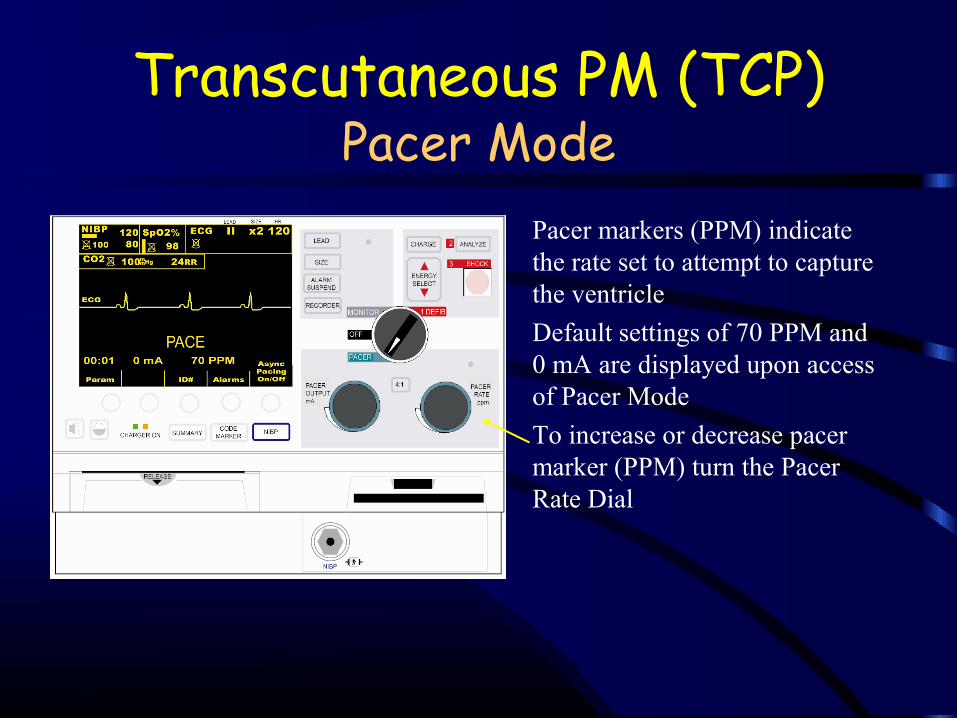
Pacer Mode• Pacer markers (PPM) indicate
the rate set to attempt to capture the ventricle
• Default settings of 70 PPM and 0 mA are displayed upon access of Pacer Mode
• To increase or decrease pacer marker (PPM) turn the Pacer Rate Dial
Transcutaneous PM (TCP)
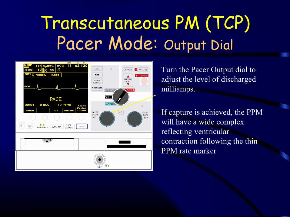
Pacer Mode: Output Dial• Turn the Pacer Output dial to
adjust the level of discharged milliamps.
• If capture is achieved, the PPM will have a wide complex reflecting ventricular contraction following the thin PPM rate marker
Transcutaneous PM (TCP)
Pulse Duration
• Pulse duration is the time of impulse stimulation. • Early TCPs used short (1-2 ms) duration impulses.
Such impulses resembled the action potential (AP) and preferentially stimulated skeletal muscle.
• In contrast, cardiac muscle APs are much longer, requiring 20-40 msec to reach maximum.
Transcutaneous PM (TCP)
Current• Using a longer pulse duration and larger electrodes
permits pts to tolerate higher applied current. • 100 mA of current applied over an average (50 Ώ
resistance) chest for 20 ms will deliver 0.1 J. This is well below the 1-2 J required to cause an uncomfortable tingling sensation in the skin.
• The force of skeletal muscle contraction, not the electric current, determines TCP discomfort. Current TCPs are capable of delivering up to 140-200 mA tolerably.
Transcutaneous PM (TCP)
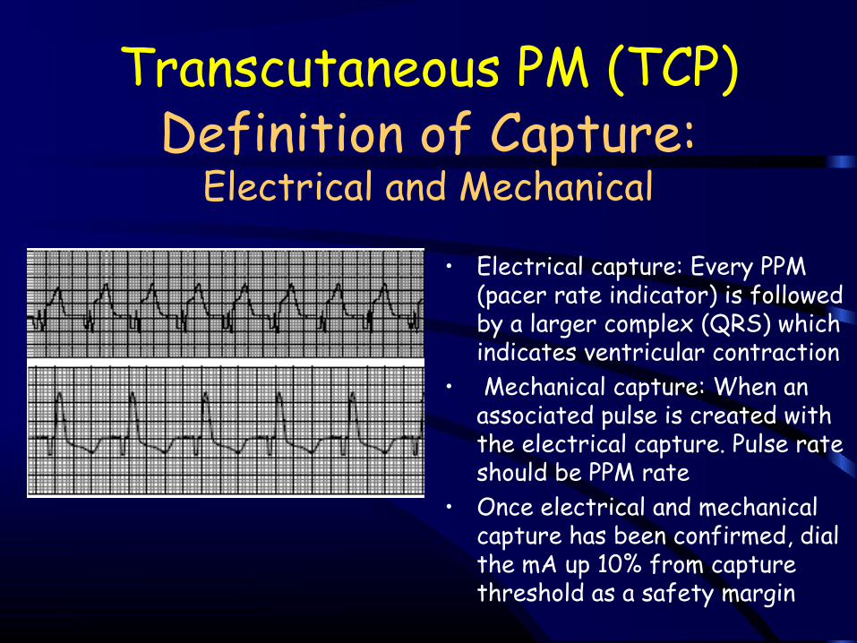
Definition of Capture:Electrical and Mechanical
• Electrical capture: Every PPM (pacer rate indicator) is followed by a larger complex (QRS) which indicates ventricular contraction
• Mechanical capture: When an associated pulse is created with the electrical capture. Pulse rate should be PPM rate
• Once electrical and mechanical capture has been confirmed, dial the mA up 10% from capture threshold as a safety margin
Transcutaneous PM (TCP)

Pacing Mode: Ability to Perform 3 Types of Ventricular Pacing
1.) Demand Pacing: Most frequent form of ventricular pacing. The PPM is set above patient’s rate (or lack thereof) and the Pacer Output dial is turned to increase the mA in attempt to obtain capture and pace the ventricles.
2.) Stand-by Pacing: Setting the PPM and Pacer output at a back-up rate less than a patient’s intrinsic heart rate. The PPM will initially be set above the patients heart rate and pacer output (mA) is increased to achieve 100% capture. The PPM is then decreased to desired rate below the patients intrinsic heart rate. Should the HR drop, the stand-by pacer will initiate impulses and begin to pace.
Transcutaneous PM (TCP)
3.) Asynchronized Pacing: Rarely used. This form of pacing is performed when no ECG electrodes can be placed due to burns, trauma or interference. The async on/off softkey button is pressed and aysnc mode is displayed. No PPM or electrical capture will be seen on the screen. Mechanical capture will only be proven by palpating a pulse if one is achieved
Pacing Mode: Ability to Perform 3 Types of
Ventricular Pacing
Transcutaneous PM (TCP)