1 Chapter 9 Muscular System Lecture 17 Visual Anatomy & Physiology First Edition Martini & Ober.
18
1 Chapter 9 Muscular System Lecture 17 Visual Anatomy & Physiology First Edition Martini & Ober
-
Upload
adrian-homer-wright -
Category
Documents
-
view
225 -
download
0
Transcript of 1 Chapter 9 Muscular System Lecture 17 Visual Anatomy & Physiology First Edition Martini & Ober.

1
Chapter 9
Muscular System
Lecture 17
Visual Anatomy & PhysiologyFirst Edition
Martini & Ober

2
Lecture Overview
• Review of how muscles are named
• Skeletal muscle actions
• What you should know for the exam about skeletal muscle actions
• Compartments and compartment syndrome
• Hernias

3
How Skeletal Muscles Are Named
• Characteristics used to name skeletal muscles– Direction
• Orientation relative to body midline
• Rectus, transverse, oblique
– Size• Relative size of muscle
• Maximus, minimus, longus, brevis, lattissimus, vastus
– Shape• Relative shape of muscle
• Deltoid, trapezius, serratus, rhomboid
See Table 11-1 (from Martini) that was distributed in class

4
How Skeletal Muscles Are Named• Characteristics used to name skeletal muscles
– Action• Principle action
• Flexor, extensor, abductor, adductor, rotator
– Number of origins• Number of tendons of origin
• Biceps, triceps, quadriceps
– Location• Temporalis, femoris
– Origin and insertion• Sternocleidomastoid, stylohyoid

5
Skeletal Muscle Actions
• origin – immovable* end• insertion – movable end
• agonist (prime mover ) – primarily responsible for movement• synergists – assist prime mover• antagonist – resist prime mover’s action and cause movement in the opposite direction
Understand these terms
Figure from: Saladin, Anatomy & Physiology, McGraw Hill, 2007

6
What You Should Know About Muscle Actions
• Given the name of an UNKNOWN muscle– Based on the naming conventions discussed previously,– And using the starred items on the chart handed out in
class (from Martini), – And using your previous knowledge about the anatomy
of the body,– You should be able to tell me what the name of a
muscle implies, e.g., where it is, what it’s attached to, is it long or short, etc.
• For the muscles on Lab Exercise 7 in your Laboratory Guide handout you should know– The name of the muscle (All)– What is does, e.g., flexes the arm at the elbow (All)– The origin/insertion (of muscles on table that are not
grayed out on the Origin/Insertion Table)

7
Examples of Naming Muscles
What would the levator scapulae do?
Figure from: Hole’s Human A&P, 12th edition, 2010

8
Examples of Naming Muscles
Near what landmark do you think the fibularis longus would be located? Would it be a short or long muscle?
Figure from: Hole’s Human A&P, 12th edition, 2010

9
Examples of Naming Muscles
What can you tell about the orbicularis oculi muscle?
Figure from: Hole’s Human A&P, 12th edition, 2010

10
Levers• Levers can give a “mechanical advantage” in two ways:
– To exert more force against a resisting object than the force actually applied
– To move the resisting object farther or faster than the effort arm is moved.
Mechanical Advantage (MA) can be stated as the ratio of the output force to the input force.
Figure from: Saladin, Anatomy & Physiology, McGraw Hill, 2007

11
Levers
Four Basic Components1. rigid bar (bones)2. Fulcrum – point on which bar moves (joints)3. Object moved against Resistance (weight)4. Effort (Force) – supplies energy for movement (muscles)
Most common type of lever in body
“FRE = 123” (Is F, R, or E in the middle?)
Effort
Effort
Effort
Effort
Effort
Effort
Figure from: Hole’s Human A&P, 12th edition, 2010

12
Levers and Movement
What types of lever are these?
Effort
Effort
Figure from: Hole’s Human A&P, 12th edition, 2010

13
Hernias
A hernia develops whenever an organ protrudes through an abnormal opening.
If organs become twisted or strangulated, ischemia (blood starvation) may result and surgical intervention is necessary to prevent further damage.
1 – Incisional hernia2 – Umbilical hernia3 – Direct inguinal hernia4 – Femoral hernia5 – Indirect inguinal hernia
Figure from: http://drgeiss.com/p_hernia.html
14
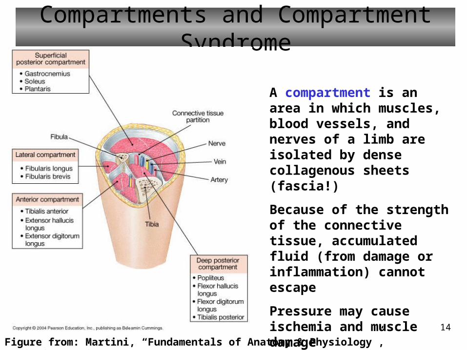
Compartments and Compartment Syndrome
Figure from: Martini, “Fundamentals of Anatomy & Physiology”, Benjamin Cummings, 2004
A compartment is an area in which muscles, blood vessels, and nerves of a limb are isolated by dense collagenous sheets (fascia!)
Because of the strength of the connective tissue, accumulated fluid (from damage or inflammation) cannot escape
Pressure may cause ischemia and muscle damage
This is called “compartment syndrome”

15
Review
• Muscles are named according to– Direction– Size– Shape– Action– Number of origins– Location– Origin and insertion

16
Review
• An origin of a muscle is the fixed end of a muscle, i.e. the end that moves the least
• An insertion of a muscle is the movable end of a muscle, i.e., the end that moves the most
• A prime mover (agonist) is a muscle whose contraction is chiefly responsible for a movement
• A synergist helps a larger agonist work efficiently
• An antagonist opposes the action of an prime mover (agonist)
17
Review
• Regarding muscle actions, you should know…– Names and actions of muscles reviewed in lab– Naming convention for muscles on Tortora
handout chart
• Levers– Be able to identify the type of lever that is
represented by a muscle/bone/joint diagram

18
Review
• Compartments of the limbs– group of muscles, blood vessels and nerves isolated by
thick CT sheets
– Inflammation or damage can cause fluid accumulation an swelling with danger of ischemia
• A hernia– Is a protrusion of organs through an abnormal opening
– Is dangerous, especially if the protruding organs become strangulated or twisted