0200-0230-Bernklau- Small Bowel - Capsule Endoscopy and...
56
Small Bowel ‐ Capsule Endoscopy and Enteroscopy and Enteroscopy Sandra L. Bernklau, MSN, FNP‐BC, CGRN Suburban Gastroenterology, Naperville, Illinois
Transcript of 0200-0230-Bernklau- Small Bowel - Capsule Endoscopy and...
Small Bowel ‐ Capsule Endoscopy and Enteroscopyand Enteroscopy
Sandra L. Bernklau, MSN, FNP‐BC, CGRNSuburban Gastroenterology, Naperville,
Illinois

ObjectivesObjectives• Identify the common causes of gastrointestinal bleeding in the
upper gastrointestinal tract.• Discuss the history and evolution of traditional evaluation of
upper gastrointestinal bleeding.upper gastrointestinal bleeding.• Explain new technologies for upper gastrointestinal bleeding:
wireless capsule endoscopy (WCE) and Double Balloon Enteroscopy (DBE)Enteroscopy (DBE).
• Discuss patient evaluation and care for patients undergoing wireless capsule endoscopy and Double Balloon Enteroscopy
• Identify nursing impact of wireless capsule endoscopy and Double Balloon Enteroscopy
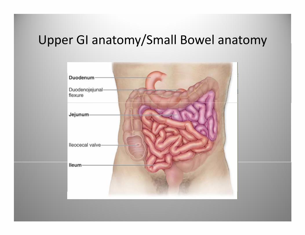
Upper GI anatomy/Small Bowel anatomyUpper GI anatomy/Small Bowel anatomy
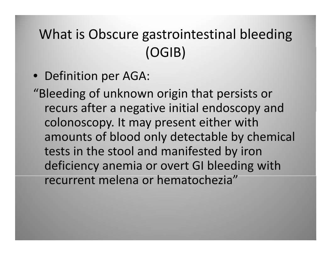
What is Obscure gastrointestinal bleeding ( )(OGIB)
• Definition per AGA:Definition per AGA:“Bleeding of unknown origin that persists or recurs after a negative initial endoscopy andrecurs after a negative initial endoscopy and colonoscopy. It may present either with amounts of blood only detectable by chemical tests in the stool and manifested by iron deficiency anemia or overt GI bleeding with
l h h i ”recurrent melena or hematochezia”
Case StudyCase Study
• 68 year old Caucasian female (Ms J) with a68 year old Caucasian female (Ms. J) with a long standing coronary artery disease status post PTCA drug alluding stent x2 in 2008 andpost PTCA drug alluding stent x2 in 2008 and 2011. She describes over the past few days increasing shortness of breath fatigue andincreasing shortness of breath, fatigue and “darker stools”. She calls the office to speak to the midlevel provider Sound familiar???the midlevel provider……Sound familiar???
Diagnosis of OGIBDiagnosis of OGIB
• The clinical history may suggest possibleThe clinical history may suggest possible location and cause of OGIB, rarely diagnostic
• Hematemesis: reliable only as bleeding aboveHematemesis: reliable only as bleeding above Ligament of Treitz
• Color of stool (melena/hematochezia)‐lessColor of stool (melena/hematochezia) less predictive of transit time
• Pain may also help with source‐esophagealPain may also help with source esophageal versus, stomach abdomen

Most common causes of OGIBMost common causes of OGIB
Esophagus:EsophagitisEsophagus:EsophagitisStomach:Cameron erosions,Dieulafoy lesionGAVE Portal HTN gastropathyGAVE,Portal HTN gastropathyDuodenum: Ampullary lesion, distal duodenal neoplasias aortoenteric fistula pancreaticneoplasias, aortoenteric fistula, pancreatic aneurysm, hemobiliaSmall bowel: Angiodysplasias, primary S a bo e g odysp as as, p a yneoplasias, metastasis, polyposis syndromes, meckel diverticulum, NSAIDS, Crohn’s disease, portal hypertensive intestinal, Osler Rendu syndrome
Etiology of Gastrointestinal BleedEtiology of Gastrointestinal Bleed
• Patient’s younger than 40‐more likely: cancer‐Patient s younger than 40 more likely: canceror Dieulafoy’s lesion– Lymphomay p– Carcinoid– Adenocarcinoma
• Older patients‐– AVMs– Erosions– Ulcers
Case study (Cont.)Case study (Cont.)
• Ms J goes to the emergency room as advisedMs. J. goes to the emergency room as advised by the midlevel provider and is found to be mildly tachycardic and have a hemoglobin ofmildly tachycardic and have a hemoglobin of 6.8. A ER physician does a rectal exam and she is occult positive A GI consult is requestedis occult positive. A GI consult is requested.

WORK UP FROM HERE?WORK UP FROM HERE?

Initial Patient work upInitial Patient work up
• EGDEGD• Colon• Enteroscopy• Bleeding scan• Small bowel follow through• EnteroclysisEnteroclysis• Angiography

Lesions most commonly MissedLesions most commonly Missed
• Upper GI tract:Upper GI tract:– Hiatal HerniaPeptic Ulcers– Peptic Ulcers
– Vascular EctasiaE h l V i– Esophageal Varix
– Persistent IDA‐rule out Celiac disease

Case studyCase study
• Ms. J under goes an upper endoscopy thatMs. J under goes an upper endoscopy that reveals gastritis and a colonoscopy that reveals two 6 mm tubular adenomas in the sigmoid colon. No old blood or signs of overt bleeding noted. She continues to pass i i l d d hintermittent melena and drops her hemoglobin to 7.2 despite receiving 4 Units PRBCs over the preceeding days ShePRBCs over the preceeding days. She undergoes a tagged RBC scan that reveals possible bleeding in the mid abdomenpossible bleeding in the mid abdomen.

Now what?
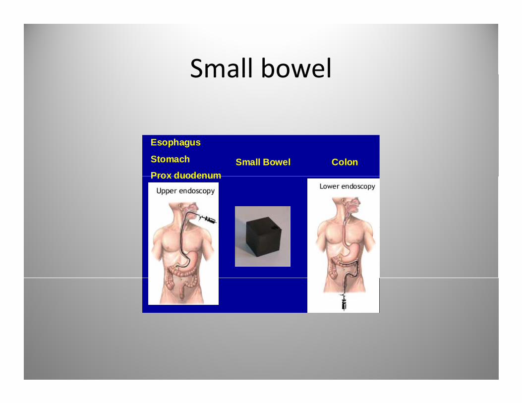
Small bowelSmall bowel
Esophagus
Stomach Prox duodenum
ColonSmall BowelProx duodenum

History of small bowel by radiologyy y gyTest Describe Visualize Yield Complic
ationsSBSeries
Pre-oral dilute barium with serial abd images
Rules out strictures
0-6% Minimal –none
imagesEntero-Clysis
Double contrast-by passing tube
Fails flatLesions-AVMs
10-20% Minimal
into prox small bowel
Radionucleidescanning
Detects bleeding
Active bleeding If bleeding-0.1-0.5 ml/min
Minimalg g
relative brisk
Angiography Not usually necessary-
Follow + bleeding scan if
44-68% Bleeding,sedation
unless severe bleeding. Can find vascular lesions
rate > 0.5 ml/min

History of endoscopic of small bowel l ievaluation
• EGD/colonEGD/colon• Push enteroscopy‐1980s and 1990s‐has shown diagnostic yield in bleeding‐shorter lengthg y g g
• Non‐surgical‐total small bowel enteroscopy– “Ropeway”(1972)‐guided string method may takeRopeway (1972) guided string method may take several days from mouth to rectum
– Sonde(1972) Endoscopy‐Unable to readvance( ) py

Surgery for Small bowel evaluationSurgery for Small bowel evaluation
• Surgery of OGIB final approachSurgery of OGIB final approach• Surgeon helps with intubation of bowel by pushing over the enteroscopepushing over the enteroscope
• Mucosal trauma common‐confuses with l bl dinormal bleeding
• Exam during insertion • Yield of 70‐93%• Disadvantage‐ invasive in natureDisadvantage invasive in nature

Therapeutic/Conservative TherapyTherapeutic/Conservative Therapy
• Supportive care:Supportive care:– Blood transfusionsEpoetin alfa– Epoetin alfa
– Iron replacementH l th– Hormonal therapy
– OctreotideA id NSAID– Avoid NSAIDs
Case study (cont.)Case study (cont.)
• Ms J undergoes a capsule endoscopy TheMs. J undergoes a capsule endoscopy. The capsule study is an adequate exam and demonstrates a bleeding “region” 10 min 45demonstrates a bleeding region 10 min 45 seconds past the ligament of treitz. She undergoes a push enteroscopy The extent ofundergoes a push enteroscopy. The extent of the exam is just distal to the ligament of treitz. No bleeding source is identified She is sentNo bleeding source is identified. She is sent back to the unit. She stabilizes and is sent homehome.

Capsule EndsocopyCapsule Endsocopy
• WCE‐Wireless capsule endoscopyWCE Wireless capsule endoscopy• Considered novel noninvasive technology for small bowelsmall bowel
• Excellent resolution 1:8 magnification.• Allows visualization of individual villi• No bowel inflation requiredq• Approved in 2000 by Given replaced by the M2A‐Pillcam SBM2A Pillcam SB.
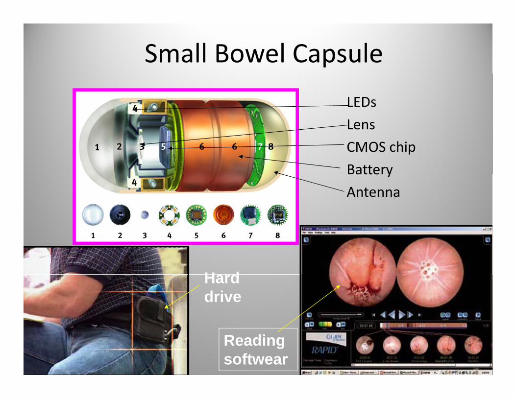
Small Bowel CapsuleLEDsLensLensCMOS chipBatteryBatteryAntenna
H dHard drive
Reading softwear
Capsule Endsocopy‐statisticsCapsule Endsocopy statistics
• Diagnostic yield of 32% to 76%Diagnostic yield of 32% to 76%• The ability of capsule study to exclude GI bleeding between 82.6% and 100%bleeding between 82.6% and 100%
• Multiple studies have demonstrated capsule better yield for GI bleedbetter yield for GI bleed
• In up to 35% of patients secondary to slow transit time.transit time.
• Possibly adding simethicone may help improve visibility.improve visibility.
Capsule Endoscopy (cont.)Capsule Endoscopy (cont.)
• System includes software and colorSystem includes software and color red(suspected blood indicator)
• Capsule takes 2 images per second• Capsule takes 2 images per second• Recorder acquires 55,000 images in 8 hours.• Review of the images 30‐90 minutes
Capsule endoscopy studies(Yield)Capsule endoscopy studies(Yield)
• WCE‐able to identify obscure GI bleeding more oftenWCE able to identify obscure GI bleeding more often than push enteroscopy (Lewis et al 2002)
• WCE yield 50‐70%.y• Largest study‐100 consecutive patients with obscure bleeding‐3‐groups‐highest diagnostic yield (92%) in g g p g g yactive bleeding (Pennazio et al 2004)
• Another study‐WCE prior to push enteroscopy‐higher diagnostic yield (de Leusse et al 2007)

Benefits of Capsule EndoscopyBenefits of Capsule Endoscopy
• Non‐invasiveNon invasive• Permits examination of most of small bowel• Very sensitive• Very sensitive• Can be performed in outpatient settingC ll ti t t k• Can allow patient to work
• No ride• Can stay on anticoagulation

Limitations of Wireless Capsule d / li iEndoscopy/complications
• Wireless capsule endoscopy depends onWireless capsule endoscopy depends on peristalsis
• Only visualization is possible no biopsy or• Only visualization is possible, no biopsy or treatment possibleL k f i i ffl i d bili i• Lack of air insufflation and ability to rinse
• 1% risk of capsule entrapment in strictures or diverticula‐good initial eval important
• Consider patency capsulep y p

Contraindications of WCEContraindications of WCE
• DementiaDementia• Gastroparesis
h l i ll i di d• Esophageal stricture or swallowing disorders• Partial or intermittent small bowel obstruction• Inoperable‐or refuse surgery• In the past pacemakers and defibrillatorsIn the past pacemakers and defibrillators
M2A patency capsuleM2A patency capsule
• Composed of lactose and 5% barium sulfateComposed of lactose and 5% barium sulfate• Can be located by radiofrequency and dissolves in 40‐100 hours after ingestiong
• Painless defecation of an intact capsule indicated safety of conventional capsule study.y p y
• Painful passage or disintegration is highly suggestive of a significant small bowel stricture
• Not considered entirely safe‐in small group of patients may cause intestinal occlusion

Case study (cont.)Case study (cont.)
• Ms J represents to the emergency room 4Ms. J represents to the emergency room 4 weeks after hospital discharge. She describes increasing fatigue over the past few days andincreasing fatigue over the past few days and melena started last night. She had been restarted on her Plavix and aspirin by herrestarted on her Plavix and aspirin by her cardiologist. Her hemoglobin is 7.1.
?PLAN?
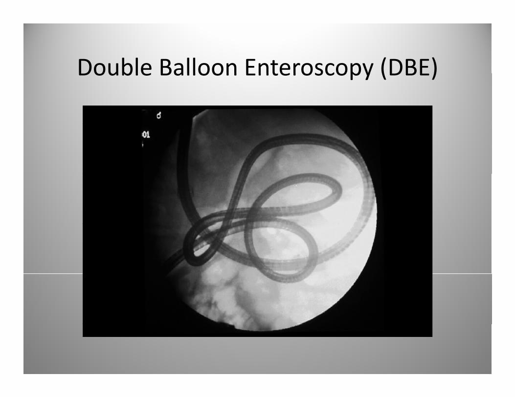
Double Balloon Enteroscopy (DBE)Double Balloon Enteroscopy (DBE)
Double Balloon EnteroscopyDouble Balloon Enteroscopy
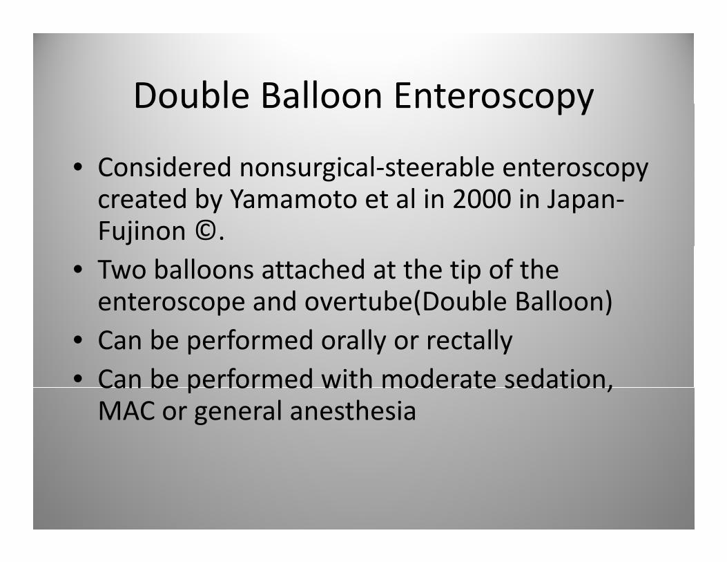
• Considered nonsurgical‐steerable enteroscopyConsidered nonsurgical steerable enteroscopy created by Yamamoto et al in 2000 in Japan‐Fujinon ©.
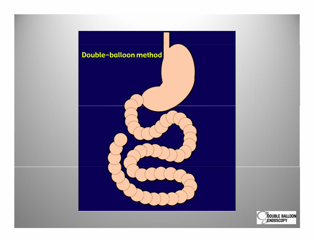
• Two balloons attached at the tip of the enteroscope and overtube(Double Balloon)
• Can be performed orally or rectally• Can be performed with moderate sedation,Can be performed with moderate sedation, MAC or general anesthesia

DBE systemDBE system
• Standard system includesy• Fuji system(Fujinon EN‐450P5)
– Thin endoscope with a 8.5 cm diameter and 200 cm working length
– 145 cm soft overtube with an outer diameter of 12.2 mm‐with a precise designed pump.p g p p
– Soft latex balloon attached to scope tip• A therapeutic DBE scope is available (Fujinon EN‐
)450T5)• The pressure in both tubes monitored accurately at 6 kPakPa.
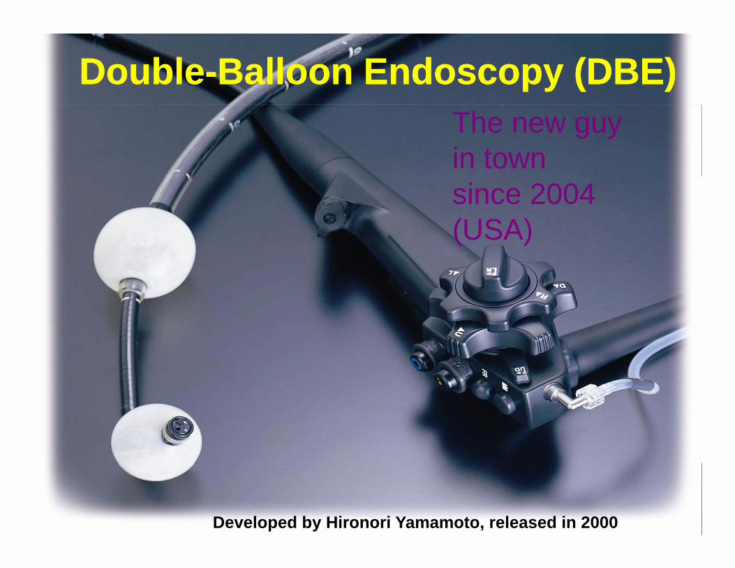
DoubleDouble--Balloon Endoscopy (DBE)Balloon Endoscopy (DBE)The new guy in townsince 2004 (USA)
Developed by Hironori Yamamoto, released in 2000


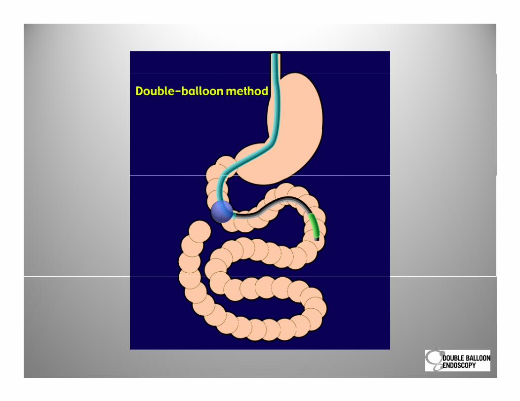

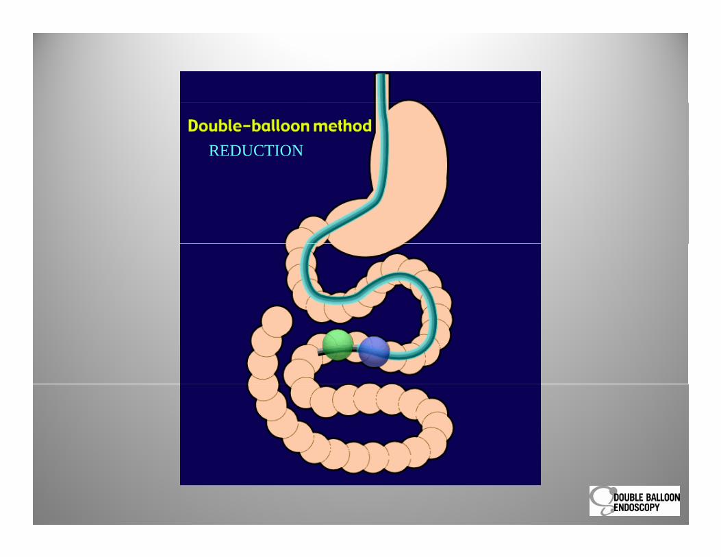
REDUCTION

REDUCTION

Therapies performed during DBETherapies performed during DBE
• Injectionj• Bicap/APC(Argon Plasma Coagulation)• Biopsy/Polypectomy/EMRp y yp y• Endoscopic clips• Balloon Dilitation• Removal of trapped capsule• Stent placement• Gastric Bypass patients/Roux en Y anastomosis‐ERCP

Double Balloon Enteroscopy(studies)Double Balloon Enteroscopy(studies)
• Study by Matsumoto et al (2005)‐positiveStudy by Matsumoto et al (2005) positive findings in 64% push enteroscopy and 82% in DBEDBE.
• A meta‐analysis of 11 studies compared DBE to capsule yield similarto capsule‐yield similar. – It was concluded that wireless CE performed first and DBE performed after positive findings onand DBE performed after positive findings on WCE.

Limitations of DBELimitations of DBE
• Need to hold anticoagulationNeed to hold anticoagulation• General anesthesia/MAC
ff f k/ d id• Day off of work/need a ride• Adhesions• Full exam at times not possible • Post op painPost op pain
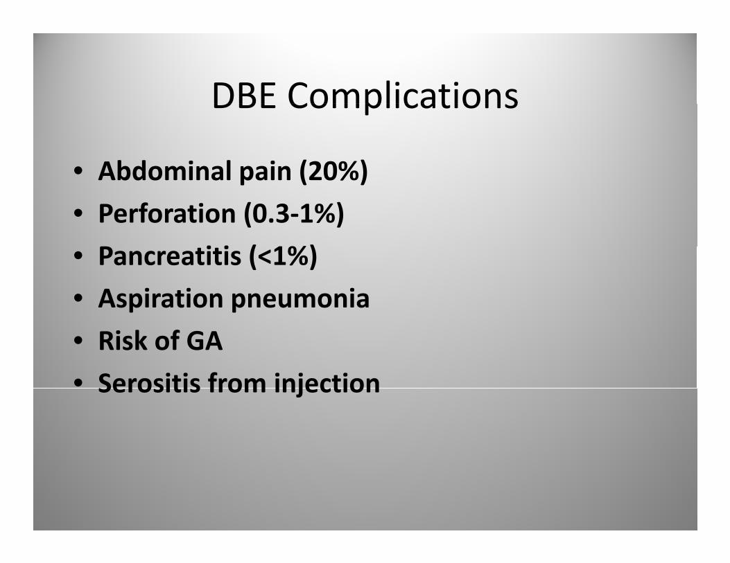
DBE ComplicationsDBE Complications
• Abdominal pain (20%)Abdominal pain (20%)• Perforation (0.3‐1%)
i i ( %)• Pancreatitis (<1%)• Aspiration pneumonia• Risk of GA• Serositis from injectionSerositis from injection
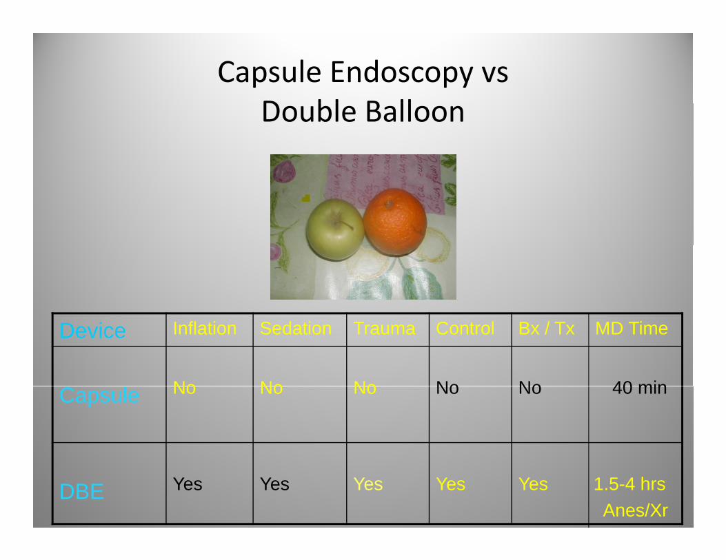
Capsule Endoscopy vs D bl B llDouble Balloon
Device Inflation Sedation Trauma Control Bx / Tx MD Time
No No No No No 40 minCapsule No No No No No 40 min
DBE Yes Yes Yes Yes Yes 1.5-4 hrsAnes/Xr
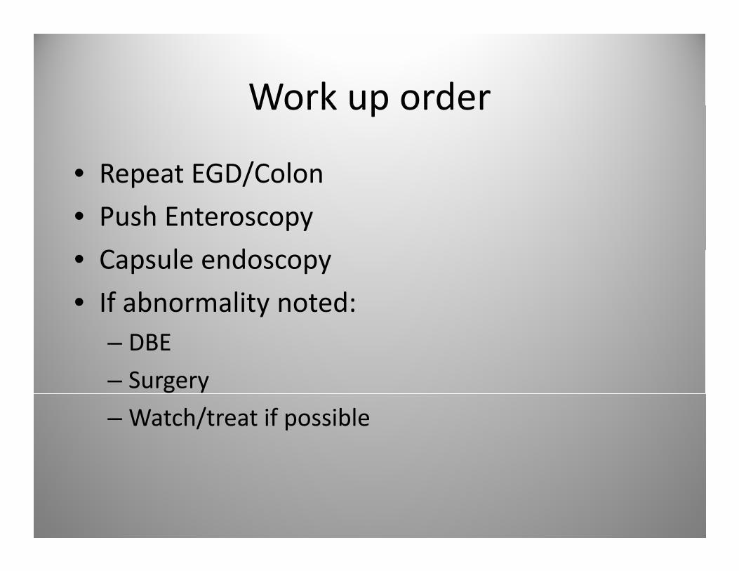
Work up orderWork up order
• Repeat EGD/ColonRepeat EGD/Colon• Push EnteroscopyC l d• Capsule endoscopy
• If abnormality noted:– DBE– Surgery– Watch/treat if possible

Patient preparation‐capsule endoscopy‐pre i larrival
• Strict NPO 10 hours prior to examp• Medication history‐review in advance• Avoid red dye in food 24 hours prior• Review patient health history‐• Prep if indicated• Pacemaker/Defibrillator no longer contraindication• Pacemaker/Defibrillator‐no longer contraindication• Can they swallow large pills????• Do they have a history of abdominal surgery‐risk of y y g ypill becoming lodged
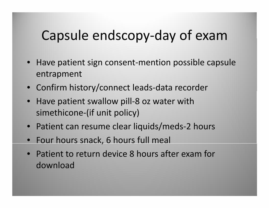
Capsule endscopy‐day of examCapsule endscopy day of exam
• Have patient sign consent‐mention possible capsuleHave patient sign consent mention possible capsule entrapment
• Confirm history/connect leads‐data recordery/• Have patient swallow pill‐8 oz water with simethicone‐(if unit policy)p y
• Patient can resume clear liquids/meds‐2 hours• Four hours snack, 6 hours full meal,• Patient to return device 8 hours after exam for download
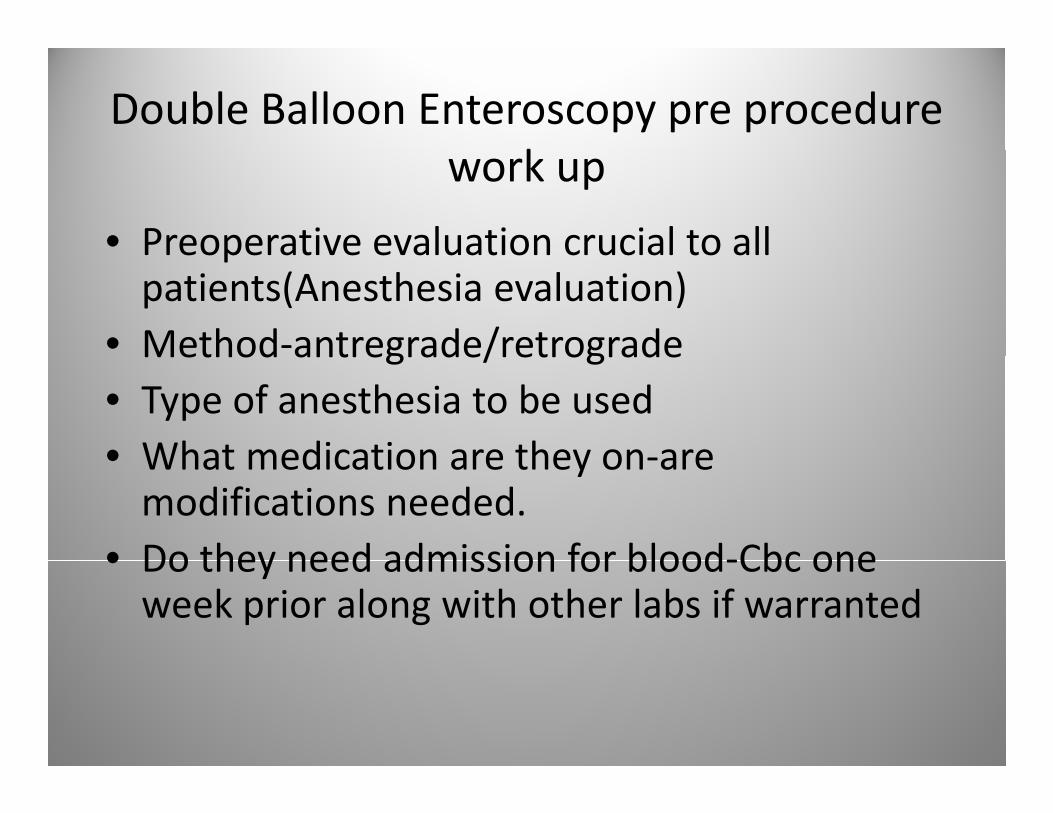
Double Balloon Enteroscopy pre procedure kwork up
• Preoperative evaluation crucial to allPreoperative evaluation crucial to all patients(Anesthesia evaluation)
• Method‐antregrade/retrogradeMethod antregrade/retrograde• Type of anesthesia to be used• What medication are they on‐are• What medication are they on‐are modifications needed.
• Do they need admission for blood‐Cbc one• Do they need admission for blood‐Cbc one week prior along with other labs if warranted
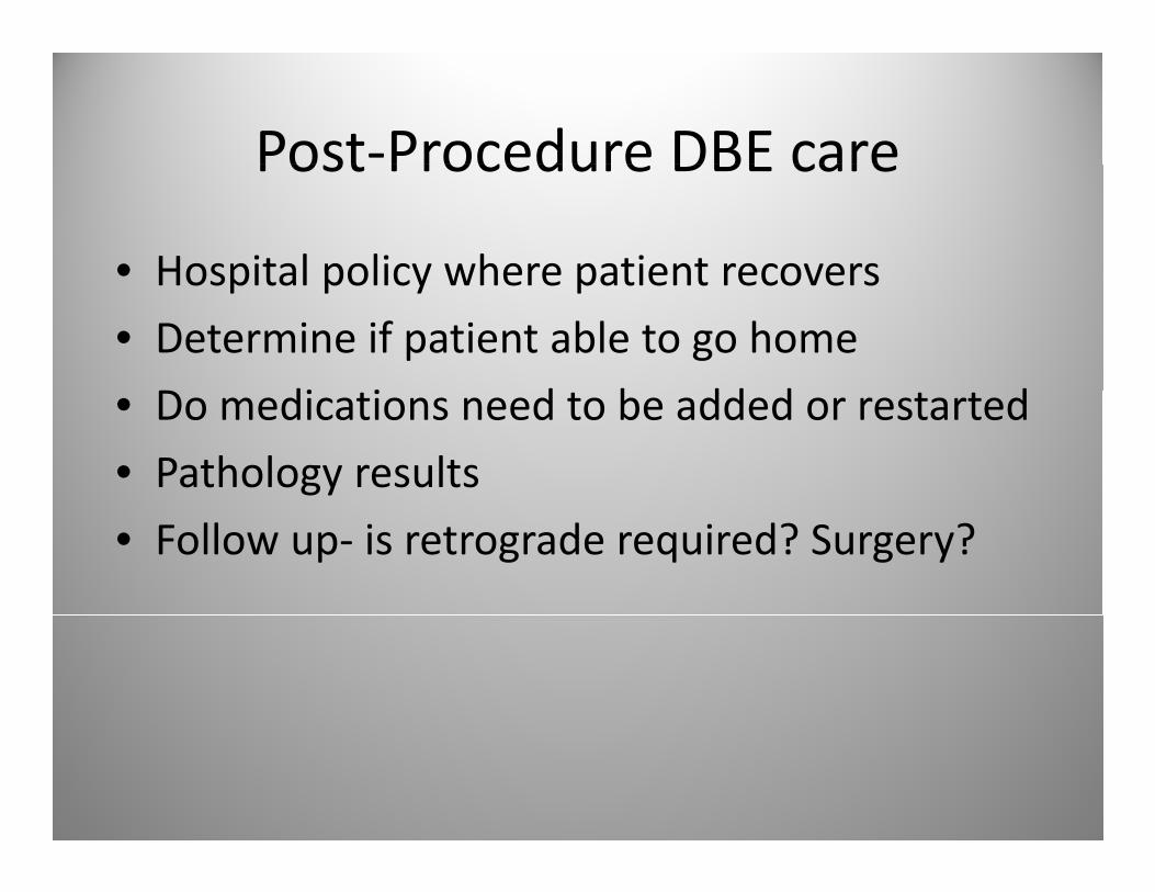
Post‐Procedure DBE carePost Procedure DBE care
• Hospital policy where patient recoversHospital policy where patient recovers• Determine if patient able to go home
di i d b dd d d• Do medications need to be added or restarted• Pathology results• Follow up‐ is retrograde required? Surgery?

Case study (cont.)Case study (cont.)
• Ms. J undergoes a antregrade DBE with general s. J u de goes a a t eg ade t ge e aanesthesia. She has a successful exam with an extent of 150 cm past the ligament of treitz. At
l h l fapproximately 40 cm past the ligament of treitz a active bleeding site is noted. The site is localized and bicap cautery is applied along with oneand bicap cautery is applied along with one endoclip. Bleeding stops instantly. Procedure completed. She is discharged home. p g
• Two week follow up hospital discharge Hgb 10.5 with no additional bleeding episodes

Questions?Questions?
• What type of follow up for this patient?What type of follow up for this patient?• Do you resume Plavix? Aspirin? Only one?
h d d if i bl d ?• What do you do if patient rebleeds?• What approach?• What are the various options?