019.gingival diseases
31
Dr Jaffar Raza Syed Page 1
-
Upload
drjaffar-raza-bds -
Category
Health & Medicine
-
view
263 -
download
0
Transcript of 019.gingival diseases

Dr Jaffar Raza Syed Page 1
Dr Jaffar Raza Syed
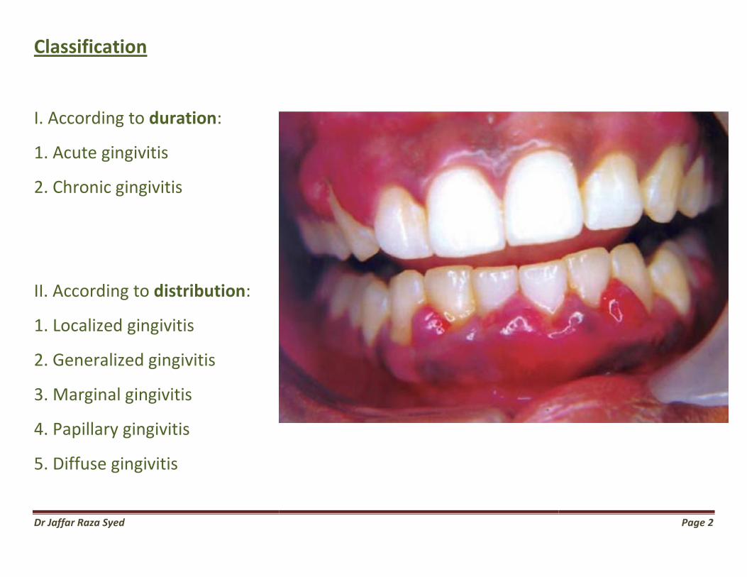
Classification
I. According to duration:
1. Acute gingivitis
2. Chronic gingivitis
II. According to distribution:
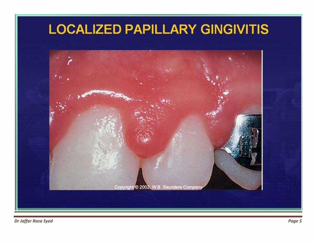
1. Localized gingivitis
2. Generalized gingivitis
3. Marginal gingivitis
4. Papillary gingivitis
5. Diffuse gingivitis
Page 2

Dr Jaffar Raza Syed Page 3

Dr Jaffar Raza Syed
Page 4
Dr Jaffar Raza Syed Page 5
Dr Jaffar Raza Syed Page 6

Dr Jaffar Raza Syed Page 7

Dr Jaffar Raza Syed
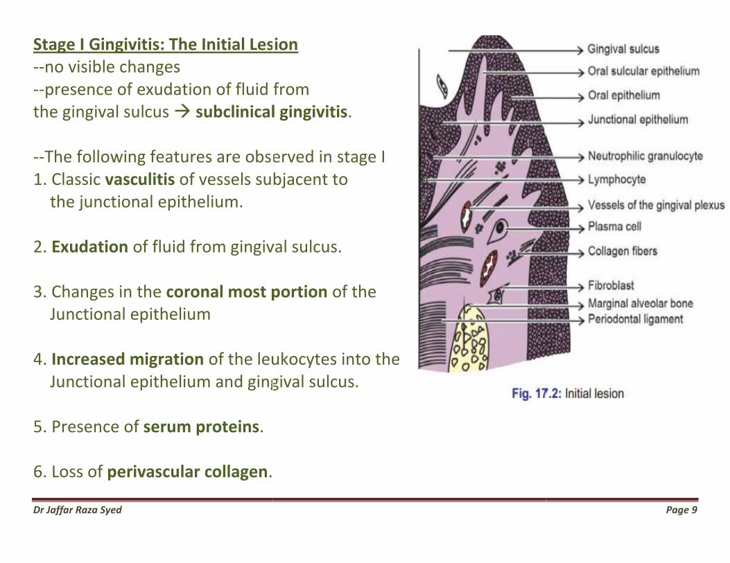
Gingival Diseases Various Stages Of Gingivitis 4 stages of gingivitis: • Stage I Initial lesion • Stage II Early lesion • Stage III Established lesion • Stage IV Advanced lesion
Initial lesion 2-4 days
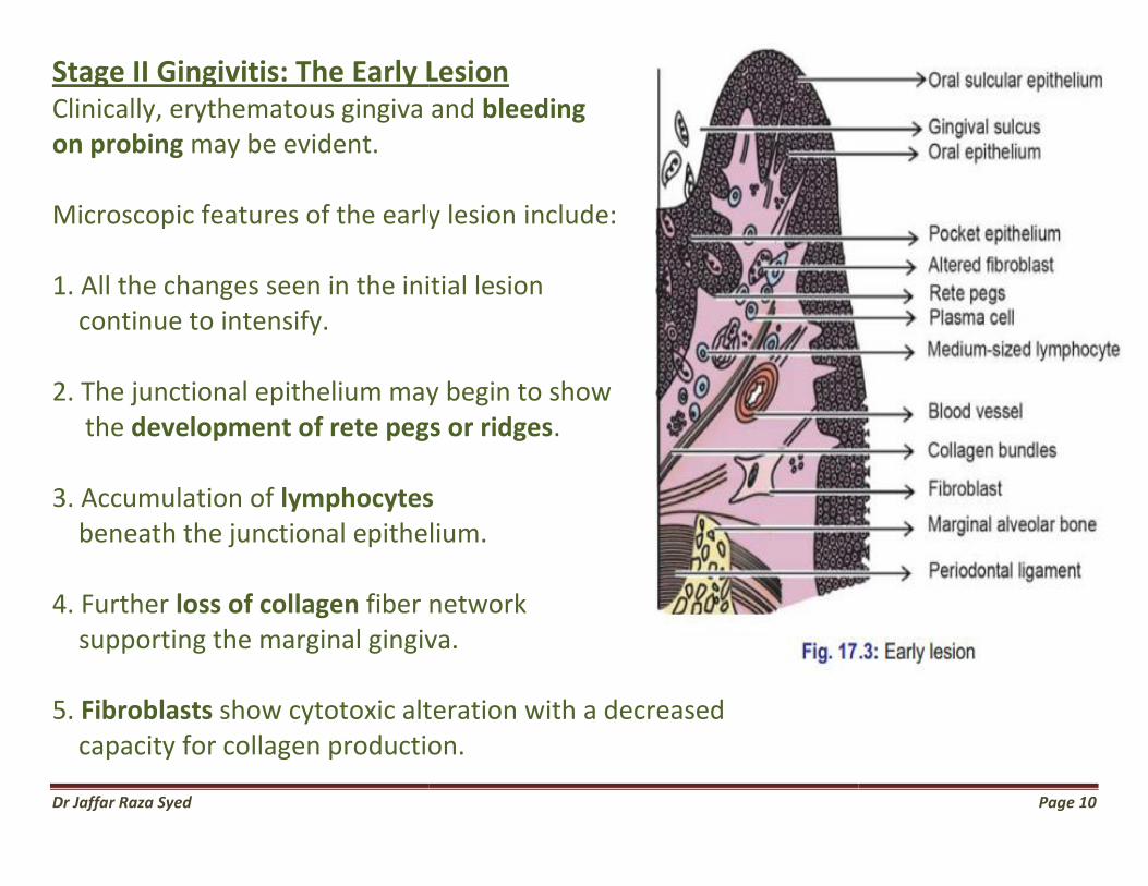
Early lesion 4-7 days
Established lesion 14-21 days
dvanced lesion
Page 8
Dr Jaffar Raza Syed
Stage I Gingivitis: The Initial Lesion--no visible changes --presence of exudation of fluid from the gingival sulcus subclinical gingivitis --The following features are observed in stage I1. Classic vasculitis of vessels subjacent to the junctional epithelium. 2. Exudation of fluid from gingival sulcus. 3. Changes in the coronal most portion Junctional epithelium 4. Increased migration of the leukocytes into the Junctional epithelium and gingival sulcus. 5. Presence of serum proteins. 6. Loss of perivascular collagen.
tage I Gingivitis: The Initial Lesion
exudation of fluid from subclinical gingivitis.
The following features are observed in stage I of vessels subjacent to
of fluid from gingival sulcus.
coronal most portion of the
of the leukocytes into the d gingival sulcus.
Page 9
Dr Jaffar Raza Syed
Stage II Gingivitis: The Early LesionClinically, erythematous gingiva and on probing may be evident. Microscopic features of the early lesion 1. All the changes seen in the initial lesion continue to intensify. 2. The junctional epithelium may begin to show the development of rete pegs or ridges 3. Accumulation of lymphocytes beneath the junctional epithelium. 4. Further loss of collagen fiber network supporting the marginal gingiva. 5. Fibroblasts show cytotoxic alteration with a decreased capacity for collagen production.
Stage II Gingivitis: The Early Lesion Clinically, erythematous gingiva and bleeding
ic features of the early lesion include:
1. All the changes seen in the initial lesion
2. The junctional epithelium may begin to show development of rete pegs or ridges.
lymphocytes beneath the junctional epithelium.
fiber network marginal gingiva.
show cytotoxic alteration with a decreased capacity for collagen production.
Page 10

Dr Jaffar Raza Syed Page 11
Stage III Gingivitis: The Established Lesion --Same as early lesion, with blood stasis --Changes are seen in color consistency and surface texture. --Bluish hue around the reddened gingiva --Proliferation, apical migration and lateral extension of junctional epithelium --Atropic areas --Plasma cells are predominant --Further loss of collagen. --Increased enzyme levels like acid and alkaline phosphatase, β glucuronidase and others.
Dr Jaffar Raza Syed Page 12
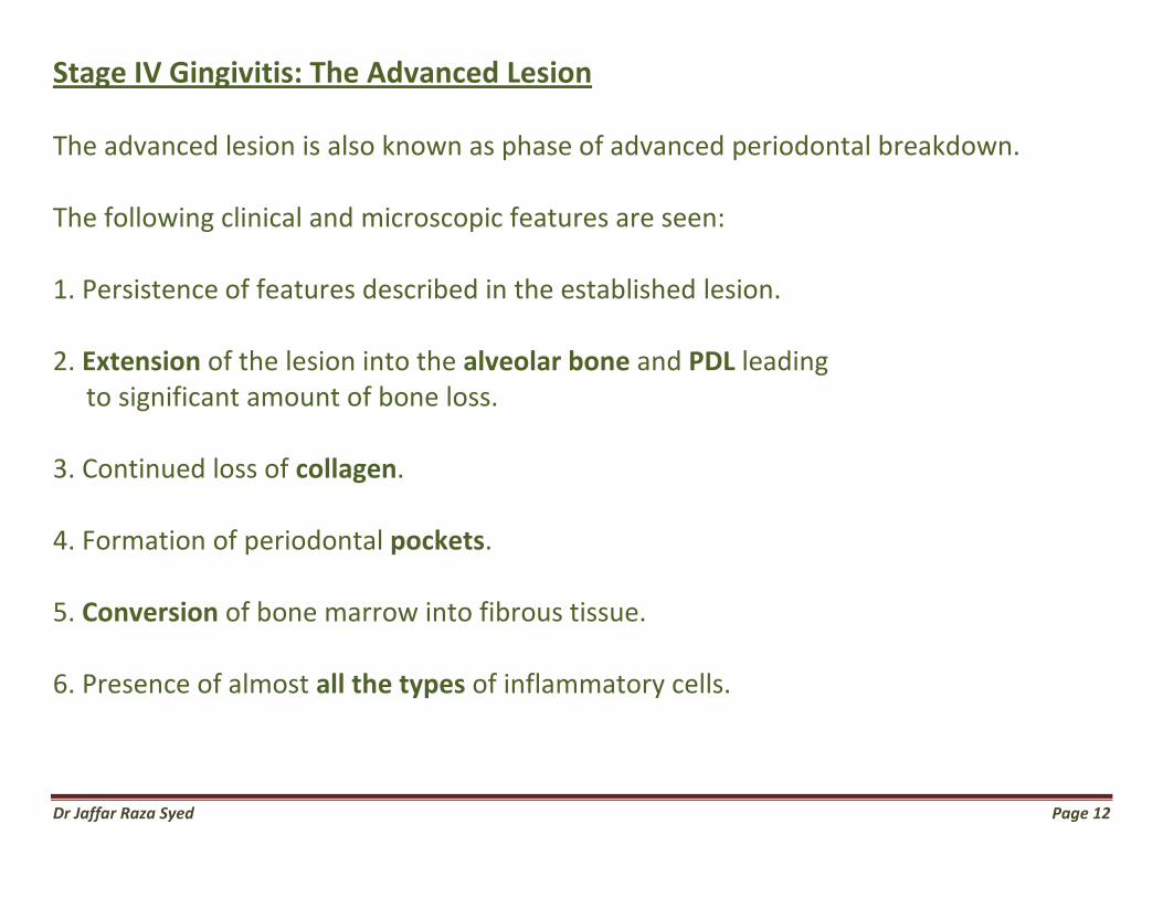
Stage IV Gingivitis: The Advanced Lesion The advanced lesion is also known as phase of advanced periodontal breakdown. The following clinical and microscopic features are seen: 1. Persistence of features described in the established lesion. 2. Extension of the lesion into the alveolar bone and PDL leading to significant amount of bone loss. 3. Continued loss of collagen. 4. Formation of periodontal pockets. 5. Conversion of bone marrow into fibrous tissue. 6. Presence of almost all the types of inflammatory cells.

Dr Jaffar Raza Syed Page 13
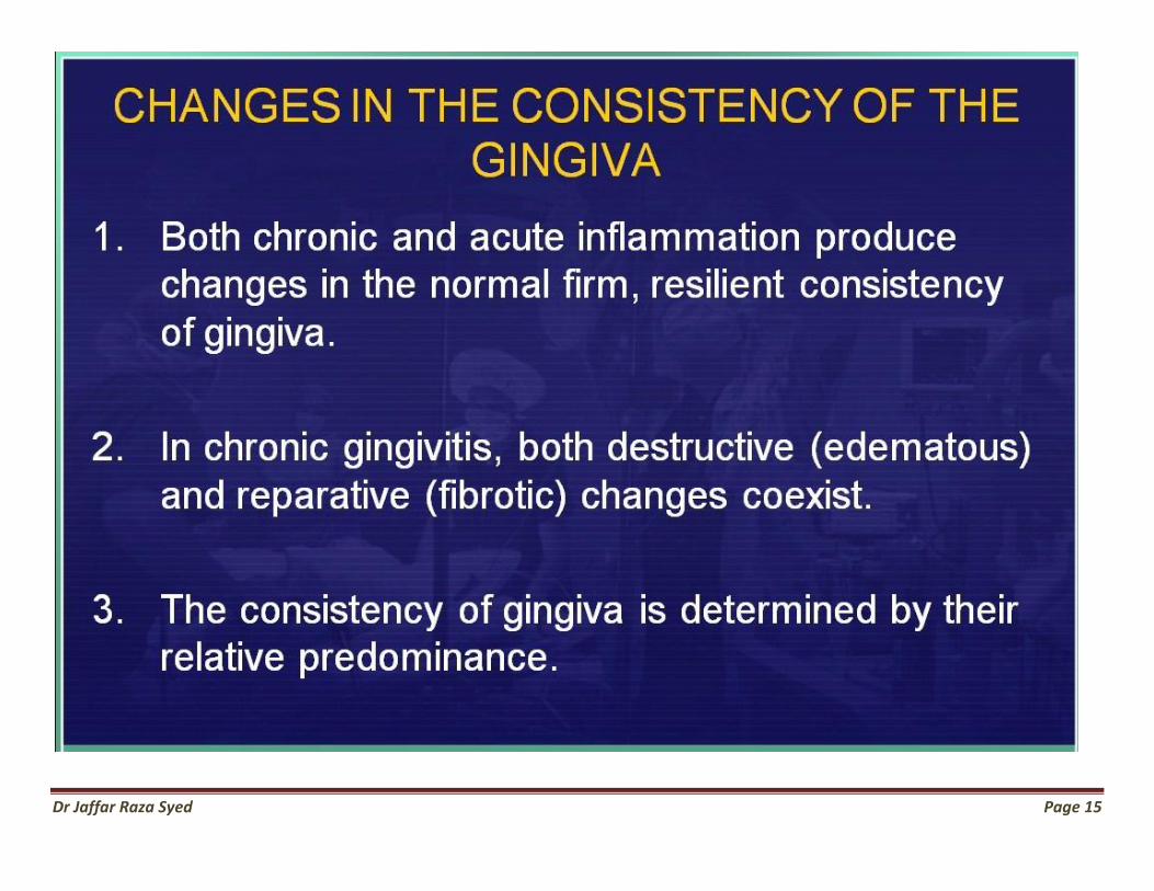
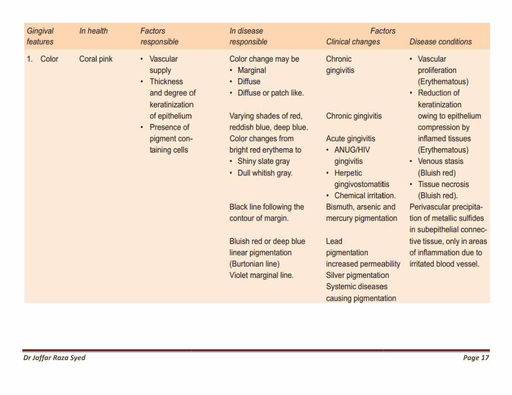
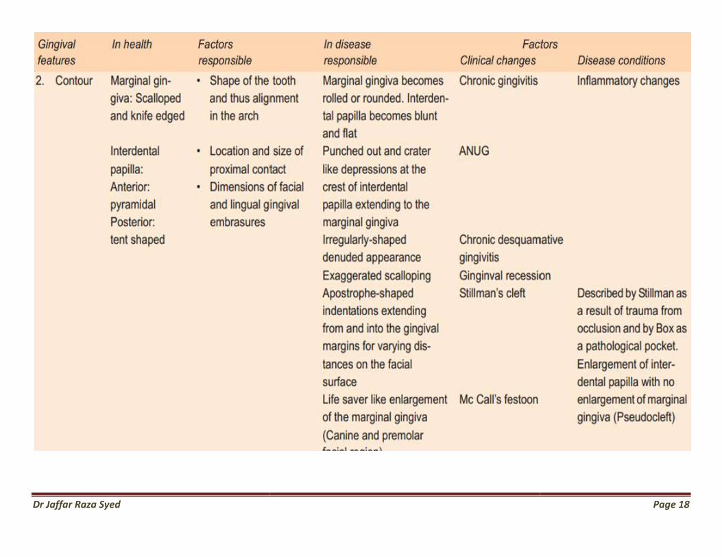
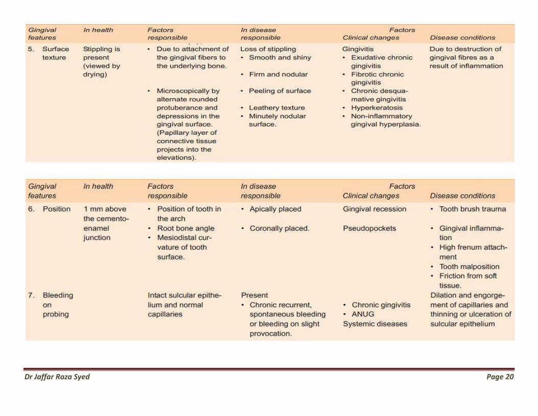
Clinical Findings
Changes in
color,
contour,
consistency,
size,
position,
severity of bleeding,
surface textur

Dr Jaffar Raza Syed Page 14
Dr Jaffar Raza Syed Page 15

Dr Jaffar Raza Syed Page 16
Dr Jaffar Raza Syed
Page 17
Dr Jaffar Raza Syed
Page 18

Dr Jaffar Raza Syed
Page 19
Dr Jaffar Raza Syed Page 20

Dr Jaffar Raza Syed
Changes in the Position of Gingiva Normal attachment CEJ In disease Coronally pseudopocket Apically gingival recession Types 1..Visible 2..hidden
Changes in the Position of Gingiva
Page 21

Dr Jaffar Raza Syed Page 22
Dr Jaffar Raza Syed Page 23

Dr Jaffar Raza Syed Page 24
PD Miller’s Classification of Gingival Recession
Dr Jaffar Raza Syed Page 25
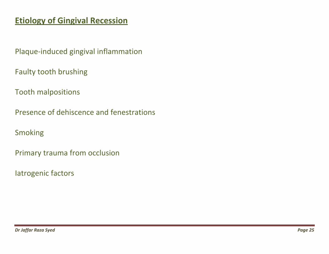
Etiology of Gingival Recession
Plaque-induced gingival inflammation
Faulty tooth brushing
Tooth malpositions
Presence of dehiscence and fenestrations
Smoking
Primary trauma from occlusion
Iatrogenic factors

Dr Jaffar Raza Syed Page 26
Clinical Significance of Gingival Recession 1. The exposed root surface may be extremely sensitive.
2. Hyperemia of the pulp may result due to gingival recession.
3. Interproximal recession creates oral hygiene problems
thereby resulting in plaque accumulation.
4. Finally, it is aesthetically unacceptable.
Dr Jaffar Raza Syed Page 27
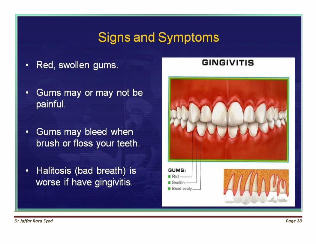
Dr Jaffar Raza Syed Page 28
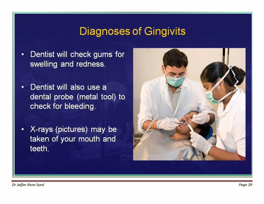
Dr Jaffar Raza Syed Page 29
Dr Jaffar Raza Syed Page 30

Dr Jaffar Raza Syed Page 31