


Languages
Pages
Legal

13th Balkan Biochemical Biophysical Days & Meeting on Metabolic Disorders’ Programme & Abstracts
13th BALKAN BIOCHEMICAL BIOPHYSICAL DAYS &
MEETING ON METABOLIC DISORDERS’ PROGRAMME & ABSTRACTS
COMMITTEES
Honorary PresidentsEngin Bermek
Şerafettin Özkurt
Executive CommitteePresiding Officials
Nazmi ÖzerFeride Severcan
SecretariatAyşe Gül ÇetinMesude İşcan
TreasurersNecla Öztürk
A.Kevser Pişkin
Members Hakan Aydın
Gürbüz ÇelebiSüleyman Daşdağ
Orhan Değer
Nezaket ErenNadide KazancıFatma Z.KutaySema T.Ozan
Turk J Biochem, 2003; 28(3), 62-224 http://www.TurkJBiochem.com62
13th Balkan Biochemical Biophysical Days & Meeting on Metabolic Disorders’ Programme & Abstracts
Scientific CommitteePekcan Ungan , Coordinator
İ.Hamdi Öğüş, Co-CoordinatorNurhan Puralı, Secretary
N.Leyla AçanNejat Dalay
Aleksandar DimovskiBurak ErmanÖmer Güzel
Mustafa GültepeVasıf N.Hasırcı
Ivan IvanovDusanka Janazic
Beki KanGüldal Kırkali
Eugenia KovacsZdravko Lalchev
Yahya LaleliAy Öğüş
H.Avni ÖktemCihan Önerİnci Özer
Asuman ÖzkaraRüstem NurtenErhan Pişkin
Zehra SayersVesna Svetlicic
Yusuf TanTijen TanyalçınAzmi TelefoncuAslıhan TolunBelma Turan
Engin UlukayaFigen Zihnioğlu
Matjas Zorko
Advisory Board
Kıymet AksoyT.Aslan AksuEmel ArınçDiler AslanErol Atalay
Sevil AtasoyUğur Atik
Ebubekir BakanOya BayındırCumhur BilgiBora BarutçuKutlay Burat
Gülden BurçakNaime CanoruçGönenç CilivSalih ÇelikÖmer ÇolakIşıl Çokur
İlhami DemirelMübeccel Durusoy
A.Kaya EmerkAhmet Eraslan
Füsun ErciyesNurten ErdalBiltan ErsözHamza EsenSina Gökçe
Şendoğan Gülenİsmail Gülen
Gül ÜnerMünire Hacıbekiroğlu
Levent KaracaBaysal KaracaHilal KaragülLevent Kayrın
Edip KehaNedret KılınçYüksel Koca
Şebnem KösebalabanMehmet KöseoğluYusuf KurtulmuşTürker Kutluay
Sevnur MandalcıTaner Onat
Asuman OrçunSinan Önen
Tomris ÖzbenMeral ÖzgüçFerit PehlivanGül SaydamAhmet Sivas
Zerrin SöylemezBolkan Şimşek
Yavuz TagaE.Ferhan Tezcan
Demir TiryakiAsuman Tokullugil
Işık TürkalpSuna TürkoğluMüjdat UysalAli Yılmaz
Piraye YargıçoğluMeral Yüce
Doğan YücelGüneş Yüreğir
Turk J Biochem, 2003; 28(3), 62-224 http://www.TurkJBiochem.com63
13th Balkan Biochemical Biophysical Days & Meeting on Metabolic Disorders’ Programme & Abstracts
SCIENTIFIC PROGRAM FOR BIOCHEMICAL & BIOPHYSICAL DAYS
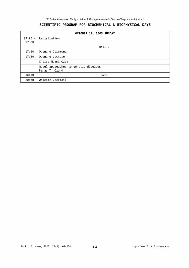
OCTOBER 12, 2003 SUNDAY
09:00 - 17:00 Registration
Hall C17:00 Opening Ceremony
17:30 Opening Lecture
Chair: Nazmi Özer
Novel approaches to genetic diseasesPınar T. Özand
18:30 Break20:00 Welcome Cocktail
Turk J Biochem, 2003; 28(3), 62-224 http://www.TurkJBiochem.com64
13th Balkan Biochemical Biophysical Days & Meeting on Metabolic Disorders’ Programme & Abstracts
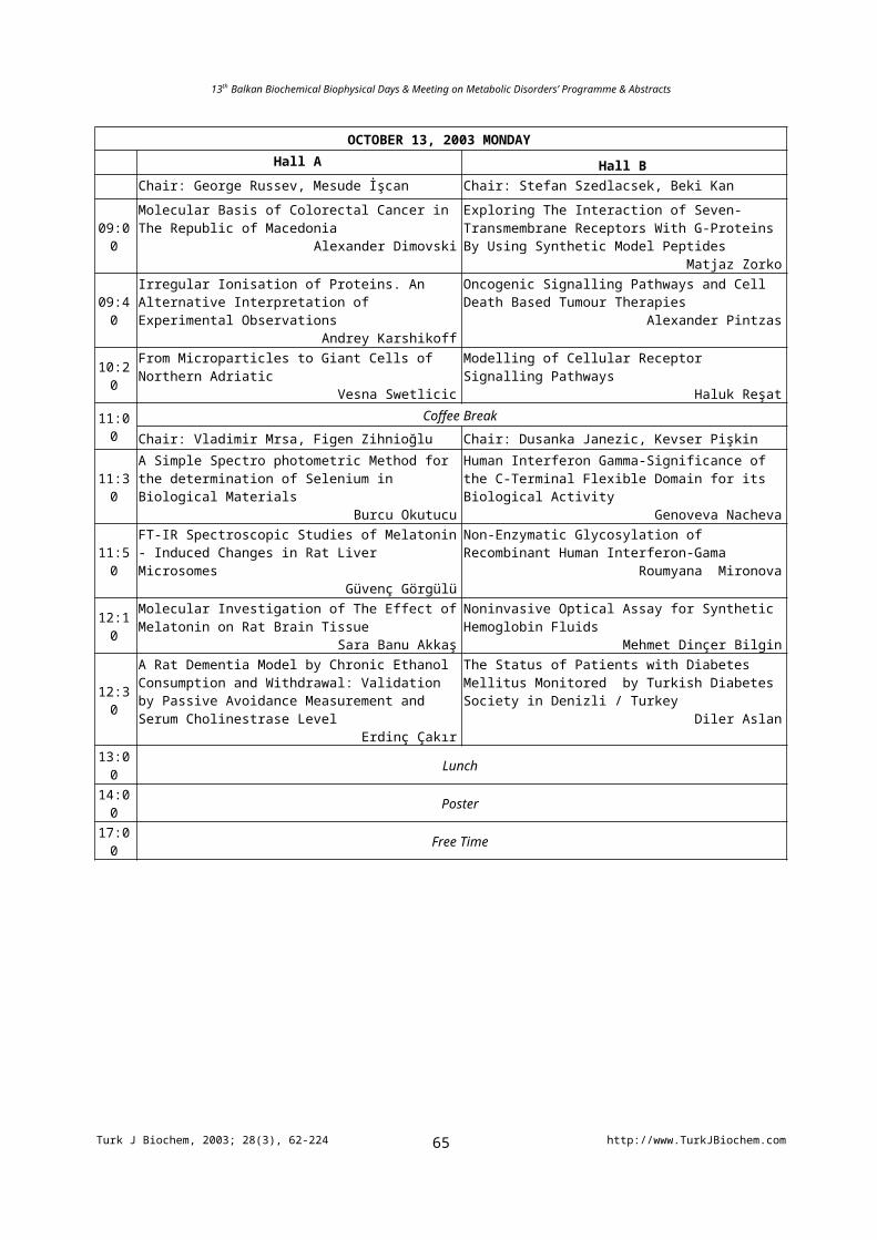
OCTOBER 13, 2003 MONDAYHall A Hall B
Chair: George Russev, Mesude İşcan Chair: Stefan Szedlacsek, Beki Kan
09:00
Molecular Basis of Colorectal Cancer in The Republic of Macedonia
Alexander Dimovski
Exploring The Interaction of Seven-Transmembrane Receptors With G-Proteins By Using Synthetic Model Peptides
Matjaz Zorko
09:40Irregular Ionisation of Proteins. An Alternative Interpretation of Experimental Observations
Andrey Karshikoff
Oncogenic Signalling Pathways and Cell Death Based Tumour Therapies
Alexander Pintzas
10:20From Microparticles to Giant Cells of Northern Adriatic
Vesna SwetlicicModelling of Cellular Receptor Signalling Pathways
Haluk Reşat
11:00Coffee Break
Chair: Vladimir Mrsa, Figen Zihnioğlu Chair: Dusanka Janezic, Kevser Pişkin
11:30A Simple Spectro photometric Method for the determination of Selenium in Biological Materials
Burcu Okutucu
Human Interferon Gamma-Significance of the C-Terminal Flexible Domain for its Biological Activity
Genoveva Nacheva
11:50FT-IR Spectroscopic Studies of Melatonin - Induced Changes in Rat Liver Microsomes
Güvenç Görgülü
Non-Enzymatic Glycosylation of Recombinant Human Interferon-Gama
Roumyana Mironova
12:10Molecular Investigation of The Effect of Melatonin on Rat Brain Tissue
Sara Banu Akkaş
Noninvasive Optical Assay for Synthetic Hemoglobin Fluids
Mehmet Dinçer Bilgin
12:30
A Rat Dementia Model by Chronic Ethanol Consumption and Withdrawal: Validation by Passive Avoidance Measurement and Serum Cholinestrase Level
Erdinç Çakır
The Status of Patients with Diabetes Mellitus Monitored by Turkish Diabetes Society in Denizli / Turkey
Diler Aslan
13:00 Lunch
14:00 Poster
17:00 Free Time
Turk J Biochem, 2003; 28(3), 62-224 http://www.TurkJBiochem.com65
13th Balkan Biochemical Biophysical Days & Meeting on Metabolic Disorders’ Programme & Abstracts
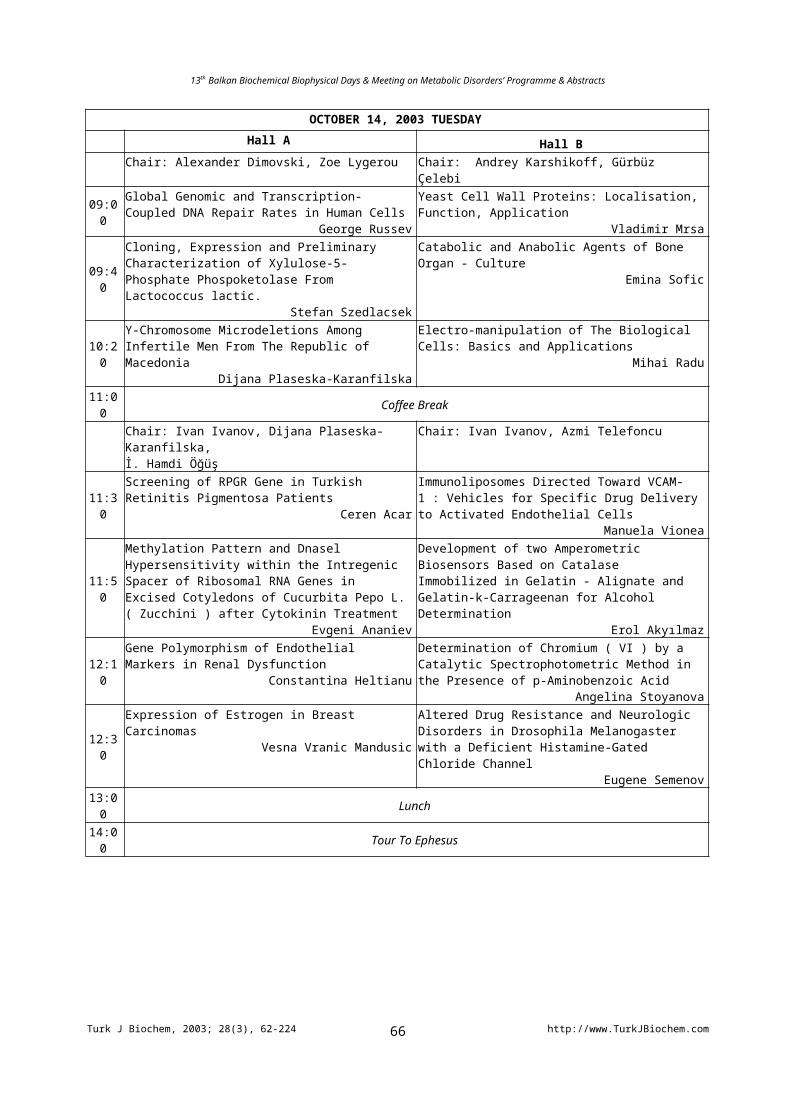
OCTOBER 14, 2003 TUESDAY
Hall A Hall BChair: Alexander Dimovski, Zoe Lygerou Chair: Andrey Karshikoff, Gürbüz Çelebi
09:00Global Genomic and Transcription-Coupled DNA Repair Rates in Human Cells
George Russev
Yeast Cell Wall Proteins: Localisation, Function, Application
Vladimir Mrsa
09:40
Cloning, Expression and Preliminary Characterization of Xylulose-5-Phosphate Phospoketolase From Lactococcus lactic.
Stefan Szedlacsek
Catabolic and Anabolic Agents of Bone Organ - CultureEmina Sofic
10:20Y-Chromosome Microdeletions Among Infertile Men From The Republic of Macedonia
Dijana Plaseska-Karanfilska
Electro-manipulation of The Biological Cells: Basics and Applications
Mihai Radu11:00 Coffee Break
Chair: Ivan Ivanov, Dijana Plaseska-Karanfilska, İ. Hamdi Öğüş
Chair: Ivan Ivanov, Azmi Telefoncu
11:30Screening of RPGR Gene in Turkish Retinitis Pigmentosa Patients
Ceren Acar
Immunoliposomes Directed Toward VCAM-1 : Vehicles for Specific Drug Delivery to Activated Endothelial Cells
Manuela Vionea
11:50
Methylation Pattern and Dnasel Hypersensitivity within the Intregenic Spacer of Ribosomal RNA Genes in Excised Cotyledons of Cucurbita Pepo L. ( Zucchini ) after Cytokinin Treatment
Evgeni Ananiev
Development of two Amperometric Biosensors Based on Catalase Immobilized in Gelatin - Alignate and Gelatin-k-Carrageenan for Alcohol Determination
Erol Akyılmaz
12:10
Gene Polymorphism of Endothelial Markers in Renal Dysfunction
Constantina Heltianu
Determination of Chromium ( VI ) by a Catalytic Spectrophotometric Method in the Presence of p-Aminobenzoic Acid
Angelina Stoyanova
12:30
Expression of Estrogen in Breast CarcinomasVesna Vranic Mandusic
Altered Drug Resistance and Neurologic Disorders in Drosophila Melanogaster with a Deficient Histamine-GatedChloride Channel
Eugene Semenov
13:00 Lunch
14:00 Tour To Ephesus
Turk J Biochem, 2003; 28(3), 62-224 http://www.TurkJBiochem.com66
13th Balkan Biochemical Biophysical Days & Meeting on Metabolic Disorders’ Programme & Abstracts
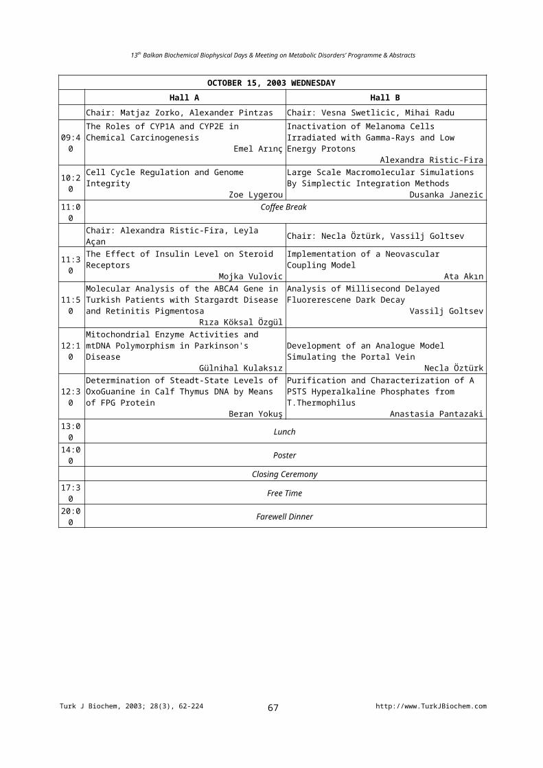
OCTOBER 15, 2003 WEDNESDAY
Hall A Hall B
Chair: Matjaz Zorko, Alexander Pintzas Chair: Vesna Swetlicic, Mihai Radu
09:40The Roles of CYP1A and CYP2E in Chemical Carcinogenesis
Emel Arınç
Inactivation of Melanoma Cells Irradiated with Gamma-Rays and Low Energy Protons
Alexandra Ristic-Fira
10:20Cell Cycle Regulation and Genome Integrity
Zoe LygerouLarge Scale Macromolecular Simulations By Simplectic Integration Methods
Dusanka Janezic11:00 Coffee Break
Chair: Alexandra Ristic-Fira, Leyla Açan Chair: Necla Öztürk, Vassilj Goltsev
11:30 The Effect of Insulin Level on Steroid ReceptorsMojka Vulovic
Implementation of a Neovascular Coupling ModelAta Akın
11:50Molecular Analysis of the ABCA4 Gene in Turkish Patients with Stargardt Disease and Retinitis Pigmentosa
Rıza Köksal Özgül
Analysis of Millisecond Delayed Fluorerescene Dark Decay
Vassilj Goltsev
12:10Mitochondrial Enzyme Activities and mtDNA Polymorphism in Parkinson's Disease
Gülnihal Kulaksız
Development of an Analogue Model Simulating the Portal Vein
Necla Öztürk
12:30Determination of Steadt-State Levels of OxoGuanine in Calf Thymus DNA by Means of FPG Protein
Beran Yokuş
Purification and Characterization of A PSTS Hyperalkaline Phosphates from T.Thermophilus
Anastasia Pantazaki13:00 Lunch
14:00 Poster
Closing Ceremony
17:30 Free Time
20:00 Farewell Dinner
Turk J Biochem, 2003; 28(3), 62-224 http://www.TurkJBiochem.com67
13th Balkan Biochemical Biophysical Days & Meeting on Metabolic Disorders’ Programme & Abstracts
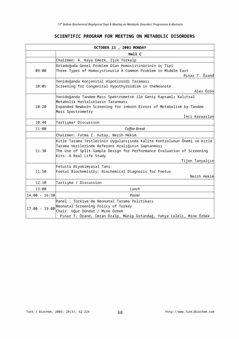
SCIENTIFIC PROGRAM FOR MEETING ON METABOLIC DISORDERS
OCTOBER 13 , 2003 MONDAY
Hall CChairmen: A. Kaya Emerk, Işık Türkalp
09:00Ortadoğuda Genel Problem Olan Homosistinürinin üç Tipi Three Types of Homocystinuria A Common Problem in Middle East
Pınar T. Özand
10:05Yenidoğanda Konjenital Hipotiroidi TaramasıScreening for Congenital Hypothyroidism in theNeonate
Alev Özön
10:20Yenidoğanda Tandem-Mass Spektrometre ile Geniş Kapsamlı Kalıtsal Metabolik Hastalıkların TaranmasıExpanded Newborn Screening for inborn Errors of Metabolism by Tandem Mass Spectrometry
İnci Karaaslan
10:40 Tartışma/ Discussion
11:00 Coffee Break
Chairmen: Fatma Z. Kutay, Nezih Hekim
11:30
Kitle Tarama Testlerinin Uygulanışında Kalite Kontrolunun Önemi ve Kitle Tarama Verilerinde Referans Aralığının SaptanmasıThe Use of Split-Sample Design for Performance Evaluation of Screening Kits: A Real Life Study
Tijen Tanyalçın
11:50Fetusta Biyokimyasal TanıFoetal Biochemistry: Biochemical Diagnosis for Foetus
Nezih Hekim
12:10 Tartışma / Discussion
13:00 Lunch
14:00 - 16:30 Poster
17:00 - 19:00
Panel : Türkiye'de Neonatal Tarama PolitikasıNeonatal Screening Policy of TurkeyChair: Uğur Dündar / Mine Özbek
Pınar T. Özand, İmran Özalp, Münip Üstündağ, Yahya Laleli, Mine Özbek
Turk J Biochem, 2003; 28(3), 62-224 http://www.TurkJBiochem.com68
13th Balkan Biochemical Biophysical Days & Meeting on Metabolic Disorders’ Programme & Abstracts
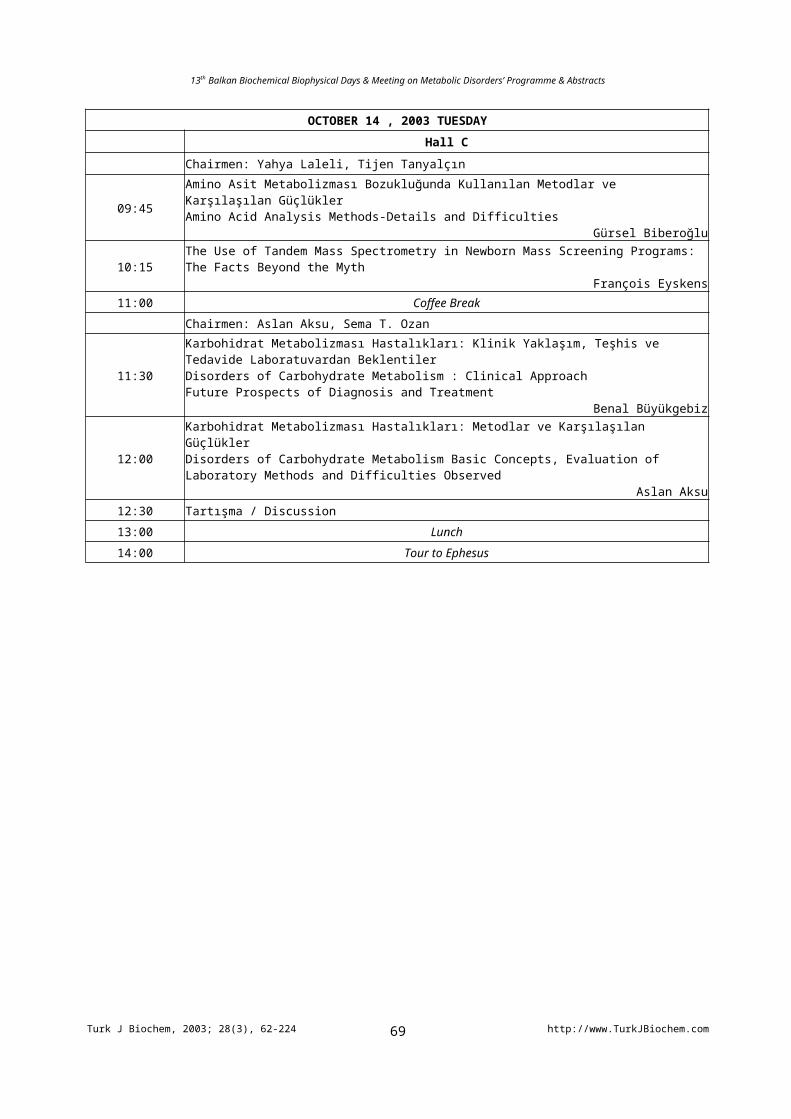
OCTOBER 14 , 2003 TUESDAY
Hall C
Chairmen: Yahya Laleli, Tijen Tanyalçın
09:45Amino Asit Metabolizması Bozukluğunda Kullanılan Metodlar ve Karşılaşılan GüçlüklerAmino Acid Analysis Methods-Details and Difficulties
Gürsel Biberoğlu
10:15 The Use of Tandem Mass Spectrometry in Newborn Mass Screening Programs: The Facts Beyond the MythFrançois Eyskens
11:00 Coffee Break
Chairmen: Aslan Aksu, Sema T. Ozan
11:30
Karbohidrat Metabolizması Hastalıkları: Klinik Yaklaşım, Teşhis ve Tedavide Laboratuvardan BeklentilerDisorders of Carbohydrate Metabolism : Clinical ApproachFuture Prospects of Diagnosis and Treatment
Benal Büyükgebiz
12:00
Karbohidrat Metabolizması Hastalıkları: Metodlar ve Karşılaşılan GüçlüklerDisorders of Carbohydrate Metabolism Basic Concepts, Evaluation of Laboratory Methods and Difficulties Observed
Aslan Aksu12:30 Tartışma / Discussion
13:00 Lunch
14:00 Tour to Ephesus
Turk J Biochem, 2003; 28(3), 62-224 http://www.TurkJBiochem.com69
13th Balkan Biochemical Biophysical Days & Meeting on Metabolic Disorders’ Programme & Abstracts
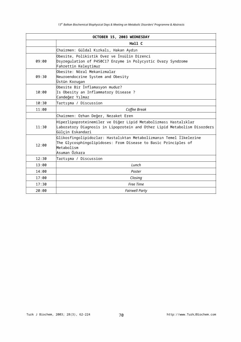
OCTOBER 15, 2003 WEDNESDAY
Hall C
Chairmen: Güldal Kırkalı, Hakan Aydın
09:00Obesite, Polikistik Over ve İnsülin DirenciDsyregulation of P450C17 Enzyme in Polycystic Ovary SyndromeFahrettin Keleştimur
09:30Obesite: Nöral MekanizmalarNeuroendocrine System and ObesityÜstün Korugan
10:00Obesite Bir İnflamasyon mudur?Is Obesity an Inflammatory Disease ? Candeğer Yılmaz
10:30 Tartışma / Discussion
11:00 Coffee Break
Chairmen: Orhan Değer, Nezaket Eren
11:30Hiperlipoproteinemiler ve Diğer Lipid Metabolizması HastalıklarLaboratory Diagnosis in Lipoprotein and Other Lipid Metabolism DisordersGülçin Eskandari
12:00Glikosfingolipidozlar: Hastalıktan Metabolizmanın Temel İlkelerineThe Glycosphingolipidoses: From Disease to Basic Principles of MetabolismAsuman Özkara
12:30 Tartışma / Discussion
13:00 Lunch
14:00 Poster
17:00 Closing
17:30 Free Time
20:00 Fairwell Party
Turk J Biochem, 2003; 28(3), 62-224 http://www.TurkJBiochem.com70
13th Balkan Biochemical Biophysical Days & Meeting on Metabolic Disorders’ Programme & Abstracts
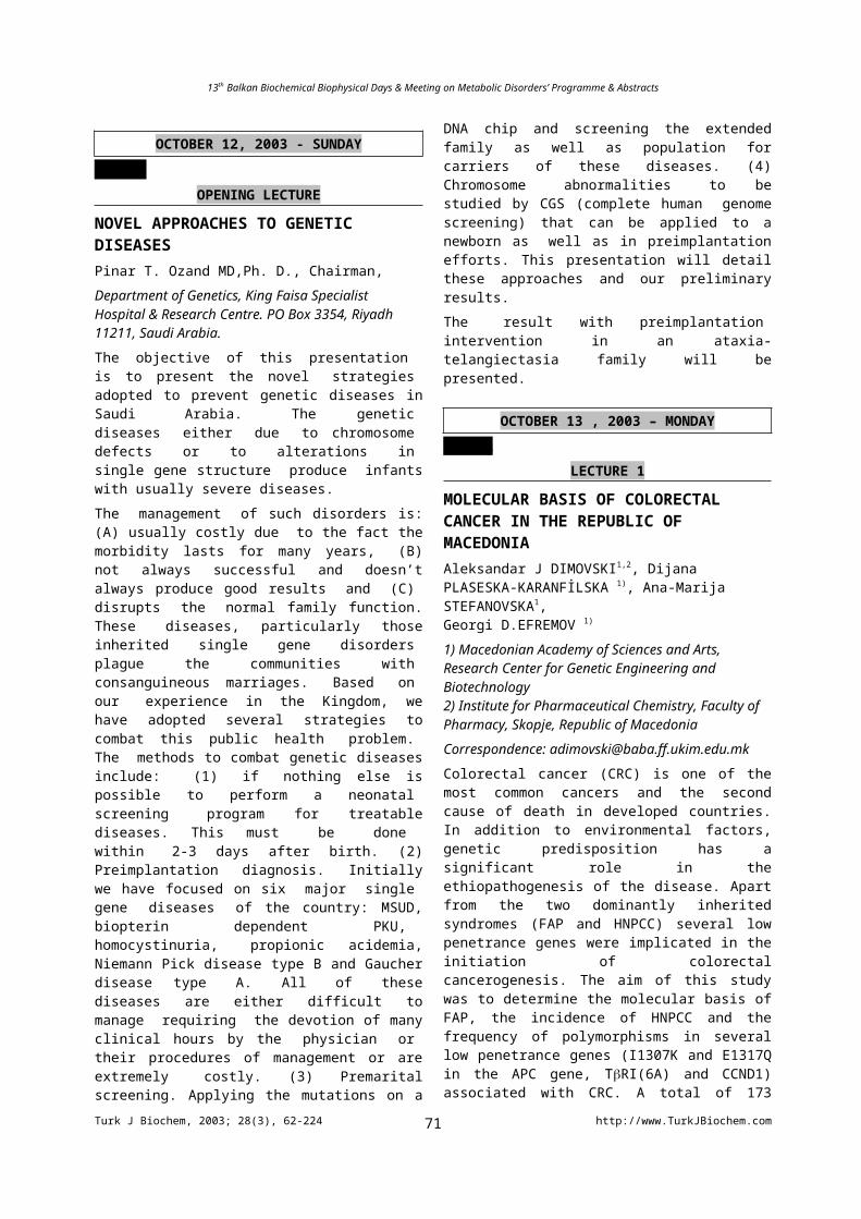
OCTOBER 12, 2003 - SUNDAY
HALL C OPENING LECTURE
NOVEL APPROACHES TO GENETIC DISEASESPinar T. Ozand MD,Ph. D., Chairman,Department of Genetics, King Faisa SpecialistHospital & Research Centre. PO Box 3354, Riyadh 11211, Saudi Arabia.The objective of this presentation is to present the novel strategies adopted to prevent genetic diseases in Saudi Arabia. The genetic diseases either due to chromosome defects or to alterations in single gene structure produce infants with usually severe diseases.The management of such disorders is: (A) usually costly due to the fact the morbidity lasts for many years, (B) not always successful and doesn’t always produce good results and (C) disrupts the normal family function. These diseases, particularly those inherited single gene disorders plague the communities with consanguineous marriages. Based on our experience in the Kingdom, we have adopted several strategies to combat this public health problem. The methods to combat genetic diseases include: (1) if nothing else is possible to perform a neonatal screening program for treatable diseases. This must be done within 2-3 days after birth. (2) Preimplantation diagnosis. Initially we have focused on six major single gene diseases of the country: MSUD, biopterin dependent PKU, homocystinuria, propionic acidemia, Niemann Pick disease type B and Gaucher disease type A. All of these diseases are either difficult to manage requiring the devotion of many clinical hours by the physician or their procedures of management or are extremely costly. (3) Premarital screening. Applying the mutations on a DNA chip and screening the extended family as well as population for carriers of these diseases. (4) Chromosome abnormalities to be studied by CGS (complete human genome screening) that can be applied to a newborn as well as in preimplantation efforts. This presentation will detail these approaches and our preliminary results.The result with preimplantation intervention in an ataxia-telangiectasia family will be presented.
OCTOBER 13 , 2003 – MONDAYHALL A
LECTURE 1
MOLECULAR BASIS OF COLORECTAL CANCER IN THE REPUBLIC OF MACEDONIAAleksandar J DIMOVSKI1,2, Dijana PLASESKA-KARANFİLSKA 1), Ana-Marija STEFANOVSKA1,Georgi D.EFREMOV 1)
1) Macedonian Academy of Sciences and Arts, Research Center for Genetic Engineering and Biotechnology
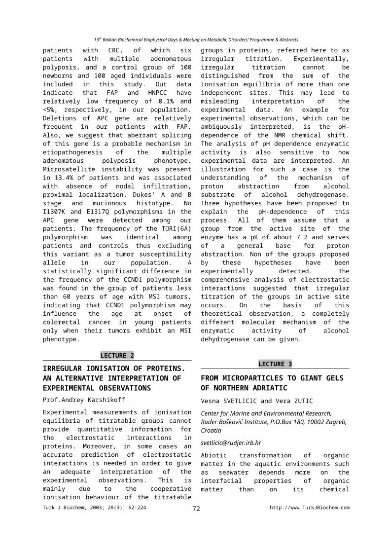
2) Institute for Pharmaceutical Chemistry, Faculty of Pharmacy, Skopje, Republic of MacedoniaCorrespondence: [email protected] cancer (CRC) is one of the most common cancers and the second cause of death in developed countries. In addition to environmental factors, genetic predisposition has a significant role in the ethiopathogenesis of the disease. Apart from the two dominantly inherited syndromes (FAP and HNPCC) several low penetrance genes were implicated in the initiation of colorectal cancerogenesis. The aim of this study was to determine the molecular basis of FAP, the incidence of HNPCC and the frequency of polymorphisms in several low penetrance genes (I1307K and E1317Q in the APC gene, TRI(6A) and CCND1) associated with CRC. A total of 173 patients with CRC, of which six patients with multiple adenomatous polyposis, and a control group of 100 newborns and 100 aged individuals were included in this study. Out data indicate that FAP and HNPCC have relatively low frequency of 0.1% and <5%, respectively, in our population. Deletions of APC gene are relatively frequent in our patients with FAP. Also, we suggest that aberrant splicing of this gene is a probable mechanism in etiopathogenesis of the multiple adenomatous polyposis phenotype. Microsatellite instability was present in 13.4% of patients and was associated with absence of nodal infiltration, proximal localization, Dukes' A and B stage and mucionous histotype. No I1307K and E1317Q polymorphisms in the APC gene were detected among our patients. The frequency of the TRI(6A) polymorphism was identical among patients and controls thus excluding this variant as a tumor susceptibility allele in our population. A statistically significant difference in the frequency of the CCND1 polymorphism was found in the group of patients less than 60 years of age with MSI tumors, indicating that CCND1 polymorphism may influence the age at onset of colorectal cancer in young patients only when their tumors exhibit an MSI phenotype.
LECTURE 2
IRREGULAR IONISATION OF PROTEINS. AN ALTERNATIVE INTERPRETATION OF EXPERIMENTAL OBSERVATIONSProf.Andrey Karshikoff
Experimental measurements of ionisation equilibria of titratable groups cannot provide quantitative information for the electrostatic interactions in proteins. Moreover, in some cases an accurate prediction of electrostatic interactions is needed in order to give an adequate interpretation of the experimental observations. This is mainly due to the cooperative ionisation behaviour of the titratable groups in proteins, referred here to as irregular titration. Experimentally, irregular titration cannot be distinguished from the sum of the ionisation equilibria of more than one independent sites. This may lead to misleading interpretation of the experimental data. An example for experimental observations, which can be ambiguously interpreted, is the pH-dependence of the
Turk J Biochem, 2003; 28(3), 62-224 http://www.TurkJBiochem.com71
13th Balkan Biochemical Biophysical Days & Meeting on Metabolic Disorders’ Programme & Abstracts
NMR chemical shift. The analysis of pH dependence enzymatic activity is also sensitive to how experimental data are interpreted. An illustration for such a case is the understanding of the mechanism of proton abstraction from alcohol substrate of alcohol dehydrogenase. Three hypotheses have been proposed to explain the pH-dependence of this process. All of them assume that a group from the active site of the enzyme has a pK of about 7.2 and serves of a general base for proton abstraction. Non of the groups proposed by these hypotheses have been experimentally detected. The comprehensive analysis of electrostatic interactions suggested that irregular titration of the groups in active site occurs. On the basis of this theoretical observation, a completely different molecular mechanism of the enzymatic activity of alcohol dehydrogenase can be given.
LECTURE 3
FROM MICROPARTICLES TO GIANT GELS OF NORTHERN ADRIATICVesna SVETLICIC and Vera ZUTIC
Center for Marine and Environmental Research, Ruđer Bošković Institute, P.O.Box 180, 10002 Zagreb, Croatia
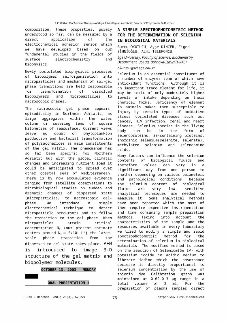
Abiotic transformation of organic matter in the aquatic environments such as seawater depends more on the interfacial properties of organic matter than on its chemical composition. These properties, purely understood so far, can be measured by a direct application of the electrochemical adhesion sensor which we have developed based on our fundamental studies in the fields of surface electrochemistry and biophysics.
Newly postulated biophysical processes of biopolymer selforganization into microparticles and mechanism of sol-gel phase transitions are held responsible for transformation of dissolved biopolymers and microparticles to macroscopic phases.
The macroscopic gel phase appears, episodically in Northern Adriatic, as large aggregates within the water column or covering tens of square kilometres of seasurface. Current views leave no doubt on phytoplankton production and bacterial transformation of polysaccharides as main constituents of the gel matrix. The phenomenon has so far been specific for Northern Adriatic but with the global climatic changes and increasing nutrient load it could be anticipated to spread over other coastal seas of Mediterranean. There is by now accumulated evidence ranging from satellite observations to microbiological studies on sudden and dramatic changes of dispersed state (microparticles) to macroscopic gel-phase. We introduce a simple electrochemical technique to detect microparticle precursors and to follow the transition to the gel phase. When micrparticles attain critical concentration Nc (our present estimate centers around Nc 5x10⁷ L-1) the large-scale phase transition from the dispersed to gel state takes place. AFM is introduced to image 3-D
structure of the gel matrix and biopolymer molecules.
OCTOBER 13, 2003 – MONDAYHALL A
ORAL PRESENTATION 1
A SIMPLE SPECTROPHOTOMETRIC METHOD FOR THE DETERMINATION OF SELENIUM IN BIOLOGICAL MATERIALSBurcu OKUTUCU, Ayşe DİNÇER, Figen ZİHNİOĞLU, Azmi TELEFONCUEge University, Faculty of Science, Biochemistry Department, 35100, Bornova-İzmir/[email protected] is an essential constituent of a number of enzymes some of which have antioxidant functions. Although it is an important trace element for life, it may be toxic of only moderately higher levels of intake depending on their chemical forms. Deficiency of element in animals makes them susceptible to injury by certain types of oxidative stress correlated diseases such as; cancer, HIV infection, renal and heart disease. Selenium species in the living body can be in the form of selenoproteins, Se-containing proteins, inorganic selenium(selenite, selenate), methylated selenium and selenoamino acids.Many factors can influence the selenium contents of biological fluids and therefore values can vary in a significant way from one person to another depending on various parameters and pathological conditions. Because the selenium content of biological fluids are very low, sensitive analytical techniques are needed to measure it. Some analytical methods have been reported which the most of them require expensive instrumentation and time consuming sample preparation methods. Taking into account the characteristics of the sample and the resources available in every laboratory we tried to modify a simple and rapid spectrophotometric method for the determination of selenium in biological materials. The modified method is based on the reaction of Selenium(Se IV) with potassium iodide in acidic medium to liberate iodine which the absorbance decrease is directly proportional to selenium concentration by the use of thionin dye Calibration graph was maintained at 0.02-0.3 g range in a total volume of 2 ml. For the preparation of plasma samples direct dilution and enzymatic digestion methods were performed and compared. Variation coefficients and standard deviations were calculated for five replicate determinations. Furthermore, as well as the accuracy, application of the method to the biological materials was investigated by using a commercial drug (Dietary Selenium Supplement) and plasma samples. Selenium in water, soil, plant materials, cosmetics, etc. can be also determined by this method.
ORAL PRESENTATION 2
FT-IR SPECTROSCOPİC STUDY OF STREPTOZOTOCIN-INDUCED DİABETİC RAT LİVER MİCROSOMESGüvenç GÖRGÜLÜ*, Sevgi GÖRGÜLÜ**, Ökkeş YILMAZ***, Tülin GÜRAY* and Feride SEVERCAN**
Turk J Biochem, 2003; 28(3), 62-224 http://www.TurkJBiochem.com72
13th Balkan Biochemical Biophysical Days & Meeting on Metabolic Disorders’ Programme & Abstracts
*Middle East Technical University, Department of Biochemistry, 06531, Ankara, Turkey**Middle East Technical University, Department of Biology, 06531, Ankara, Turkey***Fırat University, Department of Biology, Elazığ, [email protected] blood glucose levels induce metabolic disorders that initiate a sequence of events including renal, arterial, cardiac and retinal disorders. Diabetes mellitus increases oxidative stress in tissues of both humans and animals. Increased oxidative stress might play a role in the development of diabetic complications.In the present study, 8 male Wistar rats (4 diabetic and 4 controls) were used. Diabetes was induced by an injection of streptozotocin (STZ) (50 mg/kg i.p.) after a 24-h fast. Liver microsomal fractions were isolated. Lipid peroxidation in microsomes was analyzed both by TBA-RS test and FT-IR study. Protein amount in microsomal fractions of diabetic samples were significantly decreased as determined by the method of Bradford. This was further supported by FT-IR study measuring the lipid-to-protein ratio from CH2 symmetric/CH3 symmetric bands. The results of FT-IR spectral analysis of absorption and second derivative signals revealed that significant increase in wavenumber of the CH2 asymmetric (p<0.005) and the CH2 symmetric (p<0.05) stretching vibrations were obtained, indicating an increase in disorder in diabetic rat liver microsomes. The bandwidth of the CH2 asymmetric stretching band significantly increased for diabetic samples (p<0.05). However, the CH2 symmetric stretching bandwidth did not differ significantly. Olefinic band (=CH at 3012 cm-1) was analyzed by measuring the intensity of this band, which shows the degree of lipid peroxidation in the system. The intensity of 3012 cm-1 band was increased for diabetic samples at different temperatures. Secondary structure conformational changes have been investigated from the analysis of amide I band (1653 cm -1) using curve fitting procedure.Use of FT-IR spectroscopy for diagnostic purposes has been an increasing demand by researchers. This technique can be used in early diagnosis of several diseases by probing different functional groups belonging to different macromolecules such as lipids and proteins.Key Words: FTIR spectroscopy, diabetes, liver tissues, microsomal membranes, lipid peroxidation
ORAL PRESENTATION 3
MOLECULAR INVESTIGATION OF THE EFFECTS OF MELATONIN ON RAT BRAIN TISSUES. Banu AKKAŞ1, Faruk ZORLU2 and Feride SEVERCAN1
1Middle East Technical University, Department of Biological Sciences, 06531, Ankara/TURKEY2Hacettepe University, Faculty of Medicine, Department of Radiation Oncology, 06100, Ankara/[email protected]
The brain is highly vulnerable to free radical attack since it is rich in polyunsaturated fatty acids and consumes very high amounts of oxygen. Thus, the goal of this study was to examine the effects on rat brain tissue of the non-enzymatic antioxidant hormone melatonin with Fourier Transform Infrared Spectroscopy (FTIR). The results of the present study reveal that melatonin induces a significant increase in the relative lipid to protein ratio of the melatonin treated samples. Furthermore, a significant increase is observed in the frequency values of the PO2
- symmetric stretching mode. The temperature dependent studies suggest that, at lower temperatures melatonin causes a decrease in the CH2
symmetric streching vibrational mode frequency, which indicates an increase in the order and thus the trans-gauche ratio of the crude brain membrane in a probable gel phase. Then after about 29-32C, it sets off an increase in the same frequency, indicating an increase in the number of gauche conformers leading to an increase in the disorder at a likely liquid-crystalline phase. It is very important to note that body temperature is among the temperatures where melatonin induced an increase in disorder. The observation of the disordering effect of melatonin at body temperature was also supported by the increases in the frequencies of both the CH3 and PO2
- asymmetric stretching modes. Taking into account that the CH2
symmetric, CH3 asymmetric and PO2- asymmetric
strecthing modes give information about the order of the interior, deep interior and head-group regions of the lipid bilayer, respectively, the results suggest that melatonin induces a disordering effect on the lipid bilayer at physiological temperatures. This seems rational since it is believed that a higher disorder of membrane lipids might make the interaction of antioxidants with lipid radicals more efficient.
ORAL PRESENTATION 4
A RAT DEMENTIA MODEL BY CHRONIC ETHANOL CONSUMPTION AND WITHDRAWAL: VALIDATION BY PASSIVE AVOIDANCE MEASUREMENT AND SERUM CHOLINESTERASE LEVEL Erdinç ÇAKIR1, Turgay. ÇELİK2, Hakan KAYIR2, Cumhur BİLGİ1, and I. Tayfun UZBAY2
Gulhane Military Medical Academy, 1Department of Emergency Medicine, Laboratory of Biochemistry, 2Department of Medical Pharmacology, Experimental Psychopharmacology Research Unit, Etlik 06018 Ankara – [email protected] aim of the present study was to investigate if the chronic ethanol administration by liquid diet to rats may be a dementia model.Female Wistar rats (188-244 g) were used in the study. Ethanol was administered by a modified liquid diet with 4.8% (v/v) ethanol for 3 days followed by 25 days on a liquid diet in which the ethanol concentration was increased to 7.2%. Control rats were pair fed with an
Turk J Biochem, 2003; 28(3), 62-224 http://www.TurkJBiochem.com73
13th Balkan Biochemical Biophysical Days & Meeting on Metabolic Disorders’ Programme & Abstracts
isocaloric liquid diet not containing ethanol. Serum ChE activity and blood ethanol concentration were measured at the end of the 4.8% ethanol consumption and after 35 days of ethanol (7.2%) feeding and, just before, 24th and 72nd
hours ethanol witdrawal period. Cognitive functions were evaluated by step-down passive avoidance test system for 150 sec (cut-off time) in three individual groups of ethanol-administered, ethanol withdrawn (24th h withdrawal) and control rats. The data was evaluated by one-way analysis of variance followed by Tukey’s test for post-hoc comparison. The daily ethanol consumption of the rats ranged from 11.5 to 14.9 g/kg. ChE activity was found significantly increased from 3rd day of ethanol (4.8%) consumption. Serum ChE activities of the rats receiving ethanol (7.2%) also increased significantly as compared to ethanol (4.8%) ingesting rats. Blood ethanol levels were measured as 200 and 2.2 mg/dl at 35th days of ethanol consumption (just before ethanol withdrawal) and 24th h of ethanol withdrawal, respectively. Passive avoidance latency was found significantly reduced in the groups that just before and 24th h of ethanol withdrawal as compared to control rats. Our results suggest that serum ChE activity increased by chronic ethanol consumption in rats and chronic ethanol caused some marked impairments on the cognitive functions Overall the data indicated that chronic ethanol feeding might be a model for evaluation cognitive functions in rats.
OCTOBER 14 , 2003 - TUESDAYHALL A
LECTURE 1
GLOBAL GENOMIC AND TRANSCRIPTION-COUPLED DNA REPAIR RATES IN HUMAN CELLSGeorge Russev, Boyko Atanassov, Emil Mladenov, Anelyia Velkova, Boyka AnachkovaInstitute of Molecular Biology, Bulgarian Academy of Sciences, 1113 Sofia, BulgariaThe most general and for historical reasons the best-studied DNA repair pathway is the human nucleotide excision repair (NER) pathway. It includes 7 genes designated XPA through XPG. Their protein products are identified, and the pathway is reconstructed in vitro in considerable details. NER can remove a huge variety of lesions among them most of the DNA lesions caused by environmental agents by anticancer drugs. There are two subclasses of nucleotide excision repair. One is the global genomic repair (GGR) which removes lesions throughout the genome regardless of whether any specific sequence is transcribed or not. It can be studied in vitro, in cell free systems where no transcription of the repair substrate is taking place. The other is transcription-coupled repair (TCR), which removes lesions only from DNA sequences, which are transcribed. There is no general way to measure TCR in vitro so far. To study TCR we applied the following approach. Human HEK 293 cells were transfected with pEGFP and pEYFP plasmids treated with UVC light, cis-DDP, Angelicin and
Trioxsalen and 24 hours later the restored fluorescence was measured and used to calculate the combined TCR+GGR rates. In a parallel set of experiments the same plasmids were incubated in repair competent HEK293 protein extracts and the GGR repair rates were determined. From the two sets of data the TCR rates were calculated. We found out that the four lesions were repaired with very similar efficiency by TCR pathway and with quite different efficiency by GGR. In the latter case the repair rates generally corresponded to the degree of DNA helix distortion at the sites of damage. Multiple host cell reactivation assay was carried out to study the competition of the different lesions for the TCR damage recognition factors in vivo. We found out that the different lesions did not compete for the damage recognition factors, but that they compete with the transcribed genes for the transcription initiation factors. A conclusion was drawn that the stalled RNA polymerase II and not the lesions themselves are the subject of recognition in the damage recognition step of TCR, and the distortion of the DNA helix does not play a role in the process.
LECTURE 2
CLONING, EXPRESSION AND PRELIMINARY CHARACTERIZATION OF XYLULOSE 5-PHOSPHATE PHOSPHOKETOLASE FROM LACTOCOCCUS LACTISAndrei VACARU1, Alexandru S. DENES1, Daniela STANGA1, Octavian BARZU2 and Stefan E. SZEDLACSEK1
1Institute of Biochemistry, Department of Biochemistry, 77700 Bucharest/ROMANIA2Institute Pasteur, Paris/[email protected]
Heterofermentative degradation of pentoses in lactic acid bacteria takes place via the phosphoketolase pathway. Xylulose 5-phosphate phosphoketolase (EC 4.1.2.9) is the central enzyme of this pathway. In presence of inorganic phosphate, this enzyme catalyses conversion of xylulose 5-phosphate (X5P) into glyceraldehyde 3-phosphate and acetylphosphate. So far, a limited number of molecular data are available for phosphoketolases, particularly for those from lactic acid bacteria (LAB).
We report here the cloning, the expression in a prokaryotic system, and the preliminary characterization of X5P phosphoketolase of Lactococcus lactis ssp. lactis (strain IL1403), one of the most important representatives of LAB in dairy industry. Phosphoketolase gene of L. lactis (termed ptk) was cloned by using a step-by-step strategy, starting from five DNA fragments of ptk, each obtained by PCR amplification on the basis of a genomic template. The 2469 bp long sequence was then transferred into a prokaryotic expression vector. Optimized expression led finally to a soluble protein, which was purified using an affinity based approach. The protein preparation thus obtained was electrophoretically homogeneous and migrated in SDS-PAGE at 93.3 kDa, in accordance with
Turk J Biochem, 2003; 28(3), 62-224 http://www.TurkJBiochem.com74
13th Balkan Biochemical Biophysical Days & Meeting on Metabolic Disorders’ Programme & Abstracts
the theoretical value derived from the enzyme sequence. Using a spectrophoptometric, coupled assay, the preliminary kinetic analysis was also performed. It demonstrates that his enzyme is thiamine pyrophosphate-dependent, possesses a relatively high specific activity and has a specific dependence on substrates concentrations and pH values. Altogether, these features define X5P phosphoketolase of L. lactis as a novel enzyme displaying a particular set of characteristics among other phosphoketolases.
LECTURE 3
Y-CHROMOSOME MICRODELETIONS AMONG INFERTILE MEN FROM THE REPUBLIC OF MACEDONIA
Dijana PLASESKA-KARANFILSKA 1), Toso PLASESKI 2), Cedomir DIMITROVSKI 2), Branka KOCEVA 2), Georgi D.EFREMOV 1)
1) Macedonian Academy of Sciences and Arts, Research Center for Genetic Engineering and Biotechnology 2) Clinic forEndocrinology and Metabolic Disorders, Faculty of Medicine, Skopje , Republic of Macedonia
Correspondence: [email protected]
Microdeletions in the three non-overlapping regions of the Y-chromosomes are associated with male infertility and have an important role for genetic counseling of the infertile couple and for their decision concerning the therapeutic options. The main aims of this study were: to determine the prevalence of Y microdeletions among the infertile males from the Republic of Macedonia; to determine the breakpoints and the size of the deletions; to correlate the genotype with the phenotype; and to introduce the screening for Y chromosome microdeletions in the routine clinical practice. Fifty fertile males and 116 infertile males were included in the study. The PCR analysis of six STS loci was used for the screening of Y chromosome microdeletions. The breakpoints and the size of the deletions were determined by analysis of additional STS flanking markers. A total of 7 patients showed presence of Y microdeletion, of which 6 had AZFc deletions, and one AZFb+c deletion. Four of the 6 patients with AZFc microdeletion carried an identical deletion with a size of 3.5 Mb, while in the other 2 patients the deletion was larger than 3.5 Mb. A 45X0/46XY mosaicism was found in the patient with AZFb+c deletion. The Y microdeletions were detected in six patients with azoospermia and one with severe oligozoospermia. No deletion was found among the fertile males, patients with normozoospermia and oligozoospermia (>5x10⁶/ml). The prevalence of Y microdeletions among the infertile males from the Republic of Macedonia is 6.4%, among patients with azoospermia – 17.4% and among those with
severe oligozoospermia - 3.6%. Different testicular defects were found among the patients with AZFc deletions (SCOS, hypospermatogenesis and maturity arrest). Testicular volumes were reduced in the patients with Y microdeletions, FSH concentrations were high and LH and testosterone were within the normal range.
OCTOBER 14, 2003 – TUESDAYHALL A
ORAL PRESENTATION 1
SCREENING OF RPGR GENE IN TURKISH RETINITIS PIGMENTOSA PATIENTSCeren Acar1, Köksal Özgül1, Adam Reddick2, Anand Swaroop2, Cihan Öner1 and Ay Öğüş1
1Hacettepe University Faculty of Science Biology Department 06532, Beytepe Ankara/TURKEY2University of Michigan Ophthalmology and Visual Science Department Ann Arbor MI/[email protected] pigmentosa (RP) is a heterogeneous group of retinal dystrophies that is characterized by photoreceptor degeneration. RP causes night blindness, a gradual loss of peripheral visual field and eventual loss of central vision. The disease can be inherited in autosomal dominant, autosomal recessive, X-linked and digenic modes. Twenty-six genes have been identified or cloned for RP and additional 14 genes mapped but not yet identified. Five loci have been mapped for XLRP. Among these, RP3 accounts for 70%-90% and RP2 accounts for 10%-20% of genetically identifiable disease. Mutations in the RPGR gene are associated with RP3 subtype of XLRP, a severe non-syndromic form of retinal degeneration. Mutations in RPGR have been identified in exons 1-14; however, ORF15 is the hot spot for mutations accounting for 50-60% of XLRP in Europe and North America. ORF15 mutations have also been detected in simplex RP males. In Turkey, there is no reported study on the molecular genetics of XLRP. Here, we present the analysis of ORF15 in 19 patients with RP from Turkey. These patients were the only ones showing the disease phenotype in their families. DNA samples were screened for RPGR-ORF15 by sequence analysis. These DNA samples were also analyzed by SSCP analysis for the following genes: ABCA4, rhodopsin, CRX, RPE65, RDS/peripherin and linkage and haplotype studies were performed for the genes PDE6A, PDE6B, LRAT, RALBP, TULP1.
ORAL PRESENTATION 2
METHYLATION PATTERN AND DNaseI HYPERSENSITIVITY WITHIN THE INTERGENIC SPACER OF RIBOSOMAL RNA GENES IN EXCISED COTYLEDONS
Turk J Biochem, 2003; 28(3), 62-224 http://www.TurkJBiochem.com75
13th Balkan Biochemical Biophysical Days & Meeting on Metabolic Disorders’ Programme & Abstracts
OF CUCURBITA PEPO L. (ZUCCHINI) AFTER CYTOKININ TREATMENTEvgueni D. ANANIEV1, Galina ABDULOVA1, Petar GROZDANOV2, Luchezar KARAGYOZOV2
1Institute of Plant Physiology “M. Popov”, 2 Institute of Molecular Biology, Bulg. Acad. Sci., “Acad. G. Bonchev” str., block 21, 1113, Sofia/ BULGARIA [email protected] “Run-on” transcription experiments with isolated nuclei have showed that treatment of detached marrow cotyledons with the cytokinin 6-benzyladenine (BA) resulted in 2-4-fold stimulation of RNA polymerase I activity. In this work we studied the methylation pattern of the intergenic spacer (IGS) of rRNA genes as a measure of their activity using the restriction enzymes Msp I and Hpa II and the method of “indirect end labeling”. A cloned fragment containing the 5/ portion of 18S rRNA gene from flax was used as DNA probe. Results showed heavy methylation of the rRNA genes. As judged from the almost total lack of digestion with Hpa II, in the repeating rDNA units there were either no methylation free regions or just a few were observed. A hypomethylated Hpa II site near the promoter region was detected in a very small number of rDNA repeats. Digestion with Msp I affected nearly 50% of the repeating units. This suggested that in addition to –CpG- sequences, methylation in –CpNpG- might not be random. The methylation pattern of IGS was not changed upon hormonal treatment of cotyledons DNase I assay with isolated nuclei revealed defined regions of DNase I hypersensitivity in IGS which coincide with the regulatory elements. The DNase I hypersensitive sites in IGS were mapped to the active promoter (transcription initiation site –TIS), to transcription termination site (TTS) and the “spacer” promoter region. No appreciable differences in DNase I hypersensitivity of IGS were found after hormone treatment of cotyledons nor in differentiated leaves of intact seedlings. The obtained results are discussed with respect to the possible molecular mechanisms of cytokinin regulation of rRNA gene transcription.
ORAL PRESENTATION 3
GENE POLYMORPHISM OF ENDOTHELIAL MARKERS IN RENAL DYSFUNCTIONConstantina HELTIANU, Gabriela COSTACHE, Mihaela BODEANU, Alexandra DOBRIN, Kemal AZIBI*, Livia POENARU*, Maya SIMIONESCUInstitute of Cellular Biology and Pathology “N.Simionescu”, Bucharest, ROMANIA, *Department of Genetic, Faculty of Medicine Cochin Port-Royal, University Rene Descartes, Paris, [email protected]: Renin-angiotensin system is implicated in the progression of kidney disease in diabetes mellitus (DM). Another disease in which the kidneys are dramatically affected is the monogenic X-linked Fabry disease. The Latter is characterized by progressive accumulation of lipids (due to the deficiency of alpha-
galactosidase A activity) preferentially in vascular endothelial cells (EC) which become activated.Aim: The purpose of the study was to analyze whether the gene encoding endothelial markers such as endothelial nitric oxide synthase (eNOS), and angiotensin converting enzyme (ACE) influence the development of nephropathy in DM and Fabry disease. Methods: To test this hypothesis about 200 DM subjects, 28 hemizygot unrelated Fabry patients and 120 healthy individuals were genotyped for the intron 4VNTR and Glu298Asp of eNOS, and I/D variant of ACE to analyze their association with the disease. The 4VNTR eNOS and I/D ACE variants were determined by PCR amplification followed by agarose gel electrophoresis. The Glu298Asp of eNOS was assessed by RFLP-PCR technique and then separated by polyacrilamide gel electrophoresis.Results: There is no association of eNOS and ACE gene variants with the presence of renal dysfunction in diabetic patients. In the case of Fabry disease, both eNOS gene polymorphism were significantly associated with the affliction namely P=0.02, OR=2.7, 95%CI=1.1-6.4 for Glu298Asp, and P=0.05, OR=2.6, 95%CI=1.0-7.0 for intron 4 VNTR. Carriers of eNOS 298Asp allele were overrepresented (P=0.04) in Fabry subgroup with renal failure when compared with patients without it, giving a 4.64-fold increased risk (95% CI=2.43-6.85) of the incidence of renal dysfunction. The distribution of ACE D allele was similar among the Fabry patients and control subjects.Conclusion: These findings suggest that the eNOS polymorphism might influence the development of renal failure in Fabry disease. This project was financially supported by the Romanian Academy (GAR 125) and INSERM France (PECO 637).
ORAL PRESENTATION 4
EXPRESSION OF ESTROGEN RECEPTOR β IN BREAST CARCINOMASVesna VRANIĆ MANDUSIĆ1, Dusan POPOV-CELKETIĆ1, Ksenija KANJER2 and Dragica NIKOLIĆ-VUKOSAVLJEVIĆ2
1 ”Vinča” Institute of Nuclear Sciences, Laboratory for Radiobiology and Molecular Genetics, Belgrade, Serbia and Montenegro
2 Institute for Oncology and Radiology of Serbia, Belgrade, Serbia and Montenegro
To determine whether estrogen receptor beta (ERβ) expression is associated with estrogen receptor α (ERα) and progesterone receptor (PR) status we compared its expression with levels of ERα and PR. Thirty two samples of breast carcinomas were examined for ERα, ERβ and PR. Levels of ERα and PR were measured by conventional biochemical techniques. Expression of ERβ mRNA was determined by RT-PCR. Briefly, RNA from tumor samples was isolated according the method of Chomczynski and Sacchi (1987); total RNA was reverse transcribed with
Turk J Biochem, 2003; 28(3), 62-224 http://www.TurkJBiochem.com76
13th Balkan Biochemical Biophysical Days & Meeting on Metabolic Disorders’ Programme & Abstracts
oligo dT primer; PCR was performed using the specific primers positioned in exons 4 and 6. PCR (254 bp product) were analyzed on polyacrilamide gel electrophoresis. In 9 carcinomas ERβ mRNA was not detected, and 23 carcinomas (71,8%) were positive for ERβ expression. Level of expression was quantified densitometricaly and relatively ranged from 1 to 3. Results conform positive correlation between ERα and PR status. Analysis of correlation between ERα and ERβ expression as wel as ERβ expression and PR shows:
absence of any correlation between ERα and ERβ;
statistical significant (hi-square test, p=0,03) inverse correlation between ERβ and PR status.
Therefore, expression of ERα and ERβ are independent one from the other, but increased expression of ERβ is connected with decreasing of ER functionalities represented through decreased PR expression.
OCTOBER 15 , 2003 - WEDNESDAYHALL A
LECTURE 1
THE ROLES OF CYP1A AND CYP2E IN CHEMICAL CARCINOGENESISEmel ARINÇGraduate Program in Biochemistry, Department of Biology, Middle East Technical University, 06531, Ankara, [email protected] has been established that approximately 80% of human cancers are attributable to environmental agents and about 70% of the human cancers are caused by chemical compounds. The majority of the organic carcinogens is not reactive as such but must undergo enzymatic reactions to form electrophilic species. The first step in biotransformation is usually the oxidative step, catalyzed by microsomal cytochrome P450 (CYP) dependent monooxygenases. Upto now more than 2000 P450 genes are sequenced and 265 P450 families have been identified and 18 of them are found in humans. Although other P450s and other mechanisms may activate the carcinogens, CYP1A enzymes are the most significant and 90% of the known carcinogens are metabolically activated by CYP1A to the proximate and ultimate carcinogens. Persistent organic pollutions such as polyaromatic hydrocarbons (PAHs), dioxins, polychlorinated biphenyls (PCBs) specifically induce CYP1A1 through Ah receptor mediated mechanism. At the same time, CYP1A1 mostly converts PAH- and PCB-type precarcinogens to their carcinogenic epoxides or other oxygenated metabolites. These epoxides, in turn bind DNA covalently forming DNA-adducts which may cause cancer in the years to come. Thus induction of CYP1A1 is used to measure both exposure and resulting toxic carcinogenic effects of these types of organic pollutants. (Arınç, E., Şen, A., Bozcaarmutlu, A.: Pure & Appl. Chem. 72, 985-994, 2000). Greater CYP1A1 induction may result in high levels of activated carcinogens, and consequently to higher
degree of persistent DNA-adduct formation or to enhanced oxidative DNA damage. Good correlation between smoking and lung cancer is established. Individuals with a high inducible phenotype CYP1A1 show high risk of lung cancer. Gene-environment interactions are more pronounced at lower levels of cigarette exposure in which the susceptibility to lung cancer increased in the case of patients with either CYP1A1 mutated alleles and GSTM1 null gene. On the other hand, CYP1A2 activates many arylamines and amides present in food and cigarette smoking. CYP1A2 exhibits polymorphisms and inducibility. Aflatoxin B1 (AFB1) must be activated to a reactive epoxide prior to exerting carcinogenic effects. CYP1A2 is the predominant human CYP enzyme involved in the activation of AFB1. In the presence of hepatitis B virus (HBV), a relative risk for liver cancer upon exposure of AFB1 increased 30-fold. CYP2E1 is also involved in chemical carcinogenesis, both in the metabolic activation of small molecular weight organic chemicals and in the generation of ROS. N-Nitrosodimethylamine (NDMA) is a procarcinogen that is activated by CYP2E1 dependent NDMA N-demethylase. NDMA is known to be carcinogenic in liver, kidney and lung. Pyridine, constituent of tobacco and tobacco smoke, has tumor-promoting and teratogenic activities. Following in vitro pyridine treatment of rabbits, NDMA N-demethylase activity of both liver and lung is stimulated by 6.9- and 5.2-fold, respectively indicating that pyridine increases the metabolic activation of NDMA and in turn, may potentiate formation of liver and lung cancers significantly (Arınç, E., Adalı, O., Gençker-Özkan, A. M.: Arch. Toxicol., 34, 329-334, 2000). In addition, experiments carried out in our laboratory demonstrated that in the experimentally induced diabetic rabbits, both CYP2E1 and NDMA N-demethylase activity have been increased about two-fold in kidney and liver of rabbits. Therefore, it is expected that the incidence of tumor formation due to exposure to NDMA will be more pronounced in diabetic state because of CYP2E1 induction.
LECTURE 2
CELL CYCLE REGULATION AND GENOME INTEGRITYZ. LygerouDepartment of Gen Biology, Medical School, University of Patras, [email protected] cells to maintain genome integrity, S-phase must alternate with mitosis at every cell cycle. Licensing of DNA for replication takes place only after completion of mitosis and ensures once per cell cycle replication. Cdt1, a central licensing factor conserved across evolution, is tightly controlled in human cells, so that it is present only in the G1 phase, when licensing is legitimate. Over-expression of Cdt1 leads to genomic instability. We have shown that ubiquitin dependent proteolysis ensures that Cdt1 is destroyed as soon as S-phase starts. Cdt1 is undetectable in cells accumulating cyclin A, suggesting
Turk J Biochem, 2003; 28(3), 62-224 http://www.TurkJBiochem.com77
13th Balkan Biochemical Biophysical Days & Meeting on Metabolic Disorders’ Programme & Abstracts
that phosphorylation by cdk2/cdc2 – cyclin A complexes targets Cdt1 for degradation. Geminin, a molecular inhibitor of Cdt1, accumulates in cells which do not contain Cdt1, suggesting that Geminin’s inhibitory function over Cdt1 might be redundant for the majority of the mammalian cell cycle. When primary cells exit the cell cycle to G0, both Cdt1 and Geminin protein and mRNA levels are decreased. In contrast, cancer cells over-express both these proteins. In order to study the localization, dynamics and interactions of licensing factors in the living cell, we constructed fusions of Cdt1 and Geminin to Green Fluorescent Protein (GFP), Cyan Fluorescent Protein (CFP) and Yellow Fluorescent Protein (YFP) and characterized their behavior after transfection into mammalian cultured cells. Time-lapse microscopy and fluorescence lifetime imaging microscopy (FLIM) allows us to identify the location and timing of protein-protein and protein-DNA interactions as cells progress through the cell cycle.
OCTOBER 15, 2003 – WEDNESDAYHALL A
ORAL PRESENTATION 1
THE EFFECT OF INSULIN LEVEL ON STEROID RECEPTORSMojca VULOVİĆ, Goran KORIĆANAC, Snežana RADIVOJŠA‚ Zorica ŽAKULA, Nevena RIBARAC-STEPIĆLaboratory for Molecular Biology and Endocrinology, “Vinča“ Institute of Nuclear Sciences, P.O. Box 522, 11001 Belgrade/SERBİA AND [email protected] receptors are key molecules in steroid hormone action. It has been proposed that a nonsteroidal hormone such as insulin may exert an influence on steroid receptors and alter the cell responses to steroid hormone action. Diabetes mellitus is very frequent metabolic disorder that also has implications on steroid hormone action. The main goal of the presented study was to elucidate the effects of insulin treatment and insulin deficiency on steroid receptors.The experiments were performed on 3-months-old male Wistar rats. Intact and streptozotocin-pretreated rats (60mg/kg, 7 days before hormone treatment) were injected with insulin (200g/kg) or hormonal solvent 3h before sacrifice. Protein contents of estrogen (ER), progesterone (PR), androgen (AR) and glucocorticoid receptors (GR) in liver were analyzed by immunoblotting method using appropriate specific antibodies.The obtained results point out that streptozotocin did not cause significant changes in protein content of any kind of analyzed steroid receptors. Insulin injection significantly decreased the AR protein content in intact rats, while there were no changes in protein content of other three analyzed steroid receptors in rat liver. Insulin given to insulin-deficient animals did not cause changes in steroid receptor content compare to intact rats. However, in comparison to
diabetic rats there was the significant decrease in PR content, whereas protein contents of other steroid receptors were unchanged.The presented data indicate that insulin treatment and insulin deficient state did not alter the protein content of ER and GR in rat liver. The levels of PR and AR were not influenced by streptozotocin treatment, while, depend on endogenous level of insulin, hormone treatment decreased their content.The research was supported by grant from Ministry of Science, Technology and Development, Republic of Serbia: ”Insulin and steroid receptors as mediators of hormone actions and their cross-talk under physiological and nonphysiological conditions”. (Grant number 1999).
ORAL PRESENTATION 2
MOLECULAR ANALYSIS OF THE ABCA4 GENE IN TURKISH PATIENTS WITH STARGARDT DISEASE AND RETINITIS PIGMENTOSARıza Köksal ÖZGÜL1, Hakan DURUKAN2, Ayşe TURAN3, Cihan ÖNER1, Ay ÖĞÜŞ1, Debora B. FARBER4
1 Hacettepe University, Department of Molecular Biology, Beytepe-Ankara,TurkeyGülhane Military Medical Academy, Department of Ophthalmology, Ankara,TurkeyNumune Research and Training Hospital, Department of Ophthalmology, Ankara,TurkeyUniversity of California, Los Angeles (UCLA), Jules Stein Eye Institute , Los Angeles, [email protected] disease (STGD), the most prevalent autosomal recessively inherited macular dystrophy is characterized by severe reduction of central visual acuity and leading to partial or complete blindness due to photoreceptor degeneration. The responsible gene for STGD encodes a ATP-binding cassette transporter, ABCA4 which has been shown to be involved in retinoid transport in the retina. Molecular characterization of the ABCA4 gene led to the identification of many mutations in Stargardt disease, cone-rod dystrophy (CRD), autosomal recessive retinitis pigmentosa (arRP) and age- related macular degeneration (AMD). By the screening of the ABCA4 gene in various types of inherited retinal degenerations, more than two hundred disease-associated mutations reported in different populations. In this study, all fifty exons and flanking intronic sequences of the ABCA4 gene were screened by Single Strand Conformation Polymorphism (SSCP) in a cohort of 40 Turkish patients with STGD and arRP. After SSCP analysis, DNA fragments showing different migration patterns were sequenced. Our results revealed the presence of three novel mutations (C54G, T829M, IVS19-6C>A), two mutations previously reported (R212C, IVS28+4C>T) and several polymorphic changes in the ABCA4 gene among Turkish patients affected with Stargardt and arRP.
Turk J Biochem, 2003; 28(3), 62-224 http://www.TurkJBiochem.com78
13th Balkan Biochemical Biophysical Days & Meeting on Metabolic Disorders’ Programme & Abstracts
This is the first report on the mutation profile of the ABCA4 gene in Turkish patients. Further studies will be helpful in understanding of complex genotype- phenotype relationship in ABCA4 gene in our population.
ORAL PRESENTATION 3
MITOCHONDRIAL ENZYME ACTIVITIES AND MtDNA POLYMORPHISMS IN PARKINSON’S DISEASEMeltem MÜFTÜOĞLU1, Özlem DALMIZRAK1, Bülent ELİBOL2, Ayşe ERCAN1, Gülnihal KULAKSIZ1, Hamdi ÖĞÜŞ1, Turgay DALKARA2 and Nazmi ÖZER1
Hacettepe University, Faculty of Medicine, 1Department of Biochemistry, 2Department of Neurology, 06100 Sihhiye, Ankara, Turkey.Parkinson’s disease (PD) is a progressive neurodegenerative movement disorder characterized clinically by tremors, rigidity, postural instability and bradykinesia. Idiopathic PD is the most common form of parkinsonism, but its etiology is still unknown. Mitochondrial dysfunction that generates oxidative stress contributes to the etiology of idiopathic PD. The reduced complex I activity, that is one of the basic abnormalities responsible for mitochondrial dysfunction, has been reported in PD not only in the substantia nigra but also in peripheral tissues. However, there are contraversing studies about the decrease in complex I activity in peripheral tissues of idiopathic PD, since methodological factors such as the use of homogenates or purified mitochondria or age differences between patients and control group affect those enzyme activities. In order to eliminate these factors, in this study, we purified mitochondria from leukocytes of idiopathic PD patients and analyzed mitochondrial complex I and complex IV enzyme activities in these purified mitochondrial suspensions to evaluate the functional activity of the mitochondrial respiratory enzymes. In addition, ND2 subunit of complex I enzyme were analyzed to identify 5460G/A polymorphism in both leukocytes and platelets of thirthyseven Turkish idiopathic PD patients and 100 healthy subjects. We found a statistically significant decrease in complex I (55 %) and complex IV (58 %) enzyme activities in the leukocytes of idiopathic PD patients. But, there was no significant correlation between these enzyme activities and age, age of onset and duration of the disease. The frequency of 5460G/A polymorphism was found to be 0.08 (3/37) in the idiopathic PD patients and 0.10 (10/100) in the control group. Thus, no effect of ND2 G5460A genotype on complex I enzyme activity was detected. The observed respiratory chain enzyme deficiency supports the hypothesis that systemic mitochondrial dysfunction is important in the pathogenesis of idiopathic PD.
ORAL PRESENTATION 4
DETERMINATION OF STEADY-STATE LEVELS OF 8-OxoGuanine IN CALF THYMUS DNA BY MEANS OF FPG PROTEINBeran YOKUŞ1, Naime CANORUÇ2, Dilek Ülker
ÇAKIR2, YILDIZ ATAMER2, Abdurrahman KAPLAN2, Sabri BATUN2
1Dicle University, Veterinary Faculty Department of Biochemistry, Diyarbakir2Dicle University, Medical Faculty Departments of Biochemistry, Diyarbakir7,8-dihydro-8-oxoguanine (8-Oxoguanine or 8-OxoGua), a major DNA damage resulting from oxidative attack, is highly mutagenic leading to translation of GC→AT . DNA adduct are lethal if not repaired. The primary function of Base excision repair (BER) enzymes are known to recognise various types of base damage: oxidised purine, pyrimidine damages and remove these ox datively damaged bases from DNA, protecting cells from the mutagenic and lethal effects of oxidative DNA damage. Escherichia coli Fpg protein (also known as formamidopyrimidine-DNA glycosylase) is a combined DNA glycosylase-AP lyase that removes the damaged bases (fapy-pyrimidine and 8-OxoGua lesions). The oxidized DNA base 8-OxoGua has been commonly measured by enzymatic hydrolysis of DNA followed by reverse phase HPLC-EC. There has been recently a debate surrounding the validity of this approach, from which it has become clear that artifactual oxidation of the native base to 8-OxoGua that can occur at numerous stages in sample preparation. Hence, we developed an alternative/modified method to traditional enzymatic digestion of DNA, which based on the use of the base excision repair enzymes (Fpg protein) and limits the potential for artifactual oxidation and speeds up the assay. In addition, we showed that substrate specifity of fpg protein.All chemicals purchased from Sigma. Calf Thymus DNA was dissolved in 20 mM TE buffer (pH 7,4),. Different concentrations of the calf thymus DNA was incubated with 16 μl Fpg protein 37° C for 2 h and hydrolysate was analysed by HPLC for 8-OxoGua using electrochemical detection (Decade, Antec-Leyden). Guanine was detected with UV/Visible spectrophotometric (Shimadzu) detector. Results were given as 8OxoGua / Gua. Retention time of 8-OxoGua was 4,8. Km value 7 nm as calculated from the Lineweaver-Burk plot. In conclusion, excision enzymes have proved useful tools for the determination of the yield.
OCTOBER 13, 2003 – MONDAYHALL B
LECTURE 1
EXPLORING THE INTERACTION OF SEVEN-TRANSMEMBRANE RECEPTORS WITH G-PROTEINS BY USING SYNTHETIC MODEL PEPTIDESMatjaž ZORKOMedical Faculty, Institute of Biochemistry, 1000 Ljubljana, Vrazov trg 2, [email protected] model peptides derived from the intracellular parts of different seven-transmembrane receptors were
Turk J Biochem, 2003; 28(3), 62-224 http://www.TurkJBiochem.com79

13th Balkan Biochemical Biophysical Days & Meeting on Metabolic Disorders’ Programme & Abstracts
used to assign the regions of the receptors that interact with G-proteins. To determine the domains essential for G-protein coupling of the human galanin receptor type 1 (GalR1), we used GalR1 mutants and synthetic receptor-derived peptides in 125I-galanin and [35S]-GTPS binding studies. Peptides derived from the third intracellular loop (IC3), especially its N-terminal part, increased the rate of [35S]-GTPS binding to the trimeric Gi1, but not to Gs, Go, and G11; peptides corresponding to the first and the second intracellular loops (IC1 and IC2) had no such effect. IC3 also inhibited the binding of 125I-galanin to GalR1. This suggests that the N-terminal part of IC3 defines the coupling of GalR1 to Gi1 and consequently, to the signal transduction cascade.
Peptides corresponding to IC1, IC2, and IC3 of glucagon-like peptide-1 receptor (GLP-1R) showed differential effects on various G proteins. Results suggest much more complex coupling of GLP-1R to G-proteins in comparisson to GalR1. GLP-1R is coupled to Gs, Gi1, Go, and G11. IC3 is the main switch that mediates signalling via GLP-1R to G-proteins, while IC1 and IC2 are important in discrimination between different types of G-proteins. We have found a new potential level of GLP-1R modulation by the endogenous ADP-ribosylase, since IC3 peptide is a good substrate of this enzyme and it also competes with the pertusis toxin sensitive G-proteins for ADP-rybosylation.
Knowledge from the presented and other studies was used to design the receptor derived synthetic peptides with the potential to regulate some physiological processes, as demonstrated by ex vivo functional studies on the isolated tissues.
LECTURE 2
ONCOGENIC SIGNALLING PATHWAYS AND CELL DEATH BASED TUMOUR THERAPIESRoberts M. , Moumtzi, S. , Drosopoulos, C., Psahoulia, F., Psarras, I., Papadogiannakis, I. and Pintzas, A. E-mail: [email protected]
Laboratory of Signal Mediated Gene Expression, Institute of Biological Research and Biotechnology, National Hellenic Research Foundation, 48, Vas. Constantinou Avenue, 116 35 Athens, Greece.
Constitutively active forms of Ras are found in a variety of tumours (1) suggesting an important role for this pathway in cancer. Ras activates the MEK-ERK pathway and activated ERK1/2 translocate to the nucleus where they phosphorylate a variety of targets .
We have developed conditionally mouse and human cell systems of activated V12 mutant Harvey Ras oncogene expression. Here we report for the first time that in the absence of growth factors initial cellular exposure to oncogenic Ras only transiently activated the same pathway in the nucleus by a mechanism which involves the phosphotyrosine phosphatase MKP-1 (2). We have also investigated the impact of transient nuclear MAPK
activation on the cell cycle as well as to changes in global gene expression profiles by using high density (microarray) analysis. We have also compared early events in Ras signalling with late nuclear effects of Ras associated with cell transformation (3, 4). The interplay between proliferative and apoptotic signals mediated by Ras are going to be discussed.
1. Bos, J. L. (1989) Ras oncogenes in human cancer: a review. Cancer Res. 49, 4682-4689.
Plows, D., Briassouli, P., Owen, C., Zoumpourlis, V., Garrett, M. and Pintzas, A. (2002). Conditional activation of oncogenic Ras leads to transient nuclear localisation of activated ERK regulated by MKP-1. Biochem. J. 362, 305-315.
Psichari, E., Balmain, A., Plows, D., Zoumpourlis, V., and Pintzas, A. (2002). High activity of serum response factor in the mesenchymal transition of epithelial tumor cells is regulated by RhoA signaling. J. Biol. Chem. 277, 29490-29495.
4. Zoumpourlis, V., Papassava, P., Linardopoulos, S., Gillespie, D., Balmain, A., and Pintzas, A. (2000). High levels of phosphorylated c-Jun, Fra-1, Fra-2 and ATF-2 proteins correlate with malignant phenotypes in the multistage mouse skin carcinogenesis model. Oncogene 19, 4011-4021.
LECTURE 3
MODELING OF CELLULAR RECEPTOR SIGNALING PATHWAYSHaluk RESAT and H. Steven WILEY
Biological Sciences Division, Pacific Northwest National Laboratory, Richland, WA 99352 USA
Endocytic trafficking of many types of receptors can have profound effects on subsequent signaling events. Quantitative models of these processes, however, have usually considered trafficking and signaling independently. Here, we present an integrated model of both the trafficking and signaling pathway of the epidermal growth factor receptor (EGFR) using a probability weighted-dynamic Monte Carlo simulation. Our model consists of hundreds of distinct endocytic compartments and about 13,000 reactions/events that occur over a broad spatio-temporal range. By using a realistic multi compartment model, we can investigate the distribution of the receptors among cellular compartments as well as their potential signal transduction characteristics. Our new model
Turk J Biochem, 2003; 28(3), 62-224 http://www.TurkJBiochem.com80
13th Balkan Biochemical Biophysical Days & Meeting on Metabolic Disorders’ Programme & Abstracts
also allows the incorporation of physio-chemical aspects of ligand-receptor interactions, such as pH-dependent binding in different endosomal compartments. To determine the utility of this approach, we simulated the differential activation of the EGFR by two of its ligands, epidermal growth factor (EGF) and transforming growth factor- alpha (TGF-a). Our simulations predict that when EGFR is activated with TGF-a, receptor activation is biased toward the cell surface whereas EGF produces a signaling bias towards the endosomal compartment. Experiments confirm these predictions from our model and simulations. Our model accurately predicts the kinetics and extent of receptor down-regulation induced by either EGF or TGF-a. Our results suggest that receptor trafficking controls the compartmental bias of signal transduction, rather than simply modulating signal magnitude. Our model provides a new approach to evaluating the complex effect of receptor trafficking on signal transduction. Importantly, the stochastic and compartmental nature of the simulation allows these models to be directly tested by high-throughput approaches, such as quantitative image analysis.
OCTOBER 13, 2003 – MONDAY
ORAL PRESENTATION 1
HUMAN INTERFERON GAMMA: SIGNIFICANCE OF THE C-TERMINAL FLEXIBLE DOMAIN FOR ITS BIOLOGICAL ACTIVITYGenoveva NACHEVA1, Kristina TODOROVA1, Maya BOYANOVA1, Alfredo BERZAL-HERRANZ2, Andrey KARSHIKOFF3 and Ivan IVANOV1
1 Institute of Molecular Biology, Bulgarian Academy of Sciences, Acad. G. Bonchev Str., 21, 1113 Sofia, Bulgaria;2
Instituto de Parasitologia y Biomedicina "Lopez-Neyra", CSIC, Ventanilla, 11, 18001-Granada, Spain; 3Department of Biosciences at Novum, Karolinska Institutet, Huddinge, S-14157 Huddinge, [email protected] significance of the C-terminal part of human interferon gamma (hIFN) for its biological activity was studied by 3’-end gene mutagenesis. A series of 9 derivative genes
obtained by systemic deletion of 3 codons was constructed and expressed in E. coli LE392. It was shown that the yield of recombinant protein gradually decreased and the solubility gradually increased with truncation of the C-terminus. To avoid artifacts related with the imperfect folding of the proteins during purification, the biological activity of the hIFN proteins was measured in clear cell lysates containing the soluble fractions only. The deletion of the C-terminus had a two step effect on both hIFN antiviral and antiproliferative activities. Whereas the removal of the last 3, 6 and 9 C-terminal amino acids led to a gradual increase (up to 10 times) in biological activity of hIFN, the deletion of more than 9 amino acids had an opposite effect. The truncation of the whole unstructured C-terminal domain resulted in a 10-fold decrease (but not in a complete loss) in biological activity of hIFN. The latter was sequestered upon deletion of 24 amino acids, three of which belonged to the -helical domain F.
ORAL PRESENTATION 2
NON-ENZYMATIC GLYCOSYLATION OF RECOMBINANT HUMAN INTERFERON-GAMMA1Roumyana MIRONOVA, 2Toshimitsu NIWA, 1Rositsa DIMITROVA, 1Maya BOYANOVA and 1Ivan IVANOV1Department of Gene Regulations, Institute of Molecular Biology, Bulgarian Academy of Sciences, 1113 Sofia, BULGARIA2Department of Clinical Preventive Medicine, Nagoya University School of Medicine, 466-8560 Nagoya, [email protected] recently the non-enzymatic glycosylation (glycation) was thought to affect the proteins of long living eukaryotes only. However, in a recent study (Mironova, R., Niwa, T., Hayashi, H., Dimitrova, R., and Ivanov, I. (2001) Mol. Microbiol. 39, 1061-1068) we have shown that glycation takes place in Escherichia coli as well. In the present study we demonstrate that the post-translational processing (proteolysis and covalent dimerization) observed with the cysteineless recombinant human interferon-gamma (rhIFN-) is tightly associated with its in vivo glycation. Our results showed that at the time of isolation rhIFN- contained early but not advanced glycation end products (AGEs). Using RP-HPLC in conjunction with fluorescence measurements, ELISA and mass spectrometry we found that AGEs arose in rhIFN- on storage. The latter were identified mainly in the Arg/Lys rich C-terminus of the protein, which was also the main target of proteolysis. Mass spectral analysis and N-terminal sequencing revealed four major (Arg140/Arg141, Phe137/Arg138, Met135/Leu136 and Lys131/Arg132) and two minor (Lys109/Ala110 and Arg90/Asp91) cleavage sites in this region. Tryptic peptide mapping indicated that the covalent dimers of rhIFN-originating during storage were formed mainly on account of lateral cross-linking of the monomer subunits. Antiviral assay showed that the proteolysis lowered, while the covalent dimerization completely abolished the antiviral activity of rhIFN-.
ORAL PRESENTATION 3
Turk J Biochem, 2003; 28(3), 62-224 http://www.TurkJBiochem.com81
13th Balkan Biochemical Biophysical Days & Meeting on Metabolic Disorders’ Programme & Abstracts
NONINVASIVE OPTICAL ASSAY OF SYNTHETIC HEMOGLOBIN FLUIDSMehmet D BİLGİNAdnan Menderes University, Medical Faculty, Biophysics Dept., 09100 Aydın / [email protected] aim of this study is to measure oxygen saturation and methemoglobin (MetHb) content in synthetic hemoglobin fluids, which contain significant amounts of non-oxygen carrying MetHb formed by the preparation and during storage. Conventional oximeters for whole blood do not assay for MetHb. Liposome encapsulated hemoglobin (LEH) was used as a synthetic hemoglobin fluids. Total transmittance and reflectance of the LEH suspensions {oxyhemoglobin (OxyHb), deoxyhemoglobin (deoxyHb), MetHb} were measured by spectrophotometer. OxyHb, deoxyHb and MetHb concentrations were calculated singular value decomposition (SVD) and were compared with known mixtures of [OxyHb]: [MetHb]. Also, diffuse reflection measurements were done in OxyHb, deoxyHb, MetHb, and mixtures of OxyHb-MetHb and OxyHb-deoxyHb. The constituent of hemoglobin derivatives analyzed by SVD. The mean deviation of the calculated concentrations from the “as mixed” values is 2 % for OxyHb and 16 % for MetHb. In addition, the effect of thermal incubation at 40C on LEH was determined. Our experiments stated that significant loss of OxyHb occurred after 4 hrs and all OxyHB was lost after 24 hrs. Also, singlet oxygen quenchers such as imidazole, sodium azide, and ascorbic acid were not protective against thermal incubation. But radical scavenger (sodium formate) and antioxidant agents (-tocopherol) caused protection against thermal effect when they were added to the lipid mixture. For example, for a LEH mixture consisting of 70% OxyHb, 10% deoxyHb, and 20% MetHb, reflection coefficient was calculated by SVD and this calculations lead to fractionalOxyHb = 0.7 and fractionalMetHb = 0.20. This approach can employ to determine LEH substitute, containing known amounts of the hemoglobin derivatives.In conclusion, reflection measurements to analyze for fractionalOxyHb and fractionalMetHb in hemoglobin fluids were helped to develop an instrument for performing noninvasive optical measurements for OxyHb and MetHb.
ORAL PRESENTATION 4
THE STATUS OF PATIENTS WITH DIABETES MELLITUS MONITORED BY TURKISH DIABETES SOCIETY IN DENIZLI / TURKEYDiler ASLAN (1), Nalan AKALIN (1), Gamze CAN YILMAZTÜRK (1),
Galip YILDIZ (2), İbrahim AKKAN (2), Mehmet BOSTANCI (3)
(1) Pamukkale University School of Medicine Department of Biochemistry, 20200, Denizli/TURKEY
(2) Turkish Diabetes Society, 20010, Denizli/TURKEY
(3) Pamukkale University School of Medicine Dept. of Population Health Care, 20200, Denizli/TURKEY.
Introduction: Optimal control of glycemia in the Diabetes Control and Complications Trial (DCCT)in USA and the Prospective Diabetes Study (UKPDS) in U.K. showed the reduction in the incidence and progression of complications of diabetes, and the importance of glycated hemoglobin (HbA1c) for guiding therapy for Diabetes Mellitus (DM).
Objective: To determine the status of patients with DM monitored by the Turkish Diabetes Society (TBS) in Denizli and the impact of glycaemic status on the complications of DM.
Methods: The results of the biochemical tests (HbA1c, glucose, cholesterol, HDL-Chol., LDL-Chol., triglyceride, urea, creatinine, calcium, magnesium, microalbumin) of 848 patients aged 7-85 years admitted between 1999-2003 to the TDS, and the correlations between HbA1c with the other tests and the complications were evaluated. The SPSS program was used in the statistical evaluations.
Results and Discussion: The HbA1c levels were found as <7.0% (the lowest:0.9%) for 51.7% of the population, and >7.0% (the highest:18.0%) for the 48.3%. There were no significant differences between parameters measured for the two HbA1c groups except HbA1c, glucose (p=0.0001) and magnesium (p=0.030) levels. Almost half of the population seems to be monitored properly, but we didn’t observe serious examinations for the complications. The results show that more training courses should be arranged about HbA1c and the complications of DM. The other advantage of this study may be the initiation of the standardization of HbA1c measurements in Turkey.
Keywords: Diabetes Mellitus, glycated hemoglobin, Diabetes Mellitus and complications, HbA1c
OCTOBER 14, 2003 – TUESDAYHALL B
LECTURE 1
YEAST CELL WALL PROTEINS: LOCALISATION, FUNCTION, APPLICATION Renata TEPARIĆ, Igor STUPAREVIĆ, Vladimir MRŠALaboratory of Biochemistry, Faculty of Food Technology and Biotechnology, University of Zagreb, Pierottijeva 6, 10000 Zagreb, [email protected] cell wall is composed of structural polysaccharides, glucan, mannan and chitin, and a number of glycoproteins. S. cerevisiae wall mannoproteins can either be noncovalently (Scw proteins - soluble cell wall proteins), or covalently (Ccw proteins – covalently linked cell wall proteins) bound to glucan. Scws are released from the wall by hot SDS, while Ccws are usually extracted by glucanases although a group of them (so called Pir – proteins with internal repeats) can also be released from glucan by mild alkali treatment.
Turk J Biochem, 2003; 28(3), 62-224 http://www.TurkJBiochem.com82
13th Balkan Biochemical Biophysical Days & Meeting on Metabolic Disorders’ Programme & Abstracts
Different extraction methods reflect differences in localisation mechanisms of the three groups of proteins. For Scws no enzymatic attachment to wall polysaccharides was postulated and the adsorption may simply be due to chemical properties of -1,3 glucan which reacts with many proteins by hydrogen bonding. Most glucanase-extractable proteins share the localisation mechanism involving the binding of C-terminally attached GPI-anchor to -1,6-glucan, while the Pir-protein family is anchored to -1,3-glucan by a so far unexplained reaction involving the characteristic repetitive sequence of these proteins.To assess their possible role, cell wall protein mutants like SCW4, SCW10, SCW11, and SCW8/BGL2, as well as all four known PIR genes (CCW5, CCW6/PIR1, CCW7/PIR2/HSP150, and CCW8/PIR3) were constructed. All mutants were viable, however, some of them like scw4scw10 and scw4scw10bgl2, showed significantly increased fraction of dead cells in the culture. Additional scw11 mutation suppressed the observed phenotype indicating an antagonistic behaviour of Scw11p to Scw4p, Scw10p and Bgl2p.Successive mutations of PIR genes led to changes in cell morphology and stability and also to increased mortality. Young cells seem to be more affected. Microscopic investigation showed an increased fraction of cells with more than one bud and in most cases daughter cells still attached to their mothers stained with methylene blue.Some potential medical and biotechnological applications of the obtained results will be discussed.
LECTURE 2
CATABOLIC AND ANABOLIC AGENTS OF BONE ORGAN-CULTURESofıc EmınHarvard School of Public Health, Harvard University, Boston, MA, U.S.A. and Faculty of Science, University of Sarajevo, Bosnia and HerzegovinaCalvaria of five-day old mice ICR strain, were dissected aseptically to encompass part of the frontal bone and most of both parietal bones. Dulbecco’s Modified Eagle’s Medium containing glucose, glutamine, bovine serum albumin, fraction V, penicillin and streptomycin, were added to each bone culture tube. This medium was serum free. Catabolic or anabolic agents were included in the medium. The bone culture tubes were incubated in a roller aparatus for 7 or 14 days at 37 C and oxygenated with 50 % O2, 5 % CO2 and 45 % N2. The media were changed every 2-3 days and after each change of media, the used medium from each bone culture tube was analyzed individually for calcium release from the bone into the medium. Bones were fixed with formalin and processed for histological examination. Bones without fixing with formalin were hydrolyzed and analyzed for hydroxyproline. Amount of calcium measured from the bone organ culture medium after prostaglandine E2 (PGE2) or human parathyroid hormone (h-PTH) fraction 1-34 was included in the medium. Both test substances cause calcium release or bones resorption. Reliable and consistent calcium resorption from bone using 500 ng/ml PGE2 or 250 ng/ml h-PTH /1-34) have occurred.
Calcium release from the bone into the medium can be observed by using a calcium ion-selective electrode for the purpose of observation of resorption process. This method is absolutely exact. The resorption process in bone organ culture can be measured and evaluated by only measuring the calcium concentration amount in the medium. Further analysis is not required. The results, thus, indicate that calcium can be considered as an independent index of bone resorption. Reliable and consistent bone formation in bone organ culture using ascorbic acid (AA) 150 g/ml or after addition 50 ng/ml of bone morphogenetic protein 4 (BMP4) have been stimulated. Both substances stimulate osteogenesis. In this case, increased calcium release into the medium did not occur. Measuring of calcium release from the bone into the medium using a calcium ion-selective electrode for observation and estimation of formation process is insufficient. A complex method of High Pressure Liquid Chromatography with Fluorescence detection (HPLC-FD) of hydroxyproline (biomarker of colagen synthesis in bone matrix) or time-consuming and expensive histological examination for osteoides observation are necessary. However, both methods can be substituted with simple, reliable and practical method of biogravimetry, as established by Sofic and Goldhaber in 1998. Significant increase in calvarial weight at a range of about 50 %, after 14 days was recorded after adding anabolic agents AA or BMP4 to the bone organ culture. Significant decrease in calvarial weight of about 80 %, after 7 days was recorded when catabolic agents PGE2 or h-PTH were added to the bone organ culture. Neutral red 25-33 g/ml (concentration which is not toxic for bone) added into the medium can inhibit calcium release in organ culture. Light intensified inhibition effect. This fenomenon can be observed and estimated by conducting a calcium analysis.
LECTURE 3
ELECTRO-MANIPULATION OF THE BIOLOGICAL CELLS BASICS AND APPLICATIOSMihai RADUHoria Hulubei National Institute for Physics and Nuclear Engineering, Department of Life and Environmental Physics, Bucharest – Magurele, PO Box MG-6, R 76900, Romania, [email protected] accumulation of the mobile charges at the interfaces between media with different electrical properties (interfacial polarization) induces the occurrence of an electric dipole into a cell exposed to the action of an external electric field. The consequences depend on the electric field features (amplitude, frequency, spatial distribution) and on the cell electrical characteristics (conductivity and permittivity of the media which compound the cell). Some of the mechanical effects produced by the interaction between the electric field and the induced dipole led the so called “electro-manipulation” of the biological cells. On the other hand, the electrical
Turk J Biochem, 2003; 28(3), 62-224 http://www.TurkJBiochem.com83
13th Balkan Biochemical Biophysical Days & Meeting on Metabolic Disorders’ Programme & Abstracts
potential induced across the cellular membranes is able to produce local reversible increase of the permeability (electroporation) allowing to the exogenous chemical species to diffuse into the cell. The electro-mechanical techniques (dielectrophoresis, electro-rotation and electro-orientation) will be reviewed addressing the mechanisms, the theoretical models and the applications. The multi-shell model and its use to the description of the yeast cells mechanical behavior in an external electric field will be discussed. Also other techniques rely on the cell electro-mechanical behavior will be described.The electroporation of the artificial and natural membrane will be analyzed in connection with the mechanism of the phenomenon reversibility. The diffusion of the different kind of molecules will be discussed. The combination of these effects with other techniques, as laser trapping of the cell, produced other complex noninvasive methods for cells investigation and manipulation that will be briefly presented.
OCTOBER 14, 2003 – WEDNESDAYHALL B
ORAL PRESENTATION 1
IMMUNOLIPOSOMES DIRECTED TOWARD VCAM-1: VEHICLES FOR SPECIFIC DRUG DELIVERY TO ACTIVATED ENDOTHELIAL CELLS Manuela VOINEA1, Elena DRAGOMIR1, Ileana MANDUTEANU1, Monica CAPRARU2, Dorin CAPRARU2, Maya SIMIONESCU1
1Institute of Cellular Biology and Pathology “Nicolae Simionescu", Bucharest, ROMANIA, 2 Millitary Hospital "D.Gerota", Bucharest, [email protected]: The use of liposomes as carriers for selective targeting of drugs and genes to endothelial cells is an attractive strategy in the treatment of cardiovascular diseases. A potential molecular target is vascular cell adhesion molecule-1 (VCAM-1) which is over-expressed in vivo by activated endothelial cells (EC) covering the developing atheromatous plaque and has an significant role in leukocyte adhesion to these cells. Taking advantage that VCAM-1 can be induced in cultured EC in the presence of inflamatory cytokines and endotoxins, we searched for the mechanisms of interaction between activated EC and liposomes targeted to VCAM-1 expressed on the cell surface.Materials and methods: Human EC line (EAhy 926) activated with TNF- were exposed to small unilamellar liposomes, plain or coupled with anti-VCAM-1 (L-VCAM-1) or with irrelevant IgG. For binding studies the cells were incubated with fluorescently labelled liposomes for 2h at 4oC. To follow the fate of liposomes after binding to the cell's surface we analyzed the uptake and the transmigration of liposomes and the subsequently induced intracellular changes using radioactively labelled liposomes. As methods, flow cytometry, liquid scintilation
counting, fluorescence microscopy and fluorimetry were employed.Results: The data showed that: (i) liposome coupled to anti-VCAM-1 bind selectively to activated EC; (ii) the immunoliposomes are taken up by specific(e.g. receptor-mediated endocytosis) and unspecific mechanisms; (iii) binding of L-VCAM-1 to EC surface induced rearrangements of actin filaments and rises in intracellular calcium concentration; (iv) a small percent of liposomes transmigrate into subendothelial space.Conclusion: The data suggest that VCAM-1 may be an appropriate molecular target for specific delivery of drugs to activated EC using immunoliposomes. This work was supported by the Ministry of Education and Research, National Program VIASAN (Grant nr. 031/2001).
ORAL PRESENTATION 2
DEVELOPMENT OF TWO AMPEROMETRIC BIOSENSORS BASED ON CATALASE IMMOBILIZED IN GELATIN-ALGINATE AND GELATIN--CARRAGEENAN FOR ALCOHOL DETERMINATIONErol AKYILMAZ and Erhan DİNÇKAYAEge University, Faculty of Science, Biochemistry Department, 35100 Bornova-İzmir/ [email protected] is a toxic material which is very often in great need of being determined in forensic medicine and clinical toxicology especially as the alcoholic drinks are widely consumed.Biosensors are defined as an analytical device incorporating a biological sensing element such as enzyme, tissue, microorganism, cell, and DNA with a suitable transducer.In this study, two different amperometric biosensors based on catalase immobilized in gelatin-alginate and gelatin--carrageenan on a dissolved oxygen (DO) probe covered with a oxygen sensitive teflon membrane, were developed for the alcohol determination. Measurements were made by standard curves which were obtained by the determination of consumed oxygen level related to alcohol concentration according to two reactions catalyzed by catalase given below; CatalaseH2O2 + H2O2 O2 + 2H2O (1)
CatalaseCH3CH2OH + H2O2 CH3COH + 2H2O (2)
The response of the both of two biosensors depended linearly on a alcohol concentration range of 0.05 – 0.8 mM with a response time of two min. For the biosensors developed in order to optimize working conditions some optimization studies such as determination of optimum pH, temperature, the most suitable buffer system and concentration were done. In the characterization studies of the biosensors substrate specificity, reproducibility,
Turk J Biochem, 2003; 28(3), 62-224 http://www.TurkJBiochem.com84
13th Balkan Biochemical Biophysical Days & Meeting on Metabolic Disorders’ Programme & Abstracts
determination of interference effects of some substances, operational and storage stability experiments were done. Results obtained in the optimization and the characterization studies for both two biosensors were also compared eachother.
ORAL PRESENTATION 3
DETERMINATION OF CHROMIUM(VI) BY A CATALYTIC SPECTROPHOTOMETRIC METHOD IN THE PRESENCE OF p-AMINOBENZOIC ACIDAngelina STOYANOVAHigher Medical Institute-Pleven, Department of Chemistry and Biochemistry & Physics and Biophysics, 5800 Pleven/BULGARIA [email protected](VI) is a strong oxidizing agent and possesses high toxicity to humans and animals due to its carcinogenic and mutagenic properties. That is why the determination of chromium in environmental and biological samples is of great interest.In this work a catalytic spectrophotometric method for the determination of chromium(VI) is proposed. The method is based on the catalytic effect of chromium(VI) on the oxidation of sulphanilic acid (SA) by hydrogen peroxide in the presence of p-aminobenzoic acid (PABA) as an activator.The reaction was followed spectrophotometrically by tracing the formation of the reaction product at 360 nm after 15 minutes of mixing the reagents.On the bases of the investigations made, the optimum reaction conditions were established: 4.0x10⁻³ mol l-1 SA, 0.57 mol l-1 H2O2 , 1x10⁻³ mol l-1
PABA and 0.04 mol l-1 acetic acid – boric acid - orthophosphoric acid buffer solution (pH 6.6), at 50 oC.The linear range of the calibration graph was up to 140 ng ml-1 and the detection limit was 10 ng ml-1. Interferences of Cu(II) and Cr(III) ions were masked. The method was applied to the analysis of Cr(VI) in industrial water with recoveries of 95.2 - 104.3 % and a mean RSD (n=6) of 5.6%.Keywords: chromium(VI), catalytic method, sulphanilic acid, p-aminobenzoic acid, industrial water
ORAL PRESENTATION 4
ALTERED DRUG RESISTANCE AND NEUROLOGIC DISORDERS IN DROSOPHILA MELANOGASTER WITH A DEFICIENT HISTAMINE-GATED CHLORIDE CHANNELMladen IOVCHEV1, Plamen KODROV1, Adrian WOLSTENHOLME2, William L. PAK3 and Eugene SEMENOV1 1Institute of Molecular Biology, Bulgarian Academy of Sciences, Sofia 1113, BULGARIA; 2Department of Biology and Biochemistry, University of Bath, Bath BA2 7AY, UK; 3Department of Biological Sciences, Purdue University, West Lafayette, IN 47907, USA; [email protected]
The recent identification and characterization of two genes, encoding histamine-gated chloride channel subunits from Drosophila melanogaster, has confirmed that histamine is a major neurotransmitter in the visual system of the fruitfly. One of the cloned genes, hclA, corresponds to ort (ora transientless), mutations in which affect histaminergic synaptic transmission in the Drosophila visual system. We identified a mutational change (a null mutation) in the genomic and RNA copies of hclA derived from flies with the ort1 allele. This correlates with new phenotypes observed in the mutant strain. We found hypersensitivity to neurotoxins of the avermectin group in both the ort1
adult flies and third instar larvae compared to Oregon R wild-type animals. In contrast, the mutation makes male and female adults more resistant to treatment with diethyl ether, and the animals show substantially prolonged recovery from paralysis after diethylether anaesthesia, as well as an impaired recovery from paralysis after mechanical shock, as revealed by the bang sensitivity test. The examination of several other alleles ort (with identified mutations in hclAs) in the same tests revealed the allele-specific responses. Altogether, our data give direct evidence that in vivo a HCLA subunit-containing receptor has a distinct role in the response to general anaesthesia and the neurotoxins, as well as indicate that its function is not limited by the frames of the visual system.TOPICS: 1) Ion channels and membrane trafficing; 2) Metabolic disorders; 3) Molecular structure and function
OCTOBER 15, 2003 – WEDNESDAYHALL B
LECTURE 1
INACTIVATION OF MELANOMA CELLS IRRADIATED WITH GAMMA RAYS AND LOW ENERGY PROTONS Aleksandra RISTIC-FIRA1, Ivan PETROVIC1, Danijela TODOROVOC1, Miroslava VUJCIC1, Lela KORICANAC1, Sabera RUZDIJIC2, Miroslav DEMAJO1, Giacomo CUTTONE3
1Vinca Institute of Nuclear Sciences, Belgrade, Serbia and Montenegro;2The Institute for Biological Research, Department of Neurobiology and Immunology, Belgrade, Serbia and Montenegro;3Istituto Nazionale di Fisica Nucleare, LNS, Catania, [email protected] and particularly proton therapy is very efficient in eliminating malignant growths, but it is also very delicate, since healthy tissue surrounding ill tissue should not be affected at all or very little by the irradiation. The main characteristics of protons, such as their well defined range, relatively small lateral scattering, and high energy deposition density, just before the end of the range, make them particularly suitable when malignant growths are deeply embedded or are close to critical organs, where there is a high demand to minimize the destruction of the neighbouring and overlaying tissue.In order to obtain better results in eliminating malignant cells, the aim of this in vitro study was to investigate the difference in response of HTB63 human melanoma cells to
Turk J Biochem, 2003; 28(3), 62-224 http://www.TurkJBiochem.com85
13th Balkan Biochemical Biophysical Days & Meeting on Metabolic Disorders’ Programme & Abstracts
irradiation with either gamma rays or protons considering dynamics of cell growth. Single irradiation with gamma rays using doses from 2 to 20 Gy exhibited weak inactivation of human melanoma cells in vitro. The best effect, 26% of growth inhibition was obtained after single irradiation with gamma ray using dose of 16 Gy. Using the same doses of proton irradiation, with energy at the target of 22.6 MeV, significant melanoma cell growth inhibition was induced. Doses of 12 and 16 Gy provoked growth inhibition of 48.9 and 51.2% respectively. Estimated RBEs for inactivation of HTB63 cells ranged from 1.02 to 2.22. The electrophoretical analyses of DNA samples and flow cytometric evaluation have shown a small percentage of apoptotic cells after both types of irradiation.The inhibitory effect of protons on melanoma growth, in contrast to gamma rays, can be explained considering specific physical properties of protons, especially taking in account good dose distribution.
LECTURE 2
LARGE SCALE MACROMOLECULAR SIMULATIONS BY SYMPLECTIC INTEGRATION METHODSDušanka JANEŽIČNational Institute of Chemistry, Hajdrihova 19, Ljubljana/ [email protected] the main theoretical methods of investigation of the dynamic properties of biological macromolecules, such as proteins, are molecular dynamics (MD) simulation and harmonic analysis. MD simulation is a technique in which the classical equation of motion for all atoms of a molecule is integrated over a finite period of time. The resulting trajectory is used to compute time-dependent properties of the system. Harmonic analysis is a direct way of analyzing vibrational motions. Harmonicity of the potential function is a basic assumption in the normal mode approximation used in harmonic analysis. This is known to be inadequate in the case of proteins because anharmonic effects, which MD has shown to be important in protein motion, are neglected. When anharmonic effects are incorporated quasiharmonic analysis may be applied. In this method, the MD simulation is utilized to obtain effective modes of vibration from the atomic fluctuations about an average structure. These modes include the anharmonic effects neglected in a normal mode calculation [1].The role of low frequency normal modes involving global conformation changes and which have been theoretically determined for several proteins is emphasized. Low frequency modes of proteins are particularly interesting because theyare related to functional properties. The analysis of these motions in the limit of harmonic dynamics lends insight into the behavior and flexibility of these molecules. The modes presented here include the lowest modes of Bovine Pancreatic Trypsin Inhibitor (BPTI) [2, 3]. Harmonic analysis also proved useful in developing efficient symplectic MD integration methods. Symplectic integration methods are often the right way of integrating the Hamilton equations of motion. Recent advances in development of SISM
(Split Integration Symplectic Method) and HANA (Hydrogens ANAlyticaly) for combined analytical and numerical solution of the Hamiltonian system based on a factorization of the Liouville propagator are presented [4, 5].SISM and HANA use an analytical treatment of high frequency motions within a second order generalized leap-frog scheme. The computation cost per integration step for both methods is approximately the same as that of commonly used algorithms, and they allow an integration time step up to an order of magnitude larger than can be used by other methods of the same order and complexity. SISM and HANA have been tested on a variety of examples. In all cases they posses long term stability and the ability to take larger time steps while being economical in computer time. The approach developed here is general, but illustrated at present by application to the MD integration of the model system of linear chain molecules and a box of water molecules.
OCTOBER 15, 2003 – WEDNESDAYHALL B
ORAL PRESENTATION 1
IMPLEMENTATION OF A NEUROVASCULAR COUPLING MODEL Sefer Burak KACAR, Ömer ŞAYLİ and Ata AKINBogazici University, Biomedical Engineering Institute, 34342, Bebek - Istanbul, Turkeye-mail: [email protected] has been tremendous achivements in brain functional imaging techniques, namely in fMRI, MRS and more recently in functional optical imaging. However quantification and determination of the physiological and biochemical mechanisms in the neurovascular system and in neural activation remain to be challenging. For our studies in metabolic disorders and functional optical imaging, we have implemented a software package which uses Aubert and Costalat’s [1] model of neurovascular coupling in the brain. The reasons of selecting this model are that it is a compact form of the neurovascular coupling, it is recent, and it takes into account the previous models. The model is essentially a coupling model between brain electrical activity, metabolism, and hemodynamics. This model combines the interactions of the following parameters and mechanisms: (i) cerebral blood flow, (ii) intracellular sodium (iii) glycolysis (iv) ATP, PCr, and mitochondrial respiration (v) blood–brain barrier exchanges and (vi) the Balloon model. The model attempts to model the relationships between the above-mentioned parameters by means of 15 differential equations. Differential equations’ parameters were obtained basically with the use of fMRI (functional magnetic resonance imaging) and MRS (magnetic resonance spectroscopy) measurements.Our software package is developed in Matlab 6.0 environment (in MS Windows XP) and it has a user-friendly graphical user interface. Basically, it solves 15
Turk J Biochem, 2003; 28(3), 62-224 http://www.TurkJBiochem.com86
13th Balkan Biochemical Biophysical Days & Meeting on Metabolic Disorders’ Programme & Abstracts
differential equations and enables us to get the biochemical responses of the brain under different metabolic conditions.We have essentially generated a simulation environment for neurovascular coupling model of Aubert and Costalat. We plan to use it for the study of metabolic disorders as well as for the comparison of the responses measured by functional optical imaging technique to the simulation results generated by the implemented model.
ORAL PRESENTATION 2
ANALYSIS OF MILLISECOND DARK RELAXATION KINETICS OF CHLOROPHYLL A DELAYED FLUORESCENCE IN LEAVES DURING THE INDUCTION PERIOD OF DARK TO LIGHT ADAPTATIONVassilij GOLTSEV, Ivelina ZAHARİEVA1, Petar LAMBREV1, Petko CHERNEV, Chavdar SLAVOV, Ivan YORDANOV2, Reto J. STRASSER3
Department of Biophysics and Radiobiology, Faculty of Biology, St. Kliment Ohridski University of Sofia, 8, Dragan Tzankov Blvd., 1164, Sofia, Bulgaria, E-mail: [email protected]; 1 Institute of Biophysics and 2 Institute of Plant Physiology, Bulgarian Academy of Sciences, “Acad. G. Bonchev” Str., Bl.21, 1113 Sofia, Bulgaria;3 Bioenergetics Laboratory, University of Geneva, CH-1254 JUSSY – GENÈVE Switzerland
The contribution of different components of delayed fluorescence (DF) dark decay during the induction period of dark to light adaptation was analyzed. Using phosphoroscope fluorometer with high speed digitalization a prompt chlorophyll fluorescence signal was registered simultaneously with a series of dark relaxation kinetics of DF, recorded at different moments during the induction period. The dark relaxation of DF between 0.35 and 5 ms is poly-exponential and can be approximated by 3 components with life-times of about τ1 ~ 0.6, τ2 ~ 3.5 ms and τ3 ≥ 20 ms. Both the amplitudes and the life-times of the DF components drastically changed during the induction. Тhe contribution of the millisecond components with lifetimes 0.6 and 2 – 4 ms predominated during the first second of the induction period, and later the amplitudes of the tree components became approximately equal. The contribution of DF components was highly dependent on registration temperature. At low temperature (5 ºC) the main contribution in the fast phase of DF induction curve had the millisecond component, at high (38 ºС) – the sub-millisecond and at room temperature (22 ºС) the amplitudes of the both components were approximately equal. On the basis of kinetic models describing the redox reactions in the donor and acceptor side of Photosystem II, the participation of different redox states of the reaction center in the formation of separate components of DF dark decay is discussed. Acknowledgments. This work was financially supported by the Swiss National Science Foundation (SCOPES 2000–2003 grant № 7BUPJ062408.00/1).
ORAL PRESENTATION 3
DEVELOPMENT OF AN ANALOGUE MODEL SIMULATING THE PORTAL VEINNecla ÖztürkDepartment of Biophysics, faculty of Medicine, Hacettepe University, Ankara, TurkeyIt is known that the force generated by muscle cells depends on the stiffness of the cross -bridges. In addition, experiments have shown that the passive tissue mechanics modulate the force. Therefore, to understand the interaction between the force on the cross bridges and the passive tissue mechanics, mechanical models (Maxwell and Voigt) have been used. The present experiment was performed in order to find an analog model to simulate smooth muscles of the portal vein. For this purpose five different combination of Maxwell and Voigt models were designed and the stiffness-force relations of these models were obtained theoretically. Also, the stiffness-force relations of the portal vein were obtained experimentally for 7 preload levels. Stiffness was measured by applying constant amplitude 5 Hz sinusoidal length perturbations continuously to the contracting muscle preparation. It was found that during isometric contractions of the muscle, the stiffness increased linearly with the isometric force; and the slope of the stiffness-force relation is 1.26 0.08 (1/mm) and the line intersects the ordinate at 0.250.13 g/mm (n=10). In addition, it was observed that the slope of the stiffness-force relationship depends on the preload applied to the muscle. When the experimental results obtained from the portal vein were compared with the theoretical results calculated from the models, it was seen that none of the models could fully represent the portal vein. However Maxwell model can be used if it is assumed that series elastic element of the model has a preload depended stiffness properties. This study was supported by the Scientific and Technical Research Council of Turkey (SBAG-1206).
ORAL PRESENTATION 4
PURIFICATION AND CHARACTERIZATION OF A PHOSPHATE SPECIFIC TRANSPORTER HYPERALKALINE PHOSPHATASE FROM THERMUS THERMOPHILUSAnastasia A. PANTAZAKI, Gregoris P. TSOLKAS and Dimitrios A. KYRIAKIDISLaboratory of Biochemistry, Dept. of Chemistry, Aristotle University of Thessaloniki, 54124 [email protected] A soluble alkaline phosphatase from the thermophilic bacterium T. thermophilus was purified to homogeneity as a single band on SDS/PAGE with a molecular mass of 40 kDa. The enzyme exhibited an optimal pH of approximately 12.3 and highest activity at 70oC. Among the tested divalent cations Ca2+ or Mg2+ cause a small increase in enzyme activity and thermostability as well. Other cations, EDTA, pyrophosphate, vanadate and
Turk J Biochem, 2003; 28(3), 62-224 http://www.TurkJBiochem.com87
13th Balkan Biochemical Biophysical Days & Meeting on Metabolic Disorders’ Programme & Abstracts
molybdate markedly inhibited the enzyme activity. NaF, tartrate and okadaic acid had a less potent or not at all inhibitory effect. The enzyme hydrolyzed ATP, phosphoenol-pyruvate and thymidine 3´-monophosphate-p-nitrophenyl ester (ammonium salt). Its apparent Km for p-nitrophenyl phosphate was 0.1 mM, while for ATP, phosphoenol-pyruvate, and p-nitrophenyl-3΄-thymidylic acid were 0.006, 0.005, 0.080 mM, respectively. The enzyme was activated approximately 35% and 30% when p-nitrophenyl phosphate is used as substrate by the presence of 40 μM ATP and 100 μΜ of oleic acid, respectively. N-terminally characterization of protein exhibited high degree of similarity with the mature chain of alkaline phosphatases of PSTS family while partially internal sequence analysis showed that the protein showed similarities with ATPases involved. Based on the above data and the enzyme substrates specificities that work as ATPase and phosphodiesterase activity we can conclude that this enzyme belongs to the phosphate specific transporter system (PSTS) which probably participate in the DNA repair.
OCTOBER 13, 2003 - TUESDAYHALL C
LECTURE 1
THREE TYPES OF HOMOCYSTINURIA, A COMMON PROBLEM IN MIDDLEEAST. Pinar T. Ozand MD, Ph. D., Chairman, Department of GeneticsKing Faisal Specialist Hospital & Research Centre. PO Box 3354, Riyadh 11211, Saudi Arabia.Saudi Arabia has an inordinately large number of autosomal recessive diseases. A retrospective study of over 1,000 patients indicated that 5 % of them have various types of homocystinuria (HCU). In fact this is a common disease in the Middle East area. An average physician in this part of the world is not well aware of this disease and missed diagnoses lead to death and crippling. While successful treatments are available. Homocystine is an end product of methionine metabolism. It is toxic and will destroy Fibrillin. This leads to the dolicostenomelic features and cataracts of lens. It also damages vascular endothelium causing thrombosis. There are two systems that get rid of it: 1) conversion to cystathionine, 2) conversion intomethionine by methionine synthetase. The deficiency of cystathionine synthetase, the first pathway, leads to the accumulation of very high levels of homocystine. The toxic effects start appearing at 4-5 years of age with classic clinical picture of HCU. The deficiency of methionine synthetase system leads usually to very early symptomatology mainly characterized by failure of the development of CNS. This usually is caused by the deficiency of the cofactors of methionine synthetase, methylene-tetrahydrofolic (MTHF) acid and methylcobalamine. (Cbl). These latter forms have milder elevations of homocystine and very low levels of methionine and are almost always missed. The existence of a tandem MS has changed the prognosis of both forms
of this disease. In the last ten years our department didn’t encounter a single thromboembolic phenomenon in classic HCU. We have been able to prevent the delay in CNS development now in five patients with MTHF deficiencies.The metabolic pathways and rationale for treatment of HCU will be discussed.
LECTURE 2
SCREENING FOR CONGENITAL HYPOTHYROIDISM IN THE NEONATENurşen Yordam, MD*, Alev Ozon, MD**
* Professor of Pediatrics, Hacettepe University Faculty of Medicine, Department of Pediatrics, Division of Endocrinology, Ankara, Turkey**AssistantProfessor of Pediatrics, Hacettepe University Faculty of Medicine, Department of Pediatrics, Division of Endocrinology, Ankara, TurkeyCongenital hypothyroidism (CH) is a disease long been known to result in mental retardation. It occurs in 1/3000 to 1/4000 newborn infants. Early diagnosis and treatment prevents brain damage and the ensuing mental retardation. Before the era of screening, only 10 % of the affected infants were diagnosed within the first month of life.The method of screening for CH involves the determination of T4 and/or TSH on dried blood spots collected on the second through fifth days of life. Ten-year analysis of our regional screening program for CH (since 1991) using primary TSH determination revealed a prevalence of 1/2512 for CH (1/3516 for dysgenesis, and 1/8791 for dyshormonogenesis). Prevalence of transient hypothyroidism was 1/1208. The recall rate was quite high at 2.8 % reflecting the iodine status of Turkey. Forty percent of infants with ectopy, and 20 % of those with dyshormonogenesis had initial serum T4 levels within normal limits, in addition to 27.5 % of cases with transient hypothyroidism that needed hormone replacement in early life. Hence primary TSH screening is the preferred method for screening in Turkey.Early detection of CH through newborn screening proved to be one of the great successes of preventive medicine. The screening should be oriented to detection of primary hypothyroidism. Expected standard for a screening test is a sensitivity approaching 100 %. However, owing to the element of human error and the potential for biological variations, no screening test could truly achieve 100 % over long time periods. Simultaneous measurement of both T4 and TSH in dried blood spots is the most sensitive method to that effect, however it is not cost-effective. Alternatives are primary TSH, or primary T4 supplemented by TSH.
LECTURE 3
EXPANDED NEWBORN SCREENING FOR INBORN ERRORS OF METABOLISM BY TANDEM MASS SPECTROMETRYİnci KARAARSLANDüzen Laboratuvarlar Grubu, İstanbul/TURKEY
Turk J Biochem, 2003; 28(3), 62-224 http://www.TurkJBiochem.com88
13th Balkan Biochemical Biophysical Days & Meeting on Metabolic Disorders’ Programme & Abstracts
[email protected] tests relied on the “one test-one disorder” concept until the introduction of tandem mass spectrometry into newborn screening in the 1990's. Profiling of amino acids and acylcarnitines in a single analysis has enabled newborn screening programs to expand testing to include up to 30 treatable inborn error of metabolism(IEM). Besides the increase in the number of diseases covered, tandem MS has also improved testing from an analytical point of view. İt is very specific and sensitive in its identification of the compounds. The false positive rates are lowered because disorders are identified not only on the basis of quantification of metabolites but also by the screening for a pattern of metabolite abnormalities as opposed to screening for a single metabolite and also by measuring metabolite ratios.Between October 2001-August 2003, 12188 newborn (1-10 day old) were screened by tandem MS in our laboratory . %95.5 of the babies were healthy and had normal birth weight. % 4.5 of the babies either had birth weights less than 1500 gr , required neonatal intensive care, or had symptoms or familiy history of an IEM. Within the first group, three babies with PKU and one with Citrullinemia were identified. In the latter group, we identified 8 amino acid disorders, 4 urea cycle defects, 7 organic acidemias and 1 fatty acid oxidation defect. Within the same period we also screened 1853 patients (age 11 days – 14 years old) who had clinical symptoms associated with IEM. We identified 15 amino acid disorders and 13 organic acidemias The conclusions we can deduct from our experience with screening for IEM by tandem mass spectrometry are1. The overall frequency of IEM is high in our country and newborn screening for these disorders at least in a selected high risk group will be cost effective both for the family and for the society in the long run.2. Quite a number of treatable IEM can be rapidly diagnosed from a very simple sample , namely a dried blood spot which is both easy to obtain, to tranport and to store. This advantage should be made use of for screening IEM especially in states of emergency and in cases where laboratories capable of performing advanced metabolic tests are not readily available.
LECTURE 4
THE USE OF SPLIT-SAMPLE DESIGN FOR PERFORMANCE EVALUATION OF SCREENING KITS: A REAL LIFE STUDY AND EXCEL PROGRAMME FOR REFERENCE VALUE DETERMINATION OF nTSH MEASUREMENT Prof. Dr. Tijen Tanyalçın (MD, PhD)Ege University Medical School and Hospital Department of Biochemistry Bornova 35100 Izmir/TurkeyLaboratory medicine is an important discipline in health care with its remarkable effect on risk assessment, diagnosis of health and disease state and especially from newborn screening approach with its, retest, recall and
follow-up procedures. This real life trial, emphasizes the need of split sample design evaluation of newly opened test kits. Quantitative measurement of phenylalanine and nTSH (neonatal thyroid stimulating hormone) were performed in both of the laboratories. After validation of calibrations were performed in the laboratory that used these industrially prepared screening kits for the first time, the same real newborn blood spot samples were analysed for phenylalanine and nTSH measurements in both of the laboratories and the obtained results were compared non parametrically and examined by the Deming regression graph and by the difference plot. There was no problem with the phenylalanine results, similar results were obtained for the same blood spot cards from both of the laboratories (P=0,496; bias estimation was 0,13). However, nTSH values were found to be significantly higher in the laboratory that used the nTSH kit for the first time. Although the validation of calibration of the nTSH kit was valid with its own control materials, split sample results showed that there was a significant difference between two laboratories (P=0,005; bias estimation was 28,6). This work implies that acceptable comparability of split sample design analysis is strictly needed for testing the analytical performance of the industrially prepared tests kits and this can be achieved only by certified reference materials. Health-associated reference values are universally needed in clinical chemistry. The aim of this study was to establish the reference intervals of two populations from data obtained by the mass screening of newborn babies and to demonstrate how to determine 95 % confidence intervals around the lower and upper limits of reference values from values that are not normally distributed. This experiment shows a way to define the rank numbers for n>1000 and to obtain reference values with 95% confidence intervals for lower and upper reference limits.
LECTURE 5
FOETAL BIOCHEMISTRY: BIOCHEMICAL DIAGNOSIS for FOETUSNezih HekimDr.Pakize İ.Tarzi Laboratuarları Nişantaşı İstanbulClinical chemistry laboratories are going through a challenge. With the improvement of “Point of Care” system, cinical chemistry laboratories are moving to decentralization from centralization, which means that availability of bed side testing, with the help of automatization and easiness of testing at different departments at health centers with decreasing costs clinical laboratories are moving to new areas of analysis for less requested but increasing demand tests. Some of these areas are: Pre implantation genetics, detection of metabolic diseases of foetus and screening for treatable metabolic diseases of new born. Preimplantation genetics provides the opportunity to detect 105.000 single nucleotide polymorphism for many single gene disease even before fertilization. There are two main problems: For applying this method, there is need for conception with assisted reproduction techniques like IVF
Turk J Biochem, 2003; 28(3), 62-224 http://www.TurkJBiochem.com89
13th Balkan Biochemical Biophysical Days & Meeting on Metabolic Disorders’ Programme & Abstracts
and ICSI. Also, at the time, approach to disease detection is to look for mutations on the gene, but there are variations of mutations in each and every country even in the same country at different locations and sequence analysis is not a screening test. The best approach for the time seems to analyse amniotic fluid or amnion cells and/or their cultured cells during early pregnancy (CVS 15-16 week) for enzyme or metabolite with classical biochemical methods. This area is named foetal biochemistry and many biochemistry laboratories are shifting interest to this area. Without any doubt, the diagnosis of the diseases and abortion after the diagnosis will be within the frame of prenatal rules. Foetal biochemistry will be the starting point for in utero genetic treatments in near future. We have aimed to share our experiment and knowledge on foetal biochemistry with our colleagues with this presentation.
OCTOBER 14, 2003 – TUESDAYHALL C
LECTURE 1
AMINO ACID ANALYSIS METHODS – DETAILS AND DIFFICULTIESGürsel BİBEROĞLUGazi University, Faculty of Medicine, Department of Pediatric Metabolism and Nutrition, 06500, Ankara/[email protected] of amino acid metabolism constitute an important part of inborn errors of metabolism. These disorders which are mainly seen in the newborn period and early childhood are characterized by the high levels of one or more than one amino acids in the plasma or urine due to the enzyme deficiency. In amino acid metabolism disorders, like the other inborn errors of metabolism; early diagnosis is very important to prevent morbidity and permanent sequels and also for the success of the treatment. In the light of these points, accurate, sensitive and prompt amino acid analysis in biological fluids is very important. Blood, urine, cerebrospinal fluid, vitreous fluid and amnion fluid are used for the diagnosis of disorders of amino acid metabolism.The analytical techniques for the measurement of amino acids can be investigated in two parts as screening tests and quantitative methods. Screening tests including Guthrie test, thin layer chromatography, paper chromatography, photometric methods and spot tests in the urine. In the recent years a world wide and important technique, that could screen many metabolic diseases in a single analytical step named tandem mass spectrometry has been used for this purpose. Among the tests those are used for the quantitative measurement of amino acids are capillary electrophoresis, gas-liquid chromatography, high pressure liquid chromatography, ion-exchange liquid chromatography (amino acid analayzer) and tandem mass spectrometry. High resolution nuclear magnetic resonance spectroscopy and molecular analysis are also used in amino acid measurements.One of the most recent and effective technic of those is tandem mass spectrometry. Tandem mass spectrometry is a
very important analytical technique that could determine many metabolic diseases from one blood sample in a very short time in a single analytical step. Phenylketonuria, hyperphenylalaninemia, maple syrup urine disease, tyrosinemia type I and II, homocystinuria, hypermethioninemia are the disorders of amino acid metabolism those could be determined by tandem mass spectrometry. In addition to amino acid disorders fatty acid oxidation disorders, organic acidemias and urea cycle defects could also be determined by tandem mass spectrometry.In the newborn period, screening tests were begun by the screening of phenylketonuria which is a kind of bacterial inhibition test progressed by Robert Guthrie. Thin layer chromatography and paper chromatography are chromatographic methods those are used for the separation and determination of amino acids.In quantitative amino acid analysis with high pressure liquid chromatography the main steps of the method are the pre-column derivatization of the amino acids with phenylisothiocyanate, o-phthalaldehyde and other similar compounds, separation in reversed-phase column and detection with either ultraviolet or fluorescence detectors. In amino acid analayzer the main part of the system is ion-exchange column, followed by gradient elution. Long period of investigation is a disadvantage for these methods.Amino acid levels in body fluids are influenced by a number of factors, such as age, physiological changes, nutritional status, diseases, medications and toxins. Also the factors such as collection time, transportation and keeping of the samples are very important. As a conclusion to gain success in the treatment and to prevent permanent sequels due to disorders of amino acid metabolism which makes up a great part of metabolic diseases, early diagnosis and treatment is highly important. Also the importance of the amino acid measurement techniques is beyond discussion for newborn screening, genetic counselling to the family and for the follow-up of the patients.
LECTURE 2
THE USE OF TANDEM MASS SPECTROMETRY IN NEWBORN MASS SCREENING PROGRAMS: THE FACTS BEYOND THE MYTH.Eyskens François J.M.Provinciaal Centrum voor Opsporing van Metabole Aandoeningen, Lab Metabolic Diseases, Antwerp, Belgium.Tandem mass spectrometry (TMS) is an analytical technique that can be implemented in the analysis of blood spots taken shortly after birth. Rather than testing the blood for the presence of just one compound (e.g. phenylalanine in the case of phenylketonuria), this technique can simultaneously examine a large number of metabolites (>30 metabolic disorders!) in a single blood spot by “electronically” weigh these molecules. Using this new technique the screening can be changed from the “one test-one disorder” towards the “one test-many disorders”
Turk J Biochem, 2003; 28(3), 62-224 http://www.TurkJBiochem.com90
13th Balkan Biochemical Biophysical Days & Meeting on Metabolic Disorders’ Programme & Abstracts
strategy. This technology thus allows a “sea change” in newborn screening but it is important that we shouldn’t drown from it: until this moment the experience is limited and, except from some defects in fatty acid oxidation (e.g. MCAD deficiency) and some organic acidemias (e.g. glutaric aciduria type 1), the expand of the newborn screening covering a mass of metabolic disorders is not been studied thoroughly on its efficacy and utility. This technique detects well amino acids and acylcarnitines; at this moment however it is still impossible to screen for congenital hypothyroidism or biotinidase deficiency, and there is only a limited experience in screening for congenital adrenal hyperplasia using this technique.The sensitivity of the screening by TMS is high but the specificity can be rather low with a high rate of false positives resulting in a high number of retests and recalls: e.g. Belgium (Antwerp, Brussels): retests between 5 and 7% in the first two years of screening with TMS; New South Wales (Australia), Pittsburgh (USA): false positive rate of 0.15-0.26%; (for comparison: false positive rate of phenylalanine screening by an enzymatic method (Quantase, BioRad) is only 0.015%). A broad screening program also involves the detection of non-diseases (e.g. 3-methylcrotonylcarboxylase def. (Germany) and/or atypical cases whose risk of developing clinical problems is unknown (e.g. MCAD def. (Australia)). H. Levy in his editorial on “Newborn screening by Tandem Mass Spectrometry: A new era” (Clinical Chemistry (1998);44,12:2401-2) stated that until nowadays screening services are almost completely controlled by state or other governmental health departments and that these agencies generally are not distinguished by their technological innovation or by their readiness to incorporate new ideas. The great danger in my opinion of the technique of TMS is that labs that have no experience whatsoever with screening, will take over the newborn mass screening solely on the base that they possess such an instrument, without providing a screening program that serves the community, completely lacking a high technological performance (absence of quality assurance testing) and a well established structure that allows recovery of all blood cards and having good contacts with treating physicians.Last but not least one should not ignore the higher costs of TMS screening in comparison with the current used techniques (literature: $ 0.7-20; own experience: $ 6 supplementary costs/screened newborn). Conclusion: Mass screening of newborns should stay centralised in the screening labs that have the most experience and the best performance; only these labs can implement new techniques in a way that newborn screening remains on a high level.Screening is not just a job, it is a dedication.
LECTURE 3
DISORDERS OF CARBOHYDRATE METABOLISM: CLINICAL APPROACH, FUTURE PROSPECTS OF DIAGNOSIS AND TREATMENTProf. Dr. Benal Büyükgebiz
Pediatri Anabilim Dalı, Dokuz Eylül Üniversitesi Tıp FakültesiCarbohydrates are the body's sugar source. Sugars used to provide energy for the body include glucose, sucrose, fructose among many others. Some sugars need to be broken down, usually by enzymes, before they can be used by the body. If the enzymes needed are not present (usually due to an inherited disorder), these sugars can build up and cause problems. The type of problem depends on the sugar involved and the localization of the enzyme defect. Most of the inherited disorders of carbohydrate metabolism fall into a few broad clinical syndromes. Hepatomegaly, convulsions, hyperbilirubinemia, cataract, mental retardation, diarrhea, episodic lactic acidosis from early infancy, failure to thrive, and hypotonia are most common signs and symptoms. The demonstration of defective enzyme activity must serve as the basis of diagnosis and treatment.
LECTURE 4
DISORDERS OF CARBOHYDRATE METABOLISM: BASIC CONCEPTS, EVALUATION OF LABORATORY METHODS AND DIFFICULTIES OBSERVEDAslan AKSU
Akdeniz University, Faculty of Medicine, Department of Biochemistry, 07070, Antalya/TURKEY
In this oral presentation, the metabolic disorders of carbohydrates metabolism will be discussed on basic theoretical concepts and laboratory evaluation point of view. Defective absorption of carbohydrates, glycogen storage diseases, metabolic disorders causing hyperglycemia and hypoglycemia, mechanism of insulin action, insulin resistance, disorders of fructose and galactose metabolism, pentosuria, glucose-6-phosphate dehydrogenase deficiency will be reviewed on this context.
OCTOBER 15, 2003 – TUESDAYHALL C
LECTURE 1
DYSREGULATION OF P450c17 ENZYME IN POLYCYSTIC OVARY SYNDROMEFahrettin KELEŞTİMURErciyes University Medical School, Kayseri, Turkey. [email protected] ovary syndrome (PCOS) is the most common reproductive endocrinopathy of women in their childbearing years. PCOS is estimated to affect 5 % of women of reproductive age and it is associated with higher rates of cardiovascular risk factors and cardiovascular disease. Current data demonstrate that type 2 diabetes, hypertension and hyperlipidemia are more frequent among women with PCOS which is a form of functional ovarian hyperandrogenism. On the other hand, functional adrenal
Turk J Biochem, 2003; 28(3), 62-224 http://www.TurkJBiochem.com91
13th Balkan Biochemical Biophysical Days & Meeting on Metabolic Disorders’ Programme & Abstracts
hyperandrogenism (FAH) which is characterized by hyperresponsiveness of adrenal androgen production to ACTH is also seen in PCOS women. The most likely cause of increased androgen production by both the ovaries and the adrenals is abnormal regulation of 17-hydroxylase and 17-20 lyase activation of P450c17 enzyme. Insulin resistance, at least in part, is responsible for the elevated androgen production. Recent data suggest that amelioration of insulin resistance may lead to improved hyperandrogenism.
LECTURE 2
NEUROENDOCRINE SYSTEM AND OBESITYÜstün Korugan.M.D.Prof.Med.Univ of IstanbulEnergy expendıture and inhibition of appetite are increased by Hypotalamic stimulii.The hormon LEPTIN which is produced in the fat tissue binds to its own spesific receptors in the Hypothalamus and causes the inhibition of appetite and the energy loosing.One the other hand a negativ feed back system operates between CortisolLeptin and Insulin.Insulin and Cortisol stimulate the production and the secretion of Leptin.In turn Leptin inhibits the secretion of Insulin.NPY is another Hypothalamic neuropeptide which stimulates the appetite and promotes the energy expenditure in contraste to LEPTIN.NPY stimulates the Hypothalamico-Hypopyseal axis,causes the increase of ACTH and Cortisol.In turn NPY production is inhibited by ACTH and LEPTİN whose production and secretion is stimulated by Cortisol.On the other hand NPY stimulates the secretion of Insulin, and eventually the production of LEPTIN.LEPTIN inhibits the secretion of NPY. Apart from these, two groups of neuropeptideswhich are called OREXIGENIC and ANOREXIGENIC take place in the Hypothalamus.LEPTIN inhibits the OREXIGENICS and stimulates the others.Besides the Hypothalamic peptides, intestinal peptides play roles in the control of appetite.OREXINES and COLESYSTOKININ are the intestinal substances which can be found olso in the brain,enhances and supress feeling of hunger respectively.The transfer of Phenylalanine into the CSF promotes the production and secretion of SEROTONIN which inhibits the appetite and especially CARBOHYDRATE CRAVING.INSULIN which is secreted as a result of carbohydrate consumption promotes the transfer of Phenylalanin into the brain tissue.
LECTURE 3
IS OBESITY AN INFLAMMATORY DISEASE? Candeğer Yılmaz
LECTURE 4
LABORATORY DIAGNOSIS IN LIPOPROTEIN AND OTHER LIPID METABOLISM DISORDERSESKANDARI H. G.Dept. of Biochemistry, Medical Faculty. Mersin University, Mersin, TurkeyDisorders of lipid metabolism will be discussed in two major categories. One of them is composed of frequently seen lipoprotein disorders in clinical biochemistry laboratories. Different disorders of lipoprotein metabolism are triggered by apolipoproteins, enzymes, and lipoprotein receptors, some clinically characterized by hyperlipoproteinemia. These disorders can be classified under seven titles according to Frederickson and WHO. Hypolipoproteinemias which are not seen as frequent as hyperlipoproteinemias are also types of lipoprotein metabolism disorders. Routine tests such as total cholesterol, triglyceride, HDL cholesterol, LDL cholesterol, lipoprotein (a), apo AI, and apo B, and spesific tests such as HDL subfractions, lipoprotein (a) isoforms, apo E polymorphism, apo B-3500-apo CII-apo CIII mutations, and hepatic lipase-lipoprotein lipase, lechitine cholesterol acyl transferase activities are being used to determine the etiopathogenesis of lipoprotein metabolism disorders. The second category of the lipid metabolism disorders is composed of mitochondrial fatty acid oxidation defects. These disorders are caused by a group of enzyme deficiencies and transport defects, and clinically characterized by hypoglycemic-hypoketotic coma, induced by fasting. In acute phase of these disorders, serum electrolytes, glucose, ammonia, transaminase levels are routine screening tests, while carnitine-acylcarnitine levels, and acylcarnitine profiles, urinary organic acid analyses by GC-MS, measurements of enzyme activities, and mutation analyses are required in determining the etiology. With its significant incidence detected in these patients, and similar frequency as phenylketonuria in North Europe, MCAD deficiency is thought to be included in newborn screening programs. It is advised to check acylcarnitine levels, especially octanoylcarnitine by tandom-MS, in blood samples taken for screening of phenylketonuria and neonatal hypothyroidism.
LECTURE 5
THE GLYCOSPHINGOLIPIDOSES: FROM DISEASE TO BASIC PRINCIPLES OF METABOLISMH. Asuman ÖZKARAHacettepe University, Faculty of Medicine, Department of Biochemistry, 06100 Ankara/TURKEY [email protected] glycosphingolipidoses are a set of diseases that are caused by defects in the lysosomal degradation of glycolipids derived from the plasma membrane. Catabolism of these lipids contains enzymes and activator proteins. Over the past decade, biochemical and molecular genetic studies of sphingolipidoses have expanded our
Turk J Biochem, 2003; 28(3), 62-224 http://www.TurkJBiochem.com92
13th Balkan Biochemical Biophysical Days & Meeting on Metabolic Disorders’ Programme & Abstracts
understanding of underlying metabolic principles of these diseases and their genes. A new lysosomal digestion model was developed and mechanisms of glycosphingolipids hydrolysis within the lysosome was understood. The discovery of sphingolipid activator proteins was an important factor in this process. By investigating the molecular basis of the diseases, basic principles of storage disease pathology begin to understood and several mechanisms were described in the pathogenesis. However, our understanding of pathogenesis in these diseases is incomplete. The generation of mouse models and sphingolipid research on cell signaling will help to investigate the pathophysiology and have facilitated the development of new and promising therapeutic strategies for these diseases, most of which are not treatable at present. Currently few options exist for therapy. One of these is enzyme replacement therapy (ERT) that has been a highly effective therapy in type 1 Gaucher disease and more recently has been undertaken in Fabry disease. ERT is not beneficial to the neuronopathic form of glycosphingolipidoses. Gene therapy holds considerable promise for this family of diseases and evaluation in mouse models is a major way forward in evaluating different gene delivery systems and evaluating efficacy. Small-molecule drugs have recently emerged as candidate therapeutics for juvenile and adult forms of glycosphingolipidoses. The approach is to use inhibitors of glycosphingolipid biosynthesis and thereby reduce the number of glycosphingolipid molecules the cells degrade. These drugs have been evaluated multiple mouse models of glycosphingolipidoses, including those with brain involvement. In all cases efficacy has been demonstrated.It is interesting to reflect that diseases that glycolipid catabolism helped unravel the basic biochemistry of glycolipids and this knowledge, in turn, has led to new therapies for these diseases. The science has therefore gone full circle from disease to basic biochemistry to disease therapy.
13.10.2003 P1
APOE AND PON 55/192 POLYMORPHISM AND EFFECTS ON PON ACTIVITY AND LIPIDS IN NIDDMBedia AĞACHAN1, Hulya YILMAZ1, Zeynep KARAALI2, Turgay İSBİR1
1University of Istanbul, Institute of Experimental Medical Research, Department of Molecular Medicine, Istanbul, Turkey; 2Haseki State Hospital, Department of Internal Medicine, Istanbul, Turkeytisbir®superonline.comObjective: Paraoxonase (PON1) and Apolipoprotein E (ApoE) have emerged as independent risk factor for cardiovascular disease. As there are no existing data for the Turkish population, we investigated the relationship between apolipoprotein E and PON 55 / 192 polymorphisms and furthermore to assess the effect of apoE polymorphisms on lipid levels in 157 non-insulin dependent diabetes mellitus (NIDDM) individuals and 116 non-diabetic controls in Turkish subjects.Methods and Results: Apolipoprotein E and PON1
genotypes were identified by PCR amplification and subsequent restriction endonuclease digestion. Apo E 4+ genotype frequencies significantly higher in NIDDM groups (2:4,122 p:0,042) than controls. We found an associations between the 4 allele and increased total cholesterol and LDLcholesterol in the diabetes group, the 2 allele and increased triglyceride levels in NIDDM group (P<0.01). In our sample, we showed that the two PON1 polymorphisms were associated with PON1 activity, which increased in the order of the AA < AB < BB genotype in the PON1 192 polymorphism and MM < ML < LL genotype in the PON1 55 polymorphism.Discussion: We found an positive association between PON55 MM, PON192 AA haplotypes and Apo 4 alleles. Thus we assumed that presence of Apo 4 allele, subjects with low PON activity (M or A) allele carriers might be a risk factor for NIIDDM.P2
MTHFR C677T MUTATION HAS AN IMPORTANT EFFECT ON HYPERHOMOCYSTEINEMIA AND LVH IN NIDDMHülya Yılmaza , Bedia Ağaçhana, H. Arzu Ergena, Zeynep Ermiş Karaalib, Turgay Isbira
a Institute of Experimental Medical Research, Department of Molecular Medicine, University of Istanbul, Istanbul, TURKEY; b Taksim State Hospital , Department of Internal Medicine, Taksim, Istanbul, TURKEY Elevated plasma concentrations of homocysteine (Hcy) as a risk factor of coronary artery disease (CAD), essential hypertension and diabetic target organ damage. Methylenetetrahydrofolatereductase (MTHFR) gene C677T polymorphism is associated with hyperhomocysteinemia. This study was designed to investigate an association of MTHFR C677T polymorphism with homocysteine levels and diabetic complications in the Turkish population.Our study was carried out in 249 patients with type II diabetes mellitus (T2DM) (102 men, 147 women) and 214 healthy volunteers as controls (91 men, 123 women). Serum Hcy levels were measured in the fasting state by immunological assay. MTHFR C677T genotypes were determined by PCR, RFLP techniques. No differences were observed in the distribution of MTHFR genotypes or allele frequencies in cases versus controls. In the T2DM and control groups, The homozygous mutant genotype (T/T) had the highest homocysteine levels compared to wild (C/C) and heterozygous mutant (C/T) genotypes (p<0,001). We found high prevalence of left ventricul hypertrophy in T2DM who had hyperhomocysteinuria (p=0,29, X2=1,10, Odds ratio:2,40, 95% CI: 0,38-15,14). Patients with TT genotype showed a higher prevalence of left venticular hypertrophy (LVH) compared to patients with CC and CT genotypes (p=0,28, X2=1,15, Odds ratio:2,62, 95% CI: 0,43-15,81). The MTHFR C677T mutation had a significant effect on the plasma homocysteine level in Turkish T2DM patients and healthy controls. Neither hyperhomocysteinemia nor MTHFR C677T mutation found significant effects on
Turk J Biochem, 2003; 28(3), 62-224 http://www.TurkJBiochem.com93
13th Balkan Biochemical Biophysical Days & Meeting on Metabolic Disorders’ Programme & Abstracts
complications associated to T2DM. However, MTHFR TT genotypes and hyperhomocysteinemia were observed to related to LVH risk in T2DM patients.
P3
YEAST GROWTH STIMULATION AND SUPRESSION OF ARGINAZA AND ENZYMES OF PROLINE BIOSYNTHESIS WITH THE HELP OF HERBAL EXTRACTSA.Kh. AGADJANYAN, S.V.CHUBARYAN, L.R.TUMANYAN, A.A.AgGADJANYAN, A.A.NIKOYAN, A.V.MANUKYANYerevan State University, Department of Biochemistry, 375049, Yerevan/[email protected] our research we have used estracts of some herbs - motherword (Artemisia absinthium), St.John`s wort (Hypericum perforatum L.) and milfoil absint (Achilea millefolium L.) as stimulators for the Candida guilliermondii yeast growth. This brought about biomass increase 4-5 times. A strongly pronounced inverse correlation between the accumulation of yeast biomass and the content of free proline in it is established. The scientific work carried out at our laboratory based on a numhed accumulation of yeast biomass and the content of free proline in it is established. The scientific work carried out at our laboratory based on a number of research objects (haricot butterfly, pea shoot, infusorian, rat mammary gland) confirm that the intensively growing plants and animal cells oxidise the free proline at a maximal rate.By fractionating of plant extracts on Sephadex G-150 was revealed the active fraction, containing stimulators of yeast growth. At the same time supression of some enzymes and their izoenzymes activity was observed.Under the influence of these extracts there is an abrupt fall in the overall activity of arginaza and enzymes of proline biosynthesis. The activity of two arginaza izoenzymes and enzymes of proline biosynthesis of yeasts Candida guilliermondii is also sharply supressed.The activity of highmolecular and lowmolecular arginaza izoenzymes is supressed under the influence of St. John`s wort extracts 3 and 6 times respectively. Under the influence of milfoil absint, motherword and St.John`s wort extracts the activity of the enzymes of proline biosynthesis reduced 2.5, 3.4 and 7.6 times respectively. The herbs which are studied have the property of curing certain diseases. They are successfully used to cure diabetes, kidney and digestion system diseases and some others. The herbs investigated by us contain proline in considerable amount. The protector role of proline was proved in extremal conditions, in particular, in radiation destruction of the organizm.The activity of glutamate dehydrogenase also decreases about 1.5-2 times by influence of medical plants extracts.
P4
ADA2 ISOFORM OF ADENOSINE DEAMINASE FROM PLEURAL FLUIDNune A. ANDREASYAN, Hripsime L. HAIRAPETYAN, Yelizaveta G. SARGISOVA and Sona S. MARDANYANH. Buniatyan Institute of Biochemistry, National Academy of Sciences, P.Sevak 5/1 375014 Yerevan, Republic of Armenia e-mail: [email protected] deaminase (ADA, EC 3.5.4.4) catalyzes the hydrolytic deamination of (deoxy)adenosine to (deoxy)inosine. Multiple forms of ADA have demonstrated in human tissues: low- (35–40kDa) and high- (280kDa) molecular forms with the same catalytic properties, classified as ADA1, and an isoform of 100-114kDa as ADA2. ADA2 differs from ADA1 in its catalytic properties, appears to be coded by a separate genetic locus. ADA2 is a minor component of many tissues, and a major component of serum ADA activity. The physiological role of ADA2 is poorly understood. The increasing of ADA2 activity level observed at different infectious diseases suppose the role for this isoenzyme at pathology, particularly, at tuberculous pleuritis its level in pleural fluid reaches 80% of ADA activity.The goal of the present work was the investigation of molecular and kinetic properties of purified ADA2 from pleural fluid and its comparison with the serum ADA2. The chromatography methods for enzyme purification, colorimetric method of Chaney and Marbach for isoenzymes activities determination, EHNA, the selective inhibitor of ADA1, for ADA2 activity evaluation were used. The molecular weight 107kDa, pH maximum at 5.5-6.5 for purified ADA2 were determined. The Km values for adenosine and 2’-deoxyadenosine were 1.48mM and 1.55mM, respectively, very close for the both substrates, but kcat, determined as Vmax/Km, is four times higher for adenosine, showing the more effectiveness of adenosine as a substrate for ADA2. The results show the identity of ADA2 obtained and enzyme from blood serum. The K i of ADA2 for some new inhibitors of ADA1 isoenzyme, 1deaza-Ado and 3deaza-Ado, which can be used in clinics, were determined. The difference of their values for two isoforms was of the same order (1-1.5), as the difference in Km for adenosine. The results show the similarity in structural environment of isoenzymes active centers, responsible for the adenine binding.
P5
HIGH PREVALENCE OF DYSLIPIDEMIA IN PATIENTS WITH BENIGN PROSTATE HYPERPLASIAGateva P., Nikolovski M, Tzvetkov M., Mladnov D., Georgiev M.Urology department Medical University Sofiae-mail: [email protected] prostate hyperplasia (BPH) is a common disease in patients over 60 y.o. Several factors are discussed in
Turk J Biochem, 2003; 28(3), 62-224 http://www.TurkJBiochem.com94
13th Balkan Biochemical Biophysical Days & Meeting on Metabolic Disorders’ Programme & Abstracts
pathogenesis of BPH, including aging, hormonal factors and diet. All of them can affect lipid levels in the organism. So, our aim was to characterize lipid parameters in patients with BPH.Material and methods. Cross-section analysis on 78 consecutive patients (mean age 67.95 ± 8.18 years, BMI 26.66 ± 3.28 kg/m2) passed through the Urology department with BPH from April 2002 to April 2003 was performed. Total cholesterol, HDL, LDL, and triglycerides were evaluated. Data were assessed according to NCEP ATP III.Results. Borderline, high or very high values of LDL were observed in 56.41% of the patients. Borderline, high ore very high triglicerydes values – in 42.02%. Low HDL values – in 29.49%. In 56.41% of the patients total cholesterol was borderline or high.Conclusion. Our study reveals patients with BPH as a population with a high dyslipidemia prevalence. Further studies are needed to elucidate role of dyslipidemia in BPH. Nevertheless, our results indicate that attention must be paid on patients with BPH, because of the possible impairment of cardiovascular risk profile.
P6
PUTATIVE MECHANISM OF TRANSPORT OF THE SATURATED N-ACYLETHANOLAMINES IN MAMMALIAN BLOODMykhaylo V Artamonov1, Olexander D Zhukov1, Olexander Yakovenko2, Nadiya M Gula1.1 The OV Palladin Institute of Biochemistry, Kiev, Ukraine2 The Institute of Molecular Biology and Genetics, Kiev, UkraineThe long chain N-acylethanolamines (NAEs) namely N-stearoylethanolamine (NSE) and N-palmitoylethanolamine are signalling lipids with high biological activity. This compounds (autacoids) have membrane protective, antioxidative properties under degenerative, ischemic conditions and toxic damage. However the mechanism of transport of the exogenic saturated N-acylethanolamines in mammalian is not clear.The aim of the study was to investigate of the putative mechanism of NAEs transport in mammalian blood. For these purposes radioisotope label method, spectrofluorimetry, thin-layer and computing modeling were used.Results. Radiolabeled N-[9,10-3H]-palmitoyethanolamine was administrated to rats per os. Whole blood were collected and plasma was obtained. The plasma protein fractions after HPLC were collected at the scintillation vials and radioactivity was measured. The higher radioactivity was found in the albumine fraction.The Trp-fluorescence of HSA and BSA in aqueous solutions was extinguished in the presence of NSE on the dose-depending manner. This indicate a possibility of the existing of NSE binding site on albumine.
The dynamical and structural properties of HSA-NSE complex have been investigated using molecular dynamics simulations. It was shown that hydrophobic weak-charged chain of NSE was located like an arch nearby TRP 214 and displased two water molecules from local TRP surroundings. This fact can explain the extiguishing of TRP-fluorescence by NSE. At the same time hydrophylic head of NSE was positioned near GLU153, ARG257, SER287, HIS288 and formed hydrogenic bond through carbonyl oxygen to residue of ARG257.Conclusion.Our results suggest the forming of the stable complex HSA-NSE, that indicate possibility that saturated NAEs may be transported in mammalian blood by albumine molecules.
P7THE EFFECT OF N-STEAROYLETHANOLAMINE ON LIPID COMPOSITION OF RAT BRAIN AND STEROIDOGENESIS UNDER X-RAY IRRADIATIONArtamonov MV1, Zhukov OD1, Shuba IM2, Storozhuk LM2, Khmel’ TO1, Mikosha OS2, Gula NM1.1 Palladin Institute of Biochemistry of National Academy of Sciences of Ukraine, 9 Leontovicha Street, 01030, Kyiv, Ukraine2 Komisarenko Institute of Endocrinology of Academy Medical Sciences of Ukraine, 69 Vyshgorods'ka Street, 02114, Kyiv, UkraineN-stearoylethanolamine (NSE) is lipid with high biological activity which posseses autacoid properties. This compound has membrane protective, antioxidative effects under different toxic injuries and ischemic damage of heart and brain, but radioprotective properties of NSE are not studied.The aim of the study was to investigate regional distribution of exogenic NSE in rat brain and to evaluate effect of NSE on the stereroidogenesis and the lipid composistion of the rat brain under X-ray ionization.For these purposes radioisotope label method, spectrofluorimetry, thin-layer and gas-liquid chromatography were used.Results. Rats were administrated with radiolabeled NSE per os. Hypothalamus, cerebellum, brain cortex, white matter, brain stem, pituitary and adrenal glands were studied. It was found that labeled NSE were primarily accumulated in hypothalamus, pituitary and adrenal glands.Rats were irradiated by X-ray with 2 Gy dose. Through 2 weeks after irradiation the quantity of palmitic acid in brain phospholipids and plasmalogen form of phosphatydilcholine were increased, free cholesterol and diacyl-form of phosphatydilcholine were decreased and N-acylated glycerophospholipids were accumulated. NSE pretreatment prevented these changes.11-OH-corticosteroid levels in blood of irradiated rats were decreased compared with control animals. 11-OH-corticosteroid content in the adrenal tissue was not
Turk J Biochem, 2003; 28(3), 62-224 http://www.TurkJBiochem.com95
13th Balkan Biochemical Biophysical Days & Meeting on Metabolic Disorders’ Programme & Abstracts
changed. Preliminary NSE administration restored 11-OH-corticosteroid level in blood of irradiated rats.Conclusions. The accumulation of radiolabeled NSE in brain indicates it penetration through hematoencephalic barrier and speculated possible role of NSE in the brain functioning and stress response regulation by hypothalamus-pituitary-adrenal gland system. NSE reveals protective effect on brain cell membranes under the X-ray ionization.
P8
DESIGN OF A FUTURE LABORATORY INFORMATION SYSTEMS (LIS) IN A CLINICAL LABORATORYMaria Markova, Anna TzonchevaChair of Clinical Laboratory, Medical University, Sofia, BulgariaAbstract:This study presents an overview of the architectural infrastructure in which existing laboratory information systems can be made to interoperate with additional modules offering a range of advanced clinical laboratory functionalities in Chair Clinical laboratory (CCL), Hospital Aleksandrovska. The infrastructure is based on an open distributed computing platform, and its specification is described using the open distributed processing reference model. The design and specification of a framework for the interoperability of existing systems and new advanced services are describe, and consequently, concentrates on the issue of integration. Laboratory Automation is essential to release laboratory technicians from simple routine work, allowing them to make use of their time for more skilled tasks. Further improvement, however, should be possible through a more consistent user interface, better integration into the laboratory workflow, and interfaces that allow the LIS to query instruments regarding their internal operating status.
Key words: Laboratory Information Systems, network, interoperability, interface
P9
EFFECTS OF IN VITRO HIGH TEMPERATURES ON BIOCHEMICAL THYROID FUNCTION TESTSFehime BENLİ AKSUNGAR1, Aynur EREN2, Işıl KENGİL3, Esra ÖZCAN3
1 Özel Marmara Hastanesi Biochemistry Laboratory, Consultant of Maltepe University, Faculty of Medicine,
Department of Biochemistry, 81530, İstanbul/TURKEY 2 Özel Marmara Hastanesi Microbiology Laboratory, Consultant of, Maltepe University, Faculty of Medicine, Department of Microbiology, 81530, İstanbul/TURKEY 3 Özel Marmara Hastanesi Central Laboratory, 81530, İstanbul/TURKEY
Determination of thyroid stimulating hormone (TSH) concentrations in plasma is the major screening test for the evaluation of thyroid function. However, some clinicians prefer to assess Free Triiodothyronine (FT3), Free Thyroxine (FT4) and TSH together. In this study we have investigated the effects of in vitro high temperatures (39-400C) on FT3, FT4, TSH determination.
57 Sera from the patients who had normal thyroid functions were studied with two different methods: Access (Beckman-Coulter, Chemiluminisence) and Vidas (bioMérieux, ELFA). After the venipuncture, blood samples were centrifuged (g= 3000) at 250C (Group I) and 390C (Group II) for 15 minutes and immediately studied. Samples centrifuged at 390C were left at 40C for two hours (Group III) and studied again. Also total protein and albumin levels were determined from all group of samples and no significant changes were observed.
Either FT4, nor TSH were effected from temperature changes but FT3 was highly effected. In group I, all three tests were in reference limits. In group II, while FT4 and TSH levels did not change, FT3 levels were significantly higher than group I. In group III, FT3 levels fell into reference limits again.
These concentration changes in FT3, can be due to the same reasons with high temperature states in body. In infections with high body temperature, thyroid hormone binding globuline (TBG)’s affinity for T4 decreases. It is postulated that the released T4 might play a critical role in response to infection by providing a supply of iodine for antibacterial purposes. Thus, FT3 is left behind which is given to the plasma.
With these findings we conclude that, a strict temperature control must be done in FT3 determinations.P10
THE HUMORAL IMMUNE RESPONSE, THE CIRCULATING IGF SYSTEM AND PROINFLAMMATORY HORMONE CORTISOL IN PATIENTS WITH VIRAL INFECTIONSIvona BARIČEVIĆ1, Olgica NEDIĆ1, J. Anna NIKOLIĆ1 and Jasminka NEDELJKOVIĆ2
1Institute for the Application of Nuclear Energy-INEP, Banatska 31b, Belgrade-Zemun, Serbia 2 The Institute for Immunology and Virology "Torlak", Vojvode Stepe 458, Belgrade, Serbia [email protected] growth factors I and II (IGF-I and -II) and their binding proteins (IGFBP) have important anabolic roles in cell growth and metabolism. IGFBP-3 is the most abundant in serum (3 mg/L), appearing in two glycoforms of 45 and 40 kD, followed by the simple protein IGFBP-2 (34 kD, 0.3 mg/L). IGFBP-1 (31kD, 0.03 mg/mL) may be regarded as an acute-phase protein. Viral infections often alter physiological systems in the host. The aim of this work was to detect possible changes in the circulating IGF/IGFBP system in adults infected with: herpes simplex virus (HSV, n 21), cytomegalovirus (CMV, n 13), rotavirus (n 19) and
Turk J Biochem, 2003; 28(3), 62-224 http://www.TurkJBiochem.com96
13th Balkan Biochemical Biophysical Days & Meeting on Metabolic Disorders’ Programme & Abstracts
adenovirus (n 21) and to examine any relationship between the humoral immune response, cortisol and the IGF system. Viral diseases were diagnosed by the micro-complement fixation test. Serum concentrations of IGF-I, IGF-II and cortisol were determined by radioimmunoassay. The IGFBP electrophoretic patterns were visualised by autoradiography. The results are shown in Table 1. IGFBP-1 was not detected. The statistical significance of differences between groups was assessed by the nonparametric Mann-Whitney U test.
Table 1. Serum antibody titres, IGF concentrations and IGFBP patterns.Infection Antibody
titer (1:)IGF-I (nmol/L)
X SDIGF-II (nmol/L)
X SDIGFBPs (most
abundant)45-40 kD 34 kD
None - 23.1 7.99 72.0 14.41 ++ ++HSV 64-256 16.0 4.10 64.6 14.23 ++/+ ++/+++CMV 32-512 13.8 4.11 56.3 17.23 ++/+ ++Rotavirus 10-320 17.3 5.02 60.8 12.94 ++/+ ++Adenovirus 64-128 14.7 7.82 53.6 12.23 + ++
+ symbols indicate semiquantitative estimation of the IGFBP content.
Data analysis demonstrated that: (i) IGF-I, IGF-II and IGFBP-3 levels were significantly lower in all groups of patients with viral infections (p 0.05); (ii) there was no correlation between IGF levels and antibody titres and (iii) cortisol concentrations were above the reference range in patients with HSV and rotavirus.
P11THE EFFECT OF MACROMOLECULAR CROWDING ON THE NATIVE AND DENATURED ENZYMESElena GANEA1, Anca C COMAN1, Mihaela TRIFAN1 and John J.HARDING2
1 Institute of Biochemistry, Bucharest, Romania2 Nuffield Laboratory of Ophthalmology, Oxford University [email protected] most significant difference between the intracellular and the in vitro environment is the large number of soluble and insoluble macromolecules present in cytoplasm, estimated at a concentration of 80-200g/liter, whereas in vitro experiments are usually performed in the dilute solutions, to avoid protein aggregation. Therefore, media where all species of macromolecules occupy a large fraction, named “crowded” media offer more physiologically relevant conditions to study the interactions between biological molecules. Although the biophysical theory of macromolecular crowding is well developed, there are not many biochemical data on the macromolecular crowding effect on protein interactions, especially enzymes. The aim of the present work was to examine the effect of macromolecular crowding on the native and denatured enzymes. The crowding conditions have been realized with high concentrations of polysaccharide (dextran, polyethylene glycol) and proteins (BSA), as crowding agents. These agents are inert, and did not interact with any of the enzymatic activities assayed (glucose 6-phosphate dehydrogenase, leucine amino peptidase, malate dehydrogenase and lysozyme). However, after 4-8 hours of incubation in the presence of crowding agents, the enzymes showed partial inactivation or a slight increase of activity,
depending on the nature of the enzyme and of the crowding agent. The results concerning the enzymes denatured by chemical agents (GuHCl, urea), heat or glycation are also different for each denaturing procedure. None of the agents protect the enzymes against glycation-induced inactivation, whereas they significantly increase the yield of reactivation of chemically-denatured leucine amino peptidase. It is obvious from these results that the possible influence of crowding upon a particular enzymatic reaction should be considered for a proper understanding of its physiological role.
P12
INSULIN GLYCATION: A REALITY; WAYS OF INHIBITIONElena GANEAInstitute of Biochemistry, Bucharest, [email protected] is unanimously accepted that hyperglycemia induces continuous accumulation of glycation products in various tissues of the body. The deleterious cumulative effects of the advanced glycation end-products (AGEs) are felt after months or years, whereas insulin plasma half-life under normal conditions is less than 4-5 min., which may explain why insulin glycation has been ignored for so long. More recently, insulin glycation has been demonstrated in pancreatic and islet extracts from various animal models of diabetes type 2, in clonal insulin-secreting cells maintained under hyperglycemic conditions in culture and in diabetic plasma. Experimental data suggest that glycation occurs in pancreatic β-cells during synthesis and storage, before the mature granules fuse with the plasma membrane and discharge their content onto the extracellular fluid. The site of glycation was identified as the NH2-terminal Phe1 residue of β-chain of the in vitro glycated human insulin; a second, minor glycation site has been detected as Gly1 in the α-chain.It has been shown that glycation of insulin resulted in reduced biological activity (glucose transport and metabolism, cell growth, and mitogenesis), but little has been published on the effect of glycation on structural stability and integrity of insulin molecule. In the present work, insulin was in vitro glycated by various sugars (glucose, fructose and ribose) and changes such those of the absorption and fluorescence emission spectra were demonstrated. Cross-linking and aggregation have also been demonstrated in the glycated insulin. The possibility to prevent these changes has been studied using natural compounds proline, pyruvate and carnosine, as well as the drug aminoguanidine. The results indicated that these compounds partially protected insulin against the structural changes induced by glycation, at different stages of glycation, acting by different mechanisms. (oral presentation)
P13
Turk J Biochem, 2003; 28(3), 62-224 http://www.TurkJBiochem.com97
13th Balkan Biochemical Biophysical Days & Meeting on Metabolic Disorders’ Programme & Abstracts
PURIFICATION PROPERTIES AND SPECIFICITY OF NDP KINASES IN MAIZE ENDOSPERMCornillia VERGIDOU and Traianos YUPSANIS.Laboratory of Biochemistry, School of Chemistry, Aristotle University of Thessaloniki, 54124, Thessaloniki, Greece, e-mail address: [email protected] isoforms of nucleoside diphosphate kinase (NDP kinase) were purified from maize endosperm through a series of steps including ammonium sulfate precipitation, ion exchange, adsorption and filtration chromatographies, followed by SDS-PAGE, autoradiography and elution of the two isoforms, resulting in 37000-fold and 43000-fold purification of NDPK-A and B respectively. Analysis of the NDP kinases was performed by autophosphorylation in the presence of (γ-32P) ATP followed by SDS- electrophoresis and autoradiography. The NDP kinase holoenzymes consist of 6 subunits, the catalytic subunits display a low molecular weight (16.5-18K), and have acidic isoelectric points. Addition of NaCl (0.4M) or urea (6M) appeared to have no effect on the autophosphorylation of the two isoforms, which were also resisitant to heat treatment up to 80oC. Autophosphorylation of the isoforms requires no metal, but when metal is added it occurs at a wider range of pH values. Hydrolysis of the two isoforms followed by T.L.C., indicated that the autophosphorylated residue was a Histidine. Antibodies raised against each of the two isoforms were capable of reacting with both of them. The phosphorylation of the nucleoside diphosphates was studied by means of Thin Layer Chromatography (TLC). The transfer reaction exhibited two optimum pH values (pH 7 and 9) for purine and pyrimidine, ribo- and deoxyribo-diphosphonucleosides. Optimum activity for both isoforms was also exhibited in the presence of Mg2+
and Mn2+, whereas Ni2+ and the absence of metal totally eliminated the reaction. Both isoforms share the same substrate specificity, prefering UDP even at very low concentrations (km= 0.02 μM). The other substrates follow at the order: ADP, dGDP, GDP, TDP, CDP, dCDP. The substrate preference these enzymes display towards UDP suggest that they play a central role in the biosynthesis of cellulose and starch, two polysaccharides essential for the development of endosperm.
P14HISTONEH1-CHROMATIN INTERACTIONS IN HUMAN FIBROBLAST NUCLEI-AFTER H1 DEPLETION AND RECONSTITUTION WITH H1 SUBFRACTIONS
Nora N. KOSTOVA1, Ljuba N. SREBREVA1, Dimiter V. MARKOV1 and Ingemar RUNDQUIST2
1Institute of Molecular Biology, Bulgarian Academy of Sciences, BG-1113 Sofia/BULGARIA, and2Department of Biomedicine and Sugery, Faculty of Health Sciences, Linköpings Universitet, SE-58185 Linköping/[email protected]
Objective: The lysine-rich histones H1 (the so-called linker histones) are involved in the formation and maintenance of higher order chromatin structures. They also act as non-specific repressors of transcription. The number of H1 subtypes and their amino acid composition vary between different species, and subtypes are also diversely distributed in various types of tissues and cells of different maturity status. Two structurally related H1 variants (H10 and H5) have been identified as differentiation-dependent. The apparent diversity of H1 subtypes may be related to their specific role in defining functional states of chromatin in vivo. It has been suggested that the subtypes of histone H1 may differ in their ability to condense chromatin. The aim of this study was to investigate the binding properties of both H10 and H5 histones compared to the main H1 subfraction.
Methods: Cultured human fibroblasts (AG 1523) were H1-depleted by 0.7 M NaCl. Thereafter, the cells were reconstituted with purified mouse (main H1, H10) or avian H5 linker histones subfractions. The presence of H1 histones in nuclei was verified in the reconstitution experiments using Alexa-labeled H1. Reconstituted histones were extracted with salt concentrations in the range of 0.3 to 0.7 M. The affinity binding of H1 histone subfractions to chromatin was analyzed by image cytofluorometry, using DAPI as an indirect probe.
Results and Conclusions: The exogenous linker histones (H10 and main H1) bound to chromatin with lower affinity than the native ones. However, we could not detect any significant differences between the main H1 and H10
histone subfractions in their affinity for chromatin. We conclude that H1 histone interactions with chromatin are controlled by mechanisms independent from H1 histone subtype composition. On the other hand, the exogenous histone H5 is more tightly bound to chromatin, compared to the other H1 subvariants, due most probably to the relatively high content of arginine.
P15
UV-B -INDUCED COMPOUNDS AS AFFECTED BY PROLINE AND NACLIvanka FEDINA, Maya VELITCHKOVA*, Katya GEORGIEVA and Irena GRIGOROVAInstitute of Plant Physiology, Bulgarian Academy of Sciences, Acad. G. Bonchev Str. bl 21, Sofia 1113, Bulgaria, *Institute of Biophysics, Bulgarian Academy of Sciences, Acad. G. Bonchev Str. bl 21, Sofia 1113, Bulgaria. [email protected] relationship between the level of UV-B-induced compounds and UV-B tolerance of barley seedlings was investigated. Chlorophyll fluorescence and oxygen evolution was measured for evaluating the seedlings response to irradiation. 3 days old barley seedlings Hordeum vulgare L. cv. Alfa were supplied with 10-6M, 10-5 M and 5.10-5 M proline or 100, 150 and 200 mM NaCl and after 4 days were irradiated with UV-B mercury lamp with a characteristic emission in the range 280-320 nm.
Turk J Biochem, 2003; 28(3), 62-224 http://www.TurkJBiochem.com98
13th Balkan Biochemical Biophysical Days & Meeting on Metabolic Disorders’ Programme & Abstracts
From the leaves new UV-B induced colored compounds, with maximum absorbance at 438 nm (A438) were extracted. These compounds appeared 4 h after UV-B treatment, reached maximum after 24 h and then declined. The content of these compounds enhanced in the plants treated with proline before UV-B irradiation and decreased as a result of NaCl pre-treatment in a concentration depending manner. The post-treatment light regimes affect the level of A438 - decreased in the light and increased in darkness. The syntheses of UV-absorbing compounds extracted in acidified methanol continued for a long period after UV exposure and after 120 h the values of A300 are higher. The post-treatment light regimes do not affect the level of UV-absorbing compounds. Apart of high absorption at 360 nm, three different maxima at 446, 423 and 398 nm were observed. 4 hours after UV-B exposure the differences of intensities of these maxima between controls and UV-B treated samples are not very pronounced, but after 24 h they are significant. In contrast to the absorbance at 300 nm, the intensities of these bands decreased 120 h after irradiation. For control plants UV-B exposure lead to an increase by 33% of absorbance at 300 nm but at 200 mM NaCl treated samples this increase is by about 14 % and 26% for treated with 5.10-5 M proline. NaCl pre-treatment is more effective in reduction of the absorbance in the region 398-460 than proline. There was not correlation between the level of A438 and UV-B tolerance of barley seedlings. It is possible these compounds to serve as stress markers and not for stress protectors.P16ELECTROPHORETIC ANALYSIS OF DNase, RNase AND NDPK ISOFORMS IN PLANTS GROWN UNDER METAL TOXICITYTraianos A. YUPSANIS1
, Lazaros SYMEONIDIS2,
Mohamad M. ABOU AUDA2, Evangelos HATZISTAVROU1
1Laboratory of Biochemistry, School of Chemistry, 2Department of Botany, School of Biology, Aristotle University of Thessaloniki, 54124 Thessaloniki Thessaloniki, [email protected] enzymes participated in the metabolism of nucleic acids (DNases, RNases) and kinases of diphosphonucleosides (NDPK) were analyzed in active gel polymerized in the presence of nucleic acids (DNA/RNA) and in SDS-gels respectively. Nucleolytic enzymes (DNases, RNases and type I nucleases E.C.3.1.30.2) are probably participating in a variety of intracellular processes including hydrolysis, recombination, replication, transcription and repair of nucleic acids. NDPK (E.C. 2.7.4.6) catalyze the transfer of the terminal phosphate of 5 triphosphate nucleotides (NTPs) to 5 diphosphate nucleotides (NDPs), through a ping pong mechanism initiated by their autophosphorylation. According to our studies Alyssum murale a nickel-accumulator plant expressed a new endo-DNase (it showed nicking action against plasmid DNA) isoform under Ni or Mn toxicity in both root and shoot. The DNase electrophoretic patterns were similar in root and shoot and revealed four DNase
isoforms in different intensities. In contrast, different accumulation of a number of RNase isoforms were observed in roots versus shoots indicating that some RNases are controlled in an organ specific manner. With regard to NDPK isoforms they were analyzed by SDS electrophoresis in Cucumis melo L. grown under Al toxicity. Two phosphorylated bands of low molecular weight (~ 14 and 17 kDa) were revealed in the autoradiograms of C. melo shoot and root after SDS- electrophoresis. Thin layer chromatography revealed that both extracted protein bands possessed NDP- Kinase activities using GDP as substrate. With increasing Al concentrations the NDP- Kinase activities were decreased both in root and shoot. Our studies on active gel analysis and SDS-electrophoresis showed that they could be used in studying the responses of enzymes taking part in the metabolism of nucleic acids under metal toxicity.
P17PEROXIDASES AS BIOCHEMICAL MARKER DURING ADVENTITIOUS ROOTING OF EBENUS CRETICA L. CUTTINGSThomas D. SYROS1, Traianos A. YUPSANIS2, Helias ZAFIRIADIS2, Athanasios S. ECONOMOU1
1School of Agriculture, Dept. of Horticulture, 2School of Chemistry, Dept. of Biochemistry, Aristotle University, 54 124, Thessaloniki, Greece, [email protected] rooting of Ebenus cretica cuttings was studied in order to examine a) the rooting ability of different genotypes in relation to electrophoretic patterns of peroxidases. b) the activity and electrophoretic patterns of soluble and wall ionically bound peroxidases, the lignin content and anatomical changes in the control and IBA treated cuttings of ‘rooting’ and ‘non-rooting’ genotypes in the course of adventitious root formation. In addition, a fraction of soluble cationic peroxidases was separated by gel filtration chromatography from the total soluble peroxidases of a ‘rooting’ genotype. No rooting occurred in cuttings without IBA-treatment. In both genotypes, electrophoretic patterns of soluble anionic peroxidases revealed two common peroxidase isoforms, while a fast-migrating anionic peroxidase isoform (A3) appeared only in ‘rooting’ genotypes. Both genotypes showed similar patterns of soluble as well as wall ionically bound cationic peroxidase isoforms. The number of isoforms was unchanged during the rooting process (induction, initiation and expression phase) but an increase in peroxidase activity (initiation phase) followed by decrease has been found in IBA-treated cuttings. During initiation phase the lignin content was almost similar to that on day 0 in ‘rooting’ genotype while it was reduced at by about 50% in ‘non-rooting’ genotype at the respective time. Microscopic observations revealed anatomical differences between genotypes. According to our studies, the ‘rooting’ and ‘non-rooting’ genotypes display differences in anatomy, lignin content, activity of soluble peroxidases and the electrophoretic patterns of soluble anionic peroxidase isoforms. The A3-anionic peroxidase isoform could be used
Turk J Biochem, 2003; 28(3), 62-224 http://www.TurkJBiochem.com99
13th Balkan Biochemical Biophysical Days & Meeting on Metabolic Disorders’ Programme & Abstracts
as biochemical marker to distinguish ‘rooting’ and ‘non-rooting’ genotypes of E. cretica and seems to be correlated to lignin synthesis in rooting process.
P18
BENEFICIAL EFFECT OF ACTIVIN IN SCLERODERMA PATIENTS1Reni KALFIN, 2Marco CERINIC, 2Anna RIGHI, 2Angela ROSSO, 2Serena GUIDUCCI and 3Dipak DAS1Institute of Physiology, Bulgarian Academy of Sciences, Sofia 1113, Bulgaria; 2Department of Medicine, University of Florence, Italy; 3Cardiovascular Research Center, University of Connecticut School of Medicine, Farmington, CT 06030-1110, [email protected] damage of the endothelium is one of the main steps in scleroderma (known also as systemic sclerosis) pathogenesis. Recently, the increase in levels of soluble adhesion molecules (SAMs) has been proposed as a potential marker of endothelium derangement. The aim of this work, therefore, was to evaluate whether a new generation antioxidant Activin derived from the grape seed proanthocyanidins, could reduce the induction of SAMs and decrease the oxidative stress in scleroderma patients. Twenty scleroderma patients were given Activin (100 mg/day orally for one month), while another group of 25 scleroderma patients (untreated with Activin) served as control. Plasma was obtained in fasting state between 8 to 9 a.m. from both groups of patients and also from 16 healthy volunteers. SAMs including sICAM-1, sVCAM-1, sE-selectin and sP-selectin were measured by enzyme-linked immunosorbent assay. Malonaldehyde, a marker for oxidative stress, was assayed by HPLC using a Waters M-490 multichannel UV detector. Statistical analysis was performed by two-way analysis of variance for repeated measures followed by a multiple comparison Scheffe’s test. The circulating levels of SAMs except for sP-selectin were significantly increased in scleroderma patients. For example, the levels of sE-selectin and sICAM-1 were 52.3 6.9 and 359.4 38.5 ng/ml in patients as compared to only 26.7 2.7 and 195.6 10.9 ng/ml in healthy subjects, respectively. Activin significantly attenuated the increased expression of sICAM-1, sVCAM-1, sE-selectin and reduced the malonaldehyde level in the plasma of scleroderma patients. In conclusion our results demonstrate the beneficial effect of Activin, which could reduce the inflammatory response and oxidative stress developed in scleroderma patients. Acknowledgement: This study was partially supported by Bulgarian Ministry of Education and Science under Grant L-1304/03.
P19
ANGIOTENSIN CONVERTING ENZYME AND METALS IN UNTREATED ESSENTIAL HYPERTENSIONOzlem B.Ekmekci1, Orkide Donma1, Aydın Tunçkale2
Istanbul Unıversıty, Cerrahpaşa Medical Faculty, Departments of Biochemistry1and Internal Medicine2,34303, Istanbul/ TURKEYOzlembal2000yahoo.comHypertension is an important health problem throughout the World and a risk factor for many diseases. Angiotensin Converting Enzyme (ACE); a component of renin-angiotensin system has an important role in the regulation of blood pressure. Zinc (Zn), a trace element with important biological functions, is located in the catalytic site of ACE. Calcium (Ca), magnesium (Mg), sodium (Na), potassium (K) also appear to be involved in hypertension pathogenesis. In this study, plasma ACE activities, Cat, Cai, Mg, Na, K and plasma/erythrocyte Zn levels of twenty untreated patients with essential hypertension and twenty-eight healthy individuals were evaluated. ACE activities in plasma samples were determined by a quantitative kinetic method. Plasma Ca analyses were performed by the spectrophotometric measurement of the purple color of Ca- cresolphthalein complexone complex.Plasma Mg, ionized Ca, Na and K levels were determined by automated methods.Plasma and erythrocyte Zn concentrations were determined by Shimadzu atomic absorption spectrophotometerPlasma ACE activities (p<0.05) and erythrocyte Zn concentrations (p<0.001) were significantly higher in patients with essential hypertension than values of control group. No significant difference was found between plasma Zn concentrations of the groups (p>0.05). Plasma Cat
(p<0.001) and Mg levels (p<0.05) in essential hypertension were significantly lower than those of controls. Plasma Na, K and Cai levels remained normal in essential hypertension.There are complex associations between arterial pressure. Ca and Mg deficiencies seem to be associated with increased prevalence of hypertension. Increases in erythrocyte Zn may have a future potential use for diagnosis of hypertension. * This work was supported by the Research Fund of the University of Istanbul. (Project number : T-430/270697)P20
IRON, NITRIC OXIDE AND MYELOPEROXIDASE IN ASTHMATIC PATIENTSOzlem B. Ekmekci1, Orkide Donma1, Emine Sardoğan1, Nurhayat Yıldırım2, Omer Uysal3 Hande Demirel2, Tuncalp Demir2
Istanbul Unıversıty, Cerrahpaşa Medical Faculty, Departments of Biochemistry1, Pulmonary Diseases 2 and Biostatistic3, 34303, Istanbul/ [email protected] is a chronic inflammatory disease of the airways, and reactive oxygen nitrogen species (ROS/RNS) are suggested to contribute to its pathology.Plasma nitric oxide (NO), myeloperoxidase (MPO) and iron (Fe) levels were determined in bronchial asthma. The relations among these parameters in different steps of
Turk J Biochem, 2003; 28(3), 62-224 http://www.TurkJBiochem.com100
13th Balkan Biochemical Biophysical Days & Meeting on Metabolic Disorders’ Programme & Abstracts
asthma were interpreted. Association of them with airway inflammation observed in patients with bronchial asthma as well as the roles and the contributions to the pathological processes were evaluated. A total of 62 individuals, 32 asthmatics and 30 controls, were included into the scope of this study. Plasma NO, MPO and Fe levels were determined by Griess reaction, ELISA and automated TPTZ method, respectively.In the asthmatic individuals, plasma NO, MPO and Fe concentrations were 133±13 µM, 95±20 ng/ml and 159±20 µg/dl, respectively; in the control group these values were found as 82±11 µM, 62±11 ng/ml and 96±9 µg/dl. Increased values were detected for plasma MPO (p>0,05), NO (p<0,01) and Fe (p<0,01) concentrations in asthmatic individuals.Considering the facts that NO modulates the catalytic activity of MPO and induces the expression of heme oxygenase as the important contributor to the mechanisms causing free Fe release; it is concluded that elevated NO, MPO and Fe levels observed in the asthmatic group act in a harmonic manner and appear to be involved in the pathogenesis of asthma.* This work was supported by the Research Fund of the University of Istanbul. (Project number : T-1135/18062001)
P21
INTERLEUKIN-8, NITRIC OXIDE AND GLUTATHIONE STATUS IN PROLIFERATIVE VITREORETINOPATHY AND PROLIFERATIVE DIABETIC RETINOPATHYErdoğan CİCİK1, Hasan TEKİN1, Solmaz AKAR1, Özlem BALCI EKMEKÇİ2, Fulya G. GÜZELDEREN2, Orkide DONMA2, Lale KOLDAŞ3 and Şehirbay ÖZKAN1
Istanbul Unıversıty, Cerrahpaşa Medical Faculty, Departments of Ophthalmology1, Biochemistry2 and Cardiology3, 34303, Istanbul/ [email protected](IL-8), nitric oxide (NO) and glutathione (GSH) profiles in vitreous humor and blood samples in patients with proliferative diabetic retinopathy (PDR) and proliferative vitreoretinopathy (PVR) were evaluated.Nitric oxide concentrations were determined by using Greiss reaction in plasma and vitreous humor samples. Glutathione levels were determined in blood and vitreous humor samples, using DTNB, a disulfide chromogen. Vitreous IL-8 were assayed by ELISA. Twenty- three patients with PDR, 18 patients with PVR and 21 cadavers as the control group were included in the study.Plasma and vitreous NO levels were 25.6±2.1 mol/L and 36.9±3.0 mol/L in PDR, 27.0±4.7 mol/L and 34.3±2.9 mol/L in PVR and 17.4±2.7 mol/L and 15.9±1.4 mol/L in controls, respectively. Values for vitreous in both groups were significantly higher than those of controls(p<0.0001). Vitreous IL-8 levels in PDR(79.6±9.7 pg/ml) and PVR(42.2±7.3 pg/ml) were significantly higher
than those of controls(19.0±3.9 pg/ml)(p<0.0001 and p0.05, respectively). Blood and vitreous GSH levels were 5.30.4 mol/g.Hb, 0.580.16 mol/L in PDR and 8.40.5 mol/g.Hb, 15.72.2 mol/L in PVR and 12.01.1 mol/g.Hb, 0.260.03 mmol/L in controls, respectively. Vitreous and blood GSH levels were significantly lower in PDR compared to PVR(p0.0001).Elevated levels of vitreous and plasma NO and vitreous IL-8 in PDR and PVR implicate a role for these parameters in the proliferation in these ocular disorders.Vitreous and blood Glutathione concentrations of PVR and PDR patients were much less than those observed in the control group. Lower GSH concentrations detected in PDR compared to those in PVR in vitreous humor and to a lesser degree, in blood may play an important role in pathogenesis of new retinal vessel formation in PDR. This also suggests that oxidative stress may be involved in pathogenesis of PVR and particularly that of PDR. * This work was supported by the Research Fund of the University of Istanbul. (Project number : T-643/190299)
P22
SUPEROXIDE DISMUTASE ACTIVITY IN RAT BRAIN CORTEX AFTER CHRONIC AND ACUTE STRESS EXPOSUREVesna Stojiljković, Jelena Kasapović, Snežana Pejić, Dragana Filipović, Marija B. Radojčić, Snežana B. PajovićLaboratory for Molecular Biology and Endocrinology, Institute of Nuclear Sciences “Vinča”, 11000 Belgrade, Serbia and Montenegro, e-mail: [email protected] exposure to stress alters the prooxidant-antioxidant balance, which might lead to the development of various human pathological states. In order to explain the role of antioxidant response in stress-induced injury, we examined the effects of two types of acute stress as well as combined effects of chronic and acute stress on MnSOD and CuZnSOD activity in rat brain cortex. Female Wistar rats, 2.5 months old, were exposed to 3 weeks of isolation followed by immobilization or cold exposure (4C) for 2 hours, whereas animals exposed only to acute stresses served as controls.In control animals, immobilization and cold exposure for 2 hours induced significantly diverse effects on the activity of both SODs (MnSOD: 14.75±1.55 vs. 5.98±1.15, p<0.05; CuZnSOD: 28.26±3.81 vs. 17.26±4.45, p<0.05), indicating stress-specific response to acute stress. In animals preexposed to chronic stress by isolation for 21 days (MnSOD, CuZnSOD: 40.02±2.18, 121.63±14.75), both types of acute stress resulted in significant decrease of MnSOD (immobilization, cold: 19.71±4.15, 14.82±0.87, p<0.05) and CuZnSOD activity (immobilization, cold: 63.51±10.77, 30.54±1.73; p<0.05). The results indicate that adaptation to chronic stress involves the mechanisms which alter the stress-specific SOD response to acute stress.
P23
Turk J Biochem, 2003; 28(3), 62-224 http://www.TurkJBiochem.com101
13th Balkan Biochemical Biophysical Days & Meeting on Metabolic Disorders’ Programme & Abstracts
RELATIONSHIP BETWEEN CONCENTRATION OF CALCIUM AND PHOSPHORUS WITH THE AGEJovanka TUTESKA1, Velimir STOJKOVSKI2, Dance KOLAROSKA3, Zulijana VOJNOVSKA3, Sonja MITREVSKA1
[email protected] Nursing College, "St. Kliment Ohridski University", Bitola2Faculty of Veterinary Medicine "St. Kiril and Methodij University", Skopje3Biochemical Laboratory, Medical Center, Bitola MacedoniaAim: This study was undertaken to determine the correlation between concentration of calcium (Ca) and phosphorus (P) with the age - young pubertal boys and girls and persons over 65 years.Material and methods: The study included 60 persons: Ist
group (n=15) - pubertal boys; IInd group (n=15) - pubertal girls; IIIth group (n=15) - men over 65 years; IVth group - women over 65 years. The concentration of Ca was measured by CPC method and concentration of P was measured by photometric UV test (HUMAN). The results were statistically analyzed by the Student's t-test.Results: The concentration of Ca was: Ist group - 2,82 ± 0,15 mmol/l; IInd group - 2,79 ± 0,16 mmol/l; IIIth group - 2,35 ± 0,11 mmol/l; IVth group - 2,23 ± 0,07 mmol/l.The concentration of P was: Ist group - 1,88 ± 0,15 mmol/l; IInd group - 1,86 ± 0,13 mmol/l; III th group - 1,05 ± 0,16 mmol/l; IVth group - 1,04 ± 0,08 mmol/l. The concentration of Ca was for 22,5% higher in the pubertal persons compared with the persons over 65 years (p>0,01); the concentration of P was for 79,85% higher in the pubertal persons compared with the persons over 65 years (p>0,001). Conclusion: The obtained results suggest relationship between concentration of Ca and P with the age. The older persons have significant lower level of Ca and P compared with the young persons.P24
CONJUGATION OF DIFFERENT SUBSTRATES WITH GLUTATHIONE CATALYSED BY GLUTATHIONE-S-TRANSFERASES T1-1Yasemin AKSOY1, Anna-Karin LARSSON2, Bengt MANNERVIK2
1Hacettepe University, Faculty of Medicine, Department of Biochemistry, 06100, Ankara/Turkey,2Uppsala University, Biomedical Center, Departmentn of Biochemistry Uppsala/[email protected] S-transferase (GSTs; EC 2.5.1.18) is a detoxifying enzyme catalyzing the conjugation of glutathione with a variety of electrophilic substrates that can be either exogenous or endogenous. In mammals 8 different classes (alpha, kappa, mu, omega, pi, sigma, theta
and zeta) of soluble GSTs have evolved with members that promote the detoxification of many structurally different electrophiles. The evolution of proteins for novel functions involves point mutations and recombinations of domains or structural segments.Alkyl halides, epoxides and benzyl halides are substrates of GSTs. Substrates which were worked are industrial intermediates, laboratory reagents. It behaves as alkylating agents. Reports have shown them to cause the respiratory and dermal toxicity in animals and humans. It has also been reported to be carcinogenic in experimental models. Thus, the wide-spread use of these aliphatic epoxides, halides is of great concern in human health problem.In our study, we purified GST T1-1 (h T1-1) from E.coli. GST activities towards 1,2 Epoxy-3-(4-nitrophenoxy)-propane(EPNP) and 4-nitrophenethyl bromide (NPB), styrene 7,8 oxide, acrylonitrile, benzyl bromide benzyl chloride, epichlorohydrin, glycidol were measured. Reactions of substrates with glutathione were measured by following the disappearance of glutathione. Glutathione was measured colorimetrically using Ellman’s reagent (DTNB method). Results of DTNB method were compared with EPNP results. Activities were expressed as micromole of glutathione reacted per minute. The most active substrate which is epichlorohydrin with T1-1, substrate-saturation curve by varying its concentration (at constant GSH concentration) was prepared. Thus, Km and Vm values were determined.
P25
GENERATION OF IR IMAGES OF ALZHEIMER INFECTED TISSUEUtku, H1, Miller Lisa M2
1. Institute of Nuclear Sciences, Hacettepe University, Beytepe, Ankara, Turkey2. National Synchrotron Light Source, Brookhaven National Laboratory, New York, USAFluorescence-assisted synchrotron IR microspectroscopy permits us to identify target proteins with fluorochromes or fluorescent antibodies and simultaneously determine their secondary structure with IR micro-spectroscopy. Fluorescence microscopy may be used to identify amyloid plaques and tangles in the brain and other tissues of control. We have incorporated protein structure methods into Matlab to generate routines for image processing. Once the desired structural images are obtained, correlation analysis can be performed with the fluorescence microscopy images.In order to generate IR images, the IR data was reduced to a relatively compact description through cluster analysis. The principal function of clustering is to display so that the influences or causes in arriving a pathogenic state might be predicted. Desired function of clustering is to reveal the protein structures in an infected tissue. Result of the cluster analysis can contribute directly to classification schemes. If the grouping suggested by the cluster analysis is to be adopted for operational use, then it may become the basis for classifying new observations. The developed software provides Linear Discriminant Analysis (LDA) option to
Turk J Biochem, 2003; 28(3), 62-224 http://www.TurkJBiochem.com102
13th Balkan Biochemical Biophysical Days & Meeting on Metabolic Disorders’ Programme & Abstracts
perform classification based on the output of the cluster analysis.
P26
POSSIBLE ANTIOXIDANT EFFECTS OF CALCIUM CHANNEL BLOCKER IN HYPERTENSION PREGNANT WOMENDidona UNGUREANU1, Mihaela MAFTEİ1, Cristiana Filip1, Nastasia GHEORGHİTA1, Alexandrina CABA2
1.University of Medicine and Pharmacy “Gr.T.Popa”, Biochemistry Department, 700115, Iasi, Romania, [email protected].”Cuza-Voda”Hospital, First Clinic, Iasi, RomaniaArterial hypertension in third trimester pregnant women is defined as values higher then 140/85 mmHg, one of the pathogenic mechanisms involved being intracellular calcium accumulation. Nifedipine- as a calcium channel blocker- in pregnant women hypertension will prevent calcium accumulation and thus, will restore blood pressure. The study was carried out on 25 third trimester pregnant women, aged between 25 and 35 years old, diagnosed with pregnancy induced hypertension (PIH), and treated with nifedipine. The results were compared with a control group- 20 third trimester normal pregnant women. At both groups we dosaged enzymatic and nonenzymatic antioxidants parameters: superoxide dismutase (SOD), glutathione peroxidase (GPx), catalase (CAT), glutathione (GSH) and uric acid (UA). As marker of lipid peroxidation we used malondialdehyde (MDA). Under Nifedipine treatment antioxidant enzymes activities and GSH increased (p<0.05) and UA and MDA decreased (p<0.05).The increasing of antioxidant enzyme activities and GSH denotes a benefic response to nifedipine treatment and it is related to MDA decreasing, demonstrating that this drug might have antioxidant properties. UA is considered to be a “sword with two edges” because it is either a predictive factor for PIH or an antioxidant parameter, knowing that it can act directly as a scavenger for reactive oxygen species. Under treatment, UA statistically decrease, but the final value is higher than the controls. So, even UA decreased under treatment, its final value still remains higher demonstrating its predictive role in PIH. Our data suggest that nifedipine has a slightly antioxidant effect by decreasing the free radical production and lipid peroxidation (which is related to Ca2+ intracellular concentration).
P27
THE İNFLUENCE OF SOME PROSTAGLANDİNS ANALOGUES ON EXPERİMENTAL HEPATOPATHY İNDUCED BY D-GALACTOSAMİNE.Nastasia GHEORGHİTA1, Didona UNGUREANU1 , Cristiana FİLİP1, Mihai NECHİFOR2
1. University of Medicine and Pharmacy “Gr.T.Popa”, Biochemistry Department, 700115, Iasi, ROMANİA, [email protected]
2 University of Medicine and Pharmacy “Gr.T.Popa”, Pharmacology Department , Iasi, ROMANİAAbstractIcosanoids represent the widest biological system of active lipids from the living world. Among icosanoids, the prostaglandins (PG) are the most researched biological compounds knowing their involvement in normal and pathological processes. In this study we followed the efficiency of PGF2α synthetic analogue (IPEF) and PGE1
analogue (IPEE) on an experimental acute hepatopathy induced by d-galactosamine in rats. We studied the aminotransferases, total bilirubin in blood (as markers of hepatic lesions), as well as antioxidant parameters (glutathione peroxidase, glutathione) in liver homogenates, in rats treated with IPEF and IPEE. As marker of lipid peroxidation we determined malondialdehyde. We also compared the biochemical parameters with the histopathological aspects. Our data suggest that PGF2α and PGE1 analogues had a partial hepatic protection in an experimental intoxication with d-galactosamine in rats. There is no statistical difference between PGF2α and PGE1
analogues effects.P28
RESPONSE OF BARLEY SEEDLINGS TO UV-B RADIATION AS AFFECTED BY NACLIrena GRIGOROVA, Ivanka FEDINA, Katya GEORGIEVAInstitute of Plant Physiology, Bulgarian Academy of Sciences, Akad. G. Bonchev str., bl. 21, Sofia 1113, Bulgaria; [email protected] effect of pre-exposure to NaCl on barley seedlings under different light regimes and the subsequent sensitivity of the seedlings to UV-B radiation was investigated. The supposed protective role of high endogenous content of proline accumulated in the cells as a result of NaCl treatment in the light and the UV-B sensitivity of PSII as an in situ sensor for radiation stress was analyzed. 3 days old plants were supplied with 150 mmol/L NaCl and then we irradiated with UV-B mercury lamp with a characteristic emission in the range of 280-320 nm. Chlorophyll fluorescence and oxygen evolution was measured for evaluating the seedlings response to irradiation. NaCl treatment resulted in a decrease of total chlorophyll content and an increase in H2O2, free proline and lipid peroxidation, as quantified by measurement of malondialdehyde. Significantly more proline was accumulated in the light than in darkness. The combination of UV-B and NaCl treatment produced an additive effect on most of the parameters studied. UV-B radiation reduced the chlorophyll/carotenoids ratio and photochemical efficiency of PSII as estimated by chlorophyll fluorescence.NaCl pre-exposure decreased H2O2 generation and lipid peroxidation and alleviated the inhibitory effect of UV-B on PSII activity. Proline accumulated under salt stress conditions might be one the reasons for the observed tolerance of barley seedlings to UV-B radiation.
P29
Turk J Biochem, 2003; 28(3), 62-224 http://www.TurkJBiochem.com103
13th Balkan Biochemical Biophysical Days & Meeting on Metabolic Disorders’ Programme & Abstracts
PLASMA GLUTATHIONE PEROXIDASE ACTIVITY, AND GLUTATHIONE, MALONDIALDEHIDE, HYDROXYPROLINE LEVELS IN PATIENTS WITH VITILIGO.Kadir Batçıoğlu1, İ.Çetin Öztürk2, Ersoy Hazneci3, Metin Genç4 .1 İnönü University, Faculty of Pharmacy, Department of Biochemistry, 44315 Malatya/Turkey. 2 İnönü University, Faculty of Medicine, Department of Biochemistry, 44315 Malatya/Turkey. 3 İnönü University, Faculty of Medicine, Department of Dermatology, 44315 Malatya/Turkey. 4 İnönü University, Faculty of Medicine, Department of Public Health, 44315 Malatya/Turkey. [email protected] is a skin disease, characterized with melanocyte destruction. The disease is widely seen all over the world in spite of this etiology is still unknown. Recently, hydrogen peroxide accumulation and epidermal oxidative stress proposed as etiopathogenesis in vitiligo. We suggest blood antioxidant systems could be affected or may be a part of disease. We measured the GSH-Px activity, and levels of GSH, GSSG, MDA and hydroxyproline in the vitiligo patients’ plasma as a indicator of blood antioxidant system. The study was performed on 30 healthy volunteer and 30 vitiligo patients. The protein contents of samples were determined according to Lowry method. GSHPx activity measured according to the method of Lawrence. Owens method was used to measure the levels of GSH and GSSG. For MDA levels Okhawa method was used, and hydroxyproline levels was measured using Arian method.
GSHPx activity
MDAlevels
Hydroxyproline levels
GSHlevels
GSSG levels
Vitiligo patients
0,550+0,077U/mg prt
0,642+0,110 nmol/ml
16,841+ 1,856 mg/L
4,497 + 0,486 nmol/ml
4,45x10⁻² +0,6x10⁻²
nmol/mlHealthy control group
0,439+0,075U/mg prt
0,4943+0,085 nmol/ml
15,013+2,231mg/L
4,567 + 0,497 nmol/ml
4,66x10⁻² +0,55x10⁻²
nmol/ml
Statistical analysis
p<0.001 p<0,001 P<0,001 p = 0,582 p = 0,166
Results show that in Vitiligo patients’ plasma antioxidant system is affected. These changes may the result of epidermal oxidative stress, or plasma antioxidant systems involving the pathogenesis of vitiligo primarily.
P30
EFFECT OF MDA ON G6PD ACTIVITY AND ITS ERYTHROCYTE MEMBRANE PROTEIN CHANGESSedefgül YÜZBAŞIOĞLU, Şule MENZİLETOĞLU YILDIZ, Kıymet AKSOYÇukurova University, Faculty of Medicine, Department of Biochemistry, 01330, Adana/ [email protected] 6-phosphate dehydrogenase is the first and rate-limiting enzyme in the hexose monophosphate shunt that
converts NADP into reduced NADPH, which is necessary for the generation of glutatione and control of oxidative damage in erythrocytes. The main function of the shunt seems to be protect the against oxidative damage. Deficiency of G6PD is the most common inherited enzyme defect known worldwide. The proportion of G6PD enzyme deficiency found as 8.2 % in the screening of population in the Çukurova region. In this study we investigated effect of malondialdehyde on G6PD activity and membrane protein abnormalities in vitro. For the scope of this study 5 cases with 0 activity of G6PD and 3 high G6PD activity cases were choosen. G6PD enzyme were partially purified by using DE-52 anion exchange chromatography and enzyme activity was measured with Beutler’s method. Malondialdehyde levels in plasma were measured by thiobarbituric acid assay. Erythrocyte ghosts were prepared according to Dodge method and membrane proteins were separeted using 8.3 % SDS-PAGE then fraction quantities determined by a dansitometer. Although in a previous in vivo study shown that MDA inactivated the enzyme, and the amount of inactivation increased with MDA concentration, but we found high level of MDA only one case which has high enzyme activity and other cases were had normal level of MDA. In all of cases were determined deficiencies ankyrin and band 4.1 or only band 4.1 protein, except one case.
P31
GENETIC AND PROTEIN PROPERTIES OF G6PD ENZYMEŞule MENZİLETOĞLU YILDIZ, İsa ÜNLÜKURT, Kıymet AKSOYÇukurova University, Faculty of Medicine, Department of Biochemistry, 01330, Adana/ [email protected] dehydrogenase enzyme (G6PD), has a key role in the hexose monophosphate shunt. Red blood cell memrrane is protected from oxidative agents the normal period of approximately 120 days of RBC. The NADPH generated has been shown to be essential for the protection cells against free radicals. The highest prevalance rates of the G6PD deficiency were found in Çukurova. In this study investigation of proteine chemistry of G6PD enzyme and determination of variants of the family with G6PD deficiency have been intended. G6PD enzyme of 23 cases were isolated and partially purified by using DE-52 anion exchange chromatography. In protein chemistry the Km G6P, and NADP values, utilization rate of NAD,dNADP, Gal6P and 2dG6P from substrate analogs and pH , heat stability was studied. G6PD Mediterranean is the most frequent in Çukurova. The detect of G6PD Mediterranean variant was studied by using ARMS and RFLP techniques. Among the cases in the same family value of G6PD was zero in 3 cases from 23. These kinetic properties were found same of the G6PD Mediterranean. The value of G6PD of 2 cases in this family were normal but all cases pozitive for the G6PD Mediterranean variant.
Turk J Biochem, 2003; 28(3), 62-224 http://www.TurkJBiochem.com104
13th Balkan Biochemical Biophysical Days & Meeting on Metabolic Disorders’ Programme & Abstracts
P32
CALCIUM FOLINATE STABILIZES NEUROCHEMICAL INJURIES EVOKED BY METHOTREXATEAnna Shevalye, Vyacheslav Slyshenkov, Igor Zverinsky
Institute of Biochemistry NASB, 50 BLK, 230009 Grodno, Belarus
Methotrexate as immunosuppressive drug is widely used for the cancer treatment, its adverse reactions include neurological disorders. Glutathione and glutathione-dependent enzymes play central role in cellular defense against toxic agents and glutathione homeostasis can have effects on the sensitivity of cancer cells to a wide range of drugs. The aim of the present work was to study the state of glutathione system of brain tissue under administration of methotrexate alone and combined with its antidote calcium folinate. The following neurochemical injuries under administration of methotrexate in a doze of 2 mg/kg/day (i.p.) to Wistar rats for 4 days were established. The significant decrease of the total glutathione and its reduced form contents in forebrain homogenates was marked. The oxidized glutathione contents did not change. It was shown, that activity of the following enzymes – glutathione reductase and peroxidase, and acetylcholinesterase did not differ from control values, while the glutathione transferase activity increased significantly. The increase of the phospholipids content and antioxidizing capacity of brain tissue was also observed, which most probably might be explained by the activation of adaptation processes of an organism. The combined injections of calcium folinate (17.5 mg/kg) and methotrxate (i.p.) to rats changed the parameters investigated considerably, resulting in the norm values. Thus, the results obtained enable us to conclude that antitoxic effect of calcium folinate can be at least partly mediated by stabilization of glutathione homeostasis of neuronal cells.
P33EFFECT OF SOME PHENOTHIAZINES ON PMA- AND A23187 IONOPHORE- ACTIVATED MACROPHAGESHadjimitova, V.1, Traykov, T.1, Bakalova, R.2*, Petrova, V.3, Lambev, I.3, Ribarov, St.1
1Department of Medical Physics and Biophysics and 3Department of Pharmacology, Medical University, 2 Zdrave Str., Sofia 1431, Bulgaria2Natural Substance Composed Materials Group, National Institute for Advanced Industrial Science and Technology, AIST-Kyushu, 807-1 Shuku-machi, Tosu, Sagaken, JapanThe effects of some phenothiazines (promethazine, chlorpromazine, levopromazine, thioridazine, trifluoperazine) on the activation and viability of rat peritoneal macrophages were investigated. The macrophage activation was estimated by measuring of luminol-dependent chemiluminescence, induced by phorbol-12-myristate-13-acetate (PMA) – a protein-kinase
C activator, or calcium ionophore A23187 – a calmodulin activator. The viability of macrophages was determined using ATP bioluminescence as a criterion of cell viability.It was observed that all drugs, in concentrations higher than 1 mol/l, decreased markedly the chemiluminescent index of PMA- or A23187-activated macrophages in a dose-dependent manner. It was better expressed in the case of chlorpromazine, followed by trifluoperazine, thioridazine, and less expressed in the case of promethazine and levopromazine. It was established that the suppression of chemiluminescence of PMA-/A23187-activated macrophages by phenothiazines was not a result of their cytotoxic effect. Moreover, it was found that all drugs enhanced dose-dependently the viability of macrophages, estimated by ATP production. The inhibitory effects of phenothiazines on the chemiluminescence of PMA-/A23187-activated macrophages were higher than their ability to decrease KO2-induced chemiluminescence as a result of interaction with superoxide radicals. It may be supposed that the inhibitory effect of phenothiazines on PMA-/A23187-induced chemiluminescence of macrophages is not only a result of interaction between drugs and superoxide radicals, generated during the “oxidative burst” of activated cells. Presumably the drugs have an immunomodulating effect on rat peritoneal macrophages.
P34EFFECT OF ESTRADIOL ON F0F1-ATPase ACTIVITY IN RAT BRAIN SYNAPTOSOMESSnježana B. PETROVIĆ, Miroslav A. DEMAJO and Anica I. HORVATInstitute of Nuclear Sciences “VINČA”, Laboratory of Molecular Biology and Endocrinology, P.O.Box 522, 11001 Belgrade/Serbia and [email protected] effect of gonadal steroid hormone, 17- estradiol, in vitro on activity of F0F1-ATPase in mitochondria from nerve terminals of female rat brain was examined. Mitochondrial fractions of the brain synaptosomes from adult intact and ovariectomized (OVX) female rats were obtained by ficoll gradient. Estrus cycles of intact female rats were determined since female rats were in different phase of the cycle. Involving inhibitors for various ATPase in the enzyme assay it was concluded that about 80% of adenosine triphosphate hydrolysis by these preparations come from mitochondrial F0F1-ATPase. The enzymatic activity shows almost no changes between different phase of estrus cycle (in nmol of liberated phosphate/ mg of mitochondrial proteine: 182 estrus, 189 diestrus, 198 proestrus) but there was a significant increase of the enzymatic activity in the cease of ovariectomy (292 nmolPi/ mg). Estradiol at concentrations up to 1 nmol.l-1 in the preincubation mixture with mitochondria from OVX rats slightly decreased F0F1 activity while at concentrations greater then 10 nmol.l-1 decrease was significant. Half of enzyme activity (143 nmolPi/ mg) was found with estradiol 1mol.l-1. The results presented suggest that estradiol at concentrations near to physiological has no effect at
Turk J Biochem, 2003; 28(3), 62-224 http://www.TurkJBiochem.com105
13th Balkan Biochemical Biophysical Days & Meeting on Metabolic Disorders’ Programme & Abstracts
enzymatic activity of F0F1-ATPase in synaptosomal mitochondria, and that the difference in enzymatic activity between intact and OVX rats may be in some kind of dependence of another gonadal steroid hormone, progesterone.
P35
MOLECULAR PATHOLOGY OF CYP1B1 GENE IN TURKISH PATIENTSSefayet BAGİYEVA1, Rıza Köksal ÖZGÜL1, Sinan M. SARICAOĞLU 2,Cihan ÖNER1 and Ay ÖĞÜŞ1
[email protected] Hacettepe University, Department of Molecular Biology, Beytepe-Ankara,Turkey2 Numune Research and Training Hospital, Department of Ophthalmology, Ankara,TurkeyPrimary Congenital Glaucoma (PCG) or Buphthalmos (GLC3) is an autosomal recessive disorder, associated with unknown developmental defect(s) in the anterior chamber and manifests itself in early childhood, usually within the first year of life. The responsible gene for PCG phenotype is CYP1B1, the only known member of cytochrome P450 I subfamily of CYP. This gene has been reported to be responsible from 85% of cases in buphthalmos. In this study we investigated CYP1B1 gene mutations in the first locus (GLC3A), mapped to chromosome 2p21 in Turkish patients.DNA samples were isolated from total of 61 individuals (13 familial and 5 isolated cases). CYP1B1 gene was amplified by PCR. Nucleotide sequence of patients who revealed abnormal pattern in SSCP, were screened by DNA Sequence Analysis.Two different mutations previously reported, were detected in CYP1B1 gene in buphthalmos patients. The mutations are; 3987 G→A (G61E) in exon 2 and 8242 C→T (R469W) in exon 3. The frequency of these mutations in Turkish patients are % 4.5 and %9 respectively. We also detected five different polymorphisms in different combinations (3947 cgg/ggg R48G; 4160 gcc/tcc A119S; 8125 gcc/gtc A330V; 8131 gtg/ctg V432L; 8195 aac/agc N453S; 8184 gat/gac silent 449) in screened individuals. The frequency of these polymorphisms in this group are %6.6, %14.8, %24, %9.8 and %24 respectively.The detection of the mutations in CYP1B1 gene will be helpful in early diagnosis of the disease, further understanding of its genetic base and the role of CYP1B1 gene in development and differentiation.
P36
TO INVESTIGATE THE EFFECTS OF ELF MAGNETIC FIELDS ON THE UTERUS OF RATSFeyzan Akşen We aimed to investigate the effect of the very low frequency magnetic fields on the uterus of rats. Fourty-
eight female Wistar albino rats were divided into two groups; one for 50 days and the other for 100 days.Then they were also divided into two groups among themselves; one was the control group (n=12) which sham application done and the other was the experimental group (n=12).Experimental rats were put into plexiglass cages in order to exposure, at the 50 Hz frequency with 1mT intensity of magnetic field for three hours per day.The same experiment was applied to the control group without applying magnetic field for three hours. The rats died after 50 and 100 days application.The uterus of rats were examined hystopathologically under the light microscope. MDA values were found on the overs and uterus.Hystopathological results were found meaningful on the uterus between the experimental and control groups after 50 and 100 days application.The MDA results of rat overs and uterus were found statistically meaningful while compared with experimental and control groups after 50 and 100 days.Key words: ELF magnetic field, uterus, MDA, hysthopathologi
P37THE EFFECTS OF CELL SENESCENCE AND GLUCOCORTICOID TREATMENT ON HUMAN MELANOMA CELL GROWTH, CELL CYCLE AND APOPTOSISDanijela TODOROVIC1, Lela KORICANAC1, Sabera RUZDIJIC2, Miroslav DEMAJO1, Aleksandra RISTIC-FIRA1
1Vinca Institute of Nuclear Science, Laboratory of Molecular Biology and Endocrinology, Belgrade, Serbia and Montenegro2Institute for Biological Research, Department of Neurobiology and Immunology, Belgrade, Serbia and [email protected] is known that apoptosis occurs in normal and neoplastic tissue either spontaneously or in response to specific treatment. In this study, HTB140 human melanoma cells were used as a model system to study the role of cell senescence and glucocorticoid treatment (triamcinolone acetonide, TA and dexamethasone, Dex) in regulation of cell growth, cell cycle and induction of apoptotic cell death. Melanoma cells were grown in culture up to nine days post plating. Untreated HTB140 cells reach maximal growth 7 days post plating. The significant decrease of proliferative activity of untreated cells, measured by incorporation of BrdU, was observed 9 days post plating. Single treatment with synthetic glucocorticoid hormones (TA or Dex, 0.5µM final concentration), 24 hours after plating, leads to the inhibition of cell growth and DNA synthesis (21.2% inhibition). Flow cytometric analysis has confirmed these results. The spontaneous appearance of apoptotic cell death, during cell senescence of HTB140 melanoma cells, was detected in DNAs isolated from samples maintained in cultures from 6 to 9 days. Single
Turk J Biochem, 2003; 28(3), 62-224 http://www.TurkJBiochem.com106
13th Balkan Biochemical Biophysical Days & Meeting on Metabolic Disorders’ Programme & Abstracts
treatment of analyzed cells with 0.5µM Dex, induced apoptosis in HTB140 cells 24 hours after application of glucocorticoid. Early apoptosis were detected on agarose gel electorophoresis as “ladder” pattern. Flow cytometric analysis of cell samples has shown changes in cell cycle distribution. The increase of cell number in G1 phase followed by the decrease of cell number in S and G2/M phase has been detected in untreated controls, 9 days after plating. Glucocorticoid treatment induced the arrest in G2/M phase of cell cycle. The obtained results have shown that cell senescence as well as treatment with glucocorticoid hormones modulate cell growth, cell cycle distribution and induce apoptotic cell death in analyzed human melanoma cells.
P38THE ERYTHROCYTE SUPEROXIDE DISMUTASE (SOD) ACTIVITY IN PATIENTS WITH SUBCLINICAL HYPERTHYROIDISM AND THE EFFECTS OF OXIDATIVE STRESS ON MALONDIALDEHYDEErgül Belge KURUTAŞ1, Ali ÇETİNKAYA2, Mehmet Akif BÜYÜKBEŞE2, Metin KILINÇ1, Fatma İNANÇ1
KSU School of Medicine, Departments of Biochemistry1 and Internal Medicine2, Kahramanmaras [email protected] has been demonstrated that basal metabolism and oxidative metabolism via certain enzyme induction were increased by the thyroid hormons. The effects of enzymes indicating oxidative stress were also seen in subjects with hyperthyroidism. However, those effects have not been investigated sufficiently in subjects with subclinical hyperthyroidism. In this study we aimed to estimate the effects of the damage which was caused by free radicals over the activity of erythrocyte superoxide dismutase (SOD) in subjects with subclinical hyperthyroidism and to investigate the plasma levels of malondialdehyde (MDA) as a demonstration of oxidative stress. Subjects resorting to KSU School of Medicine, Department of Internal Medicine who were diagnosed as subclinical hyperthyroidism were included the study. Comparisons were made regarding with levels of erythrocyte SOD and plasma MDA levels between subjects with and without subclinical hyperthyroidism (control group). Both groups had similar age and sex. Erythrocyte SOD activity was measured via Fridovich method and plasma MDA was measured via Okawa method. Mann-Whitney U test was used as statistical analyses. Of the subjects with subclinical hyperthyroidism consisting of 16 female and 4 male, mean age was 42.45 ±11.62, mean TT3 was 1.40 ± 0.45 IU/ml, mean TT4 was 9.06 ± 1.42 ng/ml, mean TSH was 0.12 ± 0.09 mIU/ml, mean SOD was 3250 ± 963.5 and mean MDA was 3.33 ± 0.57. Of the control group consisting of 13 female and 5 male, mean age was 40.83 ± 9.79, mean TT3 was 1.35 ± 0.30 IU/ml, mean TT4 was 9.01 ± 1.40 ng/ml, mean TSH was 1.63 ± 0.78 mIU/ml, mean SOD was 2028 ± 496.4 and mean MDA was 2.10 ± 0.31. The values of SOD and MDA were found higher in subjects with subclinical hyperthyroidism than the control group
(p<0.01 and p<0.01). In conclusion; subclinical hyperthyroidism gives rise to oxidative stress, high levels of free radicals inside the cells increase the MDA levels and organism defends itself from the effects of oxidative stress by increasing SOD activity as a protection.
P39
MICRONULEI INDUCTION CAPACITY OF MAGNETIC RESONANCE IMAGING SYSTEMS AND ULTRASONOGRAPHY Funda S. PALA1, Özer PALA2, 1Trakya Univ. Medical Faculty , Dept. of Medical Biology, 22030, EDIRNE/TURKEY2Trakya Univ. Medical Faculty, Dept. of Pediatry, 22030, EDIRNE/[email protected] Resonance Imaging (MRI) Systems and Ultrasonography (US) are the most preferred diagnostic systems due to their radiation risk been free. It is well known that ionising radiation induces chromosomal aberrations in the living cells. However the biological effects of non-ionizing radiation is not clear.In this study we determined micronuclei induction capacity of MRI systems and US, to evaluate these systems’ biological effects on patients.Micronucleus appears as a separate small nucleus in the cytoplasm in addition to the main nucleus in the cell. They originated either from acentric chromatin materials or whole chromosomes that were not included into daughter nuclei during mitotic divisions. If you detect micronucleus formation, you can be quite sure that genetic damage has occurred. For this reason, this method has become particularly suitable for the investigation and understanding of the mechanism of the effect of certain agents.In order to evaluate the biological effect of MRI system, in-vitro study has been established. The static magnetic fields and its combine effects with radio frequency were examined. There was no significant contribution of radiofrequency on micronuclei yield. However, static magnetic fields slightly increased micronuclei yield depending on duration of exposure.To determine micronuclei induction capacity of US, 17 children’s micronuclei levels were compared before and after US examination. No increase was observed at micronuclei frequency after US examination. Totally 155.500 binucleated cells were scored and 60 and 61 micronuclei were observed in blood samples respectively for taken 1 h before US examination, and taken 1 day after US examination.
P40
DO MOBILE PHONES CAUSE ADVERSE HEALTH EFFECT?Funda S.PALA, Kıymet TABAKÇIOĞLU, Hakan GÜRKAN
Turk J Biochem, 2003; 28(3), 62-224 http://www.TurkJBiochem.com107
13th Balkan Biochemical Biophysical Days & Meeting on Metabolic Disorders’ Programme & Abstracts
Trakya Univ. Medical Faculty , Dept. of Medical Biology, 22030, EDIRNE/[email protected] phones have been using as a communication tool by increasing rate all around the world. A mobile phone sends and receives information (voice messages, fax, computer data, etc) by radiocommunication. Radiofrequency signals are transmitted from the phone to the nearest base station and incoming signals are sent from the base station to the phone. The possible biological effects of mobile phones and base stations is not clear yet. Therefore it is the most popular subject to research. The research studies on this subject could be classified as follows: Cancer: According to last WHO report (1), all established health effects of non-ionizing radiation exposure are clearly related to heating. It is known that heating can cause teratogenic effects (2) Many studies showed that either MF or RF can cause DNA breaks (3,4). There are conflicting epidemiological studies on mobile phones and base stations have cancer risk for human (5,6,7,8,9). Driving: Research has clearly shown an increased risk of traffic accidents when mobile phones are used while driving (10,11). Other health risks: Scientists have reported other effects of using mobile phones including changes in brain activity, reaction times, and sleep patterns.(12,13) It was found that, when human beings were exposed to the electromagnetic field of a cellular phone, their cerebral cortex biopotentials revealed an increase in the alpha-range power density. There are no obvious associations between the site of exposure and regions of the brain from which effects are reported or implied. There is also shown that mobile phones can affect short time memory (14), .In this review we attempt to compare the mobile phone studies that performed by different researcher and to make decision whether mobile phone safe for human beings or not.P41
NEURAL NETWORKS PREDICT THE BIOLOGICAL ACTIVITY OF HIV-1 PROTEASE INHIBITORSAdina MILAC1, Speranţa AVRAM2, Andrei PETRESCU1
1Institute of Biochemistry, Splaiul Independentei 296, Bucharest, Romania2Faculty of Biology, Dept. of Biophysics and Physiology, Splaiul Independentei 91-95, Bucharest, [email protected] efforts to design drugs for the inhibition of HIV-1 protease are discouraged by its ability to produce resistant mutants. This is a major problem in the anti-AIDS therapy, and precise techniques, able to analyze and predict the biological activity for new inhibitors, are needed. During the past few years the use of neural networks in the
quantitative structure-activity relation proved to be very useful for the HIV-1 protease inhibitor affinity prediction.In this work we present an analysis of the main physico-chemical properties that determine the biological activity of cyclic urea derivatives obtained using a three-layered feed-forward neural network, trained by Levenberg-Marquardt algorithm. The molecular descriptors used to define the HIV-1 protease inhibitors are: the molecular volume, hydrophobicity, dipole moment and a 'steric factor'. We based our analysis on 42 urea derived inhibitors of known biological activity. These were divided into a ‘training’ set (37 molecules) and a ‘testing’ set (5 molecules). A preliminary analysis showed that prediction is very poor for inhibitors presenting extreme values for the molecular descriptors so these items were included in the training set. We have tested 49 different architectures and 6 different combinations of molecular descriptors for the input vectors. Given the random start of the error minimization procedure, the prediction experiment was repeated 30 times. For each architecture and set of descriptors the accuracy was evaluated as the difference between the predicted and experimental biological activity. Prediction is extremely accurate on training set (99% correct prediction), while for testing set the accuracy varies between 80% and 95%, depending on the set of molecular descriptors and network architecture. Based on this, properties we found that properties such as hydrophobicity and dipolar moment are more important for protein-inhibitor interaction than volume and stericity. P42
HIGH-SPEED 3-D VISUALIZATION OF SIGNAL TRANSDUCTION IN CELL STRUCTURESMarianne Sowa Resat, Lee Opresko And H. Steven WileyCurrent studies on cell physiology are usually done using cells cultured on two-dimensional surfaces. This geometry greatly simplifies the methodology used to observe and propagate cells. However, this places great constraints on how one can investigate intercellular communication. Understanding cellular responses under more physiological conditions, however, necessitates development of technical approaches for observing signaling events under more realistic conditions. We are currently developing the tools necessary for analyzing cell signaling in a three-dimensional environment. We have designed and assembled a high-speed confocal microscope that can simultaneously acquire two color images at speeds up to 30 frames/second. To build the high-speed system, it was necessary to use a Nipkow disk confocal head that feeds into two sensitive intensified CCD cameras using a beam splitter. A secondary benefit of this design is that high quality color images can be acquired using very little excitation light. This greatly reduces cell phototoxicity and extends the time during which cells can be observed. We have tested this system using fluorescently labeled antibodies against cell surface proteins, such as the EGF receptor, and have demonstrated the ability to acquire 3D images over an extended time period. The speed, sensitivity and spectral flexibility of this system provide an ideal platform for analyzing signaling events in living cells.
Turk J Biochem, 2003; 28(3), 62-224 http://www.TurkJBiochem.com108
13th Balkan Biochemical Biophysical Days & Meeting on Metabolic Disorders’ Programme & Abstracts
P43
RESEARCH OF INFLUENCE OF CYCLOPHOSPHAMIDE ON ACTIVITY OF SOME ANTIOXIDANT ENZYMES AND REDOX POTENTIALSargsyan Naira A.189 Bashinjagyan, #13, 375078, Yerevan, [email protected] of antioxidant or prooxidant activities of anticancer preparations is important medico-biological task for contemporary medicine. It this work it was investigated the influence of cyclophosphamide on activity of peroxidase, which was responsible for splitting hydrogen peroxide formed during oxidation stress in molecular structures, and superoxide dismutase, which was responsible for inhibition of generation of superoxide radical as well as the alteration of value of redox potential under presence of cyclophosphamide, which expressed the redox balance in organism. As biological target there was used homogenate of brain of cow. Experiments were done by spectrophotometric methods of determining the activity of enzymes and potentiometric method of determining the value of redox potential. During investigations it was found out that cyclophosphamide suppressed the activity of peroxidase (by 14%) and superoxide dismutase (by 47%). It was also discovered that cyclophosphamide decreased the value of redox potential by 9% suppressing oxidation process and behaved as an antioxidant. It was very interesting to turn out the dependence of activity of cyclophosphamide on value of redox potential from pH of environment. As a result it was found out that maximal activity of this preparation was shown in pH=8,4 (decreasing of the that value by 25%). Analyzing these results we can say that cyclophosphamide suppressed the activities of peroxidase and superoxide dismutase, which played an important role in development of cancer. Decreasing the value of redox potential showed the antioxidant activity of this preparation, which can explain the defending of membrane structures and supporting redox balance in organism during cancer. P44
THE DELAYED POSTRADIATION EFFECTS ON MEDICAL THERAPEUTIC PROCEDURESAntonina GEGOVA, Nezabravka POPDIMITROVA *, Iva KOLEVA**, Genoveva ZLATEVA*, Donka VLADIMIROVA *, Elisaveta POPHRISTOVA
Medical University, Department of General and Clinic Pathology,* Department of Physics and Biophysics, ** Clinic of radio-chemotherapy, Sofia 1421,BULGARIA
The post radiation effects possibly are connected with organism reaction processes.On the other hand it would be possible to depend on some physical factors as temperature, partial oxygen pressure, chemical protection, the human organism status at the moment, like kind of
tissues, age, health, previous radioactivity exposures, space distribution of the dose obtained.
The author’s attention was directed to delayed post radiation effects and some complications months and years after irradiation radio therapy, other than cancer genesis. Two cases are presented as a typical example of delayed post radiation effects and the sequences are presented by means of polarization microscopy, electron microscopy and endoscope techniques. The two women were irradiated for cervical carcinoma .The first of them, 34 years old, had undergone several surgical interventions and radiotherapy. She has not a carcinoma recurrence, but obtained post radiation fibrosis injuries of the gastrointestinal tract, urinary bladder, vagina and pelvic fibrosis. The second woman was 62 years old having a diagnosis carcinoma recta.. The clinical and experimental results demonstrated a radiation proctitis and a severe pelvic fibrosis. Our attention was pointed out especially towards the blood vessels delayed radiation injuries and to the obtained sub intimate and muscle wall fibrosis as well as lumen narrowing. A discussion is presented on the radio biological action and its effect on the human organism processes, having in mind different physical factors of the surrounding media, as well as biological factors, analyzed in details in the paper.
P45
RESPONSE OF CHLORİNA BARLEY MUTANTS TO HEAT STRESS UNDER LOW AND HIGH LIGHTKatya GEORGIEVA, Ivanka FEDINA, Liliana MASLENKOVA, Violeta PEEVAInstitute of Plant Physiology, Bulgarian Academy of Sciences, Acad. G. Bonchev Str., Bl. 21, Sofia 1113, [email protected] aim of this study was to evaluated the effect of heat stress under low and high light intensity in plants with different pigment content and light harvesting components using wild type barley plants and chlorina f2 and chlorina 126 mutants. Chlorina f2 is devoid of chlorophyll b, causing a complete loss of LHCIIb. It lacks LHCIId, has strongly reduced amounts of LHCIIa and LHCIIc is the most abundant Lhcb protein. Chlorina 126 is a chlorophyll b-deficient mutant. It lacks 25 kDa polypeptide of LHCIIb and has a strongly reduced amount of 28 kDa polypeptide of LHCIIb and reduced amount of LHCIId. Barley plants were subjected to 42oC for 5 h at 100 µmol m-2 s-1 and 1000 µmol m-2 s-1. The second fully developed leaf was used in the experiments to measure oxygen evolution, thermoluminescence, proline, malondialdehyde and hydrogen peroxide content The exposure of plants to heat stress at 100 µmol m-2 s-1 induced enormous proline accumulation indicating that heat stress was stronger when it was combined with low light intensity. The functional activity of PSII, O2 evolution and flash-induced thermoluminescence B-band amplitude were strongly
Turk J Biochem, 2003; 28(3), 62-224 http://www.TurkJBiochem.com109
13th Balkan Biochemical Biophysical Days & Meeting on Metabolic Disorders’ Programme & Abstracts
reduced when plants were exposed to heat at low light. The results clearly showed that high light had a protective effect on photosynthetic activity when barley plants were treated with high temperature. Low proline content corresponded to the observed enhancement in the thermoresistance of barley plants at these conditions. It was observed in all investigated barley genotypes suggesting that the presence of LHCIIb is not closely related to this phenomenon. Comparison of the thermosensitivity of wild type and chlorina mutants revealed that O2 evolution in chlorina 126 and especially in chlorina f2 was more heat sensitive than in wild type.
P46
THE EFFECTS OF AGEING AND DEXAMETHASONE TREATMENT ON GLUCOCORTICOID RESPONSE ELEMENT BINDING ACTIVITY IN RAT LIVERMiroslava VUJCIC1, Natasa TERZIC1, Marija KRSTIC-DEMONACOS3, Aleksandra RISTIC-FIRA1 and Sabera RUZDIJIC2
1Vinca Institute of Nuclear Sciences, Molecular Biology and Endocrinology Lab., Belgrade/SERBIA2The Institute for Biological Research, Department of Neurobiology and Immunology, Belgrade/ SERBIA3University of Glasgow IBLS, Division of Biochemistry and Molecular Biology, Glasgow, SCOTLAND
The effects of glucocorticoid hormone on target cells occur at the transcriptional level via glucocorticoid receptor (GR) binding to a specific DNA sequence termed glucocorticoid response element (GRE). GRE consensus sequence represents an imperfect inverted repeat of GGTACAnnnTGTTCT, located in the 3′ flanking region of the rat GR gene. In our work we have studied the binding activity of the GRE consensus sequence (5′AGAGGATCTGTACAGGATGTTCTAGAT3′), in nuclear extracts from livers of rats belonging to different age groups, (3, 6, 12, 18 and 24 months old), both untreated (control) and dexamethasone-treated (DEX-treated) by electrophoretic mobility shift assays. Level of GRE binding activity was provided by densitometric analysis of detected bands on autoradiograms. The GR protein levels were assessed as intensity of immuno-detected bands after Western blot analysis using BuGR2 antibody.
The GRE binding activity values in control groups, were 43, 78, 58 and 49% for 6, 12, 18 and 24 months old animals, respectively, given as percentage of 3 months control. In DEX-treated groups of corresponding age, the GRE binding activities were 87, 55, 90, 62 and 56% for 3, 6, 12, 18 and 24 months of age, respectively. GR protein levels were reduced with ageing up to 81% in control animals, and up to 60 % in DEX-treated aged rats, both compared to the 3-months control.
The obtained results showed that GRE binding activity in rat liver decreased with ageing. Upon hormone treatment, GRE binding activity was increased in aged rats and followed with a reduction of GR protein quantity, as distinguishing from young animals where GR is down regulated by glucocorticoids.
P47
THE EFFECTS OF AGEING AND DEXAMETHASONE TREATMENT ON BINDING ACTIVITY OF GLUCOCORTICOID-CONTROLLED REGULATORY ELEMENTS OF TYROSINE AMINOTRANSFERASE GENE IN RAT LIVERMiroslava VUJCIC1, Natasa TERZIC1, Marija KRSTIC-DEMONACOS3, Aleksandra RISTIC-FIRA1 and Sabera RUZDIJIC2
1Vinca Institute of Nuclear Sciences, Molecular Biology and Endocrinology Lab., Belgrade/SERBIA2The Institute for Biological Research, Department of Neurobiology and Immunology, Belgrade/ SERBIA3University of Glasgow IBLS, Division of Biochemistry and Molecular Biology, Glasgow, SCOTLAND
Trans-activating potential of the protein-DNA interaction of tyrosine aminotransferase (TAT) gene upon ageing and glucocorticoid treatment was analyzed in nuclear extracts from rat livers of different age groups, (3, 6, 12, 18 and 24 months old), control and dexamethasone-treated (DEX-treated). The binding activity of following nucleotides located in glucocorticoid-controlled region of TAT gene was analyzed by electrophoretic mobility shift assay: TAT-GRE, containing the GRE consensus sequence; TAT-HRE, containing hormone response element; TAT-CRE, containing cAMP-response element, appearing to be the main element of the enhancer for the tissue-specificity of TAT expression in liver. Levels of binding activities were provided by densitometric analysis of detected bands on autoradiograms.
During ageing, (6-24 months), the TAT-GRE activity expressed an oscillatory binding of nuclear proteins to DNA, with a maximal peak at 12 months, comparing to the control young rats (3 months) (32, 80, 45 and 50% for 6, 12, 18 and 24 months, respectively). DEX-treatment increased TAT-GRE binding activity in all examined groups (56, 90, 61% for 6, 12 and 18 months, respectively) except in 24-months old animals where retarded complexes were decreased up to 25%. TAT-HRE binding activity shows similar patterns. The characterization of TAT-CRE activity showed an increase of retarded protein-DNA complexes under hormone treatment with a maximum in 12-months-old rats compared to untreated animals. Cross competition experiments among all three probes indicate a possible mechanism for cross-talk between cAMP and
Turk J Biochem, 2003; 28(3), 62-224 http://www.TurkJBiochem.com110
13th Balkan Biochemical Biophysical Days & Meeting on Metabolic Disorders’ Programme & Abstracts
glucocorticoid pathways in transcriptional regulation of glucocorticoid responsive-TAT genes during ageing.
P48
COPPER AND MANGANESE INDUCED BIOCHEMICAL CHANGES IN BARLEY PLANTSKlimentina DEMIREVSKA-KEPOVA*, Lyudmila SIMOVA-STOILOVA*, Zlatimira STOYANOVA*, Urs FELLER** and Regina HÖLZER***Institute of Plant Physiology, Bulgarian Academy of Sciences, Sofia 1113/BULGARIA**Institute of Plant Sciences, University of Bern, CH-3013 Bern/[email protected] toxic effect of Cu and Mn ions on the barley leaf soluble proteins is less investigated in comparison to other plant species. The present study was undertaken to identify changes in some important proteins and enzymes involved in CO2 fixation (Rubisco, Rubisco activase - RA, Rubisco binding protein - RBP), NH4 assimilation (glutamine synthetase - GS and glutamate synthase - GOGAT) and in antioxidant defense system (superoxide dismutase, ascorbate peroxidase, guajacol peroxidase and catalase) as a result of toxicity produced by Cu and Mn excess. The levels of hydrogen peroxide, oxidative damage to proteins and antioxidant components (ascorbate and non-protein sulfhydryl groups) were determined too. Barley (Hordeum vulgare L.cv. Obzor) seedlings were grown in Huffaker’s nutrient solution with 1.5 µM Cu and 18.3 µM Mn. The 7-day old plants were exposed to 10, 100 and 1000 times higher Cu and Mn concentrations and grown until 12 th day. After the treatment, first leaves from the control plants and variants were analyzed for specific activity and heterogeneity of enzymes using spectrophotometric and electrophoretic methods. SDS PAGE and immunoblotting were also performed using specific polyclonal antibodies. The results showed the complex toxic action of Cu and Mn excess on the investigated proteins, enzymes and cell components. After immunobloting in the case of Cu excess (1500 µM) Rubisco LS and SS were reduced considerably compared to variants with the highest Mn concentrations (18300 µM) where it is seeen a small decreasing of Rubisco LS. The RBP was diminished only under the higest concentrations of Cu and Mn. The intensity of RA isoforms were changed differently. GS and GOGAT were very sensible to Cu and Mn toxicity. GS decreased under highest concentration of Cu and GOGAT was absent in the same conditions. Therefore, overloading with Cu damages completely GS in barley leaves. Under Mn excess at 1830 and 18300 µM the GOGAT diminished. All enzymes participating in reactive oxygen intermediate scavenging mechanisms were changed in the same manner comparing two toxicities. Diferences exist between Cu and Mn effect on the level of hydrogen peroxide and low molecular antioxidants. The damage by Cu became evident after increasing 100 times the ion concentration in the nutrient solution. The excess of Mn (1000 times more in nutrient
solution) was not so harmfull to the investigated proteins. It was revealed without any drastic changes of the proteins and enzymes except guaiacol peroxidase which increased 4-5 times. GS and GOGAT were changed in diferent degree. The ascorbate pool and the pool of sulfhydryl groups decreased under Mn toxicity, but increased under Cu toxicity. The level of hydrogen peroxide enlarged progressively. The results confirm oxidative damage to tissues and different biochemical mechanisms of Cu and Mn excess. The major role of the level of low molecular antioxidants is discussed
P49
ANTIOXIDATIVE PROTECTION IN DARK-SENESCING BARLEY LEAVESLyudmila SIMOVA-STOILOVA, Klimentina DEMIREVSKA-KEPOVA, and Zlatimira STOYANOVAInstitute of Plant Physiology, Bulgarian Academy of Sciences, 1113 Sofia/ BULGARIA [email protected] senescence is frequently used as a reproducible model system for induction of uniform senescence symptoms. Rapid selective proteolysis of the key photosynthetic enzyme Rubisco is observed both during the early reversible stage of induced senescence and in natural senescence, however, the triggering mechanism remain unclear. It has been suggested that oxidative modification of Rubisco could be the possible signal for degradation. To check this hypothesis, some antioxidant compounds (ascorbate and non-protein sulfhydryl groups), protective enzymes (superoxide dismutase, catalase, guajacol and ascorbate-peroxidases), hydrogen peroxide and protein carbonylation levels were studied in the time-course of dark-induced senescence. Barley seedlings (Hordeum vulgare L cv. Obzor) were grown in Huffaker`s nutrient solution under 12/12h photoperiod, 27/22oC and 63W.m-2 irradiance. Senescence symptoms (decrease in chlorophyll, leaf protein and Rubisco loss) were induced by placing 10d old seedlings in continuous darkness. Control plants were kept in normal day/night cycle. Analyses were performed on first leaves` extracts. Levels of ascorbate, non-protein thyols, superoxide dismutase and ascorbate peroxidase activities were analysed both in extracts and in purified chloroplasts. Differences in the activities of antioxidant enzymes were observed comparing the early reversible and the late irreversible stages of induced senescence.
Some evidence was obtained against development of oxidative stress and Rubisco oxidative modification as a triggering mechanism for proteolysis in the early stage of induced senescence. There was lack of significant differences between controls and senescing leaves in the activities of catalase, guajacol peroxidase and ascorbate peroxidase, no accumulation of hydrogen peroxide, lower level of superoxide dismutase activity and protein carbonylation in darkness. Diminution in ascorbate and non-protein sulfhydryl (mainly glutathione) pools was observed in the time-course of the study both in the controls and in dark-treated leaves. In darkness the levels
Turk J Biochem, 2003; 28(3), 62-224 http://www.TurkJBiochem.com111
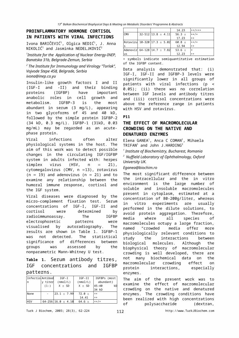
13th Balkan Biochemical Biophysical Days & Meeting on Metabolic Disorders’ Programme & Abstracts
of these antioxidant compounds were significantly lower but the percentage of reduced ascorbate was maintained high. In chloroplasts, the activity of superoxide dismutase diminished during the reversible stage of senescence, but some increase was observed later, probably reflecting the disappearance of the major chloroplastic protein Rubisco and changes in the chloroplastic protein pattern. The activity of the stromal isoform of ascorbate peroxidase declined on days 4-5th in darkness. Data concerning antioxidant compounds revealed some impairment of the ascorbate and glutathione pools in chloroplasts. The percentage of reduced ascorbate was maintained high in the chloroplasts without significant difference from the controls. Taken together, the results do not support development of oxidative stress and oxidative modification of Rubisco as a triggering mechanism for selective proteolysis in dark-induced senescence.
P50
THE EFFECT OF BUPIVACAINE ON COMPOUND ACTION POTENTIAL OF FROG SCIATIC NERVE FIBERSNizamettin DALKILIÇ1 Hülagü BARIŞKANER2 Barkın İLHAN1 İlhami DEMİREL1 Necdet DOĞAN2 1Selcuk University Meram Medical Faculty, Biophysics Department, 42080, Konya, Turkey2Selcuk University Meram Medical Faculty, Pharmacology Department, Konya, Turkey
Local anaesthetics block the initiation and propagation of the action potential by preventing the voltage-dependent increase in Na+ conductance. Bupivacaine is an amide-type local anaesthetic used for surgical, obstetric, acute and chronic pain therapy. Since a nerve is composed of many axons having different radii bound together, the change in the potential recorded extracellularly is merely an algebraic sum of fibers’ individual action potential waveforms dispersed in time, and is called the compound action potential (CAP). In this study, the effects of the local anaesthetic agent bupivacaine on individual fibers of a peripheral nerve have been documented. To accomplish this objective, CAPs were recorded from isolated frog sciatic nerves treated with bupivacaine in seven individual cases involving seven individual concentration levels. Fast Fourier Transform (FFT) and other numerical analysis involving CAP areas, latency periods, maximum and minimum derivatives were performed on these data. The results show that the area and absolute values of maximum and minimum derivatives decrease linearly as bupivacaine concentration increases. The power spectrum of CAPs, which resides in the 0 Hz-1 KHz interval, initially shifts to higher frequencies, then appears to be returning to lower frequency region again, with increasing bupivacaine concentration. Due to this result, it is thought that bupivacaine inhibits nerve fibers in a dose-dependent manner. It primarily affects the fibers having the least
myelinated sheets (motor fibers), then it begins to depress the fast conducting (neurosensorial) fibers as the bupivacain concentration increase, and finally blocks the unmyelinated C-fibers.
P51
CONDUCTION VELOCITY DISTRIBUTION IN NORMAL HUMAN PERONEAL MOTOR NERVENizamettin Dalkılıç1 Figen bayramoğlu2 İlhami Demİrel1
1 Selcuk University Meram Medical Faculty Biophysics Departmrnt, 42080, Konya TURKEY2 Selcuk University Meram Medical Faculty Neurology Departmrnt, 42080, Konya [email protected] of the practical methods used to obtain relative number of nerve fibers is the computer assisted collision method, in which a distal supramaximal stimulus (S1) is combined with a delayed proximal stimulus (S2). When the delay time between two stimuli (Inter Stimulus Interval; ISI) is relatively short, the proximally evoked orthodromic nerve action potential is cancelled by the antidromic impulse coming from the distal stimulus due to collision, and only an early action potential is observed at the recording site. By sequentially increasing ISI, an instant is reached at which the distally evoked antidromic impulse would have passed the proximal site before the proximal stimulus is delivered. Provided that the nerve fiber has recovered from the associated refractoriness, an orthodromic impulse would be initiated in response of the proximal stimulus, which in turn would evoke an additional late action potential. When the whole nerve is considered, cumulative activation of fibers can be measured by recording the CAP, as ISI is being gradually incremented. In this study, motor conduction velocity range associated with peroneal nerve is examined using collision method on 17 normal subjects. Paired supramaximal stimuli with ISI intervals of 6.4 to 20.0 ms were applied at distal and proximal points on peroneal nerve and resultant compound action potentials (CAPs) were recorded. The change in CAP amplitudes and areas with ISI were deduced, and using these data the relative number of fibers corresponding to each conduction velocity group (CVG) were computed. Conduction velocities of the peroneal motor nerve groups belonging to nerves innervating the Extensor Digitorum Brevis muscle were found in the range of 30-50 m/s and CVG innervating the greatest number appears to range between 40-48 m/s which consist of %70 of all fibers. These results show that, peroneal nerve conduction velocity groups consist relatively of more slow conducting fibers compared with median motor nerve.
P52
APPLICATION OF INHALATED PHOSPHOLIPID LIPOSOMES IN HCL – LUNG INJURY
Turk J Biochem, 2003; 28(3), 62-224 http://www.TurkJBiochem.com112
13th Balkan Biochemical Biophysical Days & Meeting on Metabolic Disorders’ Programme & Abstracts
Jordanka STENEVA, 1 Albena. JORDANOVA 3, Zdravko LALCHEV, 2, Samuil. NINIO 3,Tania NEICHEVA, 3 and Diana PETKOVA, 3*.
Anestisiology and Intensive Care, University Hospital Queen Giovanna , Sofia, Bulgaria ; 2 Biochemistry, Sofia University St. Kliment Ohridski , Sofia, Bulgaria and 3 Lipid-protein Interactions, Institute of Biohpysics, Bulgarian Academy of Sciences, 1113 Sofia, Bulgaria The aim of this study is to evaluate the application of phosphatidylcholine liposomes (PL) in HCl - induced ARDS in rabbits. Acute respiratory distress syndrome (ARDS) was induced by administration of 0.2 N HCl via intratracheal instillation for 45 min. After induced ARDS animals under artificial lung ventilation were retreated with PL for 60 min. Arterial blood gas analysis was performed at 15, 30, 45 and 60 min after PL application. Untreated animals were ventilated for the same time. Rabbits were killed with thiopental and bronhoalveolar lavage fluid (BALF) was investigated for lipid and specific surfactant protein content. The equilibrium surface tension and dymanic surface tension characteristics of monolayers obtained from BALF was determined by Wilhelmy balance. HCl- lung injury caused decrease of PaO2/FiO2 (arterial oxygen pressure/ fraction of The aim of this study was to evaluate the inhalatory application of inspired oxygen) ratio more than 50% compared to the control. We obtained high respiratory acidosis - increase of PaCO2 ( arterial pressure of CO2) and decrease of blood pH. An increase of A-a pO2 ( oxygen gradient) was also detected. The inhalation of PL led to reversion of gas exchange even at 30 min after application. Blood pH at 60 min after administration returned to the control value. HCl- lung injury caused significantly increase of total protein and cholesterol content, decrease of total phospholipids and percent participation of phosphatidylcholine and increase of that of sphingomyeline in BALF compared to the control. These altrations correlated with biophysical parameters. The sample surface tension was decreased. The hysteresis area and dynamic characteristics were also changed. The application of PL led to reverse of the biochemical and biophysical parameters to the control value.P53
ARGINASE AND ORNITHINE LEVELS IN BREAST CYST FLUIDHakan ERBAŞ, Aynur DAĞLAR, Mehmet Emin İRFANOĞLU1, Şendoğan GÜLENTrakya Üniversity, Faculty of Medicine, Department of Biochemistry, 1Surgery, 22030, Edirne/[email protected] cystic breast disease is the most common benign breast disease. Several recent studies have shown that women with palpable breast cyst may have a higher risk of developing breast cancer. There are two groups of breast cyst; lined either by apocrine epithelium (intracystic Na/K < 3) or flattened epithelium (intracystic Na/K > 3). The former group has been shown to be associated with a higher risk of breast cancer than the latter group. Arginase, a cytoplasmic enzyme, catalyses the hydrolysis of L-
arginine to urea and ornithine in the last step of mammalian urea cycle. Arginase also play an important role in the synthesis of polyamines through ornithine. Polyamines were shown to be a cell promoter and their levels have been found to be higher in malignant lesions. In addition, arginase activities were found to be higher in several carcinomas including breast, colorectal, stomach and prostate. Therefore, presence of arginase and ornithine in breast cyst fluid may help to develop breast carcinoma from the gross cystic disease of breast. In this study, we have determined the arginase enzyme activities and ornithine levels in breast cyst fluid samples. Arginase and ornithine levels were measured spectrophotometrically using thiosemicarbazide diacetylmonoksime urea and Chinard methods respectively. There was no istatistically significant difference on the arginase activities between apocrine (Range = 0.03 to 1.8 U/mg protein, m = 0.6 U/mg protein, n = 6) and flattened (Range = 0.21 to 1.9 U/mg protein, m = 0.85 U/mg protein, n = 5) cyst groups. On the other hand, ornithine levels were significanty higher (p = 0.002) in apocrine (Range = 0.023 to 0.056 mol/mg protein, m = 0.037 mol /mg protein, n = 6) than flattened (Range = 0.006 to 0.022 mol/mg protein, m = 0.013 mol/mg protein, n = 5) groups of breast cyst. As a conclusion, although we could not find any significant difference between arginase activities in the two groups may be due to small sample size, ornithine levels were significantly higher in the apocrine group which is suggested to have higher risk of developing breast carcinoma. These findings may indicate that arginase and ornithine may play a possible role on the development of breast cancer from the gross cystic disease of breast.
P54
CHRONIC EXPOSURE TO 50 Hz MAGNETIC FIELD DOES NOT AFFECT LIPID PEROXIDATION, SPERM COUNT, p53 IMMUNE REACTIVITY AND HISTOLOGY OF SOME ORGANS IN MALE SPRAGUE-DAWLEY RATSM. Zulkuf AKDAG1, Suleyman DASDAG1, Feyzan AKSEN1, Birgul ISIK2 Fahri YILMAZ3.Departments of Biophysics1, Biochemistry2 and Pathology3 Medical Faculty of Dicle University, 21280, Diyarbakir, [email protected] and magnetic fields (EMF) are a fact of daily life: they are emitted by power lines, transformers, service wires and electrical panels as well as by home appliances (such as electric blankets, clocks, shavers, and television). Electricity has been used, to greate advantage, for 100 years without society being aware of any adverse health effect, other than thermal injury and electrocution. Despite a multitude of studies, there remains considerable debate over what, if any, health effects result from exposure to EMF. There is still no clear answer to the question, "Can exposure to electric and magnetic fields resulting from the production, distribution, and use of electricity promote cancer or initiate other health problems?".This study evaluated the possible effect of a sinusoidal 50 Hz
Turk J Biochem, 2003; 28(3), 62-224 http://www.TurkJBiochem.com113
13th Balkan Biochemical Biophysical Days & Meeting on Metabolic Disorders’ Programme & Abstracts
magnetic field (1.35 mT) on the sperm count, testes, liver, kidney and brain histopathology, malondialdehyde (MDA) concentration of the tissue under investigation, p53 immune reactivity of bone marrow and some trace elements in blood of rat. Sixteen Sprague-Dawley male rats were separated into two groups of eight, sham exposed (control) and experimental. The rats in the experimental groups were exposed to Extremely Low Frequency Magnetic Field (ELF MF) 2 hr/day/ for 2 months (7 days a week). Eight rats of sham group were treated like experimental group except ELF MF exposure. The Mann-Whitney U-test was used for statistical comparisons of groups. No statistically significant alteration in any endpoints was noted except Mn+2, concentrations (p<0.001). This study found no evidence suggesting an adverse effect of ELF MF on measurement of MDA concentrations, histology of some tissues mentioned in this study, p53 immune reactivity of bone marrow and serum concentrations of Fe+3, Zn+2, and Cu+2.
P55
TAXONOMIC AND PHYLOGENETIC ANALYSIS OF TURKISH FRITILLARIA L. SPECIESMehtap Tekşen, Ayten Çelebi, Leyla AçıkThe genus, Fritillaria consist of 43 taxa, which is 36 species and 7 subspecies, in Turkey. 25 taxa are endemic to Turkey. The ratio of endemism is 58%. The genus contains many economically and medicinally important species. Some of them commonly used as medicine for diabetes, asthma, bronchitis and hearth diseases. Some of them are being cultivated but interest in this genus is very much on rise. Although some of the taxa in Turkey are defined based on morphological differences, many ambiguities are still remain. Because of the differeences in the genetic structure of the species, protein electrophoresis can be used to distinguish intra/inter species relationships. In this study, seed proteins of 29 taxon was investigated in order to distinguish inter species level. Seed protein analysis clearly diffrentiated 29 taxon but some of the results did not match with the sistematic results in Flora of Turkey. Key words: Fritillaria, Seed Proteins, PhylogenyP56
MUTATIONAL ANALYSES OF RECOMBINANT GLOBULAR HEAD REGIONS A- AND C- IN C1Q-IMMUNOGLOBULIN INTERACTIONSAlexandra. ZLATAROVA1, Michaela GADJEVA2, Ivanka. TSACHEVA1, Ljubka. ROUMENINA1, Uday KISHORE 3, 4 , Ken. B. M. REID3 and Michaela KOJOUHAROVA1
1Dept. Biochemistry, Sofia University, Bulgaria, 2Dept. of Medical Microbiology and Immunology, University of Aarhus, Denmark; 3MRC Immunochemistry Unit and 4Weatherall Institute of Molecular Medicine, University of Oxford, [email protected]
The interaction of the globular C1q heads with their ligands- aggregated IgG and IgM triggers the classical complement activation. Each globular head (gC1q) is composed of the C-terminal halves of one A- (ghA), one B- (ghB) and one C- (ghC) chain. Recent evidence suggests that the gC1q region has a modular organisation and is composed of three, structurally and functionally, independent modules which retain multivalency in the form of a heterotrimer. In the present study we have examined the contribution of several charged amino-acid residues, which are supposed to play an important role in the recognition of the immunoglobulins. The chosen residues were Arg162 and Arg156 of the A- and C-chain respectively. For reaching this goal five single-residue mutants were generated (RghA162A, RghA162E, RghC156A, RghC156E and RghC156Q) and expressed in Escherichia coli as soluble fusion proteins linked to maltose-binding protein. The abilities of the mutants to bind IgG and IgM were assessed by direct and competitive ELISA. pH dependence of IgG binding of ghA and ghC (wild types and mutants) were tested as well. The obtained results indicated that: i) the positive charge of the amino-acid residues at these positions is necessary; ii) the selected amino-acid residues contribute with up to 25% of the immunoglobulin-binding activity of wild types globular head regions; iii) a hydrophobic component in the process of interaction between ghA chain of C1q and IgG was observed.
P57
SEED PROTEIN ANALYSIS OF TURKISH VERBASCUM L. GENUS (GROUP A)Karavelioğulları F.A., Çelebi A., Açık L.,The genus Verbascum L. (Scrophulariaceae) includes about 934 species on Earth. It is also the largest second genus in Turkey, where it is represented by 233 species in 13 groups, and 126 hybrids. Partly artifical groups are used in the Flora of Turkey account, all Turkish species of Verbascum within Murbeck‘s Sect. belong to Bothrosperma Murb. A, B, C, groups in the Flora of Turkey belong to Subsect. Ebracteolate Murb. 186 species (80%) of these 233 species are endemic in Turkey. Verbascum is represented by the highest number of species in the West and Central regions of Turkey. The plants are adapted especially to steppe environment, open places and stony slopes. In this study, seed protein profiles of 21 taxa were examined. Morphological data supported with protein analysis were clarified the taxonomic status of these taxa.Key Words: Verbascum, protein profiles, taxonomy
P58
STUDY OF HYPERHOMOCYSTEINEMIA AND CAD IN BULGARIAN POPULATIONTzontcheva A., Ganev V., Horvath A.Medical University of Sofia, Chair of Clinical Laboratory and Clinical Immunology, Chair of chemistry and biochemistry, G.Sofijski str. 1, Sofia 1431, Bulgaria
Turk J Biochem, 2003; 28(3), 62-224 http://www.TurkJBiochem.com114
13th Balkan Biochemical Biophysical Days & Meeting on Metabolic Disorders’ Programme & Abstracts
There is growing evidences that Homocysteine (Hcy) is an independent risk factor for coronary artery diseases (CAD) and a sensitive marker of Cobalamine and Folate function. In this study total Homocysteine (tHcy), Folate (Fol), Vit. B12, total cholesterol, triglycerides and HDL-C were measured in 89 patients (age 35-65), survived myocardial infarction (MI), and in 70 age and sex matched control subjects. Additionally we have genotyped them for C677T MTHFR polymorphism. Serum tHcy level was determined using automated Abbott IMx fluorescence polarization assay. Fol and B12 were determined using Chemiluminescence ACS:180 assays. We find significantly higher levels of Hcy in patients, compared to controls (18.008.76 mol/L vs. 13.825.94 mol/L , p=0.001). The Fol levels were lower in patients than in controls (13.908.56 nmol/L vs. 21.6411.53 nmol/L, p<0.001). Vit. B12 levels were also lower in patients (96,2796.5 pmol/L vs. 312.8 131.9 pmol/L, p=0.008). Patients were more frequently carriers of T/T genotype, compared to controls (16 vs. 3, p=0.023). After log-transformation of values, the bivariant correlation analysis was performed to estimate the strength of association between Hcy and Fol in different groups. Statistically significant negative correlation (r=-0.359, p=0.001) was find in patients survived MI, but not in control subjects (r=-0.121, p=0.345). The same results were obtained when patients and controls were divided by age. Our results suggest that high Hcy and low Fol and vit.B12, more frequently found in patients with MI than in controls probably contribute to high cardiovascular disease risk in these patients. C677T MTHFR genotype can in part, but not fully explain high Hcy/low Fol levels.
P59
ARGINASE AND ORNITHINE IN PATIENTS WITH BENIGNANT AND MALIGNANT SKIN TUMORSSelma SÜER GÖKMEN1, A.Cemal AYGIT2, Reyhan YILDIZ1, Beyhan ÇAKIR2, Şendoğan GÜLEN1
Departments of Biochemistry1 and Plastic and Reconstructive Surgery2, Trakya University, School of Medicine, Edirne/[email protected] extrahepatic mammalian tissues, arginase is believed to supply the cell with ornithine, a precursor for biosynthesis of the polyamines. Arginase activity and ornithine level have been shown to be elevated during carcinogenesis. The aim of this study is to determine arginase activity and ornithine level in benignant and malignant skin tumors and, to evaluate whether they can be used as biological markers for distinguishing patients with benignant skin tumors from those with malignant skin tumors. Arginase activity and ornithine level were determined by the method of Geyer and Dabich, and that of Chinard, respectively. The protein content of the tissues was determined by the method of Lowry. One unit of arginase was defined as the amount of enzyme that released 1µmol of urea for 1 minute at 37 0C. The Wilcoxon two-sample test and Student’s t test were used to analyze the results. The mean arginase activity and ornithine levels in benignant tumor tissues were
10.45±3.77 U/mg protein (n=26) and 28.32±16.95 nmol/mg protein (n=27), respectively, versus 4.81±2.64 U/mg protein (n=14) and 14.09±6.66 nmol/mg protein (n=15), respectively, for normal adjacent tissues. The mean arginase activity and ornithine levels in malignant tumor tissues were 16.52±11.02 U/mg protein (n=19) and 35.04±18.21 nmol/mg protein (n=18), respectively, versus 4.87±2.75 U/mg protein (n=17) and 14.27±7.19 nmol/mg protein (n=17), respectively, for normal adjacent tissues. Arginase activity and ornithine level in benignant skin tumors (p<0.05 for arginase and p<0.01 for ornithine) and in malignant skin tumors (p<0.01 for both of them) were found to be higher than those found in adjacent normal tissues.There was also a significant difference between arginase activities of benignant and malignant skin tumors (p<0.05). As a result, we may report that, although arginase activity and ornithine levels are increased in benignant and malignant tumors of the human skin, only arginase activity may be useful for distinguishing patients with malignant skin tumors from those with benignant skin tumors.
P60
ERYTHROCYTE ARGINASE ACTIVITY IN PATIENTS WITH ACUTE MYOCARDIAL INFARCTIONSelma SÜER GÖKMEN 1, Reyhan YILDIZ 1, Fatih ÖZÇELİK 2, Zihni AKTAŞ 2, Şendoğan GÜLEN 1
1Biochemistry and 2Cardiology Departments, Trakya University, School of Medicine, Edirne, [email protected] Arginase, the enzyme catalyzing the hydrolysis of arginine to urea and ornithine, is mostly found in the liver. In extrahepatic tissues, arginase is believed to supply the cell with ornithine, a precursor of the polyamines. It has been reported that polyamines are one of the intracellular factors that contribute to isoproterenol-mediated cardiac injury in the rat. It has been suggested that determination of arginase activity in serum may serve as a useful test in early differential diagnosis of myocardial infarction. A crucial point for the clinical utility of a marker is the time from the first investigation of the patient and blood sampling until the time a result is available which is then used for clinical and therapeutic decision making by the phyician. In this setting, we think that determination of arginase activity in erythrocyte instead of serum will offer a more rapid results. The purpose of the present study is to investigate erythrocyte arginase activity in patients with acute myocardial infarction and to evaluate whether it can be used as a biological marker for diagnosis of myocardial infarction. In this study, 58 patients (age 55.46±9.63 years) and 37 healthy volunteers( age 52.24±8.71 years) were included. Arginase activity was determined by the method of Geyer and Dabich. Student’s t-test was used to analyze the results. One unit of arginase was defined as the amount of enzyme that released 1µmol of urea for 1 minute at 37 0C. Arginase activity was found to be 67.17±20.89 U/g hemoglobin in patient group and 51.51±11.36 U/g hemoglobin in control group. Erythrocyte arginase activity at 24h post-infarction in patients with acute myocardial infarction was significantly higher than those found in
Turk J Biochem, 2003; 28(3), 62-224 http://www.TurkJBiochem.com115
13th Balkan Biochemical Biophysical Days & Meeting on Metabolic Disorders’ Programme & Abstracts
control group (p<0.001). In conclusion, we may report that there is a significant increase in erythrocyte arginase activity and determination of erythrocyte arginase activity may offer a more rapid result and may be used as a biological marker for diagnosis of patients with acute myocardial infarction.
P61
SERUM TOTAL SIALIC ACID IN PATIENTS WITH BENIGNANT AND MALIGNANT SKIN TUMORSSelma SÜER GÖKMEN1, Cemal KAZEZOĞLU1, Bendigar SUNAR1, A. Cemal AYGIT2, Beyhan ÇAKIR2
Departments of Biochemistry1 and Plastic and Reconstructive Surgery2, Trakya University, School of Medicine, Edirne/[email protected] acids, a group of acylated neuraminic acids, are widely distributed in nature as terminal sugars on oligosaccharides attached to protein or lipid moieties. They impart a net negative charge to cell surface and are important in cell-to-cell or cell-to-matrix interactions. Alterations in the metabolism of sialic acid in the presence of malignancy have been reported by some investigators. The aim of the present study is to investigate whether serum total sialic acid can be used as a tumor marker for distinguishing patients with benignant or malignant skin tumors from each other and from healthy subjects. In this study, 36 patients with benignant skin tumors (17 men, age 46.28±16.90 years), 23 patients with malignant skin tumors (15 men, age 49.61±12.60 years) and 36 healthy volunteers (19 men, age 48.47±8.65) were included. Serum total sialic acid determination was performed by the thiobarbituric acid method described by Warren. We compared differences between two patient groups and, between patient and control groups using Student’s t-test.The mean serum total sialic acid levels were found to be 62.30±11.80 mg/dl in patients with benignant skin tumors, 68.31±11.27 mg/dl in patients with malignant skin tumors and 51.40±4.26 mg/dl in control group. There was a significant difference between serum total sialic acid levels of control group and patients with benignant or malignant skin tumors (p<0.001 for both of them). Serum total sialic acid levels of patients with benignant skin tumors was not different from those with malignant skin tumors. In conclusion, we may report that the measurement of serum total sialic acid levels may be useful for distinguishing patients with benignant or malignant skin tumors from healthy subjects, but it cannot be used as a tumor marker for distinguishing patients with benignant skin tumors from those with malignant skin tumors.
P62
SERUM CERULOPLASMIN AND SIALIC ACID LEVELS IN ACUTE MYOCARDIAL INFARCTIONÖzgül GÜNGÖR1, Bendigar SUNAR1, Fatih ÖZÇELİK2, Zihni AKTAŞ2, Selma SÜER GÖKMEN1
Biochemistry1 and Cardiology2 Department, Trakya University, School of Medicine, 22030, Edirne/[email protected] acid concentration is increased after myocardial infarction but the reason for this elevation remains obscure. An increased output of acute phase proteins has been reported to be responsible for an elevation in serum total sialic acid (TSA) concentration.The aim of the present study is to investigate serum TSA and ceruloplasmin levels at 24h post-infarction and to evaluate the role of ceruloplasmin, which contains sialic acid residues, in the elevation of sialic acid concentration in myocardial infarction. In this study, 45 patients (male 33) with myocardial infarction and 45 healthy volunteers (male 26) ranging in age from 40 to 70 were included. Serum TSA determination was carried out by the thiobarbituric acid method of Warren and serum aspartate aminotransferase, lactate dehydrogenase and creatine kinase-MB activities and ceruloplasmin levels were measured with an automatic biochemistry analyzer. Student’s t test and Pearson’s correlation test were used to analyze the results. The mean activity of the enzymes in patients group were higher than those found in control group (p<0.001 for all). Serum TSA and ceruloplasmin concentrations were found to be 66.47 ± 9.08 mg/dl and 66.69 ± 8.12 mg/dl in patients , and 53.81 ± 5.74 mg/dl and 30.35 ± 8.20 mg/dl in control group, respectively. Serum TSA (p<0.001) and ceruloplasmin (p<0.001) levels in patients were significantly higher than control group. Patient and control groups were also divided into two age groups: 40-54 years and 55-70 years. Serum TSA and ceruloplasmin levels in 40-54 years and 55-70 years of patients with myocardial infarction were significantly higher when compared with those found in control group (p<0.001 for all). Furthermore, there was a significant difference between the levels of ceruloplasmin in 40-54 years and 55-70 years of control group (p<0.001).There was no correlation between serum TSA and ceruloplasmin levels in patients (r=0.038, p>0.05) and in control group (r= -0.272, p>0.05). As a result, we may report that serum TSA and ceruloplasmin levels are elevated at 24h post-infarction in patients with acute myocardial infarction and an increased output of ceruloplasmin from the liver cannot be only factor responsible for an increased serum TSA concentration following myocardial infarction.P63
INSULIN-ACTIVATED GPI-TRANSAMIDASE IN HUMAN ERYTHROCYTESDragana STANİĆ1, Milan NİKOLİĆ2 and Vesna NİKETİĆ2
1 ICTM - Center for Chemistry, 11001, Belgrade/ SERBİA AND MONTENEGRO2 Department of Chemistry, University of Belgrade, 11001, Belgrade/ SERBİA AND [email protected] target cells insulin stimulate specific membrane protease and phospholipase which hydrolyse GPI (glycoinositolphospholipid) - proteins and GPI-lipids, which result in release of secondary messengers of insulin
Turk J Biochem, 2003; 28(3), 62-224 http://www.TurkJBiochem.com116
13th Balkan Biochemical Biophysical Days & Meeting on Metabolic Disorders’ Programme & Abstracts
action. We have demonstrated that in human erythrocytes prolonged hyperinsulinism, both in vivo and in vitro, caused an opposite process: covalent GPI binding to the C termini of both hemoglobin (Hb) -chains, which resulted in the formation of novel hitherto unrecognized minor hemoglobin fraction (GPI-Hb) (Niketić et al., Biochem. Biophys. Res. Commun. 239 (1997) 435). Here we demonstrate that exposure of erythrocyte membranes to insulin cause the activation of membrane protease, as well as that the formation of GPI-Hb parallels its activity. This, together with recent findings regarding biochemical pathway of GPI-protein biosynthesis suggest that the insulin-activated protease is GPI-transpeptidase which is able to catalyze, albeit slowly, the transpeptidation, i.e., the replacement of the carboxy-terminal amino acid(s) residues of Hb β-chains with GPI as an exogenous nucleophile. Using specific substrates and inhibitors, we found that this enzyme has cathepsine B-like specificity. Specific extraction procedures have shown that insulin-activated protease exists in raft membrane microdomains, which contain GPI-lipids and insulin receptors. To our knowledge the present results show for the first time that insulin-activate protease with GPI-transamidase activity and demonstrate that this enzyme may be involved in post-translational GPI binding to proteins. Results described in this work may bear relevance to studies of physiological disorders that are characterized by hyperinsulinism.
P64
THE EFFECTS OF NITRIC OXIDE AND PEROXYNITRITE ON MnSOD (E. Coli)Dragana STANİĆ1, Milan NİKOLİĆ2, Srđan STOJANOVİĆ2, Smiljana RAİČEVIĆ3 and Vesna NİKETİĆ1
1 ICTM-Center for Chemistry, 11001 Belgrade/ SERBİA AND MONTENEGRO2 Department of Chemistry, University of Belgrade, 11001 Belgrade/ SERBİA AND MONTENEGRO3 Center for food analysis, 11001 Belgrade/ SERBİA AND [email protected] oxide (NO) is ubiquitous biological messenger and cytotoxin. It is well established that some effects of NO are mediated by its reactive species instead. These include metal-generated nitrosonium (NO+) and nitroxyl (NO–) species and peroxynitrite (ONOO-), reaction product of NO and superoxide anion radical (O2
·–), of which each exhibit distinctive chemistry in biological milieu. Nitration of protein-tyrosine by ONOO- is considered to be involved in a number of pathomechanisms. We demonstrated recently that exposure of MnSOD (E. coli) to NO under anaerobic condition leads to the generation of both NO+
and NO– species, which causes inactivation and extensive structural alteration of the enzyme [1]. Surprisingly, MacMillan-Crow et al. found that ONOO-, but not NO, affect structure and activity of human recombinant MnSOD (hMnSOD) [2]. This prompted us to initiate the present study with aim to characterize in more detail effects of NO and ONOO- on the structure and activity of MnSOD (E. coli). Our results demonstrate that MnSOD (E.
coli)-stimulated generation of NO+ and NO– species is associated with nitration of enzyme tyrosine residues and dityrosine formation, which cause enzyme inactivation. This represents to our knowledge entirely knew mode of NO-mediated tyrosine nitration. Peroxynitrite treatment of MnSOD (E. coli) caused nitration of tyrosine residues and loss of activity, but not dityrosine formation. Considering high structural similarities of active centers of these two enzymes, observed differences are surprising. We assume that different reactivity of MnSOD (E. coli) toward NO and ONOO- comparing to that of hMnSOD may be (partly) explained by higher flexibility of its (dimeric) structure comparing to that of hMnSOD, which is tetramer.
P65
CHOLESTEROL BOUND TO HEMOGLOBİN İN NORMAL HUMAN ERYTHROCYTESMilan NİKOLİĆ1, Dragana STANİĆ2 and Vesna NİKETİĆ1
1 Department of Chemistry, University of Belgrade, 11001, Belgrade/ SERBİA AND MONTENEGRO2 ICTM - Center for Chemistry, 11001, Belgrade/ SERBİA AND [email protected] is generally accepted that lipids extracted from intact red blood cells (RBC) originate from red cell membrane. However, literatures values reported for the total cholesterol (Ch) and phospholipid (PL) content in normal human RBC varies greatly, which prompted us to conduct this study with aim to provide further data concerning the RBC lipid content. Fifty young healthy male subjects were screened twice in one year, at summer and winter time. Plasma and total RBC lipids, lipids from RBC membrane and hemolysates (supernatants from which membranes were carefully separated) were evaluated at each season and were compared. Our results demonstrate that in contrast to Ch and PL contents of RBC membrane which are confined to a narrow range, the lipid levels estimated in intact RBCs showed more variation: the lowest individual values for RBC lipids corresponded to those found in membrane, whereas in RBCs with higher lipid contents the “excess” was found in hemolysates. We found that “an excess” of cholesterol (associated with phospholipid) strongly binds to hemoglobin (Hb), yielding Hb-lipid adduct (Hb-Ch). Significantly higher levels of Hb-Ch in winter comparing to those in summer that parallel plasma cholesterol levels and positive correlation between %Hb-Ch and HDL-Ch levels, point to the direct influence of plasma lipoprotein metabolism on the formation of Hb-Ch. Our in vitro studies demonstrated that Hb-Ch could be formed upon incubation of (lipid-free) hemoglobin with cholesterol-phospholipid mixture as well as upon the exposure of RBCs to the excess of cholesterol-phospholipid dispersion with FCh/PL ≤ 1. It is tempting to speculate that Hb-Ch represents a new form of cholesterol in circulation, which contributes to the permanent removal of the “excess“ of unesterified cholesterol from circulation. This implies that RBCs may represent a part of the mechanisms involved in the first line “defense” against “an excess“ of free cholesterol (FCh) in circulation.
Turk J Biochem, 2003; 28(3), 62-224 http://www.TurkJBiochem.com117
13th Balkan Biochemical Biophysical Days & Meeting on Metabolic Disorders’ Programme & Abstracts
P66
INCREASING THE STABILITY of ALGINATE BEADS CROSSLINKED WITH 1,6-DIAMINOHEXANEBurcu OKUTUCU, Azmi TELEFONCUEge University, Faculty of Science, Biochemistry Department, Bornova-İzmir, [email protected] of the most common method for immobilization of proteins, enzymes, whole microbial, plant and animal cells are based on their entrapment in calcium-alginate gel beads. Alginate is a polysaccharide extracted from brown algae. Alginates are linear copolymers of -L-guluronate and -D-mannuranate. Their gelling properties derive from the cooperative binding of divalent cations localized between homopolymeric blocks of guluronate residues(termed G-blocks). Ca ions are located into electronegative cavities, like eggs in a egg-box, from this similitude arises the term egg-box model. The ionic interactions between guluronate blocks and Ca ions cause the formation of a strong termostable gel which properties largely depend on the characteristics of the polymer and the preparation method. The beads are made of to drop alginate solution to CaCl2 solution. However calcium alginate beads are very porous and present a low retention capacity of entrapped molecules. Polyelectrolite solutions like chitosan, polyethyleimine and polypropyleneimine can be used surface-coating materials by dropping alginate into them. Although chemical stability in some solutions increase but the mechanical stability and conformational changes may occur.To prevent these problems the cross-linking agents can be used for surface-covering of Ca-alginate beads. In this study we used 1,6-Diaminohexane(HDA) for this aim. The calcium alginate beads were activated by 1-ethyl-3-(3-dimethylaminopropyl) carbodiimide (EDC) and coupled with HDA to occur a surface covering. To test the beads, pH stability and release of Bovine Serum Albumine (BSA) from the beads under different conditions were investigated.P67
PHOTOINACTIVATION OF CATALASE BY ULTRAVIOLET RADIATIONMarina T. NECHIFOR, Diana DINUUniversity of Bucharest, Faculty of Biology, Splaiul Independentei 91-95, Bucharest/[email protected] aim of this study was to investigate the mechanism of catalase photoinactivation induced by ultraviolet radiation. Hence, the major questions regarding catalase photoinactivation may be formulated as follows: does the photoinactivation of cellular catalase proceed via the same pathways as photoinactivation of pure catalase? Does photoinactivation of pure catalase occur in the absence of cellular photosensitizers? To answer these questions, the effect of in vitro UVA (320-400 nm) irradiation on catalase
activity in cellular homogenates and pure catalase solutions, was studied.Samples were irradiated at various intervals of time and then assayed for enzymatic activity. Photoinactivation of purified liver catalase and of catalase from liver homogenate was found to be similar, and the kinetics of such photoinactivation obey first order processes. To evaluate a possible involvement of cellular photosensitizers in the photoinactivation mechanisms we determined the basic parameters, i.e. the photoinactivation constants and the half-life of catalase activity. The constant of photoinactivation was significantly higher, and the half-life of enzymatic activity was significantly lower for catalase in liver homogenate (ki = 0.42 h-1, t1/2 = 99.2 min) as compared to the purified catalase (ki = 0.0647 h-1, t1/2 = 630 min). These data show the enhancement of catalase photosensitivity in the presence of cellular components. A decrease of the heme Soret absorption peak at λ=405 nm was noticed, suggesting that photoinactivation is caused by the destruction of heme, as a consequence of direct absorbtion of light. Polyacrylamide gel electrophoresis analysis under denaturing conditions shows photooxidative changes in the apoprotein structure, i.e. formation of intersubunit cross-links. In conclusion, we have shown that in vitro UVA irradiation induces a decrease of enzymatic activity of catalase. Catalase from cellular homogenate is more photosensitive than purified catalase because of the presence of endogenous photosensitizers. The photoinactivation seems to involve the destruction of the porphyrin ring, but also the modification of apoprotein.
P68
CALF AORTIC AND TENDON COLLAGEN MODIFIED BY ADVANCED GLYCATIONAndreea Iren SERBAN-CAPATINA1, Eduard CONDAC2, Elena GANEA3
1University of Agricultural Science and Veterinary Medicine, Faculty of Veterinary Medicine, Bucharest, Romania²University of Bucharest, Faculty of Biology, Molecular Biology Center, Bucharest, Romania³Institute of Biochemistry, Bucharest, [email protected] diabetes and aging collagen is non-enzymatically modified by reducing sugars. The major initial product is a fructose-lysine compound resulted from the glycation of ε-amino groups. In subsequent Maillard reactions, products known as advanced glycation end products (AGEs) are formed. These AGE products include structurally characterized adducts such N- carboxymethyl lysine (CML), pentosidine and chemically unidentified compounds which induce protein binding, browning, fluorescence, and cross- linking. In the present work we detect and quantify the AGEs formed by glycation with glucose in calf tendon and aortic collagen by various assays. Collagen samples were extracted from calf aorta and tendon by delipidation followed by extensive pepsin digestion. Extracted collagen was incubated in 0.01M PBS,
Turk J Biochem, 2003; 28(3), 62-224 http://www.TurkJBiochem.com118
13th Balkan Biochemical Biophysical Days & Meeting on Metabolic Disorders’ Programme & Abstracts
pH 7.4 in the presence of 0.5 M glucose for 0, 2 and 4 weeks at 37° C. The free sugar was removed by dialysis and collagen samples were lyophilised. Collagen – linked fluorescence was measured at 370/460 nm and 335/385 nm. The relative fluorescence level in tissue glycated collagen was higher then the unglycated collagen. Fluorescence measured at 335/385 nm indicated that pentosidine level was three fold higher in glycated tendon collagen than in glycated aortic collagen. SDS-PAGE analysis showed the formation of high molecular weight compounds in aortic and tendon collagen after 2 and 4 weeks of glycation. The chromatographic pattern (FPLC, Superdex 200 column) of the aortic and tendon collagen showed, after 2 and 4 weeks of incubation with glucose the appearance of new peaks comparing with native collagen; these peaks correspond to higher molecular weight, namely 86.14 kDa for glycated aortic collagen and 130.17 kDa for glycated tendon collagen. The present study supports the hypothesis that sustained hyperglycaemia and aging can induce the formation of advanced glycation end compounds, which has deleterious consequences, such as structural modifications of the extracellular matrix.
P69
DO WHOLE-BODY EXPOSURE TO RADIATION EMITTED FROM MOBILE PHONES ACCUMULATE Bcl-2 IN BRAIN AND TESTES?Fahri Yilmaz1, Suleyman Dasdag2, Zulkuf Akdag2, Nihal Kilinc1
Dicle University, Faculty of Medicine, Patholgy1 and Biophysics2 Departments, 21280, Diyarbakır/[email protected] large family of genes that regulate apoptosis has been identified and these genes can be remembered as a series of three-letter words beginning with b. The first antiapoptotic gene identified, bcl-2 is a member of a large family of homodimerizing and heterodimerizing proteins, some of which inhibit apoptosis (such as bcl-2 itself and bcl-xL), whereas others (such as bax, bad, and bcl-xS) favor programmed cell death. Although the bcl-2 family of genes plays an important role in regulating apoptosis, at least two other cancer-associated genes are also intimately connected with apoptosis: the p53 gene and the protooncogene c-myc. Because of this importance of bcl-2 we investigated the accumulation of bcl-2 in rat brain and testes after whole-body exposure to radiation emitted from 900 MHz mobile phones.Sixteen Sprague-Dawley rats were separated into two groups of eight, one sham and one experimental. The rats were confined in Plexiglas cages (20 ´10.5 ´10 cm) with ventilation holes, and the cellular phones were placed 0.5 cm under cages. Exposure began approximately 10 minutes after transferring into the exposure cages, a period of time when rats settled down to a prone position and selected a fixed location inside the cage spontaneously. For the experimental group, the phones were in the speech condition for 20 minutes per day for 1 month. By speech condition, we mean that the phone is sending a tape of
human speech to the base station. The same phones were place under sham group rats, but the phones were turned off. Immunohistochemical staining of bcl-2 was performed according to the standardized avidin-biotin complex method.The results of this study showed that no bcl-2 accumulation was observed in the brain and testes of rats exposed to the radiation emitted from mobile phones. Finally, we emphasize that there is not any adverse effect of radiation emitted from 900 MHz mobile phones in terms of the first antiapoptotic gene, which is identified bcl-2.
P70
INTERACTION OF NUCLEAR PROTEINS WITH HAPTOGLOBIN HORMONE RESPONSIVE ELEMENT IN RAT LIVERSvetlana DINIĆ, Mirjana MIHAILOVIĆ, Desanka BOGOJEVIĆ, Svetlana IVANOVIĆ-MATIĆ, Goran POZNANOVIĆInstitute for Biological Research, Department of Molecular Biology, 29. Novembra 142, 11000 Belgrade, Serbia & Montenegro, [email protected] (Hp) is a plasma acute phase (AP) glycoprotein with the prominent role in the binding and clearance of hemoglobin. Though it was classified as a gene expressed in the liver only after birth, its expression is initiated during embryogenesis. Hp gene expression is primarily controled at the transcriptional level, depending on interactions between specific cis-acting DNA sequences and hepatocyte-enriched trans-acting regulatory factors, such as C/EBP proteins. It also appears that interactions between nuclear matrix proteins and specific DNA sequences of AP protein genes are involved in modulation of their expression. Therefore, factors controling transcriptional regulation of the Hp gene during rat liver development were assessed by the binding afinity of nuclear matrix and nuclear extract proteins to the Hp gene hormone responsive cis-element (-165/-56). South-Western analysis revealed DNA binding affinity of a common set of proteins in the 35-29 kD region in both nuclear fractions isolated from embryonal (19-days old) and postnatal (1, 3, 7, 14 and 21 day old) rat livers. Using Immuno-Western analysis, 35 kD protein was identified as an isoform of C/EBP protein in both nuclear fractions. While in the nuclear matrix fraction C/EBP was present throughout development, in the nuclear extract it was detected from day 14 of postnatal development. Our results lend further support to the assertion that the nuclear matrix takes active part in transcriptional regulation of gene expression during differentiation.
P71
EFFECT OF POLLEN AND PROPOLIS EXTRACTS ON ELASTASE SECRETION FROM KML-62 CELL LINESOrhan DEĞER1,Meltem ÇOLAK1,Yaşam BARLAK1,Yavuz TEKELİOĞLU2,Fahri UÇAR3
Turk J Biochem, 2003; 28(3), 62-224 http://www.TurkJBiochem.com119
13th Balkan Biochemical Biophysical Days & Meeting on Metabolic Disorders’ Programme & Abstracts
Departments of Biochemistry1, Histology2,and Medical Biology3, Faculty of Medicine,KTU,61080, Trabzon/[email protected] polen and propolis are apicultural products which are composed of nutritionally valuable substances and contain considerable amounts of polyphenol substances which may act as potent antioxidants. Elastase is primarily located in the azurophil granules and is an active component of the phagocytic system of neutrophils.We wanted to show if elastase secretion from KML-62 cancer cell lines could be influenced when incubated with pollen and propolis extracts or not.Pollen and propolis extracts at concentrations of 50,25,12.5 and 0 mg/ml were prepared by dimethyl sulfoxide. KML-62 cell cultures and lymphocyte cultures by preparing peripheral blood as control cells were incubated with extracts for 24 h. Elastase secretion was determined by CellProbe reagent (RGES elastase, Beckman Coulter) by using flow-cytometric fluorescence analysis. While about 85% fluorescence positivity was obtained with 0 concentrations for both KML-62 and lymphocyte cell cultures, fluorescence positivity decreased (between 1.7 and 6.9%) as concentrations of both propolis and polen extracts increased for KML-62 cell culture, but unchanged (between 55 and 76%) for lymphocyte cell culture. It was concluded that pollen and propolis extracts inhibit elastese secretion from cancer cell lines probably by their antioxidant potentials.
P72APPLICATION OF INHALATED PHOSPHOLIPID LIPOSOMES IN HCL – LUNG INJURYJordanka STENEVA, 1 Albena. JORDANOVA 3, Zdravko LALCHEV, 2, Samuil. NINIO 3,Tania NEICHEVA, 3 and Diana PETKOVA, 3*.
Anestisiology and Intensive Care, University Hospital Queen Giovanna , Sofia, Bulgaria ; 2 Biochemistry, Sofia University St. Kliment Ohridski , Sofia, Bulgaria and 3 Lipid-protein Interactions, Institute of Biohpysics, Bulgarian Academy of Sciences, 1113 Sofia, Bulgaria The aim of this study is to evaluate the application of phosphatidylcholine liposomes (PL) in HCl - induced ARDS in rabbits. Acute respiratory distress syndrome (ARDS) was induced by administration of 0.2 N HCl via intratracheal instillation for 45 min. After induced ARDS animals under artificial lung ventilation were retreated with PL for 60 min. Arterial blood gas analysis was performed at 15, 30, 45 and 60 min after PL application. Untreated animals were ventilated for the same time. Rabbits were killed with thiopental and bronhoalveolar lavage fluid (BALF) was investigated for lipid and specific surfactant protein content. The equilibrium surface tension and dymanic surface tension characteristics of monolayers obtained from BALF was determined by Wilhelmy balance. HCl- lung injury caused decrease of PaO2/FiO2 (arterial oxygen pressure/ fraction of of inspired oxygen) ratio more than 50% compared to the control. We obtained high respiratory acidosis - increase of PaCO2 ( arterial pressure
of CO2) and decrease of blood pH. An increase of A-a pO2
(oxygen gradient) was also detected. The inhalation of PL led to reversion of gas exchange even at 30 min after application. Blood pH at 60 min after administration returned to the control value. HCl- lung injury caused significantly increase of total protein and cholesterol content, decrease of total phospholipids and percent participation of phosphatidylcholine and increase of that of sphingomyeline in BALF compared to the control. These alterations correlated with biophysical parameters. The sample surface tension was decreased. The hysteresis area and dynamic characteristics were also changed. The application of PL led to reverse of the biochemical and biophysical parameters to the control value.
P73
PEROXIDASE ISOENZYMES PATTERN IN NIGELLA DAMASCENA IN VIVO CULTURE UNDER CONDITIONS SIMILAR TO EXTRATERRESTRIAL ENVIRONMENTGabriela MARINESCU, Cristina BABEANU, Elena GLODEANU, Georgeta CIOBANUUniversity of Craiova, Faculty of Chemistry, Department of Biochemistry, 1100 Craiova, [email protected] of low magnetic field effects on biological systems have drawn attention of space biologists due to the planning of long-term space flights to other planets. Yet, data are gradually accumulating and pointing to the influence of low magnetic field at different levels of living systems organization. The purpose of this paper is to improve the general knowledge of the response mechanism of living systems to low magnetic field in the presence of other physical stimuli such as screened geoelectric field, negative thermal shock and ferrofluid presence applied as modulators of peroxidase system. In this study we exposed Nigella damascena seeds to an extremely low magnetic field (200 nT) for short periods (1 to 60 min) and to a conjugated action of two or even three stimuli such as: a) 200 nT and –196°C; b) screened geoelectric field and –196°C; c) 200 nT, -196°C and ferrofluid.Seedlings growth has been observed over a 35-day period. Aerial part of seedlings has been assayed for isoperoxidases pattern and activity as well as for chlorophylls and carotene levels determination. Measured parameters were germination grade, plantlets’ viability, and growth rate.The peroxidase cationic and anionic components have shown different sensibilities to low magnetic field and other investigated factors and a correlation with the exposure time has been found. Meanwhile low-temperature shock, geoelectric field cancellation and ferrofluid treatment have induced a net effect on the individual isoforms determining a clear variation in the cationic: anionic distribution, the effect of the very low magnetic field has been manifested especially by attenuating large variations produced by the other factors. Positive
Turk J Biochem, 2003; 28(3), 62-224 http://www.TurkJBiochem.com120
13th Balkan Biochemical Biophysical Days & Meeting on Metabolic Disorders’ Programme & Abstracts
correlation has also been found between foliar organogenesis and the anionic component and between growth rate and the cationic component. Significant alterations in chlorophylls and carotene level accompanied these changes in the isoperoxidases’ pattern and activity.
P74
PROLACTIN-NEW TUMOR MARKER FOR BREAST CANCER?Zlata Mujagic, Hamza MujagicChair in Biochemistry and Chair in Clinical Oncology, Medical faculty University of Tuzla, 75000 Tuzla, Univerzitetska 1, Bosnia,E-Mail: [email protected]:The aim of this study was to assess the usefulness of prolactin as a potentially useful prognostic tool in breast cancer patients.Methods: The main experimental group consisted of 47 female patients with histologically confirmed diagnosis of breast cancer. The results were compared with prolactin levels of apparently clinically healthy women, and female patients with other locations of cancer. Results were processed by means of t-test, two way analysis of variance, and logistic linear correlation model. Results: The circulating levels of prolactin before treatment as well as their frequencies were significantly higher in breast cancer patients in comparison to controls. The average prolactin concentration in patients with metastatic disease was significantly higher than in those without detectable metastases. There also was a significant negative correlation between prolactin levels and time intervals before the occurrence of metastases in all, and especially in hyperprolactinemic patients. Of special interest is to mention the existence of highly significant negative correlation between prolactin levels after treatment and the results of treatment.Conclusions: These results suggest that prolactin may be a useful prognostic tool in breast cancer patients, but larger series of patients are necessary to be included before final judgment can be made.
P75
ASSOCIATION OF K-ras MUTATIONS WITH p16INK4A AND MGMT METHYLATION IN HUMAN COLORECTAL CANCERMilena DRAGIĆ1, Slavica KNEŽEVIĆ-UŠAJ2, Bogomir DIMITRIJEVIĆ1, Koviljka KRTOLICA1
1 Laboratory for Radiobiology and Molecular Genetics, Institute of Nuclear Sciences “Vinča”, Belgrade, Serbia and Montenegro, P. O. BOX 522, 2 Institute for Pathology, Military Medical Academy, Belgrade, Serbia and [email protected] DNA methylation has been identified as an important epigenetic mechanism for inactivation of tumor
suppressor genes and DNA repair genes such as p16INK4A
and MGMT respectively, during colorectal carcinogenesis. We examined the methylation status of CpG islands in promotor region of p16INK4A and MGMT gene in selected group of 37 patiens, with diagnosis of colorectal carcinoma and compared obtained results with presence of mutations in K-ras gene for the same tumor samples, in order to evaluate the possible association between this genetic event and aberrant methylation of p16INK4A and MGMT gene, respectively. DNA was extracted from paraffin-embedded tissue semples, using standard protocol involving proteinase K digestion, phenol/chloroform/isoamil extraction and ethanol precipitation. DNA methylation patterns were determined by chemical bisulphite modification of unmethylated, but not the methylated cytosines to uracil and subsequent PCR, using primers specific for either methylated or the modified unmethylated DNA. K-ras mutations were present in 51.4% (18 of 35) studied samples. Methylated p16INK4A was found in 54.1% (20 of 37) , and methylated MGMT gene in 45.9 % (17 of 37) of samples. Among 18 tumors with K-ras mutations, p16INK4A methylation was detected in 10 (55.6%), and MGMT methylation in 8 (44.4%) of samples. Six of 7 (85.7 %) tumors with G to A mutation in K-ras showed MGMT methylation, whereas only 2 of 11 (18.2 %) of the tumors with other kind of K-ras mutations were methylated. This demonstrate a clear association between inactivation of MGMT by promoter hypermethylation and the appearance of G to A mutations at K-ras gene. Our results suggest that p16INK4A and MGMT methylation occurs frequently in human colorectal cancers and is closely associated with K-ras mutations. These associations indicate that aberrant methylation have important interactions with genetic lesions in pathogenesis of this cancer type.
P76EFFECT OF IONIZING RADIATION ON RAT BRAIN ATPase ACTIVITIESАnica I. HORVAT, Snježana B. PETROVIĆ, Nataša A. TERZIĆ, Miroslav A. DEMAJOVinča Institute of Nuclear Sciences, Laboratory of Molecular Biology and Endocrinology, 11001 Belgrade, Serbia and [email protected] aim of this work is to study the modulation of Na,K-ATPase and Mg-ATPase activities from rat brain nerve terminals after irradiation with -rays from a 60Co source. Ionizing irradiation is widely used for diagnostic and therapeutic purposes. Despite extensive studies of ionizing radiation effects on various tissues, there is lack of information concerning brain irradiation effects. With the aim to explore early (60 min) neuromodulatory effect of -rays, we measured activities of synaptosomal Na,K-ATPase and Mg-ATPase of wholebody acute irradiated rats (9.6 Gy, 10.7 cGy/min). Female cycling (CY) and bilateraly chronically ovariectomized (OVX) rats were divided into three groups: the control group (C) were under physiological conditions, animals whole body irradiated (9,6 Gy, 10,7 cGy/min) were termed as the irradiated group (IR). During irradiation, the animals were kept in plywood boxes. Because of the immobilization stress as a positive control third group, the animals were treated as the irradiated group but without being irradiated (IM). One
Turk J Biochem, 2003; 28(3), 62-224 http://www.TurkJBiochem.com121
13th Balkan Biochemical Biophysical Days & Meeting on Metabolic Disorders’ Programme & Abstracts
hour after irradiation, membranes of nerve endings (SPM) were isolated from whole brains and the activities of ATPases were determined under in vitro conditions. Na,K-ATPase and Mg-ATPase acivity were significantly higher in CY compareed to OVX. In IM the activities of both enzymes were higher than in C of CY, but deprivation of ovarian hormones supress immobilization induced increase of Na,K-ATPase. One hour after irradiation, in CY activities of Na,K-ATPase and Mg-ATPase decrease in the respect to activity of IM as well as Mg-ATPase of OVX rats, while Na,K-ATPase was increased. It is seem that ovarian hormones modulate stress-and irradiation-induced response of Na,K-ATPase, while hormonal status does not influence neither stress nor irradiation effect on Mg-ATPase activity. It is obvious that single whole body irradiation after one hour inhibits stress-induced increased Na,K- and Mg-ATPase activity nearly reaching the control level.
P77INFLUENCE OF SIMVASTATIN ON SERUM LEVELS OF LIPOPROTEIN(a) AND HOMOCYSTEIN IN HYPERCHOLESTEROLEMIC PATIENTS WITH MYOCARDIAL INFARCTIONV.Arsova, J LavcanskaInstitute of Clinical Biochemistry – Clinical Center, Skopje, MacedoniaIntroduction: Elevated lipoprotein(a) and homocystein levels have been described as independent risk factors for coronary artery disease and venous thrombosis. Statins (simvastatin) are a major advance in the treatment of hypercholesterolemia because they act as inhibitors HMG, CoA-reductase. The aim of the study was to evaluate the potential relationship of lipoprotein(a) and serum levels before and after 12 months simvastatin therapy in hypercholesterolemic patients with acute myocardial infarction. Subjects and methods: The study group of 65 patients (average age 5214 years, 45 males, 20 females) were monitored at baseline and 12 times within 12 months, measurement of:lipoprotein(a) cut-off <30mg/dl, and total chomocystein, tCHy cut-off < 10mol/l. Serum levels of Lp(a) were measured by immoturbidimetric method on Cobas Mima (Roche); tHCy by Abbott AYSYM assay. The doses of simvastatin therapy were 40mg and 20mg depending of cholesterol levels. Results: According to our results there were not statistically significant differences at baseline and after therapy; Lp(a) mg/dl (10482 v.s 99.8683.5) p>0.05;tHCYmol/l (15.83.23 v.s 14.723.11) p>0.05. There was statistically high correlation between Lp(a) concentration at baseline and 12 months later r=0.936 p<0.0001 and of tHCy r=0.896 p<0.001. All patients had the same Lp(a) levels at baseline and 12 months follow up, more over, all the patients had approximately the same tHCy values at baseline and 12 months follow up. Conclusion: Significant reduction of total and HDLc represents good results of therapy, tHCY levels can easily be treated with vitamin supplements, while Lp(a) levels are stable over time.
P78
HID INFLUENCES ON THYROCYTE ANTIOXIDANT AND FUNCTIONAL ACTIVITIESSiarhei V. LUPACHYK, Zoya V. NIATSETSKAYA, Liliya I. NADOLNIKLaboratory of Endocrine Glands Biochemistry, Institute of Biochemistry National Academy of Sciences of Belarus, BLK-50, 230017 Grodno, [email protected]'s defined that high iodine doses (HID) induced thyrocyte apoptosis and possibly explained by oxidative stress activation. We aimed investigate HID influence on thyroid functional activity and its pro/antioxidant status.All experiments were performed on female Wistar rats (180-200 g, n=10) and treated with KI 0.7, 7 and 70 mg/kg weight (10, 100 and 1000 physiological doses, respectively and sacrificed after 24 hours). As a control we used animals receiving daily physiological iodine dose (0.07 mg/kg). Thyroid functional activity and pro/antioxidant status was determined by measuring total, free and protein-binding tissue iodine levels, thyroperoxidase activity and thyroid thiobarbituric acid reactive substances (TBARS) levels, catalase, SOD and glutathione reductase (GR) activities, respectively.Our data indicate that HID inhibits thyroid functional activity on two levels. Firstly, 1.58- and 1.18-fold total iodine levels (7 and 70 mg/kg, respectively) and 1.30-fold free iodine levels (7 mg/kg) reduction evidences iodine uptake suppression. Secondly, this accompanied by iodine organification inhibition: 3.98-fold thyroperoxidase activity (70 mg/kg) and 1.12-, 2.19- and 1.23-fold reducing the protein-binding iodine levels (0.7, 7 and 70 mg/kg, respectively). Interestingly also that KI 70 mg/kg treatment leads to significant 1.18-fold increasing of thyroid weight. HID treatment caused 1.14-, 1.14- and 1.25-fold tissue TBARS levels and 1.17-, 1.26- and 1.51-fold catalase activity rising in rat thyroids treated with 0.7, 7 and 70 mg/kg, respectively. GR activity was significantly 1.21-fold increased to only KI 70 mg/kg treated rats and SOD activity was not altered. Markedly rising catalase activity may be caused by HID induction H2O2 production by thyrocytes.Our results suggest that HID, presumable, have a toxic effect on thyroid, since even single KI treatment inhibit thyrocyte functional activity by suppression as iodine uptake as its organification. This accompanied pronounced oxidative stress activation and rising of reactive oxygen species and lipid peroxidation toxic products.
P79
EFFECT OF L-ARGININE ANALOGUE ON THE NITRIC OXIDE SYNTHESIS IN BLOODKarine Barsegyan, Sedrak Ghazaryan, Nina Movsesyan, Nina Alchujyan, Hovhannes Movsesyan, Gohar Elbakyan and Guevork Kevorkian H.Buniatian Institute of Biochemistry, National Academy of Sciences, 375014, Yerevan/Armenia [email protected]
Turk J Biochem, 2003; 28(3), 62-224 http://www.TurkJBiochem.com122
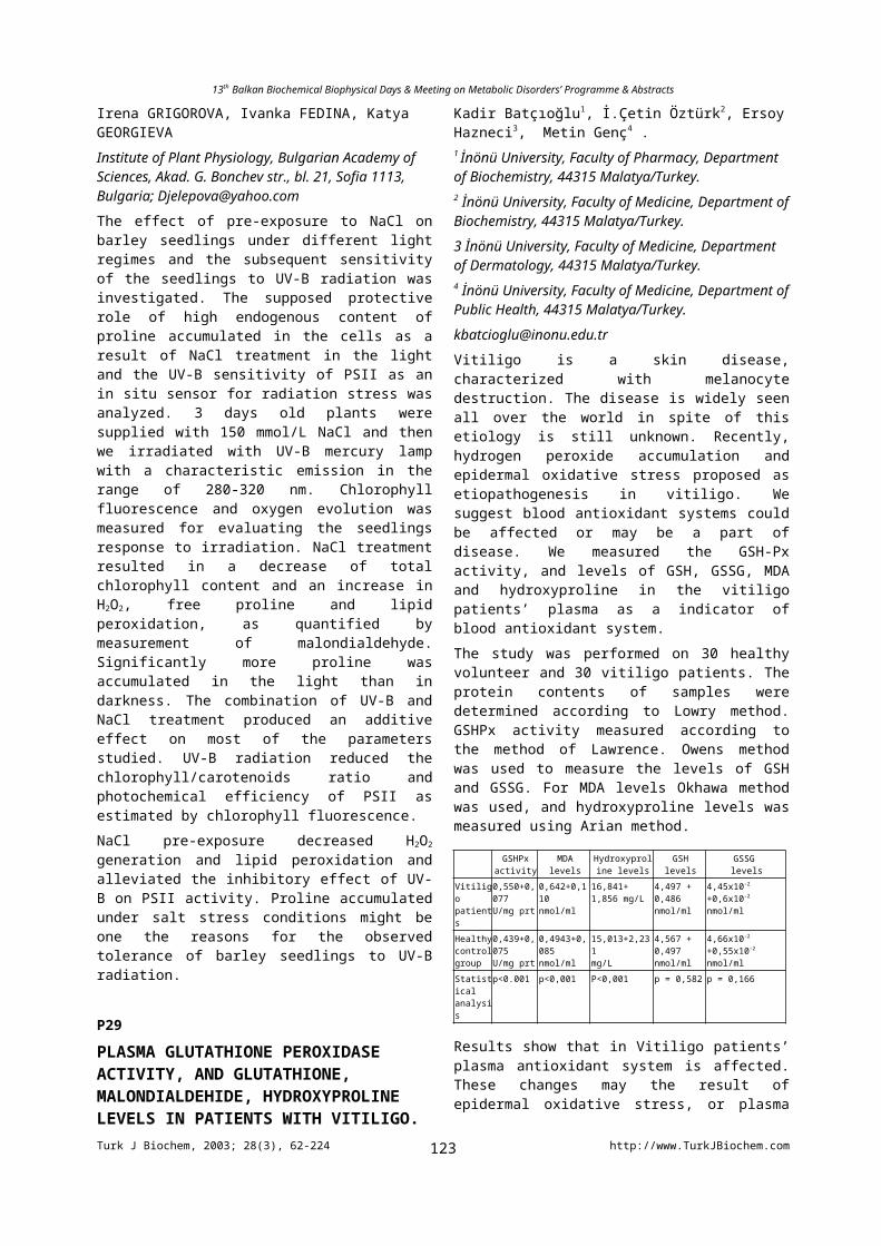
13th Balkan Biochemical Biophysical Days & Meeting on Metabolic Disorders’ Programme & Abstracts
This study was designed to elucidate the association of blood formed elements (BFE) (platelets, neutrophils, monocytes and lymphocytes) and NOS isoenzymes with the pathogenesis of familial Mediterranean fever (FMF). We have synthesized L-arginine analogue with blocked -NH2 group (CH1) and examined its effect on the NOS activity in whole blood and BFE of healthy humans (3 women of 38-51 years old) and patients with FMF (3 women of 16-50 years old). The effect of calmodulin (CaM) was also studied. No close relationship was observed between the production of NO2+/NO3+ and L- citrulline, as well as the consumption of L-arginine in whole blood and BFE. Volunteers’ studies showed the NOS activity was decreased in BFE, especially in platelets and neutrophils, whereas the plasma level of NO2+/NO3+
was increased for 1,2-1,7 times in patients with FMF, as compared to controls. It appears, the nitrite/nitrate anions caused the feedback inhibition of NOS. The elevated glucose level in plasma and erythrocytes sedimentation rate observed in patients with FMF seemed to be caused by the decrease of NOS activity, since NO/NOS is known regulate the rheological behavior of erythrocytes and partially regulate the concentration of L- arginine, which is in turn involved in the regulation of glucose and insulin levels in human blood. The addition of CH1 in the incubation mixture was mainly accompanied with shift to the Ca2+- CaM dependent NOS activity. CH1 increased markedly the dropped NOS activity in platelets and neutrophils of women with FMF. The effects of CaM and CH1 seemed to be different on various NOS isoenzymes and should further be studied.
P80
MYELOPEROXIDASE (MPO) ACTIVITIES IN BRAIN, LUNG AND RENAL TISSUES AFTER EXPOSURE TO MAGNETIC FIELDS OF 50 Hz?Ayse G. CANSEVEN1, Ummühani OZEL2 , Ayse BILGIHAN2 and Nesrin SEYHAN1
1) Department of Biophysics, Gazi University, Medical Faculty, Ankara, TURKEY2) Department of Biochemistry, Gazi University, Medical Faculty, Ankara, TURKEYMyeloperoxidase (MPO), a bactericidal enzyme secreted by activated phagocytes, specifically catalyzes the production of hypochlorous acid (HOCl) from chloride and hydrogen peroxide. The study was assessed to evaluate the influences of in vivo exposures to 10 G (Gauss) and 30 G of magnetic fields on MPO activities of lung, brain and kidney tissues in guinea pigs. MPO activity was measured as a marker of neutrophil accumulation in those tissues.Thirthy six male, 250-300 g weighted guinea pigs were used. Twenty eight guinea pigs were exposed to the fields of 50 Hz 10 G and 30 G with the period of 4 hours/day and 8 hours/day for 5 days in 4 different groups. Eight animals were served as control, keeping at the same conditions without being exposed to any magnetic field. Magnetic field was generated by a pair of Helmholtz coils. Circular coils pair of Helmholtz configuration was used. Animals
were placed pairly in plastic cages which were positioned at the center of the Helmholtz Coil during exposure conditions, to avoid any distortion of the generated magnetic field. Ambient geomagnetic field was measured as 0.3 G in the laboratory. Brain, lung and renal tissues were homogenized according to methods of Matsuo Y. et.al., Koike K. et al. and Lopez-Neblina F. et.al. respectively. MPO activities in these tissue samples of exposed and unexposed guinea pigs were determined by measuring the H2O2-dependent oxidation of o-dianisidin by the method of Glowick SP. et al. Mann Whitney-U test was applied for statistical analysis.MPO activities in brain tissues of guinea pigs exposed to the magnetic fields of 10 G and 30 G were found increased with repect to the controls for both of the exposure periods. Increased MPO activities were found in lung tissues under the effect of 10 G magnetic fields for both of the exposure periods whereas decreased MPO activities were determined for the magnetic field of 30 G. The decrease in MPO activities for 4 hours/day of exposure times was found statistically significant (p=0.002). Renal MPO activities were also found increased for both of the magnetic fields and the exposure periods. The increase in MPO activities was found statistically significant (p= 0.004) under the effect of 10 G for 5 days with the exposure period of 4 hours/day.
P81
THE EFFECT OF ELF MAGNETIC FIELD EXPOSURE ON KIDNEY MYELOPEROXIDASE (MPO) ACTIVITYAyse G. CANSEVEN1, Ummühani OZEL2 , Ayse BILGIHAN2 and Nesrin SEYHAN1
1) Department of Biophysics, Gazi University, Medical Faculty, Ankara, TURKEY2) Department of Biochemistry, Gazi University, Medical Faculty, Ankara, TURKEYMyeloperoxidase (MPO) is one of the enzymes that constitute the defence system of immune cells.The aim of the present study was to investigate the effect of 50 Hz, 20 G magnetic field on MPO activity in renal tissues of guinea pigs.Magnetic field was generated by a pair of Helmholtz coils. Circular coils pair of Helmholtz configuration was used in the vertical manner. Twenty two male, 250-300 g weighted guinea pigs were used. Fourteen guinea pigs were exposed to the field of 50 Hz, 20 G with the exposure periods of 4 hours/day and 8 hours/day for 5 days in 2 different groups. Eight animals were served as control, keeping at the same conditions without being exposed to any magnetic fields. Animals were placed pairly in plastic cages which were positioned at the center of the Helmholtz Coil during exposure conditions, to avoid any distortion of the generated magnetic field. The animals were kept in the laboratory at a room temperature of 23C, a day and night cycle of 12 hours and ambient geomagnetic field of 0.3 G. MPO activities in renal tissues of exposed and unexposed guinea pigs were determined by measuring the H2O2-dependent oxidation of o-dianisidin according to methods
Turk J Biochem, 2003; 28(3), 62-224 http://www.TurkJBiochem.com123
13th Balkan Biochemical Biophysical Days & Meeting on Metabolic Disorders’ Programme & Abstracts
of Lopez-Neblina et al. and Glowick SP. et al. Mann Whitney-U test was applied for statistical analysis.MPO activities in kidney tissues of guinea pigs exposed to the 50 Hz, 20 G magnetic field were found increased in both of the exposure periods. The increases in MPO activities of renal tissues were statistically significant both application times of 4 hours/day (p=0.014) and 8 hours/day (p=0.001). The exposure periods of 8 hours/day was found more effective.
P82
LYSINE-RICH HISTONES AS A MODEL SYSTEM TO INVESTIGATE NONENZYMATIC GLYCOSYLATION IN E. COLIGeorgi STOYNEV, Ljuba SREBREVA, Ivan IVANOVBulgarian Academy of Sciences, Institute of Molecular biology, “Acad. G. Bonchev” str. bl. 21, 1113 Sofia, Bulgariasrebreva@obzor bio21.bas.bgObjective: Nonenzymatic glycosylation (glycation) is a multystep reaction between free amino groups in the side chains of proteins and various carbonyl compounds. The initially formed unstable Schiff and Amadory products (called early glycation products) are spontaneously converted into more stable terminal adducts -advanced glycation end products (AGEs). For a long time it has been thought that AGEs are typical for the long-lived proteins in higher eukaryotes only. Recently, it has been found in our laboratory that E.coli proteins, including recombinant human interferon gamma (hrIFN-) are also subjected to glycation (Mironova, R. et al, Mol. Microbiol. (2001) 39, 1061-1068). The aim of this study is to develop an assay system for studying the glycating capability of bacterial lysates based on the lysine-rich histones H1 and/or H5 subvariant. Methods: H1 or H5 histones are extracted from rat liver or chicken erythrocytes by perchloric acid, dissolved in water and dialyzed overnight at 4o C against clear (centrifuged) bacterial lysates. Accumulation of AGEs is monitored by fluorescence (ex 365 nm and em
443 nm) and the accompanying changes in the protein structure are monitored by electrophoresis. Results and Conclusion: In kinetic studies we have detected significant accumulation of AGEs in both H1 and H5 histones and also fragmentation and cross-linking. Using known AGE inhibitors (as thiamine, pyridoxine and aspirin) we have registered a detectable decrease in the level of AGEs. Our results showed that aspirin is the most effective inhibitor of glycation. In conclusion, the in vitro incubation of lysine-rich histones with bacterial lysates produces AGEs on the native proteins.
P83
THE ROLE OF NITRIC OXIDE IN NEUROTRANSMISSION IN THE GUINEA PIG ILEUM
* Snežana RADIVOJŠA, ** Vladislav VARAGIĆ and *** Slobodan MILOVANOVIĆ* Laboratory for molecular biology and endocrinology, “Vinča” Institute of Nuclear Sciences, P.O. Box 522, 11001 Belgrade/SERBIA AND MONTENEGRO*** The Military Medical Academy, Crnotravska 17, 11000 Belgrade/SERBIA AND MONTENEGRO** Medical Faculty, Dr. Subotića 8,11000 Belgrade/SERBIA AND [email protected] role of nitric oxide in inhibitory non-adrenergic non-cholinergic neurotransmission was studied on longitudinal muscle of the guinea pig ileum. The most widely reported action of sodium nitroprusside (SNP), a nitric oxide donor compound, in the gut is relaxation of smooth muscle. The isolated segments of ileum were maintained in Tyrode solution and the addition of SNP (10-10–10-5M) to the organ bath concentration-dependently inhibited contractions, caused by electrical stimulation (8-79%) and acetylcholine (24-62%). Segments of ileum were exposed to 1M SNP during 60min to induce in vitro tolerance to SNP and then exposed to increased concentrations of SNP. The degree of relaxation of contraction caused by electrical stimulation and acetylcholine was 4-50% and 6-30%, respectively. Among the most popular theories for nitrate tolerance is the “intracellular sulfhydryl depletion hypothesis”. The influence of the N-acetylcysteine, donor of sulfhydryl group, on tolerance caused by SNP was investigated. Our results showed that N-acetylcysteine (1mM) can change the activity of SNP (electrical stimulation – 8-63%) and it was found that exogenously added thiols can partially reverse nitrate tolerance.Higher concentrations of SNP induced a biphasic response, an immediate relaxation (1-3min) followed by prolonged contraction (10min). The relaxations evoked by NO liberated from SNP were blocked 20% by methylene blue (10mM). The results suggest that relaxant responses to exogenous nitric oxide in guinea pig ileum are mediated via the activation of soluble guanylate cyclase and the formation of guanosine-3’,5’-cyclic monophosphate. The contractions evoked by SNP were inhibited 40% by atropine (1M). Concerning the obtained results it can be concluded that contraction evoked by SNP on smooth muscle cells are also mediated by activating acetylcholine release from neurons. P84
EXAMINATION OF ELECTRIC FIELD EFFECTS ON LIPID PEROXIDATION AND ANTIOXIDANT ENZYMES BY USING EXPERT SYSTEMSGöknur GÜLER1 and Fırat HARDALAÇ2
1Gazi University, Faculty of Medicine, Department of Biophysics, Ankara, Turkey2 Fırat University, Faculty of Medicine, Department of Biophysics, Elazığ, [email protected], [email protected] (MDA) and superoxide dismutase (SOD)
Turk J Biochem, 2003; 28(3), 62-224 http://www.TurkJBiochem.com124
13th Balkan Biochemical Biophysical Days & Meeting on Metabolic Disorders’ Programme & Abstracts
levels in spleen and testis tissues of guinea pigs which were exposed to different periods of Extremely Low Frequency ( ELF ) electric fields were determined and the results are applied to neural networks as learning data and the training of the feed forward neural network is realized. At the end of the training, to determine the effect of the electric field on tissues in computer without applying electric field and without using too many guinea pigs and to form a database for the researchers in this field are aimed. Five groups of 15 male white guinea pigs (150-200 g) were exposed to 50 Hz 5 kV/m ELF electric fields. Each group was exposed daily for 8 hours for 1 day, 3 days, 5 days, 7 days and 10 days. The 75 guinea pigs were examined according to the exposure periods while 15 guinea pigs, which were not exposed to any electric field, formed the control group. The effect of 50 Hz electric field exposure on MDA and SOD levels was investigated for different application periods. The increase in MDA and SOD levels of spleen and testis tissues was found to depend significantly on the type of electric field and the length of exposure.After the experiments, the prediction of the neural network is averagely 99 %. Those percentiles of the prediction performance of the neural network belonging to experiment results of electric field were so high; this fact shows that the feed forward neural networks which are used many fields could be applied in the studies of electric field too. Furthermore this study may form a database for the scientists investigating the effects of electric fields on lipid peroxidation and antioxidant enzymes.
P85
TISSUE RESPONSE TO EXTREMELY LOW FREQUENCY ELECTRIC FIELDS WITH DIFFERENT EXPOSURE PERIODSGöknur GÜLER and Nesrin SEYHANGazi University, Faculty of Medicine, Department of Biophysics, Ankara, [email protected], [email protected] has been renewed concern in recent years on the biological effects of electric fields. This interest is based on the fact that all living organisms are continuously exposed to both natural and man-made electric fields. We live a large part of our lives surrounded by a grid of wires that delivers the energy we need to power lights, electric motors, and many of the conveniences that make modern living possible.
The effects of Extremely Low Frequency (ELF) Electric Field which we are exposed in daily life was investigated in this study. Collagen synthesis under different exposure periods was studied. The effect was evaluated by assessing the amount of hydroxyproline in the lung and kidney tissues. 5 kV/m ELF electric field with 50 Hz frequency was applied to 60 guinea pigs in 5 different exposure periods being 1 day, 3 days, 5 days and 7 days with daily exposure period of 8 hours. 15 guinea pigs were also kept in the same laboratory conditions and served as control
without any electrical field application. At the end of each exposure period lung and kidney hydroxyproline contents were determined using Stegemann-Stalder’s method.
The applied electric field was found decreased the hydroxyproline amount of lung and kidney tissues significantly in all of the exposure periods with respect to the controls suggesting decreased synthesis of collagen under ELF Electric field.
Beside that, theoretical values of electric field, current intensity and current density on the surface and inside of those guinea pigs and human models were calculated.
P86
EFFECTS OF RADIATION ON LPO AND AOD IN THE THYROID OF HYPOTHYROID RATS.Olga I.VALENTSIUKEVICH, Zoya V. NIATSETSKAYA and Liliya I. NADOLNIK
Institute of Biochemistry of NAS Belarus, BLK – 50, Grodno, 230017, Belarus
It is well known, that the major action of methimazole (MMI) is to inhibit synthesis of thyroid hormones in the thyroid gland. However, recent studies have shown that MMI also has antioxidant and immunomodulatory effects.
In this study, the activity of lipid peroxidation (LPO) processes and antioxidant defenses (AOD) were measured in the thyroid gland of rats received MMI at doses of 2.5 and 10 mg/kg body weight for 2-week period. The influence of single external gamma-irradiation at a dose of 1 Gy on the animals receiving the same MMI doses was also studied. The MMI-induced hypothyroidism was accompanied by the increased activity of Cat (23.58 %), SOD (29.43 %) and of TBARs concentration (37.50 %) in rats thyroid. The radiation exposure leads to a raise of TBARs concentration by 1.34 times in the group of control animals. The single external gamma-irradiation at a dose of 1Gy may have an inhibitory effect in relation to antioxidant system activity because we have not found increasing of Cat and SOD activity neither in the thyroid tissue of control group rats, nor in animals receiving MMI. Under such conditions, a significant increase of TBARs level (by 1.69 times) was observed at a MMI dose 10 mg/kg. The above show that the MMI-induced hypothyroidism does not stimulate functioning of the antioxidant system in irradiated rat thyroid tissue. Moreover, an histological examination of thyroid gland tissue of irradiated animals receiving MMI at a dose of 10 mg/kg, we have found an area with lymphoid autoaggresion.
The results obtained indicate the presence of a complicated mechanism of MMI influence on the metabolism of thyroid cells and free radical oxidation activity. We suggest that the enhanced lipid peroxidation in MMI-induced hypothyroidism results in destruction of thyrocytes, rising of thyroid autoantigen concentrations in the blood and development of autoimmune aggression.
Turk J Biochem, 2003; 28(3), 62-224 http://www.TurkJBiochem.com125
13th Balkan Biochemical Biophysical Days & Meeting on Metabolic Disorders’ Programme & Abstracts
P87
OXIDATIVE STRESS INFLUENES ON IODINE UPTAKE AND ORGANIFICATIONLiliya I. NADOLNIK
Laboratory of Endocrine Glands Biochemistry, Institute of Biochemistry National Academy of Sciences of Belarus, BLK-50, 230017 Grodno, BELARUS
e-mail: [email protected]
Recently an increase in the number of noniodine-deficient pathologies of the thyroid was noted, which was a result of the influence of unfavourable ecologic factors, including radiation effects. We previously shown that single and chronic irradiation effects as well as emotional and pain stress reduce the thyroid status, inhibit thyroperoxidase (TPO) and disturb iodine metabolism in the rat thyroid. We aimed to study the effect of oxidative process activation on the iodine uptake and oxidation by thyrocytes of rat thyroid organic culture in vitro. Fe2+/ascorbate at concentrations of 0.1x10⁻³ - 0.1x10⁻⁴ M was used as a prooxidant system. The iodine content in the medium was assessed by a cerium arsenite method and TPO activity was measured spectrophotometrically. It was shown an increase concentrations of stable aldehyde lipid peroxidation products in the medium by 2.88-6.76 - fold. Under these conditions, the iodine uptake by thyrocytes was almost completely inhibited within 2 hours. A 31.1% decrease in TPO activity was also found in 2 hours, at Fe2+/ascorbate concentration of 0.1 x10⁻⁴M. At higher concentrations, TPO was inhibited by 30% after 5 hours and by 61.55% after 8 hours. The TPO inhibition and iodine uptake were reversible since after 24 hours the enzyme activity was recovered to the control values. The addition of the 1x10⁻² - 1x10 -4 M H2O2 concentrations leads to 24-h inhibition of iodine uptake and a decrease of TPO activity by 17.23 - 33.4%. The data obtained suggest pronounced sensitivity of thyroid hormone synthesis in the thyroid and oxidative stress activation. The disturbed iodine uptake, as well as its oxidation and organification by thyrocytes seem to be the most important mechanism of thyroid function impairment under the action of an unfavourable ecologic factor. The research was supported by a grant from the Belarusian Foundation for Fundamental Studies (Grant B01-343).
P88
THE EFFECT OF HMG-COA REDUCTASE INHIBITION ON HEMORHEOLOGICAL PARAMETERSBIKMAZ PS1, BIKMAZ K2, TAMER S1, YIGIT R1
1Istanbul University, Istanbul Faculty of Medicine, Department of Physiology, Findikzade, Istanbul / Turkey2SSK Okmeydanı Teaching Hospital, Department of Neurosurgery, Okmeydani, Istanbul / [email protected] Methyl Glutaryl Co-enzym A (HMG-CoA) reductase which is participated in cholesterol synthesis
starting with the Acetyl-coenzym A in liver cell catalyse the conversion of HMG-CoA to Mevolanat, which is a velocity limiting stage in cholesterol biosynthesis in human. HMG-CoA reductase inhibitors inhibit HMG-CoA reductase enzym competitively thus reducing the cholesterol and lipoprotein levels in liver cells. As a result, they decrease LDL cholesterol and total cholesterol level by reducing lipoprotein synthesis and also increasing the entrance and destruction of the lipoprotein containing Apo-B to the liver cells and the other cells. It is reported that, in patients with lipoprotein metabolism disorder, there are erythrocyte morphology disorders, increasing erythrocyte aggregation and decreasing blood flow. We performed this study with 31 hyperlipidemia patients and 20 healthy subjects. After 12 weeks of Atorvastatin treatment (20mg/day), the basal hemorheological parameters, lipid levels of the patients were compared to the pretreatment and the control group values. Our data suggest that, inhibition of HMG-CoA reductase with Atorvastatin treatment results in a significant decrease in total cholesterol, LDL, VLDL levels (p<0,001); a significant increase in HDL levels (p<0,001); and a significant decrease in whole blood viscosity, plasma viscosity, erythrocyte rigidity (p<0,05; p<0,05; and p<0,001 respectively). No significant changes in fibrinogen levels were observed (p>0,05). In spite of the significant decrease in plasma viscosity, there was no significant improve in fibrinogen levels those which one of the determinants of plasma viscosity. Therefore, we considered that Atorvastatin’s improving effects on lipid profile could have positive effects on other determinants of hemorheological parameters. P89ERYTHROCYTE MEMBRANE PROTEINS AND LIPID COMPOSITION IN ATORVASTATIN ADMINISTERED PATIENTS WITH HYPERCHOLESTEROLEMIABIKMAZ PS1, ALBENIZ I2, BIKMAZ B3, GOKKUSU C4, TAMER S1
1 Department of Physiology, Istanbul Faculty of Medicine, Istanbul University, Findikzade, Istanbul / Turkey 2 Department of Biophysic, Istanbul Faculty of Medicine, Istanbul University , Findikzade, Istanbul / Turkey3 Department of Neurosurgery, SSK Okmeydanı Educational Hospital, Okmeydani, Istanbul / Turkey4 Department of Biochemistry, Istanbul Faculty of Medicine, Istanbul University, Findikzade, Istanbul / [email protected] HMG-CoA (3-Hydroxy-3 Methyl Glutaryl Co enzyme A) reductase inhibitors are known as significantly effective in reducing plasma and cholesterol levels, not many studies concerning the relationship between membran lipid and protein levels are available. In our study, we aimed to examine the changes in erythrocyte membrane proteins and lipid composition in hypercholesterolemic patients administered with atorvastatin, a HMG-CoA (hydroxy methyl glutaryl Co enzyme A) reductase inhibitor. Therefore, 20 patients whose were enrolled in Okmeydani Training Hospital General Internal Diseases Clinic, were included the study.
Turk J Biochem, 2003; 28(3), 62-224 http://www.TurkJBiochem.com126
13th Balkan Biochemical Biophysical Days & Meeting on Metabolic Disorders’ Programme & Abstracts
The patients had no clinical symptom except hyperlipidemia. The hyperlipidemic patients were treated with orally administered atorvastatin (20 mg/day) during 12 weeks. At the end of this time period, the lipid composition in plasma and erythrocyte membranes (RBC) was determined using enzymatic methods whereas membrane protein levels were determined by SDS-PAGE electrophoresis method. When the findings were compared to healthy control group (n=15), a significant reduction (p<0.001) was observed in plasma and RBC total cholesterol (TC), total phospholipid (TPL) and low density lipoprotein cholesterol (LDL-C) levels after 12 weeks of treatment. Neither high density lipoprotein cholesterol (HDL-C) levels nor RBC protein fractions of hyperlipidemic patients showed any significant difference after 12 weeks of treatment, however. Our findings suggest that, orally administered atorvastatin treatment reduces plasma cholesterol levels besides membrane cholesterol levels.
P90PRO-DIABETIC CONDITIONS INDUCE CHANGES IN THE OXIDANT/ANTIOXIDANT BALANCE IN PERICYTESMonica Raicu, Adrian Manea, Doina Popov, Elena ConstantinescuInstitute of Cellular Biology and Pathology “N.Simionescu”, Bucharest, [email protected]: Recent data suggest that the oxidative stress play an important role in the pathogenesis of diabetes and its complications, i.e accelerated atherosclerosis, nephropathy, retinopathy. This study was designed to investigate the role of pro-diabetic conditions: high glucose, AGE-proteins, vasoactive factors, in the modulation of antioxidant enzyme activities, gluthatione level and reactive oxygen species (ROS) production in pericytes. Material and methods: Pericytes isolated from rat adipose microvasculature were cultured in DMEM, 10% fetal calf serum, antibiotics and low or high glucose concentrations (5 mM or 25 mM). The cells were incubated for 5 days in the presence or absence of AGE-Lysine (5 UF/ml), angiotensin II (Ang II 1 M) or their combination. For comparison smooth muscle cells (SMC) cultured in the same conditions were used. The activity of antioxidant enzymes: superoxide dismutase (SOD), catalase (CAT), glutathione peroxidase (GPx) and total gluthatione (GSH) was measured spectrofotometrically. ROS production was evidentiated by spectrofluorimetry and fluorescence microscopy. As positive control H2O2 was used. Intracellular calcium was determined using Fura 2 AM assay.Results: No changes in the antioxidant enzyme activities were detected when the cells were cultured in high glucose alone. The presence of AGE-Lys and Ang II increased CAT (17.5 2.2 vs 13.91 2.3 U/mg prot) and SOD (44.14 2.8 vs 38.8 2.1 U/mg prot) activity. Their combination decreased significantlly GPx (0.88 0.06 vs 1.03 0.05 U/mg prot) and GSH level ( 11.98 0.17 in 5 mM glucose and 14.74 0.03 in 25 mM glucose vs 16.00 0.04 nmol/mg prot). A two times increase in ROS
production and a significant deregulation of intracellular calcium homeostasis was detected in cells cultured in the presence of pro-diabetic agents, pericytes being more susceptible than SMC.Conclusion: The increase of the oxidative stress induced by high glucose in combination with advanced glycation end product and angiotensin II in pericytes, may explain the structural and functional abnormalities of these cells observed in diabetic retinopathy.This project was supported by the Romanian Academy (Grant nr. 83/2002).
P91
THE EFFECTS OF VITAMIN C ON RENAL ISCHEMIA-REPERFUSION INJURY IN THE RATSNurettin AYDOĞDU and Kadir KAYMAKDepertment of Physiology, Faculty of Medicine, University of Trakya, Edirne/[email protected] ischemia is observed in a variety of clinical situations, such as cardiac arrest with recovery, liver transplantation and heminephrectomy. Acute renal failure (ARF) observed after ischemia is characterized by decresed gomerular filtration rate, tubular necrosis and increased renal vascular resistance. Free radicals and formation of nitric oxide (NO) plays an important role in the pathophysiology of renal injury mediated by ischemia-reperfusion. The aim of this study was to estimate the protective effects of vitamin C on plasma malondialdehyde (MDA), glutathione GSH) and NO levels during ishemia-reperfusion injury of kidney. For this purpose; thirty female Spraque Dawley rats divided into three groups: Group 1; was given saline intraperitonealy (ip). Group 2; subjected to bilateral renal ischemia (60 min) folloved by reperfusion (24 h) and saline injected ip 30 min before induction of ischemia. Group 3; is also subjected to bilateral renal ischemia (60 min) folloved by reperfusion (24 h) and Vitamin C (200 mg/kg) injected ip 30 min before induction of ischemia. At the end of the reperfusion period, the rats were sacrificed. The level of plasma MDA, GSH and NO were determined. The levels of MDA and NO were significantly lower in group 1 than group 2 (p<0.001) and GSH was significantly higher in group 1 than group 2 (p<0.001). MDA levels were significantly lower in group 3 than group 2 (p<0.001), NO and GSH levels were significantly higher in group 3 than group 2.As a conclusion, Vitamin C decresed plasma MDA level and increased plasma GSH and NO levels. Therefore, these findings may indicate that Vitamin C has a protective effect on plasma during the course of renal ischemia-reperfusion injury.
P92
EFFFECT OF CAFFEIC ACID PHENETHYL ESTER MYOGLOBINURIC ACUTE RENAL FAILURE IN RATSNurettin AYDOĞDU, Gülizar ATMACA, Kadir BATÇIOĞLU1, Kadir KAYMAK
Turk J Biochem, 2003; 28(3), 62-224 http://www.TurkJBiochem.com127
13th Balkan Biochemical Biophysical Days & Meeting on Metabolic Disorders’ Programme & Abstracts
Depertment of Physiology, Faculty of Medicine, University of Trakya, Edirne/TURKEY, 1Department of Biochemistry, Faculty of Farmacy, University of Inonu, Malatya/[email protected] metabolites play an important role in renal injury during myoglobinuric acute renal failure (ARF). This study was designed to determine the protective influence of caffeic acid phenethyl ester (CAPE), an active component of propolis extract, exhibits antioxidant properties and treatment in an experimental model of myoglobinuric-ARF induced by intramuscular injection of hypertonic glycerol in rats. The rats were randomly divided into three groups: Group 1 was given saline, group 2; glycerol plus saline, and group 3; glycerol plus CAPE (10 μmol/kg). After 48 h rats were sacrificed. Kidney and liver tissue malondialdehyde (MDA) levels and plasma MDA, urea, creatinine and nitric oxide (NO) levels were determined. Plasma urea level was significantly lower in group 1 than group 2 and 3, (p<0.001) and it was lower in group 2 than group 3 (p<0.05). The levels of plasma creatinine and kidney and liver tissue MDA were significantly lower in group 1 than group 2 and 3 (p<0.001), but no significant difference found between groups 2 and 3 for the same parameters. Plasma NO levels was significantly higher in group 1 than group 2 and 3 (p<0.001), and it was significantly lower in group 3 than group 2 (p<0.001). Plasma MDA levels was significantly lower in group 1 than group 2 and 3 (p<0.001), and it was significantly lower in group 2 than group 3 (p<0.01). Nitric oxide synthase inhibition worsens, and NO suplementation protects against the glycerol ARF model. The result of this study may suggest that intraperitonealy administration of CAPE does not have a beneficial effect on prevention against impairment of renal function under these conditions in this model of myoglobinuric ARF.
P93COMPARATIVE STUDIES ON THE ADSORPTION OF Cr (VI) IONS ONTO CHITOSANAyşe BARAN, Evran BIÇAK, Şenay BAYSAL, Seçil ÖNALDepartment of Biochemistry, Faculty of Science,Ege University, Izmir 35100, TURKEYhamarat @sci.ege.edu.trThe toxic heavy metals cause serious threat to the environment, animals and humans. Many industries such as mining, iron-sheet cleanining, plating, metal processing, automobile parts manufacturing, dyeing, textile, fertilizer and petroleum industies release heavy matals such as chromium in the environment. The trivalent and hexavalent forms of chromium are environmentally important. Chitosan is a biopolymer, which is of interest to researches concerning the adsorption of metal ions on chitosan. Chitosan has been described as a suitable natural polymer for the collection of metalsions, since the amine groups and hydroxyl groups on the chitosan chain can act as chelation sites for metal ions. The adsorption of Cr (VI) ions onto chitosan has been investigated. Batch adsorption experiments were carried out as a function of pH, agitation period and concentration of Cr (VI) ions. The optimum pH
was found as 3.0. the maximum chromium sorption occured at about 30 minute. The suitability of the Freundlich and Langmiur adsorption models were also investigated. The chromium ions can be removed from sorbents rapidly by treatment with an aqueous EDTA solution and at the same time the sorbent regenerated and also can be used again to adsorb by heavy metal ions. The result showed that chitosan, which is a readiliy available, economic sorbent, was found suitable for removing chromium from aqueous solution.
P94REMOVAL OF Cr(VI) IONS FROM AQUEOUS SOLUTION ONTO IMMOBILIZED Chryseomonas luteola BIOMASSEvran BIÇAK, Ayşe BARAN, Seçil ÖNAL, Şenay BAYSALDepartment of Biochemistry, Faculty of Science,Ege University, Izmir 35100, TURKEYhamarat @sci.ege.edu.trHeavy metals are major pollutants in marine, ground, industrial and even treated waste waters. The presence of these metals in the environment has been of great concern because of their increased discharge, toxic nature and other adverse effects on receiving waters.Conventional methods to remove heavy metals from waste waters, such as chemical precipitation, electrowinning, membrane seperations, evaporation and resin ion excahange may be technologically inapplicable or very expensive from an economic point of view. Biosorption may be a suitable waste water tecnology to remove heavy metals as demonstrated by several researchers because it is possible the use cheap adsoption materials that can be competitive with conventional tecnologies. Polysaccharide gel immobilized microorganisms can be used to remove heavy metal ions from aqueous solutions, providing an alternative to physico-chemical technologies for waste water treatment. Alginate is a linear polysaccharide composed of (1 4) linked residues of -L-gulucuronic acid (G) and -D-mannuronic acid (M). Carbohydrate polymers such as alginate have been mostly used as the matrix for the immobilization of microbial cells via entrapment. These polymers are also known to bind metal ions strongly. C.luteola has been immobilised into calcium alginate beads via entrapment. Biosorption of Cr(VI) was studied for diffent pH, metal concentration and agitation time. The optimum pH was found as 4.0. The maximum Cr(VI) sorption occured at about 60 minutes. The equilibrium was described by both Langmiur and Freundlich isotherms.
P95THE LEVELS OF CYTOKINES AND NITRIC OXIDEIN THE BRUCELLOSIS AFTER THE ANTIBRUCELLAR THERAPYAysun BAY KARABULUTTurgut Ozal Medical Center of Biochemistry
Turk J Biochem, 2003; 28(3), 62-224 http://www.TurkJBiochem.com128
13th Balkan Biochemical Biophysical Days & Meeting on Metabolic Disorders’ Programme & Abstracts
The aim of the present study was to investigate possible involvement of oxidant stress and apoptosis by proapoptotic stimuli, including IL-1 , IL-2R, IL-6, IL-8 and TNF- in the brucellosis before, during and after the antibrucellar therapy.We measured the nitric oxide (NO) and IL-1, IL-2R, IL-6, IL-8 and TNF- levels in the in twenty patients before the therapy, after the third and sixth week of the therapy and twenty healthy subjects. Plasma NO levels were were higher than the control group in baseline, thirth and sixth week of the therapy (p<0.0001). IL-1β levels were increased in baseline and thirth weeks of the therapy group, but could not be detected in the other groups. IL-6 levels were found increased in brucellosis patients than that of control group.(p<0.0001) But it could not be detected in the other groups. IL_2R levels were increased in baseline but were decreased thirth and sixth week of the therapy in comparison of control. IL-8 levels were higher than control in baseline and thirth of the therapy. TNF- α levels were found to be increased in baseline, thirth week’s of the therapy decreased in sixth week’s of the therapy when compared to control group. These results showed that lymphocyte nitrite and cytokine levels may reflect the immune reactiveness of the body and could be used for evaluating the severity and therapy of the brucellosis.
P96
ELECTRON-HISTOCHEMICAL LOCALIZATION OF CATHEPSIN D ACTIVITY IN POST-PARTUM RAT UTERUSVictor RIVNEAC, Valentin GUDUMAC*, Elena RIVNEAC*Laboratory of Morphology and Laboratory of Biochemistry* “Nicolae Testemitanu” Medical and Pharmaceutical State University, MD-2004, Chisinau / MOLDOVA [email protected] D is one of the most important lysosomale aspartile proteinase, present in different cells and tissues and indispensable for proteolitic processes. The enzyme is capable to degrade in vitro the main components of the intercellular matrix: collagens, proteoglycans, glycoproteins. Aims: 1. To determine the localization of cathepsin D in uterus. 2. To reveal the participation of cathepsin D in the intracellular and/or extracellular collagen degradation in the post-partum uterus involution.Material and methods. Female rats were sacrificed in the 2 and 3 days post-partum. The activity of cathepsin D in the virgin and involuting uterus was investigated electron-histochemically. BZ-Arg-Gly-Phe-Phe-Pro-4MBNA (Bachem) served as the substratum for cathepsin D. Smith and Van Frank (1975) technique was used. Ultrathin sections were viewed without staining.Results: The reaction product was revealed as a fine granular sediment in the smooth muscle cell (SMC), macrophage and fibroblast lysosomes in both virgin and
involuting rat uterus. In involuting uterus the reaction product was detected also in macrophage and fibroblast vacuoles containing phagocyted fragments of collagen fibrils. We didn’t detected the extracellular activity of cathepsin D in miometrum during the post-partum uterus involution. Conclusions: Thus, in rat uterus cathepsin D is localized in the SMC, macrophage and fibroblast lysosomes. Cathepsin D takes an active part in the intracellular collagen degradation during the post-partum uterus involution.
P97
CONGENITAL GLUCOSE-GALACTOSE MALABSORPTION: REPORT OF TWO CASESNevenka SLAVESKA1, Stojka FUSTIK1, Velibor TASIC1, Vidanka LUKAREVSKA1, Rene SANTER2
1 University Children’s Hospital, Skopje, Macedonia2 University Children’s Hospital, Hamburg, [email protected] glucose-galactose malabsorption is a rare autosomal recessive disorder of intestinal transport of glucose and galactose.It is characterized by watery diarrhea, dehydration, failure to thrive, or early death without appropriate dietary treatment. Two cases of congenital glucose-galactose malabsorption from The Republic of Macedonia has been presented.The first patient was 15 days old male, and the second, 25 days old female, when they were admitted to the hospital because of continuous, severe, watery, acidic diarrhea and hypernatremic dehydration. The abnormal stool looses, in both of them, were recorded within first week of birth. They were followed by abdominal distension, with no vomiting, and persistent osmotic, watery diarrhea for the next few weeks. Despite management with lactose-free semielemental formula, and periodic administration of total parenteral nutrition during hospitalization, both patients developed severe malnutrition.Further laboratory investigations revealed repeted low blood sugar levels, slight intermitent glycosuria, low stool Ph, and presence of reducing substances in the feces. Oral glucose tolerance test showed flat blood glucose response. Diagnostic evaluation ruled out infectious etiology of the diarrhea, cystic fibrosis, familial chloride diarrhea, and lactose intolerance. The X-ray examination of the intestinal tract revealed no abnormality. The clinical history of the patients and performed laboratory investigations were stongly suggestive of congenital glucose-galactose malabsorption. Dramatic ceasure of the diarrhea followed when the patients were treated with a commercial glucose and galactose-free formula – Galctomine 19 (specialized fructose-based formula).A few months later the clinical diagnosis, in both patients, was confirmed by mutational analises of the SGLT1 gene.
P98
Turk J Biochem, 2003; 28(3), 62-224 http://www.TurkJBiochem.com129
13th Balkan Biochemical Biophysical Days & Meeting on Metabolic Disorders’ Programme & Abstracts
DEVELOPMENT OF ANTI-MURINE EpCAM ANTIBODIES TO USE IN CANCER THERAPHYSerap YALIN1, Elena RIPAMONTI2, Renata FERRI2, Cecilia MELANI2, Marco COLOMBO2, Dorothee Herlyn3, Silvana CANEVARI2 and Mariangela FIGINI2
1 Mersin University, Pharmacy Faculty, Biochemistry Department, Mersin, Turkey, 2Experimental Oncology Department , Istituto Nazionale Tumori, Milan, Italy, 3The Wistar Institute, Philadelphia, PA, [email protected] (epithelial cell adhesion molecule, GA733, KSI-4, KSA, EGP, Trop-1) is an approximately 40 kDa integral membrane glycoprotein that is expressed on the basolateral cell surface in most human simple epithelia and in the vast majority of carcinomas, including ovarian, breast, lung, prostate and colorectal carcinoma. EpCAM functions to mediate Ca2+ independent homotypic cell-cell adhesion through interaction of its cytoplasmic tail with the actin cytoskeleton via -actinin. EpCAM is directly involved in the proliferation and metabolism of epithelial cells. Thus, EpCAM is an interesting diagnostic and therapeutic target so several groups are using EpCAM as a valid target for monoclonal antibody based therapies. In this study we constructed scFv library (single chain variable fragment) against EpCAM by using phage display technology to use in cancer theraphy. For this purpose mRNA was extracted from mouse spleen and used for cDNA synthesis. The immunglobulin heavy and light chain variable fragments were amplified with PCR. These fragments were combined as scFv format in the pHEN2 phagemid vector. For selection four biopanning process were applied and 44 clones were selected. After phage ELISA three clones that recognize EpCAM were described. The characterization process of these clones are in progress.
P99
ACCELERATED REFOLDING OF LAP IN A DETERGENT/DEXTRIN SYSTEMAnca C.COMAN, Elena.GANEAInstitute of Biochemistry, Bucharest, [email protected] aminopeptidase (LAP) is a cytosolic exopeptidase which hydrolyzes the peptide bond adjacent to a free amino group. This enzyme is present in animals, plants and bacteria and has different tissue-specific physiological roles in the processing or degradation of peptides.The aim of the present study was to find a way to accelerate LAP refolding using an artificial chaperone method.The artificial chaperone method is a chemical approach in which small molecules, i.e. detergent and dextrin promote protein refolding from the chemically denatured state.This technique consists of a two-steps protocol involving a binding-release mechanism of the non-native protein. In the first step, the protein aggregation is prevented by the addition of the detergent molecules, which bind to the protein surface through weak detergent-protein
interactions. In the second step, the detergent molecules are stripped off by the dextrin, allowing the protein to refold.LAP was denatured by either 6M urea or by 2.5M guanidinium chloride. The enzyme refolding was performed by the dilution of the denatured enzyme in the absence and in the presence of the artificial chaperones using anionic, cationic and zwitterionic detergents respectively, followed by dextrin10 addition. The results indicate a different refolding capacity of the three detergent/dextrin systems studied (deoxycholic acid/dextrin, CTAB/dextrin and CHAPS/dextrin) as shown by the percentage of enzymatic activity recovered. The spectrophotometrical analysis showed that the simple dilution of the denatured protein leads to a little unassisted refolding whereas incubated in the presence of deoxycholic acid/dextrin, CTAB/dextrin and CHAPS/dextrin systems the enzymatic activity was recovered to 113%, 92% and 90% respectively.These data were supported by our results of Western-blot and Dot-blot analysis using specific rabbit anti-LAP antibodies.In conclusion, the results of this study show that the artificial chaperone method could be very useful for the enzyme refolding, provided using the appropriate type of detergent/dextrin system for a certain protein.
P100ELECTROPHILIC REACTIVITY OF CATIONIC TRIARYLMETHANE DYES TOWARDS PROTEINS AND PROTEIN-RELATED NUCLEOPHILESYasemin Yücel ELDEM, İnci ÖZERHacettepe University, School of Pharmacy, Department of Biochemistry, 06100, Ankara/[email protected] adduct forming (bleaching) properties of 4 cationic triarylmethane dyes (methyl green, MeG+; malachite green, MG+; pararosaniline, PR+; crystal violet, CV+) were studied at 25C, in 100 mM MOPS (pH 8) and/or 100 mM CAPS (pH 10) buffer, using simple nucleophiles (imidazole, 2-mercaptoethanol, 3-mercaptopropionic acid) or proteins (chichen ovalbumin, OA; human serum albumin, HSA; human -globulins, IgG) as addends. Among simple nucleophiles, significant adduct formation was observed only with thiols. The apparent dissociation constants (Kd’) at pH 8 for the 2-ME adducts of MeG+, MG+, PR+ and CV+
were 0.034, 0.22, 1.4 and 44 mM, respectively.Methyl and malachite green were the only dyes to be bleached by proteins at moderate concentration (150 M). Bleaching was multiphasic, summing contributions from multiple nucleophilic centers. In contrast to the trend in the reactions with simple nuclephiles, MeG+ was generally more resistant to protein-mediated bleaching than MG+: OA and HSA contributed 78 and 38%, respectively, to the total color loss in MG+; the corresponding contributions to the bleaching of MeG+ were 16 and 15%. With both dyes IgG-mediated bleaching amounted to ca. 30%. It appeared that protein-borne sulphydryl groups could add to MG+ but not to MeG+. The inferior reactivity of MeG+ towards protein-SH may arise from hindered access of this nucleophile to the central carbon of the TAM+ nucleus. The
Turk J Biochem, 2003; 28(3), 62-224 http://www.TurkJBiochem.com130
13th Balkan Biochemical Biophysical Days & Meeting on Metabolic Disorders’ Programme & Abstracts
exceptional tendency of MG+ to add protein-SH needs to be accounted for. One possibility is that SH groups, excluded from the central carbon, add to the unsubstituted phenyl ring unique to MG+.The results may be significant in relation to applied research on the use of TAM+s in the health sciences (eg. CALI).
P101
INHIBITION OF HUMAN PLASMA CHOLINESTERASE BY TRIARYLMETHANE DYESTuba TÜYLÜ, Fırat ULUTAŞ, İnci ÖZERHacettepe University, Faculty of Pharmacy, Department of Biochemistry, 06100 Ankara/[email protected] triarylmethane dyes (TAM+s) are toxic substances. As such, they are currently under study as cancer therapeutic agents. TAM+ impact on biosystems has been ascribed to DNA intercalation, effects on mitochondrial membrane integrity and redox damage to target biomolecules. A further possibility is that the dyes act as reversible effectors of protein function. The present study focuses on the potential of TAM+s to act as ligands, tested with human plasma cholinesterase (ChE) as the target protein.ChE was concentrated and partially purified by chromatography on DEAE-Trisacryl M eluted with a linear gradient of 0-0.5 M NaCl in 25 mM sodium acetate buffer, pH 4.5. The enzyme was assayed at 25°C in 100 mM MOPS buffer, pH 8, using -naphthyl acetate (NA) as substrate (Km = 1.7 ± 0.5 mM). The reactions were initiated by the addition of NA ± TAM+ and monitored through the increase in A327. A preliminary screen at 0.4 mM NA and 1 µM dye, showed the TAM+s tested (methyl green, malachite green, pararosaniline, crystal violet) to cause 40 ± 5.4 % inhibition of esterase activity. Detailed analysis at 0.4 mM NA and 0-20 µM malachite green (MG+) yielded a biphasic inhibition profile: 75 % of the esterase activity was inhibited with an apparent Ki of 0.4 µM. The Ki value for the remaining 25 % of the activity was in the mM range. Parallel studies with butyrylthiocholine as inhibitor showed the low Ki component to reflect inhibition of cholinesteratic sites.The results suggest that proteins, especially those with anionic binding sites or hydrophobic pockets, may be primary targets of TAM+ action. It is likely that the immediate toxicity of the dyes derives from their ability to impair protein function and that perturbances in overall cellular structure and function are secondary manifestations of TAM+ toxicity.
P102ENZYMATIC SYNTHESIS OF OLIGOSACCHARIDES AND ALKYLGLYCOSIDES VIA TRANSGLYCOSYLATION REACTION OF LACTOSEElena BANKOVA, Nadka BAKALOVA, Svetla PETROVA and Dimiter KOLEV
Department of Biochemistry, Faculty of Biology, Sofia University "St. Kliment Ohridski", 1164, Sofia/BULGARIA [email protected] aim of this work was to study transglycosylation activity of -galactosidase from Aspergillus orysae. Enzymatic synthesis of oligosaccharides and alkylglycosides via transglycosylation reaction of lactose was carried out. The reactions were performed in monophasic (aqueous phase) and biphasic (emulsified aqueous in isobutanol phase) system, respectively. Under optimal conditions for the transglycosylation (pH 5.5, 40 C, 15 % lactose) in monophasic system trisaccharide, tetrasaccharide and new, different from the lactose disaccharide were synthesized. The products were analyzed by TLC, using silica gel plates eluted with propanol-water- ethyl acetate (7: 2: 1) and HPLC system (AminopropylSi column).The oligosaccharides were separated by molecular-sieve chromatography on Biogel P-2 column (1.6x100cm) eluted with distilled water.The purified oligosaccharides were investigated as growth promoting factors for intestinal Bifidobacteria. The activity of the same enzyme was studied in the presence of various concentrations of isobutanol, isopropanol, secondary butanol, DMSO (dimethylsulfoxide) and DMF (N,N-dimethylformamide).Generally the activity of -galactosidase decreases proportionally to solvent concentration except in water-isobutanol mixture. Only 10% of - galactosidase activity in acetate buffer was measured in 80% (v/v) isopropanol, 50% secondary butanol, 40% DMSO and 40% DMF, whereas about the same activity was detected in 80% of isobutanol. Besides the above mentioned oligosaccharides isobutylgalactoside was synthesized in isobutanol/water biphasic system via transglycosylation reaction of lactose.
P103COMBINATION OF PLASMA LIPOPROTEIN(a) CONCENTRATION AN PLASMA TOTAL/HDL CHOLESTEROL AS A RISK FACTOR FOR ATHERTOGENECITY IN PATIENTS WIRG NON-INSULIN DEPENDENT DIABETES MELLITUSDanica LABUDOVIK, Katerina TOSHESKA, Sonja ALABAKOVASKA and Bojana TODOROVADepartment of Medical and Experimental Biochemistry, Medical Faculty, 1000 Skopje Republic of Macedonia [email protected] with non-insulin dependent diabetes mellitus (NIDDM) are at increased risk of developing atherosclerotic vascular disease. A variety of lipoprotein abnormalities have been described as being associated with this increased risk. The aim of this study was to determine whether apo(a) isoforms independently of Lp(a) levels and plasma Lp(a) concentration in association with some lipid parameters increase the relative risk for developing atherosclerosis in patients with NIDDM.Apo(a) isoforms, Lp(a) and plasma lipids were determined in 65 NIDDM patients and in 182 healthy individuals. Apo(a) isoforms were separated by 3-15% gradient SDS-
Turk J Biochem, 2003; 28(3), 62-224 http://www.TurkJBiochem.com131
13th Balkan Biochemical Biophysical Days & Meeting on Metabolic Disorders’ Programme & Abstracts
PAGE followed by immunoblotting; plasma Lp(a) concentration was measured immunochemically using DADE-Behring kits on BNA 100. Logistic analysis showed that: Lp(a) levels >30 mg/dl ( RR= 0.18, 95%CI: 0.10-0.32, p = 2 x 10⁻⁵): HTA (RR=0.30, 95% CI: 0.19-0.48; p = 1 x 10⁻⁵): LMW-S1 apo(a) isoform (RR=7.04, 95%CI: 1.40-35.40, p<0.0057) and HMW>S4:(RR= 2.59; 95%CI:1.28-5.21, p<0.0067) play a significant role in developing of atherosclerotic vascular disease in patients with NIDDM. The highest risk (RR= 6.50, 95%CI:1.73-24.38;p-1.41x10⁻⁴) was found in NIDDM patients with high Lp(a) levels >30mg/dl and plasma total/HDL Ch. ratio (4.5-5.8), then in those with plasma Lp(a) levels<15mg/dl and total/HDL Ch. ratio>5.8 (RR=3.25; 95% CI: 1.59-6.69 p -7.79 x 10⁻⁴), and at last in NIDDM patients with Lp(a) values <15mg/dl and plasma total/HDL Ch. ratio < 4.15 (RR= 0.25, 95% CI: 0.13-1.46; p< -0.001).As a conclusion it can be said that elevated Lp(a) levels, LMW S1 and HMW >S4 apo(a) isoforms, HTA and combination of increased Lp(a) levels and total/HDL cholesterol ratio increase the risk for the development of atherosclerosis in patients with NIDDM.
P104
EFFECTS OF PROLONGED ETHANOL CONSUMPTION ON LIPID PEROXIDATION AND ANTIOXIDANT ENZYMES IN RATSDiana DINU, Marina Tamara NECHIFOR and Gheorghe STOIANUniversity of Bucharest, Faculty of Biology, Splaiul Independentei 91-95, Bucharest/[email protected] consumption may result in an increased oxidative stress with formation of lipid peroxides and free radicals. Antioxidant enzymes are very important scavengers of oxygen radicals in the cell. Thus, the purpose of this study was to examine the effects of oxidative stress induced by long-term ethanol consumption on main antioxidant enzymes (superoxide dismutase, catalase and glutathione peroxidase) in rat liver and kidney. We have also measured the level of malondialdehyde content as an indicator of lipid peroxidation process. Wistar rats were given ethanol (2g/kg of body weight) daily by intragastric infusion. Rats were sacrificed in groups of 16 (8 ethanol treated and 8 controls) after 10 and 30 weeks of treatment. Malondialdehyde was determined by a colorimetric test with thiobarbituric acid. Superoxide dismutase activity was determined by using nitroblue tetrazolium as a detector of superoxide anions. Catalase activity was measured by H2O2 disappearance at 240 nm. Glutathione peroxidase activity was performed following NADPH oxidation at 340 nm. Statistical analysis was carried out using Student's t test. The content of lipid peroxidation products estimated as malondialdehyde does not present significant modification in the liver and the kidney of ethanol-treated rats. The activities of antioxidant enzymes were increased after
prolonged ethanol consumption. The results are presented in the table to follow.
Liver 10 weeks 30 weeks
Kidney10 weeks 30 weeks
Superoxide dismutase Control EthanolCatalase Control EthanolGlutathione peroxidase Control Ethanol
113.12±9.4 93.6±8.3 139.3±12.1* 116.3±9.5*
890.3±71 812±60 1083±102* 1089±95*
0.92±0.08 1.17±0.08 1.44±0.13* 1.63±0.15*
2.76±0.12 2.52±0.11 3.04±0.31 2.82±0.16
194±11 176±15 201±14 206±9*
0.034±0.0016 0.03±0.002 0.045±0.0021* 0.044±0.0023*
Note. Values are means±SEM, enzymatic activities expressed as U/mg protein, *P<0.05.
The results demonstrate an adaptive increase of antioxidant enzymes in liver and kidney after prolonged ethanol consumption. This mechanism may partly counteract the enhanced generation of pro-oxidant free radicals following ethanol intake.
P105
MEAN CORPUSCULAR VOLUME AND ENZYME ACTIVITIES IN ALCOHOLIC LIVER DISEASELejla BEGIĆ, Aida ARNAUTOVIĆ1
Medical Faculty University of Tuzla, Univerzitetska 1, 75000 Tuzla, Bosnia and Herzegovina 1Internal Clinic UKC [email protected] alcohol use is a huge socio-economic problem. Clinical features of alcoholic liver disease are: fatty liver, alcoholic hepatitis and cirrhosis. If alcohol problems are recognized at an early stage, a physician may prevent further development and progression of disease. The aim of our work was the evaluation of MCV (mean corpuscular volume), ALAT (alanine aminotransferase), ASAT (aspartate aminotransferase) and GGT (gamma-glutamyltransferase) assays in alcoholic liver disease, and their application as markers in chronic alcoholism. The investigation included 54 patients with reliable anamnesis data about chronic alcoholism. By needle biopsy and ultrasound of liver, examinees were classified into 5 groups. The MCV is determined by automatic method based on alteration of impedance. ALAT, ASAT and GGT activities in serum are determined by spectroscopic methods.In total sample, the biggest group was composed of patients suffering cirrhosis (19) followed by groups of patients suffering hepatitis (16), steatosis (12), hepatocellular carcinoma (6) and fibrosis (1). As many as 68.52% of all patients have had increased values of MCV, 75.9% had increased levels of ASAT, 55.6% had increased levels of ALAT, and 90.7% had increased GGT activity. The ratio ASAT/ALAT in our patients was 1.88.
Turk J Biochem, 2003; 28(3), 62-224 http://www.TurkJBiochem.com132
13th Balkan Biochemical Biophysical Days & Meeting on Metabolic Disorders’ Programme & Abstracts
Based on our investigation we can conclude that the GGT level in serum and the ASAT/ALAT ratio are valuable indicators of chronic excessive alcohol intake. The major shortcomings of the GGT as a marker of excessive alcohol consumption are lack of both sensitivity and specificity. Numerous other disorders and drugs can elevate the GGT and produce false positive results. The ASAT/ALAT ratio is better marker of alcoholic liver disease than separate serum levels of ASAT and ALAT.
P106
TRANSCRIPTIONAL INACTIVATION OF THE DNA-REPAIR GENE MGMT IN PATIENTS WITH ORAL CANCERTatjana DRAMIĆANIN1, Koviljka KRTOLICA1, Nasta DEDOVIĆ1, Nikola TANIĆ2, Dragana TRIFUNOVIĆ1, Miodrag GUŽVIĆ1 and Bogomir DIMITRIJEVIĆ1
1Institute “Vinca,” P.O. BOX 522, 11001 Belgrade, Serbia,1Institute for Biological Research “Siniša Stanković,” 11001 Belgrade, Serbia
email: [email protected]
Alterations in DNA methylation has been proposed as a central phenomenon underlying the neoplastic transformation. Generally, hypermethylation is one of the most important epigenetic mechanisms responsible for inactivation of gene transcription. Consequently, methylation of CpG islands in promoter region of DNA repair genes, such as MGMT, will result in loss of MGMT protein responsible for the correction of G to A point mutations. These mutational events, the consequence of MGMT hypermethylation, may generate genomic instability associated with promotion and / or progression of neoplastic phenotype.
Our aim was to determine methylation status of MGMT gene in 20 samples of planocelular cancer of lip vermilion. For that reason, we performed methylation specific PCR (MSP) based on amplification of bisulfite modified DNA with pair of primers specific for methylated and unmethylated DNA in the promoter region of this gene. Hypermethylation of CpG islands in promoter region of MGMT was found in 4 out of 20 DNA samples from oral cancer patients (20%). Our results suggest that transcriptional inactivation of MGMT by hypermethylation may participate in the pathogenesis of planocellular head and neck carcinoma.
P107THICK FILM SENSORS BASED ON LACCASE FROM Trametes versicolor IMMOBILIZED IN POLYANILINE MATRIXSuna TİMUR1, Nurdan PAZARLIOĞLU1, Roberto PILLOTON2, Azmi TELEFONCU1
1Ege University, Faculty of Science, Biochemistry Department, 35100-Bornova, Izmir/TURKEY
2ENEA C.R. Casaccia, Via Anguillarese 301-I-00060-Santa Maria di Galeria-Rome/[email protected] the past two decades, bioelectrochemistry has received increased attention. Progress of bioelectrochemistry has been integrated into analytical applications, e.g. in biosensors working as detectors in clinical and environmental analysis. The development of sensors, which are highly selective and easy to handle opens the door to the problem in analysis. On the other hand, conducting polymers have enough scope for the development of various sensors. Sensor systems based on conducting polymers also rely on sensible changes in the optical and electrical futures of this kind of materials. Biosensors have found promising applications in various fields such as biotechnology, food and agriculture product processing, health care, medicine and pollution monitoring. The combination of oxidoreductases and amperometric electrodes is by far the most commonly studied biosensor concept and through various strategies the enzyme reaction can be easily followed and sensitively measured by electrochemical means. Laccases (benzenediol: oxygen oxidoreductase, E.C. 1.10.3.2) are copper containing oxidoreductases produced by higher plants and microorganisms, mainly fungi and have wide substrate specificity and a great potential for the determination of phenolic compounds which are highly toxic, carcinogenic and allergenic and due to their toxic effects, their determination and removal in the environment are of great importance.In this work, thick film biosensors containing Trametes versicolor (TvL) laccases were developed for the determination of phenolic compounds and the measurement was based on oxygen consumption in relation to analyte oxidation. The electrodeposited organic polymer; polyaniline was used as a matrix for the immobilization of biological compounds. The systems were calibrated for different phenolic substances. A linearity was obtained in concentration range between 0.4 and 6.0 M phenol, 0.2 and 1.0 M catechol, 2.0 and 20.0 M L-DOPA, respectively in the response time of 300 sec. Furthermore, as well as sample application and accuracy, optimum pH, temperature and thermal stabilities of the proposed systems were also investigated.
P108AMPEROMETRIC BIOSENSOR FOR HYPOXANTHINE DETECTION Dilek ODACI, Suna TİMUR, Mustafa SEZGİNTÜRK, Erol AKYILMAZ, Erhan DİNÇKAYA, Azmi TELEFONCUEge University, Faculty of Science, Biochemistry Department, 35100-Bornova, Izmir/TURKIYE [email protected] control is of utmost importance in food industry. The establishment of a rapid and accurate method for the determination of fish freshness is a requirement in the marine food industry. Development of an efficient and cheap sensor to monitor the quality of fish is therefore, a desired goal. The continuing development and application of analytical methods are proceeding at a rapid pace and many methods have been proposed for the determination of trace amounts of hypoxanthine (HX) which is a major
Turk J Biochem, 2003; 28(3), 62-224 http://www.TurkJBiochem.com133
13th Balkan Biochemical Biophysical Days & Meeting on Metabolic Disorders’ Programme & Abstracts
metabolite in the degradation of adenine nucleotide, and accumulates in fish and meet continuously after death. Autodegredation of adenosine triphosphate (ATP) in fish tissue follows the pathway;ATP→ADP→AMP→IMP→INO→HXWhere ADP is adenosine diphosphate, AMP is adenosine monophosphate, IMP is inosine monophosphate, INO is inosine and HX is hypoxanthine. Whereas IMP is one of the major contributing factors to pleasant flavour of fresh fish, its degradation product HX imparts the bitter ‘off-taste’. The level of hypoxanthine is generally used in the food industry as an index for evaluating meet or fish freshness.In this study, an enzyme electrode based on xanthine oxidase (XO) was developed for the determination of HX. The HX biosensor employs the amperometric detection of oxygen consumed by the enzymatic reaction catalyzed by XO immobilizing on the oxygen electrode. The system was calibrated for both hypoxanthine and xanthine, respectively. Furthermore, as well as sample application and accuracy, optimum pH, temperature and thermal stabilities of the proposed system were also investigated.
P109
THE EFFECTS OF GINKGO BILOBA EXRACT ON TISSUE ADENOSINE DEAMINASE, XANTHINE OXIDASE, MYELOPEROXIDASE AND MALONDIALDEHYDE, NITRIC OXIDE LEVELS IN CISPLATIN-INDUCED NEPHROTOXICITYMukaddes GÜLEÇ1, Mustafa IRAZ2, Ramazan YILMAZ3, Hüseyin ÖZYURT4, Ömer AKYOL1, İsmail TEMEL1
Departments of 1Biochemistry and 2Pharmacology, Inonu University Medical Faculty, Malatya, 3Department of Medical Biology and Genetics, Suleyman Demirel University Medical Faculty, Isparta, 4Department of Biochemistry, Gaziosmanpasa University Medical Faculty, [email protected] study was carried out to determine if Ginkgo Biloba Exract (GBE) exerts a beneficial effect against cisplatin-induced renal failure in rats. Sprague Dawley rats were divided into four groups and treated as follows: 1)control, untreated rats (n=7); 2) rats treated with i.p. injection in a single dose of 7 mg/kg body wt CDDP (Cisplatin, Ebewe) (n=8); 3) rats treated with CDDP plus i.p. injection of 10 mg/kg body wt vit E (Evigen-Aksu, Turkey) (n=9); 4) rats treated with CDDP plus oral administration of GBE in the dose of 100 mg/kg body wt (n=7).CDDP was found to lead statistically significant increases in plasma BUN and creatinine levels as well as urine micro total protein (MTP) levels leading ARF in rats. Renal xanthine oxidase (XO) activities increased in all of groups. The increase of XO in CDDP+GBE-treated rats was statistically significant according to control (p<0.001) and CDDP- treated rats (p<0.001). Adenosine deaminase (AD) activities were increased in CDDP-treated rats, and decreased in CDDP+GBE and CDDP+vit E- treated rats,
regarding to controls.The results were statistically significant (p<0.041 and p<0.005 respectively). On the other hand, malondialdehyde (MDA), nitric oxide (NO) levels and myeloperoxidase (MPO) activities were increased in the kidney tissues of CDDP-treated rats. Vit E improved plasma creatinine and urine MTP levels, together with tissue MDA, NO levels, and MPO activities. But GBE had no statistically significant effect on this parameters.This results indicate that increased XO, ADA, MPO activities and MDA, NO levels play a critical role in cisplatin nephrotoxicity. To find out the definite therapeutic effect of GBE on CDDP-induced neprotoxicity, further studies with different doses, different time interval, and more animal number are needed.
P110
PLASMA PHOSPHOLIPASE A2(PLA2) AND ACETYLCHOLINESTERASE(AChE) ACTIVITIES IN TYPE 2 DIABETES MELLITUSMustafa TEKE1, Suna TİMUR1, Erdal DUMAN2, Figen ZİHNİOĞLU1, Candeğer.YILMAZ2, Azmi TELEFONCU1
1Ege Univ., Faculty of Science, Biochemistry Department, 35100, Bornova/İZMİR-TÜRKİYE2Ege Univ., Faculty of Medicine,Endocrinology and Metabolism Diseases Division, Bornova/İZMİR-TÜRKİ[email protected] propose that type 2 diabetes mellitus is due to damage to neurons in the ventromedial hypothalamus or to a defect in the action or properties of insulin or insulin receptors in brain. Phospholipase A2(PLA2; EC 3.1.1.4) is a lypolitic enzyme that catalyses, the hydrolysis of membrane phospholipids into the corresponding lysophospholipid and fatty acid, mainly arachidonic acid(AA). Arachidonic acid, which is a precursor of eicosanoids, prostaglandins, prostacyclins, tromboxanes and leukotriens, enhances the glucose uptake and glucose in turn augments acetylcholine(ACh) release. Acetylcholinesterase(AChE; EC 3.1.1.7) plays a key role in cholinergic transmission by catalysing the rapid hydrolysis of the neurotransmitter ACh into acetate and choline. Recent studies in humans indicated that the cholinergic effects of ACh on insulin secretion are mediated through muscarinic receptors, located on the beta cell plasma membrane. To date both enzymes were thought to be differentiated in diabetic patients in various conditions.The present study was undertaken to emphasize the relationship between type 2 diabetes and plasma PLA2 and AChE activities. Venous blood samples were taken from all volunteers which are female and closer age into tubes containing EDTA. Healthy and type 2 diabetic patients were grouped according to the routine biochemical analysis, glucose tolerance test and antropometric characteristics. Insulin sensitivity was also conducted by HOMA. Group 1-4 are; controls without family history of diabetes, healthy persons with family history, type 2 diabetics, 0-5 years, and more than ten years, respectively. The data was evaluated statistically. The data showed that PLA2 and AChE activities were significantly decreased depending on the duration time of the patients which may
Turk J Biochem, 2003; 28(3), 62-224 http://www.TurkJBiochem.com134
13th Balkan Biochemical Biophysical Days & Meeting on Metabolic Disorders’ Programme & Abstracts
imply that due to the lesser amounts of PLA2 activity, arachidonic acid formation is decreased and this may cause the poor release of acetylcholine which is a substrate of AChE, results in activity decrease.
P111
BIOSENSOR FOR ASPARTAME DETERMINATIONDilek ODACI, Suna TİMUR, Azmi TELEFONCUEge University, Faculty of Science, Biochemistry Department 35100, Bornova-İzmir/TURKIYE [email protected] (N-L--aspartyl-L-phenylalanine methyl ester) is a low-calorie artificial sweetener. It is composed of three smaller chemicals; aspartic acid, phenylalanine and methanol. Since phenylalanine can be neurotoxic and affect the synthesis of inhibitory monoamine neurotransmitters, the phenylalanine in aspartame could conceivably mediate neurologic effects. The neurotoxicity of methanol in primates has also been well documented. Aspartame is very widely used in foods, soft drinks and dietary products. Its increased application in food industry has given a new impetus to the development of fast and efficient methods including GC-LC and HPLC. However, these methods are costly, time consuming and require pretreatment of the samples prior to the chromatographic operation or don’t have the selectivity required for aspartame determination in some commercial samples. This study attempted to establish a biosensor for the aspartame determination in soft drinks and commercial sweetener tablets. The sensor was a bienzyme system composed of carboxyl esterase and alcohol oxidase, immobilized in gelatin membrane, subsequently combined with the dissolved oxygen electrode. The optimum operational conditions for the enzyme sensor were pH 8.0 and 37oC. A linear relationship was observed between dissolved oxygen (D.O) consumption and the aspartame concentrations in the range of 5.0´10-8 and 4.0´10-7 M. In the case of aspartame determination in commercial soft drinks and sweetener tablets by this system, the results were found to be in close agreement with the labeled values provided by manufacturer.
P112
BIOSENSOR BASED ON Helianthus tuberosus TISSUE FOR PHENOL DETECTIONDilek ODACI, Suna TİMUR, Azmi TELEFONCUEge University, Faculty of Science, Biochemistry Department 35100, Bornova-İzmir/TURKIYE [email protected] compounds are released into the environment by a large number of industrial wastes. Many of these phenolic compounds have toxic effects on animals and plants, resulting in an acute environmental problem. So the monitor and control of these pollutants are greatly important for protection of the environment. For phenol determination various spectrometric and chromatographic methods are in common use. Instead of those conventional methods, biosensor could be a cheap and easy alternative, getting increasing attention in the literature. Plant and animal tissues have been successfully employed as
biocatalytic components in the construction of biosensors for about two decades. As compared to biosensors with immobilized isolated and pure enzymes, tissue based biosensors show potential advantages of low cost, high stability, longer life time and high level activity. In this study, we described a biosensor for the determination of phenol based on Jerusalem artichoke (Helianthus tuberosus) plant tissue. The enzyme polyphenol oxidase (PPO) in the Jerusalem artichoke has been well characterized in previous works. Enzymatic reaction due to the action of polyphenol oxidases occurred in the plant tissue can be monitored amperometrically by using oxygenmeter. The tissue electrode response depends linearly on phenol concentration between 0.002 and 0.010 M in 10 min response time. Maximum electrode response was found in phosphate buffer at pH 8.0 and 35 oC. The reproducibility of the enzyme electrode was also tested by using standard phenol solutions (0.005 µM). The standard deviation (SD) and variation coefficient (cv) were calculated as 1.4x10⁻⁴ M and 3.1 %, respectively.
P113
PURIFICATION AND PARTIAL CHARACTERIZATION OF MANGANESE PEROXIDASE FROM IMMOBILIZED Phanerochaete chrysosporiumRaziye OZTURK UREK1 , Nurdan KASIKARA PAZARLIOGLU2
1Dokuz Eylul University, Faculty of Education, Department of Chemistry, 35150 Buca-Izmir/Turkey2Ege University, Faculty of Science, Department of Biochemistry, 35100 Bornova-Izmir/[email protected] culture of white rot fungus Phanerochaete chrysosporium BKMF-1767 (ATCC 24725) has been carried out, using an inert support, polystyrene foam. The suitable medium and culture conditions have been chosen to favour the secretion of manganese peroxidase (MnP; EC 1.11.1.13). MnP is an extracellular heme-containing enzyme known to catalyze the oxidation of Mn+2 to Mn+3 in a reaction requiring appropriate manganese chelator. The enzyme was isolated and purified from Phanerochaete chrysosporium and partially characterized.Partial protein precipitation in crude enzyme was affected by using (NH4)2SO4, polyethylene glycol, methanol and ethanol methods. Fractionation of MnP was performed by DEAE-Sepharose ion exchange chromatography followed by Ultragel AcA 54 gel filtration chromatography. This purification process attained 24,37 % activity yield with a purification factor of 7.96. According to data on gel filtration chromatography anda Sodium dodecyl sulfate polyacrylamide gel electrophoresis (SDS-PAGE), the molecular weight of the enzyme was 45000 1000 Da. The optimum pH and temperature of purified MnP were 4.5 and 30oC respectively. This enzyme was stable in the pH range 4.5 to 6.0, 25oC and also up to 35oC at pH 4.5 for 1-h incubation period. MnP activity was inhibited by 2 mM NaN3, Ascorbic acid, -mercaptoethanol and dithreitol. The Km values of MnP for Hydrogen peroxide and 2.6-dimetoxyphenol were 71.4 and 28.57 M at pH 4.5 respectively.
P114Turk J Biochem, 2003; 28(3), 62-224 http://www.TurkJBiochem.com135
13th Balkan Biochemical Biophysical Days & Meeting on Metabolic Disorders’ Programme & Abstracts
PARTIAL PURIFICATION OF AN EXTRACELLULAR LIPASE FROM HANSENULA NONFERMENTANSFunda KARTAL1, Yusuf BARKUT1,Ali KILINÇ1,Tansel ÖZTÜRK2, İhsan YAŞA2, Figen ZİHNİOĞLU1
1Ege University, Faculty of Science, Biochemistry Department, 35100 Bornova/İZMİR-TÜRKİYE2Ege University, Faculty of Science, Biology Department, 35100 Bornova/İZMİR-TÜRKİ[email protected][triacyl glycerol acylhydrolases (E.C.3.1.1.3)] are enzymes that catalyze the breakdown of fats and oils with subsequent release of free fatty acids, diacylglycerols, monoacylglycerols and glycerol. Many applications of lipases include resolution of racemic mixtures, synthesis of new surfactants and pharmaceuticals, oils and fats bioconversion and chemical analyses. Lipases which play a key role in the biological turnover of lipids are distributed among higher animals, microorganisms, and plants, but only microbial lipases are commercially significant.Microbial lipases today occupy a place of prominence among biocatalysts owing to their ability to catalyze a wide variety of reactions in aqueous and non-aqueous media. Therefore, these enzymes are nowadays extensively studied for their potential industrial applications. In this study, 7 different yeast induced with fish oil were checked for their lipase activity and protein content inorder to find the most appropriate microorganism for lipase production and Hansenula nonfermentans, was choosen as enzyme source. Extracellular lipase was purified 10 fold by using ammonium sulphate (25-75%) treatment, acetone precipitation(50%), ion exchange (DOWEX 1X4 –200: Strongly basic Anion Exchanger) chromatography, respectively.The purity and molecular weight of the enzyme was tested by 10% SDS polyacrylamide gel electrophoresis and the relative molecular weight was estimated approximately as 39 kDa, which seems closer to the other yeast lipases purified before.
P115IDENTIFICATION OF ESSENTIAL AMINO ACIDS FOR THE CATALYTIC ACTIVITY OF TWO ENDOGLUCANASES FROM A MUTANT STRAIN TRICHODERMA SP. M7
Svetla PETROVA, Nadka BAKALOVA and Dimiter KOLEVUniversity of Sofia “St. Kliment Ohridski”, Faculty of Biology, Department of Biochemistry, 1164, Sofia/ Bulgaria [email protected] (1,4--D-glucano-glucohydrolases, EC 3.2.1.4) attack randomly internal -1,4-glucosidic bonds of the amorphous regions of the cellulose, providing a sufficient number of chain ends for exoglucanases to act on. The mutant strain, Trichoderma sp. M7, has proved to be one of the best in producing an optimal cellulase system when grown on lignocellulosic materials and has been used commercially for the conversion of delignified waste-cellulose fibers from the paper industry. The aim of the present work was to investigate the properties endoglucanases from a mutant strain and to identify catalytically important amino acid residues by chemical modifications with specific reagents.The
purification procedure of endoglucanases included consecutive chromatographic methods using Sephadex G-75, anion-exchangers DEAE Sephadex A-50 and Mono Q HR 5/5, followed by gel-filtration step on Superose 12 HR 10/30.The kinetics of the modification of the highly purified forms of the enzymes with group specific reagents was investigated to elucidate the mechanism and to determine the kinetic constants.Two of the purified endoglucanases had Mr of 49.7 and 47.5 kDa, and estimated pI values of 3.7 and 6.35, respectively.The optimal pH and temperature values were determined to be pH 5.0 and 60 oC for the first cellulase, whereas pH 5.2 and 50 oC were optimal for the other. A water-soluble carbodiimide inactivated the one of the purified endoglucanases, while both were inhibited by jodoacetamide, indicating the involvement of carboxyl or thiol groups in the catalysis. N-Bromsuccinimide showed a strong inhibitory effect on both endoglucanases, suggesting that tryptophan residues are essential for the activity and binding to the substrate (binding of the modified endoglucanases to Avicel is reduced by 40%).The modification studies are very important because the improvement of the role of the cellulases in biotechnological applications requires a fundamental understanding of their mode of action.
P116ROLE OF LHC II PROTEINS IN THE THERMAL STABILITY OF PHOTOSYNTHETIC MEMBRANESAnelia G. DOBRIKOVA a, Ivana B. PETKANCHIN b and Stefka G. TANEVA a
a Institute of Biophysics, Bulgarian Academy of Sciences, Sofia 1113, Bulgariab Institute of Physical Chemistry, Bulgarian Academy of Sciences, Sofia 1113, [email protected] light scattering method have been used to monitor the thermal sensitivity of thylakoid membranes from wild type and Chlorina f2 mutant of barley with a strongly reduced amount of the light-harvesting complex of photosystem II (LHC II). Our recent study has suggested that the major LHC II directly contribute to the surface electric properties of thylakoid membranes – transversal charge asymmetry distribution and electric polarizability. In this study, we compared the changes in the electric dipole moments after heat treatment of the thylakoid membranes from both barley genotypes. The permanent dipole moment and the dimension of the thylakoid membranes from the mutant which is deficient in the major LHC II exhibit higher thermal stability as compared to the wild type, while the electric polarizability per unit surface sharply decreases above 50 C. The thermal-induced changes in the dipole moments and the dimension of barley wild type thylakoids after heating in the range 20-75 C might be attributed to changes in the macroorganization of LHC II, which plays a decisive role in the stabilization of the thylakoid ultrastructure.Acknowledgement: This work was supported by Contract MY-K-1003/00 from the Bulgarian National Science Fund.
Turk J Biochem, 2003; 28(3), 62-224 http://www.TurkJBiochem.com136
13th Balkan Biochemical Biophysical Days & Meeting on Metabolic Disorders’ Programme & Abstracts
P117
BENZOIC ACID DETERMINATION USING A MUSHROOM TİSSUE HOMOGENATE BASED INHIBITOR BIOSENSORTüge GÖKTUĞ, Mustafa Kemal SEZGİNTÜRK, Erhan DİNÇKAYAEge University, Faculty of Science, Biochemistry Department, 35100 Bornova-İzmir/[email protected] acid and sodium benzoate are used as food preservatives and are most suitable for foods, fruit juices, and soft drinks that are naturally in an acidic pH range. Their use as preservatives in food, beverages, toothpastes, mouthwashes, dentifrices, cosmetics, and pharmaceuticals is regulated. Benzoic acid occurs naturally in many plants and in animals. It is therefore a natural constituent of many foods, including milk products. Anthropogenic releases of benzoic acid and sodium benzoate into the environment are primarily emissions into water and soil from their uses as preservatives. Concentrations of naturally occurring benzoic acid in several foods did not exceed average values of 40 mg/kg of food. Maximum concentrations reported for benzoic acid or sodium benzoate added to food for preservation purposes were in the range of 2000 mg/kg of food.Cases of urticaria, asthma, rhinitis, or anaphylactic shock have been reported following oral, dermal, or inhalation exposure to benzoic acid and sodium benzoate. The symptoms appear shortly after exposure and disappear within a few hours, even at low doses. The information concerning skin reactions caused by benzoic acid or sodium benzoate in the general population is limited.In this study, an amperometric biosensor based on mushroom tissue homogenate was developed for the determination of benzoic asit. Mushroom (Agaricus bisporus) tissue homogenate was used as the biological material. The principle of the measurements was based on the determination of the decrease in the differentiation of oxygen level which had been caused by the inhibition of polyphenol oxidases in the biological material by benzoic acid. Characterization studies of the biosensor such as optimum substrate concentration, optimum pH, optimum temperature and thermal stability were carried out and a linearity in the benzoic acid concentration range 25-100 M was obtained when 200 M phenol was used as a substrate. The repeatability experiments were done and the average value ( x ), standard deviation (S.D.) and variation coefficient (C.V.) were calculated. P118
A CLONING STRATEGY FOR DIRECTIONAL cDNA CLONING IN TOPO BASED VECTORSİncilay SİNİCİ1, Michael B TROPAK2, Don J MAHURAN2, H. Asuman ÖZKARA1
1Hacettepe University, Faculty of Medicine, Department of Biochemistry, 06100, Ankara-TURKEY
2Research Institute, The Hospital For Sick Children, [email protected] cloning is a quick, highly efficient, widely used, cloning strategy that facilitates cloning to other mammalian expression vectors. For this purpose, TOPO based pCR2.1-TOPO is designed as having appropriate sites for restriction endonucleases in the polycloning regions. It has advantage of direct insertion of Taq polymerase-amplified PCR products. Ligase and post-PCR procedures are not required. Taq polymerase has a nontemplate-dependent terminal transferase activity that adds a single deoxyadenosine to 3’ ends of PCR products. pCR2.1-TOPO vector has single overhanging 3’-thymidine residues. This allows PCR inserts to ligate efficiently by annealing the A-T nucleotides. The insertion of cDNA can be in sense (5’ 3’) or antisense (3’ 5) orientations. It is important to check and capture the correct orientation, 5’ 3’ of inserted cDNA following the vector’s promoter, in clones for expression. In our study, while cloning PCR products into pCR2.1-TOPO vector has T7 promoter sequence. Forward (5’) primer, used for PCR, has T7 promoter sequence. Another forward (5’) primer, carrying no T7 promoter sequence, was designed for another two PCR. Both orientations were obtained in all 30 clones. A previous report is explaining that the repeats of DNA sequences acquire toxic functions and/or may direct cells into the apoptotic cycle (1). As a conclusion, if two repeats of T7 promoter sequence in the 5’ region are closer to each other, it would account for the death of cells carrying the other orientation by causing some toxic effects. If this is the situation, it is possible to propose a correct orientation cloning strategy of PCR products for TOPO cloning by designing 5’ primer having T7 promoter repeat sequence. Closer examination of the constructs led a possible potential novel technique for directional cDNA cloning using TOPO based vectors.References:1- Parniewski P, Staczek P. Molecular Mechanisms of TRS instability. Adv Exp Med Biol. 2002;516:1-25.
P119
A STRATEGY FOR BROADENING THE ANTIMICROBIAL SPECIFICITY OF LYSOZYME: MODIFICATION WITH OLEOYL CHLORIDESerap EVRAN1, İhsan YAŞA2, Azmi TELEFONCU1
1Ege University, Faculty of Science, Department of Biochemistry, 35100, Izmir / TURKEY2Ege University, Faculty of Science, Department of Biology, 35100, Izmir / [email protected] development of alternative preservatives for food, pharmaceuticals and cosmetics is necessary due to poor solubility, high toxigenic potential and narrow antimicrobial spectrum of preservative agents. Lysozyme, exhibiting a high water solubility as well as negligible toxigenic potential, is one of the most popular and safe bactericidal proteins, but it lacks an antimicrobial capability against Gram-negative bacteria. Lysozyme, a mucopolysaccharidase, acts against Gram-positive bacteria
Turk J Biochem, 2003; 28(3), 62-224 http://www.TurkJBiochem.com137
13th Balkan Biochemical Biophysical Days & Meeting on Metabolic Disorders’ Programme & Abstracts
by transforming the insoluble polysaccharides of cell wall to soluble mucopeptides. The peptidoglycan layer in Gram-negative bacteria is protected from the lytic action of lysozyme, as the outer membrane of the bacteria functions as a permeability barrier. Hence, for lysozyme to broaden the antimicrobial specificity to Gram-negative bacteria, it must overcome the outer membrane barrier. Our study was based on the strategy in which the lysozyme molecule was equipped with a hydrophobic carrier by chemical modification to enable it to penetrate the bacterial membrane. Oleoyl chloride was covalently bound to the -amino group of lysyl residues of lysozyme. Lysozyme with various degrees of modification was obtained by changing the lysozyme/oleoyl chloride mass ratio. Lysozyme derivatives were removed from the reaction environment by precipitation at their isoelectric points taking into account the shift in the isoelectric point of lysozyme. A control sample was obtained by the same procedure without the addition of oleoyl chloride. The degree of modification was determined by measuring the free amino groups of both pure lysozyme and modified lysozyme using trinitrobenzenesulfonic acid (TNBS). The effect of the lipophilization on lytic activity of lysozyme derivatives against M. lysodeikticus cells was determined according to turbidimetric method based on the decrease in turbidity of the cell suspension following the addition of lysozyme derivatives. The antimicrobial activity of lysozyme derivatives was tested against both Gram-negative and Gram-positive bacteria by determining the viable cell numbers. Percentage survival was represented with respect to control mixture (no protein added). In conclusion, lysozyme derivatives are easily accessible by a simple chemical reaction and different strategies can be attempted to convert lysozyme to be active in killing Gram-negative bacteria.
P120
PURIFICATION OF GLUTATHIONE-S-TRANSFERASE FROM PARATHION-METHYL TREATED WHEAT(Triticum aestivum)Gülseren COŞKUN, Ayşe DİNÇER, Figen ZİHNİOĞLUEge University, Faculty of Science, Biochemistry Department, 35100, Bornova-İzmir/TÜRKİ[email protected](GSTs; EC: 2.5.1.18) are a family of multifunctional enzymes involved in the metabolism of xenobiotics and reactive compounds. These enzymes catalyze the nucleophilic attack of thiol group of glutathione (GSH) at an electrophilic site of a second substrate, yielding a GSH-conjugate, which is generally less toxic than the parent compound. The enzymes are found in mammals, insects, plants and microorganisms and are thought to play a major role in the protection of these organisms from the toxic effects of wide variety of electrophilic and hydrophobic compounds. In most organisms studied, GSTs have been found to exist in multiple forms. Plant GSTs were first identified with regard to herbicide detoxification and environmental safety. Additionally the exhibit a variety of further
functions, such as auxin binding, cellular protection from oxidative stress and non-enzymatic binding and transport(ligandin function). A common feature of most of these functions in that, they are essential components of the plant’s defence system for environmental stress.In this work, wheat(Triticum aestivum), which was treated with and without parathion-methyl(1.37 mM) were harvested 12 days after planting. The roots were homogenized and centrifuged at 10000xg for 30 minutes. The supernatants were subjected to 40-80% (NH4)2SO4
precipitation and CDI(1-1’,carbonyldiimidazole) activated GSH-Sepharose 6B affinity column. GST from control and pesticide induced wheat was purified 145 and 159 fold, respectively. Optimum temperature, pH and some kinetic parameters were studied and compared. Relative molecular weights were estimated approximately as 25.2 kDa for control and 25.2 kDa and 23.7kDa for parathion-methyl induced wheat by the help of 11% SDS-gel electrophoresis. The results may imply that after induction of the plant, new isoform exists besides the control GST, which seems to be specific to the methyl-parathion. However, data obtained should be confirmed by structure analysis and substrate specificity experiments.
P121
THE EFFECTS OF KAINIC ACID AND MELATONIN ON THE RATIO OF Bcl-2 TO Bax IN THE RAT HIPPOCAMPUSAyfer YALÇIN1, , Lütfiye KANIT2 , Eser Y SOZMEN3 1Ege University, Faculty of Pharmacy, Dept of Biochemistry, 35100 Izmir/Turkey2 Ege University, Faculty of Medicine, Dept of Physiology, 35100 Izmir/Turkey3 Ege University, Faculty of Medicine, Dept of Biochemistry, 35100 Izmir/Turkeye-mail: [email protected] acid (KA) is an agonist of ionotropic glutamate receptors and excessive or persistent activation of glutamate-receptor gated ion channels (excitotoxicity) contributes to neuronal degeneration. Bcl-2 proto-oncogen is an common regulator of multiple apoptotic pathways. The active form of the Bcl-2 protein ,which promotes cell survival, is part of a heterodimer with Bax , which promotes cell death , and the ratio of Bcl-2 to Bax appears to determine the susceptibility to apoptotic stimuli. The pineal hormone melatonin is of particular interest as it can prevent neuronal degeneration induced by neurotoxins such as KA.In this present study, we assessed the effect of kainic acid and melatonin on the ratio of Bcl-2 to Bax in excitotoxic hippocampal injury. Rats were received melatonin or kainic acid as i.p injection and divided into four experimental groups as saline, melatonin alone, kainic acid alone and kainic acid plus melatonin. In the latter group melatonin was administered to rats 30 min before kainic acid injection. Total RNA isolated from hippocampus tissues by isopropanol precipitation method followed phenol-chloroform extraction. Bcl-2 and Bax mRNA were
Turk J Biochem, 2003; 28(3), 62-224 http://www.TurkJBiochem.com138
13th Balkan Biochemical Biophysical Days & Meeting on Metabolic Disorders’ Programme & Abstracts
quantified using real-time polymerase chain reaction followed reverse transcription. It is found that the ratio of Bcl-2 to Bax mRNA is significantly increased in kainic acid plus melatonin treated rats when compared to kainic acid alone treated group (p<0.01). This finding suggests that a possible neuroprotective effect of melatonin in relation to Bcl-2/Bax in excitotoxic hippocampal injury.
P122
IMMOBILIZATION OF -GLUCOSIDASE IN CHITOSAN-COATED POLY GALACTURONIC ACID(PGA) BEADSAyşe DİNÇER, Burcu OKUTUCU, Figen ZİHNİOĞLU, Azmi TELEFONCUEge University, Faculty of Science, Biochemistry Department, 35100, Bornova-İzmir/TÜRKİ[email protected] use of immobilized enzymes in industry and medicine requires the development of new methods for immobilization. The increasing use of immobilized enzymes as catalysts in various processes is mainly due to their reusability. Furthermore, immobilization affords easy recovery of the enzyme from the substrates and products, and may result in enhanced enzyme stability and alters kinetic characteristics. Currently, enzyme therapy and oral controlled delivery of protein drugs are extensively studied by means of various immobilization techniques. For this is aim some natural polymers especially polysaccharides have been widely used because of their unique advantages, such as; non-toxic, biocompatible, biodegradable and abundant properties. Entrapment in ionotropic gels is one of the simplest methods of immobilization that provides immobilization under mild conditions and therefore results in minimum denaturation of the catalyst during the process. -Glucosidases (E.C; 3.2.1.20) catalyze not only the hydrolysis of an -glucosidic linkage, but also the transglucosylation of an -glucosyl residue to various glucosyl co-substrates resulting the synthesis of new oligosaccharides, besides digestion, lysozomal catabolism of glycoconjugates and glycoprotein synthesis. Polygalacturonic acid(PGA) is a major component of naturally occurring water-soluble polysaccharide; pectin. Chitosan is a cationic polysaccharide, which consist of N-acetylglucosamine and glucosamine residues made from alkaline N-deacetylation of chitin.In this study, baker’s yeast -glucosidase entrapped in PGA(2.5%) beads prepared by using gelation of CaCl2(2%) with PGA, were coated with chitosan(0.2%). Immobilization yield and dimentions of spherical beads were determined by means of general procedures. Furthermore, optimum pH and temperature, kinetic constants(Km, Vmax), reusage, thermal, storage and pH stability of the -glucosidase beads were investigated in comparison with the free enzyme. Data showed that stability of the enzyme enhances by immobilization. Furthermore, in vitro release studies in various physiological pH’s were also investigated.
P123
Cu/Zn SOD IN BRAIN CORTEX OF RATS EXPOSED TO ACUTE AND CHRONIC STRESSDragana Filipovic, Vesna Stojiljkovic, Jelena Kasapovic, Snezana Pejic, Snezana B. Pajovic, and Marija B. Radojcic
Laboratory of Molecular Biology and Endocrinology, Institute of Nuclear Sciences”Vinca”
11000 Belgrade, Serbia and Montenegro, email: [email protected]
The role of enzymic antioxidant defence system in reparation of cell injury induced by high concentration of reactive oxygen species (ROS) generated during different stress conditions is of great importance. We examined the changes in the concentration of Cu/Zn superoxid dismutase (Cu/Zn SOD) in the brain cortex cytosol of two months old male Wistar rats. The animals were exposed for 2h to either cold (40C) or immobilization, as acute stresses, or for 21day to either isolation (LTI) or crowding (LTC) as chronic stresses. The LTI or LTC were also followed by the acute stresses. The concentration of Cu/Zn SOD was determined in the tissue cytosol fraction by Western immunoblotting and quantitative analysis on Gel Doc 1000.
The Western blot analysis of Cu/Zn SOD by the polyclonal antibodies, showed pronounced increase in the enzyme concentration in both acute stresses. The increase in Cu/Zn SOD was cca. 39% (p0.01) after 2h immobilization and cca 35% (p0.01) after 2h of cold-exposure. After the LTI and LTC, the concentration of Cu/Zn SOD in the brain cortex increased for cca. 25% vs. 12% (p0.05). The subsequent exposure of animals to acute immobilization did not affect the concentration of Cu/Zn SOD in LTI animals, but the enzyme increased for 4% (p0.01) in LTC animals. The changes in Cu/Zn SOD concentration were most pronounced after cold exposure and increased for additional 75% (p0.01) in LTI animals, and for 18% ( p0.05) in LTC animals.
The results of our measurements showed that both acute stresses lead to the significant increase of Cu/Zn SOD concentration. Also, both chronic stresses: LTI and LTC resulted in similar, but smaller increase in Cu/Zn SOD compared to the acute stresses. The additional 2h cold exposure was more potent acute stressor than immobilization, and caused the highest increase in Cu/Zn SOD concentration.
P124
THE EFFECTS OF MELATONIN ON LIPID PEROXIDATION AND ANTIOXIDANT ENZYME ACTIVITIES OF RAT LIVERH.Güneş ÖZHAN*, Çağhan KIZIL*, Güvenç GÖRGÜLÜ**, Gökhan SADİ**, Feride SEVERCAN*** and Tülin GÜRAY**
Turk J Biochem, 2003; 28(3), 62-224 http://www.TurkJBiochem.com139
13th Balkan Biochemical Biophysical Days & Meeting on Metabolic Disorders’ Programme & Abstracts
*Middle East Technical University, Dept. of Molecular Biology and Genetics, 06531, Ankara, Turkey**Middle East Technical University, Dept. of Biochemistry, 06531, Ankara, Turkey***Middle East Technical University, Dept. of Biology, 06531, Ankara, [email protected] (5-methoxy-N-acetyltryptamine) is a pineal gland hormone that takes role in phase control of internal clock, core body temperature, development and ageing. Melatonin has received growing attantion in recent years due to its putative roles in preventing free-radical-induced oxidative damage. Melatonin has implications for disease processes, for instance neurodegenerative and cardiovascular diseases, which involve free radicals. Free radicals are also known to be responsible in aging. Eight female Sprague-Dawley rats, three controls and three melatonin-treated, were employed. Effects of melatonin on activities of three antioxidant enzymes, namely Superoxide Dismutase (SOD), Catalase (CAT) and Glutathione Peroxidase (GPx), have been investigated from rat liver tissue.Furthermore, impact of melatonin on microsomal lipid peroxidation rate was also determined by TBA-RS test in order to acquire a better understanding of the role of melatonin as a free-radical scavenger and effects on microsomal membranes.We observed significant increases in CAT (p < 0,05), GPx (p < 0,05) and SOD (p < 0,005) activities for melatonin treated samples. In addition, a considerable decrease in lipid peroxidation rate ( p < 0,05) was obtained for the treated samples which was compared with the findings obtained from FTIR specta. Keywords: Melatonin, SOD, CAT, GPx, Lipid peroxidation, FTIR, antioxidant enzymes
P125
PREPARATION AND COMPARISON OF ALCOHOL BIOSENSORS BASED ON ALCOHOL OXIDASE IMMOBILIZED IN DIFFERENT IMMOBILIZATION MATERIALSErol AKYILMAZ, and Erhan DİNÇKAYAEge University, Faculty of Scienece, Biochemistry Department, 35100 Bornova-İzmir/TURKEY [email protected] the determination of alcohol two biosensors based on alcohol oxidase immobilized in gelatin-alginate and gelatin--carrageenan were developed.Alcohol oxidase (EC 1.1.3.13) catalysis the oxidation of primer aliphatic alcohols to aldehyde and hydrogen peroxide in the presence of oxygen which acts as a co-substrat for the enzyme.Commercially available alcohol oxidase from C.Boidinii was immobilized on a dissolved oxygen probe, covered with an oxygen sensitive teflon membrane, by using gelatin-alginate and gelatin---carrageenan as the immobilization material in the presence of a croos-linking agent, glutaraldehyde. In the experiments a YSI Model 57 oxygenmeter was used. Measurements were carried out at
350C with various alcohol concentrations under steady-state conditions. By using the biosensors developed the amount of oxygen consumed being proportionate to alcohol concentration was determined according th the reaction given below;
AOCH3CH2OH + O2 CH3COH + H2O2
After obtaining the linear detection limits of the biosensors some optimization and characterization studies of the biosensors were done. For this purpose some parameters such as optimum pH, temperature, substrate specificity, thermal and storage stability were investigated. In the reproducibility experiments for 0.5 mM concentration of alcohol (n=10) the standard deviation and variation coefficient were found as 0.00632 and 1.28 % for the gelatin-alginate based alcohol biosensor. For the gelatin---carrageenan based biosensor the standard deviation and variation coefficient for 0.1 mM concentration of alcohol (n=10) were found as 0.00290 and 2.98 %, respectively.
P126
ANTIMUTAGENICITY TESTING OF ORIGANUM OIL AND CARVACROL IN THE AMES ASSAYSezer OKAY1, Evrim IPEK1, Hulya ZEYTINOGLU1, Mine KURKCUOGLU2
1Anadolu University, Faculty of Science, Department of Biology, Molecular Biology Section, 26470 Eskisehir, Turkey.2Department of Pharmacognosy, Faculty of Pharmacy, Anadolu University, 26470 Eskisehir, [email protected] this study, the mutagenic and antimutagenic effects of the essential oil of Origanum onites L. and carvacrol that are used in medicine, flavouring of food and crop protection were investigated. The mutagenic and antimutagenic activities were screened using Salmonella typhimurium strains TA98 and TA100, with or without S9 metabolic activation. No mutagenicity was found in the oil to the both strains either with or without S9 mixture. The oil and its major constituent carvacrol were finally tested for their antimutagenic activity with 30 min standard preincubation time. It was shown that both of them strongly inhibited mutagenicity induced by 4-nitro-o-phenylenediamine and 2-aminofluorene in both strains with or without S9, respectively. These results indicate significant antimutagenicity of the essential oil and carvacrol in vitro, suggesting its pharmacological importance for the prevention of cancer. P127
POLYMORPHISMS OF COAGULATION AND BIOCHEMICAL RISK FACTORS AND ALTERATIONS OF LIPID PROFILES IN CAD1Ebru DÜNDAR, 2Abdi BOZKURT, 3Kahraman TANRIVERDİ, 1Abdullah TULİ, 2Esmeray ACARTÜRK
Turk J Biochem, 2003; 28(3), 62-224 http://www.TurkJBiochem.com140
13th Balkan Biochemical Biophysical Days & Meeting on Metabolic Disorders’ Programme & Abstracts
Çukurova University, Faculty of Medicine, Department of 1Biochemistry, 2Cardiology, 3Hematology, 01330, Adana/[email protected] the last decade, in addition to the traditional and acknowledged risk factors such as hypertension, smoking, gender, hypercholesterolemia, a number of biochemical compounds have been recognized as new risk factors of coronary artery disease (CAD). Homocysteine (Hcy), one of these compounds, has been revealed to be an independent risk factor for CAD. Methylenetetrahydrofolate reductase (MTHFR) is one of the three key enzymes in Hcy metabolism. The atherosclerotic potential on the mutation (677T) of MTHFR gene remains controversial. However, polymorphisms in coagulation factors, such as Factor V Leiden (FVL) have been established as a risk factor for arterial thrombotic diseases but their effects are still not clear for CAD.We aimed to study these mutations in patients with CAD and normal controls. The case-control study included 117 patients with the diagnosis of CAD and 104 controls. The DNA was extracted from whole blood by Poncz method; we used Light Cycler and Real-Time PCR for mutations analyses. Although the prevalence of prothrombin 20210A and FVL was higher in CAD patients than control subjects, the difference was not statistically significance. Our data suggests that there may be an association between the MTHFR 677T gene mutation and the presence of CAD (p<0.05). We observed that the lipid profiles (LDL-Cholesterol, Total-Cholesterol and Triglyceride) were significantly increased in CAD patients, although the HDL-Cholesterol was not found to be significant.
P128
INHIBITION OF MYELOPEROXIDASE BY TIAZOFURINTatjana MOMIĆ, Vesna VASIĆVinča Institute of Nuclear Sciences, P.O.Box 522, 11001 Belgrade, Serbia & [email protected] (MPO) is protein that exist in granulocytes and catalyse the conversion of H2O2 and chlorine into HOCl. To help clarify the role of this enzyme in bacterial cilling and inflammation, a protein inhibitor needs to be identified. The aim of this study was to investigate wheter tiazofurin, effective oncolytic agent in chronic granulocytic leukemia, has a inhibitory effect on MPO activity and to evaluate some properties of this inhibition. The inhibitory effect of tiazofurin on MPO activity was studied in human granulocytes. MPO activity was measured spectrophotometrically throught the oxidation of a syntetic substrate o-dianisidine in the presence of H2O2. Tiazofurin inhibited MPO activity in a dose-dependent but not time-dependent manner with an IC50 value of 5x10⁻² M. Using this tiazofurin concentration, the inhibitory effect was monitored at different substrate concentrations. The highest granulocytes MPO activity was obtained with 0.20x10⁻³ M H2O2. Concentrations of
substrate higher then this value inhibited the enzyme activity. Km values obtained for control (sample without inhibitor) and samples with 5x10⁻⁴ M, 1x10⁻² M, 5x10⁻² M tiazofurin were 0.12 mM, 0.098 mM, 0.086 mM and 0.050 mM, respectively. Since our results showed that increasing inhibitor concentration decreased both, Km and Vmax values, tiazofurine is a noncompetitive inhibitor for human granulocyte MPO.
P129
MOLECULAR PATHOLOGY OF CYP1B1 GENE (CYT P4501B1) IN TURKISH PATIENTSSefayet BAGİYEVA1, Rıza Köksal ÖZGÜL1, Sinan M. SARICAOĞLU 2, Cihan ÖNER1 and Ay ÖĞÜŞ1
1Hacettepe University, Department of Molecular Biology, Beytepe-Ankara,TurkeyNumune Research and Training Hospital, Department of Ophthalmology, Ankara,[email protected] Congenital Glaucoma (PCG) or Buphthalmos (GLC3) is an autosomal recessive disorder, associated with unknown developmental defect(s) in anterior chamber and manifests itself in early childhood, usually within the first year of life. Responsible gene for PCG phenotype is CYP1B1, the only known member of cytochrome P450 I subfamily of CYP1B. This gene has been reported to be responsible from 85% of cases in Buphthalmos. In this study we investigated CYP1B1 gene mutations in the first locus (GLC3A), mapped to chromosome 2p21 in Turkish patients.DNA samples were isolated from total of 61 individuals (13 familial and 5 isolated cases). CYP1B1 gene was amplified by PCR. Nucleotide sequence of patients who revealed abnormal pattern in SSCP, were screened by DNA Sequence Analysis.Two different mutations were detected in CYP1B1 gene in buphthalmos patients. The mutations are; 3987 G→A (G61→E) in exon 2 and 8242 C→T (R469→W) in exon 3. The frequency of these mutations in Turkish patients are % 4.5 and % 9 respectively. We also detected five different polimorphisms (3947 cgg/ggg R48→G; 4160 gcc/tcc A119→S; 8125 gcc/gtc A330→V; 8131 gtg/ctg V432→L; 8195 aac/agc N453→S; 8184 gat/gac silent 449) in screened individuals. The frequency of these polymorphisms are %6.6, %14.8, %24, %9.8 and %24 respectively.The detection of the mutations in CYP1B1 gene will be helpful in early diagnosis of the disease, further understanding of its genetic base and the role of CYP1B1 gene in development and differentiation.P130
THE EFFECT OF SALT STRESS ON CYTOKININS IN RNA FROM MAIZE PLANTS*Lyubomira ATANASOVA, Milena PISSARSKA
Turk J Biochem, 2003; 28(3), 62-224 http://www.TurkJBiochem.com141
13th Balkan Biochemical Biophysical Days & Meeting on Metabolic Disorders’ Programme & Abstracts
Institute of Plant Physiology, Bulgarian Academy of Sciences, 1113, Sofia/[email protected] role of cytokinin molecules present in RNA for the regulation of plant metabolism is still hypothetical. Largest variety and abundance of cytokinins occur in tRNA while in rRNA their concentrations are lower. The cytokinins in prokaryotic tRNA were found to be affected by environmental and physiological factors. The response of cytokinins in plant RNAs to changes of such factors is quite obscured yet. In this study the effect of salt stress on the cytokinins in cell RNAs from maize plants grown on liquid nutrient medium was examined. Total RNA was isolated from apical root and shoot parts; tRNA and rRNA were fractionated from the total RNA by Qiagen-anion-exchange chromatography. Cytokinins in hydrolyzed RNAs were detected by indirect competitive ELISA with polyclonal antibodies against trans-zeatin riboside (ZR) and N6-(2-isopentenyl)adenosine (iPA). The treatment of roots with 100 mM NaCl for three weeks gradually reduced root and shoot growth by a half, and decreased root total RNA content but did not change significantly shoot total RNA content. Salt stress altered the level of RNA-containing compounds that cross-reacted in anti-ZR- and anti-iPA-ELISA. An increase of these cytokinin-like compounds was determined in tRNA from roots and shoots at the second day of the stress. One week after exposure to the stress , their level in both, tRNA and rRNA, was lower compared to that in RNAs from control plants. Three weeks after the stress start, the level of cytokinin-like compounds in both RNAs from stressed roots was comparable to the level of control root RNAs and was much lower in stressed shoot RNAs.In the discussion a suggestion is made, that the abundance and the nature of the cytokinins present in RNAs act as a regulatory mechanism allowing organisms to adapt to the environmental changes.*Supported by NFSR, project B-1208/02.d..Molecular structure and functionl.. Metabolic disorders
P131
PROGESTERONE, ESTROGEN AND TESTOSTERONE HORMONES LEVELS IN RATS EXPOSED TO ELECTROMAGNETIC FIELD TO 50 HzDilek Ulker CAKIR1, Beran YOKUS2, Nuriye METE1, Cemil SERT4 Zülküf AKDAG3
1Dicle University Medical Faculty Department of Biochemistry, and 3Biophysic, Diyarbakir2Dicle University Veterinary Faculty Department of Biochemistry, Diyarbakir4Harran University Medical Faculty Department of Biophysics, SanliurfaThe effects of extremely low frequency-electromagnetic fields (ELF-EMF) on the biological functions of living organisms represent an emerging area of interest for human health. It has been thought that ELF-EMF can affect
neuroendocrine and immune systems. It has been suggested that increased exposure to EMF may have effects on the reproductive system, but evidence from epidemiological studies is inconclusive.We determined that progesterone, estrogen and testosterone hormones in rats that have experimentally been exposed to an EMF throughout 100 days in order to analyse whether electromagnetic fields have an effect on body progesterone, estrogen and testosterone hormones levels or not. Our aim is to investigate whether there is relation between these hormones and exposing time to EMF.In our study, 24 Wistar-Albino type female rats were divided two groups First group (n=12) were exposed to EMF throughout 100 days, the second group (n=12) was control group. The experiment group has been exposed to a 0.9 mT -electromagnetic field in Plexiglas boxes for 100 days, 3 hours a day. This field have been prepared with Helmholtz Bobbins. The control group have been kept in Plexiglas boxes under same conditions for 100 days. However, they have not been exposed to a magnetic field. The rats have been sacrification after these exposing periods and progesterone, estrogen and testosterone hormones have been determined in their serum samples. For statistically comparison assessed with Student’s t test using SPSS 10.0.Testosterone have clearly increased in the rats that have been exposed to the EMF on 100 days (p<0.001). But serum estrogen and progesterone levels did not significantly change.In conclusion; our result indicated that long-term exposure to ELF-EMF may affect the reproductive activity. ELF-EMF may impair endocrine homeostasis and it may cause peripheral effects.
CONTROL GROUPMean±SD
(n=12)
EXPOSED GROUPMean±SD
(n=12)PROGESTERONE (ng/ml) 24,04±8,87 26,50±11,81
ESTROGEN (pg/ml) 24,06±10,84 28,82±13,95
TESTOSTERONE (ng/ml) 0,24±0,05 0,32±0,13*
*As compared to control p<0,001
P132
RELATION BETWEEN MICROALBUMINURIA AND GLUCOSE TOLERANCEON DETECTION OF DIABETIşık TÜRKALP1, Lale UÇAKTÜRK2, Hilal SEKBAN3
1Diamed Dialysis Center, Biochemistry Laboratory, Şişli, İstanbul/TURKEY2Beykoz State Hospital, Biochemistry Laboratory, Beykoz, İstanbul/TURKEY3Haydarpaşa Education and Research Hospital, Biochemistry Laboratory, Üsküdar, İstanbul/[email protected] this survey in order to search the relationship between the tolerance of glucose and microalbuminuria we analyzed microalbuminuria levels in the first urine sample in the
Turk J Biochem, 2003; 28(3), 62-224 http://www.TurkJBiochem.com142
13th Balkan Biochemical Biophysical Days & Meeting on Metabolic Disorders’ Programme & Abstracts
morning, HbA1C levels in fasting blood and fructosamine levels in serum in 70 cases whom wanted Oral Glucose Tolerance Test (OGTT). For OGTT we analyzed the glucose levels at the 1/2th 1st, 2nd and 3rd hours after giving the cases 75 gr glucose. We evaluated the results according to the criteria of World Health Organization. In 40 cases whose OGTT results were normal we found microalbuminuria level 0.76±0.39 mg/dl, HbA1C level 4.02±0.32% and fructosamine level 186.7±18.9 µmol/L. In 10 cases whose OGTT results were impared we found the mean microalbuminuria level 1.54±0.77 mg/dl, HbA1C 5.27±0.53% and fructosamine 223±28.4 µmol/L. In 20 cases whose OGTT results were diabetic we found the mean microalbuminuria level 2.33±1.006 mg/dl, HbA1C level 6.23±0.57% and fructosamine level 233.6±32.8 µmol/L. According to the OGTT microalbuminuria levels of the diabetic cases (P<0.03) and microalbuminuria levels of the impaired OGTT cases (P<0.05) were considerably higher than the control group. Besides there was a significant difference between the diabetic group and impaired OGTT group (P<0.05). In our research in the term that diabetes hasn’t diagnosed yet the microalbuminuria levels of diabetes and impaired OGTT cases were found higher than control groups. According to these findings it was thought that the microalbuminuria assays that show diabetic nephropathy which was one of the most serious complications of DM would be useful to monitor the disease from the time that DM was diagnosed.
P133COMPARISION OF ION-SELECTIVE ELECTRODES WITH FLAME EMISION SPECTROPHOTOMETRY AND ENZYMATIC SPECTROPHOTOMETRIC METHOD FOR DETERMINATION OF SODIUM AND POTASIUM IN ABNORMAL SERUM SAMPLES WITH ENDOGENOUS INTERFERENCEIşık TÜRKALP1, Ünsal GÜNDOĞDU2, Asuman KAPTANAĞASI ORÇUN3
1Diamed Dialysis Center, Biochemistry Laboratory, Şişli, İstanbul/TURKEY2Urfa State Hospital, Biochemistry Laboratory, Şanlıurfa/TURKEY3Kartal Education and Research Hospital, Biochemistry Laboratory, Cevizli, İstanbul/[email protected] this work to investigate the effects of endogenous interferences to serum electrolyte determinations we have determined sodium (Na+) and potasyum (K+) concentration in normal 52 healty control serums and 110 abnormal serums with endogenous interference (lipemic, ichteric, uremic and hemolized) by methods ion-selective electrodes (ISE) flame emision spectrofhotometry (FES), and enzimatic spectrofhotometry (ES). In addition accuracy and precision of all these three methods were tested during 14 days with 10 different samples and the correlation with three methods was evaluated with corelation analysis. Effect of endoğgenous interference is observed in hemolized and ichteric serum samples analyses with ES method. Difference between normal control samples and hemolized and ichteric K+ samples is very significant
(P<0.0001). Except ichteric K+ results, FES method is the least affected method from endogenous interference. Moreover ISE method is affected moderately from it. It is observed that ISE results are in excellent agreement with ES results (rNa = 0,99, rK = 0,93). ISE and FES results were in agreement, except K+ results (rNa = 0,97, rK = 0,72). Also ES and FES results were found to be in agreement with each other (rNa = 0,95, rK = 0,95). FES and ISE analytical precisions fulfill the target CVanal < 0,5 Cvbio. However ES results are found to be appropriate for K+ analyses but unacceptable for Na+ analyses. As a result it has been found that ES determination method is the most affected method from endogenous interferences whereas ISE method affected moderately and FES method affected the least.
P134
THE INVESTIGATION OF THE PHENOTYPIC PROPERTIES OF BACTERIAL CULTURES ISOLATED FROM LOCAL ECOSYSTEMS ABLE TO PRODUCE INDUSTRIALLY IMPORTANT ENZYMES Hülya Ağırdemir1, A. Akın Denizci2, Dilek Kazan2,3, Altan Erarslan2,4
1Marmara University, Institute for Technical Sciences, Göztepe Campus, 81040 Ziverbey-Kadıköy, İstanbul / TURKEY2The Scientific and Technical Research Council of Turkey (TUBITAK), Research Institute for Genetic Engineering and Biotechnology (RIGEB), Marmara Research Center Campus (MRC), P.O. Box 21, 41470 Gebze – Kocaeli / TURKEY3Marmara University, Faculty of Engineering, Department of Chemical Engineering, Göztepe Campus, 81040 Ziverbey-Kadıköy, İstanbul / TURKEY4Kocaeli University, Faculty of Arts and Sciences, Department of Chemistry, Division of Biochemistry, 41300 İzmit-Kocaeli / TURKEY
In this study, 148 bacterial cultures were isolated from the composts and hot spring waters located in Marmara Region of Turkey. Isolated cultures were screened in order to estimate their ability for production of industrially important enzymes such as alkaline protease, endoglucanase, amylase and lipase.
The isolates that are able to produce alkaline protease, amylase, endoglucanase, lipase were found to be 39.1%, 21.6%, 7.4% and 3.3% of the total isolates respectively. 5.4% of the total isolates are found to be produce both of the alkaline protease and amylase. 20 isolates from 148 bacterial cultures were selected for further phenotypic characterisation. 18 alkaline protease producers were shown 80% similarity with the alkaliphilic type species of Bacillus genus. Isolates designated as GMBAE 131 and GMBAE 132 the amylase producers and 90% phenotypic similarity were observed between each other. However, only 55% similarity were found between these isolates and alkaliphilic type species.
Turk J Biochem, 2003; 28(3), 62-224 http://www.TurkJBiochem.com143
13th Balkan Biochemical Biophysical Days & Meeting on Metabolic Disorders’ Programme & Abstracts
All of the 20 selected isolates were found to be as the members of Bacillus genus. The results of the phenotypic characterisation investigations showed that except the isolates GMBAE 85, GMBAE 131 and GMBAE 132 all the other selected isolates are at the same cluster with Bacillus clarkii DSM 8720. 95% phenotypic similarity was observed between selected isolates and this type strain. On the other hand, 94% percent similarity were found to be between the isolate GMBAE 85 and Bacillus horikoshii DSM 8719.
Key Words: Phenotypic characterisation, industrially important enzymes, Bacillus genus.
P135
ISOENZYME SPECTRA OF PEROXIDASE, CATALASE AND SUPEROXIDE DISMUTASE AS MARKERS FOR RESISTANCE INDUCTION IN SCENEDESMUS INCRASSATULUS TO UNICELLULAR FUNGAL PARASITE PHLYCTIDIUM SCENEDESMIDimitrina NEDEVA, Anna NIKOLOVA, Irina PUNEVA, Christo CHRISTOV and Janet MINCHEVAInstitute of Plant Physiology ”Acad. M. Popov”, BAS, Acad. G. Bonchev Str., Bldg. 21, 1113, Sofia, BULGARIA [email protected] is well known that ROS production is stimulated by various environmental and biotic stresses such as invasion by various pathogens. Very few reports are available regarding the response of antioxidant enzymes in algae to pathogen invasion. In our previous investigation it was established that pretreatment of unicellular green algae Scenedesmus incrassatulus with ABA (10-5M for 7 days) induced resistance to the invasion of unicellular fungal parasite Phlyctidium scenedesmi. A decrease (18%) in the number of invaded cells in ABA pretreated Scenedesmus cultures compared to untreated algal cultures was observed.. Besides, Phlyctidium invasion enhanced the endogenous ABA levels in algal cells, whereas fluridon treatment in concentrations of 10-7 M for 7 days prevented this increase. Isoenzyme patterns of peroxidase, catalase and SOD were used as markers for ROS accumulation and tolerance induction during pathogen invasion. Protein profiles changed most drastically under the parasite invasion. In the presence of parasite the Rubisco band disappeared. The small Rubisco unit also lacked in the polypeptide spectra. ABA application delayed Rubisco degradation by parasite. Scenedesmus cells do not posses peroxidase activity under normal conditions but after Phlyctidium invasion two isoenzyme bands were manifested, the most active in fluridone treated cells. Catalase activity was very high in Scenedesmus cells both under the influence of ABA and fluridone as well as in the presence of Phlyctidium. A high SOD activity was manifested in Scenedesmus cells. SOD isoenzyme profile was significantly changed after parasite invasion. Data obtained were discussed in support to the suggestion that ABA is one of the growth regulators involved in plant-pathogen interaction as well as the role of antioxidant enzyme activity in pathogen resistance.
P136
EFFECT OF METHYL ESTER OF JASMONIC ACID, ABSCISIC ACID AND BENZYLADENINE ON PROTEIN PROFILE AND ISOENZYME SPECTRA OF SUPEROXIDE DISMUTASE, PEROXIDASE AND CATALASE IN EXCISED COTYLEDONS OF CUCURBITA PEPO L. (ZUCCHINI)Janet MINCHEVA, Dimitrina NEDEVA, Anna . NIKOLOVA, Evgueni. ANANIEVInstitute of Plant Physiology ”Acad. M. Popov”, Dep. Genomics and Proteomics, BAS, Acad. G. Bonchev Str., Bldg. 21, 1113 Sofia, [email protected] of excised marrow cotyledons (C. pepo L zucchini) with methyl ester of jasmonic acid (MeJA), abscisic acid (ABA), 6-benzyladenine (BA) or their equimolar mixtures resulted in significant changes in the electrophoretic pattern of soluble and termostable proteins as well as in the activity of the three studied enzymes. Duration of phytohormone treatment was for 48 h and the experiments were carried out in darkness or in the light. Results showed different electrophoretic behaviour of soluble proteins both in natural and denaturing conditions. Light, BA and to a lesser extent MeJA stimulated the protein degradation whereas ABA inhibited this process. The equimolar combination of BA+MeJA provoked the most active degradation of the reserve proteins. It must be noted that only in the case of MeJA a specific set of so called “jasmonate-induced proteins” (JIPs) were detected. Among them a polypeptide with Mw 43 kDa was most prominent in the SDS-PAAGE. As regards the system of defense complex enzymes the activities of catalase and peroxidase were strongly stimulated after hormonal treatment. Maximal stimulation was observed when cotyledons were treated with MeJA especially in the case of ascorbat peroxidase thus suggesting the role of jasmonates in the process of induced senescence. We discussed the effect of these phytohormones in relation to regulation of cotyledon development and senescence.
P137
ACTIVITY OF ALDEHIDE OXIDASE, XANTHYNE DEHYDROGENASE AND SOME ANTIOXIDANT ENZYMES IN DEVELOPING WHEAT SEEDSVaska BOJINOVA, Velichka BAIDANOVA, Dimitrina NEDEVA, Anna NIKOLOVA, Reneta VUNKOVA-RADEVAInstitute of Plant Physiology ”Acad. M. Popov”, BAS, Acad. G. Bonchev Str., Bldg. 21, 1113, Sofia/[email protected] advances have been made recently in the identification of genes encoding enzymes of the ABA biosynthetic pathway. But their regulation has mainly been studied in vegetative tissues and expression studies in seeds are still incomplete. ABA regulates concentrations of reactive oxygen species (ROS) in tissues by its influence
Turk J Biochem, 2003; 28(3), 62-224 http://www.TurkJBiochem.com144
13th Balkan Biochemical Biophysical Days & Meeting on Metabolic Disorders’ Programme & Abstracts
on expression of genes encoding some antioxidant enzymes. On the other hand elevated ABA levels may lead to oxidative stress. In the present investigation an attempt was made to study by the method of electrophoresis the correlation between the activity of ABA-aldehyde converting enzyme - ABA-aldehyde oxidase (AO) and the pattern of ABA accumulation during the formation and maturation of wheat seeds. It was established that the activity of AO was low in the whole seed at 10 day after flowering (DAF) phase and increased in the isolated endosperm at 20 DAF. The highest activity was revealed at the 30 DAF and after this stage a gradual decrease was observed. A very high activity of AO was manifested in the isolated embryos from 20 to 40 DAF. A slight decrease was seen at 50 DAF. The activity of xanthine dehydrogenase remained at least at the constant levels during seed development both in the endosperm as well as in the embryos, being higher in the embryos. SODs were very active from the beginning of seed formation to mature dry seed in the two seed parts investigated. Two very active isoenzymes of ascorbate peroxidase were revealed in the endosperm at 10 and 20 DAF and they disappeared during seed maturation. Peroxidase activity was observed in the endosperm at 10 and 20 DAF and then it disappeared. High activity and more peroxidase isoenzymes was revealed in the embryos of developing wheat seeds. The results obtained were discussed in concern with complex interactions between ABA levels and ROS destruction.
P138
ALKALINE PHOSPHATASE ACTIVITY IN THE NEUTROPHILIC GRANULOCYTESVesna M. PAVELKIC 1, Vera D. SPASOJEVIC-TISMA 2, Zeljka S. ILIC 2 1 Vinca – Institute of Nuclear Sciences, Department of Phisical Chemistry, POB. 522, Belgrade, Serbia and Montenegro2 Vinca – Institute of Nuclear Sciences, Department of Medical Protection, POB. 522, Belgrade, Serbia and [email protected] phosphatase, an enzyme that is present in different forms in many tissues. Leukocyte alkaline phosphatase (LAP) is present in the cytoplasmatic microsomes of neutrophils and is used in human medicine to characterize unsegmented neutrophile granulocytes and thereby to diagnose leukocyte reactions in terms of a leftward shift. Serial LAP activity can be useful adjunct in evaluating the activity of some disease as well as its response to therapy, and also for monitoring of biological effects of ionizing radiation.In this paper we present the kinetic method for neutrophilic alkaline phosphatase activity determination. The assay is based on the hydrolysis of nitro - 4 – phenilphosphate (4-NP) as a specific substrate for ALP. The reaction is carried out at pH 10.5 in a 0.9 M amino-methyl-2 propanol-1 buffer that contain 1 mM MgSO4.
Polymorphonuclear granulocytes were separated from whole blood using Polymorphprep. After centrifugation (450 ´ g for 30 min) separated granulocytes were rinsed in 154 mM saline, and frozen at -20C over night. After lysing 0.1 ml was used for activity determination in the final volume of working solution of 3.1 ml. The change in absorbance was followed at 410 nm and at 37C, and the change of absorbance per minute was used for the activity calculation in U/L (where U/L = mol per minute per liter). The activity was calculated via equation: ACTIVITY = A / min ´ 1771 ( U/L )Where: correction factor 1771 include molar absorptivity for p-NP at 410 nm in a 1 cm cuvet, and the whole volume of working solution.Normal reference interval was also determined and ranges from 10 to 50 U/L, and every examined blood sample (over than 60 were examined) was compared with result obtained with standard Kaplow Scoring Procedure.
P139
AN EQUIPMENT FOR INVESTIGATIONS OF PHOTOSYNTHETIC OXYGEN PRODUCTION REACTIONSYuzeir ZEINALOVInstitute of Biophysics, Acad. G. Bonchev Str., Bl. 21, 1113 Sofia, [email protected] equipment for investigation of the photosynthetic oxygen production reaction mechanisms is presented. A very convenient and fast for application polarographic oxygen rate electrode with a 100 L sample volume, equipped with a system with flash, modulated and continuous illumination is described, allowing quantitative and qualitative estimation of photosynthetic oxygen production. A special four impulse generator for ignition of groups of four different photoflash tubes with variable dark spacing between the groups and between the flashes in the groups is used. The equipment is destined for investigation of the forward and deactivation (back) reactions of the oxygen evolving centers (Si states, according to the model of Kok) and for studying of the transient effects (induction phenomena, Emerson transients) as well the enhancement effects (Emerson second effect) at photosynthesis.It should be emphasized that the presented equipment will be very helpful not only for investigations of the kinetic parameters of the oxygen production reaction, but also for estimation of the photochemical and photosynthetic activities of different species - isolated chloroplasts and chloroplast fragments as well for establishment of the physiological state of the algae suspensions during the investigation of the maximum value of the quantum efficiency of photosynthesis.Acknowledgment. This investigation was in part supported by Grant K-808/1998 of the National Science Council, BulgariaP140
Turk J Biochem, 2003; 28(3), 62-224 http://www.TurkJBiochem.com145
13th Balkan Biochemical Biophysical Days & Meeting on Metabolic Disorders’ Programme & Abstracts
NUCLEATION OF PROTEIN CRYSTALS IN A WIDE CONTINUOUS SUPERSATURATION GRADIENTAnita PENKOVA, Ivaylo DIMITROV and Christo NANEVInstitute of Physical Chemistry, Bulgarian Academy of Sciences, 1113 Sofia, [email protected] classical method for establishing the rate of crystal nucleation is based on a strict time separation of the nucleation and growth stages. This is achieved by means of the so-called double pulse technique. Applying a sufficiently high supersaturation pulse the nucleation of crystals is effected. After the (expected) onset of the nucleation the supersaturation is decreased suddenly to a value which corresponds to the metastable zone. During this second, much longer, pulse only the existing nuclei grow to visible sizes. Then the number n of crystal nuclei is plotted vs. nucleation time, t. Using a supersaturation gradient along a protein solution contained in a glass capillary tube, during the nucleation stage, we have now modified the classical double pulse technique, accelerating substantially the measurement procedure: Quantitative data for 7 to 8 dependencies for n vs. t were obtained in every run of measurements. Performing a thorough study of protein nucleation is deeper inside in some details of the nucleation processes has been achieved. Data for the number of crystal nuclei, n vs. nucleation time, t, were obtained for hen-egg-white lysozyme, since reliable solubility data are available for HEWL in the literature. The stationary nucleation rate and the nucleation time lag have been measured. Quantitative data for the work of nucleus formation, Ak = 4.1x10⁻¹³ erg and the size of the critical cluster (three molecules) were also obtained. Besides, it has been observed that Ostwald ripening seems to be acting for nucleation times longer than 150 min. Using the same technique semi-quantitative investigations were performed with trypsin from porcine pancreas. Controlling the nucleation rate we found the most appropriate conditions for the growth of relatively big crystals of this protein. Currently there are undertaken the experiments with porcine insulin with upgraded apparatus. P141
SALICYLIC ACID - INDUCED PROTECTION ON PHOTOSYNTHESIS TO PARAQUAT OXIDATIVE STRESSLosanka P. POPOVA, Elitsa A. ANANIEVA, Kaloyan N. CHISTOVAcad. M.Popov Institute of Plant Physiology, Department of Photosynthesis, Acad. G. Bonchev str., bl. 21, 1113 Sofia/ [email protected] the present work it is demonstrated that Salicylic acid (SA) provided protection on photosynthesis (A) against paraquat (Pq) stress and diminished the oxidative damages caused by Pq. Barley seedlings (12d old) were supplied with 500 M SA or 10 M Pq via the transpiration stream
and kept in the dark for 24 h. Then they were exposed to 100 molm-2s-1 PAR and samples have been taken 1,2,3, and 6 h after the light exposure. Leaf gas exchange parameters, the activity of ribulose-1,5-bisphosphate carboxylase (RuBPC), and of the photorespiratory enzymes phosphoglycolate phosphatase (PG), glycollate oxidase (GO), and catalase (CAT) were determined. Treatment of seedlings with SA alone resulted in decreased level of chlorophyll (Chl), A and transpiration. Pq treatment led to a decrease in Chl and protein contents and to a very strong inhibition of A. Pq-treatment did not affect the activity of RuBPC but highly increased the activity of the photorespiratory enzymes. Pre-treatment of seedlings with SA fully blocked the inhibitory effect of Pq on A and provided protection against subsequent Pq-induced oxidative damage. This observation was confirmed by gas exchange parameters, Chl and protein content and by changes in lipid peroxidation, H202 level, and electrolyte leakage. The relationship between SA and Pq toxicity and the degree of oxidative damage was examined by measuring the activities of several antioxidative enzymes such as SOD, APX, GR and POX. Treatment with 10 µM Pq reduced the activities of APX and GR. Pre-treatment with 500 µM SA for 24 h in dark highly improved the capacity of the antioxidative defence system and increased Pq tolerance. The data suggest that SA antagonize Pq effect via elicitation of an antioxidative response in barley plants.
P142
MEGX AS A QUANTITATIVE TEST OF HEPATIC FUNCTION IN PATIENTS WITH BENIGN HEPATIC TUMOURS: COMPARATION WITH THE ROUTINE LIVER FUNCTIONAL TESTSPetar SVORCAN1, Sanja STANKOVİC2, Daniela BOJİC1, Nadezda COLİC1, Branka DAPCEVİC1, Mirka ILİC2, Bozina RADEVİC3, Nada MAJKİC-SİNGH2
1 Zvezdara University Hospital, Department of Gastroenterohepatology, Belgrade, Serbia & Montenegro2 Clinical Center of Serbia, Institute of Medical Biochemistry, Belgrade, Serbia & Montenegro3 Dedinje Institute of Cardiovascular Diseases, Belgrade, Serbia & [email protected] (monoethylglycinexylidede) is the primary metabolite of lidocaine, which is formed by oxidative N-de-ethylation by the hepatic cytochrome P-450 system. Measurable blood concentrations of MEGX are found in patients treated with lidocaine. Due to the high extraction ration and excessive metabolism of lidocaine, it has been demonstrated that the quantitative measurement of lidocaine metabolism can serve as a sensitive indicator of the liver function. The use of MEGX test in clinical practice in patients with benign hepatic tumors is still controversial. We have studied the levels of the MEGX test in a group of 11 female patients (mean age 46) with benign hepatic tumours (6 patients with hemangioma and 5 patients with cysts). We have compared it with the routine
Turk J Biochem, 2003; 28(3), 62-224 http://www.TurkJBiochem.com146
13th Balkan Biochemical Biophysical Days & Meeting on Metabolic Disorders’ Programme & Abstracts
liver functional tests to assess liver function. Patients were received a single bolus dose of lidocaine (1mg/kg body mass weight) and blood samples were drawn 15 min after. Serum MEGX was determined by commercial kit (Abbott), based on fluorescence polarization immunoassay (FPIA), using TDX system (Abbott). Our results demonstrate that there is no statistically significant correlation (0.078 < r < 0.612, P > 0.05) between MEGX levels and values of routine liver functional tests (prothrombin time, albumin, total bilirubin, alkaline phosphatase, aspartate aminotransferase, alanine aminotransferase). The MEGX test is an index in evaluating hepatic function and it is also quick and easy to perform and capable of determining residual liver function. This test should not be used for preoperative assessment in patients with benign hepatic tumors.
P143
ALTERATIONS OF WHOLE BLOOD RESULTS IN RATS THAT HAVE BEEN EXPOSED TO LOW FREQUENCY MAGNETIC FIELDS (50 HZ)Beran YOKUS1, Dilek Ulker CAKIR2, Zülküf AKDAG3, Nuriye METE2, Cemil SERT 4
1Dicle University, Veterinary Faculty Department of Biochemistry, Diyarbakir2Dicle University, Medical Faculty Departments of Biochemistry, and Biophysic3 Diyarbakir4Harran University, Medical Faculty Departments of Biophysics, UrfaExposure to a low frequency electromagnetic field (EMF) (50 Hz) has some risks for health. One of the interaction mechanisms of magnetic fields with biological systems is free radicals. It has been suggested that 50/60 Hz magnetic fields may extend the lifetime of free radicals. If this were the case, the proportion of radicals reacting with macromolecules would increase, leading to possible adverse effects on cell function. EMF may affect the immune and haematological systems.Previously we determined that whole blood parameters in rats that have experimentally been exposed to an EMF throughout 50 days, in order to analyse whether electromagnetic fields have an effect on blood count parameter levels or not. In this study differently we investigated effect of EMF throughout 100 days. We aimed to investigate whether there is relation between whole blood parameters and exposing time to EMF. In our study, 48 Wistar-Albino type female rats were divided four groups First group (n= 12) were exposed to EMF throughout 50 days, the second group (n=12) throughout 100 days. Third and fourth groups were control groups (corresponding to first and second groups respectively). The experiment groups have been exposed to a 0.9 mT -electromagnetic field in plexiglass boxes, 3 hours a day. This field have been prepared with Helmholtz Bobbins. The control groups have not been exposed to a magnetic field. The rats have been sacrification after these exposing periods and the leukocyte and its formulae, Erythrocyte, Platelet and their indexes have been
determined in whole blood samples. Analysis of differences between exposed animals and controls on given days was done using One way ANOVA test.Haemoglobin and MPV have clearly decreased in the rats that have been exposed to the EMFon 50 days and this has been found meaningful in statistical terms (p<0.05). On the contrary, we have not found difference on 100 days, possible because of compensation by the haematopoietic system. In conclusion, it has been found that there is a relationship between exposure to an electromagnetic field and same blood count parameters. According to these findings we believe that electromagnetic fields have an important effect on blood levels and require care in daily use.
CONTROL GROUPS EXPOSED GROUPS
Day 50 Days 100 Day 50 Day 100
Mean±SD(n=12)
Mean±SD(n=12)
Mean±SD (n=12)
Mean±SD(n=12)
WBC 3.78 ±2.98 3,02 ±2,01 WBC 3,15±2,96 2,49 ±1,31NEU 0.40±0.23 0,58 ±0,44 NEU 0.40±0.33 0,42 ±0,1LYM 2.83±2,98 1,56 ±1,32 LYM 1.56±0.8 1,54 ±1,03MONO 0.24±0.22 0,43 ±0,48 MONO 0.12±0.09 0,19 ±0,1EOS 0.13±0.08 0,34 ±0,34 EOS 0.06±0.03 0,27 ±0,33BASO 0.09±0.07 0,13 ±0,13 BASO 0.09±0.12 0,05 ±0,04RBC 7.77±0.27 7,33 ±1,27 RBC 6.76±1.8 7,58 ±0,51HGB 14.9±0.97 14,04 ±1,16 HGB 13.45±1,94* 14,1 ±0,94HCT 67.69±2.42 63 ±10,9 HCT 59.08±15,4 64,6 ±4,38MCV 86,8±1,86 85,9 ±2,02 MCV 87.68±1,95 85,3 ±2,09MCH 19.20±1.36 19,7 ±3,85 MCH 22.29±9,25 18,6 ±0,44MCHC 21.73±1.91 22,98 ±4,39 MCHC 25.37±10.37 21,81 ±0,36RDW 16.66±1.83 16,79 ±1,68 RDW 15.5±0.94 16,32 ±1,33PLT 672,75±327,4 503,3 ±247,1 PLT 703.27±289,4 525,3 ±217MPV 10,04±0.89 8,44 ±1,12 MPV 8.43±0.81* 9,02 ±1,18PCT 0,77 ±0,23 0,5 ±0,15 PCT 0,65 ±0,21 0,51 ±0,09PDW 19,7 ±1,62 19,62 ±1,65 PDW 18,5 ±1,35 20 ±2,14
As compared to control *p<0.05
Key words: Electro magnetic field, Blood cell, Eritrocyte, Plathelet, Leukocyte, Haemoglobin
P144
LIPIDS ON THE ACUTE CORONARY SYNDROMES AND BEHAVIOUR ON DEPENDING DAYSDilek Ülker ÇAKIR1, Beran YOKUŞ2 , Turhan SÖĞÜTÇÜ1
1Dicle University, Medical Faculty Departments of Biochemistry, Diyarbakir2Dicle University, Veterinary Faculty Department of Biochemistry, DiyarbakirThe aim of this study is to examine consecutive levels (1st, 2nd, 3rd, and 7th days) of the plasma lipid profile in patients admitted to medical faculty of dicle university, department of Cardiology due to Acute Coronary Syndrome (ACS).The levels of LDL-C, HDL-C, VLDL-C, triglyceride, total cholesterol, Apo A1, Apo B 100 and lp(a) have been analysed. The blood has been collected in the 1st, 2nd, 3rd, and 7th days in four groups and assessed with spectrophotometer and nephelometer. Group1: patients who has suffered MI (n=37), Group2: patients with
Turk J Biochem, 2003; 28(3), 62-224 http://www.TurkJBiochem.com147
13th Balkan Biochemical Biophysical Days & Meeting on Metabolic Disorders’ Programme & Abstracts
Unstable Angina Pectoris (n=12), Group3: patients with Stable Angina Pectoris (n=28), Group4: healthy people without any health problems (n=20). For statistically comparison between two groups assessed with Mann-Whitney U method and between days assessed with Wilcoxon Sign-Ronk test. Comparisons of risk factors between control group and the other groups were made by One-Way ANOVA method and Dunnet test.The differences in the levels of ApoA1 and lp(a) between the 1st and the 4th groups and the levels of HDL-C between the 4th and the other groups have been found statistically significant (p<0,05, p<0,01,p<0,001 respectively) in the first day. In the comparison regarding the sampling days in the Group1 and 2: It has been observed that there were significantly elevation in lp(a) and ApoA1 levels between 1st and 2nd day(p<0,001 ,p<0,05 ,respectively) and the HDL-C levels in 4th day has been found significantly high when compared to the 1st day (p<0,05) and again HDL-C levels in 4th day has been found significantly high when compared to the2 nd day (p<0,01). Differences in the levels of the Apo A1, lp(a) and HDL-C has not been found significant between on the other groups and days.In conclusion, We believe that it may be important to measure the behaviour depending 1st, 2nd, 3rd, and 7th days of the lp(a), ApoA1 and HDL-C peak levels of them in classifying the patients with ACS, in determining their prognosis, in-hospital outcome, later outcome, risk stratification and in carrying out new therapeutic approaches.Key Words: Acute Coronary Syndrome, LDL-C, HDL-C, VLDL-C, triglyceride, total cholesterol, ApoA1, ApoB100, Lp(a)
P145
THE EFFECTS OF MELATONIN ON LIVER DURING ISCHEMIA-REPERFUSION INJURY OF THE KIDNEYNurettin AYDOĞDU, Hakan ERBAŞ1, Aynur DAĞLAR1
and Kadir KAYMAKDepartment of Physiology, 1Biochemistry, Faculty of Medicine, University of Trakya, Edirne/[email protected] ischemia is a major cause of acute renal failure (ARF), initiating a complex and interrelated sequence of events, resulting in injury to and the eventual death of renal cells. The prognosis is complicated by the fact that reperfusion, although essential for the survival of ischemic renal tissue, causes additional damage (reperfusion injury), contributing to the renal dysfunction and injury associated with ischemia/reperfusion of the kidney. Melatonin is the chief secretory product of the pineal gland and has a very potent antioxidant activity. The aim of this study was to estimate the protective effects of melatonin on liver arginase, ornithine, urea, malondialdehyde (MDA) and glutathione (GSH) levels during ishemia-reperfusion injury of kidney. For this purpose; thirty female Spraque Dawley rats divided into three groups: Group 1; was given saline intraperitonealy (ip). Group 2; subjected to bilateral renal
ischemia (60 min) followed by reperfusion (24 h) and saline injected ip 30 min before induction of ischemia. Group 3; is also subjected to bilateral renal ischemia (60 min) followed by reperfusion (24 h) and melatonin (10 mg/kg) injected ip 30 min before induction of ischemia. At the end of the reperfusion period, the rats were sacrificed. The level of liver arginase, ornithine, urea MDA and GSH were determined. Application of melatonin had no significant effect on arginase activities and ornithine, urea and MDA levels in the liver tissue. The liver GSH levels were found to be significantly higher in melatonin injected rats’ liver when it was compared to group 1 (p=0.02) and group 2 (p=0.016).As a conclusion, these finding may suggest that although melatonin application significantly increased liver GSH level which has been reported to be the most important intracellular protector against oxidative injury, has no effect on the other parameters in our model of study.
P146LIVER PROTECTION BY VITAMIN C DURING ISCHEMIA-REPERFUSION INJURY OF THE KIDNEYNurettin AYDOĞDU, Hakan ERBAŞ1, Aynur DAĞLAR1
and Kadir KAYMAKDepartment of Physiology, 1Biochemistry, Faculty of Medicine, University of Trakya, Edirne/[email protected] most common cause acute renal failure (ARF) is renal ischemia, which causes renal functional impairment through a combinations of renal vasoconstriction, renal tubular obstruction, tubular back leakage of glomerular filtrate and decreased glomerular permeability. Vitamin C has to potential to protect both cytosolic and membrane components of cells from oxidant damage. The aim of this study was to estimate the protective effect of vitamin C on liver arginase, ornithine, urea malondialdehyde (MDA) and glutathione GSH) levels during ishemia-reperfusion injury of kidney. For this purpose; thirty female Spraque Dawley rats divided into three groups: Group 1; was given saline intraperitonealy (ip). Group 2; subjected to bilateral renal ischemia (60 min) followed by reperfusion (24 h) and saline injected ip 30 min before induction of ischemia. Group 3; is also subjected to bilateral renal ischemia (60 min) followed by reperfusion (24 h) and vitamin C (200 mg/kg) injected ip 30 min before induction of ischemia. At the end of the reperfusion period, the rats were sacrificed. The level of liver arginase, ornithine, urea, MDA and GSH were determined. Liver tissue arginase activity was significantly lower in Vitamin C applied group compared to group 1 (p=0.02) and group 2 (p=0.003). Similarly, the ornithine and urea levels were significantly lower in group 3 when it was compared to group 1 (p<0.05) and group 2 (p<0.05). MDA levels were also found to be lower in gruop 3 than group 1 (p=0.02) and group 2 (p=0.026) and finally, GSH levels were higher in group 3 compared to group 1 (p=0.01) and group 2 (p=0.006).As a conclusion, these data suggested that vitamin C may have a possible protective effect on the liver during the course of renal ishemia-reperfusion injury in the rats.
Turk J Biochem, 2003; 28(3), 62-224 http://www.TurkJBiochem.com148
13th Balkan Biochemical Biophysical Days & Meeting on Metabolic Disorders’ Programme & Abstracts
P147THE EFFECTS OF N-ACETYLCYSTEIN ON LIVER DURING ISCHEMIC ACUTE RENAL FAILURE OF RATSHakan ERBAŞ, Nurettin AYDOĞDU1, Ayşe A. KUNDAK and Kadir KAYMAK1
Department of Biochemistry and Physiology1, Faculty of Medicine, University of Trakya, Edirne/[email protected] ischemia-reperfusion is a complex syndrome involving several mechanisms such as renal vasoconstrictions, extensive tubular damage and glomerular injury. Reperfusion after ischemia results in tissue injury due to celluler damage caused by reactive oxygen species in various organs. N-acetylcystein (NAC), a potent antioxidant by itself, may serve as a precursor for glutathione synthesis. The aim of this study was to estimate any protective effects of NAC on liver arginase, ornithine, urea, malondialdehyde (MDA) and glutathione GSH) levels during ishemia-reperfusion injury of kidney. For this purpose; twenty four female Spraque Dawley rats divided into three groups: Group 1; was given saline intraperitonealy (ip). Group 2; subjected to bilateral renal ischemia (60 min) followed by reperfusion (24 h) and saline injected ip 30 min before induction of ischemia. Group 3; is also subjected to bilateral renal ischemia (60 min) followed by reperfusion (24 h) and N-acetylcystein (300 mg/kg) injected ip 30 min before induction of ischemia. At the end of the reperfusion period, the rats were sacrificed. The level of liver arginase, ornithine, urea, MDA and GSH levels were determined. Arginase activity was significatly higher in group 3 compared to group 1 (p=0,018) and group 2 (p=0,018). Similarly, the ornithine and urea levels were higher in group 3, rather than group 1 (p<0,05) and group 2 (p<0,05). MDA levels also were significatly higher in group 3 when it was compared to group 1 (p=0,001) and group 2 (p=0,001). Liver tissue GSH levels were also foud to be higher in group 3 compared to group 1 (p<0,05) and group 2 (p<0,05).As a conclusion, these finding may suggest that although NAC significantly increased the liver GSH level, it also increased other parameters which have negative effect on liver during ischemic ARF. Therefore, in total, NAC may have not a protective effect on the liver in this model of ARF.
P148
EFFECT OF OREGANO ESSENTIAL OIL ON SOME BIOLOGICAL PARAMETERS IN LAMBSTemenuchka MODEVA, Lazar KOZELOV, Ema TONCHEVA, Yanko PROFIROV* and Mariana PETKOVAInstitute of Animal Science, Kostinbrod, * University of Forestry, Sofia – Faculty of Veterinary MedicineOregano essential oil (OEO), rich in phenols - thymol and carvacrol and other organic compounds possess a wide range of biological actions and pharmacological activities.
The aim of the current research was to study the effect of Ropadiar (5% OEO, product of Holland firm - Ropapharm) upon the contents of microbial and infusoria protein, ammonia and pH in ruminal fluid as well as on the activities of intestinal hydrolase enzymes: alkaline phosphatase (APh), leucine aminopeptidase (LAP) and disaccharidases: maltase (M), glucoamylase and trehalase (Trh) in enterocyte microvillous membranes, isolated from the proximal jejunum of lambs, wich take part in the final stage of dietary protein and carbohydrate digestion.The experiment was carried out with 36 female lambs 4 mounts of age divided in two groups: control, fed on concentrate mixture and meadow hay (40:60%) and experimental, received basal diet applicated with 0.05% Ropadiar.Up to 96 days fattening period 8 lambs (4controls and 4 experimentals) were decapitated. The results obtained about the effect of Ropadiar showed stimulation of membrane-associatedAPh (P< 0.05) and glucoamylase (P< 0.01) activities аnd insignificant effect on LAP, M and Trh activities. It was observed significant reduction in ammonia level (P< 0.05) and a tendency to decrease the microbial and infusorial protein contents in ruminal fluid of experimental lambs. pH was not changed. It was suggested decrease of protein degradation in the rumen after Ropadiar application. The biochemical conseqences coming as a result of Ropadiar application lead to the possibility for stimulation of transport processes in the epithelial cells of experimental lambs. Considerably increase of glucoamylase activity in enterocyte microvillous membrane suggest increase the dietary carbohydrates wich escape fermentation in the rumen of lambs reseived Ropadiar. Conclusion: Data obtained showed that 0.05% Ropadiar application of diet (its oregano essential oil) has a specific impact on rumen fermentation processes and on membrane digestion of dietary proteins and carbohydrates in lamb small intestine.
P149
DIFFERENTTIAL SCANNINGCALORIMETRIC STUDY OF PEA THYLAKOID MEMBRANES. EFFECT OF INCORPORATION OF MEMBRANE PERTURBING AGENTS – CHOELSTEROL AND BENZYL ALCOHOLMaya Velitchkova, Atanas BoyanovInstitute of Biophysics, Bulgarian Academy of Sciences, Acad. G.. Bonchev str. Bl. 21, 1113 Sofia/[email protected] properties of pea thyalkoid membranes and their constituents were studied by high-sensitive differential scanning calorimetry (HSDSC). Two membrane perturbing agents - cholesterol and benzyl alcohol were applied to change the fluidity and ordering of lipid phase of isolated membranes. HSDSC traces of control, non-treated membranes in the temperature range 30-980C exhibited seven endothermic transitions located at 46 0C, 60.7 0C, 64.8 0C, 69.8 0C, 74.6 0C, 82.3 0C and 89 0C. According to the literature data the most intensive maxima at 64.8 0C and 74.6 0C are related to the transition
Turk J Biochem, 2003; 28(3), 62-224 http://www.TurkJBiochem.com149
13th Balkan Biochemical Biophysical Days & Meeting on Metabolic Disorders’ Programme & Abstracts
of CF1 factor and light-harvesting complex II, respectively. All the transitions are irreversible and did not appear in the second scan. The second scan of the control thyalkoid membranes up to 98 0Cfollowing first scan up to 65 0C showed that the last two transitions reflected the denaturation of membrane constituents which are independent on the protein complexes with transitions at lower temperature. Incorporation of cholesterol, leading to rigidification of thyalkoid membranes, resulted in superimposition of more of transitions and only two maxima at 64.8 0C and 82.3 0C could be resolved. After treatment with fluidizing agent benzyl alcohol the transition at 74.6 0C and a shoulder at 89 0C were observed. Data presented indicated that the changes of physico-chemical properties and fluidity of the lipid phase of thyalakoid membrane induced by incorporation of cholesterol and benzyl alcohol affected considerably the thermodynamic parameters of pigment-protein complexes. The most probable mechanism of this action seems to be mediated by alteration of protein complexes package and their mutual organization due to the perturbations of lipid bilayer. Acknowledgements: This work is supported by Bulgarian National Council for Scientific Investigation – Research project K-807.
P150
ANTIVIRAL ACTIVITY OF LACTOFERRIN AGAINST BOVINE VIRAL DIARRHEA VIRUSAurelia RADUCANU, Cristina STOICA, Norica NICHITA, Anca ROSEANURomanian Academy, Institute of Biochemistry, 060031, Bucharest/[email protected] lactofferin (Lf) has been recognized as a potent inhibitor towards a wide range of human and animal viruses including HCV, HSV, HIV. Its mechanism of action is still under debate.This paper describes the ability of human and bovine Lfs to interfere with bovine viral diarrhea virus (BVDV) infection in Madin-Darby bovine kidney (MDBK) cells. Due to the lack of an efficient culture system to support HCV replication, BVDV has been adopted as a model organism for HCV.To investigate the antiviral activity of Lfs cells were infected with the virus and incubated in the absence and presence of different concentrations of proteins. The number of plaques resulting from infection was then determined. The level of viral protein expression was analyzed by SDS-10% PAGE under reduction conditions followed by Western blotting. Cell toxicity of Lfs was assessed by MTS cell proliferation assay.We found that both human and bovine Lfs exhibit anti-BVDV activity in dose and time-dependent manner, the highest inhibition (~100%) being obtained by preincubation of 6M Lfs with the virus for 1h at 37C before infection. The effect was shown both on the level of
virus infectivity and viral protein expression. The anti-BVDV action was found to be specific not influenced by the iron content of Lfs and due to the direct interaction to the virus.All together our results demonstrated for the first time the antiviral activity of Lf towards a pestivirus culture model.
P151
HEPATOCELLULAR CARCİNOMA AND ANTIOXIDANT SYSTEMNecmiye CANACANKATAN 1, Figen DORAN 2, Levent KAYRIN 1
Çukurova University, Faculty of Medicine, Department of Biochemistry ,01330, Adana/TURKEY
Çukurova University, Faculty of Medicine, Department of Pathology,01330, Adana/TURKEY
Hepatocellular carcinoma (HCC) is representing the third largest cause of cancer related death and its incidence is increasing day by day. In this study we aimed to study antioxidant system and malondialdehyde (MDA) in HCC. For this purpose we developed an experimental HCC model by using N-nitrosodiethylamine (DEN), a chemical carcinogenic agent. Various benign and malignant liver lesions can be induced by DEN which provides high success and also low mortality rate.
In our study a modified technique was used for inducing HCC in male rats (n=8) by administering 100 ppm DEN orally in their drinking water. At the end of treatment period rats were sacrificed by cervical dislocation. Pathological investigations were performed with using light microscope and it was observed that HCC occured at the end of 19th week.
The activities of antioxidant enzymes including glutathione peroxidase (GSH-Px), superoxide dismutase (SOD) and a well-known oxyradical scavenger reduced glutathione (GSH) and malondialdehyde (MDA) one of the end product of lipid peroxidation were determined in erythrocytes and liver. Also ALT, AST, ALP and total protein were determined in serum. GSH-Px enzyme activity and level of GSH were assayed according to Beutler methods. SOD enzyme activity and MDA content were assayed according to Mc Cord and thiobarbituric acid methods, respectively.
The level of GSH was significantly decreased (p=0,001) where as GSH-Px significantly increased(p=0,001) in erythrocytes. The level of GSH was significantly decreased(p=0,003), the level of MDA(p==,001) and SOD enzyme activity(p=0,001) were significantly incresed in liver. The levels of ALT(p=0,004), AST(p=0,017) and ALP(p=0,004) were also significantly increased in serum. But unexpectedly nonsignificant difference observed in GSH-Px(p=0,505) in liver and the level of total protein(p=0,931).
Turk J Biochem, 2003; 28(3), 62-224 http://www.TurkJBiochem.com150
13th Balkan Biochemical Biophysical Days & Meeting on Metabolic Disorders’ Programme & Abstracts
In the light of these results, it was concluded that free radicals in liver may be the one of the reason for the formation of HCC.
P152
N371Q MUTATION IN HUMAN TYROSINASE RESULTS IN AN INACTIVE FORM RETAINED INTO THE ERViorica LASTUN, Stefana-Maria PETRESCU, Andrei-J. PETRESCUInstitute of Biochemistry, Splaiul Independentei 296, Bucharest, [email protected] tyrosinase, the key enzyme of melanogenesis, is a type I membrane glycoprotein comprising 533 aminoacids, 7 potential N-glycosylation sites, 17 Cys residues grouped in two cysteine rich domains and two cooper domains. Mutation T373K was shown determine a severe form of oculocutaneous albinism type I (OCA-I) disease in humans. We show here that another mutation N371Q results in an inactive form of tyrosinase that is retained into the endoplasmic reticulum (ER). This indicates that the main cause of OCA I is impairing N-glycosylation at site 7 (N371) and suggests that OCA I in this case is a folding disease.Sequence alignment shows that N-glycosylation site 7 (N371) is strictly conserved in all tyrosinase related proteins suggesting that it has critical role. A mutant (N371Q) abolishing this site was built by site-directed mutagenesis and expressed in CHO and B16 cells. Cells lysates expressing tyrosinase cDNA wild type (WT) and N371Q were subjected to electrophoresis under reducing conditions followed by Western blotting. In WT site 7 is fully occupied, as WT migrates slower than the mutant. The native elctrophoresis DOPA-oxidase assay indicates that the N371Q mutant is inactive.Digestion with glycolytic enzymes and immunolocalization were performed to investigate the intracellular traffic. Digestion with endoglycosidase-H shows that the mutant presents only high manose glycans suggesting that the protein does not leave the ER. In addition, subcellular localisation by immunofluorescence microscopy shows that unlike WT tyrosinase the mutant is totally co-localised with calnexin, an ER resident chaperone.These data suggests that N-glycosylation at site N371 is crucial for acquiring the native conformation enabling tyrosinase to leave the ER. In the absence of a glycan at site N371, human tyrosinase can not escape the quality control mechanisms and it is retained into the ER in an inactive form.
P153
THE EFFECTS OF AC CHRONIC MAGNETIC FIELD ON BLOOD AND MECHANICAL PARAMATERS OF HEALTHY AND DIABETIC RATSIşıl ÖCAL*,Ayşe DEMİRKAZIK*,Ayşe DOĞAN**,Abdullah TULİ***and İsmail* GÜNAY
*Cukurova University, Medicine Faculty, BiophysicsDepart.Balcali/ADANA/TURKEY,**Cukurova University, Medicine Faculty, Physiology Depart.Balcali/ADANA/TURKEY***Cukurova University, Medicine Faculty, Biochemical Depart.Balcali/ADANA/[email protected] development of diagnostic and therapeutic applications of magnetic fields, especially with regard to magnetic resonance imaging(MRI), draws attention to accompanying possible adverse effects.Recent investigations revealing an increase in insulin release in diabetic rats, increase in glycogen, and decrease in glucose levels in rats exposed to magnetic fields, have provided the stimulus for the current studies. So in animals, paricularly in streptozotocin-treated rats, there is experimental evidence for an impaired endothelium-dependent relaxation, while the endothelium-independent vasodilatation remains unaltered and we have previously reported a close relation-ship between endothelial dysfunction and metabolic control. That is reason that we examined the effect of chronic alternating current(AC) magnetic field on blood and mechanical parameters of isolated thoracic aorta rings in healthy and diabetic rats. Sixty rats (Wistar albino spp) weighing between 250-300g were used. The rats were first divided into four groups. The first group was made up of the control rats (C, n=15), the second group was comprised of rats described as control+magnetic field group (C+MA, n=15), third group contained experimental diabetic rats (DIA, n=15), and the fourth group was comprised of both experimental diabetic and magnetic field group (DIA+MA, n=15). Magnetic fields of 5 mT intensity and 50 Hz frequency oriented in the north-south direction was applied to the C+MA and DIA+MA groups for 2 hours each day for one month. The rats were weighed once every week during the one-month period. The measurements were expressed in grams. After the one-month study period, we have collected blood, before rats were killed by decapitation. After the thoracic aorta dissected, and excess fats or connective tissues removed. Isometric tension measurements were recorded with the Model 7 Polygraph. We used phenylephrine for contraction responses and acetylcholine or sodium nitroprruside for relaxation responses. The contractions were calculated in grams, and the relaxations expressed as percentage peak reduction of phenylephrine contracture. We observed attenuated contraction responses to PE and elevated endothelium-dependent relaxation responses to ACh of the thoracic aorta rings of rats in the C+MA and DIA+MA groups compared to group C and DIA, while the endothelium-independent vasodilatation to SNP remains unaltered. The weights of rats in DIA+MA or C+MA groups compared to the DIA and C groups were decreased. The blood parameters of rats in DIA+MA or C+MA groups compared to DIA and C groups were decreased
P154TIAZOFURIN AFFECTS ANTIOXIDATIVE SYSTEM IN RAT ERYTHROCYTESVesna Vranic-Mandusić1, Ljubica Medic3, Olga Jozanov-Stankov1, Dušan Popov-Čeleketić1, Milan Jokanović2, and Bogomir Dimitrijevic1
Turk J Biochem, 2003; 28(3), 62-224 http://www.TurkJBiochem.com151
13th Balkan Biochemical Biophysical Days & Meeting on Metabolic Disorders’ Programme & Abstracts
1 ''Vinča'' Institute of Nuclear Sciences, Laboratory for Radiobiology and Molecular Genetics, Belgrade, Serbia and Montenegro2 Galenika AD, Research and Development Institute, Belgrade, Serbia and Montenegro3 ICN, Belgrade, Serbia and [email protected] Active metabolite of tiazofurin (TZF), tiazofurin adenine dinucleotide (TAD), was detected in different cell lines, but not in erythrocytes, so the mechanism of early erythrocytotoxicity (Vranic et al, 2000, Tricot, 1996, Roberts et al 1987) induced by this agent remains unclear. In order to investigate some of possible mechanisms leading to red cell lyses, we examined some enzymes indicated the presence of oxidative stress. Isolated rat erythrocytes were incubated with range of (TZF) concentrations in buffered medium. Erythrocyte suspension (45 % hematocrit) in HEPES medium containing TZF (60, 120, 240, 500 µM) was incubated at 37°C. Aliquots for enzymatic assay were sampled after 15, 30, 60 and 90 min and immediately frozen in dry ice with ethanol. The activity of catalase (CAT) was determined by monitoring absorbance decrease at 240 nm in the presence of 19 mM H2O2, using the method described by Aeibi, 1984. Enzyme activity was expressed in IU per liter of suspension. The amounts of TBARS in RBC were estimated by the procedure of Satoh (1978) using a modification of the method reported by Uchiyama (1978) and expressed in μmol per liter of suspension. We found that TZF affects responsiveness to the oxidative stress through inhibition of catalase and increase the rate of lipid peroxidation at high concentrations. Inhibition of catalase activity could impair the capacity of cell for metabolizing reactive oxygen species. Increased rate of lipid peroxidation may cause alteration in membrane properties. It can be proposed that these processes may lead to alteration of membrane integrity and finally, to hemolysis.
P155
CAPABILITY OF RESTORATION OF A549 CELLS AFTER TREATMENT WITH HALOTHANETanya TOPOUZOVA, Rusina HAZAROSOVA, Elena STEPHANOVADepartment of Cytology, Histology and Embryology, Faculty of Biology, Sofia University8 Dragan Tzankov Str., 1164 Sofia, BulgariaTo reach the target cells, inhalation anaesthetics pass trough the alveolar epithelial and endothelial cell membranes. Thus anaesthetics surely impair the pneumocytes and disturb their normal physiological processes. The aim of our work was to follow the capability of restoration of pneumocytes type II after exposure to inhalation anaesthetics.We have used A549 cells as an in vitro model system. Both the cell and nuclear morphology were analyzed under the light microscope. Special attention was paid on the assessment of mitotic index (MI), appearance of apoptotic features such as membrane blebbing, mitochondrial redistributions and nuclear fragmentation. Statistical processing of data was made using Microcal Origin 7.0 (P
= 0.05). To evaluate restoration of damaged DNA after treatment, neutral DNA gel electrophoresis and alkaline comet assay were applied. Nuclear fragmentation and budding were observed on the first day after administration and these events have increased three to five times in cells treated with 1% and 1.4% halothane, respectively, during the next few days. Although some cells succeeded to recover their normal features and thus contributed to renovation of cell population, statistically significant reduction of MI at both concentrations was observed (p < 0.05, n = 15). A typical for apoptosis perinuclear clustering of mitochondria was recognized in the most treated cells, but some cells were still able to retain their normal cytoplasmic localisation. DNA degradation in post-treatment period was also detected. Data from neutral DNA electrophoresis indicated partial recovery of genomic DNA only after the third day in cells treated with concentrations up to 1% halothane.
Our results clearly demonstrated that at the applied concentrations halothane has caused complex cell injury, but part of cell population managed to recover their normal features during post-treatment period, while others underwent cell death.
P156
CHANGES IN THE LEVELS OF SOME MARKERS OF PURINE AND LIPID METABOLISM IN PATIENTS WITH CHRONIC SATURNISMSonia PAVLOVA, Vera PETKOVA, Tania KUNEVA, Diana APOSTOLOVA
Multiprofile Hospital for Active Treatment “St.I.Rilski”, Faculty of Medicine, Medical University, D.Nestorov Str. 15, 1431 Sofia, Bulgaria
The study included 112 male workers with chronic exposure to lead. Biomarkers of lead exposure were measured in all subjects, namely the levels of lead in blood (PbB) and urine (PbU). The effect’s biomarkers were also measured: free protoporphyrine in erythrocytes (FEP), 5-aminolevulinic acid in urine (5-ALA), 5-aminolevulinic acid dehydratase in blood (5-ALAD) , haemoglobin (Hb) and erythrocytes (Er). At the same time some indicators of lipid metabolism (total cholesterol, triglycerides, HDL, LDL, VLDL) and purine metabolism were followed (levels of puric acid in blood and urine). The subjects were divided into four interval groups according to the PbB levels. The statistical analysis of results included alternative, variance and correlation analyses. The comparison of results between studied groups showed a marked trend toward an elevated uricaemia in subjects with increased lead absorption. A moderate correlation (r=0,33, p<0,001) of the levels of triglycerides with uric acid and the increased total cholesterol with LDL was found in the group with significant lead absorption. The role of lead exposure in the pathologic mechanisms of hyperuricaemia
Turk J Biochem, 2003; 28(3), 62-224 http://www.TurkJBiochem.com152
13th Balkan Biochemical Biophysical Days & Meeting on Metabolic Disorders’ Programme & Abstracts
and hyperlipidaemia was discussed on the basis of results obtained. P157
INVESTIGATION OF BIOLOGICAL ACTIVITY OF THYMIC FRACTIONS BY USING IN VIVO METHOD OF HEMOLYTIC PLAQUESMilena RADETA, Jasna NOVAKOVIĆ and Aleksandar PIROŽKOVGalenika a.d.-Pharmaceutical Works - Institute and Quality Control Department , Batajnički drum bb, Belgrade / SERBIA AND MONTENEGRO [email protected] is known that thymus gland plays an important role in some immunological disorders. Investigation of function and properties of this gland shows that thymus contains pharmacologically active components with immunological properties. Therefore we investigated the possibility of using the thymus extracts as potential immunomodulating pharmaceutical drug.The goal of this study was the determination of biologically active components of thymus extract.Extract of calf thymus was prepared and fractioned on lipid and nonlipid components.The lipid component was fractioned by column chromatography (1) (Silica gel 60, Merck) to neutral lipids ( 66-75%), phospholipids ( 23-28 %) and glycolipids (1-2 %). Each lipid component was characterised by thin layer chromatography and gas – mass chromatography, with FID detector. Fraction which contained biologically active peptides was isolated from nonlipid component of thymus extract, using Folch method (2).After evaporation and lyophilization of this material, peptides' content was determined by Biuret method (3). Isolated peptides were characterised by IR and NMR. Analyses of IR and NMR spectra indicated the presence of characteristic bands and peaks for peptides.Potential biological activity of isolated fractions was determined by in vivo method of hemolytic plaques. Biological investigations were performed on Wistar rats aged 13-18 months, with involuated thymus. The peptide fraction of nonlipid thymus extract component shows significant increase of hemolytic plaques.The phospholipid fraction also showed increase of hemolytic plaques. Glycolipid and neutral lipid fractions did not express significant immunological response.
P158
ESTROGEN-REGULATED PROTEINS IN BREAST CANCER:pS2 AND Cath-DDragica NIKOLIĆ-VUKOSAVLJEVIĆ and Milan MARKIĆEVIĆInstitute of Oncology and Radiology of Serbia, 11000 Beograd/Serbia
[email protected] hormone dependence, simply defined by the presence of ER and PR does not strictly indicate hormonal responsiveness, studying the expression of proteins cath-D and pS2, transcriptionally regulated by ER, may contribute to the better understanding of estrogen role in breast cancer. With regard of that, we have searched for an optimized cut-off value of cath-D and pS2 which may define a subset of patients who are more likely to respond to endocrine manipulation. The study includes 152 patients with histologically verified breast carcinoma. ER and PR were assessed in accordance with the recommendation of the EORTC. pS2 and Cath-D were determined using immunoradiometric assay, The results were analysed using non-parametric statistical methods. Estrogen-regulated cut-off value for pS2 protein (15 ng/mg) was defined on the basis of ER status- and histologic grade-, as well as menopausal-related pS2 quantitative values. No overlapping of pS2 protein values was obtained between ER-positive and ER-negative carcinomas within defined unfavourable menopausal- and histological grade-related pS2 protein expression subgroups. The highest pS2 protein level observed in ER-negative unfavourable subgroups was considered as the cut-off value. Estrogen-regulated cut-off value for cath-D protein (28 pmol/mg) was defined on the basis of SR status- and axillary lymph node status-, as well as tumor size-related cath-D quantitative values. No overlapping of cath-D protein values was obtained between SR-positive and SR-negative carcinomas within defined favourable axillary lymph node- and tumor size-related cath-D protein expression subgroups. The highest cath-D protein level observed in SR-negative favourable subgroups was considered as the cut-off value. Our results suggest functional heterogeneity in ER-positive breast carcinomas in relation to pS2 status, and in ER-positive and in ER-negative breast carcinomas in relations to cath-D status.
P159
SYNERGISTIC EFFECT BETWEEN APOLIPOPROTEIN E AND APOLIPOPROTEIN B GENE POLYMORPHISMS IN THE RISK FOR EARLY ISCHEMIC STROKESanja STANKOVİC1, Sanja GLİSİC2, Nada MAJKİC-SİNGH1, Zagorka JOVANOVİC3, Dragan ALAVANTİC 2
1 University School of Pharmacy, Department of Medical Biochemistry, Belgrade, Serbia & Montenegro2 “Vinca” Institute of Nuclear Sciences, Laboratory for Radiobiology and Molecular Genetics, Belgrade, Serbia & Montenegro3 Clinical Center of Serbia, Institute of Neurology, Belgrade, Serbia & [email protected] possible effect of the apolipoprotein (apo) E and apoB polymorphism on the development of ischemic stroke has not been sufficiently investigated. The aim of this study was to determine whether the DNA polymorphism in apoE
Turk J Biochem, 2003; 28(3), 62-224 http://www.TurkJBiochem.com153
13th Balkan Biochemical Biophysical Days & Meeting on Metabolic Disorders’ Programme & Abstracts
and apoB would be associated with occurrence of ischemic stroke in young adults. The occurrence of stroke was proven by computed tomography or magnetic resonance of the brain. The XbaI polymorphism of apoB gene and common apoE genotypes were analyzed in 65 survivors of ischemic stroke aged 65 years or less and 325 age-matched healthy controls. Genotyping was performed by polymerase chain reaction/RFLP analysis. In addition, serum lipid and apolipoprotein AI, B, E and lipoprotein (a) levels were determined. Patients affected by stroke had significantly higher frequency of E4 allele and lower E2 allele than age-matched control subjects (P<0.05). The frequencies of the X1 and X2 allele in patients were not significantly different (P>0.05) compared with controls. No significant difference (P>0.05) was observed between any of the apoB XbaI genotypes and serum lipid and lipoprotein parameters. Associations of apoE polymorphism with the lipids analyzed were consistent with the well-identified effects of apoE: E4 significantly (P<0.01) increased both total and LDL cholesterol, while E2 decreased it. No significant differences (P>0.05) were found in serum apoAI, apoE and Lp(a) by apoE alleles. The E4 allele was associated with increased serum apoB (P<0.01) with regard to E3, while the opposite happened to E2. Patients with at least one E4 allele and at least one X2 allele had 4.1 times higher risk of incident stroke compared with patients without either of these alleles. Carriers of E4 and X2 allele have significantly higher total cholesterol, apoB and Lp(a) levels. Our data suggest a synergistic effect between the apoE and apoB polymorphisms and early ischemic stroke.
P160
GLUTAMINE:FRUCTOSE-6-PHOSPHATE AMIDOTRANSFERASE (GFAT) AND UDP-N-ACETYLGLUCOSEAMINE LEVELS IN TYPE 2 DIABETES MELLITUSCandeğer YILMAZ1, Azmi TELEFONCU2, Taylan KABALAK1, Erdal DUMAN1, Seçil ÖNAL2, Figen ZİHNİOĞLU2
1Ege Univ., Faculty of Science, Biochemistry Department, 35100, Bornova/İZMİR-TÜRKİYE2Ege Univ., Faculty of Medicine,Endocrinology and Metabolism Diseases Division, Bornova/İZMİR-TÜRKİ[email protected] concentrations of glucose induce insulin resistance, impair insulin secretion and affect hepatic glucose production in a manner that mirrors type 2 diabetes and hexosamines mimic many of these effects. This has led to the hypothesis that cells use hexosamine flux as a glucose and staiety-sensing pathway. Glucose metabolism through the overactivity of the hexosamine biosynthesis pathway has been hypothesized to mediate many of adverse effects of hyperglycemia and to be involved in the pathogenesis of type 2 diabetes and “glucose toxicity” or glucose-induced insulin resistance. This pathway, which accounts for 2-3% of cellular flux, converts fructose-6-phosphate to glucosamine-6-phosphate, a precursor of UDP-N-acetylglucoseamine by the transfer of an amide group from
glutamine. The first and rate limiting step in this pathway is catalyzed by the enzyme glutamine: fructose-6-phosphate amidotransferase(GFAT). The end product of hexosamine pathway is UDP-N-acetyl glucosamine which is formed via series of enzymatic steps serves as substrate for multiple glycosylation reactions. To test the role of hexosamine metabolism in type 2 diabetes, we determined GFAT activity and UDP-GlcNAc levels in human blood. All volunteers (n=44) are female, with closer age. Fasting blood glucose, insulin HbA1c and glucose tolerance test were determined besides the other biochemical parameters. Insulin sensitivity was measured by HOMA. Antropometry and body composition measurements were made by standard procedures and the patients were classified in four groups (1.Controls without family history of diabetes, 2.Positive family history, 3.Type 2 diabetics; duration 0-5 years, 4.Type 2 diabetics ; duration 10 years). The results indicated that both GFAT activity and UDP-GlcNAc levels were significantly increase in Type 2 diabetes patients with duration more than 10 years in comparison with controls. Also less increase in the levels of other two groups were observed. Correlation between all data was evaluated statistically by means of other biochemical parameters. Data in this work raise all possibility that overactivity of the hexosamine pathway may contribute to glucose toxicity. They also imply that the magnitude of insulin resistance can be determined by GFAT and UDP-GlcNAc besides many factors. This work was supported by SERVIER Pharm. Comp.
İSTANBUL/TÜRKİYE
P161
PLASMA FREE FATTY ACIDS; A LINK BETWEEN TYPE 2 DIABETES AND INSULIN RESISTENCE Candeğer YILMAZ1, Azmi TELEFONCU2, Taylan KABALAK1, Erdal DUMAN1, Seçil ONAL2, Figen ZIHNIOĞLU2
1Ege Univ., Faculty of Science, Biochemistry Department, 35100, Bornova/İZMİR-TÜRKİYE2Ege Univ., Faculty of Medicine,Endocrinology and Metabolism Diseases Division, Bornova/İZMİR-TÜRKİ[email protected] elevations of plasma FFA inhibit acutely as well as chronically insulin stimulated glucose uptake in a dose dependent fashion. This situation caused at least two distinct biochemical defects; inhibition of insulin stimulated glucose transport and/or phosphorylation and inhibition of muscle glycogen synthase activity which is a rate limiting enzyme in glycogen synthesis. Thus higher levels of plasma FFAs produce peripheral and probably also hepatic insulin resistance in healthy subjects and in patients with type 2 diabetes. This study was designed to determine if plasma FFA levels in Type 2 diabetes correlate with metabolic parameters; such as insulin, glucose, triglyceride and total cholesterol. For this aim, four groups of volunteers (n=44) which were classified after their routine biochemical analysis, glucose tolerance test, antropometric and body composition measurements.
Turk J Biochem, 2003; 28(3), 62-224 http://www.TurkJBiochem.com154
13th Balkan Biochemical Biophysical Days & Meeting on Metabolic Disorders’ Programme & Abstracts
Insulin sensitivity was measured by HOMA. The groups are; 1. controls without family history, 2. healthy; positive family history, 3. type 2 diabetics 0-5 years, 4. type 2 diabetics 10 years. Plasma total FFA levels were determined by half-micro enzymatic colorimetric assay(Roche) as mmol FFA/ L plasma. The results showed that increasing levels of plasma concentrations was observed from group 2 to 4 compared with controls. Patients with duration year 10 have the most significant increase. Besides this correlations between plasma FFA levels of type 2 diabetics and certain variables were statistically evaluated. The results imply the evidence of putative pathogenic role of circulating FFA in the pathogenesis of type 2 diabetes appears compelling, however, the effect of various factors and differentiation in FFA composition other than total FFAs levels should be noticed. This work was supported by SERVIER Pharm. Comp.
İSTANBUL/TÜRKİYE
P162
DETECTION OF MALIGNANT TUMORS BASED ON HARMONIC ANALYSISOnur OSMAN1 İmran GÖKER2,+
1 Istanbul Commerce University, Ragip Gumuspala Cad. No.84 Eminonu, Istanbul, Turkey [email protected] Yeditepe University, Kayisdag, İstanbul, Turkey [email protected]+Bogazici University, Institute of Biomedical Engineering, Bebek, Istanbul, Turkey.To diagnose the malignant tumors as soon as possible is obligatory to apply an effective treatment for the survival of the patients. Usually, the evaluation of the histopathological observations of the biopsy materials based on the microscopic studies is used for that purpose. However, the success of these evaluations depends on the individual experiences of the pathologists. In this study, an harmonic analysis of cell boundary that will provide more objective evaluation is presented in order to accomplish the early diagnosis of the malignant disease.Our proposed model is based on tracing the cell boundary and constituting its function according to the locations of its pixels. This function is a type of distorted sine wave. This distortion depends on the cell differentiation. Function belonging to a healthy (undifferentiated) cell is very similar to a sine wave. However, that of a differentiated cell has some notches on it that might be considered as harmonics. Applying the Fourier Transform to find the effects (i.e. the amplitudes) of the harmonics is convenient method to determine the distortions. We applied this proposed method to three types of cells from endometrial tissue. These are normal, simple differentiated, and complex hyperplasic cells. First of all, functions of the cells are obtained. Then Fourier Transform is applied to these functions. The evidences of the harmonics of these three cells increase from normal to complex hyperplasic cells.
To compare these evidences, computing the mean square of the harmonics is convenient for that purpose. Mean squares of the first thirty harmonics of these three cells are 180, 388, and 476 respectively. As a result, mean square of the harmonics of the cell function indicates cell differentiation level clearly.P163EFFECT OF LACTOFERRIN ON MURINE MELANOMA B16-F1 CELLSAnca ROSEANU1,Paula PRUNESCU2, Florica CHELU1, Aurelia RADUCANU1, Mihaela Trif1, Carol-Constantin PRUNESCU2, Jeremy H.BROCK3
1Institute of Biochemistry, 060031 Bucharest/Romania2Institute of Biology, 060031 Bucharest/Romania3 Glasgow University, Department of Immunology, G11-6NT Glasgow/UK [email protected] studies pointed out the capacity of lactoferrin (Lf) to inhibit cell proliferation and supress tumor growth through a mechanism not fully elucidated.In this study we have investigated the interaction of bovine Lf with a metastatic murine melanoma B16-F1 cells and its effect on the cell growth and morphology.Cells were plated in 24 well microplates containing coverslips and incubated for 24h and 48h at 37C with different concentrations (0-500 g/ml) of either free (Apo-Lf) or saturated (Fe-Lf) forms of Lf. Cell proliferation and viability were assessed by a nonradioactive quantitative colorimetric (MTS) assay and by Trypan Blue exclusion. The morphological changes were visualized by optical microscopy using Hemalum-Eosin staining. The apoptosis was evaluated by TUNEL method. Binding and internalization of Lf into B16-F1 cells were investigated by immunofluorescence assay.We found that Lf specifically reduced the growth of B16-F1 cells in a dose and time- dependent manner. Thus the number of living cells was reduced by 80% after 48h incubation with 500 g/ml of Lf. Cells exposed to Lf – especially to high concentrations-displayed typical apoptotic characteristics such as chromatin condensation, DNA fragmentation. Fe-Lf was less effective compared to Apo-Lf, suggesting a more complex mechanism than a simply iron deprivation by protein. We have also found that Lf is internalized in B16-F1 cells following its binding to the cell surface.Our results demonstrated the ability of Lf to affect the tumor cell growth and to induce morphological modifications associated with apoptosis. The interaction of protein with cells could be an important step in the mechanism of its action. P170THE GLU298ASP POLYMORPHISM OF ENDOTHELIAL NITRIC OXIDE SYNTHASE GENE AND DIABETES MELLITUSSimona-Adriana BALAN, Alexandra DOBRIN, Cristian GUJA*, Marian BURTEA*, Constantin IONESCU-TIRGOVISTE*, Constantina HELTIANU Institute of Cellular Biology and Pathology "Nicolae Simionescu", Bucharest, ROMANIA ,* Institute of
Turk J Biochem, 2003; 28(3), 62-224 http://www.TurkJBiochem.com155
13th Balkan Biochemical Biophysical Days & Meeting on Metabolic Disorders’ Programme & Abstracts
Diabetes, Nutrition and Metabolic Diseases "N. C. Paulescu", Bucharest, ROMANIAe-mail : [email protected]: Endothelial nitric oxide synthase (eNOS) is the enzyme involved in the synthesis of nitric oxide (NO) with role in the regulation of the vascular tone. The gene encoding eNOS has different polymorphism (VNTR 4b/a and Glu298Asp variant). The patients with diabetes mellitus (DM) manifest a major risk for renal complications. Diabetic nephropathy is present in both type 1 diabetes mellitus (T1DM) and type 2 diabetes mellitus (T2DM). The aim of this study was to examine the Glu298Asp variant for eNOS gene in DM with or without nephropathy.Methods: The subjects (n = 252) were classified thus: 140 diabetic patients, 46 nondiabetic with nephropathy and 66 normal controls. The diabetic patients were divided in four subgroups: (i) T1DM, (ii) T1DM with nephropathy (T1DN), (iii) T2DM and (iv) T2DM with nephropathy (T2DN). The genotyping of Glu298Asp eNOS variant was determined by RFLP-PCR technique and the DNA products were separated by gel electrophoresis. The frequencies of the genotypes and alleles were calculated and the significance of mutant genotype or allele in patients compared with the control group was evaluated by chi-square test. Results: Analysis of this missense mutation of the eNOS gene showed that the frequency of T allele was significantly associated with T1DM (P=0.03) and T2DM (P=0.006). The frequencies of mutant genotypes and alleles for Glu298Asp variant of the eNOS gene is uniform distributed between subgroups of patients with diabetic nephropathy.Conclusions: The findings of the case-control studies indicate that the differences in the DNA sequence of eNOS gene stand for the risk of diabetes mellitus. This project was financially supported by the Romanian Academy (GAR 62/2003).
P171
ACTIVATED PROTEIN C INHIBITS LIPOPOLYSACCHARIDE-INDUCED TUMOR NECROSIS FACTOR-ALPHA PRODUCTION BY INHIBITING ACTIVATION OF BOTH NUCLEAR FACTOR-kappaB AND ACTIVATOR PROTEIN-1 IN HUMAN MONOCYTESMehtap YÜKSEL*,†,§, Kenji OKAJIMA†, Mitsuhiro UCHIBA†, Gül GÜNER*,‡, Hiroaki OKABE†
[email protected]*Research Laboratory and ‡Department of Biochemistry, Dokuz Eylül University School of Medicine, İzmir, TURKEY; †Department of Laboratory Medicine and §Department of Biochemistry, Kumamoto University School of Medicine, Kumamoto, JAPAN [Objectives] Activated protein C (APC) is an important natural anticoagulant which is converted from protein C by
the action of the thrombin-thrombomodulin complex on endothelial cells. APC regulates the coagulation system by a proteolytic inactivation of activated forms of coagulation factors V and VIII. APC is also involved in regulation of inflammatory responses by inhibiting lipopolysaccharide (LPS)-induced tumor necrosis factor-alpha (TNF-alpha) production by monocytes. APC was shown to significantly reduce the mortality of patients with severe sepsis. To elucidate the mechanism(s) by which APC inhibits LPS-induced TNF-alpha production, we examined the effect of APC on LPS-induced activation of nuclear factor-kappaB (NF-kappaB) and activator protein-1 (AP-1) in human monocytes in vitro. [Methods] Monocytes were isolated from human buffy coats. Monocytes were activated by LPS and APC was added 30 minutes before LPS stimulation. TNF-alpha levels in the supernatant were measured by enzyme-linked immunosorbent assay. The binding of NF-kappaB and AP-1 to target sites were determined by electromobility shift assay. Degradation of IkappaB and phosphorylation of IkappaB, c-Jun N-terminal kinase (JNK) and p38 mitogen-activated protein kinase (p38 MAPK) were determined by Western blot analysis. [Results] APC inhibited LPS-induced TNF-alpha increase 4 hours after stimulation in a concentration dependent manner. APC significantly inhibited LPS-induced binding of NF-kappaB and AP-1 to target sites. APC also significantly inhibited degradation of IkappaB and phoshorylation of IkappaB, JNK and p38 MAPK. [Conclusion] These observations suggested that APC could regulate LPS-induced monocytic production of TNF-alpha by inhibiting activation of both NF-kappaB and AP-1. These results would at least partly explain the mechanism(s) by which APC exerts its therapeutic effects in patients with sepsis.
P172
STUDIES ON PSEUDOCHOLINESTERASE: EVALUATION OF REFERENCE VALUES1Ergül BELGE KURUTAŞ, 2Lülüfer TAMER, 1Metin KILINÇ, 1Fatma İNANÇ, 3Şermin GÜL1Kahramanmaraş Sütçü İmam University, Medical Faculty, Department of Biochemistry, Kahramanmaraş, Turkey 2Mersin University, Medical Faculty, Department of Biochemistry, Mersin, Turkey 3Çukurova University, Art&Science Faculty, Department of Chemistry, Adana, Turkey
The analytical, intra-individual and inter-individual variations were determined for serum pseudocholinesterase, and the reference values were established. A total of 290 apparently healthy people, 150 male and 140 female, were were randomly selected from villages and cities of the southern part of Turkey. The distribution was Gaussian and no significant difference was observed between the male and the female subjects. The mean (standard deviation) of the population investigated for pseudocholinesterase was 14.2 (2.9) U/mL. The analytical, intra-individual and inter individual variations were assessed in 15 apparently healthy subjects and were
Turk J Biochem, 2003; 28(3), 62-224 http://www.TurkJBiochem.com156
13th Balkan Biochemical Biophysical Days & Meeting on Metabolic Disorders’ Programme & Abstracts
found to be 1.6%, 3.1% and 17.5% respectively. The results of the index of individuality showed that reference values of pseudocholinesterase could not be used for diagnostic purpose. Therefore, screening using reference values will not detect latent or early disease in many subject. P173EFFECT OF CONVULSANT DOSES OF KAINIC ACID ON TRX mRNA , MITOCHONDRIAL CoQ10 AND LIPID PEROXIDATION IN RAT HIPPOCAMPUSAyfer YALÇIN1, Emrah KILINÇ2, Semra GEZER3, Halil RESMİ3, Eser Y SOZMEN4
1 Ege University, Faculty of Pharmacy, Dept of Biochemistry, 35100 Izmir/Turkey2 Ege University, Faculty of Pharmacy, Dept of Analytical Chemistry, 35100 Izmir/Turkey3 Dokuz Eylul University, Faculty of Medicine, Dept of Biochemistry , Izmir/Turkey4 Ege University, Faculty of Medicine, Dept of Biochemistry, 35100 Izmir/Turkeye-mail: [email protected] stress is an important participant in the process of excitotoxicity, which is thought to play a critical role in epileptic brain damage and, mitochondria seems to be an important source of reactive oxygen species (ROS). Kainic acid (KA) is an excitatory neuro-toxic substance and capable of generating ROS. The administration of kainic acid to rodents can trigger characteristic limbic seizures and selective neuronal death in the hippocampus. Thioredoxin (Trx) plays several important biological roles both in intracellular and extracellular compartments with its redox-regulating and ROS scavenging activities.In this present study, we investigated the effect of convulsant doses of kainic acid (15 mg/kg) on the expression levels of Trx , mitochondrial levels of Coenzyme Q10 (CoQ10) and malondialdehyde (MDA), as an index of lipid peroxidation, in rat hippocampus. Total RNA and mitochondria were isolated from hippocampus using phenol-chloroform extraction/isopropanol precipitation and Percoll density gradient centrifugation, respectively. CoQ10 and MDA levels were determined using electrochemical (EC) and UV-HPCL methods, respectively. Trx mRNA was quantified by real-time polymerase chain reaction followed reverse transcription. It is found that mRNA expression of Trx is significantly up-regulated and the levels of MDA are increased in hippocampus by convulsant doses of kainic acid (p<0.01). CoQ10 levels are unsignificantly decreased in kainic acid treated group when compared to control group (p>0.01). These results suggest that excitotoxic hippocampal injury induced by convulsant doses of KA leads to oxidative stress in mitochondria and, the up-regulation of Trx may be related its ROS scavenging function during this process.
P174HIGH GLUCOSE EFFECT ON GELATINASES SECRETION BY ENDOTHELIAL CELLS
Manuela NICOLAE, Magdalena TIRCOL, Dorin ALEXANDRUInstitute of Cellular Biology and Pathology “N.Simionescu”,Laboratory of Extracellular Matrix, 79691,Bucharest/ [email protected] is one of the complications that appear in diseases like diabetes. Two members of MMP family, MMP-2 and MMP-9 play an important role in neovascularization process. Recent studies have revealed that some drugs (acetylsalicylic acid, captopril, statins), that are not specific inhibitors of MMPs, are potent inhibitors of angiogenesis. The aim of this study was to determine the influence of high glucose condition on the secretion of MMP-2 and MMP-9 and the effect of acetylsalicylic acid on the profile of MMP-2 secretion in endothelial cells. Materials and methods. We have used EA.hy926 cell line cultured in DMEM with 4.5‰ and 6‰ glucose supplemented with 10% FCS. After reaching confluence, the cells were grown in conditioned medium in the presence of acetylsalicylic acid (5mM) for 48h. In all experiments, cells grown in DMEM with 1‰ glucose were used as control. For identifying the profile of MMP-2 in both media and cellular homogenate we performed SDS-PAGE gelatin zymography and Western blotting experiments. RT-PCR was done to examine the level of gene expression.Results. Zymography and Western blotting experiments have revealed an increase of the 66kDa active form of MMP-2 for the cells grown in DMEM with 4.5‰ and 6‰ glucose vs. control. Incubation with acetylsalicylic acid determined a decreased secretion of the 66kDa active form of MMP-2 for the cells grown in high glucose condition. RT-PCR experiments showed an increased expression of MMP-2 for the cells grown in DMEM with high glucose, while the incubation with acetylsalicylic acid determined a decrease of gelatinase A expression. Conclusions. These data suggest that high glucose conditions determined an increase of the 66kDa active form of MMP-2; acetylsalicylic acid caused lesser activation of MMP-2 66kDa form, revealing an inhibitory effect. This study was supported by Romanian Ministry of Education and Research (CERES Grant).
P175
THE INVESTIGATION OF THE EFFECT OF MARAS POWDER (SMOKELESS TOBACCO) ON HEMATOLOGICAL PARAMETERSMetin Kılınç*, Erdoğan Okur**, Fatma İnanç*, Ergül Belge Kurutaş*, İlhami Yıldırım**Kahramanmaras Sutcuimam University, Faculty of Medicine, Departments of Biochemistry* and Otolaryngology**. 46050 Kahramanmaras/TURKEY. [email protected]: Nicotine is used in different forms including smokeless tobacco. A special kind of smokeless tobacco also known as Maras Powder (MP) is widely used in South
Turk J Biochem, 2003; 28(3), 62-224 http://www.TurkJBiochem.com157
13th Balkan Biochemical Biophysical Days & Meeting on Metabolic Disorders’ Programme & Abstracts
eastern region especially Kahramanmaras, Gaziantep and other south eastern cities of Turkey. It is obtained from a tobacco species, Nicotina rustica L. (NRL) and ash of oak or grapevine wood. The aim was to investigate the effect of nicotine on haematological parameters in MP users.Method: Sixty-nine MP users from Kahramanmaras and its environs and 30 healthy controls who did not use MP were included in the study. We measured Soluble transferrin reseptor (sTfR), transferrin (tf), ferritin, iron, iron binding capacity (TIBC), white blood cell (WBC), neutrophil, lymphocyte, monocyte, eosinophil, basophil, hemoglobin (hgb), hematocrit (hct), MCV, MCH, MCHC, RDW, Platelet levels in the blood samples of MP users and controls. Results: Our results showed that while iron and WBC levels were higher in MP users than the controls (p< 0.001), monocyte and platelet counts were lower (p<0.05 and p<0.001, respectively). Other hematological parameters were found not to be significantly different in MP users than control group (p>0.05).Conclusion: Increased leukocyte counts in MP users may be an indicator of the present inflammatory events in various tissues. So, we assume that MP, because of either high nicotine content or high tobacco-specific nitroso amines levels (TSNA), causes chronic inflammatory changes in various cells, organs and systemic circulation.Keywords: Hematological parameters Maras Powder, smokeless tobacco.
P176
LIPID PEROXIDATION, ANTIOXIDANT DEFENCE SYSTEM AND ACID-BASE STATUS IN PLACENTAL TISSUE ACCORDING TO THE ROUTE OF DELIVERY1Fatma INANC, 2Alanur GUVEN, 1Metin KILINC, 1Ergul BELGE KURUTAS, 3Gurkan CIKIM, 2Hakan KIRANDuring labour it is known that increased oxidative stress disturbs the balance between the oxidant-antioxidant systems. Even though there are various publications that labour has an increasing effect on oxidative stress the information about the effect of the mode of delivery on oxidant and antioxidant systems is not decisive and sufficient yet. The aim of this study was to find out the degree of oxidative stress which the newborn is exposed to during delivery and to investigate the state of the antioxidant system and to see whether this showed any changes according to the mode of labour.This study included 36 elective cesarean section and 37 normal vaginal deliveries. All of the patients had normal singleton pregnancies between 37 and 42 weeks gestation. Immediately after delivery a segment of umbilical cord was double clamped and blood was drawn from both the umbilical artery and umbilical vein into separate 5-ml pre-heparinized plastic syringes. The blood samples were analyzed within 5-15 minutes of collection on a blood gas
analyzer for pH, carbon dioxide (pCO2), oxygen (pO2), bicarbonate, oxygen saturation and base excess. Placental samples were collected immediately on ice, washed with cold 0,9 per cent NaCl and stored at -20ºC. The placental lipid peroxidation levels, superoxide dismutase (SOD) and catalase (CAT) enzyme activities were evaluated spectrophotometrically.The levels of lipid peroxidation and the activities of CAT and SOD increased in the plasental samples of cesarean section compared to normal vaginal deliveries (p<0.05). As a conclusion, the route of the delivery had an important effect on oxidative stress.P177
SUPEROXIDE DISMUTASE, CATALASE AND MALONDIALDEHYDE IN HUMAN URINE: BIOLOGICAL VARIATIONS AND REFERENCE VALUESErgül BELGE KURUTAŞ1, Fatma İNANÇ1, Metin KILINÇ1, , Şermin GÜL2
1Kahramanmaraş Sütçü İmam University, Medical Faculty, Department of Biochemistry, Kahramanmaraş, TURKEY 2Çukurova University, Art&Science Faculty, Department of Chemistry, Adana, TURKEYThe analytical, intra-individual and inter-individual variations were determined for urine superoxide dismutase, catalase and also malondialdehyde, and the reference values were established. A total of 143 apparently healthy people, 70 male and 73 female, were randomly selected from villages and cities of the southern part of Turkey. No significant difference was observed between the male and the female subjects. The mean (standard deviation) of the population investigated for superoxide dismutase was 5.36 (2.72) U/mg protein, for catalase was 0.52 (0.33) U/mg protein, and also for malondialdehyde was 0.27 (0.12) nmol/mg protein, respectively. The analytical, intra-individual and inter individual variations were assessed in 15 apparently healthy subjects and were found to be; superoxide dismutase: 4.0%, 8.6%, and 31.0%, catalase: 3.5%, 15.0% and 29.5%, malondialdehyde 7.2%, 54.0% and 14.9%, respectively. The results of the index of individuality showed that reference values of malondialdehyde could be used for diagnostic purposes except superoxide dismutase and catalase.
P178
BIOLOGICAL CHARACTERIZATION OF CELL LINES ESTABLISHED FROM MC29 VIRUS-INDUCED TRANSPLANTABLE HEPATOMA IN CHICKENRadostina ALEXANDROVA1, Ivaylo ALEXANDROV1, Evelina SHIKOVA1, Ivan IVANOV1, Plamena. JORDANOVA1, Weselina TSENOVA2, Martina POTURNAJOVA3 Kristina HLUBINOVA3, Andrea PASTORAKOVA3, Veronika ALTANEROVA3, Cestmir ALTANER3
1Institute of Experimental Pathology and Parasitology, Bulg. Acad. Sci., Acad. G. Bonchev Str., Bl. 25, Sofia 1113,
Turk J Biochem, 2003; 28(3), 62-224 http://www.TurkJBiochem.com158
13th Balkan Biochemical Biophysical Days & Meeting on Metabolic Disorders’ Programme & Abstracts
Bulgaria, 2Queen Ioanna Hospital, 8 Bialo More Str., Sofia 1527, Bulgaria, 3Cancer Research Institute, SAS, Bratislava, [email protected] permanent cell line LSCC-SF-Mc29 was established from a transplantable chicken hepatoma induced by the myelocytomatosis virus Mc29. The cell line was cloned and subcloned and four sublines were isolated: E7, E10, G9B4 and D6E10. The aim of the present study was to evaluate the biological characteristics of these cell lines. LSCC-SF(PR2) cell line obtained by Sovova et al. (Avian Pathol. 10: 462-469, 1981) from the same tumour model was also used in some of the experiments for comparative investigations. It was found that these cells differ from each other in morphology, karyotype, in vitro and in vivo growth properties. Based on their potential to induce tumours in 7-14 days old inbred 15I White Leghorn chickens, they were graded as follows: E7 > LSCC-SF(Mc29) > D6E10 > E10 > G9B4. The most tumorigenic were E7 cells: 85 – 100 % of the inoculated chickens developed tumours at the site of injection after 4-14 days latent period. G9B4 cells exhibited the lowest tumorigenic potential – tumour growth appeared in only 12-25 % of implanted chickens. While many organs were examined tumour nodules were observed only in the liver (3 cases) and pancreas (1 case) of four chickens implanted with E7 cells. E7 cells were also found to induce tumours in 5-6 weeks old nude mice when administered subcutaneously at doses of 5, 7.5, 10 and 20 x 106 cells/mouse. The electron-microscopic investigations showed that the cells from all lines were virus-producing. Liver and kidney tumours were observed after intravenous inoculation of cell-free culture fluid from the cell cultures in 1-day old 15I White Leghorn chickens. The presence of v-myc gene was detected by PCR in all avian cell lines and in mice tumours induced by E7 cells as well. Acknowledgement: Partially supported by grant MU-CC-1/2000 from NSF, Sofia, Bulgaria.
P179
OXIDATIVE INJURY IN CEREBRAL ISCHEMIA REPERFUSION EXPOSED TO DIABETIC RATSPınar ATUKEREN(1), M.Koray GUMUSTAS(1), Meral YUKSEL(2), Ugur AKSU(3), A.Suha YALCİN(2)
(1)Istanbul University, Cerrahpasa Faculty of Medicine, Dept. of Biochemistry, (3)Faculty of Sciences, Dept. of Biology, (2)Marmara University, School of Medicine, Dept. of Biochemistry,Istanbul, [email protected] oxygen species (ROS) are believed to be involved in the pathogenesis of a variety of central nervous system disorders, including cerebral ischemia-reperfusion (I/R) injury. During cerebral ischemia a number of events may occur that predispose the brain to the formation of oxygen free radicals. After reperfusion, these events can set off a cascade of other biochemical and molecular sequale, such as the xanthine/ xantine oxidase reaction and phospholipase activation, leading to free radical production, especially superoxide anion (O2
-), and causing
additional central nervous system damage. On the other hand diabetes accelerates maturation of neuronal damage, increases infarct volume, and induces postischemic seizures. The aim of the present study was to investigate the oxidative damage in diabetic rats exposed to cerebral I/R injury by measuring chemilumiscence (CL). Male Wistar Albino rats were divided into 4 groups as : control, control+I/R, diabetic and diabetic+I/R. Diabetes was induced by a single dose streptozotocin (65 mg/kg i.p.) injection and after 4 weeks rats were anesthetized with sodiumpentobarbital (100 mg/kg i.p.), both common carotid arteries were exposed and cerebral ischemia induced by clamping each of the common carotid arteries with a vascular clamp for 30 min. Reperfusion was initiated by removing the clamps. The animals were sacrificed 30 min. after the restoration of the blood flow. Sham controls received similar treatment except for occlusion of the carotid blood flow. ROS were determined by the CL technique in the fresh brain tissue samples. Specimens were put into vials containing PBS-HEPES buffer. ROS were quantitated after the addition of luminol (quantitates H2O2, OH-, HClO) and lucigenin (selective for O2
-) for a final concentration of 0.2 Mm. Counts were obtained at 15 sec. intervals and the results were given as the area under curve (AUC) for a counting period of 5 min and corrected for wet tissue weight (rlu/mg tissue). Both luminol and lucigenin CL counts were significantly higher in all groups when compared with the healthy controls. For the diabetic+I/R samples lucigenin CL measurements were significantly increased with respect to both diabetic (p<0.001) and I/R control (p<0.001) tissues. Luminol enhanced CL were found to be significantly increased in diabetic+I/R samples when compared with the I/R group (p<0.05), but no significant difference was found when compared with the diabetic control group. There was no significant difference in between the diabetic and I/R groups in both luminol and lucigenin CL measurements (p>0.05, p>0.05). It is clear that, the oxidative injury in cerebral ischemia reperfusion becomes intensified with diabetes through excessive ROS generation.
P180
SINCREASED LEVELS OF NITRIC OXIDE DERIATIVES IN INDUCED SPUTUM IN PATIENTS WITH CHRONIC OBSTRUCTIVE LUNG DISEASE (COPD)Aysun Bay Karabulut1, Süleyman Savaş Hacıevliyagil2, Levent Cem Mutlu2, İsmail Temel1, Hakan Günen2 , Muzaffer Koçer1
Inonu University, Faculty of Medicine, Department of Biochemistry1 and Pulmonary Diseases 2, Malatya, Turkey. e.mail: [email protected], [email protected] Obstructive Lung Disease (COPD) is a major cause of chronic morbidity and mortality throughout the world. One of the prevalent theory concerning of
Turk J Biochem, 2003; 28(3), 62-224 http://www.TurkJBiochem.com159
13th Balkan Biochemical Biophysical Days & Meeting on Metabolic Disorders’ Programme & Abstracts
pathogenesis of COPD is that nitric oxide (NO) plays an important role as an inflammatory mediator in the airways. In this study, sputum induction was performed in 25 patients with COPD and 13 normal control subjects. Level of NO was measured in sputum samples. Total nitrite levels in induced sputum were significantly higher in patients with COPD than in normal controls (341,5 164,8 mmol/L vs 95,028,2 mmol/L, p<0,001).
There were a negative correlations between NO level and FVC, FEV1, pO2, SaO2 (r: -0,782, -0.611, -0.743, -0.869-p<0.05); and positive correlations between NO levels and pCO2 (r.: 0.542, p<0.05).
NO are major inflammatory mediators and, the levels of NO are found high in bronchial secretions of the patients with COPD. We think that this condition contributes to the pathogenesis of COPD.
P181
CLONING, EXPRESSION AND PRELIMINARY CHARACTERIZATION OF XYLULOSE 5-PHOSPHATE PHOSPHOKETOLASE FROM LACTOCOCCUS LACTISAndrei VACARU1, Alexandru S. DENES1, Daniela STANGA1, Octavian BARZU2 and Stefan E. SZEDLACSEK1
1Institute of Biochemistry, Department of Biochemistry, 77700 Bucharest/ROMANIA2Institute Pasteur, Paris/FRANCE [email protected]
Heterofermentative degradation of pentoses in lactic acid bacteria takes place via the phosphoketolase pathway. Xylulose 5-phosphate phosphoketolase (EC 4.1.2.9) is the central enzyme of this pathway. In presence of inorganic phosphate, this enzyme catalyses conversion of xylulose 5-phosphate (X5P) into glyceraldehyde 3-phosphate and acetylphosphate. So far, a limited number of molecular data are available for phosphoketolases, particularly for those from lactic acid bacteria (LAB).
We report here the cloning, the expression in a prokaryotic system, and the preliminary characterization of X5P phosphoketolase of Lactococcus lactis ssp. lactis (strain IL1403), one of the most important representatives of LAB in dairy industry. Phosphoketolase gene of L. lactis (termed ptk) was cloned by using a step-by-step strategy, starting from five DNA fragments of ptk, each obtained by PCR amplification on the basis of a genomic template. The 2469 bp long sequence was then transferred into a prokaryotic expression vector. Optimized expression led finally to a soluble protein, which was purified using an affinity based approach. The protein preparation thus obtained was electrophoretically homogeneous and migrated in SDS-PAGE at 93.3 kDa, in accordance with the theoretical value derived from the enzyme sequence. Using a spectrophoptometric, coupled assay, the
preliminary kinetic analysis was also performed. It demonstrates that his enzyme is thiamine pyrophosphate-dependent, possesses a relatively high specific activity and has a specific dependence on substrates concentrations and pH values. Altogether, these features define X5P phosphoketolase of L. lactis as a novel enzyme displaying a particular set of characteristics among other phosphoketolases.
P182
INVESTIGATION OF RELATIONSHIPS WITH AUTOIMMUNE ANTIBODIES, ADHESION MOLECULES AND LEUKOCYTE FUNCTIONS IN THYROID DYSFUNCTIONM. Kamil ARIKAN, Ahmet ALVER, S. Caner KARAHAN, Cihangir EREM, Orhan DEĞER, Ilgın HOŞVER
Karadeniz Technical University, Faculty of Medicine, Department of Biochemistry, 61080, Trabzon/TURKEY
Thyroid gland diseases include situations including hyper- and hypothyroidism and mass lesions of the gland. The lymphocytes accumulate in the gland markedly in autoimmune thyroid diseases and other thyroiditis. Adhesion molecules play a key role for extravasation of leukocytes from blood and migration to the target tissue. The cytokines released from inflammatory region in tissue with thyroiditis and leukocytes increase synthesis and presentation of adhesion molecules. Increased production of cytokines and free radicals lead to endothelial dysfunction. In the present study, thyroid function tests, haemogram, some adhesion molecules (sICAM-1, sVCAM-1, sE-Selectin, fibronectin), leukocyte activation parameters (IL-6, CRP), and nitrate and nitrite blood levels were determined in patients with thyroid dysfunction including Hashimoto's thyroiditis (n:20), Toxic multinodulary goiter (TMNG) (n:20) and Graves diseases (n:20), and healthy subjects (n:30) and compared with each other. In addition, sensitivities and specifities of the parameters were calculated and importance for establishment of diagnosis and prognosis were investigated. The mean blood sICAM-1, sVCAM-1, sE-selectin and fibronectin levels in patients groups were found to be higher than those in healthy subjects (p<0.001). IL-6 levels, however, were found higher only in Hashimoto group (p<0.01) and Graves group (p<0.05). CRP level was higher only in Hashimoto group (p<0.001) than that of healthy subjetcs. Nitrate levels in Hashimoto and TNMG groups (p<0.01) and nitrite levels in Hashimoto (0<0.001) and Graves (p<0.01) groups were found to be higher than those in health subjects. According to ROC analyses, parameters with the highest diagnostic specificity were antiTGAb and sVCAM-1 for Hashimoto's throiditis, fibronectin for TMNG group, and sICAM-1 for Graves disease.It was thought that a damage to a vessel endothelium and hence a background for atherosclerotic/thromboembolic events may occur by a
Turk J Biochem, 2003; 28(3), 62-224 http://www.TurkJBiochem.com160

13th Balkan Biochemical Biophysical Days & Meeting on Metabolic Disorders’ Programme & Abstracts
beginning process with increased lymphocyte activation, antithyroid antibodies, cytokines, adhesion molecules in patients with autoimmune hypo- or hyperthyroidism. In addition, adhesion molecules, nitrate and nitrite levels increased in non-autoimmune TMNG patients , therefore it was concluded that thyroid hormone levels may affect on the levels of the parameters and also endothelial damage and tissue destruction may be occured for TMNG patients.
P183LIPOSOME BASED DRUG DELIVERY SYSTEMS FOR THE TREATMENT OF ARTHRITISMihaela TRIF1, James M. BREWER2, Anca ROSEANU1, Jeremy H. BROCK2
1. Romanian Academy, Institute of Biochemistry, 060031, Bucharest / ROMANIA2. University of Glasgow, Department of Immunology, G11 6NT, Glasgow / [email protected] have investigated the ability of liposome-entrapped lactoferrin to suppress joint inflammation and to modulate the cytokines response of T lymphocytes in DBA/1 mice with collagen-induced arthritis (CIA).CIA generated in mice is a suitable model to study the mechanisms and effects of anti-arthritic drugs in the treatment of diseases. Lactoferrin (Lf) has a therapeutic potential in arthritic diseases after intra-articular (i.a.) injection. In order to protect Lf from enzymatic degradation and to maintain adequate concentration in the joint, liposomes have been used as carriers for controlled drug delivery. Our previous studies have shown that the stability of liposome-Lf system and the therapeutic availability of the encapsulated agent can be increased by modifying the proprieties of liposomes. The anti-inflammatory effect of Lf was more pronounced by its association with positive liposomes obtained from dipalmitoyl-phosphatidyl-ethanolamine (DPPE), cholesterol (Chol) and stearylamine (SA), in 5:5:1 molar ratio. This effect persisted for at least 12 days, much longer than that seen with free Lf both in terms of arthritic score and joint swelling. In order to determine whether the amelioration of CIA observed after administration of liposomal Lf is accompanied by changing in T cells activity we investigated Th1/Th2 cytokines production by lymph node T cells. Our results indicated a reduction of proinflammatory cytokines, TNF- and IFN-, compared to untreated mice, suggesting that liposome-Lf down regulated the ongoing Th1 response to collagen type II. However, a compensatory anti-inflammatory response (Th2) was observed by increased production of IL 5 and IL 10. The distribution of liposomal systems in LN after i.a injection was examined.Our results suggested that liposomes could have a great potential as a controlled delivery system in the treatment of RA. The anti-inflammatory effect of liposomal system is correlated with the inhibition of Th1 immune response in the treated mice.
P184
DYNAMIC BEHAVIOR OF MONOLAYERS FROM DPoPE UNDER COMPRESSION / DECOMPRESSION Albena G. JORDANOVA, Boris G. TENCHOV and Zdravko I. LALCHEV*Institute of Biophysics, Bulgarian Academy of Sciences, Sofia 1113, Bulgaria*Biological Faculty, Sofia University “St. Kliment Ohridski”, 8 Dragan Tsankov str., Sofia 1164, BulgariaE-mail: [email protected] to their amphiphilic structure the membrane phospholipids are able to spread at interfaces and to form stable monolayers. On the other hand the aqueous lipid dispersions display rich variety of different phases depending on the lipid composition, content of water and temperature. Since the binding energy in the lipid phases varies one may expect different dynamic monolayer behaviour of these phases during compression / decompression. The aim of the present study is to study dipalmitoleoylphosphatidylethanolamine (DPoPE) monolayers at the air/water interface, which can form lamellar (L) and non-lamellar (inverse hexagonal HII and inverse bicontinuous cubic QII) phases. Surface tension before compression (max) and after compression to 20% of the initial area (min) was measured during six consecutive compression/decompression cycles. Monolayers with two different initial surface concentrations (200Å2 and 100Å2
per lipid molecule in 100% of initial monolayer area) in the three phase states of DPoPE were studied. A comparative analysis of the results demonstrate that the lamellar state shows the best molecular spreading of DPoPE monolayers, higher molecular repulsion in the plane of the monolayer and smaller lost of material from the surface during cycling. In much less extend these effects were observed in the non-lamellar phase states, where the lipid molecules are assembled not into bilayers but into cylinders (HII) and cubes (QII). According to the quantitative parameters measured (max and min) the presently studied QII phase retains an intermediate position between the Lα and HII phases. The bicontinuous cubic QII phases are the subject of intense recent investigations aiming to clarify their possible role in the living cells.
P185
HAPLOTYPE ANALYSIS USING LARGE PEDIGREES FOR IDENTIFICATION AND TESTING OF MULTIPLE LOCI IN HEREDITARY SPASTIC PARAPLEGIARıza Köksal ÖZGÜLHacettepe University, Child Health Institute, TUBITAK DNA/Cell Bank and Gene Research Laboratory, 06100, Ankara/[email protected] of large families are very valuable for the genetic testing of heterogeneous diseases. By analyzing candidate
Turk J Biochem, 2003; 28(3), 62-224 http://www.TurkJBiochem.com161

13th Balkan Biochemical Biophysical Days & Meeting on Metabolic Disorders’ Programme & Abstracts
chromosomal regions with linkage analysis , there is no risk for missing disease causing mutations and this method supports the mutation screening methods as a preliminary test.Hereditary spastic paraplegias (HSPs) are a heterogeneous group of neurodegenerative disorders characterized by progressive spasticity of the lower limbs. The disease has been described with an autosomal dominant, autosomal recessive and X-linked inheritance forms. The genetic basis and the heterogeneity of the disease is expanding rapidly with the use of linkage studies in large affected families. To date, genetic analysis of autosomal recessive HSP families led to identification of nine different loci for arHSPs.The aim of this study was to collect large autosomal recessive HSP (arHSP) families from Turkey which served as a guide for haplotype studies for genetic testing of the identified autosomal recessive HSP loci. Dinucleotide and tetranucleotide repeat markers were choosen for arHSP loci according to their map location. The selected tightly linked markers to the loci were genotyped and haplotype analysis were performed in families.In our cohort of arHSP families, one large pedigree demonstrated segregation with the disease allele on the chromosomal region 15q13-15. Howard et al, 2002 mapped the disease causing gene (KCC3) to this locus for arHSP. This locus has been found to be allelic with the Anderman syndrome. Using this strategy we were able to exclude other loci for the disease in this familias and the family that shown to be linked to chromosome 15q13-15 is a potential candidate for mutation screening in the KCC3 gene for future molecular studies.AcknowledgementsI would like to thank Dr. H. Topaloğlu who performed clinical evaluations of most of the patients. I also thank the patients and their familias for their cooperation.
P186
THE CORRELATION BETWEEN ANTIOXIDANT ENZYME ACTIVITIES AND LIPID PEROXIDATION LEVELS IN MENTHA PULEGIUM ORGANS GROWN IN Ca2+, Mg2+, Cu2+, Zn2+ AND Mn2+ STRESS CONDITIONSNilgün CANDAN a and Leman TARHANb
aChemistry Department, Dokuz Eylul University, Faculty of Art and Science Buca, 35160 Izmir, TurkeybChemistry Department, Dokuz Eylul University, Education Faculty, Buca,35150 Izmir, [email protected] variations of antioxidant enzyme activities and lipid peroxidation levels were investigated in all Mentha pulegium organs grown in the excess and absence of Ca2+, Mg2+ as macronutrients; Cu2+, Zn2+ and Mn2+ as micronutrients and control conditions. The decreasing of all antioxidant enzyme activities from roots to leaves, except
for AsA-dep and Gua-dep POD activities under Ca2+ stress conditions caused increases of LPO levels under both Ca2+
and Mg2+ stress conditions. Absence of Ca2+, Mg2+ and Zn2+
caused higher values of SOD and CAT in all organs of M. pulegium than control and maximum increases were obtained in roots as 213.6 4.2 and 45.5 1.3 IU/mg; 139.9 2.7 and 29.2 0.5 IU/mg; 140.4 3.0 and 28.0 0.6 IU/mg in the absence of Ca2+, Mg2+ and Zn2+, respectively. Whereas the activities increased above control levels under excess Ca2+ and Mg2+ stress conditions, the values were lower than control under excess Zn2+
conditions. Whereas AsA-dep POD activities in Ca2+, Mg2+ and Zn2+ stress conditions were usually lower than control, the lower Gua-dep POD activity values were obtained only in leaves. All these antioxidant enzyme activities correlated positively with increasing Cu2+
concentrations in all M. pulegium organs. SOD and CAT activities under excess Mn2+ conditions were higher, whereas they were lower in the absence of Mn2+ than control. AsA-dep and Gua-dep POD activities were inversely related to SOD and CAT activities. All of stress conditions caused higher LPO levels in all M. pulegium organs than control, except for roots under Ca2+ stress conditions. Whereas absence of Ca2+ and Mg2+ caused maximum LPO levels in leaves, the maximum increases were obtained under excess of Cu2+, Zn2+ and Mn2+ in roots.
P187
CARDIOVASCULAR RISK FACTORS IN PATIENTS WITH END STAGE RENAL DISEASEMirjana PEROVİC 1, Sanja STANKOVİC 2, Nada MAJKİC-SİNGH 2
1 Health Center, Clinical Biochemical Laboratory, Vrbas, Serbia Montenegro2 Clinical Center of Serbia, Institute for Medical Biochemistry, Belgrade, Serbia [email protected] disease (CVD) is the most important cause of mortality in patients with end stage renal disease (ESRD). This finding can be attributed to the traditional risk factors for atherosclerosis such as lipid and apolipoprotein levels abnormalities, hypertension, diabetes, smoking. Also, markers of inflammation (C-reactive protein (CRP), fibrinogen), malnutrition (albumin), and hypercoagulability (fibrinogen) have been linked to en excessive risk of cardiovascular morbidity and mortality. The aim of this study was to examine the association between potential cardiovascular risk factors and CVD in patients with ESRD. The concentrations of total cholesterol (TC), high density lipoprotein cholesterol (HDL-C), low density lipoprotein cholesterol (LDL-C), triglycerides (TG), apolipoprotein AI (apo AI) and apolipoprotein B (apoB), CRP, fibrinogen, albumin were measured in the samples of 60 patients (25 with chronic renal failure (CRF), 35 hemodialysed (HD) patients) and 50 sex and age-matched healthy controls. TG, CRP, fibrinogen levels were significantly higher and HDL-C, apo AI significantly lower compared to the control group (P<0.05). The levels of TC, LDL-C and apoB were higher and albumin lower in
Turk J Biochem, 2003; 28(3), 62-224 http://www.TurkJBiochem.com162
13th Balkan Biochemical Biophysical Days & Meeting on Metabolic Disorders’ Programme & Abstracts
CRF patients than in healthy controls (P>0.05). Cardiovascular events were noted in 21 patient. Hypertension prevalence and CRP concentrations were also higher in ESRD patients with CVD. All these abnormalities in lipid/apolipoprotein status, elevated serum CRP concentrations, and hypertension may act synergistically with smoking, hypercoagulability and other classical cardiovascular risk factors and contribute to the cardiovascular events in ESRD patients. P188DIFFERENTIATION OF K562 CELL LINE UNDER THE EFFECT OF HIGH EXTRACELLULAR MAGNESIUM AND EXTREMELY LOW FREQUENCY ELECTROMAGNETIC FIELDSG.Ayşe İNHAN1, Şule ÖNCÜL1.M.Tunaya KALKAN2
1 Marmara Univ.School of Medicine,Biophysiscs Dept.,34716,İstanbul,Turkey.2 İstanbul Univ.Cerrahpasa Faculty of Medicine,Biophysiscs Dept.,İstanbul,[email protected], the second most abundant cation in the intracellular environment is involved in a large variety of metabolic functions. In the last years researches have shown that magnesium is also involved in regulation of cell proliferation and differentiation. The erythroleukemia ( chronic myeloid leukemia) cell line K562, was induced to differentiate with hemin. In order to evaluate the effect of high extracellular magnesium on differentiation cells were kept above physiological levels ranging from 0.75mM to 2.00mM. Cells were then stained with trypan blue and counted on the fourth day of induction with hemacytometer. In contrast to the results obtained with promyelocytic leukemia HL-60 cell line an increase in differentiation (%10-%40) and also a moderate increase in proliferation (%10) were observed. However these results imply that magnesium is able to change the differentiation pattern. The cells were also exposed to extremely low frequency electromagnetic field (ELF-EMF), 50 Hz, 5mT, in similar extracellular magnesium concentrations at different time sequences. One hour exposure at the time of hemin induction caused a decrease in differentiation on the other hand when the cells were exposed each day for one hour an increase in differentiation was observed. These results demonstrate that the impact of ELF-EMF on living systems depends on the exposure time sequence, which reflects the non-linear character of the interaction, and imply that this is affected by extracellular magnesium concentration.
P189PURIFICATION AND CHARACTERISATION OF ALKALINE PROTEASE FROM NEWLY ISOLATED Bacillus clausii GMBAE 42 Dilek Kazan1,2, A. Akın Denizci1, Mine N. Kerimak Öner3, Altan Erarslan1,4
1The Scientific and Technical Research Council of Turkey (TUBITAK), Research Institute for Genetic Engineering and Biotechnology (RIGEB), Marmara Research Center Campus (MRC), P.O. Box 21, 41470 Gebze – Kocaeli / TURKEY
2Marmara University, Faculty of Engineering, Department of Chemical Engineering, Göztepe Campus, 81040 Ziverbey-Kadıköy, İstanbul / TURKEY3Kocaeli University, Köseköy Technical Collage, Division of Fermentation, 41135 İzmit – Kocaeli /TURKEY4Kocaeli University, Faculty of Arts and Sciences, Department of Chemistry, Division of Biochemistry, 41300 İzmit-Kocaeli / [email protected] extra-cellular alkaline protease producer Bacillus strain capable of growing under highly alkaline conditions was isolated from compost. Strain was identified as Bacillus clausii according to the investigations on the physiological properties, cellular fatty acid composition, G + C content of genomic DNA and 16SrRNA gene sequences analysis and designated as GMBAE 42. 16S rRNA sequence data of the isolate GMBAE 42 have been submitted to NCBI, NLM, NIH and GenBank nucleotide sequence databases under the accession number AY152839. Enzyme production carried out by shaking flask cultivation of the strain at 37°C and pH 10.5 in protein rich medium. The highest alkaline protease activity was observed at the late stationary phase of cell cultivation. The extra-cellular alkaline protease in culture filtrate was highly purified by DEAE-cellulose anion exchange chromatography followed by ammonium sulfate precipitation step. 57% of enzyme activity available in the culture filtrate was obtained by 16-fold purification. The molecular weight of enzyme was found to be 64 kDa SDS-PAGE analysis. Michaelis-Menten constant (Km) and turnover number (kcat) of enzyme was estimated as 1.8 mg ml-1 Hammarsten casein and 14.47 min-1 (specific activity: 4628 U mg-1, protein concentration: 0.144 mg ml-1), respectively. Optimum temperature of enzyme was found to be 60°C, however it is shifted to 70°C after addition of Ca2+ ions in 5 mM concentration. The enzyme was stable between 30-40°C intervals when incubated for 2 hrs at pH 10.5. Only 14% activity loss was observed at 50°C at the same incubation time and pH. Optimal pH of the enzyme was found to be 11.3. Enzyme did not show any activity loss at pH values between 9.0 to 12.2 when incubated for 24 hours at 30°C. 38 and 76% activity losses were observed at pH values 12.7 and 13.0 respectively at the same incubation time and temperature. The activation energy of the Hammarsten casein hydrolysis by purified enzyme was found to be 10.59 kcal mol-1. The treatment of enzyme by active site inhibitors iodoacetate, ethylacetimidate, phenylglyoxal, iodoacetimidate, n-ethylmaleimidate, n-bromosuccinate, diethylpyrocarbonate, n-ethyl-5-phenyl-iso-xazolium-3'-sulfonate did not affected the enzyme activity. The strong inhibition of enzyme by phenylmethanesulfonyl-fluoride (PMSF) treatment suggested that enzyme is a serine alkaline protease. Enzyme was stable in the presence of the 1% concentration of Tween-20, 40, 60, 80 and 0.2% SDS after 1 hour incubation at 30°C and pH 10.5. Only 10% activity loss was observed by 1% sodium perborate (SPB) at the same incubation conditions.Key Words: Alkaline protease; Bacillus clausii; enzyme purification; kinetic properties, active site inhibitors, enzyme stability.
P190DETERMINATION OF CHROMIUM(VI) BY A CATALYTIC SPECTROPHOTOMETRIC
Turk J Biochem, 2003; 28(3), 62-224 http://www.TurkJBiochem.com163
13th Balkan Biochemical Biophysical Days & Meeting on Metabolic Disorders’ Programme & Abstracts
METHOD IN THE PRESENCE OF p-AMINOBENZOIC ACIDAngelina STOYANOVAHigher Medical Institute-Pleven, Department of Chemistry and Biochemistry & Physics and Biophysics, 5800 Pleven/BULGARIA [email protected](VI) is a strong oxidizing agent and possesses high toxicity to humans and animals due to its carcinogenic and mutagenic properties. That is why the determination of chromium in environmental and biological samples is of great interest.In this work a catalytic spectrophotometric method for the determination of chromium(VI) is proposed. The method is based on the catalytic effect of chromium(VI) on the oxidation of sulphanilic acid (SA) by hydrogen peroxide in the presence of p-aminobenzoic acid (PABA) as an activator.The reaction was followed spectrophotometrically by tracing the formation of the reaction product at 360 nm after 15 minutes of mixing the reagents.On the bases of the investigations made, the optimum reaction conditions were established: 4.0x10⁻³ mol l-1 SA, 0.57 mol l-1 H2O2 , 1x10⁻³ mol l-1
PABA and 0.04 mol l-1 acetic acid – boric acid - orthophosphoric acid buffer solution (pH 6.6), at 50 oC.The linear range of the calibration graph was up to 140 ng ml-1 and the detection limit was 10 ng ml-1. Interferences of Cu(II) and Cr(III) ions were masked. The method was applied to the analysis of Cr(VI) in industrial water with recoveries of 95.2 - 104.3 % and a mean RSD (n=6) of 5.6%.Keywords: chromium(VI), catalytic method, sulphanilic acid, p-aminobenzoic acid, industrial water
P191
MOLECULAR MECHANISMS OF INTERACTION BETWEEN C1Q COMPLEMENT SUBCOMPONENT AND IMMUNOGLOBULINS/Michaela KOJOUHAROVA1 , Michaela GADJEVA1, Ivanka. TSACHEVA1, Alexandra. ZLATAROVA1, Ljubka. ROUMENINA1, Magdalena CHORBADJIEVA, Boris ATANASOV 2 , Uday KISHORE 3, 4 and Ken. B. M. REID3 1Dept. Biochemistry, Sofia University, Bulgaria, 2Institute of Organic Chemistry, Bulgarian Academy of Sciences, BG 3MRC Immunochemistry Unit and 4Weatherall Institute of Molecular Medicine, University of Oxford, [email protected] is a recognition subunit of the classical complement cascade. The interaction of the globular C1q heads with their ligands- IgG or IgM triggers the classical pathway. Each globular head (gC1q) is composed of the C-terminal parts of A-, B- and C- chain ( ghA,ghB and ghC). Recent evidence suggests that the gC1q region has a modular
organisation and is composed of three, structurally and functionally, independent modules which retain multivalency in the form of a heterotrimer. We have expressed ghA, ghB and ghC and their single-residue mutants (ghAR162A, ghAR162E; ghBR114A, ghBR114E, ghBR114Q, ghBR129A, ghBR129E, ghBR163AghB163E, ghBH117A, ghB117D, ghCR156A and ghCR156E in E. coli as soluble fusion proteins linked to maltose-binding protein. The functional activity of the wild types and mutants were examined by several kinds of ELISA assays. Our obsarvations lead to the conclusion that the interaction of ghB and ghC with immunoglobulins have mainly electrostatic nature, whereas in ghA the hydropfobic interactions are involved as well. Our results highlight the importance of arginine and histidine residues within gC1q domain in the interaction between C1q and IgG and IgM. The main role of ghBArg114 was proved.
P192
THE LEVELS OF LEPTIN IN TIP I DIABETES MELLITUS AND OBESITYBerna ASLAN,Alev KURAL,Şebnem Ciğerli,Nezaket ERENThe Biochemistry and Clinical Biochemistry Laboratory of Sisli Etfal Education and Research Hospital The prevalance of type 2 DM has shown a dramatical increase in the last 20 years. The increase in obesity, decrease in the physical activity level and the changes in the feeding habits are thought to be responsible. Besides being a serious disorder obesity plays a significant role in the pathogenesis of other disorders. For those reasons we examined a new hormon called leptin in a patient population including obesity and type 2 DM. 80 patients consisting of obese and nonobese type 2 DM patients and obese and nonobese nondiabetic patients are examined in our study. BMI is calculated in those patients. Using leptin, blood sugar, HbA1c, lipid profile( total kol, HDL-Kol, LDL-Kol.), insuline values in hunger and HOMA-IR formule, IR values are facind. The results of all parameters are are coralated with leptin and groups.As a conclusion we determined that serum leptin level varies according to sex, it is higher in females than males, that it has positive corelation with BMI: We also determined that leptin which has multifactoriel effects has no relation with type 2 DM but is a parameter dependent on BMI in obese patients.As a conclusion more studies must be achieved in order to clarify the effects and the interactions with other molekules of that hormon which has been identifical recently and thus new steps should be taken in the pathogenesis and treatment of obesity and accompanying diseases like DM.
P193
BIOCHEMICAL EFFECTS OF DIAZINON ON ANTIOXIDANT DEFENCE SYSTEM, LIPID PEROXIDATION AND ACETYLCHOLINESTERASE ACTIVITY IN DIGESTIVE GLAND OF Cyprinus carpio L.
Turk J Biochem, 2003; 28(3), 62-224 http://www.TurkJBiochem.com164
13th Balkan Biochemical Biophysical Days & Meeting on Metabolic Disorders’ Programme & Abstracts
Elif ÖZCAN ORUÇ and Demet USTACukurova University, Faculty of Arts and Sciences, Department of Biology, 01330, Balcali- Adana/[email protected]
We investigated the effects of diazinon, at different concentrations and exposure times in fish, Cyprinus carpio to elucidate the possible mechanism related to oxidative stress as well as the inhibitory effect of diazinon on acetylcholinesterase activity. Cholinesterase inhibition is considered a specific biomarker of exposure and effect for organophosphorus pesticides. Biochemical studies were recorded spectrophotometrically in fish exposed to 0.036 ppb, 0.18 ppb, 0.36 ppb sublethal concentrations for 5, 15, 30 days. Digestive gland was chosen because of its important role in the first pass of biotransformation of lipophilic xenobiotics. After 5 days diazinon exposure, superoxide dismutase, glutathione peroxidase and catalase enzyme activities decreased and malondialdehyde content increased, while 15 and 30 days of treatment caused no further changes in the parameters. Acetylcholinesterase activity remained constant in all the treatment groups compared with controls. An induction of antioxidant enzyme activity and malondiladehyde content, as observed in 5 days of diazinon exposure, may represent a first response in this study, followed by an adaptation of antioxidant system to pesticide exposure. Since malondialdehyde content increased after diazinon exposure it is thought that diazinon toxicity may be possitively correlated to oxidative stress. Results of this study also indicate that diazinon exposure may not essentially alter the acetylcholinesterase activity, but may enhance lipid peroxidation to fish digestive gland.
P194
THE EFFECTS OF RICH OF MONOUNSATURATED OIL ACID HAZELNUT OIL AND RICH OF POLIUNSATURATED OIL ACID FISH OIL ON THE LIPID PROFILE OF HEMODİALYSIS PATIENTS.Şebnem CİĞERLİ,Nurcan ÖZYÜREK, Nezaket EREN, Aysun TOKER
The biochemistry and Clinical Biochemistry Laboratory of Sisli Etfal Hastanesi
Cardiovascular disease is considered one of the most important mortility reasons in end stage renal failure.One of the factors responsible for atherosclerosis related to üremia is hyperlipidemia.It is known that hyperlipidemia increases the risk of cardiovascular disease.
In our research we have divided hiperlipidemia hemodialytic patients into 3 groups according to their lipid profiles.And then ın each group we divided then into subgroups according to usage of two different fish oil and hazelnut oil we administered these oils at specific doses for 2 months.Triglycerides , cholesterol, VLDL , HDL, LDL
levels have been measured.At the end we didn’t find statistically significant difference in all groups and subgroups for cholesterol ve VLDL between pre-treatment of fish and hazelnut oil(P>0,05).However , LDL cholesterol and HDL cholesterol levels were statistically significant betwen pre- and post treatment (P<0,001). We found that while HDL cholesterol level was increasing , LDL cholesterol level decreased.As a result we conclude that hazelnut oil and fish oil have a positive effect in preventing atherosclerosis formation in hyperlipidemic hemodialysis patients.
P195
EFFECTS OF ALTERNATING MAGNETIC FIELD ON THE BIOMECHANIC PARAMETERS AND HEMATOLOGİCAL OF STREPTOZOTOCIN-INDUCED DIABETIC RAT DIAPHRAGMAyşe DEMİRKAZIK*, Işıl ÖCAL*, Ayşe DOĞAN**, Abdullah TULİ***, İsmail GÜNAY*
* Cukurova University, Faculty of Medicine, Department of Biophysics, 01330, Adana/TURKEY
** Cukurova University, Faculty of Medicine, Department of Physiology, 01330, Adana/TURKEY
*** Cukurova University, Faculty of Medicine, Department of Biochemistry, 01330, Adana/TURKEY
e-mail: [email protected]
Electromagnetic fields can modify molecular structure, and they play an important role in diverse physilogical processes. Evidence obtained indicates that eloctromagnetic fields can influence man and a wide range of animals. The effects of acute and chronic magnetic field on live organism have taken part in current researches.
Diabetes mellitus is metabolic disorder that is characterized mainly with high blood glucose concentration. On the other hand the effects of alternating (AC) macnetic field on skeletal mucsle biomechanic in diabetic patients have not been identified yet. With this in mind, we aimed to evaluate magnetic field could be used as therapeutic tools.
In the present study, totally 40 rats Wistar Albino weighing 271±12 gr were used. They were divided into four groups; control (n=10), control group exposed to AC magnetic field (n=10), diabetic groups(n=10) and diabetic group exposed to AC magnetic field (n=10). 20 rats were in the experimental group exposed to AC magnetic field, 10 of them (control) having magnetic field and the other ten were diabetic group. The rats in the experimental groups were exposed to AC Magnetic field with 5 mT force in 50 Hz frequency during four weeks 3 hours a day. Then rats were anaesthesized, blood samples were taken from their heart ventricule, and diaphragm muscle strips (0.053 ±.0,06 g.) were taken from the rats in the each group.
That blood glocose concentration of the diabetic group exposed to AC magnetic field is compared with diabetic groups, plasma glucose level was significantly lower. Muscle twitch measured by isometric transducers was
Turk J Biochem, 2003; 28(3), 62-224 http://www.TurkJBiochem.com165
13th Balkan Biochemical Biophysical Days & Meeting on Metabolic Disorders’ Programme & Abstracts
observed through digital storage oscilloscope, and put into computer in order for analysis. Of the isometric twitch tensions (Ps), contraction time (CT), one-half relaxation time (½RT) were determined.
In conclusion, the data analysed revealed that there was a significant difference between the isometric twitch parameters CT, ½Rt and isometric contraction force P s
taken from muscle strips belonging to the four groups and there were significant difference between the other parameters measured.
P196
AN INVESTIGATION ON THE EFFECTS OF WATER EXTRACT OF USNEA LONGISSIMA ON THE ANTIULCEROGENIC, AND SOME ANTIOXIDANT ENZYMES ACTIVITIES ON THE MODEL OF INDOMETHACINE-INDUCED ULCER IN RATSMesut HALICI a,*, Fehmi ODABASOGLU a Halis SULEYMAN b, Ali ASLAN c, Ahmet CAKIR d, Fatma GOCERb, OsmanYUCELe, and Yasin BAYIR a. aAtaturk University, Faculty of Pharmacy, Department of Biochemistry, 25240, Erzurum, Turkey. b Ataturk University, Faculty of Medicine, Department of Pharmacology, 25240, Erzurum, Turkey.cAtaturk University, Kazım Karabekir Education Faculty, Dep. Biology, 25240, Erzurum, Turkey.dAtaturk University, Kazım Karabekir Education Faculty, Dep. Chemistry, 25240, Erzurum, Turkey.e Ataturk University, Sağlik Hizmetleri Meslek Yuksek Okulu, 25240, Erzurum, [email protected] this study, the antiulcerogenic effect of water extract obtained from a lichen species, Usnea longissima in ulcer models induced by indomethacin was investigated, in vivo. In the experimental groups that consisted of 6 rats, the antiulcerogenic activity of water extract at 50, 100 and 200 mg/kg doses was determined by comparing the negative control groups (only treated with indomethacin) and ranitidine groups (positive control). 100-mg/kg dose of the water extract of Usnea longissima exhibited a siginificant antiulcerogen activity as compared to negative control groups. In order to discuss the relationship between antiulcerogen and antioxidant defence systems, total antioxidant activity of water extract of Usnea longissima was evaluated by using the thiocyanate method. The water extract of Usnea longissima showed a moderate antioxidant activity when compared with the trolox and ascorbic acids, which were used as positive antioxidant controls. In addition, the activities of antioxidant enzymes such as superoxide dismutase (SOD), catalase (CAT) and glutathione s transferase (GST) were determined in the gastric damaged stomach tissues of rats and compared with that of the negative and positive control groups to expound the effects of antioxidant enzymes on the antiulcerogenic activity. When the activities of SOD and GST in
indomethacin-administrated tissues reduced, contrarily the CAT activity increased. These results suggested that free radicals are produced in the gastric mucosal damage and indomethacin effects the activities of the enzymes, that play an important role in antioxidant defence systems, negatively. In contrast to indomethacine-adminisrated tissues, the decrease in the activities of SOD and GST and the increase in the activity of CAT in the water extract of Usnea longissima and ranitidine administrated-tissues, supports that the reduction of negative effects of reactive oxygen (ROS) radicals, produced in gastric mucosa. Keywords: Usnea longissima – antioxidant activity- antioxidant enzymes - antiulcerogenic activity – ranitidine – rat.
P197
A NEW MOLECULE POSSIBILY CAUSING COMPLICATIONS AFTER MYOCARD INFARCTION (MI): METHYLMALONIC ACIDÖmer ÖZCAN*, Mustafa GÜLTEPE*, O. Metin İPÇİOĞLU*, Turgay ÇELİK**,Ejder KARDEŞOĞLU***GATA Haydarpaşa Training Hospital, Department of Biochemistry /İstanbul-TURKEY, **GATA Haydarpaşa Training Hospital, Department of Cardiology /İstanbul-TURKEYe-mail: [email protected]: The complications after MI are the biggest problems that could cause most of the post-MI morbidities and mortalities. However there is still not any appropriate laboratory parameter that is significantly related to post-MI complications.Objectives: To investigate concentrations of methylmalonic acid (MMA) that was reported to be elevated in cerebral ischemic events, in MI states and to show whether the relationship between MMA and complications of MI are significant or not. Materials and Methods: Among the patients applied to emergency room with chest pain were evaluated for MI by using serum Troponin I, cardiac enzymes, myoglobin and EKG measurements. MI was determined in 46 of patients. 36 of healthy individuals in the same ages of patient group were selected as control group. Urine MMA (uMMA) measurements were made by using the photometric method reported by Gültepe et al ( Clin Biochem. 2003) in the spot urine samples normalizing with urine creatinine levels. Additionally in the patient group serum C-Reactive Protein (CRP) levels of patient group were determined with nephelomethry. The patients were monitored in the intensive care unit and the developing complications were observed. The amount of effected coronary vessels was evaluated by coronary angiographies. Statistical analyzes were calculated by using SPSS for Windows (Ver. 11.0)Results: uMMA concentrations of patient and control group were found to be 9.31 8.38 mmol/mol cre. (Mean SD) and 5.25 1.34 (mean SD) respectively. The difference of uMMA levels between two groups was found to be statistically significant (p<0.01). There was a positive
Turk J Biochem, 2003; 28(3), 62-224 http://www.TurkJBiochem.com166
13th Balkan Biochemical Biophysical Days & Meeting on Metabolic Disorders’ Programme & Abstracts
correlation between CRP and uMMA and between CRP and Troponin I levels. The relationship between uMMA and CRP was found to be independent from Troponin I levels. There was a significant statistical difference in uMMA levels (p<0.001) and less significantly in CRP levels (p<0.05) between complication developed group and uncomplicated group in the post MI period. Conclusion: This is the first study describing elevations of MMA levels in MI in the literature. The elevated MMA levels were also correlated with CRP that is a marker for inflammation. The relationship of CRP and uMMA with post-MI complications could demonstrate that MMA that is reported to be elevated by free radicals of ischemia might be attending to the inflammation. Furthermore MMA could also make complications by blocking Complex II in the mitochondrial respiratory chain or any other unknown mechanisms.
P198
ALTERATIONS IN FOCAL ADHESION COMPLEXES IN RESPONSE TO HALOTHANERalitza V. Valtcheva1, Elena V. Stephanova1, Roumen G. Pankov2, George P. Altankov3, Zdravko I. Lalchev4
1Faculty of Biology, Sofia University "St. Kliment Ohridski", 8 Dragan Tsankov blv, Department of cytology, histology and embryology, 1164 Sofia, Bulgaria; 2Craniofacial Developmental Biology and Regeneration Branch, National Institute of Dental and Craniofacial Research, National Institutes of Health, Department of Health and Human Services, Bethesda, MD 20892-4370; 3Institute of Biophysics, Bulgarian Academy of Sciences, 1113 Sofia, Bulgaria; 4Faculty of Biology, Sofia University "St. Kliment Ohridski", 8 Dragan Tsankov blv, Department of biochemistry, 1164 Sofia, Bulgariae.mail for correspondence: [email protected] halogenated hydrocarbons, such as halothane, are among the currently used anesthetics in clinics. Because of their lypophilic properties, the first effect on cellular level is expressed as direct interaction with membrane lipids, and therefore influence membrane fluidity affecting the cell surface receptors. Among the most important cell surface receptors responsible for outside-in signaling pathways are a family of glycoproteines, realizing the contact between extracellular matrix and cellular cytoskeleton. Integrin receptors are known to form clusters in so called focal adhesion complexes, along with other proteins, such as vinculin, paxillin, focal adhesion kinase and so on, which contact to F-actin stress fibers. The aim of our work was to estimate the effect of volatile anesthetic halothane on focal adhesion formation, when it is applied to lung carcinoma cells A 549 in clinically relevant concentrations. Wе saturated culture medium – DMEM, supplemented with 10 % FBS, with halothane and achieved a final concentration of saturated solution 3.0 mM. A 549 cells were grown in 0.9, 1.5 and 2.1 mM halothane for 2 hours at 37 0 C, 5 % CO2 and humidified atmosphere. After treatment we fixed the cells with 4 % paraformaldehyde
and using indirect immunofluorescense approaches, we showed distribution of F-actin, vinculin and paxillin. Our results indicated that the sub-toxic clinically relevant concentration of 0.9 mM induced disruption of focal adhesion complexes. These results were confirmed for both vinculin and paxillin, even there were no detectable damages on cell periphery and F-actin stress fibers formation. Higher concentrations of 1.5 and 2.1 mM halothane, however induced cell shrinkage and these results were consistent with our previous data, for induction of cell detachment and loss of adhesion. These alterations in signal transduction proteins, could serve as a suitable marker for initiation of cellular disorders, caused by anesthetics
P199
ERYTHROCYTE AND TISSUE ANTIOXIDANT ENZYME ACTIVATION VARIATIONS IN COMPARTMENT SYNDROME: A STUDY IN RATSÖmer Selim YILDIRIM a, Sait KELEŞ b, Fehmi ODABASOGLU c, Mesut HALICI d, Ramazan MEMISOGULLARI a,aAtatürk Üniversitesi Tıp Fakültesi Ortopedi ve Travmatoloji ABD, Erzurum-TÜRKİYE.bAtatürk Üniversitesi Tıp Fakültesi Biyokimya ABD, 25240, Erzurum-TÜRKİYE.cAtaturk University, Faculty of Pharmacy, Department of Biochemistry, 25240, Erzurum, Turkey. [email protected] compartment syndrome, activation variations of erythrocyte and tissue antioxidant enzymes [malondialdehyde (MDA), catalase (CAT), glutathione peroxidase (GPx), superoxide dismutase (SOD), and glutathione S transferase GST)] were ascertained. This study was performed in Medical Experimental Application and Research Center. Extremities of 10 Sprag Dawley rats tightly encased in plaster and compartment syndrome was empirically formed. Erythrocytes and necrotic tissues; obtained from rat blood, in which compartment syndrome development is confirmed clinically and histopathologically was used as research material. Blood and tissues of 10 healthy rats were used as control group. Erythrocytes were obtained with the method of washing and centrifuging. Activations of enzymes were spectrophotometrically compared with the control and study groups. Results were statistically evaluated with Mann-Whitney-U test. Burn severe trauma, violent traumas, compartment syndrome developed by big elective surgical and etc. ; are serious conditions that result with tissue damages and deceases. In our study we established; increased MDA enzyme activity, decreased SOD, GPx and CAT activities and not influenced GST activity in tissues. Keywords: Compartment syndrome, antioxidant enzymes, rat
P200
MONOCLONAL ANTIBODY AGAINST A CELL WALL ANTIGEN IN
Turk J Biochem, 2003; 28(3), 62-224 http://www.TurkJBiochem.com167
13th Balkan Biochemical Biophysical Days & Meeting on Metabolic Disorders’ Programme & Abstracts
MICROCLUSTER CELLS FROM EMBRYOGENIC SUSPENSION CULTURES OF DACTYLIS GLOMERATA L.Ivelin PANCHEV, Stanimir KJURKCHIEV, Mariela ODJAKOVA and Magdalena TCHORBADJIEVASofia University, Faculty of Biology, Department of Biochemistry, 1164 Sofia/[email protected] embryospecific genes are low copy genes. Their isolation is frequently hampered. One approach to solve this problem is the generation of antibodies against marker proteins for somatic embryogenesis. A monoclonal antibody of the IgG1 subclass (mAb-3G2) was isolated from the supernate of hybridoma line obtained with splenocytes from a mouse immunised with extracellular proteins secreted in the medium of D. glomerata embryogenic suspension culture. Immunoblotting of extracellular proteins from embryogenic suspension culture after 2D gel electrophoresis identified a single 48 kDa acidic glycoprotein with pI 5.2. It was found in the medium and the cell wall of the most early morphological structures - microclusters of embryogenic suspension cultures only and thus could be used as a potential early marker for somatic embryogenesis. Indirect immunofluorescence showed that the 3G2 epitope is localised in the cell wall. A variable labelling of particular microcluster cells has been observed. We propose that gp 48 marks the cells which develop further into somatic embryos. The addition of mAb-3G2 to microclusters had a very strong impact on their morphology and further development. The microclusters were dispersed into single enlarged and expanded cells thus forming a culture of proliferating nondiferentiating cells. We suppose that gp 48 takes part in cell adhesion at a defined stage of somatic embryogenesis. Since cell position of population of meristematic cell determins cell fate the broken tight cell contact seems to be the reason a huge part of the cells to loose information for their development. To our knowledge gp48 is the first marker for embryogenic competence in Gramineae.
P201ANTINEOPLASTIC ACTION OF POLYPHENOLIC ANTIOXIDANT - ELLAGIC ACID ON SOME DIFFERENT TUMOR CELL LINESŽeljko Žižak, Tatjana Stanojković, Tatjana Petrović, Zorica JuranićDepartment of Experimental Oncology, Institute for Oncology and Radiology of Serbia, Pasterova 14, Belgrade, Serbia and Montenegro. [email protected] the present study, we investigated the in vitro antineoplastic activity of proven anti-cancer preventive agent ellagic acid, against LS174 colon carcinoma cell line, MDA-MB-361 and MDA-MB-453 human breast carcinoma cell lines. Ellagic acid is a member of the group of polyphenolic antioxidants, present in some vegetables, fruits (raspberries, strawberries, pomegranate), and beverages (tea, red wine). Antitumor activity of ellagic acid was assessed using Kenacid Blue R (KBR) dye
binding method, after 72 h of continuous agent action, on 7000 cells per well of the 96 tissue culture well plate. Five different concentrations of ellagic acid were added to the wells to the final concentrations from 6.25μM to 100μM, except to the control wells, where only a nutrient medium was added to the cells. Ellagic acid exerted a dose dependent antiproliferative action towards LS174, MDA-MB-361, and MDA-MB-453 cell lines. Concentration required for 50% growth inhibition (IC50) obtained from two independent experiments were: (59.0±11.4)μM for LS124 cells, (79.2±0.6)μM for MDA-MB-361 cells, and >100μM for MDA-MB-453 cells. Results obtained showed that ellagic acid could not be solely cancer-preventive agent, but could also possess anticancer activity. These findings point to the importance of elaborating in vivo studies to further elucidate the antitumor action of ellagic acid.
P202CHARACTERIZATION OF NON-SPECIFIC LIPID TRANSFER PROTEINS FROM DACTYLIS GLOMERATA L. EMBRYOGENIC SUSPENSION CULTURESMagdalena TCHORBADJIEVA, Ivanka TSACHEVA, Mima PETKOVA, Ivelin PANCHEV and Mariela ODJAKOVA Sofia University, Faculty of Biology, Department of Biochemistry, 1164 Sofia/[email protected] transfer proteins (LTPs) are a class of small proteins which are capable of transferring phospholipids between membranes in vitro. One of the roles assigned to these proteins is the transfer and deposition of cutin monomers from their site of secretion or synthesis toward the growing cutin polymer. LTPs could be involved in the early steps of somatic embryogenesis by participating in the formation of a protective layer around the young somatic or zygotic embryo. We studied the presence and the localization of LTPs in embryogenic and non-embryogenic suspension cultures with the aim to use them as potential embryogenic markers. For the purpose we used antibodies against LTP from D. carrota. SDS-PAAGE and immunoblotting of extracellular proteins from embryogenic suspension cultures showed a strong cross-reaction of the anti-LTP with one 12 kDa protein. However, on a 2D PAAGE the antiserum recognise five isoforms of LTPs. All plant LTPs are small basic proteins with pI 8.8-10. It is intriguing that the LTPs in embryogenic cultures of D. glomerata are acidic ones with pIs 4.1; 4.3, 4.5, 5.3 and 6.4. The presense of LTP with pI 4.1 in all embryogenic suspension cultures defines the latter as a marker for embryogenic potential. The identified LTPs were separated on 2D PAAGE, blotted and eluted from PVDF membrane and used to select phagemids with anti-LTP-binding properties from the Synthetic scFv library Nissim. Anti-LTP polyclonal phage population was produced after four rounds of immuno-panning against the eluted LTPs and their specificity was confirmed by ELISA and Western blotting.
P203
Turk J Biochem, 2003; 28(3), 62-224 http://www.TurkJBiochem.com168
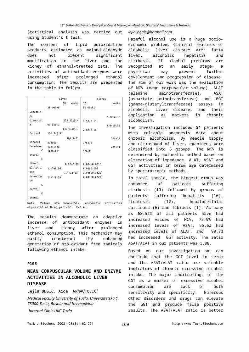
13th Balkan Biochemical Biophysical Days & Meeting on Metabolic Disorders’ Programme & Abstracts
INFLUENCE OF SELENIUM INTAKE ON OXIDATIVE STRESS PARAMETERS IN THE BLOOD OF RATS TREATED WITH ADRIAMYCIN OR 5-FLUOROURACIL*Olga Jozanov-Stankov, *Miroslav Demajo, **Ivana Djujic, *Ljiljana Markovic*Institute of Nuclear Sciences “Vinca”, P.O.Box 522, Belgrade, **Institute of Chemistry, Technology and Metallurgy - Center of Chemistry, Belgrade, Serbia and Montenegro, [email protected]
Most of the drugs used today in the therapy of malignant diseases express their effects either through inducing or by the direct production of reactive oxygen species (ROS). Increase ROS generation causes additional engagement of the antioxidant defense system (AODS) in the organism, as well as inducing changes in parameters indicating to the presence of oxidative stress. Being non-specific, these cytostatics, beside the neoplasm also affect healthy tissue. In our investigation we compared AODS compounds in blood after two frequently applied cytostatics, Adriamycin (doxorubicin, ADR) and 5-Fluorouracil (5-FU), that were injected into healthy experimental rats (ADR in one i.v. dose and 5-FU i.p. in 5 equal daily doses, each total amount corresponding to one standard human therapeutic cycle or dose), under conditions of (1) moderate microelement Selenium (Se) deficiency and (2) under optimal intake of Se. In Se supplementation, the animals received organically bound Se with drinking water (dose which corresponds to about 100g/day) for a month before treatment with ADR or 5-FU. The experimental results showed that treatment with ADR induced a significant increase of glutathione peroxidase (GSH-Px) activity in erythrocytes (RBC) and plasma and RBC catalase (CAT) activity. 5-FU had a direct counter effect in both cases for GSH-Px activity while CAT not significantly enhanced. Se supplementation significantly increased GSH-Px activity in blood but did not change CAT activity. In animals with adequate Se intake treatment with ADR or 5-FU resulted in the same changes in GSH-Px activity, but in lesser extent, while the CAT activity higher increased in those groups. These results show that ADR and 5-FU have a different mechanism of AODS engagement and that an optimal intake of Se may improve the defense of organism by diminishing the changes in AODS enzymes induced by those drugs.
P204
ELEVATION OF FIBRINOGEN AND D-DIMER LEVELS OF TYPE II DIABETES MELLITUS PATIENTS WITH NEPHROPATHYPolat ÇALIŞKANER*, Ayşenur ATAY*, Zeliha HEKİMSOY**, Mehmet H. Köseoğlu**Ataturk Training Hospital , Department of Biochemistry and Clinical Biochemistry, 35360, Izmir /TURKEY**Celal Bayar University, Faculty of Medicine, Department of Endocrinology, Manısa /[email protected]
Renal failure develops in 5 to 10 % of Type II Diabetes Mellitus (DM) patients. Diabetic nephropathy (DN) is one of the important causes for end stage renal failure. Atherothrombosis is a major cause of death in renal failure. Hyperlipidemia in diabetes produces hypercoagulability or hypofibrinolysis. We investigated the levels of some coagulation proteins in type II diabetes mellitus wıth nephropathy in this study. Thirty-four type II DM patients with diabetic nephropathy and 58 Type-II DM patients without diabetic nephropathy were included in this study. Fibrinogen, D-dimer, antithrombin III, protein C and protein S, PT, APTT were determined on ACL Futura Plus coagulatıon analyser with commercial assay kits.According to the rate of urinary albumin excretion in the 24 –h urine collection, patients were divided into 3 groups; Group I- Macroalbuminuria >300mg/24 h (n=10)Group II- Microalbuminuria 30-300 mg/24 h (n=24)Group III- Normoalbuminuria <30 mg/24 h (n=58)SPSS (Version 11.0) for Windows XP program was used for statistical analysis. Measurable plasma variables were analyzed with One Parameter Kolmogorov-Simirnov test, One Way Anova, Kruskall-Wallis multiple comprasions test techniques. Fibrinogen levels were significantly higher in macroalbuminuria group (535.59+/-125.85 mg/dl, p<0.000 ) than normoalbuminuria (403.58+/-55.38 mg/dl). D-dimer levels were significantly higher in macroalbuminuria group (839.77+/-128.68 ng/ml, p<0.000 ) than normoalbuminuria (231.08+/-176.19 ng/ml )Due to inflammatory response of diabetic nephropathy, fibrinogen levels were increased as an acute phase response. Increased free radical and Advanced Glycosylation End products in diabetic nephropathy may be led to endothelial damage so that plasma fibrinogen levels increase. PT, APTT, AT III, Protein C and Protein S activity levels were not significantly different. This findings showed that D-dimer and plasma fibrinogen levels in diabetic nephropathy increase due to their positive acute phase response behavior, depending on inflammatory response and renal dysfunction has effects on them.
P205
REGULATION OF MUSCARINIC RECEPTOR EXPRESSION IN K562 CELLS BY CHRONIC AGONIST EXPOSURE.Hülya CABADAK1,Banu AYDIN1,Beki KAN1
Marmara University School of Medicine, Department of Biophysics Tıbbiye Cad.No 49 34716- İstanbul/[email protected] receptors which are members of G protein coupled receptors , mediate a variety of cellular responses, including inhibition of adenylate cyclase, breakdown of phosphoinositide and modulation of K channels. Many cells express a mixture of muscarinic receptor transcripts. Agonist induced loss of muscarinic receptors has been reported in a number of cell lines. In this study, we have investigated the effect of agonist exposure on m2 and m3
muscarinic receptor transcripts, using RT-PCR assay.
Turk J Biochem, 2003; 28(3), 62-224 http://www.TurkJBiochem.com169
13th Balkan Biochemical Biophysical Days & Meeting on Metabolic Disorders’ Programme & Abstracts
K562 cells were grown in suspension using RPMI medium supplemented with 10% fetal calf serum at 37C in a 5% CO2 humidified atmosphere. Cells were usually seeded at a density of 105 cells/ml and passaged every 4-5 days. K562 cells were challenged with 100M carbachol for different times. The cells were washed twice , resuspended in phosphate buffered saline (PBS) and were centrifuged at 700g for 5 min at room temperature .Total RNA was isolated by the guanidium thiocyanate-phenol-chloroform extraction method, as previously described (Chomsky and Sacchi ,1987). Purity and quantitation were assessed by A 260/A280 ratios . RNA samples were analysed by RT-PCR . Analysis of PCR reactions was performed on 2% agorose gels stained with ethidium bromide.RT-PCR analysis showed that each muscarinic transcript was differentially regulated. m3 was expressed as much higher levels than m2 in K562 cells. The levels of m3 and m2 mRNA's were compared at 1,3,5, 24 and 48 hours of agonist challenge. When compared to the level of expression at one hour after carbachol treatment, a decrease in mRNA transcripts was observed for m2 and m3
receptors after five hours of challenge.Acknowledgement:This work was supported by grant from L'ORĒAL TURKEY (UNESCO)
P206
THE EFFECT OF SYSTEMIC ADMINISTRATION OF ALENDRONATE ON PLASMA GLUTATHIONE AND LIPID PEROXıDE LEVELS FOLLOWING TOOTH EXTRACTİON IN RATSAzize ŞENER1, Hatice ALTUNDAL2, Bahar GÖKER1, Ertuğrul YURTSEVER1
1Marmara University, Faculty of Pharmacy, Department of Biochemistry, 34668, İstanbuL/TURKEY2Yeditepe University, Faculty of Dentistry, Department of Oral Surgery, 34668, İstanbuL/TURKEYA wound can be described as the damage to a tissue integrity. The tissue damage can result from several factors. One of the significant mechanism in cell damage is the destruction due to free radicals. Eventhough the mechanism of the oxygen free radicals formation is thoroughly underrstood, the role of these compounds on healing process in wounded tissue has not yet been clearly elucitated.The aim of this study was to investigate the effect of the free radicals formation on plasma lipid peroxide and glutathione levels during soft tissue healing following tooth exraction in rats. In addition, the effect of alendronate, which is applied to prevent alveolar bone loss following tooth exraction, on plasma glutathione and lipid peroxide level was investigated.In this experimental study, 7-8 weeks old male Wistar albino rats were used. The rats were divided in to three group: baseline group, saline treated groupand alendronate-treated group. Both saline and alendronate treated groups were divided into two subgroups: 14 and 28 day follow up
groups. In the baseline group, blood samples were collected before tooth extraction. The right mandibular first molars were extracted under general anaestehsia. The two alendronate treated subgroups, were administered with daily amount of 0,25 mg/kg alendronate (Merc Sharp & Dohme) subcutaneously for 2 and 4 weeks respectively. The saline treated subgroups were given a daily saline solution for 2 and 4 weeks respectively, as well. The rats in the saline and alendronate treated groups were sacrificed 14, 28 days following tooth exraction. Before sacrification, blood samples were collected.The levels of plasma glutathione and lipid peroxide were measured.A increase in the level of plasma lipid peroxide was observed in saline and alendronate treated groups on day 14 as compared with the baseline group. The level of plasma lipid peroxide on day 28 lower than on day 14. In the saline and alendronate treated groups, the level of plasma glutathione decreased on day 14 and an increase was observed onday 28 as compared with 14 day values. These decreases and increases were not significant istatistically. In alendronate treted group, alendronate did not cause a sifnificant difference in the level of plasma glutathione and lipid peroxide both on day 14 and 28 as compared with saline treated group.
P207
ATHEROSCLEROTIC POLYMORPHISMS IN POSTMENOPAUSAL WOMEN WITH ESTABLISHED CORONARY DISEASELale AFRASYAP1, Ibrahim BARIS2
1Mugla University, Health High School, Department of Biochemistry, Mugla ; 2 Bogazici University, Department of Molecular Biology and Genetic, Istanbul/ [email protected] incidence of coronary disease risk due to atherosclerosis is higher in men and postmenopausal women than in premenopausal women. Although the polymorphisms of the MTHFR (C677T and A1298C) and eNOS (G894T) genes were investigated in different population groups with coronary disease, very few studies have addressed about the association between these polymorphisms and coronary disease in postmenopausal women. The aim of study is to investigate if genetic mutations increase the risk of coronary disease in postmenopausal women. The study was organized for 40 postmenopausal women with an intact uterus. They were divided into two groups, according to angiography results. 1- 25 women with >50% stenosis affecting at least one artery were included in group with coronary heart disease (patients) 2-15 women with < 20 % stenosis were enrolled in group without disease (controls). Mean ages of patients and controls were 64,06±8,65 and 66,12±6,80, respectively. After DNA was extracted from whole blood samples with salting-out method, genotypes were analyzed by polymerase chain reaction-restriction fragment length polymorphism. Statistical analyses were computed by SPSS 11,5 version, using nonparametric tests. Although
Turk J Biochem, 2003; 28(3), 62-224 http://www.TurkJBiochem.com170
13th Balkan Biochemical Biophysical Days & Meeting on Metabolic Disorders’ Programme & Abstracts
the prevalences of 1298CC and 1298CC/AC were higher in patients with respect to controls (p=0.009; p=0,016, respectively),the significant difference was not observed in the prevalences of the other genotypes between the groups. There was the positive correlation between coronary disease and the frequency of 1298CC ( r=0,447 p=0,017) The odds ratio was 1,71 (p=0,038, 95% CI, 1,00 to 2,92) in the patients with 1298CC mutation with respect to without. It was also 1,71 ( p=0,026, 95% CI, 1,00 to 2,66) for 1298 CC as compared with 1298 AA/ AC combination. The high prevalence of the 1298 CC genotype might be effective on the genesis of the disease itself and an important risk factor in the occurrence of coronary disease in postmenopausal women. P208
DATABASES IN BIOINFORMATICSÇağla AKMEMİŞ, Azmi TELEFONCUDepartment of Biochemistry, Faculty of Science, Ege University, Bornova/İzmir, [email protected] progresses in molecular biology have revealed the large fractions of the genome sequences during the last decades. Public sequence databases have been growing at exponential rates. Storage, organization and cataloging of this information became indispensable by information science methods under the field called bioinformatics.Bioinformatics deals with the recording, storage, annotation, analysis and retrieval of sequence and structural information. Thus, these databases provide to reach various information in internet. The main function of biological databases is to make biological data available to scientists in computer-readable form. Published data may be difficult to find access, and collecting it from the literature is very time consuming. And not all data is actually published explicitly in an article (genome sequences!). Therefore having the data in computer-readable form (rather than printed on the paper) is necessary first step, since analysis of biological data almost always involves computers. These databases and its contents are being increased by received information from laboratories in all around the world day by day. Eventually, this pioneering new field aims to enable the discovery of new biological insight as well as to create a global perspective of virtual cells that can be used as models for disease prediction, diagnosis and treatment.
P209
CONCENTRATION AND TEMPERATURE DEPENDENT STUDIES OF INTERACTION OF MELATONIN WITH LIPID MEMBRANESİpek ŞAHİN1, Nadide KAZANCI1 and Feride SEVERCAN2
1Ege University, Faculty of Science, Department of Physics, 35100, Bornova-İzmir/TURKEY2Middle East Technical University, Department of Biology, 06531, Ankara / TURKEY
Melatonin is an lipophilic antioxidant drug which is widely used for the prevention from several diseases. In the present study we will report the results of melatonin induced changes occuring in dipalmitoyl phosphatidylcholine (DPPC) membranes using Fourier transform infrared (FTIR) spectroscopy and differential scanning calorimetry (DSC) . Infrared spectra were obtained using a Bomem 157 FTIR Spectrometer which was continiously purged with dry air. The spectra were recorded in the 4000-1000 cm-1 region with CaF2 window using 12 m path length. Interferograms were accumulated for 50 scans at 2 cm -1
resolution. The Grace-Specac temperature controller unit was used for temperature regulation. Bomem Easy software was used for all FTIR data manipulations. For DSC studies, a TA Q100 DSC instrument was used with a heating rate of 1C/min.The infrared spectra of DPPC multilamellar liposomes, both pure and containing different concentration of melatonin were investigated as a function of temperature. The C-H stretching, the C=O stretching and PO-
2
antisymmetric stretching mode were considered. The results of both FTIR and DSC studies reveal that melatonin changes the physical properties of the DPPC bilayers by decreasing the main phase transition temperature, abolishing the pretransition, ordering the system in the gel phase, increasing the dynamics of the system and causing strong hydrogen bonding in between the C=O and PO-
2 groups of DPPC and either melatonin or the water molecules, both in the gel and liguid crystalline phases. Furthermore melatonin, at high concentrations, induced phase separation in DPPC membranes.This work has been partially supported by Ege University Research Fund AFP: 2002 Fen 025
P210
THE EFFECTS OF SOIL FLOODING ON THE ANTIOXIDATIVE ENZYMES IN BARLEY PLANTSRusina YORDANOVA-ZLATANOVA, Kaloyan CHRISTOV and Losanka POPOVAAcad. M. Popov” Institute of Plant Physiology, BAS, 1113, Sofia/[email protected] deprivation is the primary stress factor in flooded soils. In most cases oxygen shortage affects directly the roots and indirectly the shoots. When tissues are hypoxic or anoxic the oxygen-dependent pathways are suppressed, the functional relationships between roots and shoots are disturbed, and both carbon assimilation and photosynthate utilization are suppressed. As a consequence of perturbed photosynthetic activity and lowered photon utilizing capacity, reactive oxygen species (ROS) are photoproduced. When the formation of ROS is in excess of antioxidant scavenging capacity thus creates oxidative stress. Our major aim was to investigate the impact of root hypoxia on the scavenging system against active oxygen in leaves of barley plants (Hordeum vulgare cv. Alfa). Effects of soil flooding on the activity of foliar antioxidative
Turk J Biochem, 2003; 28(3), 62-224 http://www.TurkJBiochem.com171
13th Balkan Biochemical Biophysical Days & Meeting on Metabolic Disorders’ Programme & Abstracts
enzymes - superoxide dismutase (SOD), catalase (CAT), peroxidase (POD), ascorbate peroxidase (APX), and glutathione reductase (GR) were studied. Seventy two to 120 h of soil flooding decreased the activity of SOD and the decrease was mainly due to progressive reduction in the activity of Fe-containing SOD located in chloroplasts. It was lowered by about 55% at 120 h after the start of the treatment whereas the chloroplastic SOD in the control plants remained unchanged. The activity of POD significantly increased and 120 h after flooding it was 2 fold higher than the control. The changes in the activity of CAT followed the same tendency as for POD activity. Soil flooding affected differently the activity of GR and APX. GR activity was insignificantly influenced over the course of treatment. Both total soluble and thylakoid-bound APX activity increased in all flooded plants. Regardless of the increased activity of hydrogen peroxide scavenging enzymes, flooding treatment caused a substantial rise in total endogenous peroxide content. It is suggested that root oxygen deficiency caused photoxidative damage to barley leaves via an increased generation of active oxygen species.
P211MESOMORPHIC AND MORPHOLOCIG PROPERTIES OF THE BINARY LYOTROPIC LIQUID CRYSTAL SYSTEMÖzgür Masalcı, Nadide KazancıEge Universty, Faculty of Science, Department of Physics, 35100, Bornova- İzmir/ TURKEYLyotropic liquid crystal are constructed by amphiphilic molecules which are consisted of polar head groups and apolar long hydrocarbon chains. When they are dissolved in water they form micellar aggregates . Different mesophases can be obtained by increasing amphiphile concentration. Lyotropic liquid crystal in nature especially is in living system. Many structures of living system are made up of lyotropic liquid crystal; such as membrane, lipids, hemoglobin, polypeptide, albumin, etc. Lyotropic liquid crystal systems could be used as a model for investigation on biological structures.In this work, the phase states of the tetradecyltrimethly ammonium bromide (TTAB)+water binary lyotropic systems have been investigated. The mesomorphic and morphologic properties have been determined. During the experimental part of this study, in order to be determined this properties, polarizing polythermic microscopy technique was used. The experimental instrument which was used is Olympus BX-P polarized microscop. Prepared samples which are different concentration were examined under the this polarized microscope. The objects of our research were the isotropic micellar phase, nematic-calamatic and hexagonal mesophase formed in the TTAB+water binary lyotropic systems. We also investigated the behavior of electro conductivity in large temperature intervals. In order to measure electroconductivity inoLab Cond Level 3 conductivity measuring system was used. The connection morphologic and electro conductivity properties of determined mesophases will be analyzed.
P212INFULENCE OF IRON AND MANGANESE CONCENTRATION ON METALS UPTAKE, ANTIOXIDANT ENZYMES RESPONSE AND MEMBRANE LİPİD PEROXIDATION LEVELS BY FUSARIUM EQUISETI AND F.ACUMINATUMHulya AYAR KAYALI and Leman TARHANUniversity of Dokuz Eylul, Faculty of Education, Department of Chemistry, 35150 Buca, [email protected] relationship between metal uptake, antioxidant enzyme activity, membrane lipid peroxidation level variations and manganase-iron concentrations in F.equiseti and F.acuminatum medium were investigated with respect to incubation period. Intracellular iron contents of F.equiseti and F.acuminatum were significantly increased with increase in iron concentration. In this growth medium, intracellular magnesium and zinc levels of both Fusarium species have been efficiently decreased with respect to iron concentration in the medium, although intracellular manganese levels have been increased up to 3.7 µM Fe2+. On the other hand, intracellular manganese and magnesium levels were increased with respect to increase in manganase concentration while iron levels were increased up to 5.9 µM Mn2+. Maximum SOD activity of F.equiseti were determined in medium containing 5.9 µM Mn2+ as 78.66 ± 1.49 while the value of F.acuminatum was 141.7 ± 4.53 IU/mg in 30 µM Mn2+ supplemented medium. In addition, the maximum CAT activities of F.equiseti and F.acuminatum were observed at 5.9 µM Mn2+ as 324.2 ± 8.5 and 225.1 ± 5.63 IU/mg, respectively. On the other hand, LPO level variations of both F.species showed negative correlation with SOD and CAT activities
P213
THE AMOUNTS OF HEAVY METALS IN COW’S RAW MILK SAMPLES COLLECTED FROM THRACETulay Engizek, Betül Yurttaş, Saim Ergenç, Süreyya Günebakan, Melek Özlem Kolusayın Ozarİstanbul University, Faculty of Sciences, Biology Department, ISTANBULIt has been known that in most countries, lackness of trace elements and heavy metals cause metabolic disorders in insufficient nutrited children. Because of that, international studies point out the importance about the studies which carry on the amounts of trace elements and heavy metals existing in essential foods. Besides, trace elements are very important in toxicologic issues. Development in industry especially in industrialized countries, instead of maternal milk cow and sheep milk or prepared nutrients for babies are preferred. Within these nutrients, cow milk has a widespread use.In our investigation , the amounts of zinc, copper and lead in cow’s raw milk samples, collected from area of Hayrabolu, Kırklareli, Malkara and Çatalca, that have been sent to the Industrial Association of Milk (İstanbul)
Turk J Biochem, 2003; 28(3), 62-224 http://www.TurkJBiochem.com172
13th Balkan Biochemical Biophysical Days & Meeting on Metabolic Disorders’ Programme & Abstracts
were measured with atomic absorption spectrophotometer. Annual average amounts of zinc, copper and lead in milk were 2.65±0.169 ppm, 0.11±0.011 ppm, 0.05±0.008 ppm in Hayrabolu, 2.54±0.141 ppm, 0.13±0.022 ppm, 0.04±0.005 ppm in Kırklareli, 2.59±0.165 ppm, 0.13±0.003 ppm 0.03±0.005 ppm in Malkara, 2.64±0.177 ppm, 0.12±0.009 ppm, 0.04±0.006 ppm in Çatalca, respectively. The values are compared with similar studies carried out for other countries.We conclude that measured zinc amount is twenty fold than copper and measured copper amount is twice than lead amount according to the mean values of each areas.Key Words: Milk, Zinc, Copper, Lead
P214THE NATURE OF DELAYED CHLOROPHYLL FLUORESCENCE INDUCTION MAXIMA APPEARED DURING FIRST ONE SECOND OF TRANSITION FROM DARK TO LIGHT ADAPTED STATE OF BARLEY LEAVESIvelina ZAHARİEVA1, Stefka TANEVA1, Reto J. STRASSER2, Vassilij GOLTSEVDepartment of Biophysics and Radiobiology, Faculty of Biology, St. Kliment Ohridski University of Sofia, 8, Dragan Tzankov Blvd., 1164, Sofia, Bulgaria, E-mail: [email protected]; 1 Institute of Biophysics, Bulgarian Academy of Sciences, “Acad. G. Bonchev” Str., Bl.21, 1113 Sofia, Bulgaria;2 Bioenergetics Laboratory, University of Geneva, CH-1254 JUSSY – GENÈVE Switzerland
The kinetic components of delayed chlorophyll fluorescence (DF), decayed from 0.35 to 5.5 ms dark interval, are analyzed during first one second of actinic illumination of dark adapted barley leaves. DF is represented by three components: a sub-millisecond one, with lifetime of ~ 0.6 – 0.9 ms, a millisecond with lifetime of ~ 1.2 – 3.5 ms and a slow with lifetime of >> 5.5 ms. The DF changes during induction are compared with simultaneously registered chlorophyll fluorescence transients and 820 nm absorption changes that correlate with P700 reduction. Both amplitudes and lifetimes of DF components are modified typically during induction. It was shown, that the first DF maximum, I1, appeared at 20–30 ms after beginning of illumination, is produced by both sub- and millisecond DF components. It correlates with formation of high relative concentration of opened Photosystem II (PS II) reaction centers with secondary quinone acceptor at reduced or semi-reduced state as well as with transmembrane electrical gradient formation. The second maximum, I2, is observed at 100–150 ms of illumination and includes predominantly millisecond DF component. Its rise is associated with reaction center reopening as a result of QB
= reoxidation. At the end of the first second of illumination a minor peak, I3, was registered that is associated with slowest component of light emission from closed PS II reaction centers. We are supposed that the emission of observed DF components is a result of charge recombination in PS II reaction centers at different redox states: Z+QA
–QB– – for sub-millisecond, Z+QA
–QB= –
for millisecond and SiZQA–QB
= – for slowest DF component, respectively.Acknowledgments. This work was financially supported by the Swiss National Science Foundation (SCOPES 2000–2003 grant № 7BUPJ062408.00/1).
P215A MATHEMATICAL MODEL OF THE KINETIC COMPONENTS OF MILLISECOND DARK DECAY OF DELAYED CHLOROPHYLL A FLUORESCENCE IN LEAVES DURING THE FIRST SECOND OF INDUCTIONPetko CHERNEV, Vassilij GOLTSEV, Reto J. STRASSER1
Department of Biophysics and Radiobiology, Faculty of Biology, St. Kliment Ohridski University of Sofia, 8, Dragan Tzankov Blvd., 1164, Sofia, Bulgaria, E-mail: [email protected]; 1 Bioenergetics Laboratory, University of Geneva, CH-1254 JUSSY – GENÈVE SwitzerlandA kinetic model that describes the redox reactions in the donor and acceptor side of Photosystem II (PS II) during the first second of transition from dark to light adapted state is designed. The model curves of the prompt and delayed fluorescence (DF) signal are fitted to experimental ones obtained by a phosphoroscope fluorometer that registers both signals simultaneously. The participation of different redox states of the reaction center in the formation of fluorescence signals is analyzed. A correlation between the redox states concentrations and the different components of DF dark decay is shown. The poly-exponential dark relaxation of DF between 0.35 and 5 ms
Turk J Biochem, 2003; 28(3), 62-224 http://www.TurkJBiochem.com173
13th Balkan Biochemical Biophysical Days & Meeting on Metabolic Disorders’ Programme & Abstracts
is approximated by 3 components with life-times of about τ1 ~ 0.6 ms (sub-millisecond component), τ2 ~ 3.5 ms (millisecond one) and a slow component with a life-time τ3
≥ 20 ms. The DF emission for the first kinetic component is associated with charge recombination in PS II reaction centers at redox state Z+QA
–QB–. Their concentration rises
up to 20–30 ms after the beginning of illumination. This DF component forms mostly the first peak, I1, of the DF induction curve. The time course of the concentration of centers in the Z+QA
–QB= redox state shows that these centers
take part in the formation of both I1 and I2 (which is observed after 100–150 ms of illumination), and are suggested to correspond to the millisecond component of DF dark relaxation. The possible role of transmembrane electrical gradient formation for the appearance of DF maximums is discussed.Acknowledgments. This work was financially supported by the Swiss National Science Foundation (SCOPES 2000–2003 grant № 7BUPJ062408.00/1).
P216
PROMPT AND DELAYED CHLOROPHYLL FLUORESCENCE OF INTACT LEAVES IN THE PRESENCE OF PHOTOSYNTHETIC HERBICIDESPetar LAMBREV1, Vassilij GOLTSEV, Reto J. STRASSER2
Dept. Biophysics and Radiobiology, Faculty of Biology, “St. Kliment Ohridski” University of Sofia,8 “Dragan Tzankov” Blvd., 1164 Sofia, Bulgaria. E-mail: [email protected] Institute of Biophysics, Bulgarian Academy of Sciences, “Acad. G. Bonchev” Str., Bl.21, 1113 Sofia, Bulgaria. E-mail: [email protected] Bioenergetics Laboratory, University of Geneva, CH-1254 Jussy – Genève SwitzerlandPrompt fluorescence (PF) and delayed fluorescence (DF) of chlorophyll a are extremely sensitive intrinsic probes for the function of the photosynthetic apparatus in vivo, however, they are still not sufficiently understood. We studied the effects of photosynthetic herbicides that block the Photosystem 2 electron transport on the PF and millisecond DF induction curves and the DF decay kinetics. The herbicides diuron and atrazine were applied in various concentrations to 14-days-old pea plants through their stems. PF and DF were measured simultaneously from detached leaves using a phosphoroscope fluorometer. High-resolution fluorescence induction transients (OJIP curves) were registered using a HandyPEA fluorometer (Hansatech, UK). The presence of diuron or atrazine in the leaves had a profound effect on both luminescence types that could be measured quantitatively. The herbicides diminished the second peak in the fast phase of the DF induction curve, I2, which is supposed to be related to the intersystem electron transport. The first maximum, I1, was less sensitive, in accordance with the concept that it arises due to a transiently generated transmembrane electrical gradient. The second component of the slow-phase DF peak, I5, was strongly inhibited, suggesting that it is a result
of a partial reopening of the reaction centres in non-treated samples. Only I4, which is supposed to reflect the thylakoid membrane energization, was expressed in the slow phase at high herbicide concentrations. In this case I4 might be related to a proton gradient built by Photosystem 1 via cyclic electron transport. PF was progressively quenched by increasing the herbicide concentration even when the electron transport was completely inhibited. This effect could not be fully explained by the now-accepted quenching by oxidised plastoquinone. Acknowledgments. This work was financially supported by the Swiss National Science Foundation (SCOPES 2000–2003 grant № 7BUPJ062408.00/1).
P217
GENERATION OF SPECIFIC ANTIBODIES AGAINST STRESS-RELATED PROTEINS FROM NaCL ADAPTED EMBRYOGENIC CALLUS OF DACTYLIS GLOMERATA L.Mima I. PETKOVA, Magdalena I. CHORBADJIEVA and Mariela ODJAKOVASofia University, Faculty of Biology, Department of Biochemistry, 1164 Sofia/[email protected] physiological and biochemical changes in plant tissue in response to different types of osmotic stresses are not completely understood. Despite extensive research of desiccation tolerance in plants, little is known about the genes and their related proteins involved in stress-defense mechanisms. The presence of some stress proteins at different stages of embryonic response shows that besides their protective function, they are important for embryogenic competence, too.The aim of our research is to generate specific antibodies against stress-related proteins We studied the changes in the protein profiles of intracellular, extracellular and ionically-bound cell wall proteins from embryogenic callus from Dactylis glomerata L. genotype Embryogenic P, selected and maintained on SH30 medium with different NaCl concentrations. Proteins from different cell compartments were isolated and subjected to 2D-PAAG electrophoresis. Upon comparing the protein pattern of secreted and ionically bound cell wall proteins we observed the appearance and accumulation of certain specific proteins from salt adapted lines. Phage display has been used in order to generate specific antibodies against some proteins characteristic for NaCl-selected lines. Proteins were transferred to a nitrocellulose membrane after 2D PAAGE and protein spots were used for the selection by phage display. Specific antibodies were selected after 5 round of panning from human synthetic single-chain Fv (scFv) phage display library (Griffin 1). Futher investigations are in progress in order to elucidate the possible use of these antibodies as markers for adaptation to salt stress.
Research was supported by grant 3457 from the Science Fund of Sofia University
Turk J Biochem, 2003; 28(3), 62-224 http://www.TurkJBiochem.com174
13th Balkan Biochemical Biophysical Days & Meeting on Metabolic Disorders’ Programme & Abstracts
P218
INTEGRATING MUTATION DATA AND STRUCTURAL ANALYSIS OF THE G6PD ENZYMEErdinç A. YALIN, Kıymet AKSOY
Çukurova University, Faculty of Medicine, Department of Biochemistry, 01330, Adana/Turkey
Glucose-6-phosphate dehydrogenase (G6PD, MIM# 305900) is a cytosolic enzyme encoded by a house keeping X-linked gene whose main function is to produce NADPH, a key electron donor in the defence against oxidizing agents and in reductive biosynthetic reactions. Hereditary deficiency of human G6PD is one of the most common human enzyme deficiency. The deficiency affects an estimated 400 million people worldwide with gene frequencies ranging from 5% to 25%. G6PD deficiency is very prevalent in the Çukurova Region of Turkey, a gene frequency about 8.2 % has been documented.
Beside about 440 different G6PD variants have been described based on biochemical and clinical characteristics, over 125 distinct mutations of G6PD have been identified to date. The relational databases integrates up-to-date mutations and structural data from various databanks. These databases and recently developed procedures provides insights into the molecular aspects and clinical significance of G6PD deficiency for researchers and clinicians, and these web-based functions as a knowledge base relevant to the understanding of G6PD deficiency and its management.
More than 50% of the mutations in the G6PD gene have been reported to be in severe (Class I) deficiency, and these affect dimer interface and/or coenzyme binding cleft, resulting in partial or complete loss of enzyme activity. Among the 104 distinct mutations we analysed 53 (50.9 %) mutations considered in Class I variants. We report here the results of systematic analysis of the effect of 53 mutations corresponding Class I variants, which can be explained in structural terms by their predicted effects on protein stability.
P219
THE EFFECT OF CAFFEIC ACID PHENETHYL ESTER (CAPE) ON CISPLATIN-INDUCED TOXICITY IN RAT LIVER TISSUESElif ÖZEROL1, Mukaddes GÜLEÇ1, Ersin FADILLIOĞLU2, Mustafa IRAZ3, Seda AĞLAMIŞ3, Ömer AKYOL1
Inonu University, Faculty of Medicine, Departments of 1Biochemistry,2 Physiology and 3Pharmacology, 44069 Malatya, Turkey
High doses of cisplatin have been known to produce hepatotoxicity. However, little is known about pathophysiology of cisplatin-induced liver injury. The present study was designed to determine the effect of cisplatin on liver oxidant/antioxidant system and the possible protective effect of caffeic acid phenethyl ester (CAPE) on liver toxicity induced by cisplatin. Adult female Wistar albino rats were divided into four groups (n=6 per group): Control, Cisplatin, CAPE, and Cisplatin+CAPE. Cisplatin was injected intraperitoneally (a single dose of 16 mg/kg bwt) to the second and the last groups of rats. CAPE was applied to the rats with a dose of 10 mol/kg/day (i.p.) one day before and 5 consecutive days after cisplatin injection. At the 5th day of cisplatin injection the experiment was finished and liver tissue was removed to study the activities of catalase (CAT), superoxide dismutase (SOD), glutathione peroxidase (GSH-PX), myeloperoxidase (MPO), xanthine oxidase (XO), adenosine deaminase (ADA), and levels of malondialdehyde (MDA) and nitric oxide (NO) in liver tissue.
The activities of SOD and GSH-Px were increased in Cisplatin+CAPE and CAPE groups in comparison with Cisplatin groups. The activity of CAT was higher in Cisplatin+CAPE group than other three groups. The activity of XO was lower in Cisplatin group than control group. Also, the activity of MPO was increased in Cisplatin group in comparison with control and CAPE groups. There were positive correlations between SOD and MPO, SOD and MDA, NO and ADA in Cisplatin group. There were positive correlations between SOD and CAT, ADA and XO, and negative correlations between CAT and MDA, SOD and MDA in Cisplatin+CAPE groups. There was a positive correlation in CAPE group between NO and CAT.
It can be concluded that CAPE prevents oxidative injury due to cisplatin in the liver tissue by increasing antioxidant enzyme activities and preventing MPO dependent reactive oxygen species production.
Key words: toxicity, cisplatin, antioxidant, caffeic acid phenethyl ester, rat, liver.
P220
THE ANTIULCEROGENIC EFFECT OF USNIC ACID ISOLATED FROM USNEA LONGİSSİMA ON INDOMETHACINE-INDUCED GASTRIC ULCER IN RATSFehmi ODABASOGLU a,* Halis SULEYMAN b, , Ahmet CAKIR c, Ali ASLAN d, Yasin BAYIR a, Mesut HALICI a, Osman YUCELe, Cavit KAZAZ f and Erdal DEMIRCEYLANa
aAtaturk University, Faculty of Pharmacy, Department of Biochemistry, 25240, Erzurum, Turkey. b Ataturk University, Faculty of Medicine, Department of Pharmacology, 25240, Erzurum, Turkey.
Turk J Biochem, 2003; 28(3), 62-224 http://www.TurkJBiochem.com175
13th Balkan Biochemical Biophysical Days & Meeting on Metabolic Disorders’ Programme & Abstracts
c Ataturk University, Kazım Karabekir Education Faculty, Dep. Chemistry, 25240, Erzurum, Turkey.dAtaturk University, Kazım Karabekir Education Faculty, Dep. Biology, 25240, Erzurum, Turkey.e Ataturk University, Sağlik Hizmetleri Meslek Yuksek Okulu, 25240, Erzurum, Turkey.fAtaturk University, Faculty of Science, Department of Chemistry, 25240, Erzurum, [email protected] the present study, the antiulcerogenic effect of usnic acid (UA) (a prototype of the dibenzofuran derivatives), isolated from diethyl ether extract of Usnea longissima on indomethacine-induced gastric ulcers in rats was investigated and compared with ranitidine. A total of 48 male, albino Wistar rats, weighing 180-190 g, have been used for the experiments. UA and ranitidine were administrated to the assigned groups of rats per orally, then the animals were sacrificed with high dose anaesthesia (thiopental sodium, 50 mg/kg). The stomachs of rats were removed and the gastric damage (or ulcer) in the stomachs was macroscopically evaluated. For the activity studies, 5, 10, 25, 50, 100 and 200 mg/kg doses of usnic acid were tested. Antiulcer effect of UA were determined by comparing to the results obtained from ranitidine (150 mg/kg dose), used as positive control, and control groups. In general, gastric damage in the rat groups treated with usnic acid and ranitidine was less than that of the control groups. While the mean damage areas in rats receiving the doses of 5, 10, 25, 50, 100 and 200 mg/Kg of usnic acid was 33.2±6.0, 22.9±5.3, 20.8±3.5, 16.7±11.7, 6.8±2.9, 18.2±8.6 mm2, respectively, they were 36.0±11.7 and 6.0±2.1 mm2 in the control and ranitidine groups, respectively. Among the treated doses, 50, 100 and 200 mg/kg doses of usnic acid showed potent antiulcerogenic activity in comparison with control groups and also 100 mg/kg dose of usnic acid was most effective in preventing of the gastric damage in rats. On the other hand, the antiulcerogenic acitivity of 100 mg of usnic acid was roughly the same as ranitidine, and as statistically it was not significant (p<0.05). These results suggest that usnic acid isolated from Usnea longissima have a significant antiulcer effect when assessed in indomethacine-induced ulcer model. Although the mechanism underlying this antiulcerogenic effect remains unknown, it seems to be related to an increase of the defensive mechanisms of the stomach such as prostaglandin synthesis. The good yield of usnic acid obtained from Usnea longissima, as well as its antiulcerogenic activity, suggest that this compound should be submitted to pharmacological research as a potential new antiulcerogenic drug.Keywords: Usnea longissima - usnic acid - antiulcerogenic activity – ranitidine – rat
P221
GABEXATE MESILATE INHIBITS LIPOPOLYSACCHARIDE-INDUCED TUMOR NECROSIS FACTOR-alpha PRODUCTION BY INHIBITING ACTIVATION OF BOTH NUCLEAR
FACTOR-kappaB AND ACTIVATOR PROTEIN-1 IN HUMAN MONOCYTESMehtap YÜKSEL*,†,§, Kenji OKAJIMA†, Mitsuhiro UCHIBA†, Gül GÜNER*,‡, Hiroaki OKABE†
[email protected]*Research Laboratory and ‡Department of Biochemistry, Dokuz Eylül University School of Medicine, İzmir, TURKEY; †Department of Laboratory Medicine and §Department of Biochemistry, Kumamoto University School of Medicine, Kumamoto, JAPAN [Objectives] Gabexate mesilate is a synthetic protease inhibitor that has anticoagulant activities. Gabexate mesilate was shown to be effective in treating patients with disseminated intravascular coagulation associated with sepsis. Gabexate mesilate inhibits lipopolysaccharide (LPS)-induced tumor necrosis factor-alpha (TNF-alpha) production by monocytes. To elucidate the mechanism(s) by which gabexate mesilate inhibits LPS-induced TNF-alpha production, we examined the effect of gabexate mesilate on LPS-induced activation of nuclear factor-kappaB (NF-kappaB) and activator protein-1 (AP-1) in human monocytes in vitro. [Methods] Monocytes were isolated from human buffy coats. Monocytes were activated by LPS and TNF-alpha levels in the supernatant were measured by enzyme-linked immunosorbent assay. The binding of NF-kappaB and AP-1 to target sites were determined by electromobility shift assay. Degradation of IkappaB and phosphorylation of IkappaB, c-Jun N-terminal kinase (JNK) and p38 mitogen-activated protein kinase (p38 MAPK) were determined by Western blot analysis. [Results] Gabexate mesilate inhibited LPS-induced TNF-alpha increase 4 hours after stimulation in a concentration dependent manner. Gabexate mesilate significantly inhibited LPS-induced binding of NF-kappaB and AP-1 to target sites. Gabexate mesilate also significantly inhibited degradation of IkappaB and phoshorylation of IkappaB, JNK and p38 MAPK.[Conclusion] These observations suggested that gabexate mesilate could regulate LPS-induced monocytic production of TNF-alpha by inhibiting activation of both NF-kappaB and AP-1. These results would at least partly explain the mechanism(s) by which gabexate mesilate exerts its therapeutic effects in patients with sepsis.P222
DIABETES MELLITUS: CLINICAL SIGNIFICANCE OF LIPID DISTURBANCESD.Pap, S.Bugarinovic1, V.Canic2
Clinical-biochemical Laboratory, Medical Center Sremska Mitrovica, Yugoslavia1 Psychiatric Hospital, Kovin, Yugoslavia2Health Center, Sokobanja,YugoslaviaThe purpose of this study was to investigate diagnostic relevance of disturbances in lipid metabolism, which is suitable for predictive coronary risk assessment in diabetes mellitus (DM). The studied subjects were the control group (136 healthy subjects) and the experimental group (188
Turk J Biochem, 2003; 28(3), 62-224 http://www.TurkJBiochem.com176
13th Balkan Biochemical Biophysical Days & Meeting on Metabolic Disorders’ Programme & Abstracts
DM patients). The DM patients were divided in 4 groups: M1 – 54 non-insulin dependent DM patients (NIDDM) on diet; M2- 68 NIDDM patients on oral antidiabetes therapy; M3 –32 NIDDM patients on insulin; M4 –34 insulin dependent patients (IDDM).We examined: levels of total cholesterol (TCH), triglycerides (TG), HDL-c, LDL-c, VLDL-c, the index of atherosclerosis IA (LDL-c/HDL-c) and established risk factors RF (TCH/HDL-c). While HDL-c was found to be significantly lower in all patients with DM, LDL-c was significantly higher in M4 and M2 group, but the difference wasn΄ t significant in M1 and M3 in comparison to controls. Values of TCH were not significant in all groups of DM regardless to their therapy. The values of TG were significantly higher in all groups (p< 0,05); expect in group of NIDDM on insulin in comparison to control group. Values of IA and RF were indicated a high risk of atherosclerosis in all groups of DM (p< 0,01). These data suggest that low levels of HDL-c are good predictors of atherosclerotic risk in all groups of DM, regardless to their therapy.
P223
ALTERATIONS IN ERYTHROCYTE TRANSMEMBRANE ANION TRANSPORT OF OPERATORS IN RADIO AND TV STATIONSMargarita KOUZMANOVA1, Miroslava HRISTOVA1, Katja VANGELOVA2
1Department of Biophysics and Radiobiology, Biological Faculty, Sofia University, 8 Dragan Tzankov blvd.,1164 Sofia, Bulgaria2 National Center of Hygiene, Medical Ecology and Nutrition, 15 D. Nestorov blvd., 1341 Sofia, [email protected] frequency electromagnetic fields (EMF) are widely used for transmitting of radio and TV signals, in wireless communications, etc. More and more people are exposed to EM radiation not only while at work but also at home. This study was designed to investigate the changes in anion transport across the erythrocyte membrane of the staff of radio and TV stations.The blood samples were taken from workers at different radio and TV transmitting stations. Each of these stations has some antennas emitting on various frequencies in MHz and GHz range. Three experimental groups have been set according to working time. The people, working on shifts (24 hours on work, 3 days away) were divided in two groups. In the first group blood samples were taken at the beginning of the next shift. The people from the second group were tested right after the end of the 24-hours workday. The third group included people that work 8 hours/day and the blood was taken at the beginning of work time. The measurements were performed using the modified pH-metric method of Glaser. The results obtained from the pH-test fit closely to sigmoidal logistic (dose-response) curve. This curve is defined by four parameters, which can be compared to Student-Fisher’s t-test.Statistically significant alterations in the calculated parameters were registered among the three groups
working at one and the same station, as well as between the groups from different stations. The alterations observed at the end of 24-hours shift are temporary and reversible. They were not detected at the beginning of the next shift after 72 hours away. These alterations are probably a result not only from the action of EM radiation, but also from long-lasting sleeplessness and exhausting of the organism. The exposure to EM radiation with various frequencies and intensities leads to variations in the investigated parameters among the workers in the different radio and TV stations. The obtained results show that working for years in environment with EM radiation could cause permanent alteration in the ion transport across the erythrocyte membrane.
P224
A SITE-DIRECTED MUTANT OF THE DEHYDROQUINATE SYNTHASE SHOWS A DECREASE IN ENZYMATIC ACTIVITY BY IN VIVO COMPLEMENTATIONA. Günel-Özcan1 and K.A. Brown2
1Kırıkkale University, Medical School Medical Biology Department, Kırıkkale, 71100 Turkey; 2 Imperial College London, Department of Biological Sciences, Centre for Molecular Microbiology and Infection, London SW7 2AZ, UKDHQS (5-dehydroquinate synthetase) is the second enzyme of the shikimate pathway and catalyzes the conversion of DHAP to DHQ (dehydroquinate). DHQS requires both NAD+ and Zn2+. In this study, we carried out site-directed mutagenesis on the Salmonella typhimurium DHQS encoded by the aroB gene .One of the highly conserved regions of DHQS contains the following motıf GHXXGHAXEXXXXXXXXXHG which includes residues important for Zn2+-binding. Based upon the crystal structure of DHQS from Aspergillus nidulans, two histidine residues (247, and 264) are predicted to form part of the zinc-binding site. A site-directed mutant was created which replaced the histidine side-chain at position 247 to a leucine in S. typhimurium DHQS. A PCR-based method was used requirıng four primers and three PCR reactions. The amplification primer carried three mismatches compared to the wild-type template sequence. Two of these mismatches were silent mutations which created an extra restriction enzyme site that could be used to screen for the presence of the mutation. The generated restriction enzyme site was StuI. Pfu polymerase was the enzyme of choice for the PCR. This site-directed mutant was cloned into the pET21d expression vector. The aroB gene encoding wild-type S. typhimurium DHQS was also cloned into the pET21d expression vector. Both of these contructs were transformed into an E.coli aroB-(DE3) strain for protein production. Wild-type S. typhimurium DHQS, which is 93.9% identical to E. coli DHQS, grew on minimal medium lacking aromatic amino acid supplementation. This result indicated that the S. typhimurium DHQS could complement E. coli DHQS activity. However, the H247L mutant failed to complement E. coli DHQS activity suggesting that this histidine side chain is essential for
Turk J Biochem, 2003; 28(3), 62-224 http://www.TurkJBiochem.com177
13th Balkan Biochemical Biophysical Days & Meeting on Metabolic Disorders’ Programme & Abstracts
correct folding and/or activity of the S. typhimurium enzyme.
P225
MODULATING EFFECT OF FULLERENOL C60(OH)20O2H2 ON CYTOTOXICITY INDUCED BY ANTITUMOR DRUGS ON SELECTED HUMAN CARCINOMA CELL LINESVesna KOJIĆ1, Dimitar JAKIMOV1, Aleksandar ĐORĐEVIĆ2, Gordana BOGDANOVIĆ1, Mirjana VOJINOVIĆ-MILORADIV2.1 Institute of Oncology Sremska Kamenica, Novi Sad, Yugoslavia2 TEMPUS Centar, University of Novi Sad, Novi Sad, YugoslaviaE-mail: [email protected] fullerene C60 derivatives - fullernols have attracted much attention due to their numerous biological characteristics. Most of these biological features are based on the ability of fullerenols to scavenge free radicals.Investigated antitumor drugs have various mechanisms of action. For most of them toxic activity can be explained by formation of free radicals.The aim of this paper was to investigate activity of fullerol C60(OH)20O2H2 on growth of human breast cancer cell lines and its modulatory effect on Adriamycin (ADR), Cisplatin (cis-Pt), Taxol, and Thiazofurine - induced cytotoxicity on the same cell lines. Growth inhibition was evaluated by colorimetric SRB essay.The cell growth was investigated on two cell lines: MCF7 (human breast adenocarcinoma; estrogen receptor positive (ER+)) and MDA-MB-231 (human breast adenocarcinoma, estrogen receptor negative (ER-)). Cell lines were treated with fullerenol at concentrations 0.9-3.9g/ml. Fullerol was given alone and in combination with cytostatics at various concentrations ranging from 10-4 to 10-8 M during 2 hours. Cytotoxic effect was evaluated 24h or 48 h after treatment .Fullerol mildly inhibits growth of both cell lines (3-15%). Combination of fullerenol and cytostatics, given simultaneously, resulted in various growth inhibition depending on fullerenol concentration, type of antitumor drug and cell line as well.Fullerol differently modulates cytotoxic effects of given cytostatics.The protective action of the cytostatic drugs was more pronounced in comparison to Taxol,whose action is based on formation of free radicals.Key Wordts: fullerol C60(OH)20O2H2, Cytotoxicity, Adriamycin, Cisplatin, Taxol, Thiazofurine, Breast cancer cell line.
P226
DETERMINATION OF STEADY-STATE LEVELS OF 8-OxoGuanine IN CALF THYMUS DNA BY MEANS OF FPG PROTEIN
Beran YOKUŞ1, Naime CANORUÇ2, Dilek Ülker ÇAKIR2, YILDIZ ATAMER2, Abdurrahman KAPLAN2, Sabri BATUN2
1Dicle University, Veterinary Faculty Department of Biochemistry, Diyarbakir2Dicle University, Medical Faculty Departments of Biochemistry, Diyarbakir7,8-dihydro-8-oxoguanine (8-Oxoguanine or 8-OxoGua), a major DNA damage resulting from oxidative attack, is highly mutagenic leading to translation of GC→AT . DNA adduct are lethal if not repaired. The primary function of Base excision repair (BER) enzymes are known to recognise various types of base damage: oxidised purine, pyrimidine damages and remove these ox datively damaged bases from DNA, protecting cells from the mutagenic and lethal effects of oxidative DNA damage. Escherichia coli Fpg protein (also known as formamidopyrimidine-DNA glycosylase) is a combined DNA glycosylase-AP lyase that removes the damaged bases (fapy-pyrimidine and 8-OxoGua lesions). The oxidized DNA base 8-OxoGua has been commonly measured by enzymatic hydrolysis of DNA followed by reverse phase HPLC-EC. There has been recently a debate surrounding the validity of this approach, from which it has become clear that artifactual oxidation of the native base to 8-OxoGua that can occur at numerous stages in sample preparation. Hence, we developed an alternative/modified method to traditional enzymatic digestion of DNA, which based on the use of the base excision repair enzymes (Fpg protein) and limits the potential for artifactual oxidation and speeds up the assay. In addition, we showed that substrate specifity of fpg protein.All chemicals purchased from Sigma. Calf Thymus DNA was dissolved in 20 mM TE buffer (pH 7,4),. Different concentrations of the calf thymus DNA was incubated with 16 μl Fpg protein 37° C for 2 h and hydrolysate was analysed by HPLC for 8-OxoGua using electrochemical detection (Decade, Antec-Leyden). Guanine was detected with UV/Visible spectrophotometric (Shimadzu) detector. Results were given as 8OxoGua / Gua. Retention time of 8-OxoGua was 4,8. Km value 7 nm as calculated from the Lineweaver-Burk plot. In conclusion, excision enzymes have proved useful tools for the determination of the yield.Key words: 8-OxoGuanine, 8-OxoGua, Oxidative DNA damage, Free radicals, DNA repair, Fpg proteinP227
DIABETES STIMULATES THE FORMATION OF BIOACTIVE COMPOUNDS BY INDUCING p-NITROPHENOL HYDROXYLASE ACTIVITY IN RABBIT LIVERŞevki ARSLAN, Orhan ADALI, Emel ARINÇBiochemistry Joint Graduate Program, Department of Biology, Middle East Technical University, 06531, Ankara, TURKEYFormation of catechols from benzene and nitrobenzene has been implicated in the carcinogenic activity of these chemicals. p-Nitrophenol, an intermediate of p-
Turk J Biochem, 2003; 28(3), 62-224 http://www.TurkJBiochem.com178
13th Balkan Biochemical Biophysical Days & Meeting on Metabolic Disorders’ Programme & Abstracts
nitrobenzene, is used in the production of acetaminophen, parathion insecticides, fungicides and dyestuffs. p-Nitrophenol is metabolized by the cytochrome P450 dependent mixed function oxidases (MFO) specially known as CYP2E1-associated p-nitrophenol hydroxylase and the product 4-nitrocatechol is oxidized to semiquinone and quinone. These substances are bioactive metabolites that have the ability of binding to macromolecules such as DNA and proteins, which ultimately cause cellular necrosis, mutagenesis and malignant transformation. The aim of this work is to determine the effect of diabetus mellitus on rabbit liver p-nitrophenol hydroxylase activity.p- Nitrophenol 4- Nitrocatechol CONJUGATION
CONJUGATION Semiquinone Quinones
Binding of macromoleculesDNA, RNA and proteins
Cellular necrosis MutagenesisMalignant transformation
In this study, 3 months Adult male New Zealand white rabbits (1.5-2 kg) were injected with a single dose of alloxan (100 mg/kg) to induce diabetes. Six weeks later, diabetic and control rabbits were killed by decapitation and liver microsomes were prepared by differential centrifigation and stored in liquid nitrogen tank. The hydroxylation of p-nitrophenol to 4-nitrocatechol was determined in an assay medium containing 100mM Tris-HCl buffer, pH 6.8, 0.25 mM p-nitrophenol, 1.5 mg microsomal protein and 0.5 mM NADPH generating system in a final volume of 1.0 ml. The reaction was carried out at 37 oC for a period of 10 minutes. Western-blot analysis of control and diabetic rabbit liver microsomes were carried out using anti-rabbit CYP2E1 antibodies. Student’s t-test was used for statistical interpretations of the results, and P<0.05 was chosen as the level of significance.The fasting blood glucose levels of control and diabetic animals was found to be 140 ± 0.58 mg /dl (mean ± SEM, n=3) and 506 ± 56 mg/dl (n=10), respectively. The results showed that the intravenous injection of single dose of alloxan induced the diabetus mellitus as determined by about 4-fold increase in the blood glucose level. Induction of diabetes caused a significant 1.3-fold increase in the cytochrome P450 content of liver microsomes. Induction of specific CYP2E1 was demonstrated by western-blot analysis and a single protein cross reactive with anti-rabbit P4502E1 antibody was observed in diabetic and control liver microsomes. The intensity of this protein band was markedly increased in the diabetic rabbit liver microsomes compared to control. An average p-nitrophenol hydroxylase activities were found to be 0.63 ± 0.05 nmole product/min/mg protein (n=10) for liver microsomes obtained from the diabetic rabbits and 0.35 ± 0.026 nmole product/min/mg protein (n=3) for control rabbits. These results showed that, diabetes caused significant 1.8-fold increase in p-nitrophenol hydroxylation rates of rabbit liver microsomes. Therefore, p-nitrophenol hydroxylase induction in diabetes may cause stimulation of semiquinone and quinone formation in turn this may
increase the risk of hepatocellular damage, mutagenesis and malignant transformation.
P228POLYMORPHISMS OF HUMAN DRUG METABOLIZING ENZYMES: STUDY OF DraI GENETIC POLYMORPHISM OF CYP2E1Gülen ULUSOY, Emel ARINÇ, Orhan ADALIBiochemistry Joint Graduate Program, Department of Biology, Middle East Technical University, 06531, Ankara, [email protected], an isozyme of P450 superfamily, is induced by ethanol, benzene and pyridine and catalyzes chlorzoxazone-6-hydroxylation reaction. It also takes role in the metabolism of N-nitrosamines, benzene, urethane, acetone, halogen hydrocarbons and other low molecular weight compounds. It has been shown that CYP2E1 increases the cancer risk as it converts procarcinogens into carcinogenic forms. This enzyme shows polymorphisms which are thought to be associated with incidence of cancer risk. One type of CYP2E1 polymorphism is the SNP at position 7632T>A in intron 6, which can be detected by RPLF using DraI restriction enzyme. Studies have shown an association between DraI polymorphism and incidence of lung, breast cancer and renal carcinoma.In this study, DNA was isolated from blood samples belonging to healthy Turkish individuals by phenol:chloroform:isoamylalcohol extraction method. Qualitative analysis of DNA was performed by 0.4% agarose gel electrophoresis and the concentration and purity of the samples were determined spectrophotometrically by measuring the absorbance values at 260 and 280 nm. To amplify the DraI restriction site on intron 6 of CYP2E1 gene, the optimized PCR medium contained 10mM Tris-HCl, 50 mM KCl, 0.08% Nonidet P40, 1.5 mM MgCl2, 0.2 mM dNTP mixture, 20 pmol of each primer (5’-TCGTCAGTTCCTGAAAGCAGG-3’ and 5’-GAGCTCTGATGCAAGTATCGCA-3’), 0.4 U of Taq DNA polymerase and 200 ng of DNA. The cycles of PCR were optimized as follows: 94C, 3 min. for denaturation; 61C, 1 min for annealing; 72C, 1 min for extension and a final extension at 72C for 6 min. The PCR product of nearly 1000 base pairs was detected by 1.5% agarose gel electrophoresis. For the detection of polymorphism, RFLP was performed on the PCR products. 20 µL of PCR product was incubated with 4 U of restriction enzyme DraI at 37C for 18 hours and the results were visualized by 1.8% agarose gel electrophoresis.Results showed that amplified region on intron 6 contained one DraI restriction enzyme site in wild type. After DraI digestion, and gel electrophoresis; the individuals were classified as DD for homozygous wild type, CC for homozygous mutant type and CD for heterozygotes according to the band patterns on the gel. Homozygous wild type, homozygous mutant type and heterozygote individuals gave two bands of 600 bp and 300 bp, a band of 900 bp and three bands of 900, 600 and 300 bp upon digestion with DraI, respectively. In all cases, PCR products gave a minor 100 bp band when digested with DraI.Among the 200 blood samples collected, DraI RFLP analysis for 36 have been completed. 31 of the subjects had
Turk J Biochem, 2003; 28(3), 62-224 http://www.TurkJBiochem.com179
13th Balkan Biochemical Biophysical Days & Meeting on Metabolic Disorders’ Programme & Abstracts
DD genotype, 5 of them had CD genotype and none had CC genotype. The wild type allele frequencey was found as 93.1%, whereas the mutant allele (CYP2E1*6) frequency was 6.9%.
P229BIOCHEMICAL MONITORING OF TOXIC AND CARCINOGENIC ORGANIC POLLUTANTS ALONG THE İZMİR BAY AFTER THE GREAT CANAL PROJECT AND POSSIBLE HEALTH EFFECTSTuğba BOYUNEĞMEZ, Azra BOZCARMUTLU, Emel ARINÇGraduate Program in Biochemistry, Department of Biology, Middle East Technical University, 06531, Ankara, [email protected] İzmir Bay is located on the Aegean sea of the Mediterranean Sea. Industrial wastes, urban and agricultural run off, discharges from ships, and waters of rivers have contaminated the Bay by the heavy metals and the organic chemicals including PAHs, PCBs, dioxins and pesticides. Amoung these pollutants, PAHs, PCBs, dioxins induce one family of P450, P4501A in fish liver. P4501A oxidatively metabolize these toxic compounds, precarcinogen/ carcinogens to their epoxides and other oxygenated metabolites which in turn bind to DNA and form DNA adducts leading to membrane impairment, cellular toxicity, mutation or even carcinogenesis. The induction of hepatic CYP1A and its monooxygenase activity 7-ethoxyresorufin O- deethylase (EROD) in fish by PAHs, PCBs and dioxins has been suggested as an early warning system, a “most sensitive biochemical response” for assessing environmental contamination conditions. This has implications for human fish consumption, as well as for the health status of the organisms.This study was carried out to determine if there exists a decrease in the concentrations of PAHs, PCB and dioxins after the Great Canal Project in the İzmir Bay by measuring induction of cytochrome P4501A associated EROD activity and to compare these results with the previous studies. (Arınç and Şen, Marine Environmental Research.; 48, 147, 1999, Arınç, Şen and Bozcaarmutlu, Pure Appl.Chem.; 72, 985, 2000.).In this study, two types of fish species, Leaping Mullet (Liza saliens) as a pelagic fish and Annular sea bream (Diplodus annularis) as a benthopelagic fish were examined for cytochrome P4501A associated 7- ethoxyresorofin O-deethylase (EROD) activity. Fish were captured on November 2002 from different sites of the Bay. Microsomes were prepared from liver by differential centrifugation. EROD activity of microsomes were determined spectrofluorometrically. Mullet caught from outer site of Bay had very low EROD activity (34 pmol/min/mg, n=2). Fishes from the Inner Bay (Liman) which has a long term history as sinks for petroleum hydrocarbons, showed elevated EROD activity (2258840, pmol/min/mg, n=15); about 66 times higher with respect to the value of outer site of the Bay. Mullet caught along a pollutant gradient at two other region: Pasaport (Inner Bay) and Üçkuyular (Middle Bay) also
displayed highly elevated EROD activities which were 1813951 (n=11) and 14001039 (n=7) respectively and about 53 and 41 times higher than those obtained from the outer site of the Bay. EROD activities of Annular sea bream that caught from the Outer Bay were low (14664 pmol/min/mg, n=7), while EROD activities of fish captured from Pasaport (Inner Bay) and Üçkuyular (Middle Bay) were highly elevated (758336 n=8 , 706372 n=8 pmol/min/mg respectively).Although, İzmir Great Canal Project has been going on to treat and protect the İzmir Bay from the contamination of domestic and industrial wastes since 2000, highly elevated liver EROD activities of mullet and seabream in the Inner and Middle Bay of İzmir indicated that concentrations of PAHs and PCB type toxic and carcinogenic pollutants are as high as before the Great Canal Project. Human consumption of fish from Inner and Middle Bay may result toxic effects and even carcinogenecity in humans.
P230
THE EFFECTS OF ALCOHOL AND SMOKING ON SERUM, SALIVA, AND URINE SIALIC ACID LEVELSNaciye Kurtula, Aynur Arıcana, Ebubekir Bakanb
aDepartment of Chemistry, Faculty of Science, KSU, Kahramanmaraş/TURKEYbDepartment of Biochemistry, Medical School, Atatürk University, Erzurum/TURKEY [email protected] this study, we aimed to investigate the effect of smoking and alcohol on serum, saliva, and urine sialic acid (TSA) levels. Serum, saliva, and urine TSA levels were measured with the modified Warren method, GGT, AST and ALT activities were measured by commercial kits.We have found that serum SA levels were higher in smokers (p<0.001) and alcohol drinkers (p < 0.005) than those in non-smokers and non-drinkers (control) subjects. There was no statistically significant difference in saliva TSA levels between smokers and non-smokers (p > 0.05), whereas we have observed that saliva TSA levels were higher in alcohol drinkers than those in controls (p < 0.05). We have determined that there was no statistically significant difference in urine TSA levels between smokers and non-smokers (p > 0.05) but urine TSA levels were much higher in alcohol drinkers than those in healthy subjects (p<0.001) and smokers (p < 0.001).We have observed that serum GGT activities were high in smokers (p < 0.005) and alcohol drinkers (p < 0.001) and there was no statistically significant difference in serum AST levels between smokers and non-smokers (p > 0.05) and also serum AST levels were higher in alcohol drinkers than those in control subjects (p < 0.001) and smokers (p < 0.01). We have determined that serum ALT levels were higher in smokers (p < 0.001) and alcohol drinkers (p < 0.01) than those in healthy subjects.Our results indicate that serum TSA were affected by, and possibly related to, smoking, and that serum GGT, AST, ALT and also serum, urine, and saliva TSA can be used as a marker for monitoring of alcohol abuse.
Turk J Biochem, 2003; 28(3), 62-224 http://www.TurkJBiochem.com180
13th Balkan Biochemical Biophysical Days & Meeting on Metabolic Disorders’ Programme & Abstracts
P231
CATALASE AND NUCLIEC ACID IN TREE SEEDLINGS EXPOSED TO IONIZING RADIATIONIoan Creanga*, Ana Andreea Arteni**, Vlad Artenie***Territorial Forestry Office, 28 Moara de Vant, Iasi, Romania, [email protected]**Univ. Al. I. Cuza, Fac. of biology, 20 A, Bd. Carol I, Iasi, RomaniaThe effects of ionizing radiation in tree seedlings biochemistry were studied. Catalase acitivity and nucleic acid content were measured by iodometric titration and respectively by spectral measurements. Oak and black locust seedlings, 3 months and 6 months old were used. Radiation exposure was carried out by means of a Cobalt laboratory source with very low radioactivity (10 mCi). In 3 months old oak seedlings the catalase activity is rapidly enhanced (to the exposure time enhancing) while for 6 months old oak seedlings it is linearly diminished. Two major effects of radiation may be invoked: direct action upon enzyme hydrogen bonds that are partially destroyed and indirect action, mediated by the water radicals generated throgh water radiolysis – possible agents of biochemical reaction rate influencing. Living cell being able to adapt their biochemical functions (self adjusting phenomena known as feed-back reactions), we might say that small radiation doses stimulate catalase biosynthesis even when part of molecules are inactivated by direct action. In 6 monyhs oak seedlings the direct destruction action is dominating. For relatively short exposure time a significant decrease of nucleic acid content was obtained revealing the damages of radiations at the level of biomolecule secondary structure. In 3 months old black locust seedlings catalase activity did not present a clear dependence on the exposure time while peroxidase activity (supplementarly assayed by spectrophotometric measurement) was considerably amplified for the longest exposure time. In 6 months old seedlings of black locust catalase activity as well as the nucleic acid content were exponentially degraded to the increase of the exposure time. This means that self-adjusting phenomena responsible to the recovering of losses produced by radiation action are not sufficiently activated to compensate damages caused by radiation at molecular level. So, low radiation doses, may influence cell biochemistry depending on the tree age and species.P232
NONLINEAR DYNAMICS IN ERG SIGNAL OF INSECT EYEDorina Emilia CREANGAUniv. Al. I. Cuza, Fac. of Physics, 11A Bd. Carol I, 6600 Iasi, Romania, [email protected] purpose of the experimental investigation was to reveal putative metabolic disorders induced by microwaves in the dynamics of invertebrate visual system by means of semi-quantitative computational tests able to characterize the system dynamics. Analysis of visual system dynamics
was carried out using ERG (electroretinographic) signal recorded in Drosophila melanogaster adults that were exposed to low power density microwaves. In high intensity light abnormal small amplitude of the ERG component named receptor potential or exaggerated high amplitude of pre-potential were observed while high amplitude of lamina-on-transient appeared in other situations. Specialized soft package utilized for the characterization of ERG dynamics revealed qualitative and quantitative differences between normal and microwave exposed insects. The state space portrait appears as a double loop in normal flies while in exposed flies a single, fuzzy loop appears. The auto-correlation dimension is enhanced and Lyapunov exponent and auto-correlation time are also modified in the exposed fly lot in comparison to the control. The shape of probability distribution histogram changed its symmetry and auto-correlation function is damping more rapidly to zero. Protein kinase C being responsible for the eye adaptation to light intensity, it is possible that microwaves affected its activity either influencing its tertiary structure or influencing directly phosphoinositide metabolism, since protein kinase C is controlled by diacil glycerol, resulted from 4,5 phosphoinositol diphosphate. Never the less, protein kinase C is controlled by calcium ions delivered by intracellular stores attached to endoplasmic reticule. These calcium ions circulate by ion channels activated by 1,4,5 inositol triphosphate resulted also by 4,5 phosphoinositol diphosphate decomposition. This is why it is possible to assign the modifications observed in the visual system dynamics in high intensity light to the perturbation of phosphoinositide cascade. Since microwave exposure affected the eye response to high intensity light, phosphoinositide metabolism that is controlling protein kinase C kinetics (responsible for light intensity adaptation), seems to be troubled during electromagnetic treatment.
P233HPLC DETERMINATION OF RIBAVIRIN IN THE RAT BRAIN
Slavica M. Ristic, Jelena S. Tasic, Vesna D. Piperski, Branka Janac* Milan JokanovicGalenika a.d. Institute, Center for Biomedical Research, Pasterova 2, 11000 Belgrade, Serbia and Montenegro, [email protected]* Institute for Biological Research, 29. Novembra 142, 11000 Belgrade, Serbia and Montenegro
Ribavirin (1 - - D – ribofuranosyl - 1, 2, 4 – triazole – 3 -carboxamide) is a broad spectrum antiviral drug, active in vitro and in vivo against many RNA and DNA viruses. Clinical studies have demonstrated ribavirin significant efficiency on neonatal respiratory syncitial pneumonia virus, influenza A and B infections and Lassa fever virus. However, administration of ribavirin caused certain neurological symptoms such as headache, irritability, insomnia and mood lability. In order to further investigate the effects of this nucleoside analogue on central nervous system, the necessity for ribavirin determination and quantification in brain tissue became evident.
Turk J Biochem, 2003; 28(3), 62-224 http://www.TurkJBiochem.com181
13th Balkan Biochemical Biophysical Days & Meeting on Metabolic Disorders’ Programme & Abstracts
Previously described literature methods for ribavirin quantification in different tissues included radioactive labelling of this nucleoside analogue and RIA methods, which further complicated sample preparation and decreased analysis throughput. Therefore, a new, rapid HPLC method for ribavirin quantification was developed and validated. The sensitive and selective separation is based on isocratic elution, while the mobile phase without organic solvents protects the operator, as well as the environment. Sample preparation was fast and simple and, together with short run-time, enabled analysis of a large number of samples. The applicability of the method was tested by quantification of ribavirin in the brains of male Wistar rats treated with 125 mg/kg of ribavirin i.p. Ribavirin was detected 20 minutes after administration, reached its maximal concentration after 60 minutes and was still present in the brain tissue after 24 hours.
P234
BIOCHEMICAL DISORDERS IN BARLEY PLANTS SUBJECTED TO SOIL FLOODINGLosanka POPOVA and Russina YORDANOVABulgarian Academy of Sciences, “Acad. M. Popov” Institute of Plant Physiology, Department of Photosynthesis, 1113 Sofia/ [email protected]
Effects of soil flooding on important physiological processes and on the antioxidative capacity of barley plants were studied. Barley plants (Hordeum vulgare L., cv. Alfa) were grown for two weeks in soil in a growth chamber: irradiance 160 mol m-2s-1 PAR, 12 h - photoperiod, temperature 24 oC, and relative humidity of 60%. When the plants were at the second to third - leaf stage half of the plants were flooded in the early morning by placing the pots inside larger glass containers filled with tap water to 25 mm above the level of the soil surface. Control plants remained well watered (60 % soil moisture) during the period of the experiment. Samples were taken 72, 96 and 120 h after the start of flooding treatment Seventy two to 120 h of soil flooding decreased the rate of C02
assimilation, chlorophyll and leaf protein content, activity of RuBP carboxylase, and of the photorespiratory enzymes phosphoglycolate phosphatase and glycolate oxidase. The activity of PEP carboxylase increased in all flooded plants. Soil flooding increased stomatal resistance without appreciably changing ci values. A decrease in the level of RuBP carboxylase and its two subunits was also observed, the effect being more pronounced on the small subunit. Although no abundant expression of specific proteins was observed, we detected that soil flooding led simultaneously to down regulation of the expression of several proteins and up-regulation of others. The hypoxic treatment of barley roots caused an oxidative stress in their leaves. Increased levels of H202, electrolyte leakage and lipid peroxidation was found in flooded plants. Antioxidant capacity and the rate of ROS scavenging enzymes: SOD (superoxide dismutase), CAT (catalase), POD (peroxidase), APX (ascorbate peroxidase) and GR (glutathione reductase) were studied. Seventy two to 120 h of soil flooding decreased the activity of SOD and that decrease was mainly due to progressive reduction in the
activity of Fe-containing SOD located in chloroplasts. The activity of slow moving and the most active isoperoxidases remarkably increased and 120 h after the start of flooding their total activity was 2 fold higher than the control. The changes in the activity of catalase followed the same tendency as for POD activity. Soil flooding differently affected the activity of GR and APX. Seventy two h of soil flooding the activity of GR was slightly increased and then gradually declined during the next 96 and 120 h. Both total soluble and thylakoid-bound APX activity increased in all flooded plants. Regardless of the increased activity of H2O2
scavenging enzymes, flooding treatment caused a substantial rise in total peroxide content. It is suggested that root oxygen deficiency caused photoxidative damage on barley leaves via an increased generation of active oxygen species.
P235
USE OF HUMAN EF PROMOTER SIGNIFICANTLY INCREASE SUCCESS IN ESTABLISHING STABLE CELL LINES FOR EXPRESSION -SUBUNIT OF HEXOSAMINIDASEİncilay SİNİCİ1 Michael B TROPAK2, Don J MAHURAN2, H. Asuman ÖZKARA1
1Hacettepe University, Faculty of Medicine, Department of Biochemistry, 06100, Ankara-TURKEY2Research Institute, The Hospital For Sick Children, Toronto-CANADA
DNA sequence to which RNA polymerase binds to initiate transcription of a gene is called promoter. A human cytomegalovirus (HCMV) immediate-early regulatory DNA sequence, termed the CMV promoter, was shown to be capable of significantly increasing the expression of a wide variety of genes. In our study, cDNA of beta-subunit of Hexosaminidase (Hex) were cloned into three different sets of expression vectors: pIRES2-EGFP and pCDNA3.1D/V5-His-TOPO vectors, carrying CMV promoter; pEFNEO vector having elongation factor (EF) promoter. All the cloned vectors were permanently and stable transfected to CHO and COS cells. Although low reporter gene, enhanced green fluorescent protein (EGFP), expression could be observed in the stabile transfected CHO cells with pIRES2-EGFP, expression of wild and mutant Hex could not be detected in Western Blot analysis. Following pcDNA3.1D-V5-His-TOPO transfection, mutant Hex still could not be detected, only barely detectable levels of wild Hex were observed. To examine the influence of different promoters on expression of Hex, pEFNEO construct was prepared. Western Blot analysis of pEFNEO wild and mutant Hex stabile transfected CHO cells clearly demonstrated high levels expression of the proteins. These observations suggest that extremely low levels of expression observed in CHO cells transfected using pIRES2-EGFP and pcDNA3.1D-V5-His-TOPO vectors may be due to weak CMV promoter. Although, reports testify to the utility and efficacy of constructs that carry CMV promoter, a previous report
Turk J Biochem, 2003; 28(3), 62-224 http://www.TurkJBiochem.com182
13th Balkan Biochemical Biophysical Days & Meeting on Metabolic Disorders’ Programme & Abstracts
proved the instances of inexplicable failure to establish cell lines, having inducible expression of the cDNA under study, by using CMV promoter (1). As a conclusion, pIRES2-EGFP and pCDNA3.1D/V5-His-TOPO vectors carrying CMV promoter are not suitable enough for expression Hex. Vectors having EF promoter are useful to obtain high expression of Hex. For establishing Hex cell lines this may be taken account. References:1. RV Gopalkrishnan, KA Christiansen, NI Goldstein, RA DePinho, PB Fisher. Use of the human EF-1 promoter of expression can significantly increase success in establishing stable cell lines with consistent expression: a study using the tetracycline-inducible system in human cancer cells. Nucleic Acids Res, 1999; 27(24): 4775-4782.
P236
EFFECT OF TEMPERATURE ON ELECTRICAL, MECHANICAL, FATIGUE AND RECOVERY POST-FATIGUE IN ISOLATED RAT DIAPHRAGM MUSCLEMustafa EMRE İsmail GÜNAY Servet KAVAKDepartment of Biophysics Çukurova University Medical Faculty, Balcalı-Adana- Turkey
The aim of this work was to study the effects of temperature on electrical, mechanical fatigue and recovery post-fatigue characteristics of diaphragm muscle. Diaphragm muscles were placed in 10 ml organ-bath containing Krebs Hanseleit solution. Isometric peak twitch tension (Pt) and peak tetanic tension (Po) parameters of the diaphragm muscle were recorded in vitro direct stimulation and at different temperatures (between 22 and 37 ºC). Fatigue was elicited by using 40 Hz fatigue protocol. Membrane and action potential parameters (amplitude, overshoot, depolarization time and half-repolarization time) were recorded and measured by using the conventional microelectrode technique. When temperature was increased, the membrane resting potential (MRP) of diaphragm muscle didn’t change. The MRP was about –75.22.4 mV and –73.82.7 mV at 22 and 37 degrees C, respectively. The amplitude (Vmax) and overshoot (Vos) of action potential markedly reduced (6% Vmax and 18% Vos, between 22 and 37 degrees C). Similarly, depolarization time (DT) and half– repolarization time (1/2 RT) of action potential markedly decreased with increasing temperature from 22 ºC to 37 ºC (65 % DT and 64 % ½ RT between 22 and 37 degrees C). The isometric contractile parameters [Peak twitch tension (Pt), contraction time (CT), half-relaxation time (1/2RT), peak rate of tension development (+dp/dt) and decline (-dp/dt)] of rat diaphragm muscle produced a significant (p<0.01) reduction with increasing temperature. The isometric contractile functions and recovery post-fatigue were stable with time at 22 and 25 ºC but decreased with time as a function of bath temperature above 25 degrees C. The decrease of Pt and Po at temperatures above 35 ºC may in part be due to inadequate O2 diffusion for the oxidative requirements of the muscle at the higher temperature. In addition, increase of protein degradation may be faster than synthesis with in vitro muscle preparations at temperatures above 35 ºC.
P237
THE EFFECTS OF GINKGO BILOBA EXTRACT ON TISSUE OXIDANT/ANTIOXIDANT SYSTEM IN CISPLATIN NEPHROTOXICITYİsmail TEMEL1, Mukaddes GÜLEÇ1, Ramazan YILMAZ2, Mustafa IRAZ3, Sadık SÖĞÜT4, Hüseyin ÖZYURT5, Ömer AKYOL1
Departments of 1Biochemistry and 3Pharmacology, Inonu University Medical Faculty, Malatya, 2Department of Medical Biology and Genetics, Suleyman Demirel University Medical Faculty, Isparta, 4Department of Biochemistry, Mustafa Kemal University Medical Faculty, Hatay, 5Department of Biochemistry, Gaziosmanpasa University Medical Faculty, [email protected] (CDDP) is a broad-spectrum antineoplastic agent. It has not commonly been used as a therapeutic agent because of its nephrotoxicity risk. Because the main underlying mechanism in nephrotoxicity has been attributed to reactive oxygen species (ROS), considering the use of antioxidant agent in those pathological processes seems to be a reasonable approach. Gingko biloba extract
Turk J Biochem, 2003; 28(3), 62-224 http://www.TurkJBiochem.com183
13th Balkan Biochemical Biophysical Days & Meeting on Metabolic Disorders’ Programme & Abstracts
(GBE, Egb 761) has been shown to be effective on some organ and tissue pathologies induced by ROS. The aim of this experimental study was to determine whether antioxidant GBE has a preventive effect on ARF induced by CDDP through oxidative damage.Male Sprague Dawley rats (60 days) were used in the experiments performed in accordance with “Guide for the Care and Use of Laboratory Animals, DHEW Publication No. (NIH) 85-123, 1985. Rats were randomly assigned to one of four groups: control untreated rats (n=7); rats treated with i.p. injection in a single dose of 7 mg/kg body wt CDDP (Cisplatin, Ebewe) (n=8); rats treated with CDDP plus i.p. injection of 10 mg/kg body wt vit E (Evigen-Aksu, Turkey) (n=9); and rats treated with CDDP plus oral administration of GBE in the dose of 100 mg/kg body wt (n=7). After 10 days of experimental procedure, animals were killed by bleeding, kidneys were removed and the oxidant and antioxidant parameters were studied to determine the effects of agents applied.CDDP was found to lead statistically significant increases in plasma BUN and creatinine levels as well as urine N-acetyl--D-glucosaminidase leading ARF in rats. Antioxidant enzyme activities were found to be decreased in the kidney tissues of CDDP-treated rats. In the case of vit E application together with CDDP, glutathione peroxidase (GSH-px) and plasma creatinine levels were improved. GBE had no effect on the parameters studied after CDDP application. To find out the definite therapeutic effect of GBE on CDDP-induced neprotoxicity, further studies with different doses, different time interval, and more animal number are needed.
P238
ASSESSMENT OF MAGNESIUM STATUS IN DIABETES MELLITUSD.Pap1,S.Bugarinovic Psychiatric Hospital, Kovin,Serbia and Montenegro1Clinical-biochemical Laboratory, Medical Center Sremska Mitrovica, Serbia and MontenegroThere is emerging evidence that low intake of Mg or abnormal Mg metabolism are associated with etiologic factors in various metabolic diseases as well as diabetes mellitus. Mg2+ is a co-factor in more than 325 enzymes systems in cells and is the second most abundant intracellular cation. Decrements in the enzymatic activities of several metabolic pathways are seen in diabetes mellitus (DM) as a result of magnesium deficiency. We decided to test changes of total Mg2+concentration in plasma of 40 healthy subjects and 40 patients with DM. Patients were divided in two groups: NIDDM on oral hypoglycemic therapy and NIDDM on insulin. Glucose levels were measured with standard enzymatic methods. Mg2+ in plasma was measured by calmagit assay. Statistically significant differences in Mg plasma levels and glucose concentrations were detected between controls and patients, with significantly lower in patients than controls (p 0,01). The obtained results demonstrated an inverse relationship between plasma Mg levels and fasting blood glucose levels in both DM patients regardless to their
therapy. There was no correlation between Mg and glucose concentrations in both groups. Neither age nor sex influenced these results in both groups. The results suggest potential mechanism whereby low Mg status may contribute to the pathogenesis of DM while Mg may beneficially alter outcomes in DM patients and interest in Mg supplementation is in the hopes of preventing long –term complications of diabetes.
P239
KINETİCS OF GLUCOSE-6-PHOSPHATE DEHYDROGENASE FROM SİX-MONTH-OLD LAMB KİDNEY CORTEXUlusu N.N., Tandoğan B., Tezcan E.F.Hacettepe University, Faculty of Medicine, Department of Biochemistry, 06100 Ankara/TurkeyGlucose can be metabolised by a pathway other than glycolysis, and this is the pentose phosphate pathway, also known as the hexose monophosphate shunt. The role of pentose phosphate pathway is the generation of NADPH and ribose-5-phosphate. Glucose-6-phosphate dehydrogenase (D-Glucose-6-phosphate: NADP+
oxidoreductase EC 1.1.1.49) is the key regulatory enzyme of the pentose phosphate pathway and the products of this enzyme are NADPH and 6-phosphogluconate. In this study, the kinetic properties of glucose-6-phosphate dehydrogenase were examined. The enzyme was purified from lamb kidney cortex, about 3640 fold with an overall yield 26.32 % using a simple and rapid method. The kinetic assays were done at 37°C in 100 mM Tris/HCl buffer, pH 8.0, containing 10 mM MgCl2, 11.25 mM KCl and various concentrations of NADP+ as varied substrate and glucose-6-phosphate as fixed substrate or glucose-6-phosphate as varied substrate and NADP+ as fixed substrate. Analysing the data by using Statistica Module Switcher shown that, the behaviour of the enzyme does not fit Ping-Pong Bi Bi mechanism and it fits sequential mechanisms (such as Ordered Bi Bi, Theorell Chance or Random Bi Bi). To identify the mechanism of the enzyme, product inhibition studies were performed since in the absence of products, the velocity equations of these three mechanisms are the same for the forward reaction in steady-state. From the product inhibition studies it was found that the enzyme follows ‘‘Ordered Bi Bi’’ kinetics. The kinetic constants have been obtained as Kia = 0.029 mM, KmA = 0.039 mM and KmB = 0.018 mM.Key words: Glucose-6-phosphate dehydrogenase, lamb kidney cortex, product inhibition and kinetics
P240
TAXOL’S EFFECT ON AGONIST INDUCED PLATELET AGGREGATION*A.Seda KANMAZ-DEMİRCİOĞLU, *Derya ÖZSAVCI, *Şermin TETİK, *Rabia OBA, **Mehmet AĞIRBAŞLI, *Turay YARDIMCI*Marmara University, Faculty of Pharmacy, Department of Biochemistry
Turk J Biochem, 2003; 28(3), 62-224 http://www.TurkJBiochem.com184
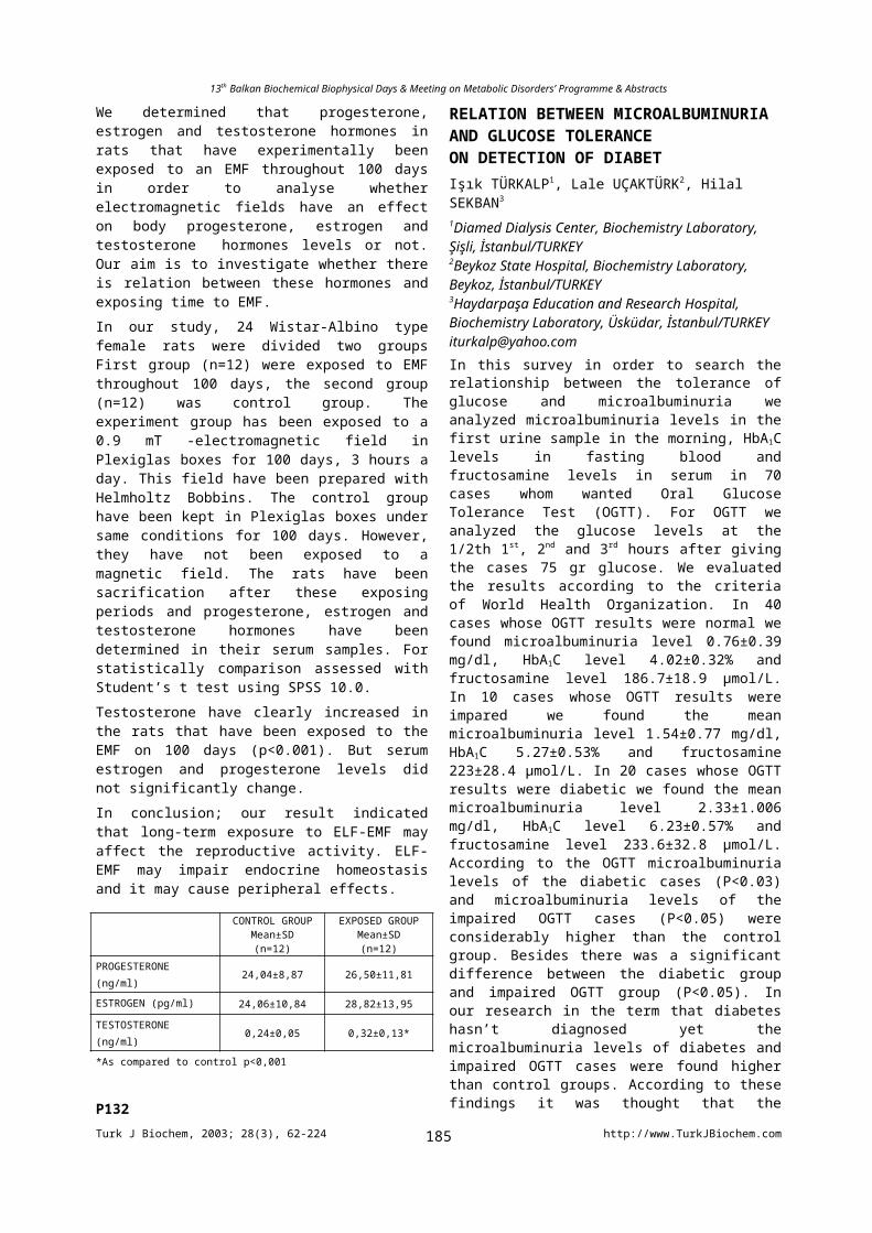
13th Balkan Biochemical Biophysical Days & Meeting on Metabolic Disorders’ Programme & Abstracts
**Marmara University, Faculty of Medicine, Department of [email protected] Taxol is an anti-cancer drug which stabilizes microtubules and blocks Go\G1 cell cycle cell during mitosis and inhibits cell proliferation.In recent years Taxol has been used in intraarterial stent coating and inhibits neointimal hyperplasia and subacute thrombosis which are the cause of restenosis after stent implantation.Platelet aggregation and activation are important in the formation of subacute thrombosis. In this study, we have examined the Taxol’s effect on platelet aggregation by aggregometer. Taxol’s effects on ADP, collagen and adrenalin induced platelet aggregation were studied in the presence of different Taxol concentrations (30, 70, 140, 280 M). From six healthy subjects whole blood samples were taken into 1\9 citrated tubes. PRP (platelet rich plasma) samples were prepared from whole blood by santrifugation at 150g for 15 minutes. PPP (platelet poor plasma) was prepared by santrifugation at 300g for 15 minutes. PPP was used to calibrate the aggregometer. For platelet aggregation studies PRP samples were used. To observe agonist induced platelet aggregation, ADP, collagen and adrenalin induced platelet aggregation was measured with and without Taxol. Taxol was used in increased concentrations.As a result Taxol (especially 140, 280 M concentrations of Taxol) inhibited collagen, Adrenaline and ADP induced primary and secondary platelet aggregation (p<0.01).Our results support Taxol’s inhibition effect on subacute thrombosis after Taxol coated stent implantation.
P241
TAXOL’S EFFECT ON PLATELET FIBRINOJEN RECEPTOR AND PLATELET ACTIVATION DETECTED BY FLOW CYTOMETRYDerya OZSAVCI**, A.Seda KANMAZ-DEMİRCİOĞLU**, Gülderen YANIKKAYA-DEMİREL*, Nezih HEKİM* and Turay YARDIMCI****Marmara University, Faculty of Pharmacy, Department of Biochemistry, Istanbul/TURKEY, *Dr. Pakize I. Tarzi Laboratory, İstanbul/ [email protected] is an anti-cancer drug which inhibits cell processes that are dependent on microtubule turnover, including mitosis, cell proliferation and cell migration.
In recent years Taxol has been used in intraarterial stent coating and inhibitsneointimal hyperplasia and subacute thrombosis which are the cause of restenosis after stent implantation. Platelet aggregation and activation are important in the inhibition of subacute thrombosis. In this study, we have examined Taxol’s effects on fibrinogen receptor GpIIb\IIIa and platelet activation in whole blood and PRP by using flow cytometry. In recent years, flow cytometry has permitted the detection of activation antigens on platelets. Citrated whole blood samples were obtained from seven healthy subjects and PRP (platelet rich plasma) was prepared by santrifugation at 150g for 15 minutes. Whole blood and PRP samples were incubated with Taxol for 10 minutes .To study the platelet activation, CD42b (GpIb) , to examine the effect of Taxol on platelet GpIIb/IIIa, CD61(GpIIIa) antibodies added to the samples and incubated for 15 minutes. All samples were fixed by PFA and analyzed on flow cytometer.In different Taxol concentration (0, 0.01, 0.245, 0.5, 2.5, 5 mM), CD61 percentage in whole blood was found to decrease significantly (0.5, 2.5, 5.0 mM Taxol), whereas CD42 percentage also decreased.In PRP, CD61 and CD42b percentages decreased after incubation with different concentrations of Taxol. In our preliminary studies, in addition to Taxol’s effect on platelet activation, we have observed Taxol’s apoptotic effect on platelets (unpublished results). In this study, the striking observation was that while Taxol was increasing the platelet activation, it decreased CD61 binding to platelets. We have also observed that when platelet activation with Taxol increased, the fibrinogen binding sites (GpIIIa) might have been suppressed by Taxol. These findings suggest that the binding sites of Taxol on platelets might be GpIIb-IIIa.
P242
NEUROCHEMICAL MONITORING: PLATELET MONOAMINE OXIDASE (MAO) ACTIVITY AS A MARKER OF BRAIN FUNCTIONS. Bugarinovic ,D.Pap1 ,G. Prtenjak 2
Psychiatric Hospital, Kovin,Serbia and Montenegro1 Clinical-biochemical laboratory, Medical Center, Sremska Mitrovica,Serbia and Montenegro2Institute of Oncology, Sremska Kamenica, Serbia and MontenegroMonoamine oxidase(MAO)is one of the primary enzymes regulating metabolism of biogenic amines. Two distinct isoforms of the enzyme, MAO A and MAO B,have different substrate and inhibitor specificities.These enzymes are reportedly involved in the pathogenesis of Parkinsons disease (PD) through the production of oxygen radicals from catabolism of dopamine and activation of exogenous neurotoxins, such as MPTP and its analogues and also possible association of MAO gene polymorphisms with PD remains to be elucidated. This study was aimed at investigating the relationship between platelet MAO
Turk J Biochem, 2003; 28(3), 62-224 http://www.TurkJBiochem.com185
13th Balkan Biochemical Biophysical Days & Meeting on Metabolic Disorders’ Programme & Abstracts
activity in patients with PD and their clinical status (age, onset of the disease, duration of the therapy, dose of L-dopa) as well as with their clinical scores(NWUDS,HY,Hamilton scale and MMSE),with attempt to characterize platelet MAO as marker of disturbed neurotransmission system(s) in PD. Subjects of the study were the control(35 healthy volunteers) and the experimental group (44 patients). Platelet MAO activity was measured by spectrophotofluorimetric procedure with kynuramine as a substrate. MAO activity in healthy women was significantly higher than in men. The sole correlation among the platelet MAO activity in patients and their clinical status and clinical scores was established for Hamilton scale (depression). Platelet MAO activity may indicate a vulnerability to depression in PD, what would suggest to state of hyposerotonergic function in the brain, respectively low platelet MAO activity reflects low brain serotonin capacity, what is related to depression in PD.
P243
GUIDELINES FOR POSSIBLE DIAGNOSTIC STRATEGY IN ANEMIAS.Bugarinovic and D.Pap1
Psychiatric Hospital, Kovin,Serbia and Montenegro 1Clinical-biochemical Laboratory, Medical Center Sremska Mitrovica, Serbia and MontenegroAnemia is defined as the inability of the blood to supply tissue with adequate oxygen for proper metabolic function. Anemia is usually associated with decreased levels of hemoglobin and haematocrit. The worlds population at all ages suffering from anemia in roughly 30%. Around half of these cases are caused by iron deficiency, while approximately 20% are caused by vitamin B12 and folate deficiencies. Anemia is a sign of an underlying pathology, the recognition of which requires the identification of the mechanism and causes of the red blood cell deficit. Determining the specific cause of anemia is important in order for physician to apply appropriate therapy. The primary diagnosis of anemia is made by referring to patient history, signs and symptoms and hematological laboratory findings. The aim of the study was to investigating anemia and iron metabolism disorders, diseases of iron deficiency, diseases of iron overload, anemia of chronic disease, vitamin B12 and folic acid deficiency and to present hematological and biochemical markers as a guidelines for the differential diagnosis of anemia.
P244
STUDIES OF THE DOSE-DEPENDANT ANTİOXİDANT ACTİVİTY OF ARTEMİSİA ABSİNTHİUM L. EXTRACTS USİNG İN VİVO MODELAstghik R. SUKIASYANIX Part of New Nork 24, apt.48, Yerevan, Armenia, [email protected] plant Artemisia absinthium L. is widely employed in Armenian popular medicine as a stimulator, a tonic and a remedy for digestion debility. Several other plant products
with similar therapeutic applications may have such effect based on the antioxidant content of flavonoids. The aim of this work was to investigate the effect of Artemisia absinthium L. water extracts on the antioxidant defenses of cow brains. During influence of preparations of Artemisia absinthium L. with ended concentrations 1, 5, and 10mg/ml. it was noticed its suppressed activity by decreasing of the concentration of first products of lipid peroxidation (tiene conjugate) on 63, 54, and 38%, and by decreasing of the concentration of second products of lipid peroxidation (malone dialdehyde) on 71, 50, and 46%. While investigation the concentration of flavonoids in these preparations it was found that during increasing the volume of Artemisia absinthium L. extracts by 1, 5, and 10mg. quantity of flavonoids increased by 0,08, 0,15, and 0,6%. We assume that this decreasing of inhibitory activity of plant is connected with increasing the concentration of flavonoids. These results can be useful in medicine.
P245
FRACTAL ANALYSIS OF THE EFFECT OF THE STRETCH ON VASCULAR SMOOTH MUSCLESÖztürk N.Hacettepe University, Faculty of Medicine, Department of Biophysics, Ankara, TurkeyIt is known that spontaneous oscillations (vasomotion) observed on the wall of the small arteries play an important role on the regulation of blood flow to various organs. Hormonal changes and stretch due to increased pressure affect these oscillations. Stretch increases the frequency and decreases the amplitude of these spontaneous contractions. It is proposed that the stretch induces these effects by depolarizing the membrane and causing the opening of the voltage dependent calcium channels. Studies have also shown that stretch causes an increase in the level of IP3 which is a factor that stimulates contractions. Dimension of a system gives the number of independent parameters that must be used in order to define that system. Therefore, in a number of studies, fractal dimension of the arterial system has been calculated by means of the spontaneous oscillations in order to assess the mechanisms behind the vasomotion. This study was carried out to find whether the increase in the frequency of the spontaneous contractions after stretch is due to the involvement of a new mechanism in addition to the mechanisms that generate the spontaneous contractions. Experiments were carried out on the portal vein isolated from the guinea-pigs. The preparations were immersed into a bath containing the Krebs solution at 37 oC. Initially a preload of 0.5 g was applied. The length of the muscle was measured and designated as Lo. After an adaptation period of about half an hour at that condition, the spontaneous contractions were recorded for about an hour. Then the length of the preparation was increased to a length of 1.2 of the previous length (Lo). After ½ hr spontaneous contractions were recorded for that length. The same procedure was repeated for 1.4xLo. After the experiments the power spectral density functions of the spontaneous contractions were calculated using the FFT algorithm, for each length. Fractal dimensions of the same records were calculated according to Grassberger-Procacci. It was observed that the fractal
Turk J Biochem, 2003; 28(3), 62-224 http://www.TurkJBiochem.com186
13th Balkan Biochemical Biophysical Days & Meeting on Metabolic Disorders’ Programme & Abstracts
dimensions of the portal vein were: 4.36 0.45 at Lo, 4.22 0.43 at 1.2xLo and 4.410.51 at 1.4xLo ( n=6). There was no significant difference between the dimension values. It was concluded that the increase in the frequency of the spontaneous contractions to higher frequencies occurs without addition of the new mechanisms. Rather the contribution if different mechanisms to the oscillations is varying during stretch.This study was supported by the Scientific and Technical Research Council of Turkey (SBAG 1929)
P246
BIOCHEMICAL MARKERS IN PATIENTS WITH BREAST CARCINOMAVioleta FILIPCE1, Beti DEJANOVA3, Venko I. FILIPCE2
and Andrej ARSOVSKI2
1Biochemistry Unit, 2Clinic of Surgery, 3Institute of Physiology, Medical Faculty, Skopje 1000, Republic of MACEDONIAe-mail: [email protected] assessment of prognosis in patients with breast cancer remains unclear. The aim of the study was to determine the level of some biochemical markers in both, patients without and with carcinoma, before and after the surgery.A number of 47 patients (female) were divided in 2 groups: I group-20 patients with benign breast tumor at age of 50.712 years; II group-27 patients with breast carcinoma at age of 47.18 years. All examinations in both groups were done before and after the surgery. Sedimentation and white blood cell count (WBC) were done by routine biochemical methods; albumin and C-reactive protein (CRP) by the biochemical analyzator Integra 700; tumor markers, cancer antigen (CA15-3) and carcino-embrional antigen (CEA) by the immunochemical analyzator Vitros-ECI-OrthoDiagnostics with enhanced chemiluminiscence.Increased sedimentation rate was noticed in all patients, 26.814mm in I group and 45.234mm in the II group before the surgery, but the level of WBC did not show increased value, as well as the albumin level. Regarding sedimentation, WBC, and albumin level, nor significant difference was found between the values of different groups, neither between the values before and after the surgery. There was significant difference for CRP between the two groups before the surgery, 4.254mg/L in I group vs. 21.119mg/L in II group (p<0.05) and after the surgery, 8.324mg/L in I group vs. 17.39mg/L in II group (p<0.01). After the surgery, there was significantly higher value of CA15-3 in II group, 28.79U/mL when compared to I group, 19.810U/mL (p<0.05). For CEA values, statistically significance was found between the two groups before the surgery, 0.150.1 ng/mL for the I group and 0.970.9ng/mL for the II group (p<0.01).From the obtained results, we can conclude that tumor markers should be accompanied by acute phase proteins for better view and evaluation of the patient’s status.
P247
OXIDATIVE STRESS IN HEMODIALYSIS PATIENTS
Beti DEJANOVA1, Tatjana RUSKOVSKA3, Ratka MANCEVSKA3, Aleksandar SIKOLE2, Petar DEJANOV2, Vesela MALESKA1 and Suncica PETROVSKA1
1Institute of Physiology, 2Clinic of Nephrology, Medical Faculty, 3Military Hospital, Skopje 1000, Republic of MACEDONIAe-mail: [email protected] on regular long term of hemodialysis (HD) have high incidence of premature cardiovascular disease. The aim of the study was to determine the association between HD and the level of oxidative stress (OS). A number of 30 patients (16 men and 14 women, mean age 4811 years) undergoing HD were compared to sex and age matched 34 healthy subjects as a control group. All the patients were dialysed 3 times per week, less than 6 hours of duration, on bicarbonate mode of HD, using cuprophan type of HD membrane. The blood for analysis was withdrawn from the cubital vein, before the HD session. Activities of red blood cell (RBC) superoxide dismutase (SOD) and RBC glucose-6-phosphate dehydrogenase (G-6-PD), whole blood gluthation peroxidase (GPx), plasma gluthation reductase (GR) and the total antioxidative status (TAS) were assayed by the commercial kits from Randox, Crumlin, UK. Lipid peroxidation, determined trough the end product malonyldialdehyde (MDA) in serum, was measured by the thiobarbituric acid-reactive substances using the fluorimetric method. Lower enzyme activity level was found in HD patients: for SOD, 1232243 U/grHb (p<0.01); for G-6-PD, 12020 mU/109 RBC (p<0.01); and for GPx, 47.914 U/grHb (p<0.05). Plasma antioxidant level was found increased: for GR, 81.515 U/L (p<0.001) and for TAS, 1.570.2 mmol/L (p<0.01). The level of MDA was higher in HD patients, 5.020.99 mol/L vs. 3.520.99 mol/L in control subjects (p<0.001).These findings suggest OS in HD patients, due to the low level of antioxidative enzymes and high level of MDA, indicating oxidative damage that can be a reason for developing atherosclerosis and/or anaemia, having low quality of life in these patients.
P248IS ANTIBIOTIC PROFILACTION THERAPY JUSTIFIED IN WOMEN IN PERIOD OF PUERPERIUM AFTER SURGICAL DELIVERY?Dobri FILIPCE1, Violeta FILIPCE2, Jovanka DONEVSKA2, Zorica POPOVSKA1, Elena NIKOLOVSKA1
Clinic for Gynecology and Obstetrics , Biochemistry Unit, Clinic of Surgery, Skopje, 1000 Republic of MACEDONIAe-mail: [email protected] aim of the study was to investigate proofs for justifications of systemic prophylactic antibiotic therapy in cases of surgical delivery and to clarify the sensitivity for different biochemical markers for early detection of inflammation.In 77 women after the surgical delivery with cesarean section and vaginal obstetrical interventions and applied therapy of Lendacin or Amoxiclav, the 2-nd (3-rd) day were examined biochemical markers for early inflammation: C-reactive protein (CRP) – Vitros 250, leucocytes (Le), granulocytes (Gr) - Cobas Mira OT8 and
Turk J Biochem, 2003; 28(3), 62-224 http://www.TurkJBiochem.com187
13th Balkan Biochemical Biophysical Days & Meeting on Metabolic Disorders’ Programme & Abstracts
-1 antitripsin (-1 At) – Integra 700-Roche. After surgical delivery, the outcome of X1 sd for CRP value was 11077.5 g/L; for Le count was 14.24.9; for Gr count was 3.10.57 mg/L; and for -1 At level was 79.95.8 g/L. The outcomes of X2 sd for CRP was 88.2%, for Le was 91.8%, for Gr was 97.4%; for -1 At level was 92.2%. The highest sensitivity for the outcomes for these biological markers is ranged in subsequent manner: Gr; -1 At; Le; CRP. From our results we could clarify high sensitivity for biological markers for early inflammation that confirmed our protocol for antibiotic use, before, during and after the surgical period. It showed that in women after the surgical way of birth, without symptoms of high temperature or increasing temperature, giving antibiotics in first three days and monitoring the biochemical markers for inflammation were justified.
P249
THE ISOLATION OF RAT LIVER PEROXISOMES BY DENSITY GRADIENT CENTRIFUGATION TECHNIQUE AND PROOFING THE PURITY BY ELECTRONE MICROSCOPY AND MARKER ENZYME ANALYSISTijen TANYALÇIN1, Remziye DEVECİ2, Güneş AK BAŞOL1, Dilek TAŞKIRAN3, Sabire KARAÇALI2, Fatma Z KUTAY1
1Ege University Medical School and Hospital Department of Biochemistry, IZMIR2 Ege University Science Faculty Department of Molecular Biology, IZMIR3 Ege University Medical School and Hospital Department of Physiology, [email protected], single-membrane bound cytoplasmic structures, are present virtually in all eukaryotic cells. They contain hydrogen-peroxide producing oxidases and catalase that decomposes hydrogen peroxide. Peroxisomes are required for specific functions such as -oxidative chain shortening of fatty acids, synthesis of ether-phospholipids, cholesterol and bile acids. In the present study, peroxisomes were characterised biochemically and morphologically by electrone microscopy. They were isolated by density gradient centrifugation technique and Nycodenz was used as a gradient material. All the fractions obtained were used for the determination of the activites of glucose-6-phosphatase, 5’ nucleotidase, Na+,K+ ATP’ase, succinate cytochrome-c reductase and catalase. Protein determination of the fractions were performed by bicinhconinic acid method. During the isolation steps, E was considered as the nuclear while PS as the light mitochondrial fraction. Mitochondrial pellet was found to be rich from mitochondria and named as heavy mitochondrial fraction. Following density gradient centrifugation, three layers were obtained; the surface fatty, plasma membrane and peroxisomal layers. Plasma membranes banded at the density interface of 1.16-1.18 while the peroxisomes at 1.18-1.24. The highest activity of Na+,K+ ATP’ase was found as 0,6 molP/mgprotein/hour and 0,4 molP/mgprotein/hour at the surface fatty and
plasma membrane layers, respectively. Succinate cytochrome-c reductase activity was 20 nmolP/mgprotein/minute at mitochondrial pellet and 26.5 nmolP/mgprotein/minute at plasma membrane layer. Glucose-6-phosphatase was determined as 6.45 molP/mgprotein/hour at PS fraction where, this fraction is rich from microsomes, peroxisomes, lysosomes. 5’ NT was 6.11 molP/mgprotein/hour at the plasma membrane fraction. Finally, catalase activity was found as 888.77 U/mg at peroxisomal fraction indicating that purity was obtained at a rate of 13365 %. In conclusion, marker enzymes can be the indicator of purity of the tissue fractions and this biochemical approach can be helpful where electrone microscopy could not be eligible.
P250
THE EFFECTS OF MELATONIN ON TORSION-DETORSION INJURY IN RAT OVARY: BIOCHEMICAL AND HISTOPATHOLOGIC EVALUATIONYusuf TÜRKÖZ, Önder CELİK, Şeyma HASÇALIK, Yılmaz ÇİĞREMİŞ, Mehmet HASÇALIK, Bülent MIZRAK, and Saim YOLOĞLUInonu University, School of Medicine and Art and Science, Departments of Biochemistry, Obstetrics and Gynecology, Anaesthesiology, Pathology, Biostatistics, and Biology, 44069, Malatya/[email protected]: This experimental study was designed to determine the changes in ovarian malondialdehyde, reduced glutathione and xanthine oxidase levels, and the effect of melatonin on these metabolite levels after adnexial torsion/detorsion in rats.Method: Thirty-two adult female albino rats were divided into four groups: sham operation, torsion, torsion-detorsion (ischemia-reperfusion=I/R) plus saline and torsion-detorsion plus melatonin. Rats in the sham operation group underwent a surgical procedure similar to the other groups but the adnexa was not occluded. Rats in the torsion group were killed after 360º clockwise adnexial torsion for 3 h. Melatonin was injected intraperitoneally 30 min before detorsion in the I/R plus melatonin group and saline, contained 0.5% ethanol, was administered in the I/R plus saline group. After 3 h of ovarian detorsion in both of these groups, the rats were killed and ovaries were removed. The tissue levels of malondialdehyde, reduced glutathione and xanthine oxidase were measured.Results: Malondialdehyde and xanthine oxidase levels in the I/R plus saline group increased signifcantly when compared to torsion and sham operation groups (p<0.001). Malondialdehyde and xanthine oxidase levels in the I/R plus melatonin group were lower than I/R plus saline and differences between the two groups were statistically significant (p<0.001). Reduced glutathione levels in the I/R plus saline group decreased significantly when compared to ischemia and sham operation groups (p<0.001). Reduced glutathione levels in the I/R plus melatonin group were higher than I/R plus saline and ischemia groups, and differences between the two groups were statistically
Turk J Biochem, 2003; 28(3), 62-224 http://www.TurkJBiochem.com188
13th Balkan Biochemical Biophysical Days & Meeting on Metabolic Disorders’ Programme & Abstracts
significant (p<0.001). Morphologically, polymorphonuclear neutrophil infiltration and vascular dilatation were obvious in the I/R damaged ovary, and the changes also partially reversed by melatonin.Conclusions: The pineal hormone melatonin protects the ovaries against oxidative damage associated with reperfusion following an ischemic insult.Key words: Ovarian torsion/detorsion, Rat, Melatonin, Lipid peroxidation products, Histopathology.
P251
NUCLEASE ACTIVITIES OF ALFALFA UNDER PARAQUAT OR ROUNDUP STRESSTraianos YUPSANIS1, Michael MOUSTAKAS2, Antoanela PATRAS1,3, Anastasia YUPSANI1, Alexander CHRISTOU1 and Peter FRANGOPOL4
1Laboratory of Biochemistry, School of Chemistry, Aristotle University of Thessaloniki, 54124 Thessaloniki, Greece, 2Department of Botany, School of Biology, Aristotle University of Thessaloniki, 54124 Thessaloniki, Greece, 3University of Agricultural Sciences and Veterinary Medicine, 6600 Iasio, Romania, 4Department of Physical Chemistry, School of Chemistry, University of Cluj-Napoca, 3400 Cluj-Napoca, Romania e mail: [email protected] exposure to herbicides, plant cells undergo substantial metabolic alterations. Paraquat (PQ) and Roundup (RD) are known herbicides widely used in agriculture. Paraquat exerts its toxic effect by catalyzing the transfer of electrons from photosystem I (PS I) to molecular oxygen, producing oxygen radicals, leading to lipid peroxidation and membrane damage. On the other hand, Roundup interferes in the shikimate pathway in a wide variety of plants and organisms. We have examined whether nuclease activities in alfalfa are subjected to alteration following PQ or RD stress.Alfalfa seeds were germinated on moist filter paper in Petri dishes in the dark at 22oC for 1, 3 and 5 days with either H2O, PQ or RD. The in vivo specific activities of nucleases (acidic and neutral) decreased under PQ or RP stress. Both acidic and neutral nucleases were purified and separated from alfalfa seeds that germinated in H2O. Electrophoretic analysis revealed that purified neutral nuclease was capable to hydrolyze PQ treated ssDNA, while acid nuclease was unable. In contrast both nucleases were capable to hydrolyze RNA. Roundup (RD) treated ssDNA formed strong complexes that were unable to be hydrolyzed by both nucleases. Acidic and neutral nucleases were capable of nicking and linearizing PQ treated plasmid –DNA. However in the presence of RD only neutral nucleases were capable of nicking and linearizing plasmid-DNA. The results lead to the conclusion that PQ or RD caused dramatic consequences on alfalfa nucleases activities in vivo and in vitro.
P252
FUNCTIONING OF PHOTOSYSTEM II (PS II) IN AN ALUMINIUM (AL) TOLERANT
AND NON TOLERANT WHEAT CULTIVAR UNDER AL STRESSMichael MOUSTAKAS and Elektra SPERDOULIDepartment of Botany, School of Biology, Aristotle University of Thessaloniki, 54124 Thessaloniki, Greece e mail: [email protected] (Al) toxicity is a serious agricultural problem in acid soils, which make up about 40% of the world’s arable land. Al3+, the phytotoxic species, inhibits root growth and the uptake of water and nutrients, which ultimately results in a production decrease, although the toxicity mechanism is poorly understood. On the other hand, some plant species and cultivars of the same species have developed strategies to avoid or tolerate Al toxicity. Al resistance can be divided into mechanisms facilitating Al exclusion from the root apex (Al exclusion) and mechanisms conferring the ability of plants to tolerate Al in the plant symplasm (Al tolerance). Transfer of Al into cells, and sequestration in the vacuoles might be an Al-tolerance mechanism. Differential sensitivity of species and genotypes to Al has been extensively documented. Analysis of chlorophyll fluorescence and mineral content were conducted in two wheat cultivars differing in their tolerance to aluminium stress. The Ca2+ and Mg2+
concentration in the leaves of the two wheat cultivars exhibited a significant decrease during Al-treatment. However, the more tolerant cultivar retained larger concentrations of Mg2+ and Ca2+ in the leaves as a percentage of the control (-Al). Under similar Al stress conditions, plants of the tolerant cultivar were able to keep a larger fraction of the PS II reaction centres in an open configuration, i.e. a higher ratio of oxidized to reduced QA (the primary, stable quinone acceptor of PS II), than plants of the relatively non tolerant wheat cultivar. Four times higher aluminium concentrations were required for tolerant cultivar plants than for non tolerant plants in order to establish the same proportion of oxidized to reduced QA.
P253
RNAi ANALYSIS OF CAENORHABDITIS ELEGANS MRG15Abdullah OLGUN1, Demetrios K. VASSILATIS2, Olivia M. PEREIRA-SMITH3
Roy M. and Phyllis Gough Huffington Center on Aging, Baylor College of Medicine, Houston, Texas 77030-3498, USAPresent addresses:1Department of Biochemistry and Clinical Biochemistry, Gülhane School of Medicine, Etlik-06018, Ankara, Turkey2Primal, Inc., Seattle, Washington, USA3Sam and Ann Barshop Center for Longevity and Aging Studies, University of Texas Health Science Center at San Antonio, STCBM, 15355 Lambda Drive, San Antonio, TX 78245-3207, USAImmortal cell lines were assigned into four complementation groups based on the dominance of senescence over immortality in cell fusion experiments.
Turk J Biochem, 2003; 28(3), 62-224 http://www.TurkJBiochem.com189
13th Balkan Biochemical Biophysical Days & Meeting on Metabolic Disorders’ Programme & Abstracts
Mortality factor on chromosome 4 (MORF4) was found to induce senescence in group B. The expressed MORF-related genes were found localized on chromosome 15 (MRG15) and X (MRGX). We silenced C. elegans y37d8a.9 gene, the ortholog of human MRG15, and y37d8a.11 because of its similarity to y37d8a.9, using double-stranded RNA-mediated interference technique(RNAi). We observed no phenotype after y37d8a.11 RNAi. Whereas y37d8a.9 RNAi caused sterility in all; body wall defects, vulval protrusion and posterior developmental defects in a small percentage of worms. Our results suggest possible transcription factor like function of y37d8a.9 in different cell types and demonstrates its role in oogenesis and development.
P254
MATHEMATICAL MODEL OF LIPID BILAYER ELECTROPORATIOND.E.Creanga, R. Leahu, Maria AnitaUniv. Al. I. Cuza, Fac. of Physics, 11 A Bd. Carol I, Iasi, e-mail: [email protected] mathematical model proposed inhere describes the permeability of the lipid bilayer under electrical constraint in dependence on the rate of hydrophilic pore formation. Since artificial membranes consistent with lipid bilayers are convenient biophysical models used for the study of charged species transport, lipid bilayer behaviour was studied by various experimental and theoretical methods. The proposed model is starting from the dependence of the rate of hydrophilic pore formation on the activation energy, k, that is depending on the area of a single lipid molecules as well as on the area of whole lipid membrane and is decreased under the action of an electrical field. The main differential equation intended for the mathematical model development led finally to a cubic solution that takes various graphical forms for different values of the rate k, in the same range of the independent variable. The interpretation was based both on 3D and 2D graphical representations. Monotone curve obtained in 2D, corresponds to the case of two complex solutions, the hysteresis like curve corresponds to the case of three distinct real solutions for certain subintervals of independent variable values while the turning point curve reveals two branches, one of them presenting also negative slope. The negative slope of the hysteresis type curve may be taken as an indication of the self-adjusting phenomena underlying the charged species transport phenomena through membrane pores under the electrical filed influence. The two branches of the bifurcated curve suggest that the system can pass from a stable state to an unstable one for certain ranges of the electrical field intensity. Though not fitted yet with experimental data, the model may be useful in the study of therapeutical protocols where drug substances are ionized molecules.
P255
FT-IR SPECTROSCOPIC INVESTIGATION OF DIFFERENCES BETWEEN BACILLUS AND MICROCOCCUS SPECIES
Araz Zeyniyev, Ayşe Gül Çetin and Feride SevercanDep. of Molecular Biology & Genetics, Middle East Technical University, 06531, Ankara, TurkeyCorrespondence: [email protected] [email protected] [email protected] transform infrared spectroscopy (FT-IR) has been developed and widely used in many disciplines. FTIR is a nondestructive technique and allows the rapid characterization of structural features of biological molecules and complex materials such as intact bacteria. Organisms can be probed by FTIR in a single experiment using simple, uniform procedures that are applicable to all bacteria. In our study we examined the potential of FTIR technique in discriminating between four bacterial species, three of which were isolated from Salt Lake located in Central Turkey. The four bacterial samples were Bacillus licheniformis, Bacillus circulans, Bacillus subtilis (reference strain) and Micrococcus luteus. Mid-infrared (MIR) regions (400-4000 cm-1) of the species were analyzed. Bacterial isolates were grown at 36oC for 24 hours. The cultures were centrifuged and the pellets were washed in sodium phosphate buffer 100mM, pH 7,0. The analysis of lyophilized samples indicated that there is a unique peak for the Micrococcus luteus at 800 cm-1
differentiating it from Bacilli. Also there observed a peak located at 606 cm-1 specific to B. circulans. We also found out that lyophilized bacterial samples do not lose their spectral properties at least a month in –18oC freezer. Therefore, FTIR seems to be a useful tool for rapid detection or identification of the species from environmental samples. P256
ANTIOXIDANT ACTIVITIES OF THE ETHANOL EXTRACTS OF AESCULUS HIPPOCASTANUM COMPONENTSGülçin SAĞDIÇOĞLU, Nursen ÇORUHMidde East Technical University, Department of Chemistry, 06531, Ankara/TÜRKİ[email protected] hipocastanum commonly known as horse chestnut trees are one of the well-known medicinal plants which are grown at all the regions of Turkey. Seed extracts of the horse chesnut have been used for medicinal remedies since the ancient times. Some of the compounds in seeds such as aescin are known to have valuable medicinal applications in chronic venous insufficiency treatments. The antioxidant activities of some compounds found in horse chestnut are also known, however, in the literature we have not come across with any information related to the antioxidant capacity of crude extracts of that plant. The aim of this work is to investigate the antioxidant capacities of the ethanol extracts obtained from the horse chestnut tree components such as seeds, barks, leaves, and flowers with respect to each other. Horse chestnut extracts were prepared by overnight solvent extraction by using 5gr of each component with ethanol in 1/10 solid to solvent ratio at room temperature. Filtered extracts were centrifuged and dried completely and
Turk J Biochem, 2003; 28(3), 62-224 http://www.TurkJBiochem.com190
13th Balkan Biochemical Biophysical Days & Meeting on Metabolic Disorders’ Programme & Abstracts
weighed. Extracts were redissolved in ethanol and their antioxidant capacities were measured via the inhibition of iron induced lipid peroxidation capacity on sheep liver microsomal membranes with the application of Thiobarbituric Acid Test. IC50 values are obtained as follows: IC50 for Flowers Ethanol Extract = 1.250 mg/ml, IC50 for Seed Ethanol Extract = 0.500 mg/ml, IC50 for Leaves Ethanol Extract = 0.200 mg/ml and IC50 for Bark Ethanol Extract = 0.026 mg/ml. The percentage inhibiton of lipid peroxidation was determined by comparing the results of the test compounds with those of controls not treated with the extracts. All the ethanol extracts of horse chestnut parts have shown significant antioxidant effect. Ethanol bark extract was found to have the highest antioxidant efficiency. Isolation studies are in progress to reveal further antioxidant compounds from the extracts.
P257
ELECTRICAL, MECHANICAL AND ENZYME ACTIVITY OF SKELETAL MUSCLE AFFECTED BY MICROWAVE ELECTROMAGNETIC FIELDNicolina I. RADICHEVA*, Radoi IVANOV**, Teodora I. VUKOVA* and Katya N. MILEVA****Institute of Biophysics, Bulgarian Academy of Sciences, Acad. G. Bonchev str. Bl. 21, Sofia 1113, BULGARIA, **Department of Physiology, Sofia University, ***SESRC, London South Bank University, UK, [email protected] work summarises the results of studying the influence of microwave electromagnetic field (MMW, 2.45 GHz, 20 mW/cm2) on isolated frog muscle fibre fatigability as well as on the activity of some enzyme systems (acetylcholinesterase - AChE, Mg2+,Ca2+- and Na+,K+- ATPase in membrane fraction and Mg2+,Ca2+- and NaHCO3-stimulated Mg2+-ATPase in mitochondrial fraction from muscle homogenate) measured by phosphomolibdenum spectrophotometric method. Infrared (IR) spectroscopy and amino acid (AA) analyses were performed on samples of lyophilised membrane fraction. Standard micro- and semimicro-electrode methods were used to register intra- and extra- cellular action potentials, and twitch contractions of single muscle fibres. Their time-amplitude and spectral characteristics obtained after MMW exposure were compared to those obtained after a sham exposure. The action potentials amplitude and propagation velocity were significantly higher, the rising time was shorter, the membrane potential was more negative, and twitch time parameters were shorter in irradiated fibres. The rate of parameter changes during uninterrupted continuous activity was significantly delayed after exposure. A dose-dependent (10 and 20 mW/cm2 – increase and decrease, respectively) and prolonged (up to 48 hrs) effect of MMW field on AChE activity at a constant temperature of 2oC was found. The rate of the other enzyme systems activity in a temperature range of 18-20oC showed a delayed decrease during one hr irradiation. We concluded that the reduced development of muscle fatigue, the changes in enzyme activity and the
conformational changes in protein structure suggested by the IR spectroscopy and AA analysis are caused by the specific non-thermal effect of the studied MMW.
P258COMPARATIVE ACTIVITY DETERMINATION OF AST, ALT, CK, LDH AND - GT BY DIMENSION RxL AND FLEXOR ANALYSERSceta S., E. Suljević, Coric J., Tanic M. Institute for Clinical Chemistry and Biochemistry, Clinical Centre of the University in Sarajevo Bosnia and HerzegovinaThis work was performed in Labaratory for enzyme and isoenzyme determination in Institute for Clinical Chemistry, Clinical Centre of the University in Sarajevo. We compared a determination of the enzyme activity by biochemical analyzer Dimension RxL - DADE Behring and Flexor analyzer - AVL.The purpose of comparing is to determine whether statsticaly relevant diferences exist, concerning the optained data, catalitical activities enzymes survey. It has been worked on two different types of analyzers with reagents beloning to two different manufacturers: DADE BEHRING-DIMENSION RxL and CHRONOLAB -FLEXOR.We have analyzed 50 serums of patient having different diseases. All analyses were done under the same working conditiones and carried out on the same working temperature 37 C.Preciseness of such measurements was investigated on both analyzers for catalytic activity of AST, ALT, CK, LDH and -GT enzymes. The results of all measured parameters were favorable. Coefficient of variation was 0,33% to 3,10% on both analyzers. Comparing results of the preciseness between series we find no differences. Correlation coefficient between series for all parameters were between 0,974 and 0,997.As the enzyme activity determination was conducted in accordance with specific methodology proposed by IFCC results of this work showedthat both analyzers are precise, correct and highly reliable machines for catalytic activity determination of different enzymes.
P2591-ACYL-SN-GLYCEROL-3-PHOSPHATE ACYLTRANSFERASE AFFECTS CYTOCHROME CBB
3 OXIDASE
FUNCTION IN Rhodobacter capsulatus Semra Aygun1, Sevnur Mandaci1, Howard Goldfine2& Fevzi Daldal3
1The Scientific and Technical Research Council of Turkey (TUBITAK), Research Institute of Genetic Engineering and Biotechnology (RIGEB), 41470 Gebze / KOCAELI, TURKEY2Department of Microbiology, School of Medicine, University of Pennsylvania, Philadelphia-PA, 19104, USA
Turk J Biochem, 2003; 28(3), 62-224 http://www.TurkJBiochem.com191
13th Balkan Biochemical Biophysical Days & Meeting on Metabolic Disorders’ Programme & Abstracts
3Department of Biology, Plant Science Institute, University of Pennsylvania, Philadelphia-PA, 19104, USA
Rhodobacter (Rb) capsulatus contains a cbb3-type cytochrome oxidase (cbb3-Cox). In addition to the functional genes of this complex, several others are required for its maturation. Earlier studies using Rb. capsulatus Cox-minus mutants have already uncovered the ccoGHIS operon, whose gene products seem to play a major role in the biogenesis process. In this study, a cbb3-Cox mutant (IJ1) was analyzed in order to identify novel gene(s) required for the biogenesis of this oxidase. In this mutant, no cbb3-Cox activity can be detected. Complementation data revealed that the mutation in IJ1 was not localized in the known biogenesis genes, ccoGHIS. The plasmid pMRC that complemented this mutant contained a 6 kb DNA, encompassing the ilvD, plsC138 and proA loci, and six additional open reading frames (ORFs) of unknown function. Insertional inactivation of these ORFs revealed that plsC138 can restore cbb3-Cox activity in IJ1. Chromosomal inactivation of the plsC138 yielded SA1 mutant, demonstrated that this gene is involved in the biogenesis of the cbb3-Cox enzyme in Rb. capsulatus. plsC138 has been annotated as 1-acyl-sn-glycerol-3-phosphate acyltransferase (AGPAT), which is an enzyme involved in phospholipid biosynthesis in bacteria. In order to probe its function, the plsC138 was used for complementing an E. coli PlsC-minus mutant. Its heterologous expression in E. coli indicated that plsC138 of Rb. capsulatus (PlsC138-Rc) is the functional homologous of the PlsC of E. coli. However, based on the total amounts of monoacylated (LPA) or diacylated (PA) phosphatidic acid fractions, similar GPAT and AGPAT activities were found in both wild type and SA1 mutant. Therefore, it suggests that additional component distinct from PlsC138-Rc is present in this bacterium.
This work has been supported by ICGEB Project No:CRP/TUR99-01 and TUBITAK-TBAG-2128 (102T002) grants to Sevnur Mandaci and NIH grant GM 38237 to Fevzi Daldal
P260
THE ANTIOXIDANT EFFECTIVENESS OF -TOCOPHEROL IN OXIDATIVE STRESS IN ERYTHROCYTESM. Betül YERER, Sami AYDOGANUniversity of Erciyes, Medical Faculty, Dept. Of Physiology, 38039, Kayseri/[email protected] radicals attacking biomembranes can lead to the oxidative damage of the membrane lipids an proteins. Furthermore, reactive oxygen species avidly reacts with nitric oxide (NO) producing cytotoxic reactive nitrogen species capable of nitrating proteins and damaging other molecules which leads to the reduction of erythrocyte deformability. The aim of this investigation was to assess the importance of - tocopherol (Vit-E) in the total
antioxidant status of the erythrocytes in Sodium nitroprussid (SNP), a nitric oxide donor, induced oxidative stress and its relation to erythrocyte deformability. Male Swiss Albino rats were used in 4 groups, comprising of 10 animals in each group. The first group was the control, and the other groups were administered SNP (10mg/kg, i.p ), Vit E (10mg/kg, i.p) + SNP, and SNP + L-NAME (10mg/kg, i.p), respectively. Relative filtration rate (RFR), Relative filtration time (RFT) and relative resistance (Rrel) were determined as the indexes of erythrocyte deformability. In addition, Malondialdehyde (MDA, as an index of lipid peroxidation) and nitric oxide levels and the antioxidant activities of Glutathione peroxidase (GSH-Px), Superoxide dismutase (SOD) and Catalase (CAT) were also determined in the red blood cells of all groups revealing the oxidant-antioxidant activity.RFT and the Rrel of the erythrocytes of the SNP-treated rats increased significantly (p<0.05) whereas the RFR of the erythrocytes decreased (p<0.05) in comparison to all groups reflecting the impaired deformability. Lipid peroxidation was suppressed by Vit-E and L-NAME significantly, where the red blood cell deformability was improved. Furthermore, SOD and CAT activities were significantly stimulated with SNP treatment (p<0.05), where as GSH-Px remained unchanged. In the contrary, GSH-Px activity was triggered significantly by Vit-E administration, whereas the SOD and CAT activities were reduced (p<0.05). As a result, these data reveal that Vit-E improves the erythrocyte deformability in SNP-induced oxidative stress by its antioxidant effects on the lipid peroxidation and antioxidant enzyme activities.P261
INVITRO EFFECTS OF MELATONIN ON THE FILTRABILITY OF ERYTHROCYTES IN OXIDATIVE STRESSSami AYDOGAN, M. Betul YERERUniversity of Erciyes, Medical Faculty, Dept. Of Physiology, 38039, Kayseri, [email protected] deformability is one of the most important charactheristics of erythrocytes for an effective microcirculatory function and is affected from a number of factors, including the oxidative-damage-induced by nitric oxide (NO). This study was performed to investigate the effects of in vitro melatonin incubation on the antioxidant status and deformability of erythrocytes in sodiumnitroprusside (SNP), a nitric oxide donor, induced oxidative stress. 40 blood samples taken from the adult healthy people were divided into 4 groups randomly and incubated with saline, SNP (1mM), Melatonin (MEL, 1mM), MEL+SNP and SNP+L-NAME (5mM) respectively. Relative filtration rate (RFR), Relative filtration time (RFT) and relative resistance (Rrel) were determined as the indexes of erythrocyte filterability. In addition, Malondialdehyde (MDA, as an index of lipid peroxidation) and the antioxidant activities of Glutathione Peroxidase (GSH-Px), Superoxide dismutase (SOD) and catalase (CAT) were also
Turk J Biochem, 2003; 28(3), 62-224 http://www.TurkJBiochem.com192
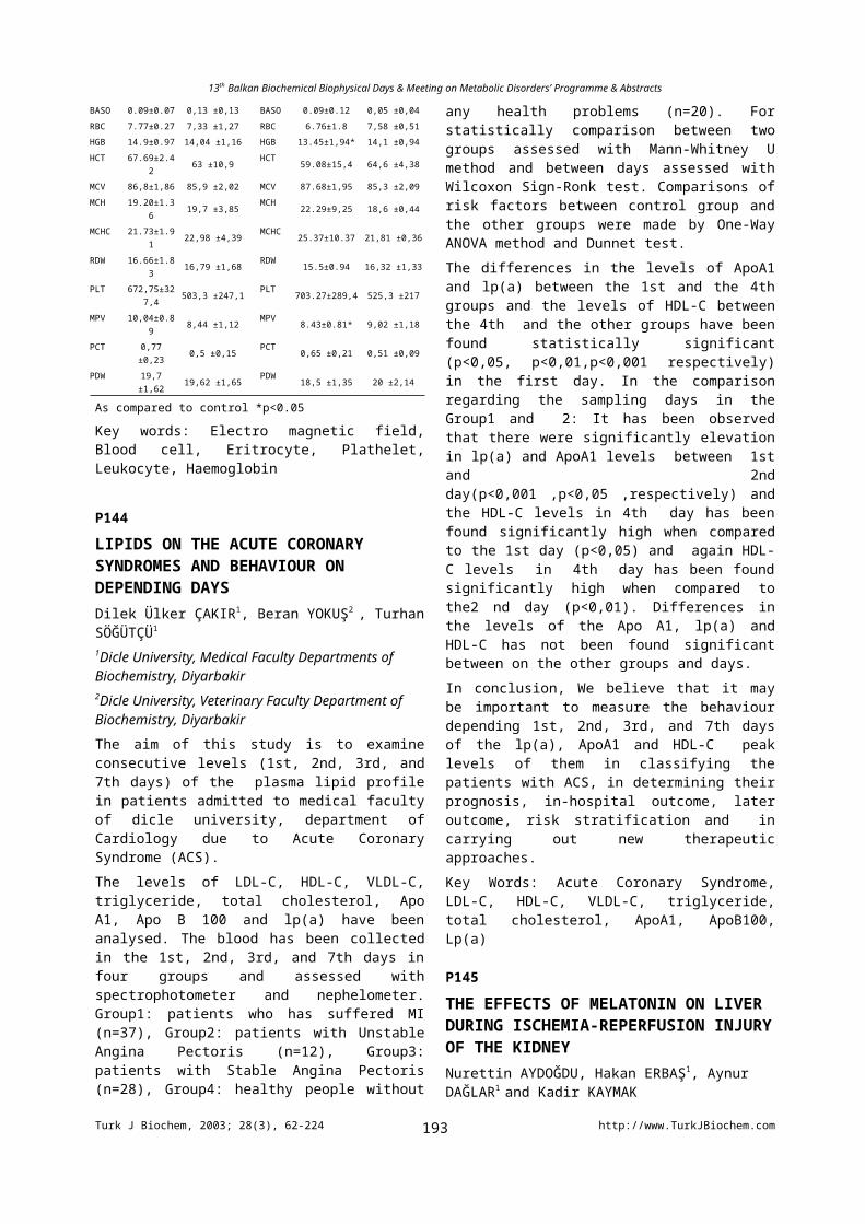
13th Balkan Biochemical Biophysical Days & Meeting on Metabolic Disorders’ Programme & Abstracts
determined in the red blood cells of all groups revealing the oxidant-antioxidant activity. RFT and the Rrel of the erythrocytes incubated with SNP increased significantly (p<0.05) whereas the RFR of the erythrocytes decreased (p<0.05) in comparision to all groups. This reduction in RFR was prevented with both L-NAME or MEL incubation. Furthermore, MEL was found to be significantly efficient in preventing the erythrocytes from lipid peroxidation in these groups. In addition, GSH-Px and SOD activities were elevated with SNP incubation reflecting the oxidative stress in erythrocytes, whereas the CAT activity remained unchanged. Melatonin has no significant effect on the GSH-Px and Cat activity but, it caused a significant decrease in SOD activity (p<0.05). These results reveal that, Melatonin can protect the erythrocytes from impaired deformability in SNP-induced oxidative stress due to antioxidant effects as revealed by lipid peroxidation and antioxidant enzyme activities.
P262
PURIFICATION AND CHARACTERIZATION OF BUTYRYLCHOLINESTERASE FROM RAT SMALL INTESTINEYıldız Ö., Bodur E., Çokuğraş A.N., Özer N.Department of Biochemistry, Faculty of Medicine, Hacettepe University, Ankara / TurkeyButyrylcholinesterase (BChE; E.C. 3.1.1.8) plays an important function in toxicology and pharmacology as a detoxification enzyme. In toxicity assesment investigations, rat is the most commonly used animal. It is clear that orally introduced toxic compounds will first be faced with intestinal BChE so, we purified and characterized the soluble isoform of BChE from the rat small intestine. In this study, small intestines were obtained from female Wistar rats killed for students' laboratory coursework at Hacettepe University Medical School. BChE was purified from soluble fraction of rat intestine by chromatography on Sephadex G-25 following repeated chromatography on affinity gel, procainamide-Sepharose 4B with a purification fold of 260. Purity and purified molecular form(s) were controlled by nonreducing polyacrylamide gel electrophoresis. Consecutive protein staining with Commassie Brillant Blue R-250 followed by AgNO3 gave two bands; a major band corresponding to tetrameric globular form of rat BChE and a minor faster moving protein band; presumably an impurity or degraded BChE. In the activity staining gel with BTCh as substrate only the major protein band was stained for BChE activity. The migration of rat BChE activity band was slower than tetrameric globular forms of both human and horse serum butyrylcholinesterases. In the activity staining gel, in human BChE lane, activity bands corresponding to monomeric, dimeric and tetrameric forms of BChE were observed.Acetylcholinesterase (E.C. 3.1.1.7) contamination was controlled by using ethopropazine, the specific inhibitor of BChE. It is found that purified soluble isoform of the enzyme from rat intestine was completely pure BChE. The
optimum pH value was determined as 7.2 after eliminating the ion strenght effect on activity by zero-buffer extrapolation. The optimum temperature of the enzyme was examined as 37C after eliminating the effect of time on activity by zero-time extrapolation.
P263
STEADY-STATE KINETICS OF RAT INTESTINAL BUTYRYLCHOLINESTERASE Yıldız Ö., Çokuğraş A.N., Özer N.Department of Biochemistry, Faculty of Medicine, Hacettepe University Ankara / TurkeySteady-state kinetics of soluble isoform of butyrylcholinesterase (BChE; E.C.3.1.1.8.), purified from rat small intestine, were determined when acetylthiocholine (ATCh), propionylthiocholine (PTCh) butyrylthiocholine (BTCh) and benzoylcholine (BzCh) were used as substrates. The plots of velocity versus substrate concentrations did not give the normal hyperbolic Michaelis-Menten curves for all substrates studied. [S]0.75 / [S]0.50 ratios were found to be approximately 4 for ATCh, PTCh, BTCh, which indicate substrate activation, whereas to be 2 for BzCh, which indicates subtrate inhibition at high substrate concentrations. So, steady-state data were fit to the equation for excess substrate activation / inhibition.The calculated parameter b in this equation reflects the efficiency of product formation from ternary complex (SES). When b>1, there is substrate activation. Rat BChE purified from soluble fraction of the intestine showed marked substrate activation with acyl-choline substrates, ATCh, PTCh and BTCh as reflected in b values obtaining as 2.75, 2.15 and 1.63, respectively. But, for BzCh, we found b value as 0.426. If b is less than 1, there is substrate inhibition. As a measure of catalytic efficiency, kcat / Km values were determined as 16 210, 25 650, 46 150 for ATCh, PTCh, BTCh, respectively. When the catalytic efficiencies were compared, soluble isoform of rat intestinal BChE became increasingly efficient as the size of acyl portion of the substrate increases; BTCh >PTCh >ATCh. Differently, the enzyme showed subtrate inhibition for BzCh and kcat / Km values was found to be 21 190. It can be said that BChE is equally efficient with BzCh and PTCh.The inhibitory effect of Triton X-100 on rat intestinal and human serum BChEs were compared. It is found that Triton X-100 at higher concentrations than its critical micellar concentration (70mM) inhibited 30% of the rat intestinal BChE activity whereas 15% of human serum BChE activity when BTCh was used as substrate.
P264
ANTIOXIDANT CAPACITIES OF BARK EXTRACTS FROM Aesculus hippocastanum L.Erkin AYDIN 1, Nursen ÇORUH 2
1 Graduate School of Natural and Applied Sciences, Biochemistry Division, Middle East Technical University, 06531, Ankara, TURKEY
Turk J Biochem, 2003; 28(3), 62-224 http://www.TurkJBiochem.com193
13th Balkan Biochemical Biophysical Days & Meeting on Metabolic Disorders’ Programme & Abstracts
2 Department of Chemistry, Middle East Technical University, 06531, Ankara, TURKEY e-mail: [email protected] hippocastanum L., commonly known as horse chesnut tree is an ornamental and medicinal tree widely found in the flora of Turkey. This plant has a long history of use in different medicinal preperations. Saponins, flovanoids and coumarines are the most significant examples. The aim of this work was preparation of several extracts from the barks of horse chestnut trees, then to decide on their inhibitory effects over microsomal lipid peroxidation. After the antioxidant capacity of crude extracts were determined, the effective components of the extracts were isolated through simple separation techniques. The ground bark was extracted with a rotary evaporator, using methanol, ethyl acetate or water as solvents in 1:6 bark to solvent ratio. Each extract was tested for their antioxidant capacities through the inhibition of lipid peroxidation. Thiobarbutiric acid (TBA) test was applied for the measurement of lipid peroxidation. Lipid peroxidation was induced by Fe(II) on the microsomes isolated from sheep liver. 50 percent inhibitory concentrations (IC50) of the extracts were found as 0.008 mg/ml for ethyl acetate extract, and 0.022 mg/ml and 0.012 mg/ml for the water and methanol extracts. Ethyl acetate extract was observed to be the most efficient antioxidant among the others, a simple chromatography method was applied to separate its individual antioxidant components. Lipophilic sephadex (LH-20) was used as the column material and column was eluted with methanol. Antioxidant capacity of the three seperable individual isolates were determined as IC50 values of 0.080 mg/ml, 0.140 mg/ml, and 0.050 mg/ml according to the order of elution. The isolate with the most potent antioxidant capacity was identified as esculetin (6, 7-dihydroxy coumarin) using spectroscopic (UV-VIS, IR, NMR) techniques. Ethyl acetate extract was deduced as having higher antioxidant capacity. A simple LH-20 chromatography revealed an important antioxidant component from the bark of Aesculus hippocastanum L. which was identified as esculetin.
P265THE EFFECT OF FLUDARABINE ON MYELOPEROXIDASE ACTIVITYA. Lale DOĞAN1, Ayça DOĞAN2, Övünç DÜZGÜNÇINAR2, Ediz DEMİRPENÇE2
1Hacettepe University Institute of Oncology, Department of Basic Oncology, and2Hacettepe University Faculty of Medicine, Department of Biochemistry, Ankara.Fludarabine is an adenine nucleoside analog that induces leukemic cell apoptosis and shows high efficacy in the treatment of chronic lymphocytic leukemia. However, severe bone marrow supression, notably anaemia, thrombocytopenia and neutropenia, has been reported in patients treated with Fludarabine. An impairment of the
neutrophil function in addition to neutropenia would certainly worsen the situation. Neutrophils are phagocytic cells that contain myeloperoxidase. During phagocytosis, myeloperoxidase catalyzes the formation of hypochlorous acid from hydrogen peroxide and chloride ion. This is the main microbicid system in phagocytes. The aim of our study was to evaluate the effect of Fludarabine on neutrophil function through myeloperoxidase activity.Cultured peripheral blood leukocytes and HL-60 cells were used in this study. Leukocytes were isolated, put into culture and incubated with or without Fludarabine for 48 hours. At the end of the incubation period, myeloperoxidase activities were measured. To assess a possible dose-dependent direct effect of Fludarabine on myeloperoxidase activity, HL-60 cells were used. Myeloperoxidase activity was measured by a spectrophotometric method using tetramethyl benzidine as synthetic substrate. Enzyme activity was expressed as unit/mg protein. Experiments were performed in triplicate and results are given as mean±SD. Myeloperoxidase activity was significantly increased in leukocytes incubated with Fludarabine (control 1,58±0,18, Fludarabine 7,80±0,85). This could be due to a direct effect of Fludarabine on the enzyme or an increase in myeloperoxidase expression. Since HL-60 cells contain substantial amount of myeloperoxidase, we used them to evaluate the dose-dependent direct effect of Fludarabine on the enzyme activity. Fludarabine was added at concentrations ranging between 10 M and 2 mM. There was no significant change in myeloperoxidase activity at any Fludarabine concentration. It has been shown that hypochlorous acid (product of the reaction catalyzed by myeloperoxidase) triggered apoptosis. We suggest that increased myeloperoxidase expression might be involved in the mechanism of Fludarabine-induced apoptosis.
P266EFFECTS OF TWO SYNTHETIC COMPOUNDS AS ANTIDOTES FOR CHLORSULFURON IN CORNGergana STOILKOVA, Petranka YONOVAAcad. M. Popov Institute of Plant Physiology, Bulgarian Academy of Sciences, Acad. G. Bonchev St, Bl.21, 1113 Sofia, [email protected] efficacy of two new synthetic compounds – 1-[4-fluorophenyl(thio)carbamoyl]-4-methyl-piperazines, B-6 and B-3 – as protectants of corn (Zea mays, L.) against injury fom preemergence application of the herbicide chlorsulfuron was determined. The corn seeds were impregnated with B-6 and B-3 by soaking the seeds for 5h in their solutions (0.5 and 1.0mM, respectively). Chlorsulfuron was applied at 10.0M for 5h immediately after compounds treatment. At least three independent parallel experiments were carried out in each case. The significant differences between mean values were evaluated by Student’s t-test. Differences were considered to be significant at p<0.05.Corn shot growth in length and fresh weight was partialy protected by B-6 from the phytotoxic effect of the
Turk J Biochem, 2003; 28(3), 62-224 http://www.TurkJBiochem.com194
13th Balkan Biochemical Biophysical Days & Meeting on Metabolic Disorders’ Programme & Abstracts
herbicide. Protein and free amino acids contents were significantly increased in corn leaves treated with B-3 or B-6 and herbicide. These changes were accompanied by decreases in the activities of GPOA and CAT in samples treated with B-3 and herbicide, while a reverse relation was found in samples treated with B-6 and chlorsulfuron. Decreases in AsPOA activity was observed for the both compunds.The results have demonstrated the protective effect of both compounds against chlorsulfuron, furthermore B-6 (with thiocarbamoyl group) was more active than B-3.
P267
HPYERLIPIDEMIA TREATMENT WITH ATORVASTATIN : HOMOCYSTEINE AND NITRIC OXIDEÇiğdem YENİSEY1, Nihat ÖZGEL2, Mukadder SERTER1, Zahit BOLAMAN2
1 Adnan Menderes University, Faculty of Medicine, Department of Biochemistry and2 Department of Internal Medicine, 09100 Aydın/TURKEY, [email protected]: To determine the efficacy of atorvastatin to mild hyperlipidemia, hypercholesterolemi, and hyperhomocysteinemia. and to test the hypothesis that endothelial NO elaboration is impaired in hypercholesterolemi, and hyperhomocysteinemi.METHODS: Patients (n=44) were randomly assigned to 6 months of treatment with atorvastatin (10 mg/day) and also control individuals (n=30) were selected according to their serum parameters except homocysteine measurements. Homocysteine (HCY), B12 and folate levels were determined via chemiluminescent enzyme immunometric method via IMMULATE I hormone analyzer. RESULTS: Part of our results as follows:
Parameters Before reatment After treatment Control
HYC(mol/L) 14.47 8.75 16.03 8.95 10.99 2.26a
B12 (pg/ml) 350 120.14 396.26 107.28c 400.67 78.63b
Folate (ng/ml) 9.45 3.25 9.82 3.73d 11.21 2.16d
T. Kol (mg/dl) 301 34.59 199.9 28.23f 171.53 20.03e
TG (mg/dl) 198.77 87.79 138.72 53.94h 94.66 21.25g
NO (nitrite+nitrate) (mol/L) 4.54 16.92 40.8 22.4 14.54 3.06ı
(a=0.015, b=0.034, d=0.007, e=0.000, g=0.000, ı=0.000) as compared with before treatment
(c=0.021, f=0.000, h=0.000) as compared with before treatment
CONCLUSIONS: Treatment with atorvastatin for 6 months was effective in reduction of T. Kol and TG levels, but not in mild hyperhomocysteinemia. Also, atorvastatin was improved endothelial dependent vasodilatation which indicating that bio-availability of NO is decreased in those with hypercholesterolemi.
P268
HYDROPHOBIC NATURE of RAT LYMPH CHYLOMICRONS
Tayfun GÜLDÜR1,Aysun Bay KARABULUT1,Nihayet BAYRAKTAR1, Özgür KAYNAR2
1 Department of Biochemistry, Faculty of Medicine,İnönü University (Malatya/ TURKEY)2 Department of Biochemistry ,Institute of Health Sciences, Fırat University (Elazığ/TURKEY)Some of the hydrophobic characteristics of rat lymph chylomicrons were investigated. Thoracic duct was cannulated and the lymph was collected overnight. Chylomicrons (>100nm) were isolated by ultracentrifugation at 4x10⁶g.min. Since the particle aggregation is a characteristic of hydrophobic nature of lipoproteins, as an index of aggregation, the turbidity generated by vortexing and storage of chylomicrons was measured spectrophotometrically at 680nm. In contrast to LDL, neither shaking nor prolonged storage at 4oC produced an increase in the optical density of chylomicron solution indicating no aggregation took place. In a second series of experiment, ability of chylomicrons to interact with five different hydrophobic interaction chromatography media (phenyl sepharose high performance, phenyl sepharose 6 fast flow (low substance), phenyl sepharose 6 fast flow (high substance), butyl sepharose 4 fast flow and octyl sepharose 4 flow. Typical elution profiles of chylomicrons through octyl, phenyl (high substance) and butyl sepharose columns showed two peaks. Peak I material emerged with 4M NaCl in a position corresponding to the void volume and peak II material eluted with water. Phenyl sepharose (high performance) media exhibited the maximum binding strength towards chylomicrons among the five different media.In the case of phenyl sepharose (low substance) column, an additional material was eluted with 3 M NaCl between peak I and II. These results indicate the heterogeneity of chylomicron surface hydrophobicity. Since the particle aggregation is a characteristics of hydrophobicity of lipoproteins and the aggregation is believed to be the underlying cause of atherosclerosis, fractionation of lipoproteins by hydrophobic interaction chromatography may introduce a new approach into the assessment of lipoprotein atherogeneicity.Key Words: Aggregation, chylomicron, hydrophobicity, atherosclerosis, chromatography
P269
FRACTAL ANALYSIS IN MEDICAL IMAGISTICM. Tudorie, D.E. Creanga, C. Murgulet*Univ. Al. I. Cuza, Fac. of Physics, Biophysics and Medical Physics, 11 A, Bd. Carol I,, Iasi-Romania, e-mail:[email protected]*University Hospital, Iasi, RomaniaFractal analysis was carried out using adequate soft packages for the calculation of fractal dimension for two types of medical images: tomography films and electro graphical recordings (variant of Kirlian images obtained in the radiology clinic of the University Hospital and respectively in the Medical Physics Laboratory). The mathematical method utilized for the fractal dimension calculation was based on the ‘box-counting’ algorithm.
Turk J Biochem, 2003; 28(3), 62-224 http://www.TurkJBiochem.com195
13th Balkan Biochemical Biophysical Days & Meeting on Metabolic Disorders’ Programme & Abstracts
The main type of tomographical image was the result of brain investigation on the basis of a Siemens Computer Tomograph; it was revealed that brain tumors leads to the increase of fractal dimension with about 10%. The electro graphical images were obtained using an electrostatic device designed and assembled by us, hands and feet of normal subjects in comparison to pathological being studied by means of the fractal dimension. Statistical analysis of the differences between the values provided by groups of normal and pathological cases showed significant results for brain tomography images as well as for electro graphical images of the hands but non-significant differences were revealed for feet images.References1] B. Mandelbrot, The Fractal Geometry of Nature, Academic Press, 1975, New York[2] G. Hartvigsen, The analysis of leaf Shape using Fractal geometry, The American Biology Teacher, 62, (9), 2000, 663-665
P270
A RAT DEMENTIA MODEL BY CHRONIC ETHANOL CONSUMPTION AND WITHDRAWAL: VALIDATION BY PASSIVE AVOIDANCE MEASUREMENT AND SERUM CHOLINESTERASE LEVELErdinç ÇAKIR1, Turgay. ÇELİK2, Hakan KAYIR2, Cumhur BİLGİ1, and I. Tayfun UZBAY2
Gulhane Military Medical Academy, 1Department of Emergency Medicine, Laboratory of Biochemistry,
2Department of Medical Pharmacology, Experimental Psychopharmacology Research Unit, Etlik 06018 Ankara – [email protected] aim of the present study was to investigate if the chronic ethanol administration by liquid diet to rats may be a dementia model.Female Wistar rats (188-244 g) were used in the study. Ethanol was administered by a modified liquid diet with 4.8% (v/v) ethanol for 3 days followed by 25 days on a liquid diet in which the ethanol concentration was increased to 7.2%. Control rats were pair fed with an isocaloric liquid diet not containing ethanol. Serum ChE activity and blood ethanol concentration were measured at the end of the 4.8% ethanol consumption and after 35 days of ethanol (7.2%) feeding and, just before, 24th and 72nd
hours ethanol witdrawal period. Cognitive functions were evaluated by step-down passive avoidance test system for 150 sec (cut-off time) in three individual groups of ethanol-administered, ethanol withdrawn (24th h withdrawal) and control rats. The data was evaluated by one-way analysis of variance followed by Tukey’s test for post-hoc comparison. The daily ethanol consumption of the rats ranged from 11.5 to 14.9 g/kg. ChE activity was found significantly increased from 3rd day of ethanol (4.8%) consumption. Serum ChE activities of the rats receiving ethanol (7.2%) also increased significantly as compared to ethanol (4.8%)
ingesting rats. Blood ethanol levels were measured as 200 and 2.2 mg/dl at 35th days of ethanol consumption (just before ethanol withdrawal) and 24th h of ethanol withdrawal, respectively. Passive avoidance latency was found significantly reduced in the groups that just before and 24th h of ethanol withdrawal as compared to control rats. Our results suggest that serum ChE activity increased by chronic ethanol consumption in rats and chronic ethanol caused some marked impairments on the cognitive functions Overall the data indicated that chronic ethanol feeding might be a model for evaluation cognitive functions in rats.
P271
AN OBSERVATION ON THE RAT PNEUMOCARDIOGRAM WITH NONLINEAR STRUCTURETamer ZEREN1, Mustafa ÖZBEK2, Nuran EKERBİÇER2, K.Gediz AKDENİZ3
Celal Bayar University, Medical School, Departments of Biophysics1 and Physiology 2, ManisaUniversity of İstanbul, School of Science, Department of Physics3, İ[email protected]: The cardiorespiratory system function requires the harmony between heart and lung: The volume changes of these two organs is an evidence of vitality of organism and it has a very significant functional value for respiratory gas exchange.Not only the volume changes of the lung but also the cardiac actions can be recorded on the external airway: The pneumocardiogram (PNCG) is a non-invasive record of the pulsative air flow in the trachea coincident with hearth motions [1]. The PNCG signals was previously considered to be useful for measurements of the respiratory mechanics and a model has been developed to explain dynamic respiratory impedance changes of external airway [2].Material and Methods: Recently, the tracheal air flow created by heart actions of the spontaneously breathing rats could be obtained [3]. And, it has been emphasized that the PNCG method may be useful for physiological studies of circulation system in the small laboratory animals: Briefly, this technique was required high sensitive air flow signals because of the small magnitude of cardiac air flow oscillations of rat (PNCG). Conclusion: We investigate the nonlinear behaviour of pneumocardiographic complex signals in Ref. [3]. It has been suggested that a non-linear model to be necessary for understanding of the fractals and dynamic behaviour of PNCG [4]. In this presentation we propose a new nonlinear model which may help to determine the reasons and/or importance of chaotic dynamic structure of cardiorespiratory functions and which could let us to obtain simulate data.[1] J.A Reitan and A. Lim; "Automated measurement and frequency analysis of the pneumocardiogram", Anesth Analg. Nov-Dec;57(6), 647-52 (1978)
Turk J Biochem, 2003; 28(3), 62-224 http://www.TurkJBiochem.com196
13th Balkan Biochemical Biophysical Days & Meeting on Metabolic Disorders’ Programme & Abstracts
[2] E. Bijaoui, P.F. Baconnier and J.H.T. Bates; "Mechanical output impedance of the lung determined from cardiogenic oscillations", J Appl Physiol. 91: 859-865 (2001)[3] M. Özbek, N. Ekerbiçer, M. Pehlivan, A. Akay, A. G. Karakurt and T. Zeren; "Anestezi altında spontan solunum yapan sıçanlarda pnömokardiografi", to be presented in 29th National Physiology Congress, Ankara-Turkey (01-05 September 2003)[4] T. Zeren, M. Özbek, N. Ekerbiçer and K.G. Akdeniz; "Pnömokardiogramda gözlenen düzensiz dinamik yapıların incelenmesi", to be presented in 15th National Biophysics Congress, Denizli-Turkey (08-12 October 2003)
P272
SERUM LIPIDS IN CHRONIC OBSTRUCTIVE PULMONARY DISEASE*ASLIHAN B. KARUL, **FİSUN KARADAĞ, *MUSTAFA ALTINIŞIK, **ORHAN ÇİLDAĞ*Adnan Menderes University, School of Medicine, Department of Biochemistry,**Adnan Menderes University, School of Medicine, Department of Chest Diseases, Aydin, TurkeyIn earlier studies, body mass index (BMI) of chronic obstructive pulmonary disease (COPD) patients was found lower than healthy controls and serum total cholesterol was inversely associated with hospitalization and death due to respiratory diseases. But there is no adequate data on serum lipids in COPD patients. The aim of this study is to evaluate serum tryglyceride (TG), total cholesterol (Chol),LDL-cholesterol (LDL) and HDL-Cholesterol (HDL) levels in COPD patients and correlate their serum levels to the severity of COPD. Fifty-two clinically stable male COPD outpatients (age 62.4±6.9) with no concomitant disease were admitted to the study. The patients were evaluated with clinical findings, pulmonary function tests and arterial blood gas analyses and subgrouped according to the severity of COPD (FEV1; forced expiratory volume in one second, % predicted). Serum Chol, TG, LDL and HDL levels were measured by ILLab 1800 autoanalyser (ILLab test kits). The statistical analyses were done by Pearson correlation coefficients and independent samples t-test. p<0.05 was accepted as significant. BMI of two groups were similar (25.04±4.02 and 25.62±3.67kg/m2, (p=0.591). Serum lipid levels in severe (FEV1<50%) and mild-moderate (FEV1>50%) COPD patients were: Chol: 203±58.9, 218.6±54 mg/dl; TG: 126.8±100.8, 161.56±75.3 mg/dl; HDL: 43.1±13.2, 46±9.1 mg/dl; and LDL: 57.5±51.2, 109±69.3 mg/dl consequtively. Serum tryglyceride (p= 0.012 ) and LDL (p=0.018). levels of severe COPD patients were lower than mild-moderate patients A positive correlation was found between serum cholesterol (p=0.001, r=0.473), LDL(p=0.027, r=0.337) and severity of COPD.
P273
INTRACELLULAR TRAFFICKING OF PH-SENSITIVE LIPOSOMESRoxana C. MUSTATA, Stefana M. PETRESCUDepartment of Molecular Glycobiology, Institute of Biochemistry, Romanian Academy, Spl. Independentei 296, 060031 Bucharest, [email protected] are used as drug delivery system, with the purpose of reducing substance toxicity and/or increase its pharmacological efficacy. Although a number of liposome formulations are already patented, not many data have been reported on the intracellular trafficking and fate of liposomes.Previous studies have shown that N-butyldeoxinojirimycin (NB-DNJ), an N-glycosylation inhibitor, had a better efficiency following inclusion in liposomes as compared to the free drug, added in the culture medium.The aim of this study was to investigate the intracellular trafficking of pH-sensitive liposomes used as drug carriers for NB-DNJ. We have shown that a concentration of 50 micromolars of liposome-included NB-DNJ decreased DOPA-oxidase activity of tyrosinase to 57% as compared to 95% activity in B16-F1 cells incubated with the same concentration of free NB-DNJ. Western-blot analyses of tyrosinase have shown that, in the presence of 50 micromolars liposome-loaded NB-DNJ the formation of complex glycans is prevented and tyrosinase migrates at lower molecular weight.We have also performed in vivo fluorescent microscopy experiments using both a lipid membrane and internal, aqueous compartment markers, to visualize intracellular trafficking of pH-sensitive liposomes in B16-F1 and MDBK cells. We found out that the liposomes enter into the cells via the endocytic pathway and the liposome-encapsulated material is released into the cytoplasm. After 24 hours liposome lipids partially colocalized with the Golgi apparatus in MDBK cells.Taken together our results suggest that the pH-sensitive liposomes cross the plasma membrane and deliver their content to the cytoplasm and to the secretory pathway.
P274
COMPARISION OF MDA LEVELS MEASURED BY USING TWO DIFFERENT HPLC DETECTORS CONCURRENTLY Memduh Bülbül*, Hilal Koçdor*, Zahide Çavdar*, Mehmet Eminoğlu*, Mehtap Yüksel*, Halil Resmi*, Gül Güner*[email protected]*Dokuz Eylül University, Learning Resources Center Research Laboratory (ARLAB),35340 İnciraltı-İzmir[Objectives] Malondialdehyde (MDA), a biomarker of lipid peroxidation, is commonly used in conditions associated with oxidative stress. Since thiobarbituric acid (TBA) reacts with many other compounds, it has been suggested that high-performance liquid chromatography
Turk J Biochem, 2003; 28(3), 62-224 http://www.TurkJBiochem.com197
13th Balkan Biochemical Biophysical Days & Meeting on Metabolic Disorders’ Programme & Abstracts
(HPLC) seperation might be more spesific, providing a relevant assay for MDA. In this study, we compared the linearity of calibration curves and the reproducibility and the recovery of a MDA method with organic phase step by using two different detectors, UV-VIS and fluorenscence detectors. [Methods] UV-VIS detector (=532 nm) and fluorescence detector (Ex=515 nm, Em=553 nm) were connected in series in our HPLC procedure. Erythrocytes were used in the study. To eliminate the effect of interfering substances, pyridine-butanol extraction step was performed. The peaks of TBA-MDA complex were obtained within 4.988 minutes with UV-VIS detector and within 5.003 minutes with fluorescence detector. The sensitivity of two detectors and the linearity of calibration curves were compared. The reproducibility (n=15) was calculated on days 1, 2 and 3. The recovery (n=10) was calculated at concentrations of 5, 20 and 50 mol/L.[Results] The correlation coefficients of UV-VIS and fluorescence detectors in the graphics were 0.99518 and 0.985388, respectively. The results obtained from erythrocytes were found to be within the range of 20-30 mol/L and the recovery was 96% in this range. The intra-assay reproducibility was 10%. The intra-assay variation and the inter-day variation of the retention time of TBA-MDA peaks were 1.71% and 1.14%, respectively.[Conclusion] The most important difference between chromatograms of two detectors was that the area obtained with fluorescence detector was 10 times larger than the area obtained with UV detector for the same MDA concentrations. Our results showed that both detectors could be used successfully. However, the fluorenscence detector appeared to be more sensitive for the samples with low MDA levels. P275
ADVANCED GLYCATİON ENDPRODUCTS (AGES) İN EXPERİMENTAL DİABETİC NEPHROPATHY: IS SUPPLEMENTATİON WİTH THİAMİNE PYROPHOSPHATE OR PYRİDOXAL 5’PHOSPHATE BENEFİCİAL?Suat H. KÜÇÜK1,2 , M. Mert ÖZDOĞAN1, Gülden BURÇAK1
1Departman of Biochemistry, Cerrahpaşa Medical Faculty, İstanbul University, 2Centro laboratories, İstanbul, TurkeyIncreased advanced glycation end product (AGE) formation is the major mechanism implicated in diabetic nephropathy (DN). Limiting the rate of AGE formation has been suggested as a new theraupeutic approach in DN. Thiamine pyrophosphate (TPP) and Pyridoxal 5’Phosphate (PLP) have been shown to inhibit advanced glycation in vitro. In this study we firstly questioned for their benefits in DN. Wistar albino male rats (n=62) ageing 8 months were allocated to “diabetic”, “diabetic+TPP”, “diabetic+PLP”, “diabetic+insulin”, “control”, “PLP” and “TPP” groups. The administered doses were as follows: STZ 70 mg/kg, ip; TPP (50 mg/kg) and PLP (50 mg/kg) in drinking water and insulin (4 U/day, subcutan).
Glucose, HbA1c and as nephropathy indices kidney weight/body weight ratio, urinary volume, creatinine clearance (GFR), microalbuminuria and β2microglobulinuria were measured. AGE-peptides were measured in plasma and kidney, and the activity of aldose reductase (AR), an enzyme for detoxification of reactive dicarbonyl compounds was measured in the kidney. The data revealed the establishment of nephropathy in the diabetic rats. AGE-peptides were observed to be significantly increased both in the kidney and in the plasma of diabetic rats. Plasma and kidney AGE-peptide values were correlated. Insülin treatment caused significant decreases in all parameters except renal hypertrophy and plasma AGE-peptide levels. PLP supplementation improved microalbuminuria and caused nonsignificant reductions in AGE-peptide levels of plasma and kidney. TPP supplementation had not any effect. AR activity didn’t displayed any sinificant difference between the groups.In conclusion, PLP treatment slowed the progression of diabetic nephropathy and decreased glomerular injury. PLP supplementation may prevent the AGE-related damage in diabetic nephropathy. Plasma AGE-peptide levels may be considered as a marker for tissue AGE levels in diabetic rats.
P276
COMPARISON OF URINE SEDIMENTS BY TWO DIFFERENT METHODSOytun PORTAKAL, Gülşen HASÇELİKClinical Pathology Laboratory, Hacettepe University Medical School 06100 Ankara/TURKEY [email protected] present study was designed to compare urine sediments of the patients by two different methods in Clinical Pathology Laboratory of Hacettepe University Medical School from November 2001 to December 2001. We compared UF-100 (ROCHE, Germany) with IRIS-900 (DPC, USA) and manual method as a gold standart. This study was carried on five following days and one hundred urine samples examined per day to detect RBC, WBC, calcium oxalate and uric acid crystals, casts and yeasts. It was observed 66, 34, 11 urine samples (normal, pathological, discordant samples, respectively) in the 1st
day, 52, 47, 9 urine samples in the 2nd day, 49, 52, 6 urine samples in the 3rd day, 43, 57, 8 urine samples in the 4 th
day, 41, 54, 11 urine samples in the 5th day. Fourty seven discordant samples were separated to examine by manually and they reexamined in IRIS-900 and in UF-100. Among 495 urine sediments, sensitivity, specificity and positive predictive values were 92.6%, 91.2% and 91.1% for UF-100 and were 99.2%, 98.8% and 98.8% for IRIS-900 respectively. False negativity and false positivity were 7.4% and 8.8% for UF-100, 0.8% and 1.2% for IRIS-900. Kappa value was 0.851. When all urine samples considered IRIS-900 and UF-100 results were observed to be consistent to each other.
P277
QUALITY ASSURANCE PROGRAM İN NEWBORN SCREENİNG
Turk J Biochem, 2003; 28(3), 62-224 http://www.TurkJBiochem.com198
13th Balkan Biochemical Biophysical Days & Meeting on Metabolic Disorders’ Programme & Abstracts
Yahya LALELİDüzen Laboratuvarlar Grubu, Ankara/[email protected] screening using dried-blood-spot (DBS) collected at birth for identification of biochemical or other inherited conditions can effectively prevent the mental retardation, other disabilities and/or death associated with these disorders. Factors such as the public health infrastructure, the financial resources available, the technological capabilities, the differences in disease prevalences and even the public awareness of newborn screening have all affected panel of diseases covered in newborn screening programs and there is yet no uniform universal newborn screening program upon which a consensus has been made. The published newborn screening program guidelines define a six part system of education, screening, follow-up, diagnostic confirmation, treatment/management and evaluation. The application of quality assurance to the screening component will be the subject of this presentation.A successful newborn screening program should produce accurate and timely reported results, should avoid missing cases (false negative) and should minimize false positive results that can cause parental anxiety. The means for a laboratory to maintain and enhance the quality of its test results is to participate in a quality assurance and proficiency testing program and to document its practice in quality assurance. Our laboratory participates in the Newborn Screening Quality Assurance Program (NSQAP) operated by Centers for Disease Control . The program provides quality control and proficiency testing DBS material for detection of congenital hypothyroidism, phenylketonuria, tyrosinemia, maple syrup urine disease, homocystinuria, galactosemia, biotinidase deficiency, congenital adrenal hyperplasia and sickle cell disease and also a separate program for detection of amino acid , fatty acid oxidation and organic acid metabolic disorders by tandem mass spectrometry . It uses certified DBS materials and consists of two DBS distribution components: Quality control (QC) materials for periodic use and quarterly proficiency testing (PT). For the QC part, NSQAP distributes DBS materials at 6-month intervals. Participants return quantitative results from five different analytical runs of the QC materials. The proficiency testing part of the program provides laboratories with quarterly panels of blind-coded DBS specimens that participants analyze once. They return their analytical results and clinical assessments. The program gives the laboratory an independent external assessment of its performance .The congenital hypothyroidism and phenylketonuria newborn screening program organized and operated by the Ministry of Health in Turkey can be achieved only through the establishment and harmonious collaboration of central and local committees and the quality assurance of the program can be guaranteed by the practice of efficient quality control and proficiency testing programs described in the above perspectives.[1]QC programs for thyroxine (T4), thyroid-stimulating hormone (TSH), phenylalanine (Phe), total galactose (Gal),
17 alpha-hydroxyprogesterone (17-OHP), leucine (Leu), methionine (Met), tyrosine (Tyr), valine (Val), and citrulline (Cit). We recently began offering QC materials for acylcarnitines (C2, C3, C4, C5, C6, C8, C14 and C16). [2][3]PT programs for T4, TSH, Phe, Gal, 17-OHP, Leu, Met, biotinidase, galactose-1-phosphate uridyltransferase, and sickle cell disease (SCD) and other hemoglobinopathies.[4]
P278
RELEVANCE OF PROCALCITONIN AS AN EARLY INDICATOR OF SEPSIS1Oytun PORTAKAL, 2Nuriye FIŞGIN1Hacettepe University Medical School, Clinical Pathology Lab. 06100, Ankara/ TURKEY2Ankara Numune Education and Research Hospital, Department of Infection Disease and Clinical Microbiology, Ankara / [email protected] main goal of the present study was to outline the efficacy of procalcitonin (PCT) at early diagnosis of sepsis with compared to C-reactive protein (CRP) in an adult intensive care unit. Thirty patients who were diagnosed with sepsis and SIRS according to the American Collage of Chest Physicians/ Society of Critical Care Medicine criteria were participated into the study. Patients were classified.as sepsis- group (sepsis, severe sepsis and septic shock, n=19), SIRS-group (sepsis origin or not, n=11) and control-group (not sepsis or SIRS, n=13). Serum concentrations of PCT and CRP were determined within 24 h after clinically onset of diseases. PCT levels were 7.8 ng/ml, 96.4 ng/ml, 0.7-403 ng/ml (median, mean, min-max levels, respectively) in sepsis-group, 3.8 ng/ml, 19.4 ng/ml, 0.94-144.3 ng/ml in SIRS- group and 0.52 ng/ml, 0.56 ng/ml 0.1-1,7 ng/ml in control-group. CRP levels were 110.0 mg/L, 94.5 mg/L, 0.0-171 mg/L (median, mean, min-max levels, respectively) in sepsis-group, 72.0 mg/L, 67.4 mg/L, 0-169.0 mg/L in SIRS-group and 0.01 mg/L, 0.043 mg/L, 0.0-0.5 mg/L in controls. While a significant rise was observed for PCT (p<0.001) in sepsis-group, serum PCT was not different from controls (p>0.05) in SIRS-group. For CRP, a significant increase was found in both sepsis and SIRS-groups (p<0.001), (p<0.001), respectively. Diagnostic accuracy was evaluated by using receiver operating characteristic (ROC) curve. The area under the ROC curve was 0.947 for PCT (95% CI, 0.874-1.0) and 0.867 for CRP (95% CI, 0.755-0.980). In this study, PCT was observed to be a useful marker for early diagnosis and management of sepsis in intensive care units.
P279
EXPANDED NEWBORN SCREENING FOR INBORN ERRORS OF METABOLISM BY TANDEM MASS SPECTROMETRYİnci KARAARSLANDüzen Laboratuvarlar Grubu, İstanbul/TURKEY
Turk J Biochem, 2003; 28(3), 62-224 http://www.TurkJBiochem.com199
13th Balkan Biochemical Biophysical Days & Meeting on Metabolic Disorders’ Programme & Abstracts
[email protected] tests relied on the “one test-one disorder” concept until the introduction of tandem mass spectrometry into newborn screening in the 1990’s. Profiling of amino acids and acylcarnitines in a single analysis has enabled newborn screening programs to expand testing to include up to 30 treatable inborn error of metabolism(IEM). Besides the increase in the number of diseases covered, tandem MS has also improved testing from an analytical point of view. It is very specific and sensitive in its identification of the compounds. The false positive rates are lowered because disorders are identified not only on the basis of quantification of metabolites but also by the screening for a pattern of metabolite abnormalities as opposed to screening for a single metabolite and also by measuring metabolite ratios.Between October 2001-August 2003, 12188 newborn (1-10 day old) were screened by tandem MS in our laboratory . %95.5 of the babies were healthy and had normal birth weight. % 4.5 of the babies either had birth weights less than 1500 gr, required neonatal intensive care, or had symptoms or family history of an IEM. Within the first group, three babies with PKU and one with Citrullinemia were identified. In the latter group, we identified 8 amino acid disorders, 4 urea cycle defects, 7 organic acidemias and 1 fatty acid oxidation defect.Within the same period we also screened 1853 patients (age 11 days — 14 years old) who had clinical symptoms associated with IEM. We identified 15 amino acid disorders and 13 organic acidemias.The conclusions we can deduct from our experience with screening for IEM by tandem mass spectrometry are1. The overall frequency of IEM is high in our country and newborn screening for these disorders at least in a selected high risk group will be cost effective both for the family and for the society in the long run.2. Quite a number of treatable IEM can be rapidly diagnosed from a very simple sample , namely a dried blood spot which is both easy to obtain, to transport and to store. This advantage should be made use of for screening IEM especially in states of emergency and in cases where laboratories capable of performing advanced metabolic tests are not readily available.
P280
INTERACTIONS OF FUNCTIONAL VITAMIN B12 DEFICIENCY ASSESSED BY URINE METHYLMALONIC ACID DETERMINATIONS WITH THE ERYTHROCYTE MEMBRANE MALONDIALDEHYDE , CHOLESTEROL AND SERUM PHOSPHOLIPASE A2 ACTIVITY IN THE PSYCHOTIC DISORDERSÖmer ÖZCAN*, Mustafa GÜLTEPE*, Osman Metin İPÇİOĞLU** GATA Haydarpaşa Educational Hospital, Department of Biochemistry, İstanbul, Turkey.
Background: There are some cell membrane abnormalities and functional vitamin B12 deficiencies in neuropsychiatric diseases. However, there is not such data defining the relationship between cobalamin metabolism and cell membrane alterations in psychiatric disorders. In this study, our aim is to investigate the markers contributing cell membrane abnormalities and cobalamin state and to detect any existing interaction between those changes in schizophrenic and antisocial individuals. Material and Methods: 18 schizophrenic, 27 antisocial and 20 healthy individuals (control group) in the same age and sex distribution, were involved to this study. In the erythrocyte membrane, malondialdehyde (MDA), cholesterol, protein and phospholipid classes were determined. Serum vitamin B12, plasma tHcy, serum folate, serum phospholipase A2 (PLA2) and serum carnitine levels were measured in both groups. The urine methylmalonic acid (uMMA) determinations of patient and control groups were made in the fasting urine samples in the morning by using a simple photometric method described by Gültepe et al (Clin Biochem 2003). Statistical analyzes were calculated by using SPSS for windows (ver. 11.0) software.Results: In schizophrenic group, uMMA, serum PLA2, membrane MDA, membrane cholesterol, membrane phosphatidylinositol and phosphatidylserin levels were found to be statistically higher than the control group’s values. There was a significant positive relationship between uMMA concentrations and membrane MDA and a negative correlation with membrane cholesterol and serum PLA2 levels (p<0.05). Membrane cholesterol content was also showing a positive correlation with PLA2 activities in the schizophrenic group. In the antisocial group, vitamin B12 and folate levels were found to be lower than the control group’s (p<0.01). In the membrane phospholipids, phosphatidyletanolamin was higher than the control group’s while phosphatidylinositol was found to be lower than control group’s values. Conclusion: In the schizophrenic individuals, the elevated membrane cholesterol, decreased phosphatidylserin levels and increased PLA2 activity causes reduced membrane fluidity. The correlations of uMMA with PLA2 and membrane cholesterol levels could be related to decreased entrance of vitamin B12 into the cytoplasm. Free radicals, increase in the erythrocyte membrane that may cause membrane damage, are also related to elevated uMMA levels while the serum vitamin B12 concentrations are in the reference range. In antisocial individuals the membrane changes were lower than the schizophrenics but there were significant changes in phospholipids against control groups. There is a strong relationship between membrane abnormalities and functional vitamin B12 deficiency in schizophrenic group. However in the antisocial group neither functional B12 deficiency nor relationship with membrane changes was found.
P281
THE RELATION OF ZINC AND SELENIUM WITH THYROID HORMON LEVELS IN DOWN’S SYNDROMEGülden BAŞKOL1, Cemil ÇELİK2, Sabahattin MUHTAROĞLU1 and Ramazan AMANVERMEZ2
Turk J Biochem, 2003; 28(3), 62-224 http://www.TurkJBiochem.com200
13th Balkan Biochemical Biophysical Days & Meeting on Metabolic Disorders’ Programme & Abstracts
1Erciyes University, Faculty of medicine, Department of Biochemistry Kayseri/Turkey2Ondokuzmayıs University, Faculty of medicine, Department of Biochemistry Samsun/[email protected]’s Syndrome (DS) is usually due to changes in chromosome number 21. Trace elements are very important for a healthy life and in their absence or deficiency whole metabolic pathways in the organism are effected and these may cause even death of organism. Trace elements are essential for growth tissue repair and many metabolic events. It is thought that zinc and selenium levels affect thyroid hormon metabolism. Coexisting deficiencies of these elements elements can impair throid thyroid functions. The relationship between DS and thyroid disease is well defined. In our study we measured zinc, selenium, FT3, FT4 and TSH levels in 35 children with DS and try to examine, if there is any correlation between thyroid hormon levels and trace elements.Plasma zinc and selenium concentrations are measured with atomic absorbtion spectrophotometer. FT3, FT4 and TSH analyses are performed with Advia Centaur, according to the chemiluminescence method.Zinc and selenium levels are detected 12,373.6 mol/L, 0,580,17 mol/L respectively in children with DS. In control group levels of these elements were found 15,886,42 mol/L, 0,660,15 mol/L respectively. Zinc and selenium levels were decreased statiscally significantly in DS. FT3, FT4 and TSH levels were found 3,210,82 pg/mL, 1,160,22 ng/dL and 4,43 2,45 U/mL (mean SD)respectively in DS. FT3 level was found significantly low, TSH level were high in DS. In addition, we found that there is no corelation between FT3, FT4 and TSH levels and zinc-selenium levels.Adequate selenium nutrition supports efficient thyroid hormone synthesis and metabolism so that supplementation of these elements in diet of DS patients may prevent them from severe thyroid fuction.
P282
PURIFICATION OF CYTOSOLIC GSTT2-2 FROM BOVINE LIVERİşgör Belgin1, İşcan Mesude 1, Çoruh Nursen2
1 Middle East Technical University, Faculty of Arts and Science, Department of Biological Sciences, Ankara/Turkey2 Middle East Technical University, Faculty of Arts and Science, Department of Chemistry, Ankara/[email protected] glutathione S-transferases (GSTs) (EC.2.5.1.18) are enzymes that participate in cellular detoxification of endogenous as well as foreign electrophilic compounds. They function in the cellular detoxification systems and protect cells against reactive oxygen metabolites by conjugating the reactive molecules to the nucleophile scavenging tripeptide glutathione (GSH, -glu-cys-gly). The GSTs are found in all eukaryotes and prokaryotic systems, in the cytoplasm, on the microsomes, and in the mitochondria. Cytosolic GSTs have been grouped into
seven distinct classes as: alpha (), mu (), pi (), sigma (), omega, theta () and zeta (). Soluble forms of GSTs are homo or heterodimers of different subunits with distinct substrate specificities having molecular weight from 20.000 to 25.000. In comparison with other GSTs, class theta enzymes have proven difficult to isolate and characterize. Two distinct theta GSTs have been identified in man, GSTT1-1 and GSTT2-2 three in the rat rGST1-1, rGSTT2-2 and 13-13 and one in the mouse .In this study, GST T2-2 was isolated and purified form bovine liver by sequential application of bovine liver cytosol into the various liquid chromatography columns starting from DEAE cellulose anion exchanger liquid chromatography column, S-hexylglutathione agarose affinity column, dye binding orange A.The enzyme activities towards CDNB, 4-nitrobenzylchloride (NBC) and 1-menapthyl sulfates were measured as described by Habig and Jacoby. The purification table was prepared with the yield of 41.3 after the orange A column. The specific activity of the purified fraction was checked against 1-MS and pNBC. The purified fraction from orange A column was found as electrophoretically and immunologically almost pure after western blotting.
P283
A. thaliana G PROTEIN γ-SUBUNIT GENE: CLONING, CHARACTERIZATION AND EXPRESSIONÇağdaş SEÇKİN, and Zehra SAYERSSabancı University, Faculty of Engineering & Natural Sciences, Biological Sciences & Bioengineering Program, 34956, İstanbul/[email protected] G-proteins belong the large G-protein (Guanine nucleotide binding protein) family and are a part of the signal transduction pathway in a wide range of systems including fungi, plants and mammals. The heterotrimer consists of alpha, beta and gamma subunits. Recently, 13 alpha, 7 beta and 2 gamma subunits were identified in plant systems (Assmann, 2002). The plant G- alpha has been identified in Arabidopsis thaliana and its role in light response, seed development and regulation of ion channels have been shown. Plant beta and gamma subunits on the other hand, have been identified on the basis of A. thaliana genome sequencing as well as through studies using yeast two hybrid technique (Weiss et al., 1994; Mason and Botella 2000, 2001). There are no reports in the literature on cloning and expression in a prokaryotic organism of plant G-protein gamma subunit genes (AGG1 and AGG2). Structural studies on AGG proteins are also lacking.In this study the AGG1 gene coding for the Arabidopsis thaliana G protein gamma subunit was amplified by PCR and subcloned in E. coli for verification and analyses of the cDNA sequence. Following source sequence verification AGG1 was inserted into different expression vector for overexpression of the recombinant protein. These vectors included pGEX-4T2, pGFPuv, pTrcHis-TOPO and pT7/NT-TOPO. Different E. coli strains including
Turk J Biochem, 2003; 28(3), 62-224 http://www.TurkJBiochem.com201
13th Balkan Biochemical Biophysical Days & Meeting on Metabolic Disorders’ Programme & Abstracts
BL21(DE3) and BL21(DE3)pLysS, were tried as host cells. Expression of AGG1-his tag fusion protein using pTrcHis-TOPO in BL21(DE3)pLysS cells was demonstrated. AGG1 protein was detected by Western blott and coomassie blue staining of polyacrylamide gels. This study is the first report of AGG1 expression and synthesis of the gene product in a bacterial cell and it provides characterization of different AGG1 gene containing constructs.
P284
BIOCHEMICAL CHARACTERIZATION OF GLUTATHIONE S-TRANSFERASE FROM HELICOVERPA ARMIGERAKonuş Metin 1, Uğurlu Sakine 2 , İşcan Mesude 1
1Middle East Technical University, Faculty of Arts and Science, Department of Biological Sciences Ankara/Turkey2Ankara Plant Protection Central Research Institute Ankara/TurkeyThe cotton bollworm, Helicoverpa armigera (Hübner)(Lep. Noctuidae) is one of the most important insect pests of many agricultural plants in Asia, Africa and Australia. It is a polyphagous insect that causes major damage to more than 60 kinds of crops, including cotton, legumes, cereals and vegetables and more than 67 kinds of wild plants. Due to excessive selection pressure by the intensive use of insecticides on cotton and other crops, the field populations of H. armigera have become resistant to synthetic pyrethroids by mainly three mechanisms, including reduced penetration through the cuticle, decreased nerve sensitivity and enhanced metabolism by the detoxification enzymes especially glutathione S-transferases.GSTs are a family of multifunctional enzymes involved in the cellular detoxification of a broad range of electrophilic xenobiotics and reactive compounds of the oxidative stress. All mammalian cytosolic GSTs occur as homo or heterodimers of the kinetically independent subunits. Mammalian GSTs are subdivided into eight species according to their sequence homologies and enzymatic, physicochemical and immunological properties as alpha, kappa, mu, omega, pi, sigma, zeta and theta, which are independent gene classes. In insects, GSTs are recognized for their importance in the metabolic detoxication of insecticides of allelochemicals from host plants, in protecting insects from the toxic effects of active oxygen species and for the practical role of GST induction in turning on the detoxifying enzymes enhancing the defense machinery, speeding the development of resistance and causing cross-tolerance to other pesticides.In this study, gut sections from H. armigera sensitive samples obtained from Israel, which had not been exposed to any insecticides, were used as GST source. Each gut section homogenized separately in 1,0 ml, 0,1 M, pH 6,5 phosphate buffer and centrifuged at +4°C, 10000g max, 30 minutes. After centrifugation supernatant was used as GST source. GST activity was determined using CDNB (1-chloro-2, 4-dinitrobenzene) as substrate. The reaction conditions were optimized. The enzyme activity was linear
with enzyme amount up to 7, 4µ g proteins in reaction medium and 7.4 μg/ml were chosen as optimum protein concentration. The maximum enzyme activity was reached at final concentration of 20 mM and that of pH was 7.5 in phosphate buffer. The optimum temperature was determined as 30◦C. The enzyme was saturated with its substrate CDNB at concentration of 1 mM. Km was calculated as 0,22mM and Vmax was obtained as 217,4 nmole/min/mg protein. The optimum cofactor GSH (Glutathione) concentration was determined as 0,2mM.
P285
ONE OF THE HOSPITAL DATA MANAGING STUDY: NEWBORN SCREENING FOR CONGENITAL HYPOTHYROIDISMYaşar ENLİ (1), Diler ASLAN (1), Mehmet TÜRK (1), Serap SEMİZ (2), Hacer ERGİN(2)(1)Pamukkale University, Faculty of Medicine, Department of Biochemistry, 20200,Denizli / TURKEY(2)Pamukkale University, Faculty of Medicine, Department of Pediatrics, 20200, Denizli / [email protected] laboratories are data managing centers in hospitals. Clinical utility from laboratory tests and cost effectiveness analyses are the main responsibilities of laboratories. The laboratory data must be evaluated, systematically. In order to give an answer about what we can do for data managing studies in our hospital, we planned this retrospective study. Thyroxine affects the function and development of many body systems and is essential for normal growth and development. Thyroid hormone is especially necessary in the first three years of life when it plays an important role in ensuring normal brain growth and nervous system development. By measuring thyroid hormone levels (thyroid stimulating hormone-TSH, Total thyroxine-TT4) in all babies after birth and between 2-6 weeks of age, newborn screening programs are able to identify infants with low thyroid hormone levels who may have congenital hypothyroidism even before there are any signs or symptoms of hypothyroidism. Prompt and appropriate treatment of infants with congenital hypothyroidism with thyroxine allows normal growth and intellectual development. In this retrospective study, we used the data taken from our laboratory information system (LIS) (Bilfo Bilgisayar ve Bilişim Sistemleri). According to this data, totally 291 newborns (149 male, 142 female) were screened after birth for congenital hypothyroidism between July 10, 2002 and July 11, 2003. TSH levels were determined higher than 20 IU/mL for 10 children. TT4 levels were determined lower than 6.5 g/dL for 5 of these 10 children. In our LIS, there is no information about gestational and postnatal age. We must know gestational and postnatal age, in order to make a decision for thyroid hormone levels high or low, correctly and to make medical decision. We
Turk J Biochem, 2003; 28(3), 62-224 http://www.TurkJBiochem.com202
13th Balkan Biochemical Biophysical Days & Meeting on Metabolic Disorders’ Programme & Abstracts
decided that our hospital information system and LIS should be improved for data management, effectively. Key Words: Clinical laboratory, data management, newborn hypothyroidism. SCREENING METHODPrimary T4 With Backup TSH MeasurementsThis approach will detect infants with primary hypothyroidism (low or low-normal T4 with elevated TSH concentrations). In addition, if the T4 result is reported, this approach can also identify infants with thyroxine-binding globulin (TBG) deficiency or hypothalamic-pituitary hypothyroidism. Programs that quantify high T4 values also have the potential to identify infants with hyperthyroxinemia. Screening programs employing a primary T4 with TSH backup approach will follow-up on infants with a low T4 and elevated TSH screening result.Primary TSH MeasurementsA majority of European and Japanese programs favor screening by means of primary TSH measurements, supplemented by T4 determinations for those infants with elevated TSH values. With this approach, infants with TBG deficiency, hypothalamic-pituitary hypothyroidism, and hypothyroxinemia with delayed TSH elevation will be missed.Twenty-five percent or more of newborns are now discharged in the first 24 hours and 40% in the second 24 hours of life and would therefore have their first screening specimen obtained before 48 hours of age, when the normal TSH level may exceed the 20 mU/L cutoff value. This would result in an unacceptably high recall rate for this group of infants unless the TSH cutoff was adjusted for age.Results from specimens collected in the first 24 to 48 hours of life may lead to false-positive TSH elevations using any screening test approach.It is highly desirable that the blood be collected when the infant is between 2 and 6 days of age. Newborns in the United States, perform newborn screening on specimens routinely collected at two time periods, initially in the first 5 days of life, and later at the first return visit, usually between 2 and 6 weeks of age.Infants with CH detected at the later screening time tend to be mildly affected, often with compensated hypothyroidism, or to have delayed TSH elevations.Some will have thyroid dysgenesis (ectopia, aplasia, or hypoplasia) on thyroid scanning, while others appear to have increased uptake and a large gland, suggestive of dyshormonogenesis, similar to disorders detected by the first screening. [7] Some may represent acquired primary hypothyroidism secondary to iodine overload or other causes.Screening programs in which routine second specimens are obtained (when the infant is 2 to 6 weeks of age) have indicated that approximately 10% of hypothyroid infants will have screening T4 values in the normal range, either with an elevated TSH concentration or with an initially low
TSH value and delayed TSH increment; these infants will be missed on the initial screening test.Any infant with a low T4 level and TSH concentration greater than 40 mU/L is considered to have primary hypothyroidism until proved otherwise.In cases in which the screening TSH concentration is only slightly elevated, above 20 mU/L but less than 40 mU/L, another filter paper specimen should be obtained for a subsequent screening test.A small number of infants with abnormal screening values will have transient hypothyroidism as demonstrated by normal T4 and TSH concentrations on the confirmatory (follow-up to screening) laboratory tests. Transient hypothyroidism frequently results from intrauterine exposure to antithyroid drugs (including iodine), maternal antithyroid antibodies, or endemic iodine deficiency. Cases also have been reported with prenatal or postnatal exposure to excess iodides (povidone iodine, iodinated contrast materials). The practice of using liberal quantities of iodine-containing solutions as disinfectants in newborn nurseries should be balanced against the potential for producing transient hypothyroidism.Transient hypothyroidism occurs more commonly in Europe (1/200 to 1/8000), most likely associated with postnatal iodine exposure in infants born in Europe's areas of low iodine environment. Idiopathic transient hypothyroidism in cases associated with postnatal iodine exposure is 30 times more common among premature neonates. Other features that suggest a transient condition are relatively modest elevation of TSH levels (20 to 60 mU/L), male sex, and a eutopic gland on radioisotope scanning.There is now ample evidence that infants with CH can be born with low T4 concentrations and normal-range TSH values (1/100 000 newborns). Serum TSH values in these infants increase during the first few weeks of life to levels characteristic of primary hypothyroidism. It is unclear whether infants with this delayed TSH elevation have an abnormality of pituitary-thyroid feedback regulation, or whether some may have an early acquired form of hypothyroidism. It is important, therefore, that screening be repeated in infants with overtly low T4 concentrations, eg, those less than 3 mug/dL (39 nmol/L) or in any infant with suggestive signs of hypothyroidism. As indicated earlier, the possibility that infants with low T4 and a delay in elevation of TSH values, in addition to those with normal T4 concentrations and elevated TSH values, might be missed on initial screening has prompted some programs to institute a routine second screening test at 2 to 6 weeks of age.
P286
COMPARATIVE QUANTATIVE ANALYSIS OF ZN, MG AND CU CONTENT IN SCALP HAIR OF BREAST CANCER PATIENTS
Turk J Biochem, 2003; 28(3), 62-224 http://www.TurkJBiochem.com203
13th Balkan Biochemical Biophysical Days & Meeting on Metabolic Disorders’ Programme & Abstracts
Eser Kilic1, Asuman Demiroglu3, Recep Saraymen1, Zeki Yılmaz2, Sabahattin Muhtaroglu1, Gulden Baskol1, Engin Ok2
Erciyes University, Medical Faculty, Departments of 1Biochemistry and Clinical Biochemistry, 2General Surgery, 38039-Kayseri/Turkey3Gebze Institute of Technology, Department of Biology, 41400-Gebze/Turkey
Hair, as a biological tissue, is unique in that it remains isolated from human metabolic activities and indicates concentration profiles of elements in an individual at a particular time period. There are a few studies trace elements in hair for pathognesis of cancer, generally studies are restricted to serum. Advantages of study reported here high trace element levels in hair, which make analysis easy, slow metobolic turnover rate of hair. The aim of this investigation was to use scalp hair as a possible indicator of element abnormality in breast cancer and to determine whether or not quantative differences in their levels might occur due to breast cancer. Quantative elemental analysis of scalp hair of breast cancer patients (n:26) and controls (n:27) was used to study to find out correlation and possible changes, between breast cancer and healthy controls. Atomic absorption spectrophotometer analysis of quantative method was used for the determination of Cu and Mg, Zn element levels. Mg concentration showed no difference (p>0.05) both in breast cancer patients and healthy subjects. However, comparison of mean elemental contents of the breast cancer patients with controls shows a significant enhancement of Cu (p<0.05) but declining trends for Zn (p<0.05) in breast cancer patients. The usefulness and significance of these biomarkers of element status can be discussed more detailed in the light of the most this recent data.
Key Words: Breast cancer, hair, trace element, atomic absorption spectrophotometer
P287
REFERENCE VALUES OF GALACTOSE 1 PHOSPHATE in TURKISH NEONATESTanyalçin T1, Lefevere M2, Eyskens F2, Büyükgebiz B3
1Ege University Medical School and Hospital Dept ofBiochemistry Izmir, TURKEY 2PCMA (Provinciaal Centrum voor de Opsporing vanMetabole Aandoeningen) Doornstraat 331- 2610 Antwerpen (Wilrijk) Belgium3 Dokuz Eylül University Department of Pediatry, Metabolism Unit Balçova ,Izmir ,TurkeyThere is a widespread practice for inborn error of metabolism in order to diagnose and monitor these disorders. Biochemical analysis of metabolites, hormones or certain proteins provide an approach to identification of affected infants shortly after birth and before life threatening or other serious metabolic complications arise. Galactose 1 phosphate (Gal1P) is one of these metabolites
that accumulate in erythrocytes and other tissues in galactosemia. The aim of this study is to determine reference values from Turkish neonates under mass screening program for phenylketonuria. The blood samples were analyzed in PCMA metabolism laboratory in antwerp, Belgium. Gal1P concentration in erythrocytes was measured by a colorimetric microassay method based on the method that alkaline phosphatase hydrolyses Gal1P to galactose, which is converted to galactonolactone and NADH/H+ by beta galactose dehyrogenease. NADH reduces the colourless iodonitrotetrazolium salt to the red formazan, a reaction catalyzed by the enzyme diaphorase. The optical densities were measured using a microplate reader at 492 nm with a reference wave length of 620 nm. 455 neonates were included in the study. Reference intervals were calculated by using the guideline of NCCLS C28-A. Distribution of data was not Gaussian type, significance level obtained by the One-sample Kolmogorow-Smirnov test was p=0.000. Therefore non parametric evaluation of the frequency distribution of gal1P was performed Data distribution ranges between 2,5% and 97,5% percentiles were calculated. 90% confidence intervals of the percentiles were also calculated by the rank numbers obtained from the rank number table (IFCC).
N 455 0
Mean ,17709
Median ,17280
Mode ,158
Range ,164
Minimum ,103
Maximum ,267
Percentiles 2,5 ,13676 97,5 ,23238
Gal1P mmol /L Lower limit Upper limit
Turkish neonates 0,136 0,233
n=455 90 %CI 0,130 0,146 0,221 0,254
Gal1P levels of healthy neonates were lower than the value 0,254 mmol/L.
Turk J Biochem, 2003; 28(3), 62-224 http://www.TurkJBiochem.com204
13th Balkan Biochemical Biophysical Days & Meeting on Metabolic Disorders’ Programme & Abstracts
P288
A MARKED DIFFERENCE BETWEEN TWO POPULATION UNDER MASS SCREENING OF NEONATAL hTSH AND BIOTINIDASE ACTIVITYT. Tanyalçın1 (MD,PhD), François Eyskens2 (MD, PhD), Eddy Philips2,1 Ege University Med,cal School and Hospital Department of Medical Biochemistry Bornova 35100 Izmir ,Turkey2 PCMA (Provinciaal Centrum voor de Opsporing van Metabole Aandoeningen) Doornstraat 331- 2610 Antwerpen (Wilrijk) Belgium 3.Dokuz Eylül University Department of Pediatry,Metabolism Unit Balçova /Izmir /TurkeyThe aim of this study is to determine the biotinidase activity and neonatal hTSH levels of Turkish population and compare the data with known levels of Belgium population that is routinely under mass screening program for these analytes.Belgium population (n=4187), Turkish population (n=1663) were screened for the presence of congenital hypothroidism and biotinidase defiency. Neonatal hTSH levels were determined by time-resolved fluoroimmunoassay method and biotinidase assay was performed by semi-quantitative fluorometric method based upon the released product, 6-amidoquinoline by the effect of biotinidase on substrate biotin 6- amidoquinoline. Histograms of the both population show us that the distribution is non parametric. Step by step, the distribution of the two population are examined after excluding the outliers according to 3 sd where the 95 % of the values are found between the limits.There is a significant difference between the groups (Nonparametric Mann-Whitney Test was used, P=0,000).Box–plot graphs also indicate the significant difference between low and high value groups .Biotinidase activity was measured in 260 Belgium, 332 Turkish neonates. The value distribution in both population is normal but the difference is significant. Mean value of Belgium biotinidase activity is 191,9337,66 U and Turkish activity is 163,1070,95 U.(1nmol diazotized PABA /ml/min/blood spot=50 UReference levels of Biotinidase (U)in Belgium & Turkish population with 90 % confidence intervals
Belgium population N=257 (90 %CI)
124,39
120,14 134,16
264,59
253,94 275,80
Turkish population
N=332 (90% CI)
61,38
51,37 66,92
275,58
268,45 281,55
(1nmol diazotized PABA /ml/min/blood spot=50 UWe came to a conclusion that in this mass screening data, we observed that the distribution of the values of nTSH levels and biotinidase activity indicate the presence of significant divergency in healthy neonati. The possible
causes such as enviromental, genetical factors will be the subject of further investigation.
P289
THE EVALUATION OF SWEAT TEST RESULTS OF THE SUSPECTED POPULATION AND THE PREVALENCE OF BORDERLINE SWEAT TESTTanyalçin T, Webster L, Tanyalçin OEge University Medical School and Hospital Department of Medical Biochemistry Bornova 35100 Izmir /Turkey ,Wescor INC, 459 South Main Street Logan, Utah 84321 USA,Tanyalcin Tip laboratuvari 1359 sokak no: 4/3Alsancak 35220 Izmir/ TurkeyCystic fibrosis (CF) is a life-shortening, autosomal recessive inherited disease with a frequency of 1:1500 and 1:2000 live births in Caucasian communities. The disease affects the way that salt and water move into and out of the body's cells. The most important effects of this problem are in the lungs and the digestive system (especially the pancreas), where thick mucus blocks the small tubes and ducts. The lung problem can lead to progressive blockage, infection, and lung damage, and even death if there is too much damage, while the pancreatic blockage causes poor digestion and poor absorption of food, leading to poor growth and undernutrition. The sweat glands are also affected, in that they make a much saltier sweat than normal. Most parts of the body that make mucus are also affected including the reproductive tract in men and women with CF.The sweat test has been the gold standard for diagnosing CF for over 40 years and when done by an experienced, reliable laboratory, the sweat test is still the best test for CF. Sweat test analysis was performed by conductivity measurement following induction of sweat glands by pilocarpincarpine iontophoresis. The CV of between days measurement of three levels of control materials are as follows, (40 ± 2 mmol/l, 2%; 71 ± 3 mmol/l, 0,79; 123 ± 2 mmol/l, 0,9 %). Although diagnosis relies primarily on sweat testing, many patients with clinical symptoms may have borderline sweat test results. The aim of this study is to evaluate our patients’sweat test results by establishing the reference values of whole group and age dependent group and to find out the reproducubility of our borderline results. All sweat tests performed between October 2001 and February 2003 were compiled. The sweat test results of 434 patients were performed in the laboratory and the results of 424 patients were below 72 mmol/lt. 10 patients having test results above 71 mmol/lt have the values between (72-104). Sweat test results of 424 patients have a normal distribution 31,61 ± 7,31 mmol/L (p=0,161). A total of 14 results (3,2%) were borderline, 410 (94,47%) were normal and 10 (2,30%) were positive. Age related differences was also evaluated in this experiment. Molecular diagnostic testing failed to diagnose CF in any of the patients with borderline results and 4 of the patients with positive sweat test result could not be confirmed by genetic analysis due to the huge number of possible mutations and the rest of 6 patients with positive results are considered cystic fibrosis without
Turk J Biochem, 2003; 28(3), 62-224 http://www.TurkJBiochem.com205
13th Balkan Biochemical Biophysical Days & Meeting on Metabolic Disorders’ Programme & Abstracts
genetic analysis and they are currently under support treatment.
P290
VASCULAR ENDOTHELIAL GROWTH FACTOR AND OXIDATIVE DAMAGE IN PATIENTS WITH BENIGN PROSTATE HYPERPLASIAAslan ARDIÇOĞLU1, Necip ILHAN2, Dilara SEÇKIN2 Nevin ILHAN2, Veysel YUZGEÇ1 İhsan HALİFEOĞLU2
Fırat University, Faculty of Medicine, Department of Biochemistry1 and Urology 2 Elazığ /[email protected] endothelial growth factor (VEGF), also known as vascular permeability factor (VPF) and VEGF-1, is a homodimeric cytokine that plays an important role in endothelial cell proliferation, vascular permeability, and the physiologic and pathophysiologic regulation of angiogenesis.To date, little is known regarding the existence and role of VEGF in beningn prostate hyperplasia (BPH). Oxidative stress is a potent factor in vascular cell proliferation and VEGF stimulates nitric oxide (NO) production by endothelial cells in vitro and in vivo. Currently, we compare VEGF levels with that of free radicals (MDA, NO), another putative regulator of angiogenesis. In the present study, we investigated the levels of MDA, NO and VEGF in the plasma of BPH patients. Twelve healthy, cancer-free individuals and 38 patients with BPH were analyzed in this study. Blood was drawn in the same fashion from all individuals and deposited in tubes containing K3EDTA as anticoagulant. Plasma was extracted and VEGF concentrations were determined using a quantitative sandwich enzyme immunoassay technique. Our results indicate that significant elevated levels of VEGF and MDA are present in BPH. Mean plasma VEGF was 38,6212,73 pg/mL in patients with BPH and 14,165,68 pg/mL in controls. Plasma MDA levels were 4,780,68 nmol/ml in patients and 2,36 1,19 nmol/ml in controls. These differences were statistically significant (P<0.001). Although, elevated levels of NO in BPH patients were not statistically significant compared to healthy controls. In conclusion, our study indicates that patients with BPH have higher plasma VEGF and MDA levels than healthy controls.Key Words: BPH, VEGF, MDA, NO
P291THE CHANGES OF VASCULAR ENDOTHELIAL GROWTH FACTOR , NITRIC OXIDE AND MALONDIALDEHYDE LEVELS FOLLOWİNG PERCUTANEOUS TRANSLUMINAL CORONARY ANGIOPLASTYErdoğan ILKAY1, Nevin ILHAN2, Dilara SEÇKIN2, Mustafa YAVUZKIR1, Necip ILHAN2
Fırat University, Faculty of Medicine, Department of Cardiology1 and Biochemistry2 Elazığ /[email protected]
Vascular endothelial growth factor (VEGF), a potent angiogenic mitogen, is known to be induced in response to ischaemia as well as being secreted from tumour cells. However, the precise mechanism of vascular endothelial growth factor release in acute myocardial infarction and the effects of coronary reperfusion on the circulating levels of vascular endothelial growth factor are still unknown. VEGF stimulates nitric oxide (NO) production by endothelial cells in in vitro and in vivo. VEGF has been found to be upregulated by conditions asssociated with the generation of free radicals and reactive oxygen intermediates. In our study, we investigated the levels of Malondialdehyde (MDA), NO and VEGF in the plasma of coronary hearth patients following percutaneous transluminal coronary angioplasty(PTCA).VEGF, NO and MDA levels were measured before and after PTCA in 15 patients with coronary hearth disease. The levels of VEGF was determined by using ELISA. Plasma NO and MDA levels were determined spectrophotometrically. Results were analyzed statistically using student’s t test.MDA and NO levels were significantly increased while VEGF levels were significantly decreased following the PTCA (p<0.02).Our results indicate that oxidative stress and lipid peroxidation are accelerated in patients with untreated coronary hearth disease. The changes of these parameters levels can be useful for following and therapy in patients with PTCAKey Words: Coronary hearth disease, PTCA, VEGF, NO
P292VASCULAR ENDOTHELIAL GROWTH FACTOR AND OXIDATIVE DAMAGE IN PATIENTS WITH PROSTATE CANCERNecip ILHAN1, Aslan ARDIÇOĞLU2, Nevin ILHAN1, Veysel YUZGEÇ2 Dilara SEÇKIN1, İhsan HALİFEOĞLU1
Fırat University, Faculty of Medicine, Department of Biochemistry1 and Urology 2 Elazığ /[email protected] endothelial growth factor (VEGF) may be an indicator for the angiogenic potential of a tumor and stimulates Nitric oxide (NO) which plays complex roles in cancer. Angiogenesis the formation of new blood vessels from existing vasculature, is necessary for tumor growth and progression and also involved in metastasis. In our study, we investigated the levels of malondialdehyde (MDA), NO and VEGF in the plasma of patients with prostate cancer.The levels of VEGF was determined by using ELISA. Plasma MDA and NO levels were determined spectrofotometrically. Plasma MDA, NO and VEGF levels were measured in 26 patients with prostate cancer and in 11 healthy subjects.Plasma VEGF, MDA and NO levels of the patients were significantly higher than those of the healthy subjects (p<0.001).We conclude that increased levels of VEGF, MDA and NO levels of in patients with prostate cancer can be useful parameters for following and assessing the therapys of prostate cancer.Key Words: Prostate cancer, VEGF, NO
Turk J Biochem, 2003; 28(3), 62-224 http://www.TurkJBiochem.com206
13th Balkan Biochemical Biophysical Days & Meeting on Metabolic Disorders’ Programme & Abstracts
P293ASSESSMENT OF MYOSİN HEAVY CHAİN AND CONNEXİN 43 PROTEİN EXPRESSİONS IN CULTURED FETAL AND ADULT BLADDER SMOOTH MUSCLE CELLSKemal BAYSAL 1, Parvaneh ASSA 2, Hayrünisa KAHRAMAN 3, Zelal ADIGÜZEL 1, Mevlit KORKMAZ4, Haluk B.GÜVENÇ 3
[email protected] TUBITAK Research Institute for Genetic Engineering and Biotechnology RIGEB, P.K. 21 Gebze 41470, Kocaeli, Turkey 2 Marmara University, Faculty of Arts and Sciences, Department of Chemistry, Göztepe Istanbul 81040, Istanbul, Turkey 3 Kocaeli University, Medical Faculty, Department of Pediatric Surgery, Kocaeli, Turkey 4 Afyon Kocatepe University, Medical Faculty, Department of Pediatric Surgery, Kocaeli, TurkeyIntroductionThe methodology for the isolation and culture of fetal and adult bladder smooth muscle cells has been reported in our recent study. The growth rates of these cells in culture were also compared. In studies by other researchers, the expression of myosin heavy chain (MHC) isoforms was shown to differ between these two types of cells. A comparison of connexin 43, the major connexin protein expressed by these cells, has not been reported.In this study, cultured fetal and adult bladder smooth muscle cells were compared regarding the expression of MHC isoforms at the mRNA and connexin 43 expressions at the protein level.Materials and methodsAdult and 26 day old fetal cells were isolated and cultured in Dulbecco’s Minimum essential medium (DMEM) containing 10 % fetal calf serum. For fetal and adult cells, experiments were performed at passages 0-7 and 3-6, respectively. Total RNA was isolated from the cells using a commercial RNA isolation kit. The expressions of MHC isoforms were investigated by reverse transcriptase, polymerase chain reaction (RT-PCR). Subsequent to isolation, the proteins were electrophoresed in 10% PAGE, transferred to a PVDF membrane, incubated with an anti-connexin 43 antibody. Bands were visualized using a chemiluminescence kit and quantified using a gel documentation system.ResultsSmooth muscle cells express isoforms from amino- and carboxyl- domains of the MHC protein. The carboxyl- domain isoforms SM1 and SM2 were investigated in this study. A High SM1/SM2 ratio was observed in cultured adult cells and fetal cells between passages 0 and 5 presented a similar pattern of expression. No significant differences in connexin 43 expressions were observed between adult and fetal cells in our experiments.
P294
GLUCOSE-6-PHOSPHATE DEHYDROGENASE (G6PD) DEFİCİENCY İN PROLONGED PATHOLOGİC JAUNDİCEYaşar ENLİ (1), Diler ASLAN (1), Hacer ERGİN (2), Mehmet TÜRK (1)
(1)Pamukkale University, Faculty of Medicine, Department of Biochemistry, 20200,Denizli / TURKEY(2)Pamukkale University, Faculty of Medicine, Department of Pediatrics, 20200, Denizli / [email protected] role of the clinical laboratory in prognosis, and particularly in outcome assessment of the newborn is an evolving field. Therefore, the data management in the laboratory is important issue in this context. The laboratory information system should be designed in order to get the appropriate data for outcome analyses and cost effectiveness. Labor, delivery and the first week of life are times of many changes for the fetus. There may be some causes of abnormal development such as disorders intrinsic to the fetus-inborn errors of metabolism. For infants with prolonged jaundice (lasting longer than seven days) or hyperbilirubinemia according to days, additional investigation and management may be required. One of the possible causes may be G6PD deficiency. In this study, our aim was to determine G6PD deficiency for pathological hyperbilirubinemic infants by using laboratory information system and laboratory records, retrospectively. Data was reviewed for G6PD deficiency between May 18, 2000 and July 11, 2003. We found that, out of 139 infants (73 males, 66 females) 3 infants had G6PD deficiency (<4.6 U/g Hb). In the review of the laboratory results, we couldn’t find the days of hyperbilirubinemia since delivery. In order to analyse the data effectively and get to the evidence, we decided that our hospital information system and laboratory information system should be improved for the preanalytical information about patients, and dialogue with the clinicians would be very helpfull. Key Words: G6PD deficiency, jaundice.
P295
COMPARISON OF THE EFFECTS OF GAMMA IRRADIATION ON HAZELNUT TISSUE PREPARED BY HOMOGENATE MEMBRANE AND PELLET METHODSUSING FOURIER TRANSFORM INFRARED SPRECTROSCOPYAYÇA DOĞAN1*, GALİP SİYAKUŞ2, FERİDE SEVERCAN3
*[email protected] Program of Biotechnology, Middle East Technical University, 06531 Ankara, TURKEY2 Turkish Atomic Energy Authority Food Irradiation and Sterilization Unit Sarayköy, Ankara
Turk J Biochem, 2003; 28(3), 62-224 http://www.TurkJBiochem.com207
13th Balkan Biochemical Biophysical Days & Meeting on Metabolic Disorders’ Programme & Abstracts
3Department of Biology, Middle East Technical University, 06531 Ankara, TURKEYFood irradiation is a physical process as food preservation method. This process is currently approved in over 40 different countries as a means of enhancing the hygenic quality, extending shelf-life, reducing the incidence of food-borne diseases, and eliminating quarantine pests. Its ultimate goal is the prevention of adverse changes by undesirable microbial or biochemical action, which provides better preserving quality.Fourier transform infrared (FT-IR) spectroscopy can be considered as a potentially effective tool for quality control applications in the food industry. In this study, 1.5kGy and 10kGy gamma irradiated hazelnut tissue were examined in comparison to the control groups at molecular level by FTIR spectroscopy. For this purpose, two different methods (tissue level and homogenate membrane level) were used. In tissue level studies, the FTIR spectra revealed differences in the signal intensity values and their ratios between the irradiated and control tissues. At 1.5kGy irradiated tissues, an increase in the total lipid content, especially in the level of fatty acid, differences in the packing of ester groups, a decrease in the nucleic acid content were seen. No significant change was observed in protein bands. In 10kGy irradiated tissue, a decrease in the total lipid content, and an increase in dehydrated phosphate group of nucleic acids were also observed. In the homogenate membrane method, all the observed interactions were also valid and in agreement with the results of tissue level studies. Some additional information was obtained from various FTIR spectral bands. For example, free C=O groups and hydrogen bonds were seen in the lipid parts causing a change in the polarity of the lipid. Moreover, the structure of cellulose changed in 10kGy irradiated tissue.Thus, FTIR spectroscopy appears to be useful to rapidly investigate the structural and conformational alterations induced by -irradiation in hazelnut.*Present adress: Kocaeli University Hereke Ömer İsmet Uzunyol M.Y.O. Hereke / KocaeliP296
A COMPARATIVE STUDY ON EFFECTS OF SODIUM SELENITE IN ALTERED ANTIOXIDANT DEFENCES OF HEART AND LIVER IN DIABETESN. Nuray Ulusu1, N. Leyla Acan1, Belma Turan2
1Department of Biochemistry, Faculty of Medicine, Hacettepe University, and 2Department of Biophysics, Faculty of Medicine, Ankara University, 06100, Ankara, Turkey.
There is substantial evidence that oxidative stress occurs during the course of diabetes Among the antioxidant protections, it has been shown that selenium has some beneficial effects on diabetic dysfunctions. The present study was aimed to investigate and compare the effects of sodium selenite treatment on antioxidant defence system and ultrastructure of liver and heart tissues of streptozotocin (STZ)-induced diabetic rats. Sodium
selenite (5 mol/kg/day) treatment was applied to the diabetic rats (STZ, 50 mg/kg body weight) for four weeks.
Treatment of the diabetic animals with sodium selenite caused an increase in the GSH levels and decrease in glucose-6-phosphate dehydrogenase activities in both heart and liver tissues of the diabetic rats significantly (p<0.05). Glutathione reductase, glutathione peroxidase and glutathione S-transferase activities were significantly (p<0.05) increased in the heart tissues of the diabetic rats while they were decreased in the liver tissues of the same rats. Sodium selenite treatment significantly (p<0.05) reversed these parameters to the normal levels while slightly but significantly decreased the blood glucose level with no effect on the reduced plasma insulin and selenium levels. Electron microscopic morphometry of diabetic heart and liver tissues revealed typical diabetic alterations consisting an increase in collagen content, myofibrillary degeneration, swollen mitochondria and dilatation in the endoplasmic reticulum. Sodium selenite treatment could prevent the loss of myofibrills and reduction of myocyte diameter. Alterations of the discus intercalaris, degenerations seen in myofilaments and Z-lines, and decreased numbers of mitochondria were reversed by this treatment.
P297
INVESTİGATİON OF THE PEPTİDE SECONDARY METBOLİTES PRODUCED BY MYCOBACTERİUM PHLEİBağnu Orhan1, Esra Büber1, Zeynep Sarıbaş2, Selçuk Keskin3 Meral Özalp4, Alpaslan Alp2, Haluk Özen3, N. Leyla Açan1
1Faculty of Medicine, Department of Biochemistry, 2Faculty of Medicine, Department of Microbiology, 3Faculty of Medicine, Department of Urology, 4Faculty of Pharmacy, Department of Pharmaceutic Technology, Hacettepe University, 06100 Ankara, Turkey
Under stress, many microorganisms produce secondary metabolites which have specialized functions not related to intermediary metabolism. Apart from their physiological functions these secondary metabolites have several pharmacological activities to be consumed as antibiotics, chemotherateutics, pesticides, immunosuppressive, etc. Strains of mycobacteria produce secondary metabolites with a peptide structure such as exochelin and mycobactin. The aim of this study is to isolate the peptide secondary metabolites produced by mycobacteria and investigate their biological activities. Mycobacterium phlei is selected for this purpose since this strain is not a recognized pathogen for mammals. Mycobacterium phlei is grown in Middlebrook 7H9 broth, under stressed conditions. The cells were removed by centrifugation at 27000xg and the supernatant was brought to 85% ammonium sulphate saturation. The precipitate was dissolved in minimal concentration of 50 mM potassium phosphate buffer, pH 7.0, and dialyzed against the same buffer. After acetone extraction and concentration of the samples, antifungal and
Turk J Biochem, 2003; 28(3), 62-224 http://www.TurkJBiochem.com208
13th Balkan Biochemical Biophysical Days & Meeting on Metabolic Disorders’ Programme & Abstracts
antibacterial activities were investigated by microbroth dilution test. MIC values for several Candida species were 42.8 μg/ml. MIC values for Escherichia coli and Enteroccus faecalis were 21.4 μg/ml Staphylococcus aureus and Pseudomonas aeruginosa were 10.9 μg/ml. The work in further purification of the metabolites and the elucidation of the structure that cause antimicrobial effects are under study.
This work is a part of the project (03G31) supported by Hacettepe University Research Unit.
P298
THE ASSOCIATION BETWEEN BONE METABOLISM AND PHYSICAL ACTIVITY IN MIDDLE-AGED MEN IN THE BIOCHEMICAL PARAMETERS POINT OF VIEW1Cevval ULMAN, 2Gürbüz Büyükyazı, 1Fatma TANELİ, 1 Serdar Seven, 1Bekir Sami Uyanık1Celal Bayar University, Faculty of Medicine, Dept of Biochemistry, Manisa2Celal Bayar University, School of Pysical Education and Sports, ManisaBackground: Physical activity has been suggested to be one of the determinants of bone turnover and to prevent age-related bone loss. To examine this we measured the serum levels of osteocalcin, alkaline phosphatase, acid phosphatase, phosphorus, calcium and parathyroid hormone as indices of bone metabolism in the middle-aged master athletes, recreational athletes and sedentary controls. Because the role of long term exercise in bone metabolism is unclear, the aim of this study was to clarify the association between long-term physical training and biochemical markers without mineral density assessment.Methods: Twelve male master athletes (MA), 12 male recreational athletes (RA) (>10 yr), and 12 male sedentary controls (CG) participated in the study. Baseline serum calcium, phosphorus, alkaline phosphatase, and acid phosphatase levels were estimated by a spectrophotometric method in Integra 400 autoanalyzer, and osteocalcin and parathyroid hormone was estimated by a electrochemiluminescence assay by Elecsys 2010, Roche diagnostics, USA. Results: MA and RA had higher levels of VO2max, lower percent body fat (p< 0.01) than CG. BMI was lower in MA than RA and CG (p< 0.05), however, BMI was not significantly different between RA and CG. Master athletes had higher serum concentration of osteocalcin (P = 0.014) than the recreational athletes. There was no significant difference among the groups in terms of serum-ALP, calcium, phosporus, and parathyroid hormone values were within normal ranges.Conclusion: Our study indicates that intensively trained athletes have an indication of higher bone formation as measured by biochemical markers. Intensive training is more useful than moderate training in stimulating osteoblastic proliferation. Nevertheless, well controlled
longitudinal research is warranted to define further its clinical implications.
P299
PROSTATE-SPECIFIC ANTIGEN AND PHYSICAL ACTIVITY IN MIDDLE-AGED MEN1Gürbüz BÜYÜKYAZI, 2Cevval ULMAN, 2Fatma TANELİ, 2Bekir Sami UYANIK1 Celal Bayar University, School of Pysical Education and Sports, Manisa, Turkey2 Celal Bayar University, Faculty of Medicine, Dept of Biochemistry, Manisa, [email protected] antigen (PSA) is an important tumor marker in detecting and monitoring prostate cancer. Because the role of exercise in prostate cancer is unclear, the aim of this study was to clarify the association between long-term physical training and serum PSA concentrations in the middle-aged master athletes, recreational athletes and sedentary controls.Methods: Twelve male master athletes (MA), 12 male recreational athletes (RA) (>10 yr), and 12 male sedentary controls (CG) participated in the study. Baseline serum total PSA and Free PSA levels of the participants were measured by electrochemiluminescence immunoassay. Group baseline comparisons were made using a Kruskal-Wallis test.Results: MA and RA had higher levels of VO2max, lower percent body fat (p< 0.01) than CG. BMI was lower in MA than RA and CG (p< 0.05), however, BMI was not significantly different between RA and CG. There was no significant difference among the groups in terms of total and free PSA and the values were within normal ranges (0.0-4.0, 0.03-0.5 ng/ml, respectively). Free/total PSA ratio was lower in MA compared to RA (p<0.05). Conclusion: Significant differences between both exercise groups and CG in VO2max, percent body fat, and BMI (except RA vs. CG) suggest that those who engage in a lifestyle of regular endurance exercise have a more desirable metabolic fitness than inactive people. That there was no statistical significance in total and free PSA levels among the three groups, gives the idea that exercise and also different type of exercise alone does not have a role in reducing risk for prostate cancer. Some other factors affecting PSA levels (having health consciousness, i.e. being non-smoker, not taking alcohol and taking a healthy diet) are as important as physical activity. Nevertheless, more research is warranted to define further its clinical implications.
P300
DOES OBESITY ALTER SERUM LIPIDS IN CHILDREN?1Cevval ULMAN, 1Fatma TANELİ, 2 Betül ERSOY, 2Dilek YILMAZ, 1Özlem TUNCER, 1Zeki ARI, 1Bekir Sami UYANIK
Turk J Biochem, 2003; 28(3), 62-224 http://www.TurkJBiochem.com209
13th Balkan Biochemical Biophysical Days & Meeting on Metabolic Disorders’ Programme & Abstracts
1Celal Bayar University, Faculty of Medicine, Dept of Biochemistry, Manisa, Turkey2Celal Bayar University, Faculty of Medicine, Dept of Pediatrics, Manisa, [email protected] is generally accepted as a risk factor for atherogenesis for adults, and it is frequently accompanied by increased serum lipids. To test whether this is also true in children, we conducted a case control study in a group of children in our region.We assessed the relation between level of obesity and apolipoproteins (Apo A-I, ApoB), Lipoprotein (a) (Lip(a), and serum lipids. The study group 7.5-17.2 years old (mean 12.9yrs), included 21 obese children, and 83 controls (male/female: 53/52). The groups were similar regarding age and sex, but body mass index (BMI) was significantly higher in the obese children. In the non-obese children, Apo A-I levels positively correlated with total cholesterol, high (HDL) and low-density lipoprotein (LDL) cholesterol, but Apo B levels correlated only with cholesterol and LDL cholesterol. In the obese children, Apo A-I levels correlated only with cholesterol, and ApoB levels positively correlated with cholesterol and LDL cholesterol. Lip (a) levels did not correlate with cholesterol, trigliserid, HDL cholesterol and LDL cholesterol in obese and non-obese childrenThe Apo A-I, and ApoB levels were not different between obese and non-obese boys, while these were significantly lower in obese girls (p=0.000, p=0.003 respectively). However the Lip(a) and Apo A-I:B ratios were not different between study groups in either sex (p >0.05 ).Apo A-I, Apo B, and Lip (a) levels did not correlate with the level of obesity (BMI) in obese and non-obese children.These results suggest that, most of the serum parameters used classically in the assessment of the risk for atherogenesis is not pertinent in childhood. Obesity may not necessarily alter these parameters in childhood, and making prospects for the prevention of early onset atherogenesis in adulthood becomes difficult. Thus, research for new parameters to assess the risk in children is required. P301
INHIBITION of HUMAN CATALASE by TRIARYLMETHANE DYESEda TOPALOĞLU, Gülberk UÇARHacettepe University, Faculty of Pharmacy, Department of Biochemistry, 06100, Ankara/[email protected] (TAM+) dyes are extensively used in inks, used as a dye for wood, silk, and paper, used as a biological stain, a blood purging agent in blood transmission, as microbicide, and anthelmintic, and also used traditionally as an antifungal agent in aquaculture although they are not currently approved for use by the Food and Drug Administration (FAD). Since TAM+ dyes have been recently shown to interact with plasma proteins and cellular components to cause some irreversible redox
changes in their targets possibly mediated by TAM+-derived free radicals and to have toxic and carcinogenic effects on mammalian tissues, three TAM+ dyes, malachite green (MG+), leucomalachite green (LMG+) and gentian violet (GV+), were tested for their inhibitory actions on human catalase. Catalase, one of the key protective enzymes against the reactive oxygen species (ROS) produced in the cell, was isolated from human erythrocytes with a spesific activity of 160 U/mg. The Km and the Vmax
values of the crude enzyme were found as 27 mM and 200 mmol/min/mg protein, respectively. All of the TAM+ dyes tested inhibited the human catalase non-competetively and irreversibly by incubating the enzyme with inhibitors at the various concentrations for 0-60 minutes at 370C. Ki values were found as 8-20 mM at these conditions. The enzyme was completely inactivated with the relatively high concentrations of MG+ and LMG+ up to 40 mM . Although the mode of the inhibition of catalase with TAM+ dyes appeared as non-competitive, the mechanism seemed complex. These preliminary results suggested that irreversible inhibiton of erythrocyte redox enzymes by TAM+ dyes which might lead an increase in the ROS production and lipid peroxidation in the cell could play an important role in cell demage in human.
P302
EFFECT OF STATIN TREATMENT ON INSULIN-LIKE GROWTH FACTOR IN POSTMENOPAUSAL WOMEN1Fatma TANELI, 2Canan TIKIZ, 1Cevval ULMAN, 2Zeliha ÜNLÜ, 3Hakan TIKIZ, 1Bekir Sami UYANIK, 2Çiğdem TÜZÜNCelal Bayar University, Faculty of Medicine, Departments of 1Biochemistry, 2Physical Medicine and Rehabilitation, 3Cardiology, 45020, Manisa/[email protected] growth factor-I (IGF-I) is an essential factor for longitudinal bone growth and stimulation of both proliferation and differentiation of osteoblasts. Early epidemiologic studies examining the association of 3-hydroxy-3methylglutaryl coenzyme A reductase inhibitors (statins) in preventive therapy of osteoporotic hip fractures produced encouraging results. In the present study, we aimed to investigate the early serum changes in IGF-I and IGF binding protein-3(IGFBP-3), which is the major binding protein of IGF-1, levels before and after three months of statin medication. Thirty women with untreated postmenopausal osteoporosis were taken into the study. Blood samples were obtained before and after 3 months of statin treatment. Serum IGF-I and IGFBP-3 levels were assessed by enzyme-linked immunosorbent assay method by DSL (Diagnostic Systems Laboratories, Inc. Webster, Texas, USA) reagents. Bone turnover markers of osteocalcin, parathyroid hormone, and C-telopeptide of type 1 collagen (CTX) levels were assessed on serum samples by automated chemiluminescence method by commercial reagents on autoanalyzer (E170 Modular System, Roche Diagnostics Corporation, Indianapolis, USA). Total cholesterol, triglyceride, HDL cholesterol, LDL cholesterol, calcium, phosphorus and total alkaline
Turk J Biochem, 2003; 28(3), 62-224 http://www.TurkJBiochem.com210
13th Balkan Biochemical Biophysical Days & Meeting on Metabolic Disorders’ Programme & Abstracts
phosphatase were assessed by enzymatic methods on autoanalyzer (Integra Roche Diagnostics Corporation, Indianapolis, USA). Bone alkaline phosphatase was assessed by heat inactivation method. Bone mineral density was assessed by dual energy X-ray absorbtiometry. We found significant (p<0.05) difference in IGFBP-3 levels before and after statin treatment. However we did not find any significant difference between the remaining biochemical bone turnover markers. In conclusion, although our results revealed a positive effect of statin on osteoporosis in postmenopausal women, we are of the opinion that the long term effects of statin medication should be further studied.P303
SALIVA LEPTIN AND DIABETESS. Aydin, F. Erman, N. Kilic, *Ozkan Y, S. P.Guzel, Halifeoglu I
Department of Biochemistry and Clinical Biochemistry, *Department of Intermal Medicine Firat University, Elazig, TURKEY 23119
LEPTIN, the product of the ob gene, is a hormone secreted primarily by adipocytes, and recently, leptin has been also identified in saliva. The aim of this study was to determine whether saliva leptin concentrations was the same with serum leptin concentrations or not.
Serum,and saliva leptin concentrations were examined in 17 clinically healthy men and 16 men with type I diabetes. Blood and saliva samples were collected from subjects between 8.00am and 10.00 am following a 12-hour fast. The extracted blood and saliva samples were then centrifuged at 4000 rpm for 10 minutes and stored at –70 degrees until the assay was performed. Serum and saliva leptin levels were determined by enzyme immunoassay technique.
The mean leptin and saliva level were statistically greater (P < .001; P < .002, respectively) in the diabetic men than in the healthy men (8.43 +/- 0.56 ng/mL; 7.31+/- 0.44 ng/mL vs. 21.07 +/- 1.42 ng/mL; 16.03 +/- 1.21 ng/mL respectively). The concentrations of serum and saliva leptin were similar, but sometimes saliva leptin concentrations indicated some diparity.
Our results suggest that saliva leptin concentrations may be used clinically instead of serum leptin concentrations because of being sample collection not invasive
P304
EFFECTS OF SUBCHRONİC TREATMENT OF THİOCARBAMİDE ON HAEMATOLOGİCAL AND BİOCHEMİCAL CONSTİTUENTS OF RATSİsmail ÇELİK, PhD Hanefi ÖZBEK, MD, PhD * and Yasin TÜLÜCEYuzuncu Yil University, Science and Art Faculty, Deparment of Biology, Van, Turkey
*Yuzuncu Yil University, Medicine Faculty, Deparment of Pharmacology, Van, TurkeyThe effects of sublethal concentration of Thiocarbamide on various haematological and biochemical costituents of rat was investigated under laboratory conditions. 250-ppm of Thiocarbamide was administered orally to 8 rats ad libitum during the tests for 25 days cosecutively.Various haematological and biochemical costituents of rat were determined after treatment. According to results, the treatment of Thiocarbamide caused significant increases in lactate dehydrogenase (LDH) and creatine phosphokinase (CPK), while the level of alanine aminotransferase (ALT) was decreased. Aspartate aminotransferase (AST) and amylase did not change. On the other hand, the treatment of Thiocarbamide on rat also resulted in a different effect on the level of blood costituents in comparision to that of control rats. While the levels of white blood corpuscles (WBC) and thrombocyte (PLT) were increased significantly by Thiocarbamide, the other parameters did not change. With regard to the biochemical characteristics, while the level of total cholesterol (TC) and high density lipoproteine cholosterole (HDL-C) were increased significantly by Thiocarbamide, the other consituents did not change. It is concluded from this study, that Thiocarbamide may cause toxicity on different tissues in rats.
P305
INACTIVATION OF UBIQUINOL OXIDASE IN RB. CAPSULATUS AND CHARACTERISTICS OF THE NULL MUTANTSÖzlem AKKAYA1, Sevnur MANDACI2, Fevzi DALDAL3, Yavuz ÖZTÜRK2
1Gebze Institute of Technology, Faculty of Sciences, Gebze, Kocaeli, 41400, Turkey, 2TUBITAK-Research Institute For Genetic Engineering and Biotechnology, Gebze, Kocaeli, 41470, Turkey, 3Department of Biology, Plant Science Institute, University of Pennsylvania, Philadelphia, PA 19104, [email protected] facultative photosynthetic bacterium Rhodobacter capsulatus can grow through respiration using two different metabolic pathways that are branched after the quinone pool. In the main respiratory chain, electrons are first transported to cytochrome bc1 complex, then to cytochrome c2/cy and finally to cytochrome cbb3 oxidase, the last electron acceptor. The other metabolic pathway is an alternative respiration pathway in which electrons are transported from the quinone pool to the ubiquinol oxidase (Qox) irrespective of any electron carrier.
In this study, to characterize respiratory function of the novel electron carriers, several chromosomal knockout Qox
-
mutants which eliminated the alternative respiratory pathway were obtained. To construct ubiquinol oxidase chromosomal knockout mutants, inactivation of cydAB genes encoding ubiquinol oxidase was achieved. Structural cydAB genes were inactivated using a gentamicin marker.
Turk J Biochem, 2003; 28(3), 62-224 http://www.TurkJBiochem.com211
13th Balkan Biochemical Biophysical Days & Meeting on Metabolic Disorders’ Programme & Abstracts
Cloning 2.7-kb cydAB::GmR fragment from pOZ1 plasmid containing inactive cydAB::GmR on pRK vector, yielded pYOZ1. pYOZ1 was conjugally transferred from E. coli strain into the Gen Transfer Agent (GTA) producing R. capsulatus Y262 strain and resulted in pYOZ1/Y262. The cydAB::GmR fragment was introduced into the various different electron carrier bearing R. capsulatus strains by homologous recombination via GTA cross, and yielded ubiquinol oxidase chromosomal knockout mutants. It was observed that all of these mutants grew via cytochrome cbb3 oxidase by respiration, but had some differences in their photosynthetic phenotypes.
The cydAB::GmR chromosomal knockout mutants which were obtained from this research are important to determine efficiency of the novel electron carriers during the mitochondrial type respiration. Moreover these mutants will be important to investigate the in vivo function of the ubiquinol oxidase in photosynthesis.
*This study was supported by ICGEB grant (TUR99-01) to S.M.
P306
ERYTHROPOIETIN INCREASES NEUROTROPHIC FACTORS IN MICROGLIA CELLSFiliz KURALAY1 , Basak BINGOL1, Sermin GENC2, Kursat GENC3
Dokuz Eylul University, School of Medicine, Department of Biochemistry1 , Medical Biology2, and Physiology3, İzmir, Turkey.
Microglial cells play an important role in the development of neurons and in the repair processes of the central nervous system (CNS) injuries. Through such growth factors as erythropoietin(EPO) and interferon(IFN) gamma which they produce and release, microglias provide survival support. Some of these growth factors are neurotrophic agents and the best known are neurotrophic factor 3(NT3),neurotrophic factor 4(NT4), and brain derived neurotrophic factor(BDNF). In this study, we aimed to investigate whether IFN+Lipopolysaccharide (LPS) and amyloid beta(AMY) as toxic stimulator agents and also EPO as a neurotrophic agent have an effect on the production of neurotrophic factors. which are BDNF, NT3 and NT4 in microglial cell lines. For obtaining microglial cells, 8 neonatal 0.day BALB/C mouse brains were used. After mechanical separation, polilizin covered culture flasks were used for sowing in DMEM/F12 medium containing 10% fetal bovine serum. At various doses, recombinant mouse EPO, LPS, IFN gamma and AMY beta were added to the culture medium. LPS, IFN gamma and AMY beta were added at the dose of 1 microgram/ml, 100 U/ml and 50 microgram/ml, respectively; while EPO was used at the three different concentration(0.1, 1, and 5 U/ml). No cytokine addition was used for control culture. Experimental research was conducted on 9 groups, involving 3 cell lines in every one, as follows: 1) Control, 2) LPS (1 microgram/ml) + IFN gamma(100 U/ml),
3)AMY (50 microgram/ml), 4) EPO(0.1 U/ml), 5) EPO (1 U/ml), 6) EPO(5 U/ml), 7) LPS (1 microgram/ml) +IFN gamma (100 U/ml) + EPO (0.1 U/ml), 8) LPS (1 microgram/ml) +IFN gamma (100 U/ml) + EPO(1U/ml), 9) LPS (1 microgram/ml) +IFN gamma (100 U/ml) + EPO (5 U/ml). After 24 h incubation, BDNF, NT3 and NT4 levels were measured by means of ELISA methods(Promega Inc, USA). Both cell alone group and LPS group were used to consist of control groups to compare with the others. In BDNF and NT3 levels, there were no significant difference between the control group and the other groups. LPS+IFN , AMY , microglial activation by LPS+IFN+EPO and EPO treatment enhanced the NT4 levels compared to the basal levels. That AMY enhanced the NT4 expression in microglial cells made us think about this increase might be a reactive response against to the damage of toxic agent. On the other hand, the effect of EPO treatment might be probable by augmenting neurotrophic factor expression in microglia cells which supports neuronal survival.
P307
BIOPHYSICAL STUDIES OF PROGESTERONE-MODEL MEMBRANE INTERACTIONSFiliz KORKMAZ1, Feride SEVERCAN2
1Atılım University and Department of Physics, Graduate Program, METU, 06531, Ankara/TURKEY2Department of Biology, METU, 06531, Ankara/TURKEY
Interactions of progesterone with zwitterionic dipalmitoyl phosphatidylcholine (DPPC) multilamellar liposomes (MLVs) were investigated as a function of temperature and progesterone concentration by using three non-invasive techniques namely Fourier transform infrared (FTIR) spectroscopy, turbidity at 440 nm and differential scanning calorimetry (DSC). DSC and turbidity studies and the investigation of the C-H, C=O and antisymmetric double stretching modes in FTIR spectra reveal that progesterone changes the physical properties of the DPPC bilayers by decreasing the main phase transition temperature, abolishing the pretransition, disordering the system in both gel and liquid crystal phase, increasing the dynamics of the system for low concentrations whereas stabilizing the acyl chains for high concentrations and inducing phase separation for low concentrations. Progesterone does not cause any hydration in C=O groups, whilst it makes significant hydrogen bond between groups and the water molecules around. Results lead to the conclusion that progesterone intercalates into the hydrophobic core of the membrane.
P308
Turk J Biochem, 2003; 28(3), 62-224 http://www.TurkJBiochem.com212
13th Balkan Biochemical Biophysical Days & Meeting on Metabolic Disorders’ Programme & Abstracts
PURIFICATION OF GLUCOSE-6-PHOSPHATE DEHYDROGENASE FROM SIX-MONTH-OLD LAMB KİDNEY CORTEXUlusu N.N., Tandoğan B.
Hacettepe University, Faculty of Medicine, Department of Biochemistry, 06100 Ankara/Turkey
Glucose-6-phosphate dehydrogenase (D-Glucose-6-phosphate: NADP+ oxidoreductase EC 1.1.1.49) catalyses the first and rate limiting step in the pentose phosphate pathway. We purified glucose-6-phosphate dehydrogenase from six-month-old lamb kidney cortex for the first time. By the other authors, a variety of methods consisting of numerous steps have been applied to obtain a reasonable amount of pure enzyme from other organisms and tissues. In this study the purification procedure composed of two steps after ultrasantrifugation. We used 2’, 5’-ADP Sepharose 4B affinity and DEAE Sepharose Fast Flow anion exchange chromatography for rapid and easy purification. Previously, we used this procedure for the purification of glucose-6-phosphate dehydrogenase from bovine lens. One problem was to overcome the separation of glucose-6-phosphate dehydrogenase from 6-phosphogluconate dehydrogenase since the latter enzyme could be bound to 2’, 5’-ADP-Sepharose 4B column. But, this time 6-phosphogluconate dehydrogenase was not bound to the affinity column. The enzyme was purified from lamb kidney cortex, about 3640 fold with an overall yield 26.32 %. The enzyme was stable at 4ºC for a week.
Key words: Glucose-6-phosphate dehydrogenase, lamb kidney cortex and purification
P309
IS INSULIN LIKE GROWTH FACTOR-1 RELATED WITH THYROID FUNCTION TESTS ?Alev USLU, Ayşenur ATAY, Banu ARSLAN, Erkan SOGUT , Füsun ERCIYAS
Ataturk Traınıng Hospıtal , Department of Biochemistry and Clinical Biochemistry, 35360, Izmir/TURKEY
Fusunerciyas @ ttnet.net.tr
Recent experimental studies have shown that there may be some differential effects of IGF-1 like as IGF-1 induced mitogenic effects on thyroid epitelial cell. Insülin, IGF-1 and TSH receptors have been linked to synergistic cascade response system of the thyroid involving growth, thyroglobulin biosynthesis and thyroid hormone formation. The aim of the present study was to investigate the relatioship between serum IGF-1 and thyroid hormone levels in euthyroid, hypothyroid and hyperthyroid patients.
Thirty patients were divided into three groups according to their TSH levels and clinical manifestations ; Group I- Hyperthyroid patients, TSH<0.35 uIU/ml (n=11); Group II- Euthyroid patients, TSH= 0.35-5.5 uIU/ml (n=10) ;
Group III- Hypothroid patients, TSH> 5.5 uIU/ml (n=9) . Serum thyroid function tests were determined with electrochemiluminesans assay on ACS Centaur autoanalyser and IGF-1 levels were measured by non extraction IRMA.
SPSS (Version 6.0) for Windows was used for istatistical analysis with Kruskall-Wallis, One way ANOVA and Mann Whitney U tests.
A strong inverse relationship between age and IGF-1 levels (r= 0.6767, p=0.000) and T3 and IGF-1 (r=0.4965, p= 0.031) by Pearson correlation. We found the negative correlation between TSH and IGF-1(r=-0.343, p=0.05) in thirty patients. There weren’t any correlations between IGF-1 and gender, T4, FT3, FT4. IGF-1 levels were significantly higher in Group I (668.25+/-278 ng/ml, p<0.009 ) than Group III (386.35+/-142 ng/ml). IGF-1 levels were significantly higher in Group II (619.35+/-222 ng/ml, p<0.026) than Group III. There was a significant difference (p= 0.0194 )among three groups for IGF-1 levels.
These results suggest that IGF-1 levels are affected by thyroid dysfunction. We demonstrated serum IGF-1 levels were positively related with T3 and negatively related with TSH. IGF-1 may be a naturel TSH inhibitor and T3 may stimulate the hepatic production of IGF1. In future, more research is needed to determine the mechanism of these hormone effects.
P310
DETERMINATION OF THE REFERENCE RANGES OF CEA, CA 19-9, CA 125,CA15-3,CA 724, AFP TESTS LIVING IN KONYA REGION.Mehmet AKÖZ, Mehmet GÜRBİLEK, Volkan KOCABAŞ, Cemile TOPÇU
Selcuk University Meram Medical Faculty,Departments of Biochemistry, Konya , TÜRKİYE
When making comment on the laboratory results the values accepted as normal value is very valuable. İndepented laboratories wish to obtain and use their own referance ranges.
This values and their limits cover 95% of the population and accepted and evaluated within the mean values and standart deviations. So many parameters may effect and change the levels and limits; for example age, sex, race, enviroment, daily habits and siclus changes, food and drugs intake and excercises. In this study, we study to determine the reference ranges of CEA, Ca 15-3, Ca 19-9, Ca 125, Ca 72-4, AFP tests performed in our laboratory.
Patients admitted to our hospital for diagnostik purposes were divided in to subgroups regarding to their age, sex and other faetures. Blood samples were obtained early morning with vacutainer tup. The serum and plasma were seperated as soon as possible. The reliability of the results were checked by internal and external control programs.
Turk J Biochem, 2003; 28(3), 62-224 http://www.TurkJBiochem.com213
13th Balkan Biochemical Biophysical Days & Meeting on Metabolic Disorders’ Programme & Abstracts
The Analysis were performed by Chemiluminisans Immunassay method using Immulite one and ımmulıte 2000 hormon analyser and DPC hormon kits.
If the analysis results are over SD3 the cases eliminated from the study. CEA for man: n=2250, =2.63, M=2.0, SS=1.88, S1=0.12-3.88, for woman: n=2250, =1.98, M=1.4, SS=1.63, S1=0,01-3,03. for Ca 15-3; n=2700,=27.62, M=27.4, SS=10.67, S1=16,73-38,07. for Ca 19-9; n= 2700, =11.39, M=9.4, SS= 7.14, S1=2,26-16,54. for Ca 125; n=2700, =8.99, M=8.3, SS=3.93, S1=4,37-12,23. for Ca 72-4; n= 1210, = 2.24, M=1.74, SS= 1.85, S1=0,01-3,59.for AFP; n= 3150, = 1.92, M=1.51, SS= 1.37, S1=0,14-2,88.
The reference ranges of the tumor marker tests are determined in Konya region.
P311
THE EFFECT OF SULFITE OXIDASE DEFICIENCY ON RAT HIPPOCAMPUS ANTIOXIDANT STATUSVural Kucukatay1, Feyza Savcıoğlu1, Gülay Hacıoğlu1, Melek Bor-Küçükatay1, Piraye Yargıçoğlu2, Aysel AĞAR1
1Akdeniz University, Faculty of Medicine, Department of Physiology2Akdeniz University, Faculty of Medicine, Department of Biophysics
Sulfites are added to foods for a variety of important technical purposes, including the control of enzymatic and non-enzymatic browning, antimicrobial actions. Considerable quantities of sulfite are also generated in the body by normal catabolic processing of sulfur-containing-amino acids and other sulfur-containing compounds. Regardless of the source, Sulfite is a toxic molecule and can react with a variety of humoral and cellular component and can cause toxicity. Little information is available about the mechanism of sulfite toxicity in the body, but its damaging effect to the cellular components may involve formation of sulfur- and oxygen centered free radicals. For this reason it is oxidized to sulfate ion, a reaction catalysed by the enzyme sulfite oxidase. There are significant differences among species in their sulfite oxidase activity. Most notable is the difference between rat and man, with the latter reported to possess only about 5-10 % of hepatic activity of the rat. Although the rats have been used predominantly in the past for evaluation of sulfite toxicity, this species may not be the most appropriate model available for the prediction of sulfite toxicity in man. Rat tissues can be depleted of sulfite oxidase activity by maintaining animals on a regimen high in tungsten and low in molybdenum. It has been suggested that these rats might be used as a model for the prediction of sulfite toxicity in human.
The main objective of this investigation was to study effect of sulfite oxidase deficiency protocol on rat hippocampus antioxidant status. This study was conducted in normal and sulfite oxidase deficient rats. The rats were made deficient in sulfite oxidase by the administration of a high-tungsten/low molybdenum regimen for a period 21 days. At the end of this period, both groups of rats were killed by exsanguinations under urethane anesthesia. Their livers and hippocampus were removed for assessing sulfite oxidase, antioxidant enzymes (SOD, CAT and GPx) and Thiobarbituric acid Reactive Substance (TBARs) levels.
Deficency protocol was very effective in reducing sulfite oxidase activity. Sulfite oxidase activity in rats treated with high-tungsten/low molybdenum regimen decreased about approximately level of 1 % of the control group on 21 th. No significant antioxidant enzymes (SOD, CAT GPx) activities and Thiobarbituric acid Reactive Substance (TBARs) levels were seen in both of groups. These parameters were not changed by deficiency protocols. In summary, we proposed that sulfite oxidase deficiency has no effect on rat hippocampus antioxidant status.
P312
THE EFFECT OF RESTRAINT STRESS AND SULFITE ON BRAIN ANTIOXIDANT STATUS AND LIPID PEROXIDATION.Piraye Yargıçoğlu1 , Selçan Aydın1, , Narin Derin1, YakupAlıcıgüzel3, Osman Elmas.3 Aysel Ağar2
Akdeniz University, Faculty of Medicine, Department of Biophysics1
Akdeniz University, Faculty of Medicine, Department of Physiology2
Akdeniz University, Faculty of Medicine, Department of Biochemistry3
There is accumulating evidence to indicate that stress can stimulate numerous pathways leading to an increased production of free radicals. The other factor that leads to lipid peroxidation, is sulfite compounds that are widely used as preservatives in foods, beverages and pharmaceuticals. The effects of sulfite and stress together on the lipid peroxidation and antioxidant status have not been previously studied. Therefore, the present study was undertaken to investigate the effects of stress and/or sulfite on lipid peroxidation and antioxidant status. Forty male albino rats, aged three months, were equally divided into four groups :Control (C), the group exposed to restraint stress (R), the group treated with sulfite (S) and the group exposed to stress and treated with sulfite (RS). Rats were exposed to 1 hour of restraint stress daily for 21 days by placing the animals in a 25x7 cm plastic bottle .Sodium metabisulfite (520 mg/kg/day) was given by gavage to the S and RS groups for 21 days. After the end of the experimental period, Cu, Zn-superoxide dismutase (Cu, Zn-SOD), glutathione peroxidase (GSH-Px), catalase (CAT) and thiobarbituric acid-reactive substances (TBARS) levels of brain were measured. TBARS levels
Turk J Biochem, 2003; 28(3), 62-224 http://www.TurkJBiochem.com214
13th Balkan Biochemical Biophysical Days & Meeting on Metabolic Disorders’ Programme & Abstracts
were significantly increased in all experimental groups with respect to the C group, but higher in the RS group than in the R and the S groups. Cu, Zn-SOD activity was found to be decreased in the R group, but increased in the S group.compared with the C group. However, in the RS group it was unaltered. Brain GSH-Px activity was significantly decreased in the R and S groups with respect to the C group. Statistically significant decrement in the brain Cu, Zn-SOD level was not detected in the RS group compared with the C group. Brain CAT activity was observed to be lower in the R and HS groups than the C and S groups. Our results show that stress and sulfite resulted in an increase in the lipid peroxidation process, that is accompanied by changes of antioxidant enzymes.
P313
CONCENTRATION AND TEMPERATURE DEPENDENT STUDIES OF INTERACTION OF MELATONIN WITH LIPID MEMBRANESİpek ŞAHİN1, Nadide KAZANCI1 and Feride SEVERCAN2
1Ege University, Faculty of Science, Department of Physics, 35100, Bornova-İzmir/TURKEY2Middle East Technical University, Department of Biology, 06531, Ankara / TURKEY
Melatonin is an lipophilic antioxidant drug which is widely used for the prevention from several diseases. In the present study we will report the results of melatonin induced changes occuring in dipalmitoyl phosphatidylcholine (DPPC) membranes using Fourier transform infrared (FTIR) spectroscopy and differential scanning calorimetry (DSC) .
Infrared spectra were obtained using a Bomem 157 FTIR Spectrometer which was continiously purged with dry air. The spectra were recorded in the 4000-1000 cm-1 region with CaF2 window using 12 m path length. Interferograms were accumulated for 50 scans at 2 cm -1
resolution. The Grace-Specac temperature controller unit was used for temperature regulation. Bomem Easy software was used for all FTIR data manipulations. For DSC studies, a TA Q100 DSC instrument was used with a heating rate of 1C/min.
The infrared spectra of DPPC multilamellar liposomes, both pure and containing different concentration of melatonin were investigated as a function of temperature. The C-H stretching, the C=O stretching and PO-
2
antisymmetric stretching mode were considered.
The results of both FTIR and DSC studies reveal that melatonin changes the physical properties of the DPPC bilayers by decreasing the main phase transition temperature, abolishing the pretransition, ordering the system in the gel phase, increasing the dynamics of the system and causing strong hydrogen bonding in between the C=O and P=O groups of DPPC and either melatonin or
the water molecules, both in the gel and liguid crystalline phases. Furthermore melatonin, at high concentrations, induced phase separation in DPPC membranes.
This work has been partially supported by Ege University Research Fund AFP: 2002 Fen 025
P314
LIPID PEROXIDATION (LPX) AND SUPEROXYDE DİSMUTASE (SOD) ACTİVİTY İN İNDUCİBLE NİTRİC OXİDE SYNTHASE (İNOS) İNHİBİTED AND/OR TOXOCARA CANİS İNFECTED BALB/C MİCEGargılı, A1, Kandil, A.2, Çetinkaya, H3, Atukeren, P4., Demirci, C2., Gümüştaş, M.K4.1 Istanbul University, Cerrahpaşa Medical Faculty, Dept. of Microbiology and Clinical Microbiology, 2Science Faculty, Dept. of Biology, 3Veterinary Faculty, Dept. of Parasitology, 4 Cerrahpaşa Medical Faculty, Dept. of Biochemistry, Istanbul, Turkey.
Toxocara canis is a nematod found in dog intestine. Paratenic hosts including human beings can be infected by the ingestion of embrionated eggs of the nematode shed with the dog faeces, via contaminated food or water. Larvae migrate in various organs such as liver, lungs, brain and eyes. During larval toxocariosis, migrating larvae cause pathological disorders which are known as Visceral Larva Migrans (VLM) in humans and other paratenic hosts.
The aim of this study is to evaluate the oxygen radical metabolism of liver and plasma in T. canis infected mice and to check if inducible nitric oxide (iNO) have an effect in the related metabolism. Each mouse was infected with 2000 larvated eggs of T.canis by oral inoculation in VLM groups. Specific inducible nitric oxide synthase (iNOS) inhibitor, Aminoguanidine (AG) was injected intraperitonally to the infected and normal mice at 100 mg/kg dose for 2 days at 8 hours intervals and then once in a day until 7th day. Physiologic saline was injected to the control mice in the same schedule as AG. Liver and blood samples were taken from the anesthesized mice of all groups at 1, 2 and 7 days after egg inoculation. Livers were examined for the precence of the larvae by the pepsin-HCl digestion technique for the confirmation of larval toxocariosis. LPx values and SOD activity were determined by thiobarbituric acide and the nitrobluetetrazolium inhibition method respectively.
LPx (liver tissue) nmol/gr wet tissue LPx (plasma) nmol/ml
24 th hour 48 th hour 7 th day 24 th hour 48 th hour 7 th day
AG 55.07± 0.74 62.37± 2.79 53.65± 2.50 3.48± 0.13 3.99± 0.25 4.76± 0.22
T.canis 57.45± 1.98 82.31± 2.28 60.91± 2.09 3.82± 0.14 5.21± 0.37 5.72± 0.12
AG+ T.canis
75.04± 2.67 97.49± 7.03 78.20± 2.15 6.61± 0.39 5.33± 0.12 5.69± 0.32
Control 32.86± 3.09 40.21± 0.40 46.62± 1.32 3.17± 0.19 3.74± 0.20 5.32± 0.10
Turk J Biochem, 2003; 28(3), 62-224 http://www.TurkJBiochem.com215
13th Balkan Biochemical Biophysical Days & Meeting on Metabolic Disorders’ Programme & Abstracts
SOD (liver tissue) U/gr wet tissue SOD (red blood cell) U/gr haemoglobin
24 th hour 48 th hour 7 th day 24 th hour 48 th hour 7 th day
AG 22.31± 0.77 28.30± 0.73 23.73± 0.60 142.45± 13.41 86.51± 4.21 94.6± 6.3
T.canis 27.36± 2.13 28.37± 0.69 26.09± 1.93 140.51± 16.13 103.11± 9.30 54.25± 4.38
AG+ T.canis 26.37± 0.71 27.71± 0.36 22.31± 1.92 120.38± 5.97 82.71± 5.12 87.1± 5.09
Control 26.50± 0.40 27.64± 0.80 26.98± 1.17 101.87± 3.30 97.62± 3.02 98.74± 3.50
Oxidative stress was elevated and LPx values were found to be higher according to the control mice in the liver tissues of VLM groups and AG administered groups (p<0.001). Larval toxocariosis led to oksidative stress elevation in plasma. AG application caused slightly elevation of oxidative stress in the first 24th and 48th hours , but it led to significant decrease in oxidative stress in 7th day (p<0.001). This result can be explained by the cause of oxidative stress elevation triggered by the experimental applications during the course of the study. As the result of anti-oxidan production, SOD activity was measured as decreased in liver tissues of AG administered mice in the first 24th hour of the experiment. In 48th hour, SOD activity was elevated by the oxidative stress increase. AG application during the larval toxocariosis did not cause any significant effect in SOD activity. Red blood cell SOD activity was significantly high in 24th hour in both VLM group and AG administered group (p<0.001). In the VLM+AG group SOD activity was not measured as high as these separate groups. Red blood cell SOD activity was measured as decreased in all groups according to control animals in 48th day. In the 7th day, SOD activity was significantly decreased in VLM group. AG application led to increase in SOD activity and therefore AG can be considered to have a protective effect in larval toxocariosis.
P315
THE EFFECT OF TRIMETAZIDINE ON ACETIC ACID INDUCED COLITIS IN FEMALE SWISS RATS
FILIZ KURALAY1, COŞKUN YILDIZ, OMER OZUTEMIZ, HURAY ISLEKEL1, SEZER CALISKAN1, BASAK BINGOL1, SERMIN OZKAL2
Dokuz Eylül University School of Medicine, Departments of Biochemistry1 and Pathology2, Ege University School of Medicine, Department of Gastroenterology, Izmir, Turkey. e-mail: [email protected]
Induction of colitis by acetic acid(AA) in the rat is widely used experimental model of inflammatory bowel disease(IBD) and ulcerations. AA as an irritant induces colitis involving infiltration of colonic mucosa with neutrophils and increased production of inflammatory mediators, such as hydrogen peroxide(H2O2), nitric oxide(NO), myeloperoxidase activity(MPO), tumor necrosis factor(TNF-) levels. Trimetazidine(TMZ), an antianginal compound, was administered to investigate if its cytoprotective features in cardiac tissue are also effective in AA-colitis where ischemic injury contributes to colitis.
Administration of TMZ via IP improved the macroscopic and microscopic score alterations produced by AA. AA administration significantly elevated colonic MPO activities, however treatment with TMZ significantly lowered this enzyme activity compared to AA. AA administration significantly enhanced SOD activities, except for AA+TMZ-IR. TMZ treatment significantly lowered nitrate levels; but increased these levels. As for the TNF- levels, AA administration markedly lowered TNF- levels, but TMZ treatment elevated these levels to control. This result supports the findings that overproduction of NO may be involved in the immunosuppression observed during acute AA-induced rat colitis. In conclusion, TMZ treatment was more effective via the IP compared to IR route, and may be beneficial in therapy of colitis.
P316
MOLECULAR PATHOLOGY OF CYP1B1 GENE IN TURKISH PATIENTSSefayet BAGİYEVA1, Rıza Köksal ÖZGÜL1, Sinan M. SARICAOĞLU 2, Cihan ÖNER1 and Ay ÖĞÜŞ1
1 Hacettepe University, Department of Molecular Biology, Beytepe- Ankara,Turkey
2 Numune Research and Training Hospital, Department of Ophthalmology, Ankara,Turkey
Primary Congenital Glaucoma (PCG) or Buphthalmos (GLC3) is an autosomal recessive disorder, associated with unknown developmental defect(s) in the anterior chamber and manifests itself in early childhood, usually within the first year of life. The responsible gene for PCG phenotype is CYP1B1, the only known member of cytochrome P450 I subfamily of CYP. This gene has been reported to be responsible from 85% of cases in buphthalmos. In this study we investigated CYP1B1 gene mutations in the first locus (GLC3A), mapped to chromosome 2p21 in Turkish patients.
DNA samples were isolated from total of 18 PCG subjects. CYP1B1 gene was amplified by PCR. Nucleotide sequence of patients who revealed abnormal pattern in SSCP, were screened by DNA Sequence Analysis.
Two different mutations were detected in CYP1B1 gene in buphthalmos patients. The mutations are; 3987 G→A (G61E) in exon 2 and 8242 C→T (R469W) in exon 3. The frequencies of these mutations in Turkish patients are 11%. We also detected five different polymorphisms in different combinations (3947 cgg/ggg R48G; 4160 gcc/tcc A119S, 8131 gtg/ctg V432L; 8195 aac/agc N453S; 8184 gat/gac silent 449) in screened individuals.
The detection of the mutations in CYP1B1 gene will be helpful in early diagnosis of the disease, further understanding of its genetic base and the role of CYP1B1 gene in development and differentiation.
P317
Turk J Biochem, 2003; 28(3), 62-224 http://www.TurkJBiochem.com216
13th Balkan Biochemical Biophysical Days & Meeting on Metabolic Disorders’ Programme & Abstracts
ATHEROSCLEROTIC POLYMORPHISMS IN POSTMENOPAUSAL WOMEN WITH ESTABLISHED CORONARY DISEASELale AFRASYAP1, Ibrahim BARIS2
1Mugla University, Health High School, Department of Biochemistry, Mugla ; 2 Bogazici University, Department of Molecular Biology and Genetic, Istanbul/ TURKEY
The incidence of coronary disease risk due to atherosclerosis is higher in men and postmenopausal women than in premenopausal women. Although the polymorphisms of the MTHFR (C677T and A1298C) and eNOS (G894T) genes were investigated in different population groups with coronary disease, very few studies have addressed about the association between these polymorphisms and coronary disease in postmenopausal women. The aim of study is to investigate if genetic mutations increase the risk of coronary disease in postmenopausal women. The study was organized for 40 postmenopausal women with an intact uterus. They were divided into two groups, according to angiography results. 1- 25 women with >50% stenosis affecting at least one artery were included in group with coronary heart disease (patients) 2-15 women with < 20 % stenosis were enrolled in group without disease (controls). Mean ages of patients and controls were 64,06±8,65 and 66,12±6,80, respectively. After DNA was extracted from whole blood samples with salting-out method, genotypes were analyzed by polymerase chain reaction-restriction fragment length polymorphism. Statistical analyses were computed by SPSS 11,5 version, using nonparametric tests. Although the prevalences of 1298CC and 1298CC/AC were higher in patients with respect to controls (p=0.009; p=0,016, respectively),the significant difference was not observed in the prevalences of the other genotypes between the groups. There was the positive correlation between coronary disease and the frequency of 1298CC ( r=0,447 p=0,017) The odds ratio was 1,71 (p=0,038, 95% CI, 1,00 to 2,92) in the patients with 1298CC mutation with respect to without. It was also 1,71 ( p=0,026, 95% CI, 1,00 to 2,66) for 1298 CC as compared with 1298 AA/ AC combination. The high prevalence of the 1298 CC genotype might be effective on the genesis of the disease itself and an important risk factor in the occurrence of coronary disease in postmenopausal women.
P318
DETERMINATION OF MEASLES VIRUS RECEPTOR ON LYMPHOCYTESA.K. Pişkin*, P. Akpınar*, B. Anlar **
* Hacettepe University, Faculty of Medicine, Department of Biochemistry, Ankara, 06100, Turkey
** Hacettepe University, Faculty of Medicine, Pediatric Neurology Department, Ankara, 06100, Turkey
Introduction
Measles virus ( MV) causes an acute childhood disease which still claims roughly 1 million lives a year. Signalling lymphocyte activating molecule ( SLAM) is an immunoglobulin-like membrane glycoprotein which is expressed on activated T and B lymphocytes is recently shown to be the receptor for MV. This provided an explanation for the immunosupressive effects of MV. Therefore, detection and characterization of SLAM , which is also known as CD 150 or IPO-3 is rather important not only for understanding the molecular basis of MV related pathologies but also for new diagnostic and therapeutic targetting strategies . This study aims at demonstrating the expression of SLAM on lymphocytes by biochemical methods.
Materials and Methods
Leukocytes were separated from 10 ml whole blood obtained from healthy volunteers by Ficoll density gradient. Cells were cultured in RPMI 1640 supplemented with 10% FCS . Culture medium contained 16 units/ml LPS and 20 mg/ml Con A. Lymphocytes were collected after 48 hours and cell pellet was lysed in medium containing 1mM Tris HCl, 0.15 mM NaCl, 1mM PMSF and % 0.5 Triton X-100. Lymphocytes that are not activated and HL-60 cells are also cultured to be used as controls. Cell lysates were applied to % 10 SDS- PAGE electrophoresis run at 50 mA for 3 hours. Proteins were transfered to nitrocellulose membrane by Western blot and treated with anti IPO-3 monoclonal antibody. Native gels were also used for separation of lymphocyte proteins and also detected by immunoblotting. Bands were detected by peroxidase staining.
Results and discussion
In SDS-PAGE, SLAM band is found to be approximately 70 kD and in activated lymphocytes was larger as compared to that of unstimulated lymphocytes or HL-60 cells. This is in accordance with the stimulous dependent expression of the molecule. SLAM yielded single bands under denaturing and nondenaturing conditions . This indicates that it consists of a single chain and has no subunits. Furthermore, mercaptoethanol treatment did not cause any shift in the size of the molecule. However, SLAM moved very slowly in native gels as compared to that of SDS-PAGE indicating that the glycosilated part consists basicly of neutral glycolipids and/or the protein core is not rich in negatively charged amino acids. It is also found that immunodetection of SLAM is much more efficient when native gels are used. It is concluded that SLAM as an immunodominant molecule may be suppresed by MV enfections and further studies on the characterization of SLAM may reveal insights in molecular immunological mechanisms.
References
1. Tatsuo, H., Ono, N., Tanaka, K. and Yanagi,Y., SLAM is a cellular receptor for measles virus, Nature, 406: 893-897, 2000.
Turk J Biochem, 2003; 28(3), 62-224 http://www.TurkJBiochem.com217
13th Balkan Biochemical Biophysical Days & Meeting on Metabolic Disorders’ Programme & Abstracts
2. Movaddat, N., Mason, D.W., Atkinson, P.D. et.al, Signalling lymphocytic activation molecule ( CDw150) is homophilic but self-associates with very low affinity. J
Biol. Chem., 275(36): 28100-28109, 2000
P319
MONOCLONAL ANTIBODIES AGAINST HUMAN MYOGLOBULIN; FUNCTION ANALYSIS WITH BIOSENSOR TECHNIQUESOzeren, Muserref
Marmara University, Arts and Sciences Faculty, Deparment of Biology, Goztepe Kampusu, Istanbul, Turkey
The interaction between human heart myoglobulin and eight specific monoclonal antibodies (mAbs) was investigated with real time biomolecular interaction analysis (RT BIA), using Surface Plasmon Resonance (SPR). The purpose of this study was selection of high affinity mABs for the Nycocard, a rapid quantitative immunoassay format. Analysis of association and dissociation kinetics was monitored in real time, with unlabelled reactants. Antibody isotyping was rapid and simple.
Monoclonal antibodies with four different epitope specificities and optimal binding function were selected for a myoglobulin sandwich assay with enhanced sensitivity.
P320
INTERACTION OF ELONGATION FACTOR 2 WITH ACTINKıvanç ERGEN1, Aynur KARADENİZLİ2, Fethiye KOLAYLI2, Bent RIIS3
1Kocaeli University Medical Faculty, Department of Biophysics, 41900, Derince-Kocaeli, TURKEY. [email protected] University Medical Faculty, Department of Microbiology, 41900, Derince-Kocaeli, TURKEY3Department of Animal Product Quality, danish Institute of Agricultural Sciences, Research Centre Foulum, P.O. Box 50, DK-8830 Tjele, DENMARK
It has been indicated that several components of protein synthetic machinery can bind to actin microfilaments and their interactions with cyotoskeleton can play a role in programmed cell death. One of these components, Elongation Factor 2 (EF-2) is a protein involved in eukaryotic polypeptide chain elongation and promotes transloction in this process. In this study, actin and EF-2 interaction was investigated and an interaction was shown in nearest in vivo conditions using cell homogenates by anti-actin antibody and protein electrophoresis, contributing to the in vitro findings of an interaction between EF-2 and actin.
P321
THE EXAMINATION OF THE FUNCTIONAL PROPERTIES OF ADENOVIRUS TYPE 5 E4 ORF3 PROTEINEmre Koyuncu, Orkide Özge Dirlik, Thomas Dobner
Institut für Medizinische Mikrobiologie und Hygiene, LandshuterStr. 22, 93047, Regensburg/Germany
Adenovirus is one of the mostly used gene therapy vectors to date. However, a major problem is the oncogenic potential of these viruses. E4 ORF3 protein is one of the oncoproteins of Adenovirus Type 5. Besides its oncogenic properties, previous reports have shown that it takes part in viral DNA replication, late viral protein synthesis, and shut-off of host protein synthesis. For this reason, in order to develop a more efficient and safe gene therapy vector, the functions of this protein should be well understood.
Most of our knowledge about the functions of ORF3 protein comes from its mutational analysis. It has never been expressed and purified successfully. In this extand, we tried to express ORF3 as a Glutathione-S-Transferase fussion protein in E. coli. At the end, we successfuly expressed and purified not only the wild type ORF3 but also four mutant forms, two of which lacking different regions in N-terminus, and the other two in C-terminus.
Moreover, by GST-capture assays, we determined interacting regions of ORF3 with the proteins that have been shown to bind ORF3 in vivo. Our results showed that,
C-terminus of ORF3 was important for protein interactions, at least for the proteins we have tested so far. This was also in agreement with the data we had from immunoflourescent analysis. One of the mutant ORF3 having a single amino acid exchange in C-terminus showed completely different localization from the wild type in the cell. The effects of this mutation and other mutations in the same region on viral growth and ORF3’s interacting partners is currently under our investigation. The results of this research will contribute to understanding of the functional properties of ORF3 which is very important for developing adenovirus vectors.
Turk J Biochem, 2003; 28(3), 62-224 http://www.TurkJBiochem.com218
13th Balkan Biochemical Biophysical Days & Meeting on Metabolic Disorders’ Programme & Abstracts
P322
PURIFICATION AND CHARACTERIZATION OF RAT SMALL INTESTINE GLUCOSE-6-PHOSPHATE DEHYDROGENASEAli DANIŞAN, Deniz CEYHAN, Hamdi ÖĞÜŞ and Nazmi ÖZER
Hacettepe University, Faculty of Medicine, Department of Biochemistry, 06100, Ankara/TURKEY
Glucose-6-phosphate dehydrogenase (G-6-PD) catalyzes the first step of the pentose phosphate patway which generates NADPH as reductive power for antioxidant systems and anabolic patways. G-6-PD was purified from the rat small intestine by successively use of homogenization, ultracentrifugation (105000 xg), ion exchange chromatography (DEAE-Fast Flow), dialysis and affinity chromatography (2’,5’-ADP-Sepharose 4B) with a specific activity of of 126 UI/mg protein and a yield of 21 %. PAGE showed three bands on protein staining, only the fast moving band had G-6-PD activity. Km values of enzyme for its substrates , NADP and glucose-6-phosphate, were calculated to to be 22,5 and 61,9 micromolar, respectively.
Rat small intestine G-6-PD has a ph optimum of 8.3. The activation energy, activation enthalpy and Q10 for the enzymatic reaction were calculated to be 8516 cal/mole, 7902 cal/mole and 1.59, respectively.
Turk J Biochem, 2003; 28(3), 62-224 http://www.TurkJBiochem.com219
13th Balkan Biochemical Biophysical Days & Meeting on Metabolic Disorders’ Programme & Abstracts
ABDULOVA G. 75ABOU AUDA M.M. 98ACAN N.L. 207ACAR C. 75ACARTURK E. 140ACIK L. 113, 114ADALI O. 178ADIGUZEL Z. 205AFRASYAP L. 170, 215AGACHAN B. 93AGADJANYAN A.KH. 93AGAR A. 212, 213AgGADJANYAN A.A. 93AGIRBASLI M. 183AGIRDEMIR H. 143AGLAMIS S. 174AK BASOL G. 186AKALIN N. 82AKAR S. 100AKDAG M.Z. 113, 118, 141, 146AKDENIZ K.G. 195AKIN A. 86AKKAN I. 82AKKAS S.B. 73AKKAYA O. 210AKMEMIS C. 170AKOZ M. 212AKPINAR P. 216AKSEN F. 106, 113AKSOY K. 104, 174AKSOY Y. 102AKSU A. 91AKSU U. 158AKTAS Z. 115, 116AKYILMAZ E. 84, 133, 139AKYOL O. 133, 174, 182ALABAKOVASKA S. 131ALAVANTIC D. 153ALBENIZ I. 126ALCHUJYAN N. 122ALEXANDROV I. 158ALEXANDROVA R. 158ALEXANDRU D. 156ALICIGUZEL Y. 213ALP A. 207ALTANER C. 158ALTANEROVA V. 158ALTANKOV G.P. 166ALTINISIK M. 195ALTUNDAL H. 169ALVER A. 160
AMANVERMEZ R. 199ANACHKOVA B. 74ANANIEV E.D. 75, 144ANANIEVA E.A. 145ANDREASYAN N.A. 94ANITA M. 188ANLAR B. 216APOSTOLOVA D. 152ARDICOGLU A. 204, 205ARI Z. 208ARICAN A. 179ARIKAN M.K. 160ARINC E. 77, 178, 179ARNAUTOVIC A. 132ARSLAN B. 213ARSLAN S. 178ARSOVA V. 121ARSOVSKI A. 185ARTAMONOV M.V. 94, 95ARTENI A.A. 180ARTENIE V. 180ASLAN A. 165, 175ASLAN B. 164ASLAN D. 82, 201, 206ASSA P. 205ATAMER Y. 79, 177ATANASOV B. 163ATANASOVA L. 141ATANASSOV B. 74ATAY A. 168, 211ATMACA G. 127ATUKEREN P. 158, 214AVRAM S. 108AYAR KAYALI H. 171AYDIN B. 169AYDIN E. 192AYDIN S. 209, 213AYDOGAN S. 191AYDOGDU N. 127, 147, 148AYGIT A.C. 114, 115AYGUN S. 190AZIBI K. 76•BABEANU C. 120BAGIYEVA S. 105, 141, 215BAIDANOVA V. 144BAKALOVA N. 131, 135BAKALOVA R. 105BAKAN E. 179BALAN S.A. 155BANKOVA E. 131
BARAN A. 127, 128BARICEVIC I. 96BARIS I. 170, 215BARISKANER H. 111BARKUT Y. 136BARLAK Y. 119BARSEGYAN K. 122BARZU O. 74, 159BASKOL G. 199, 202BATCIOGLU K. 103, 127BATUN S. 79, 177BAY KARABULUT A. 128, 159, 194BAYIR Y. 165, 175BAYRAKTAR N. 194BAYRAMOGLU F. 112BAYSAL K. 205BAYSAL S. 127, 128BEGIC L. 132BELGE KURUTAS E. 106, 156, 157,
158BENLI AKSUNGAR F. 96BERZAL-HERRANZ A. 81BIBEROGLU G. 89BICAK E. 127, 128BIKMAZ B. 126BIKMAZ K. 126BIKMAZ P.S. 126BILGI C. 73, 194BILGIHAN A. 122, 123BILGIN M.D. 81BINGOL B. 210, 214BODEANU M. 76BODUR E. 191BOGDANOVIC G. 177BOGOJEVIC D. 119BOJIC D. 146BOJINOVA V. 144BOLAMAN Z. 193BOR-KUCUKATAY M. 212BOSTANCI M. 82BOYANOV A. 149BOYANOVA M. 81BOYUNEGMEZ T. 179BOZCARMUTLU A. 179BOZKURT A. 140BREWER J.M. 160BROCK J.H. 155, 160BROWN K.A. 176BUBER E. 207BUGARINOVIC S. 176, 183, 184BULBUL M. 196
Turk J Biochem, 2003; 28(3), 62-224 http://www.TurkJBiochem.com220
AUTHOR INDEX
13th Balkan Biochemical Biophysical Days & Meeting on Metabolic Disorders’ Programme & Abstracts
BURCAK G. 197BURTEA M. 155BUYUKBESE M.A. 106BUYUKGEBIZ B. 91, 203BUYUKYAZI G. 207, 208•CABA A. 102CABADAK H. 169CAKIR A. 165, 175CAKIR B. 114, 115CAKIR D.U. 79, 141, 146, 147, 177CAKIR E. 73, 197CALISKAN S. 214CALISKANER P. 168CAN YILMAZTURK G. 82CANACANKATAN N. 150CANDAN N. 161CANEVARI S. 129CANIC V. 176CANORUC N. 79, 177CANSEVEN A.G. 122, 123CAPRARU D. 83CAPRARU M. 83CAVDAR Z. 196CELEBI A. 113, 114CELIK C. 199CELIK I. 210CELIK O. 187CELIK T. 73, 166, 194CERINIC M. 99CETIN A. 189CETINKAYA A. 106CETINKAYA H. 214CEYHAN D. 217CHELU F. 155CHERNEV P. 86, 172CHISTOV K.N. 145, 171CHRISTOU A. 187CHRISTOV C. 143CHUBARYAN S.V. 93CICIK E. 100CIGERLI S. 164CIGREMIS Y. 187CIKIM G. 157CILDAG O. 195CIOBANU G 120COKUGRAS A.N. 191, 192COLAK M. 119COLIC N. 146COLOMBO M. 129COMAN A.C. 97, 129CONDAC E. 118CONSTANTINESCU E. 126CORIC J. 190CORUH N. 189, 192, 200
COSKUN G. 137COSTACHE G. 76CREANGA D.E. 180, 188, 194CREANGA I. 180CUTTONE G. 85•DAGLAR A. 113, 147, 148DALDAL F 190, 210DALKARA T. 78DALKILIC N. 111, 112DALMIZRAK O. 78DANISAN A. 217DAPCEVIC B. 146DAS D. 99DASDAG S. 113, 118DEDOVIC N. 132DEGER O. 119, 160DEJANOV P. 186DEJANOVA B. 185, 186DEMAJO M. 85, 105, 106, 121, 168DEMIR T. 100DEMIRCEYLAN E. 175DEMIRCI C. 214DEMIRCIOGLU A.S. 183, 184DEMIREL H. 100DEMIREL I. 111, 112DEMIREVSKA-KEPOVA K. 110,
111DEMIRKAZIK A. 150, 165DEMIROGLU A. 202DEMIRPENCE E. 193DENES A.S. 74, 159DENIZCI A.A. 143, 162DERIN N. 213DEVECI R. 186DIMITRIJEVIC B. 121, 132, 151DIMITROV I. 145DIMITROVA R. 81DIMITROVSKI C. 75DIMOVSKI A.J. 71DINCER A. 72, 137, 138DINCKAYA E. 84, 133, 136, 137, 139DINIC S. 119DINU D. 118, 131DIRLIK O.O. 217DJUJIC I. 168DOBNER T. 217DOBRIKOVA A.G. 136DOBRIN A. 76, 155DOGAN A. 206DOGAN A.L. 193DOGAN Ayca 193DOGAN Ayse 150, 165DOGAN N. 111DONEVSKA J. 186
DONMA O. 100DORAN F. 150DORDEVIC A. 177DRAGIC M. 121DRAGOMIR E. 83DRAMICANIN T. 132DROSOPOULOS C. 80DUMAN E. 134, 154, 153DUNDAR E. 140DURUKAN H. 78DUZGUNCINAR O. 193•ECONOMOU A. 99EFREMOV G.D. 71, 75EKERBICER N. 195EKMEKCI O.B. 100ELBAKYAN G. 122ELDEM Y.Y. 130ELIBOL B. 78ELMAS O. 213EMINOGLU M. 196ENGIZEK T. 172ENLI Y. 201, 206ERARSLAN A. 143, 162ERBAS H. 113, 148, 147ERCAN A. 78ERCIYAS F. 211EREM C. 160EREN A. 96EREN N. 164ERGEN H.A. 93ERGEN K. 216ERGENC S. 172ERGIN H. 201, 206ERMAN F. 209ERSOY B. 208ESKANDARI H. 92EVRAN S. 137EYSKENS F. 90, 203, 204•FADILLIOGLU E. 174FARBER D.B. 78FEDINA I. 98, 103, 109FELLER U. 110FERRI R. 129FIGINI M. 129FILIP C. 102, 103FILIPCE D. 186FILIPCE V. 185, 186FILIPCE V.I. 185FILIPOVIC D. 101, 139FISGIN N. 198FRANGOPOL P. 187FUSTIK S. 129•
Turk J Biochem, 2003; 28(3), 62-224 http://www.TurkJBiochem.com221
13th Balkan Biochemical Biophysical Days & Meeting on Metabolic Disorders’ Programme & Abstracts
GADJEVA M. 114, 163GANEA E. 96, 97, 118, 129GANEV V. 114GARGILI A. 214GATEVA P. 94GEGOVA A. 109GENC K. 210GENC M. 103GENC S. 210GEORGIEV M. 94GEORGIEVA K. 98, 103, 109GEZER S. 156GHAZARYAN S. 122GHEORGHITA N. 102, 103GLISIC S. 153GLODEANU E. 120GOCER F. 165GOKER B. 169GOKER I. 154GOKKUSU C. 126GOKTUG T. 136, 137GOLDFINE H. 190GOLTSEV V. 86, 172, 173GORGULU G. 72, 139GORGULU S. 72GRIGOROVA I. 98, 103GROZDANOV P. 75GUDUMAC V. 128GUIDUCCI S. 99GUJA C. 155GUL S. 156, 158GULA N.M. 94, 95GULDUR T. 194GULEC M. 133, 174, 182GULEN S. 113, 114, 115GULER G. 124GULTEPE M. 166, 199GUMUSTAS M.K. 158, 214GUNAY I. 150, 165, 182GUNDOGDU U. 142GUNEBAKAN S. 172GUNEL-OZCAN A. 178GUNEN H. 159GUNER G. 155, 175, 196GUNGOR O. 116GURAY T. 72, 139GURBILEK M. 212GURKAN H. 107GUVEN A. 157GUVENC H.B. 205GUZEL S.P. 209GUZELDEREN F.G. 100GUZVIC M. 132•HACIEVLIYAGIL S.S. 159
HACIOGLU G. 212HADJIMITOVA V. 105HAIRAPETYAN H.L. 94HALICI M. 165, 167, 175HALIFEOGLU I. 204, 205, 209HARDALAC F. 124HARDING J.J. 96HASCALIK M. 187HASCALIK S. 187HASCELIK G. 197HATZISTAVROU E. 98HAZAROSOVA R. 151HAZNECI E. 103HEKIM N. 89, 184HEKIMSOY Z. 168HELTIANU C. 75, 155HERLYN D. 129HLUBINOVA K. 158HOLZER R. 110HORVAT A.I. 105, 121HORVATH A. 114HOSVER I. 160HRISTOVA M. 176•ILHAN B. 111ILHAN Necip 204, 205ILHAN Nevin 204, 205ILIC M. 146ILIC Z.S. 144ILKAY E. 205INANC F. 106, 156, 157, 158INHAN G.A. 162IOCHEV M. 85IONESCU-TIRGOVISTE C. 155IPCIOGLU O.M. 166, 199IPEK E. 140IRAZ M. 133, 174, 182IRFANOGLU M.E. 113ISBIR T. 93ISCAN M. 200ISGOR B. 200ISIK B. 113ISLEKEL H. 214IVANOV I. 81, 123, 158IVANOV R. 189IVANOVIC-MATIC S. 119•JAKIMOV D. 177JANAC B. 180JANEZIC D. 85JOKANOVIC M. 151, 180JORDANOVA A.G. 112, 119, 160JORDANOVA P. 158JOVANOVIC Z. 153JOZANOV-STANKOV O. 151, 168
JURANIC Z. 167•KABALAK T. 153, 154KACAR S.B. 86KAHRAMAN H. 205KALFIN R. 99KALKAN M.T. 162KAN B. 169KANDIL A. 214KANIT L. 138KANJER K. 76KAPLAN A. 79, 177KAPTANAGASI ORCUN A. 142KARAALI Z.E. 93KARAARSLAN I. 88, 198KARACALI S. 186KARADAG F. 195KARADENIZLI A. 216KARAGYOZOV L. 75KARAHAN S.C. 160KARANFILSKA D.P. 71, 75KARAVELIOGULLARI F.A. 114KARDESOGLU E. 166KARSHIKOFF A. 71, 81KARTAL F. 135KARUL A.B. 195KASAPOVIC J. 101, 139KAYIR H. 73, 194KAYMAK K. 127, 148, 147KAYNAR O. 194KAYRIN L. 150KAZAN D. 143, 162KAZANCI N. 170, 171, 213KAZAZ C. 175KAZEZOGLU C. 115KELES S. 167KELESTIMUR F. 91KENGIL I. 96KERIMAK ONER M.N. 162KESKIN S. 207KEVORKIAN G. 122KHMEL T.O. 95KILIC E. 202KILIC N. 209KILINC A. 135KILINC E. 156KILINC M. 106, 156, 157, 158KILINC N. 118KIRAN H. 157KISHORE U. 114, 163KIZIL C. 139KJURKCHIEV S. 167KNEZEVIC-USAJ S. 121KOCABAS V. 212KOCDOR H. 196
Turk J Biochem, 2003; 28(3), 62-224 http://www.TurkJBiochem.com222
13th Balkan Biochemical Biophysical Days & Meeting on Metabolic Disorders’ Programme & Abstracts
KOCER M. 159KOCEVA B. 75KODROV P. 85KOJIC V. 177KOJOUHAROVA M. 114, 163KOLAROSKA D. 101KOLAYLI F. 216KOLDAS L. 100KOLEV D. 131, 135KOLEVA I. 109KONUS M. 200KORICANAC G. 78KORICANAC L. 85, 106KORKMAZ F. 211KORKMAZ M. 205KORUGAN U. 91KOSEOGLU M.H. 168KOSTOVA N.N. 98KOUZMANOVA M. 176KOYUNCU E. 217KOZELOV L. 148KRSTIC-DEMONACOS M. 109KRTOLICA K. 121, 132KUCUK S.H. 197KUCUKATAY V. 212KULAKSIZ G. 78KUNDAK A.A. 148KUNEVA T. 152KURAL A. 164KURALAY F. 210, 214KURKCUOGLU M. 140KURTUL N. 179KUTAY F.Z. 186KYRIAKIDIS D.A. 87•LABUDOVIK D. 131LALCHEV Z.I. 112, 119, 160, 166LALELI Y. 197LAMBEV I. 105LAMBREV P. 86, 173LARSSON A.K. 102LASTUN V. 150LAVCANSKA J. 121LEAHU R. 188LEFEVERE M. 203LUKAREVSKA V. 129LUPACHYK S.V. 122LYGEROU Z. 77•MAFTEI M. 102MAHURAN D.J. 181MAJKIC-SINGH N. 146, 153, 162MALESKA V. 186MANCEVSKA R. 186MANDACI S. 190, 210
MANDUTEANU I. 83MANEA A. 126MANNERVIK B. 102MANUKYAN A.V. 93MARDANYAN S.S. 94MARINESCU G. 120MARKICEVIC M. 152MARKOV D.V. 98MARKOVA M. 95MARKOVIC L. 168MASALCI O. 171MASLENKOVA L. 109MEDIC L. 151MELANI C. 129MEMISOGULLARI R. 167MENZILETOGLU S.Y. 104METE N. 141, 146MIHAILOVIC M. 119MIKOSHA O.S. 95MILAC A. 108MILEVA K.N. 189MILLER L.M. 102MILOVANOVIC S. 124MINCHEVA J. 143, 144MIRONOVA R. 81MITREVSKA S. 101MIZRAK B. 187MLADENOV E. 74MLADNOV D. 94MODEVA T. 148MOMIC T. 140MOUMTZI S. 80MOUSTAKAS M. 187, 188MOVSESYAN H. 122MOVSESYAN N. 122MRSA V. 82MUFTUOGLU M. 78MUHTAROGLU S. 199, 202MUJAGIC H. 120MUJAGIC Z. 120MURGULET C. 194MUSTATA R.C. 196MUTLU L.C. 159•NACHEVA G. 81NADOLNIK L.I. 122, 125NAIRA S. 108NANEV C. 145NECHIFOR M.T. 118, 131NECHIFOR Mihai 103NEDELJKOVIC J. 96NEDEVA D. 143, 144NEDIC O. 96NEICHEVA T. 112, 119NIATSETSKAYA Z.V. 122, 125
NICHITA N. 149NICOLAE M. 156NIKETIC V. 116, 117NIKOLAVA A. 143, 144NIKOLIC J.A. 96NIKOLIC M. 116, 117NIKOLIC-VUKOSAVLJEVIC D. 76,
152NIKOLOVSKA E. 186NIKOLOVSKI M. 94NIKOYAN A.A. 93NINIO S. 112, 119NIWA T. 81NOVAKOVIC J. 152•OBA R. 183OCAL I. 150, 165OD Z. 95ODABASOGLU F. 165, 175, 167ODACI D. 133, 134ODJAKOVA M. 167, 168, 173OGUS A. 75, 141, 215OGUS H. 78, 217OK E. 202OKABE H. 155, 175OKAJIMA K. 155, 175OKAY S. 140OKUR E. 157OKUTUCU B. 72, 117, 138OLGUN A. 188ONAL S. 127, 128, 153, 154ONCUL S. 162ONER C. 75, 78, 141, 215OPRESKO L. 108ORHAN B. 207OS M. 95OSMAN O. 154OZALP M. 207OZAND P.T. 71, 87OZAR M.O.K. 172OZBEK H. 210OZBEK M. 195OZCAN E. 96OZCAN O. 166, 199OZCAN ORUC E. 164OZCELIK F. 115, 116OZDOGAN M.M. 197OZEL U. 122, 123OZEN H. 207OZER I. 130OZER N. 78, 191, 192, 217OZEREN M. 216OZEROL E. 174OZGEL N. 193OZGUL K. 75
Turk J Biochem, 2003; 28(3), 62-224 http://www.TurkJBiochem.com223
13th Balkan Biochemical Biophysical Days & Meeting on Metabolic Disorders’ Programme & Abstracts
OZGUL R. 78, 105, 141, 161, 215OZHAN H.G. 139OZKAL S. 214OZKAN S. 100OZKAN Y. 209OZKARA H.A. 92, 181, 137OZON A. 88OZSAVCI D. 183, 184OZTURK I.C. 103OZTURK N. 87, 185OZTURK T. 135OZTURK UREK R. 135OZTURK Y. 210OZUTEMIZ O. 214OZYUREK N. 164OZYURT H. 133, 182•PAJOVIC S.B. 101, 139PAK W.L. 85PALA F.S. 107PALA O. 107PANCHEV I. 167, 168PANKOV R.G. 166PANTAZAKI A.A. 87PAP D. 176, 183, 184PAPADOGIANNAKIS I. 80PASTORAKOVA A. 158PATRAS A. 187PAVELKIC V.M. 144PAVLOVA S. 152PAZARLIOGLU N.K. 133, 135PEEVA V. 109PEJIC S. 101, 139PENKOVA A. 145PEREIRA-SMITH O.M. 188PEROVIC M. 162PETKANCHIN I.B. 136PETKOVA D. 112, 119PETKOVA M. 148PETKOVA M.I. 168, 173PETKOVA V. 152PETRESCU A.J. 108, 150PETRESCU S.M. 150, 195PETROVA S. 131, 135PETROVA V. 105PETROVIC I. 85PETROVIC S.B. 105, 121PETROVIC T. 167PETROVSKA S. 186PHILIPS E. 203PILLOTON R. 133PINTZAS A. 80PIPERSKI V.D. 180PIROZKOV A. 152PISKIN A.K. 216
PISSARSKA M. 141PLASESKI T. 75POENARU L. 76POPDIMITROVA N. 109POPHRISTOVA E. 109POPOV D. 126POPOVA L. 145, 171, 181POPOV-CELEKETIC D. 76, 151POPOVSKA Z. 186PORTAKAL O. 197, 198POTURJANOVA M. 158POZNANOVIC G. 119PROFIROV Y. 148PRTENJAK G. 184PRUNESCU C.C. 155PRUNESCU P. 155PSAHOULIA F. 80PSARRAS I. 80PUNEVA I. 143•RADETA M. 152RADEVIC B. 146RADICHEVA N.I. 189RADIVOJSA S. 78, 124RADOJCIC M.B. 101, 139RADU M. 83RADUCANU A. 149, 155RAICEVIC S. 116RAICU M. 126REDDICK A. 75REID K.B.M. 114, 163RESAT H. 80RESAT M.S. 108RESMI H. 156, 196RIBARAC-STEPIC N. 78RIBAROV ST. 105RIGHI A. 99RIIS B. 216RIPAMONTI E. 129RISTIC S.M. 180RISTIC-FIRA A. 85, 106, 109, 110RIVNEAC E. 128RIVNEAC V. 128ROBERTS M. 80ROSEANU A. 149, 155, 160ROSSO A. 99ROUMENINA L. 114, 163RUJDIJIC S. 85, 106, 109, 110RUNDQUIST I. 98RUSKOVSKA T. 186RUSSEV G. 74RUZDIJIC S. 85, 106•SADI G. 139SAGDICOGLU G. 189
SAHIN I. 170, 213SANTER R. 129SARAYMEN R. 202SARDOGAN E. 100SARGISOVA Y.G. 94SARGSYAN N. 108SARIBAS Z. 207SARICAOGLU S.M. 105, 141, 215SAVCIOGLU F. 212SAYERS Z. 200SAYLI O. 86SCETA S. 190SECKIN C. 200SECKIN D. 204, 205SEKBAN H. 142SEMENOV E. 85SEMIZ S. 201SENER A. 169SERBAN-CAPATINA A.I. 118SERT C. 141, 146SERTER M. 193SEVEN S. 207SEVERCAN F. 72, 73, 139, 170, 189,
206, 211, 213SEYHAN N. 122, 123SEZGINTURK M.K. 133, 136, 137SHEVALYE A. 104SHIKOVA E. 158SHUBA I.M. 95SIKOLE A. 186SIMIONESCU M. 76SIMOVA-STOILOVA L. 110, 111SINICI I. 137, 181SIYAKUS G. 206SLAVESKA N. 129SLAVOV C. 86SLYSHENKOV V. 104SMIONESCU M. 76, 83SOFIC E. 82SOGUT E. 211SOGUT S. 184SOGUTCU T. 147SOZMEN E.Y. 138, 156SPASOJEVIC-TISMA V.D. 144SPERDOULI E. 188SREBREVA L. 98, 123STANGA D. 74, 159STANIC D. 116, 117STANKOVIC S. 146, 153, 162STANOJKOVIC T. 167STEFANOVSKA A.M. 71STENEVA J. 112, 119STEPHANOVA E.V. 151, 166STOIAN G. 131STOICA C. 149
Turk J Biochem, 2003; 28(3), 62-224 http://www.TurkJBiochem.com224
13th Balkan Biochemical Biophysical Days & Meeting on Metabolic Disorders’ Programme & Abstracts
STOILKOVA G. 193STOJANOVIC S. 116STOJILJKOVIC V. 101, 139STOJKOVSKI V. 101STOROZHUK L.M. 95STOYANOVA A. 84, 163STOYANOVA Z. 110, 111STOYNEV G. 123STRASSER R.J. 86, 172, 173STUPAREVIC I. 82SUER GOKMEN S. 115, 114SUKIASYAN A.R. 185SULEYMAN H. 165, 175SULJEVIC E. 190SUNAR B. 115, 116SVETLICIC V. 72SVORCAN P. 146SWAROOP A. 75SYMEONIDIS L. 98SYROS T.D. 99SZEDLACSEK S.E. 74, 159•TABAKCIOGLU K. 107TAMER L. 156TAMER S. 126TANDOGAN B. 183, 211TANELI F. 207, 208, 209TANEVA S. 136, 172TANIC M. 190TANIC N. 132TANRIVERDI K. 140TANYALCIN O. 204TANYALCIN T. 89, 186, 203, 204TARHAN L. 161, 171TASIC J.S. 180TASIC V. 129TASKIRAN D. 186TCHORBADJIEVA M. 163, 167, 168,
173TEKE M. 134TEKELIOGLU Y. 119TEKIN H. 100TEKSEN M. 113TELEFONCU A. 72, 117, 133, 134,
137, 138, 153, 154, 170TEMEL I. 133, 159, 182TENCHOV B.G. 160TEPARIC R. 82TERZIC N.A. 109, 121TETIK S. 183TEZCAN E.F. 183TIKIZ C. 209TIKIZ H. 209TIMUR S. 133, 134TIRCOL M. 156
TO K. 95TODOROVA B. 131TODOROVA K. 81TODOROVIC D. 85, 106TOKER A. 164TONCHEVA E. 148TOPALOGLU E. 209TOPCU C. 212TOPOUZOVA T. 151TOSHESKA K. 131TRAYKOV T. 105TRIF M. 155, 161TRIFAN M. 96TRIFUNOVIC D. 132TROPAK M.B. 181TSACHEVA I. 114, 163, 168TSENOVA W. 158TSOLKAS G.P. 87TUDORIE M. 194TULI A. 140, 150, 165TULUCE Y. 210TUMANYAN L.R. 93TUNCER O. 208TUNCKALE A. 100TURAN A. 78TURAN B. 207TURK M. 201, 206TURKALP I. 142TURKOZ Y. 187TUTESKA J. 101TUYLU T. 130TUZUN C. 209TZONTCHEVA A. 95, 114TZVETKOV M. 94•UCAKTURK L. 142UCAR F. 119UCAR G. 209UCHIBA M. 155, 175UGURLU S. 200ULMAN C. 207, 208, 209ULUSOY G. 178ULUSU N.N. 183, 207, 211ULUTAS F. 130UNGUREANU D. 102, 103UNLU Z. 209UNLUKURT I. 104USLU A. 211USTA D. 164UTKU H. 102UYANIK B.S. 207, 208, 209UYSAL O. 100UZBAY I.T. 73, 194•VACARU A. 74, 159
VALENTSIUKEVICH O.I. 125VALTCHEVA R.V. 166VANGELOVA K. 176VARAGIC V. 124VASIC V. 140VASSILATIS D.K. 188VELITCHKOVA M. 98, 149VELKOVA A. 74VERGIDOU C. 97VLADIMIROVA D. 109VOINEA M. 83VOJINOVIC-MILORADIV M. 177VOJNOVSKA Z. 101VRANIĆ MANDUSIĆ V. 76, 151VUJCIC M. 85, 109, 110VUKOVA T.I. 189VULOVIC M. 78VUNKOVA-RADEVA R. 144•WEBSTER V. 204WILEY H.S. 80, 108WOLSTENHOLME A. 85•YAKOVENKO O. 94YALCIN A. 138, 156YALCIN A.S. 158YALIN E.A. 174YALIN S. 129YANIKKAYA-DEMIREL G. 184YARDIMCI T. 183, 184YARGICOGLU P. 212, 213YASA I. 135, 137YAVUZKIR M. 205YENISEY C. 193YERER M.B. 191YIGIT R. 126YILDIRIM I. 157YILDIRIM N. 100YILDIRIM O.S. 167YILDIZ C. 214YILDIZ G. 82YILDIZ O. 191, 192YILDIZ R. 114YILMAZ C. 92, 134, 153, 154YILMAZ D. 208YILMAZ F. 113, 118YILMAZ H. 93YILMAZ O. 72YILMAZ R. 133, 182YILMAZ Z. 202YOKUS B. 79, 141, 146, 147, 177YOLOGLU S. 187YONOVA P. 193YORDAM N. 88YORDANOV I. 86
Turk J Biochem, 2003; 28(3), 62-224 http://www.TurkJBiochem.com225
13th Balkan Biochemical Biophysical Days & Meeting on Metabolic Disorders’ Programme & Abstracts
YORDANOVA-ZLATORAVA R. 171, 181
YUCEL O. 165, 175YUKSEL Mehtap 155, 175, 196YUKSEL Meral 158YUPSANI A. 187YUPSANIS T.A. 97, 98, 99, 187YURTSEVER E. 169YURTTAS B. 172YUZBASIOGLU S. 104
YUZGEC V. 204, 205•ZAFIRIADIS H. 99ZAHARIEVA I. 86, 172ZAKULA Z. 78ZEINALOV Y. 145ZEREN T. 195ZEYNIYEV A. 189ZEYTINOGLU H. 140ZHUKOV O.D. 94, 95
ZIHNIOGLU F. 72, 134, 135, 137, 138, 153, 154
ZIZAK Z. 167ZLATAROVA A. 114, 163ZLATEVA G. 109ZORKO M. 79ZORLU F. 73ZUTIC V. 72
ZVERINSKY I. 104
Turk J Biochem, 2003; 28(3), 62-224 http://www.TurkJBiochem.com226
Top Related