Cerebral in hereditary haemorrhagic telangiectasia: report ... · J. Neurol. Neurosurg. Psychiat.,...
5
J. Neurol. Neurosurg. Psychiat., 1967, 30, 563 Cerebral abscess in hereditary haemorrhagic telangiectasia: report of two cases in a family N. H. DYER From the London Hospital The diagnosis of cerebral abscess can be difficult and requires a high index of suspicion: thus, any associ- ated syndrome with characteristic and easily recognized features may be a help. Hereditary haemorrhagic telangiectasia is a familial disorder which is characterized by a hereditary tendency, the presence of typically distributed telangiectases, and episodic haemorrhage from those telangiectases (Osler. 1901). Cerebral abscess may occur as a complication of hereditary haemorrhagic telangi- ectasia (Lancet, 1960), and so its stigmata may suggest the possibility of this condition. Two patients with hereditary haemorrhagic telangiectasia who developed cerebral abscess are described. Both recovered, and the diagnosis of the second case was facilitated by the knowledge of the first. The patients were second cousins, and were brie.fly mentioned in the family study of Penfold and r Lipscomb (1943: cases IV 1 and III 5). This com- plication has not been reported previously in two members of the same family although in the large study of Hodgson, Burchell, Good, and Clagett (1959), one case of abscess was mentioned together with another case in which the brain contained an area of porencephaly, which probably resulted from FIG. 1. Case 1. Chest radiograph at the ag infarction, but might have represented a healed Pulmonary arteriovenous fistula in the right ko abscess. adjacent to the cardiac silhouette. CASE REPORTS CASE 1 M.R. suffered from occasional epistaxis at the age of 14, and small telangiectases were present on the lips and tongue. A shadow was noticed on chest radio- graphy at the age of 20 (Fig. 1). In 1955 (aged 27), he suddenly noticed loss of feeling around both sides of the mouth which lasted for a few minutes. Twelve hours later, he developed numbness of the tips of the fingers of the left hand and drooping of the left corner of the mouth. Thirty-six hours later, the entire left hand had become numb, and he complained of a right frontal headache which became more severe and he was admitted to hospital. Examination revealed an alert man with typical tel- angiectases of the face and fauces, and clubbing of the finger and toe nails. He was not cyanosed. Tone was slightly increased in the left arm but there were brief episodes of marked hypertonia and tremor. Power was reduced in the left hand but normal elsewhere apart from a left lower facial weakness. The left hand was ataxic. Reflexes were increased in the left limbs with an extensor plantar response. Position sense was severely impaired in the left fingers but not in the toes, and there was a small area of hypalgesia on the left hand. Haemoglobin was 16-3 g. per 100 ml. The red blood cell morphology was normal. The white cell count was 14,600 per c.mm. (78 % neutrophils). Radiography of the skull was normal. A single blood culture was sterile. He developed occasional spikes of fever to 99-50F. (37-5°C.), drowsiness, bilateral papilloedema, and pro- gressive left hemiparesis. On the sixth day, exploration was decided on, as he had developed neck stiffness and signs of brainstem compression with bilateral ptosis, 563 ...,. ?e of 20. gwer zone guest. Protected by copyright. on August 27, 2019 by http://jnnp.bmj.com/ J Neurol Neurosurg Psychiatry: first published as 10.1136/jnnp.30.6.563 on 1 December 1967. Downloaded from
Transcript of Cerebral in hereditary haemorrhagic telangiectasia: report ... · J. Neurol. Neurosurg. Psychiat.,...

J. Neurol. Neurosurg. Psychiat., 1967, 30, 563
Cerebral abscess in hereditary haemorrhagictelangiectasia: report of two cases in a family
N. H. DYER
From the London Hospital
The diagnosis of cerebral abscess can be difficult andrequires a high index of suspicion: thus, any associ-
ated syndrome with characteristic and easilyrecognized features may be a help. Hereditaryhaemorrhagic telangiectasia is a familial disorderwhich is characterized by a hereditary tendency, the
presence of typically distributed telangiectases, andepisodic haemorrhage from those telangiectases
(Osler. 1901). Cerebral abscess may occur as a
complication of hereditary haemorrhagic telangi-ectasia (Lancet, 1960), and so its stigmata may
suggest the possibility of this condition.
Two patients with hereditary haemorrhagic
telangiectasia who developed cerebral abscess are
described. Both recovered, and the diagnosis of the
second case was facilitated by the knowledge of thefirst. The patients were second cousins, and were
brie.fly mentioned in the family study of Penfold and rLipscomb (1943: cases IV 1 and III 5). This com-
plication has not been reported previously in twomembers of the same family although in the largestudy of Hodgson, Burchell, Good, and Clagett(1959), one case of abscess was mentioned togetherwith another case in which the brain contained an
area of porencephaly, which probably resulted from FIG. 1. Case 1. Chest radiograph at the ag
infarction, but might have represented a healed Pulmonary arteriovenous fistula in the right ko
abscess. adjacent to the cardiac silhouette.CASE REPORTS
CASE 1 M.R. suffered from occasional epistaxis at theage of 14, and small telangiectases were present on thelips and tongue. A shadow was noticed on chest radio-graphy at the age of 20 (Fig. 1).
In 1955 (aged 27), he suddenly noticed loss of feelingaround both sides of the mouth which lasted for a fewminutes. Twelve hours later, he developed numbness ofthe tips of the fingers of the left hand and drooping of theleft corner of the mouth. Thirty-six hours later, the entireleft hand had become numb, and he complained of a
right frontal headache which became more severe and hewas admitted to hospital.
Examination revealed an alert man with typical tel-angiectases of the face and fauces, and clubbing of thefinger and toe nails. He was not cyanosed. Tone was
slightly increased in the left arm but there were briefepisodes of marked hypertonia and tremor. Power was
reduced in the left hand but normal elsewhere apart froma left lower facial weakness. The left hand was ataxic.Reflexes were increased in the left limbs with an extensorplantar response. Position sense was severely impaired inthe left fingers but not in the toes, and there was a smallarea of hypalgesia on the left hand.Haemoglobin was 16-3 g. per 100 ml. The red blood
cell morphology was normal. The white cell count was
14,600 per c.mm. (78% neutrophils). Radiography of theskull was normal. A single blood culture was sterile.He developed occasional spikes of fever to 99-50F.
(37-5°C.), drowsiness, bilateral papilloedema, and pro-gressive left hemiparesis. On the sixth day, explorationwas decided on, as he had developed neck stiffness andsigns of brainstem compression with bilateral ptosis,
563
...,.
?e of 20.gwer zone
guest. Protected by copyright.
on August 27, 2019 by
http://jnnp.bmj.com
/J N
eurol Neurosurg P
sychiatry: first published as 10.1136/jnnp.30.6.563 on 1 Decem
ber 1967. Dow
nloaded from

N. H. Dyer
FIG. 2. Case 1. Chest radiograph at the age of 31. Thearterio venous fistula in the right lower lobe has increased insize. Early lesions can just be seen in the left lung.
defective conjugate deviation of the eyes, and absentconvergence. Craniotomy was performed (Mr. D. W. C.Northfield), and a right fronto-parietal abscess was found.It contained 40 mi. of thick green pus, from which non-haemolytic streptococci were cultured. Six weeks later, theabscess capsule was excised. It was situated above themiddle of the posterior limb of the Sylvian fissure at adepth of 2 cm. and measured 5 x 2 5 x 1-5 cm.Review of the pulmonary lesion at the age of 31 showed
a bilobed opacity in the right lower lobe with feedervessels, and further shadows in the left lower lobe (Fig. 2).
Pulmonary angiography confirmed the presence ofarteriovenous fistulae in both lungs. Surgery was notperformed because of the bilateral nature of the disease.However, at the age of 33, he had his first haemoptysis,and an arteriovenous fistula 5 cm. in diameter wasremoved by a right basal segmentectomy (Mr. J. R.Belcher).
Apart from finger clubbing, this patient did not showthe typical signs of a pulmonary arteriovenousfistula, as he was not cyanosed, was not significantlypolycythaemic, and had never had a pulmonary bruit.However, a pulmonary shadow in the presence ofhereditary haemorrhagic telangiectasia suggestedthe diagnosis. In this case excision of the largestfistula was performed for haemoptysis and notbecause of the risk of abscess.
CASE 2 L.H. as a boy suffered from epistaxes which wereinitially severe but have become progressively less profuse.He also developed winter bronchitis which has becomemore disabling.At the age of 21 he had several haemoptyses, and chest
radiography revealed a shadow in the right middle zone,which was diagnosed as a pulmonary arteriovenousfistula after extensive investigation. Small telangiectaseswere now present on the nasal septum. There was clubbingof the finger nails. He appeared plethoric and wasmoderately polycythaemic with haemoglobin valuesvarying from 17 8 to 19-8 g. per 100 ml. (120-135%) andred cell counts ranging from 5 76 to 6-13 million perc.mm.At this time, a blood film was found to contain
abundant oval and elliptical red cells, and elliptocytosiswas diagnosed. This was associated with splenomegalyand evidence of increased haemolysis.Over the years, further arteriovenous fistulae have
appeared in both lungs and increased in size (Figs. 3 and4), but he has had no more haemoptyses. Moreover, thepolycythaemia has disappeared owing to haemorrhagefrom the telangiectases and haemolysis of the elliptocytes,so that the packed cell volume has varied from 41 to 45 %.At the age of 24, he had a sudden, severe episode of
retrosternal pain but an E.C.G. excluded cardiac infarc-tion. Between the ages of 23 and 25, he developed six orseven episodes of unconsciousness. Some were syncopalbut others were epileptiform. On one occasion, there wereleft-sided clonic movements, right-sided headache, andvomiting which was followed by transient left-sidedweakness. The cerebrospinal fluid contained 60 mg.protein per 100 ml. but was otherwise normal. None ofthese episodes followed haemoptysis.At the age of 45, he developed a left upper quadrantic
visual field defect which was preceded by a right temporalheadache. E.E.G. showed a focal right-sided abnormality.Pulmonary function studies indicated a right-to-leftshunt and mild airways obstruction.When aged 50, there was sudden onset of severe frontal
headache, 'muddled thinking', dim vision, malaise, andfever. He was admitted to hospital the next day afteran epileptic convulsion.
Examination revealed a flushed man with a low-gradefever up to 100°F. (37 8°C.), He had typically distributedcutaneous and mucosal telangiectases. His finger nailswere clubbed. There was no cyanosis. Pulse rate was100 per minute. Blood pressure was 120/70 mm. Hg. Hischest was emphysematous and contained scatteredrhonchi and crepitations, and a systolic murmur washeard at the right base. Although he was alert andorientated, he showed evidence of a left-sided post-central lesion with global dysphasia, dysgraphia, dyslexia,finger agnosia, inversion of objects on copying, right-sided sensory and visual inattention, astereognosis, andslight right hemiparesis. There was no neck stiffness andthe optic fundi were normal.Haemoglobih was 14-9 g. per 100 ml. The white cell
count was 11,000 per c.mm. Two blood cultures weresterile. The E.S.R. (Westergren) was 5 mm. in one hour.Skull radiography was normal. The chest radiograph
564
guest. Protected by copyright.
on August 27, 2019 by
http://jnnp.bmj.com
/J N
eurol Neurosurg P
sychiatry: first published as 10.1136/jnnp.30.6.563 on 1 Decem
ber 1967. Dow
nloaded from

Cerebral abscess in hereditary haemorrhagic telangiectasia: report of two cases in a family
FIG. 3. Case 2. Aged 24. Chest radiograph. Arterio venousfistula in right middle lobe.
FIG. 4. Case 2. Aged 38. Chest radiograph. Enlargementofright-sidedfistula. Smallfistula in left mid-zone (arrow).
showed arteriovenous fistulae in both lower zones and inthe right upper lobe. An E.E.G. revealed a focal dis-turbance in the left temporal region. Lumbar punctureproduced cerebrospinal fluid under normal pressure,which contained 55 mg. protein per 100 ml. but wasotherwise normal.The headache and fever persisted, and his right hand
became numb and clumsy. On the sixth day, he hadanother epileptic convulsion, developed a higher temper-ature, and became confused. There was neck stiffness andincrease in the hemiparesis. Lumbar puncture revealedturbid cerebrospinal fluid which contained 10,000 whitecells per c.mm., most ofwhich were neutrophils, 1,000 mg.protein, 15 mg. glucose and 582 mg. chloride per 100 ml.No organisms were seen or cultured. Bilateral carotidarteriography and echo-encephalography showed no shiftof midline structures. He was treated with antibiotics butremained febrile, although the hemiparesis improved.
Ventriculography was attempted, and during explora-tion through a parietal burr hole, the abscess was locatedin the left temporoparietal region, and 20 ml. of thickgreen pus was aspirated. A few organisms were seen onthe smear but proved difficult to culture and were thoughtto be attenuated streptococci. The abscess cavity com-municated with the lateral ventricle. He made a slowrecovery. Further aspirations yielded no pus and noother abscesses were located. The abscess cavity was notexcised.
As a young man he exhibited the classical signs ofa pulmonary arteriovenous fistula together withtransient cerebral disturbances and chest pain. Hedeveloped a permanent visual field defect later. Thecorrect diagnosis was suspected because of experi-ence with case 1. The diagnosis had to be pursuedrelentlessly and was not confirmed until the abscesswas needled during ventriculography. Surgicalremoval of the multiple pulmonary lesions is notfeasible.
DISCUSSION
The connexion between these two uncommonconditions is the presence of a pulmonary arterio-venous fistula, which is now a well-recognizedassociated lesion of hereditary haemorrhagic tel-angiectasia. There are no reported cases of cerebralabscess complicating it in which a pulmonary lesionis stated to be absent (Table I). Thus abscess is acomplication of the fistula rather than the telan-giectases. The first report of cerebral abscess com-plicating a pulmonary vascular shunt is usuallyaccredited to Reading (1932), although Jaffd (1929)had previously described an infant who had multiplehaemangiomata ofmany organs, including the lungs,and died of cerebral abscess and meningitis; neithercase had hereditary haemorrhagic telangiectasia.Latour, Puech, Hertault, and Grolleau (1965) found
565
I
guest. Protected by copyright.
on August 27, 2019 by
http://jnnp.bmj.com
/J N
eurol Neurosurg P
sychiatry: first published as 10.1136/jnnp.30.6.563 on 1 Decem
ber 1967. Dow
nloaded from
N. H. Dyer
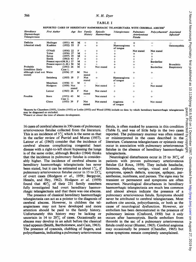
TABLE IREPORTED CASES OF HEREDITARY HAEMORRHAGIC TELANGIECTASIA WITH CEREBRAL ABSCESS"
First Author Age Sex Family EpisodicHistory Haemorrhage
Telangiectases Pulmonary PolycythaemiaW AssociatedArteriovenous InfectionsFistula
Definite Hedinger (1951) 46 M +(classical triad) Kushlan (1952) 55 F +
Probable(condition likelyalthough triad notstated)
O'Neill (1956) 22Taber (1956) 27Syrop (1957) 35Smith (1963) 43Present report(M.R.) 27Present report (L.H.) 50Stern (1953) 26
Case 1.
F +M +M +F +M +M +M +
Weiss (1954) 27 M Notstated
Steinberg (1955) 39 F Notstated
Meacham (1958) 11 MHodgson (1959) Not
statedLatour (1965) 26 F
Stern (1953) 43 MCase 2.
Glenn (1953) 39 F
Notstated
Notstated
+±
+++±±
Not stated +
Not stated
Not stated
Not stated +
Not stated Angiomaof tongue
'Reports by Chambers (1955), Linden (1953) or Lodin (1952) and Wood (1956) include no data by which hereditary haemorrhagic telangiectasiamay be diagnosed or excluded."Present at about the time of abscess development.
16 cases of cerebral abscess in 350 cases ofpulmonaryarteriovenous fistulae collected from the literature.This is an incidence of 5% which is the same as thatin the earlier review of Purriel and Muras (1957).Latour et al. (1965) consider that the incidence ofcerebral abscess complicating congenital heartdisease with a right-to-left shunt bypassing the lungsis of the same order, although Boczko (1964) thinksthat the incidence in pulmonary fistulae is consider-ably higher. The incidence of cerebral abscess inhereditary haemorrhagic telangiectasia has neverbeen stated, but it can be estimated as about 1%, ifpulmonary arteriovenous fistulae occur in 15 to 23%of overt cases (Hodgson et al., 1959; Bergqvist,Hessen, and Hey, 1962). Hodgson et al. (1959)found that 40% of their 231 family membersfully investigated had overt hereditary haemor-rhagic telangiectasia and that there was one abscess.The presence of classical hereditary haemorrhagic
telangiectasia can act as a pointer to the diagnosis ofcerebral abscess. However, in children the tel-angiectases may not have developed and moreattention should be paid to the family history.Unfortunately this history may be lacking oruncertain in 14 to 20% of cases. Occasionally anabscess may develop in an adult before the classicaltriad appears (Smith, Bartholomew, and Cain, 1963).The presence of cyanosis, clubbing of fingers, andpolycythaemia, indicating a pulmonary arteriovenous
fistula, is often masked by anaemia in this condition(Table I), and was of little help in the two casesreported. The pulmonary murmur was often missedor misinterpreted in the cases described in theliterature. Cutaneous telangiectases or epistaxis mayoccur in association with pulmonary arteriovenousfistulae in the absence of hereditary haemorrhagictelangiectasia.
Neurological disturbances occur in 25 to 30% ofpatients with proven pulmonary arteriovenousfistulae (Le Roux, 1959). They include headache,faintness, diplopia, vertigo, visual and auditorysymptoms, speech defects, syncope, epilepsy, par-aesthesiae, numbness, and pareses. The signs may betransient or permanent and symptoms are oftenrecurrent. Neurological disturbances in hereditaryhaemorrhagic telangiectasia are much less commonand almost always indicate the presence of apulmonary arteriovenous fistula. Symptoms shouldnever be attributed to cerebral telangiectases. Mostauthors cite anoxia, polycythaemia, or both as thecause of neurological dysfunction. However, airembolism has been demonstrated in the presence ofpulmonary lesions (Crafoord, 1950) but it onlyoccurs after haemoptysis. Sterile embolism fromthrombi in the sac of a pulmonary arteriovenousfistula could occur (Hunter, 1965). Cerebral angiomamay occasionally be present (Chandler, 1965) butsome symptoms remain completely unexplained.
566
HereditaryHaemorrhagicTelangiectasia
OsteomyelitisHaemangiomaof tongue
+
+
Haemangiomaof tongue+
Not stated+++Borderline
Borderline
Possible
+
Not stated+++
BronchitisRespiratory
Not stated
+
Not stated
guest. Protected by copyright.
on August 27, 2019 by
http://jnnp.bmj.com
/J N
eurol Neurosurg P
sychiatry: first published as 10.1136/jnnp.30.6.563 on 1 Decem
ber 1967. Dow
nloaded from

Cerebral abscess in hereditary haemorrhagic telangiectasia: report of two cases in a family
Cerebral abscess is thus only responsible for asmall proportion of the neurological disturbances inhereditary haemorrhagic telangiectasia, but as itcarries a high mortality without treatment, earlydiagnosis is essential, and it should always beexcluded by appropriate investigations. In the twocases reported here, the abscesses probably resultedfrom bacteria or septic emboli bypassing thepulmonary filter and lodging in the brain. Neithercase was significantly polycythaemic at the time ofthe disaster. Therefore secondary bacterial invasionof an area of encephalomalacia resulting fromcerebral thrombosis is unlikely (Stern and Naffziger,1953). However, anoxia causing focal cerebralnecrosis without vascular occlusion (Berthrong andSabiston, 1951) or sterile emboli from the peripheralvessels or sac of the fistula may have played a part.Maier, Himmelstein, Riley, and Bunin (1948) andStevenson (1953) have suggested that infection couldarise in the pulmonary arteriovenous fistula itself,which then discharges septic emboli, but such'endocarditis' has not been demonstrated histo-logically. Often a primary source of infection cannotbe found (Table I), but a number of cases havesuffered from bronchitis, as in case 2, or chronicosteomyelitis (Muri, 1953). The absence of anteced-ent infection and the preferential localization in thebrain with no abscesses at other sites has also beencommented on in congenital heart disease (Sancettaand Zimmerman, 1950).
SUMMARY
Two cases of cerebral abscess complicating heredi-tary haemorrhagic telangiectasia are described. Thepatients were members of the same family. Reportedcases of cerebral abscess associated with hereditaryhaemorrhagic telangiectasia have been examined andthe pathogenesis discussed. Other causes of neuro-logical disturbance in this condition are brieflyreviewed, and it is emphasized that cerebral abscessshould always be considered.
I would like to thank Professor C. Wilson, Dr. K. M. A.Perry, Mr. J. R. Belcher, and Mr. D. W. C. Northfieldfor permission to publish these cases, and Dr. R. A.Henson for helpful criticism of the paper.
REFERENCES
Bergqvist, N., Hessen, I., and Hey, M. (1962). Arteriovenous pul-monary aneurysms in Osler's disease. Acta med. scand.,171, 301-309.
Berthrong, M., and Sabiston, D. C. (1951). Cerebral lesions incongenital heart disease. Bull. Johns Hopk. Hosp., 89, 384-401.
Boczko, M. L. (1964). Neurological implications of hereditaryhemorrhagic telangiectasis. J. nerv. ment. Dis., 139, 525-536.
Chambers, W. R. (1955). Brain abscess associated with pulmonaryarterio-venous fistula. Ann. Surg., 141, 276-277.
Chandler, D. (1965). Pulmonary and cerebral arteriovenous fistulawith Osler's disease. Arch. intern. Med., 116, 277-282.
Crafoord, C. (1950). In Discussion of Lindskog et al., Ann. Surg.,132, 591.
Glenn, F. (1953). In Discussion of Stern and Naffziger. Ann. Surg.,138, 531.
Hedinger, C., Hitzig, W. H., and Marmier, C. (1951). Ober arterio-venose Lungenaneurysmen und ihre Beziehungen zur Osler-schen Krankheit. Schweiz. med. Wschr., 81, 367-374.
Hodgson, C. H., Burchell, H. B., Good, C. A., and Clagett, 0. T.(1959). Hereditary hemorrhagic telangiectasia and pulmonaryarteriovenous fistula: survey of a large family. New Engl. J.Med., 261, 625-636.
Hunter, D. D. (1965). Pulmonary arteriovenous malformation: anunusual cause of cerebral embolism. Canad. med. Ass. J.,93, 662-665.
Jaffe, R. H. (1929). Multiple hemangiomas of the skin and of theinternal organs. Arch. Path., 7,44-54.
Kushlan, S. D. (1952). Hereditary hemorrhagic telangiectasia withpulmonary arteriovenous aneurysm: possible etiology andprophylaxis. Conn. med. J., 16, 505-513.
Lancet (1960). Pulmonary arteriovenous fistula in hereditary haemor-rhagic telangiectasia. Lancet, 1, 158-159.
Latour, H., Puech, P., Hertault, J., and Grolleau, R. (1965). Abc&sdu cerveau revelateur d'un anevrysme arterio-veineux pul-monaire. Arch. Mal. Coeur, 58, 1503-1510.
Le Roux, B. T. (1959). Pulmonary arteriovenous fistulae. Quart. J.Med., 28, 1-19.
Linden, L. (1953). Hjarnabscess som foljd av paradoxal emboli.Nord. Med., 49, 255-257.
Lodin, H. (1952). Tomographic analysis of arteriovenous aneurysmsin the lung. Acta radiol. (Stockh.), 38, 205-211.
Maier, H. C., Himmelstein, A., Riley, R. L., and Bunin, J. J. (1948).Arteriovenous fistula of the lung. J. thorac. Surg., 17, 13-26.
Meacham, W. F., and Scott, H. W. (1958). Congenital pulmonaryarterio-venous aneurysm complicated by Bacteroides abscessof brain: successful surgical management. Ann. Surg., 147,404-408.
Muri, J. (1953). Arterio-venous aneurysma of the lung. Dis. Chest,24,49-61.
O'Neill, T. J. E., Fisher, H., McDowell, D. E., and Lauby, V. W.(1956). Pulmonary arteriovenous fistula in sisters. J. thorac.Surg., 31, 286-297.
Osler, W. (1901). On a family form of recurring epistaxis, associatedwith multiple telangiectases of the skin and mucous membranes.Bull. Johns Hopk. Hosp. 12, 333-337.
Penfold, J. B., and Lipscomb, J. M. (1943). Elliptocytosis in manassociated with hereditary haemorrhagic telangiectasia.Quart. J. Med., 12, 157-167.
Purriel, P., and Muras, 0. (1957). Aneurismas arteriovenosos depulm6n. T6rax, 6, 101-158.
Reading, B. (1932). A case of congenital telangiectasis of the lung,complicated by brain abscess. Texas St. J. Med., 28, 462-464.
Sancetta, S. M., and Zimmerman, H. A. (1950). Congenital heartdisease with septal defects in which paradoxical brain abscesscauses death. Circulation. 1, 593-601.
Smith, C. R., Bartholomew, L. G., and Cain, J. C. (1963). Hereditaryhemorrhagic telangiectasia and gastrointestinal hemorrhage.Gastroenterology 44, 1-6.
Steinberg, I., and McClenahan, J. (1955). Pulmonary arteriovenousfistula. Amer. J. Med., 19, 549-568.
Stern, W. E., and Naffziger, H. C. (1953). Brain abscess associatedwith pulmonary angiomatous malformation. Ann. Surg.,138, 521-531.
Stevenson, F. H. (1953). Infected arteriovenous fistula in the lung.Lancet, 1, 626.
Syrop, H. M. (1957). Hereditary hemorrhagic telangiectasia. OralSurg., 10, 253-261.
Taber, R. E., and Ehrenhaft, J. L. (1956). Arteriovenous fistulae andarterial aneurysms of the pulmonary arterial tree. Arch.Surg., 73, 567-577.
Weiss, E., and Gasul, B. M. (1954). Pulmonary arteriovenous fistulaand telangiectasia. Ann. intern. Med., 41, 989-1002.
Wood, P. (1956). Diseases of the Heart and Circulation, 2nd. ed.,p. 912. Eyre and Spottiswoode, London.
567
guest. Protected by copyright.
on August 27, 2019 by
http://jnnp.bmj.com
/J N
eurol Neurosurg P
sychiatry: first published as 10.1136/jnnp.30.6.563 on 1 Decem
ber 1967. Dow
nloaded from