


Languages
Pages
Legal


Gulf Coast Research Center for Evacuation and Transportation Resiliency
LSU / UNO University Transportation Center
Improving the Self-Healing Properties of Concrete Materials by using Composite Actions with Fiber Reinforced Polymers
Final Report Michele Barbato – LSU Department of Civil and Environmental Engineering Marwa Hassan – LSU Department of Construction Management Louisiana State University and A&M College Baton Rouge, LA 70803
Sponsoring Agency United States Department of Transportation Research and Innovative Technology Administration
Washington, DC Project # 12-01 June 2013
GULF COAST RESEARCH CENTER FOR EVACUATION AND TRANSPORTATION RESILIENCY
The Gulf Coast Research Center for Evacuation and Transportation Resiliency is a collaborative effort between the Louisiana State University Department of Civil and Environmental Engineering and the University of New Orleans' Department of Planning and Urban Studies. The theme of the LSU-UNO
Center is focused on Evacuation and Transportation Resiliency in an effort to address the multitude of issues that impact transportation processes under emergency conditions such as evacuation and other types of major events. This area of research also addresses the need to develop and maintain the ability of transportation systems to economically, efficiently, and safely respond to the changing demands that may be placed upon them.
Research The Center focuses on addressing the multitude of issues that impact transportation processes under emergency conditions such as evacuation and other types of major events as well as the need to develop and maintain the ability of transportation systems to economically, efficiently, and safely respond to the changing conditions and demands that may be placed upon them. Work in this area include the development of modeling and analysis techniques; innovative design and control strategies; and travel demand estimation and planning methods that can be used to predict and improve travel under periods of immediate and overwhelming demand. In addition to detailed analysis of emergency transportation processes, The Center provides support for the broader study of transportation resiliency. This includes work on the key components of redundant transportation systems, analysis of congestion in relation to resiliency, impact of climate change and peak oil, provision of transportation options, and transportation finance. The scope of the work stretches over several different modes including auto, transit, maritime, and non-motorized. Education The educational goal of the Institute is to provide undergraduate-level education to students seeking careers in areas of transportation that are critical to Louisiana and to the field of transportation in general with local, national and international applications. Courses in Transportation Planning, Policy, and Land use are offered at UNO, under the Department of Planning and Urban Studies. In addition to the program offerings at UNO, LSU offers transportation engineering courses through its Department of Civil and Environmental Engineering. The Center also provides on-going research opportunities for graduate students as well as annual scholarships.
Technology Transfer The LSU/UNO UTC conducts technology transfer activities in the following modes: 1) focused professional, specialized courses, workshops and seminars for private sector entities (business and nonprofits) and government interests, and the public on transport issues (based on the LSU-UNO activities); 2) Research symposia; transport issues (based on the LSU-UNO activities); 3) Presentations at professional organizations; 4) Publications. The Center sponsors the National Carless Evacuation Conference and has co-sponsored other national conferences on active transportation.
Disclaimer The contents of this report reflect the views of the authors, who are solely responsible for the facts and the accuracy of the material and information presented herein. This document is disseminated under the sponsorship of the U.S. Department of Transportation University Transportation Centers Program in the interest of information exchange. The U.S. Government assumes no liability for the contents or use thereof. The contents do not necessarily reflect the official views of the U.S. Government. This report does not constitute a standard, specification, or regulation
i
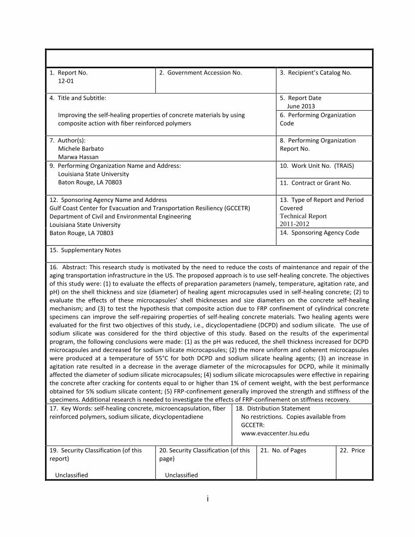
1. Report No. 12-01
2. Government Accession No.
3. Recipient’s Catalog No.
4. Title and Subtitle: 5. Report Date June 2013
Improving the self-healing properties of concrete materials by using composite action with fiber reinforced polymers
6. Performing Organization Code
7. Author(s): Michele Barbato Marwa Hassan
8. Performing Organization Report No.
9. Performing Organization Name and Address: Louisiana State University Baton Rouge, LA 70803
10. Work Unit No. (TRAIS)
11. Contract or Grant No.
12. Sponsoring Agency Name and Address Gulf Coast Center for Evacuation and Transportation Resiliency (GCCETR) Department of Civil and Environmental Engineering Louisiana State University Baton Rouge, LA 70803
13. Type of Report and Period Covered Technical Report
2011-2012
14. Sponsoring Agency Code
15. Supplementary Notes
16. Abstract: This research study is motivated by the need to reduce the costs of maintenance and repair of the aging transportation infrastructure in the US. The proposed approach is to use self-healing concrete. The objectives of this study were: (1) to evaluate the effects of preparation parameters (namely, temperature, agitation rate, and pH) on the shell thickness and size (diameter) of healing agent microcapsules used in self-healing concrete; (2) to evaluate the effects of these microcapsules’ shell thicknesses and size diameters on the concrete self-healing mechanism; and (3) to test the hypothesis that composite action due to FRP confinement of cylindrical concrete specimens can improve the self-repairing properties of self-healing concrete materials. Two healing agents were evaluated for the first two objectives of this study, i.e., dicyclopentadiene (DCPD) and sodium silicate. The use of sodium silicate was considered for the third objective of this study. Based on the results of the experimental program, the following conclusions were made: (1) as the pH was reduced, the shell thickness increased for DCPD microcapsules and decreased for sodium silicate microcapsules; (2) the more uniform and coherent microcapsules were produced at a temperature of 55°C for both DCPD and sodium silicate healing agents; (3) an increase in agitation rate resulted in a decrease in the average diameter of the microcapsules for DCPD, while it minimally affected the diameter of sodium silicate microcapsules; (4) sodium silicate microcapsules were effective in repairing the concrete after cracking for contents equal to or higher than 1% of cement weight, with the best performance obtained for 5% sodium silicate content; (5) FRP-confinement generally improved the strength and stiffness of the specimens. Additional research is needed to investigate the effects of FRP-confinement on stiffness recovery.
17. Key Words: self-healing concrete, microencapsulation, fiber reinforced polymers, sodium silicate, dicyclopentadiene
18. Distribution Statement No restrictions. Copies available from GCCETR: www.evaccenter.lsu.edu
19. Security Classification (of this report)
Unclassified
20. Security Classification (of this page)
Unclassified
21. No. of Pages
22. Price
ii
Acknowledgements
This project was funded by the Gulf Coast Center for Evacuation and Transportation Resiliency
(CETR) at Louisiana State University, Baton Rouge, LA 70803.
The authors gratefully acknowledge the Louisiana Transportation Research Center (LTRC) for
granting access to their laboratory. Additional support from the Department of Construction
Management and the Department of Civil and Environmental Engineering is also greatly
acknowledged.
iii
Table of Contents
ACKNOWLEDGEMENTS ......................................................................................................... II
TABLE OF CONTENTS ........................................................................................................... III
LIST OF FIGURES .................................................................................................................... IV
EXECUTIVE SUMMARY ............................................................................................................ 1
ABSTRACT................................................................................................................................... 5
1 INTRODUCTION .............................................................................................................. 7
2 LITERATURE REVIEW .................................................................................................. 7
3 METHODOLOGY ............................................................................................................. 9 TASK 1: EVALUATION OF THE EFFECTS OF PREPARATION PARAMETERS 3.1
OF SELF-HEALING MICROCAPSULES ..................................................................... 9
3.1.1 Test materials ......................................................................................................... 11
3.1.2 Test methods .......................................................................................................... 11
TASK 2: EVALUATION OF THE EFFECTS OF MICROCAPSULES’ 3.2
PROPERTIES ON CONCRETE SELF-HEALING MECHANISM........................... 12
TASK 3: EVALUATION OF FRP-CONFINEMENT EFFECTS ON SELF-3.3
HEALING CONCRETE ................................................................................................. 13 3.3.1 Test materials ......................................................................................................... 13
3.3.2 Experimental matrix ............................................................................................... 13 3.3.3 Loading test protocol ............................................................................................. 14
4 RESULTS ........................................................................................................................ 15 EVALUATION OF THE EFFECTS OF PREPARATION PARAMETERS OF 4.1
SELF-HEALING MICROCAPSULES .......................................................................... 15 4.1.1 Effects of pH on morphology and shell thickness ............................................. 15
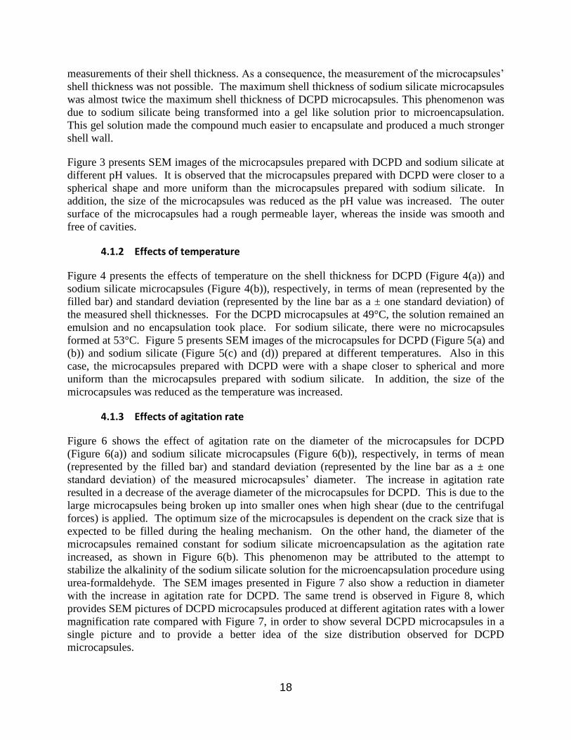
4.1.2 Effects of temperature ........................................................................................... 18
4.1.3 Effects of agitation rate .......................................................................................... 18
EVALUATION OF THE EFFECTS OF MICROCAPSULES’ PROPERTIES ON 4.2
CONCRETE SELF-HEALING MECHANISM............................................................. 24 EVALUATION OF FRP-CONFINEMENT EFFECTS ON SELF-HEALING 4.3
CONCRETE .................................................................................................................... 27
5 CONCLUSIONS ............................................................................................................. 31
APPENDIX A: DCPD MICROENCAPSULATION PROCEDURE ..................................... 32
APPENDIX B: SODIUM SILICATE MICROENCAPSULATION PROCEDURE ............. 33
REFERENCES ........................................................................................................................... 35
iv
List of Figures
Figure 1: Schematic of the Components of a Microcapsule ................................................ 10
Figure 2: Effect of pH Values on the Shell Thickness for: (a) DCPD Microcapsules, and (b) Sodium Silicate Microcapsules .................................................................................. 16
Figure 3: Effect of pH Values on the Morphology of Microcapsules for: (a) DCPD at pH = 3.1, (b) DCPD at pH = 3.7, (c) Sodium Silicate at pH = 3.0, and (d) Sodium Silicate at pH = 3.2 ........................................................................................................................... 17
Figure 4: Effect of Temperature on the Shell Thickness for: (a) DCPD Microcapsules, and (b) Sodium Silicate Microcapsules ................................................................... 19
Figure 5: Effects of Temperature on the Morphology of Microcapsules for: (a) DCPD at T = 55°C, (b) DCPD at T = 52°C, (c) Sodium Silicate at T = 55°C, and (d) Sodium Silicate at T = 51°C ................................................................................ 20
Figure 6: Effect of Agitation Rate on the Diameter for: (a) DCPD Microcapsules, and (b) Sodium Silicate Microcapsules .......................................................................... 21
Figure 7: Effects of Agitation Rate on the Morphology of Microcapsules for: (a) DCPD at 250 rpm, (b) DCPD at 549 rpm, (c) Sodium Silicate at 257 rpm, and (d) Sodium Silicate at 551 rpm ................................................................................. 22
Figure 8: Qualitative Size Distribution for DCPD Microcapsules as a Function of Agitation Rate: (a) 350 rpm, and (b) 450 rpm ................................................................ 23
Figure 9: Effect of Preparation pH of Microcapsules on Concrete Modulus of Elasticity before and after Healing .................................................................................................... 24
Figure 10: Effect of Amount of Microcapsules (% of Cement Weight) on Concrete Modulus of Elasticity before and after Healing .............................................................. 26
Figure 11: Crack Healing after 1-Week Recovery: (a) DCPD before Healing, (b) DCPD after 1-Week Healing, (c) Sodium Silicate (1%) before Healing, and (d) Sodium Silicate (1%) after 1-Week Healing ................................................................................. 26
Figure 12: Stress-Strain Response for Plain Concrete Specimens: (a) Unconfined, and (b) FRP-Confined ............................................................................................................... 29
Figure 13: Stress-Strain Response for Concrete Specimens with 1.0% Sodium Silicate: (a) Unconfined, and (b) FRP-Confined ........................................................................... 29
Figure 14: Stress-Strain Response for Concrete Specimens with 2.5% Sodium Silicate: (a) Unconfined, and (b) FRP-Confined ........................................................................... 30
Figure 15: Stress-Strain Response for Concrete Specimens with 5.0% Sodium Silicate: (a) Unconfined, and (b) FRP-Confined ........................................................................... 30
Figure 16: Concrete Stiffness Before and After Healing ...................................................... 31
1
Executive Summary
Background and Objectives
The aging civil infrastructure in the US represents a serious challenge for maintenance and repair
using only limited available resources. For example, the average age of a bridge in the US is 43
years, with a design lifetime usually assumed equal to 50 years. The American Association of
State Highway and Transportation Officials (AASHTO) estimated that the cost to repair every
deficient bridge in the country would be approximately $140 billion. This repair cost does not
include the cost associated with mandated annual bridge inspections, or the cost associated with
traffic restrictions on structurally-deficient or functionally obsolete bridges. Immediate
improvement of this situation is difficult due to the economic crisis that the US has suffered in
recent years. It is envisioned that a long-term solution of this problem can only be achieved
through new and creative transformative approaches that can significantly reduce the costs
associated with inspection, maintenance, and repair of infrastructure elements. One solution for
this problem involves the use of a new paradigm known as “self-healing concrete.” Self-healing
in concrete can be defined as the ability of concrete to autonomously heal cracks that may
develop throughout its structure. By incorporating self-healing properties into concrete mixes, it
is expected that concrete quality design and control methods will improve, with the goal of
positively impacting concrete construction processes as a whole.
The specific objectives of this study were: (1) to evaluate the effects of preparation parameters
(namely, temperature, agitation rate, and pH) on the shell thickness and size (diameter) of
microcapsules of healing agents for use in self-healing concrete; (2) to evaluate the effects of
microcapsules’ shell thickness and size diameters on the concrete self-healing mechanism; and
(3) to test the hypothesis that composite action due to FRP confinement of cylindrical concrete
specimens can improve the self-repairing properties of self-healing concrete materials.
The goal of this research was to significantly advance the self-repairing capability of RC bridge
components and systems. This capability is currently limited to the closure of small surface
cracks produced in a controlled environment. This self-healing property mimics the self-healing
of human skin after small cuts. The composite action due to confinement with fiber reinforced
polymers (FRPs) can help close larger cracks in the concrete. In comparison with the human
body, this self-healing capability is equivalent to the cicatrization of deep cuts and the healing of
bone fractures. This research explored a completely innovative and untested idea, since it
represents the first attempt of using composite action to enhance the performance of self-healing
concrete materials.
Research Outcomes and Results
Three sets of experimental tests were developed and performed to achieve the objectives of this
research. Two healing agents were evaluated for the first two objectives of this study, i.e.,
dicyclopentadiene (DCPD) and sodium silicate. The use of sodium silicate was considered for
2
the third objective of this study. Based on the results of the experimental program, the following
conclusions were made: (1) as the pH was reduced, the shell thickness increased for DCPD
microcapsules and decreased for sodium silicate microcapsules; (2) the more uniform and
coherent microcapsules were produced at a temperature of 55°C for both DCPD and sodium
silicate healing agents; (3) an increase in agitation rate resulted in a decrease in the average
diameter of the microcapsules for DCPD, while it minimally affected the diameter of sodium
silicate microcapsules; (4) sodium silicate microcapsules were effective in repairing the concrete
after cracking for contents equal or higher than 1% of cement weight, with the best performance
obtained for 5% sodium silicate content; (5) FRP-confinement generally improved the strength
and stiffness of the specimens. Additional research is needed to investigate the effects of FRP-
confinement on stiffness recovery.
Other Project Outcomes:
Students support:
A PhD and a Master student were supported on this project. The PhD student Yueqiang Sui
works under the supervision of the PI, Dr. Michele Barbato, and his PhD research is in progress.
The Master student was Lt. Cmdr. James Gilford III and he worked under the supervision of the
Co-PI, Dr. Hassan. He was a US Naval officer. Gilford’s tuition was provided by the US Navy
but support for Gilford included building an experimental setup and all the supplies he needed to
conduct his research. He graduated with a MSc. in Engineering Science in summer 2012 with
the following thesis:
Gilford III, J. (2012). “Microencapsulation of self-healing concrete properties.” Master thesis,
Department of Engineering Science, LSU, Baton Rouge, LA.
Publications:
This project resulted in one Transportation Research Board (TRB) paper presented at the 92nd
Annual Meeting and one ASCE journal paper currently under review:
Gilford III, J., Hassan, M.M., Rupnow, T., and, Barbato, M. (2013). “Evaluation of
Microencapsulation of Dicyclopentadiene (DCPD) and Sodium Silicate for Self-Healing
Concrete.” Paper #13-1172, 92nd
Transportation Research Board Annual meeting, National
Research Council, Washington, D.C.
Gilford III, J., Hassan, M.M., Rupnow, T., Barbato, M., Okeil, A., and, Asadi, S. (2013). “DCPD
and Sodium Silicate Microencapsulation for Self-Healing of Concrete.” ASCE Journal of
Materials in Civil Engineering, under review.
It is expected that the results of this research will be used to prepare an additional journal
publication.
3
Proposals submitted:
The project produced preliminary data that allowed the team to submit the following
collaborative NSF proposal between LSU and Texas A&M Kingsville to the division of CMMI,
SMM:
COLLABORATIVE RESEARCH: A NEW GENERATION OF INFRASTRUCTURE
ELEMENTS MIMICKING SELF-HEALING MECHANISMS OF LIVING ORGANISMS. PI:
Dr. Marwa Hassan, Co-PI’s Dr. Michele Barbato, & Dr. Ayman Okeil. Requested funding:
$266,543. Status: Declined.
The PI and Co-PI intend to re-submit a modified version of the previous proposal with stronger
focus on the material characterization and the chemical/mechanical interaction between
microcapsules of self-healing agents and concrete.

4
5
Abstract
This research study is motivated by the need to reduce the costs of maintenance and repair of the
aging transportation infrastructure in the US. The proposed approach is to use self-healing
concrete. The objectives of this study were: (1) to evaluate the effects of preparation parameters
(namely, temperature, agitation rate, and pH) on the shell thickness and size (diameter) of
healing agent microcapsules used in self-healing concrete; (2) to evaluate the effects of these
microcapsules’ shell thicknesses and size diameters on the concrete self-healing mechanism; and
(3) to test the hypothesis that composite action due to FRP confinement of cylindrical concrete
specimens can improve the self-repairing properties of self-healing concrete materials. Two
healing agents were evaluated for the first two objectives of this study, i.e., dicyclopentadiene
(DCPD) and sodium silicate. The use of sodium silicate was considered for the third objective of
this study. Based on the results of the experimental program, the following conclusions were
made: (1) as the pH was reduced, the shell thickness increased for DCPD microcapsules and
decreased for sodium silicate microcapsules; (2) the more uniform and coherent microcapsules
were produced at a temperature of 55°C for both DCPD and sodium silicate healing agents; (3)
an increase in agitation rate resulted in a decrease in the average diameter of the microcapsules
for DCPD, while it minimally affected the diameter of sodium silicate microcapsules; (4) sodium
silicate microcapsules were effective in repairing the concrete after cracking for contents equal
to or higher than 1% of cement weight, with the best performance obtained for 5% sodium
silicate content; (5) FRP-confinement generally improved the strength and stiffness of the
specimens. Additional research is needed to investigate the effects of FRP-confinement on
stiffness recovery.

6
7
1 Introduction
The aging civil infrastructure in the US represents a serious challenge for maintenance and repair
using only limited available resources. For example, the average age of a bridge in the US is 43
years, with a design lifetime usually assumed equal to 50 years. The American Association of
State Highway and Transportation Officials (AASHTO) estimated that the cost to repair every
deficient bridge in the country would be approximately $140 billion (AASHTO 2008). This
repair cost does not include the cost associated with mandated annual bridge inspections, or the
cost associated with traffic restrictions on structurally-deficient or functionally obsolete bridges.
Immediate improvement of this situation is difficult due to the economic crisis that the US has
suffered in recent years. It is envisioned that a long-term solution of this problem can only be
achieved through new and creative transformative approaches that can significantly reduce the
costs associated with inspection, maintenance, and repair of infrastructure elements. One solution
for this problem involves the use of a new paradigm known as “self-healing concrete.” Self-
healing in concrete can be defined as the ability of concrete to autonomously heal cracks that
may develop throughout its structure. By incorporating self-healing properties into concrete
mixes, it is expected that concrete quality design and control methods will improve, with the goal
of positively impacting concrete construction processes as a whole.
The main objective of this research project was to test the hypothesis that composite action in
reinforced concrete (RC) structures can drastically improve the self-repairing properties of self-
healing concrete materials. The goal of this research was to significantly advance the self-
repairing capability of RC bridge components and systems. This capability is currently limited to
the closure of small surface cracks produced in a controlled environment. This self-healing
property mimics the self-healing of human skin after small cuts. The composite action due to
confinement with fiber reinforced polymers (FRPs) can help close larger cracks in the concrete.
In comparison with the human body, this self-healing capability is equivalent to the cicatrization
of deep cuts and the healing of bone fractures. This research explored a completely innovative
and untested idea, since it represents the first attempt of using composite action to enhance the
performance of self-healing concrete materials.
2 Literature Review
Considerable interest has been given in recent years to the utilization of self-healing materials in
concrete (Sharp and Clemena 2004). This has led to the introduction of a new class of smart
materials that have the ability to heal after damage. Self-healing applications in concrete have led
to the introduction of bacteria-based self-healing concrete and microcapsule-based self-healing
concrete. Bacteria-based self-healing concrete uses mineral-producing bacteria, which were
found able to seal surface cracks (Jonkers 2011). The concept of microcapsule healing is based
on a healing agent being encapsulated and embedded in the concrete. When the crack propagates
and reaches the microcapsule, the capsule breaks and the healing agent is released into the crack
to repair it. Self-healing concrete provides a proactive approach rather than a reactive
8
countermeasure for cracks that develop within concrete structures. Self-healing concrete
materials have been proven effective in terms of reduction of permeability (Reinhardt and Jooss
2003), as well as recovery of strength and stiffness under controlled cracking of the material in
laboratory conditions (Li et al. 1998). However, the results in field applications have not yet been
as satisfactory, since it is very difficult to limit the size of the cracks in the concrete to less than
150 m (size beyond which the self-healing mechanism cannot be activated, see Yang et al.
2009). An emerging research direction focused on improving the performance of self-healing
concrete is the development of engineered cementitious composites that can limit the average
crack size due to the intrinsic material properties (Yang et al. 2009).
Since their introduction in the 1950s, microencapsulation has been evaluated in numerous
construction materials including mortar, lime, cement, marble, sealant, and paints (Boh and
Sumiga 2008). It has also been patented and tested in the food, chemical, textile, and
pharmaceutical industries. The most common mechanism to trigger microcapsule-healing is
through external pressure, which ruptures the microcapsule and releases the healing agent from
the core. Therefore, the microcapsule must be sufficiently stiff to remain intact during
processing, concrete mixing, pouring, and setting but it must break during damage of the
concrete. In addition, the microcapsule shell provides a protective barrier between the catalyst
and the healing agent to prevent polymerization during the preparation of the composite. There
are three main methods for preparation of microcapsules (Boh and Sumiga 2008): (1) the
mechanical method, which mechanically applies the microcapsule around the healing agent; (2)
the coacervation method, in which the microcapsule wall solidifies around a core made of the
healing agent; and (3) the polymerization method where the healing agent is applied as an
emulsion, which then solidifies at the interface between water and healing agent to form the
microcapsule wall.
FRP composites have found increasingly numerous applications in structural engineering due to
their high strength-to-weight and stiffness-to-weight ratios, high corrosion resistance, and
potentially high durability (Einde et al. 2003). One of these applications is the confinement of
reinforced concrete (RC) columns with FRP to improve their structural performance in terms of
ultimate load bearing capacity and ductility (Nanni and Bradford 1995, Seible et al. 1997). FRP
confinement of RC columns presents numerous advantages compared to other strengthening
techniques, e.g., strengthening using steel jackets. Some of these advantages include small
increase in structural size and weight, easy transportation, and good resistance to corrosion and
other degradation processes due to harsh environmental conditions (Bakis et al. 2002). This
strengthening method has been widely used in retrofitting of bridges and buildings in the past
few decades (Flaga 2000, Pantelides et al. 2000, Mertz et al. 2003, Monti 2003, Motavalli and
Czaderski 2007).
The use of FRP confinement for RC cylindrical columns derives from the fact that, when
concrete is subjected to an axial compression load, the concrete tends to expand laterally and to
load the FRP confining jacket in axial tension along the radial direction. Thus, the concrete core
of the column becomes subjected to a three-dimensional (3-D) compressive stress condition,
which can significantly increase the compressive strength and the ductility of otherwise brittle
concrete. Similar behavior has been observed also for RC columns with square or rectangular
cross-section, for which a lower level of efficiency can be reached due to the fact that the stress
9
in the FRP is not uniform. When subjected to an axial compression load, FRP-confined RC
columns behave differently than steel-confined RC columns. In fact, while steel jackets provide a
constant confinement after the yielding point of the material is reached, FRP jackets provide a
linearly increasing confinement force, which better contrasts the expansion of the concrete in the
radial direction and significantly reduces the volumetric expansion of the concrete. This
phenomenon produces a significantly more ductile behavior of the concrete, which is evidenced
by the distinct bilinear shape of the monotonic stress-strain curve, with a smooth transition zone
beginning at a stress level close to the strength of the unconfined concrete. It is noticed here that,
while extensive research is available to document the positive effects on strength and ductility
due to FRP confinement of RC columns subjected to axial and bending actions, no information is
available in literature regarding the effects of FRP confinement on self-healing concrete.
3 Methodology
This research consisted of three main tasks: (1) evaluation of the effects of self-healing
microcapsules’ preparation parameters, namely, temperature, agitation rate, and pH on the shell
thickness and size (diameter) of the microcapsules; (2) evaluation of the effects of
microcapsules’ shell thickness and size diameters on the concrete self-healing mechanism
through mechanical experimental testing performed in laboratory; and (3) verification of the
hypothesis that composite action due to FRP confinement of cylindrical concrete specimens can
improve the self-repairing properties of self-healing concrete materials through mechanical
experimental testing performed in laboratory.
Task 1: Evaluation of the effects of preparation parameters of self-healing 3.1
microcapsules
There are three main methods for preparation of microcapsules (Boh and Sumiga 2008): (1) the
mechanical method, which mechanically applies the microcapsule around the healing agent; (2)
the coacervation method, in which the microcapsule wall solidifies around a core made of the
healing agent; and (3) the polymerization method where the healing agent is applied as an
emulsion, which then solidifies at the interface between water and healing agent to form the
microcapsule wall. The polymerization method, which was used in this study, is categorized as
either in-situ polymerization, in which the healing agent is added to the liquid phase of an
emulsion, or as interfacial polymerization, in which the healing agent is dissolved into the liquid
phase. In this study, in-situ polymerization was selected for preparation of the microcapsules.
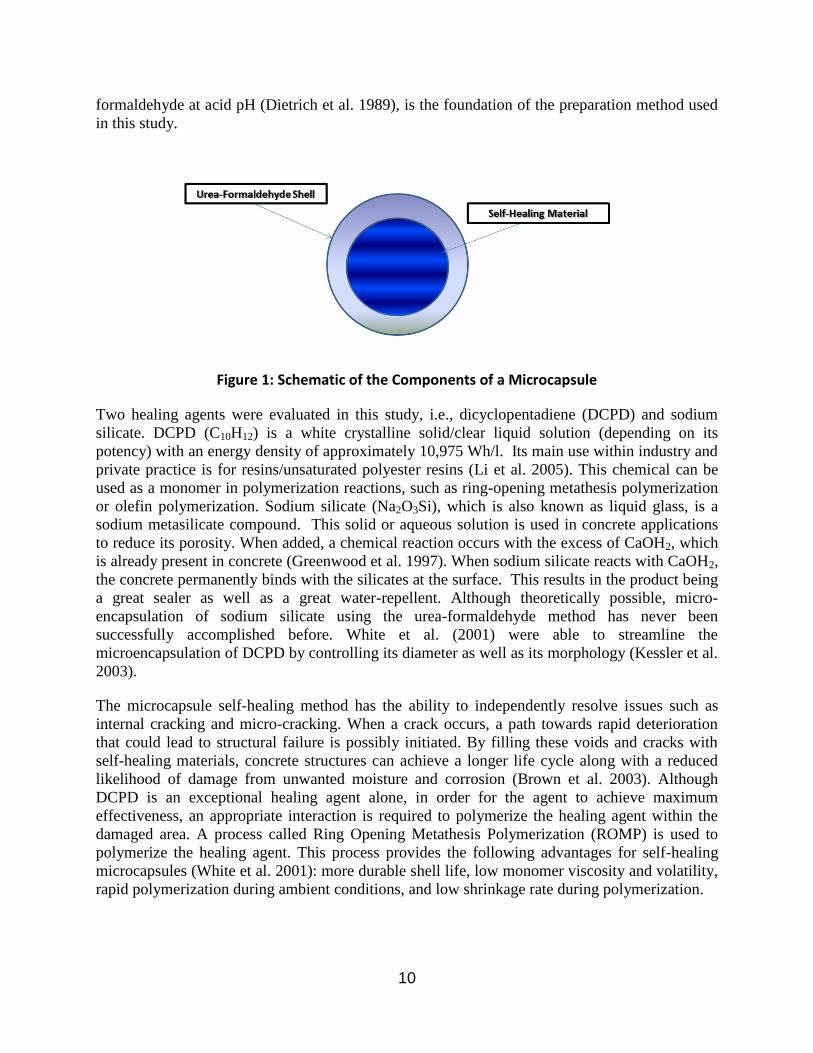
As shown in Figure 1, the two main design parameters of interest during microcapsule
preparation are shell thickness and microcapsule size (diameter). Microcapsule walls that are too
thin would fail during the manufacturing process, concrete mixing, pouring, and setting (Tseng et
al. 2005). In contrast, capsule shells that are too thick will not allow breaking or fracturing of the
shell as the crack penetrates through the microcapsules plane. A well-developed process of
microencapsulation using the urea-formaldehyde method was developed by Brown et al. (2003).
The in-situ encapsulation method for water-immiscible liquids, by the reaction of urea with
10
formaldehyde at acid pH (Dietrich et al. 1989), is the foundation of the preparation method used
in this study.
Figure 1: Schematic of the Components of a Microcapsule
Two healing agents were evaluated in this study, i.e., dicyclopentadiene (DCPD) and sodium
silicate. DCPD (C10H12) is a white crystalline solid/clear liquid solution (depending on its
potency) with an energy density of approximately 10,975 Wh/l. Its main use within industry and
private practice is for resins/unsaturated polyester resins (Li et al. 2005). This chemical can be
used as a monomer in polymerization reactions, such as ring-opening metathesis polymerization
or olefin polymerization. Sodium silicate (Na2O3Si), which is also known as liquid glass, is a
sodium metasilicate compound. This solid or aqueous solution is used in concrete applications
to reduce its porosity. When added, a chemical reaction occurs with the excess of CaOH2, which
is already present in concrete (Greenwood et al. 1997). When sodium silicate reacts with CaOH2,
the concrete permanently binds with the silicates at the surface. This results in the product being
a great sealer as well as a great water-repellent. Although theoretically possible, micro-
encapsulation of sodium silicate using the urea-formaldehyde method has never been
successfully accomplished before. White et al. (2001) were able to streamline the
microencapsulation of DCPD by controlling its diameter as well as its morphology (Kessler et al.
2003).
The microcapsule self-healing method has the ability to independently resolve issues such as
internal cracking and micro-cracking. When a crack occurs, a path towards rapid deterioration
that could lead to structural failure is possibly initiated. By filling these voids and cracks with
self-healing materials, concrete structures can achieve a longer life cycle along with a reduced
likelihood of damage from unwanted moisture and corrosion (Brown et al. 2003). Although
DCPD is an exceptional healing agent alone, in order for the agent to achieve maximum
effectiveness, an appropriate interaction is required to polymerize the healing agent within the
damaged area. A process called Ring Opening Metathesis Polymerization (ROMP) is used to
polymerize the healing agent. This process provides the following advantages for self-healing
microcapsules (White et al. 2001): more durable shell life, low monomer viscosity and volatility,
rapid polymerization during ambient conditions, and low shrinkage rate during polymerization.
11
ROMP utilizes a Grubbs catalyst (transition metal catalyst), which incorporates a high metathesis
method. The use of this catalyst allows multiple chemical groups to be utilized within the
chemical process (such as oxygen and water). When DCPD encounters the Grubbs catalyst,
polymerization occurs (Brown et al. 2005). Sodium silicate, however, does not require a matrix
and can be used as an individual healing component. The first reaction consists of sodium silicate
reacting with calcium hydroxide, which is a product of cement hydration (Nonat 2004). The
second reaction occurs between sodium hydroxide and silica. In both processes, the mending
agent that resides in an aqueous environment within the microcapsule itself is essential (Nonat
2004). Water enables the hydration of the damaged cement paste and allows further bonding of
the mending agent. The products of both reactions fill the crack and subsequently permit
recovery of strength. Both processes support the presence of the aqueous mending agent, which
also provides further integrity of the concrete by creating a bond and healing the crack (Brown et
al. 2005).
3.1.1 Test materials
The chemicals utilized in the preparation of the microcapsules based on the in-situ
polymerization method are presented in Table 1. The two microencapsulation laboratory
procedures that were utilized in this study for preparation of DCPD and sodium silicate
microcapsules are presented in Appendices A and B, respectively.
Table 1: Required chemicals for interfacial polymerization synthesis
Chemical Function Manufacturer
Urea Creates endothermic reaction in water The Science Company
Ammonium Chloride Assists with curing Process The Science Company
Resorcinol (Technical Grade Flake)
Reacts with formaldehyde and is a
chemical intermediate for the
synthesis process
NDSPEC Chemical
Corporation
ZeMac E60 Copolymer Improves mechanical properties Vertellus Specialties, Inc.
ZeMac E400 Copolymer Improves mechanical properties Vertellus Specialties, Inc.
Octanol Prevents surface bubbles Oltchim
Hydrochloric Acid Lowers pH The Science Company
Sodium Silicate Reacts with Ca(OH)2 The Science Company
Sodium Hydroxide Increases pH The Science Company
Formaldehyde Reacts with urea during synthesis
process The Science Company
Grubbs Catalyst Reacts with DCPD and polymerizes Materia, Inc.
DETA (diethylenetriamine) Mix with
EPON 828
Used in synthesis of catalysts, epoxy
curing agent, and corrosion inhibitors Huntsmann
DCPD Selected Resin to Heal Concrete
Crack
Texmark- 87% & 89% Purity
Cymetech- 99% Purity
3.1.2 Test methods
An experimental program was developed to evaluate the effects of preparation parameters
(namely temperature, agitation rate, and pH) on the shell thickness and size of the microcapsules
using Scanning Electron Microscopy (SEM). Microscopic analysis was conducted using a FEI
Quanta 3D SEG Dual Beam SEM with focused ION beam at an acceleration voltage of 15kV
12
and in the Backscattered Electron Imaging Mode. The images were stored as 1,290×968 TIFF
files. Using image analysis software (Image J), the average particle diameter and shell thickness
was measured and calculated. Measured microcapsules were selected by random sampling from
each developed batch. The samples were coated with a thin layer of platinum conducting film by
sputtering. Each sample was sputtered for 4 minutes to ensure an even distribution of the coating
around each shell.
Table 2 presents the experimental matrix followed in this task. Two healing agents were
evaluated, i.e., DCPD and sodium silicate. During synthesis, the agitation rate, temperature, and
pH were varied one at a time. The agitation rate was varied at six levels for the DCPD synthesis
and at four levels for the sodium silicate synthesis, while the temperature and pH were kept
constant. Similarly, to evaluate the effect of temperature, three levels were used for both DCPD
and sodium silicate, while the pH and agitation rate were kept constant. Three pH levels were
considered for both DCPD and sodium silicate, while the temperature and agitation rate were
kept constant. The constant reference levels of temperature, pH, and agitation rate were: 55°C,
3.7, and 550 rpm, respectively, for the DCPD; and 55°C, 3.0, and 550 rpm, respectively, for the
sodium silicate. This experimental matrix resulted in a total of 10 synthesis methods tested using
DCPD and 8 synthesis methods tested using sodium silicate.
Table 2: Experimental test matrix for Task 1
Task 2: Evaluation of the effects of microcapsules’ properties on concrete 3.2
self-healing mechanism
The incorporation of the prepared microcapsules in concrete’s response to loading was evaluated
in the laboratory. Concrete cylinder specimens with height equal to 8 in (20.32 cm) and diameter
equal to 4 in (10.16 cm) were prepared using a standard ready-mix concrete with a water/cement
ratio of 0.5 and a nominal compressive strength of 4,000 psi (28 MPa). Sodium silicate
microcapsules, prepared at a pH value of 3.1, were added to the mixing water at a content of 0.5,
1.0, 2.5, and 5.0% by weight of cement. Sodium silicate microcapsules were also prepared at
three pH values (3.0, 3.1, and 3.2) in order to vary the shell thickness and were added to the
mixing water at a content of 5.0% by weight of cement. DCPD was used at a content of 0.25%
by weight of cement for microcapsules prepared at a pH of 3.1, 3.4, and 3.7 to vary the shell
thickness. The cylinders were steam-cured in a temperature and humidity-controlled chamber.
The heat and moisture penetrated the specimens quickly, fully hydrated the concrete material,
and strengthened the concrete cylinders so that they could be used directly after accelerated
curing. Cylindrical concrete specimens were de-molded after 24 hours and were cured by
applying steam curing at 20 to 25°C for six days.
Parameters DCPD Sodium silicate
Agitation rate 250, 350, 450, 550, 800,
and 1000
250, 350, 450, and 550
Temperature 49, 52, and 55 51, 53, and 55
pH value 3.1, 3.4, and 3.7 3, 3.1, and 3.2
13
Specimens were tested based on a modified version of ASTM C 469, Standard Test Method for
Static Modulus of Elasticity and Poisson’s Ratio of Concrete in Compression, by applying 70%
of the peak concrete strength. The maximum load was increased to 70% of the peak strength
instead of 40% as required in ASTM C 469 to induce damage in the concrete specimens and to
observe the effect of the microcapsules on the healing process. Specimens were loaded and
unloaded for three cycles and were then left in the curing room for 48 hours to heal. After the
healing period, specimens were then retested using the same test protocol. The initial tangent
modulus, which is defined as the slope of the tangent to the stress-strain curve at the origin, was
calculated before and after healing. Three replicates were prepared for each testing condition
with an average coefficient of variation of 10% in the modulus of elasticity.
Task 3: Evaluation of FRP-confinement effects on self-healing concrete 3.3
Based on the results obtained from Tasks 1 and 2, sodium silicate was investigated as the healing
agent in Task 3. Optimum microcapsule preparation parameters (namely temperature, agitation
rate, and pH) to control the shell thickness and size of the microcapsules were also chosen based
on the results from Tasks 1 and 2 as: temperature = 55°C; agitation = 350 rpm; and pH = 3.1.
3.3.1 Test materials
The chemicals utilized in the preparation of the microcapsules using the in-situ polymerization
method are listed in Table 1. The micro-encapsulation laboratory procedures that were utilized in
this study for preparation of sodium silicate microcapsules are presented in Appendices A. The
concrete utilized in this research was QUIKRETE Pro Finish high strength concrete mix, with a
nominal compressive strength of 5,000 psi. The FRP sheet used was SikaWrap Hex-100G
uniaxial E-glass fiber fabric designed specifically for structural strengthening. The properties of
the fiber are listed in Table 2. SikaDur 300 impregnating resin was used as adhesive with a
volume mixing ratio of A:B 2.82:1.
Table 3: Mechanical properties of the glass fiber reinforced polymer (SikaWrap Hex-100G)
Tensile
Strength
Ksi
Tensile
Modulus
Ksi
Elongation
%
Density
Nominal
Thickness
in.
330 10,500 4 0.092 0.014
3.3.2 Experimental matrix
A total of 12 plain concrete and 18 self-healing concrete cylinders of 4-in (10.16 cm) diameter
and 8-in (20.32 cm) height were prepared for uniaxial compression tests. For the plain concrete
specimens, six specimens were left unconfined and six specimens were wrapped with one layer
of FRP. For the self-healing concrete specimens, the content of sodium silicate microcapsules
was varied as 1%, 2.5% and 5% of cement weight. Three unconfined specimens and three FRP-
confined specimens were prepared for each level of sodium silicate content.
The concrete cylindrical specimens were prepared using the QUIKRETE Pro Finish concrete
mix with a water/cement ratio of 0.5. Sodium silicate microcapsules were added to the mixing
14
water at a carefully measured content of 1.0, 2.5, and 5.0% by weight of cement. The cylinders
were de-molded after 24 hours and submerged into water at a temperature between 20-25°C for
28 days to be fully cured. For the confined specimens, the concrete surface was carefully cleaned
before being impregnated with the resin. One layer of glass fiber was fully impregnated with
resin and then wrapped around the concrete cylinders with the glass fiber aligned along the hoop
direction. To avoid anchorage rupture, an overlap length of a quarter of the circumference was
adopted along the fiber direction, as recommended by the FRP producer.
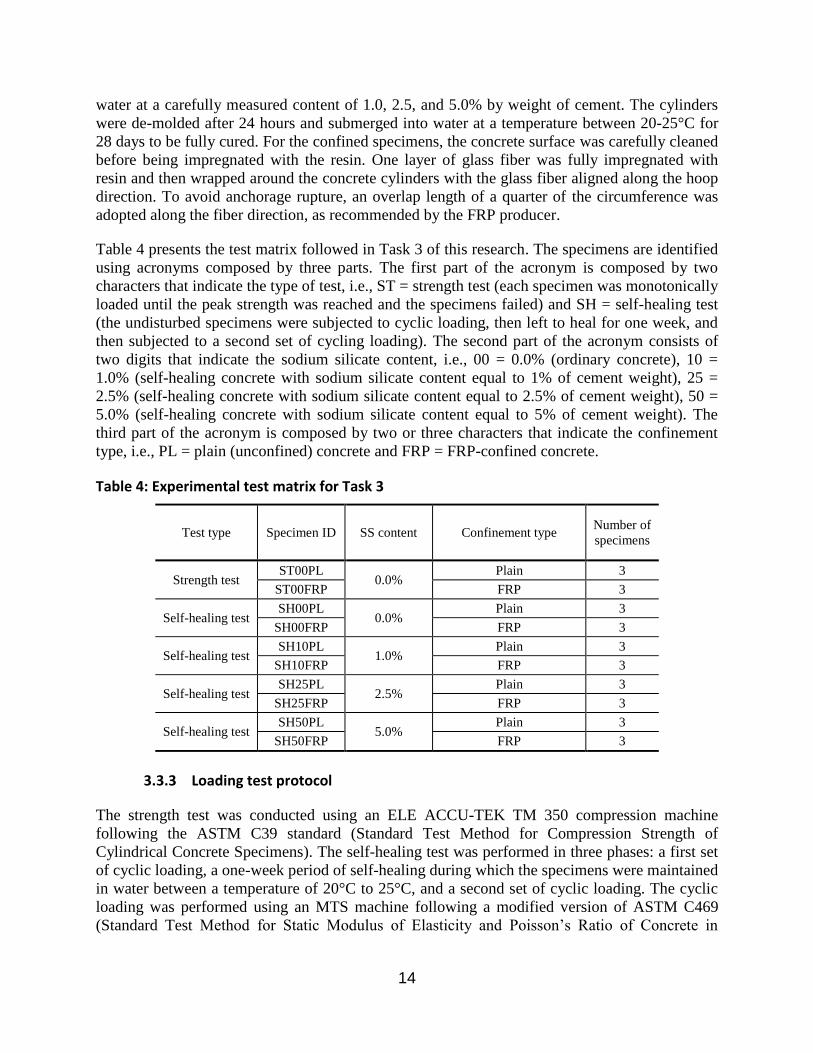
Table 4 presents the test matrix followed in Task 3 of this research. The specimens are identified
using acronyms composed by three parts. The first part of the acronym is composed by two
characters that indicate the type of test, i.e., ST = strength test (each specimen was monotonically
loaded until the peak strength was reached and the specimens failed) and SH = self-healing test
(the undisturbed specimens were subjected to cyclic loading, then left to heal for one week, and
then subjected to a second set of cycling loading). The second part of the acronym consists of
two digits that indicate the sodium silicate content, i.e., 00 = 0.0% (ordinary concrete), 10 =
1.0% (self-healing concrete with sodium silicate content equal to 1% of cement weight), 25 =
2.5% (self-healing concrete with sodium silicate content equal to 2.5% of cement weight), 50 =
5.0% (self-healing concrete with sodium silicate content equal to 5% of cement weight). The
third part of the acronym is composed by two or three characters that indicate the confinement
type, i.e., PL = plain (unconfined) concrete and FRP = FRP-confined concrete.
Table 4: Experimental test matrix for Task 3
Test type Specimen ID SS content Confinement type Number of
specimens
Strength test ST00PL
0.0% Plain 3
ST00FRP FRP 3
Self-healing test SH00PL
0.0% Plain 3
SH00FRP FRP 3
Self-healing test SH10PL
1.0% Plain 3
SH10FRP FRP 3
Self-healing test SH25PL
2.5% Plain 3
SH25FRP FRP 3
Self-healing test SH50PL
5.0% Plain 3
SH50FRP FRP 3
3.3.3 Loading test protocol
The strength test was conducted using an ELE ACCU-TEK TM 350 compression machine
following the ASTM C39 standard (Standard Test Method for Compression Strength of
Cylindrical Concrete Specimens). The self-healing test was performed in three phases: a first set
of cyclic loading, a one-week period of self-healing during which the specimens were maintained
in water between a temperature of 20°C to 25°C, and a second set of cyclic loading. The cyclic
loading was performed using an MTS machine following a modified version of ASTM C469
(Standard Test Method for Static Modulus of Elasticity and Poisson’s Ratio of Concrete in
15
Compression). The specimens were loaded and unloaded for one cycle up to 20% of their
compressive strength (estimated from the results of the previously performed strength tests), one
cycle up to 40% of their compressive strength, and then three cycles up to 75% of their
compressive strength. The stress time history was recorded from a loading cell and the strain
time history was calculated using an LVDT attached to the specimens through a
compressometer. The tangent stiffness modulus (computed as the slope of the stress-strain curve
between 2% and 10% of the estimated peak strength of the specimens) and the residual strain at
end of each loading cycle (computed as the strain reached during unloading at 2% of the
estimated peak strength of the specimens) were calculated before and after healing.
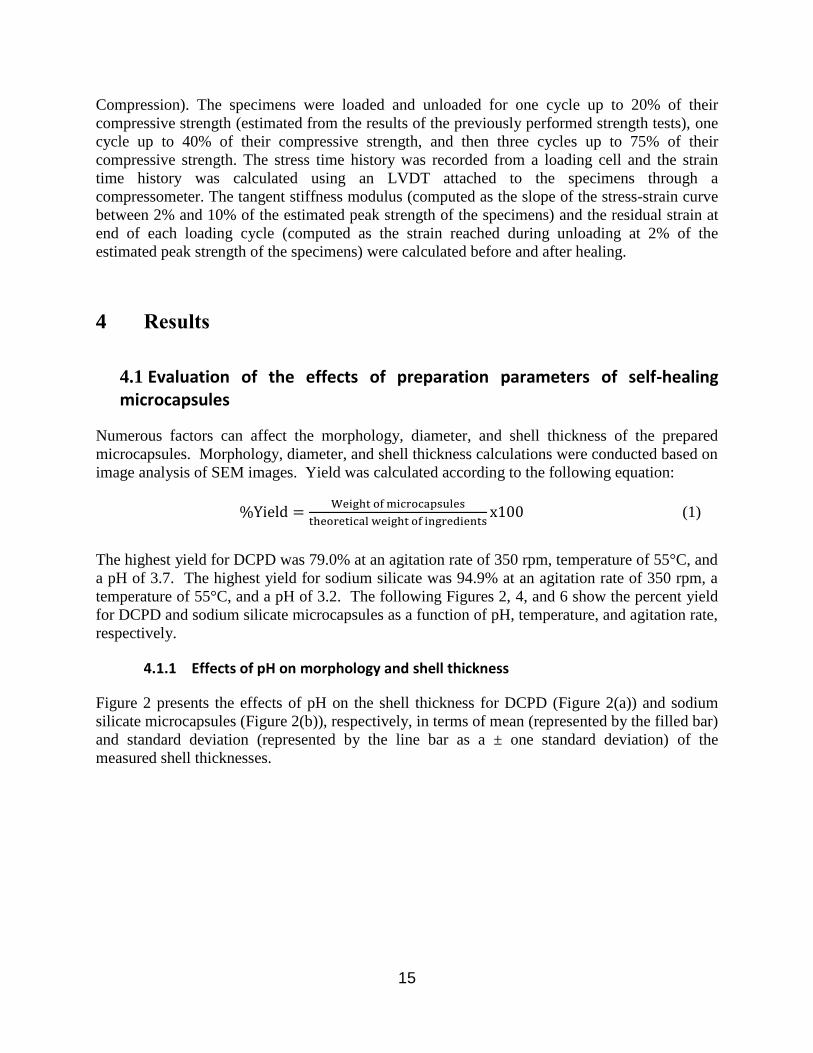
4 Results
Evaluation of the effects of preparation parameters of self-healing 4.1
microcapsules
Numerous factors can affect the morphology, diameter, and shell thickness of the prepared
microcapsules. Morphology, diameter, and shell thickness calculations were conducted based on
image analysis of SEM images. Yield was calculated according to the following equation:
(1)
The highest yield for DCPD was 79.0% at an agitation rate of 350 rpm, temperature of 55°C, and
a pH of 3.7. The highest yield for sodium silicate was 94.9% at an agitation rate of 350 rpm, a
temperature of 55°C, and a pH of 3.2. The following Figures 2, 4, and 6 show the percent yield
for DCPD and sodium silicate microcapsules as a function of pH, temperature, and agitation rate,
respectively.
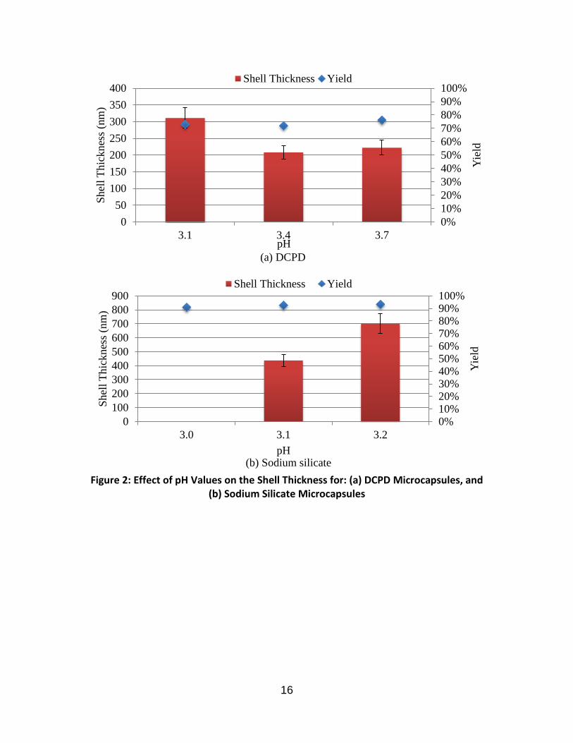
4.1.1 Effects of pH on morphology and shell thickness
Figure 2 presents the effects of pH on the shell thickness for DCPD (Figure 2(a)) and sodium
silicate microcapsules (Figure 2(b)), respectively, in terms of mean (represented by the filled bar)
and standard deviation (represented by the line bar as a ± one standard deviation) of the
measured shell thicknesses.
16
Figure 2: Effect of pH Values on the Shell Thickness for: (a) DCPD Microcapsules, and
(b) Sodium Silicate Microcapsules
0%
10%
20%
30%
40%
50%
60%
70%
80%
90%
100%
0
50
100
150
200
250
300
350
400
3.1 3.4 3.7
Yie
ld
Shel
l T
hic
knes
s (n
m)
pH
Shell Thickness Yield
(a) DCPD
0%10%20%30%40%50%60%70%80%90%100%
0
100
200
300
400
500
600
700
800
900
3.0 3.1 3.2
Yie
ld
Shel
l T
hic
knes
s (n
m)
pH
Shell Thickness Yield
(b) Sodium silicate
17
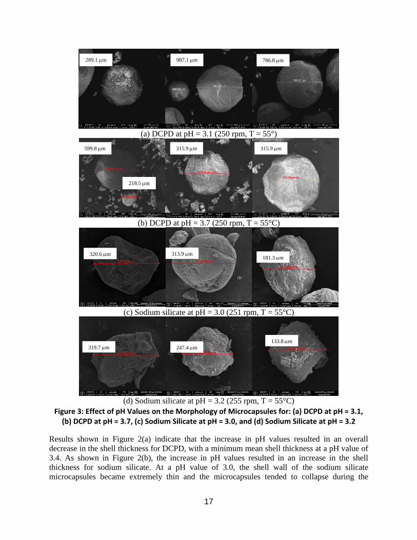
Figure 3: Effect of pH Values on the Morphology of Microcapsules for: (a) DCPD at pH = 3.1,
(b) DCPD at pH = 3.7, (c) Sodium Silicate at pH = 3.0, and (d) Sodium Silicate at pH = 3.2
Results shown in Figure 2(a) indicate that the increase in pH values resulted in an overall
decrease in the shell thickness for DCPD, with a minimum mean shell thickness at a pH value of
3.4. As shown in Figure 2(b), the increase in pH values resulted in an increase in the shell
thickness for sodium silicate. At a pH value of 3.0, the shell wall of the sodium silicate
microcapsules became extremely thin and the microcapsules tended to collapse during the
(a) DCPD at pH = 3.1 (250 rpm, T = 55°)
(b) DCPD at pH = 3.7 (250 rpm, T = 55°C)
(c) Sodium silicate at pH = 3.0 (251 rpm, T = 55°C)
(d) Sodium silicate at pH = 3.2 (255 rpm, T = 55°C)
786.8 m 987.1 m 289.1 m
599.8 m
218.5 m
315.9 m 315.9 m
320.6 m 313.9 m 181.3 m
319.7 m 247.4 m
133.8 m
18
measurements of their shell thickness. As a consequence, the measurement of the microcapsules’
shell thickness was not possible. The maximum shell thickness of sodium silicate microcapsules
was almost twice the maximum shell thickness of DCPD microcapsules. This phenomenon was
due to sodium silicate being transformed into a gel like solution prior to microencapsulation.
This gel solution made the compound much easier to encapsulate and produced a much stronger
shell wall.
Figure 3 presents SEM images of the microcapsules prepared with DCPD and sodium silicate at
different pH values. It is observed that the microcapsules prepared with DCPD were closer to a
spherical shape and more uniform than the microcapsules prepared with sodium silicate. In
addition, the size of the microcapsules was reduced as the pH value was increased. The outer
surface of the microcapsules had a rough permeable layer, whereas the inside was smooth and
free of cavities.
4.1.2 Effects of temperature
Figure 4 presents the effects of temperature on the shell thickness for DCPD (Figure 4(a)) and
sodium silicate microcapsules (Figure 4(b)), respectively, in terms of mean (represented by the
filled bar) and standard deviation (represented by the line bar as a ± one standard deviation) of
the measured shell thicknesses. For the DCPD microcapsules at 49°C, the solution remained an
emulsion and no encapsulation took place. For sodium silicate, there were no microcapsules
formed at 53°C. Figure 5 presents SEM images of the microcapsules for DCPD (Figure 5(a) and
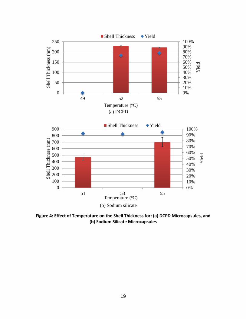
(b)) and sodium silicate (Figure 5(c) and (d)) prepared at different temperatures. Also in this
case, the microcapsules prepared with DCPD were with a shape closer to spherical and more
uniform than the microcapsules prepared with sodium silicate. In addition, the size of the
microcapsules was reduced as the temperature was increased.
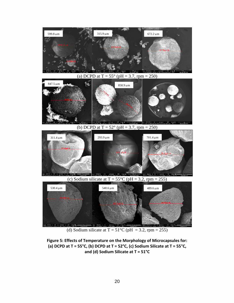
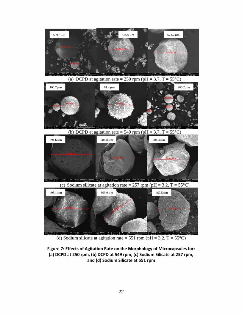
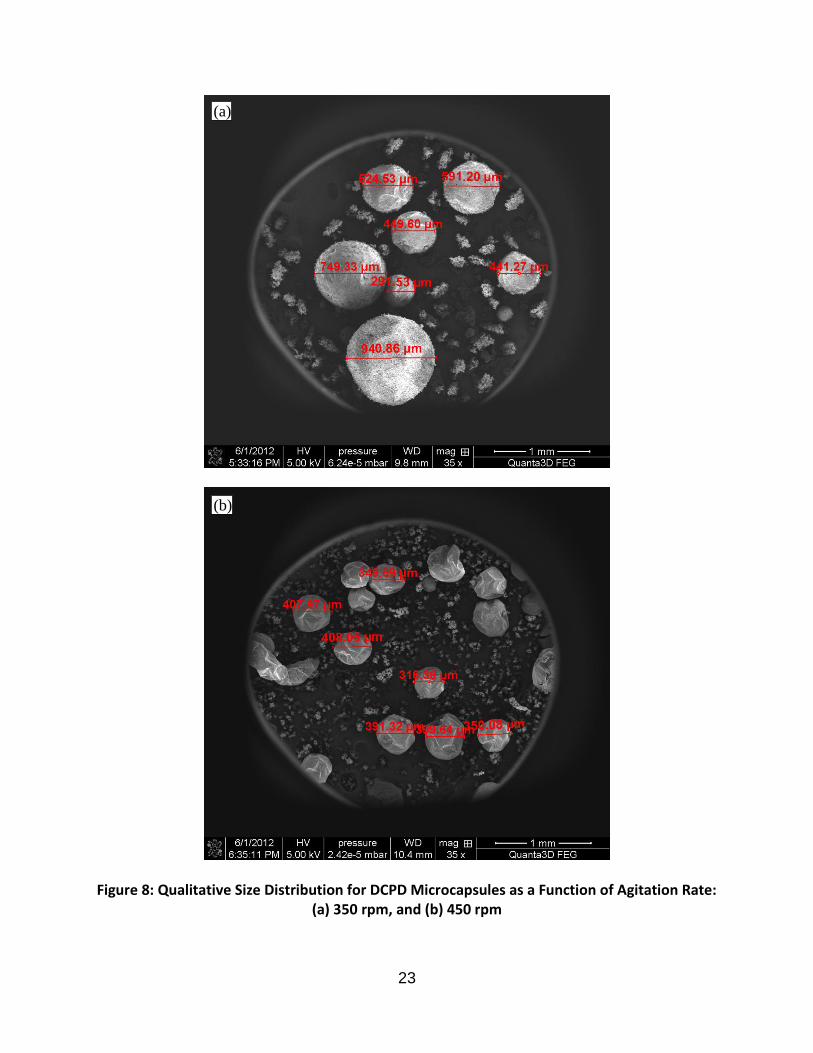
4.1.3 Effects of agitation rate
Figure 6 shows the effect of agitation rate on the diameter of the microcapsules for DCPD
(Figure 6(a)) and sodium silicate microcapsules (Figure 6(b)), respectively, in terms of mean
(represented by the filled bar) and standard deviation (represented by the line bar as a ± one
standard deviation) of the measured microcapsules’ diameter. The increase in agitation rate
resulted in a decrease of the average diameter of the microcapsules for DCPD. This is due to the
large microcapsules being broken up into smaller ones when high shear (due to the centrifugal
forces) is applied. The optimum size of the microcapsules is dependent on the crack size that is
expected to be filled during the healing mechanism. On the other hand, the diameter of the
microcapsules remained constant for sodium silicate microencapsulation as the agitation rate
increased, as shown in Figure 6(b). This phenomenon may be attributed to the attempt to
stabilize the alkalinity of the sodium silicate solution for the microencapsulation procedure using
urea-formaldehyde. The SEM images presented in Figure 7 also show a reduction in diameter
with the increase in agitation rate for DCPD. The same trend is observed in Figure 8, which
provides SEM pictures of DCPD microcapsules produced at different agitation rates with a lower
magnification rate compared with Figure 7, in order to show several DCPD microcapsules in a
single picture and to provide a better idea of the size distribution observed for DCPD
microcapsules.
19
Figure 4: Effect of Temperature on the Shell Thickness for: (a) DCPD Microcapsules, and (b) Sodium Silicate Microcapsules
0%10%20%30%40%50%60%70%80%90%100%
0
50
100
150
200
250
49 52 55
Yie
ld
Shel
l T
hic
knes
s (n
m)
Temperature (oC)
Shell Thickness Yield
(a) DCPD
0%
10%
20%
30%
40%
50%
60%
70%
80%
90%
100%
0
100
200
300
400
500
600
700
800
900
51 53 55
Yie
ld
Shel
l T
hic
knes
s (n
m)
Temperature (oC)
Shell Thickness Yield
(b) Sodium silicate
20
Figure 5: Effects of Temperature on the Morphology of Microcapsules for: (a) DCPD at T = 55°C, (b) DCPD at T = 52°C, (c) Sodium Silicate at T = 55°C,
and (d) Sodium Silicate at T = 51°C
(a) DCPD at T = 55° (pH = 3.7, rpm = 250)
(b) DCPD at T = 52° (pH = 3.7, rpm = 250)
(c) Sodium silicate at T = 55°C (pH = 3.2, rpm = 255)
(d) Sodium silicate at T = 51°C (pH = 3.2, rpm = 255)
599.8 m 315.9 m 672.2 m
847.5 m 858.9 m
311.4 m 293.9 m 701.4 m
530.4 m 540.6 m 489.6 m
21
Figure 6: Effect of Agitation Rate on the Diameter for: (a) DCPD Microcapsules,
and (b) Sodium Silicate Microcapsules
0%
10%
20%
30%
40%
50%
60%
70%
80%
90%
100%
0
100
200
300
400
500
600
700
250 349 451 549 800 1000
Yie
ld
Dia
met
er (
mm
)
Agitation Rate (rpm)
Diameter Yield
(a) DCPD
0%10%20%30%40%50%60%70%80%90%100%
0
100
200
300
400
500
600
700
250 350 451 552
Yie
ld
Dia
met
er (
mm
)
Agitation Rate (rpm)
Diameter Yield
(b) Sodium silicate
22
Figure 7: Effects of Agitation Rate on the Morphology of Microcapsules for: (a) DCPD at 250 rpm, (b) DCPD at 549 rpm, (c) Sodium Silicate at 257 rpm,
and (d) Sodium Silicate at 551 rpm
(a) DCPD at agitation rate = 250 rpm (pH = 3.7, T = 55°C)
(b) DCPD at agitation rate = 549 rpm (pH = 3.7, T = 55°C)
(c) Sodium silicate at agitation rate = 257 rpm (pH = 3.2, T = 55°C)
(d) Sodium silicate at agitation rate = 551 rpm (pH = 3.2, T = 55°C)
599.8 m 315.9 m 672.2 m
165.7 m 81.4 m 265.2 m
591.6 m 786.8 m 701.4 m
498.1 m 609.9 m 467.5 m
23
Figure 8: Qualitative Size Distribution for DCPD Microcapsules as a Function of Agitation Rate: (a) 350 rpm, and (b) 450 rpm
(a)
(b)

24
Evaluation of the effects of microcapsules’ properties on concrete self-4.2
healing mechanism
A set of laboratory tests was performed to measure the modulus of elasticity of plain concrete
with and without self-healing microcapsules before and after a 1-week healing period. The
objective of this experimental investigation was to obtain preliminary information on the relation
between production parameters of the microcapsules of self-healing agents (which affect the
morphology and shell thickness of the microcapsules) and the effectiveness of the microcapsules
in enhancing the concrete self-healing properties.
Figure 9 presents the effects on the concrete modulus of elasticity before and after healing of
DCPD (with a content of 0.25% of the cement weight) and sodium silicate (with a content of 5%
of the cement weight) microcapsules prepared at different pH values. Error bars showing the
average variability (about ±10%) that was observed in the measurements are also provided.
Figure 9: Effect of Preparation pH of Microcapsules on Concrete Modulus of Elasticity before
and after Healing
The following observations are made based on the results presented in Figure 9:
1) As expected, no self-healing process was detected for the control specimens (i.e., without
self-healing), for which a small decrease of the modulus of elasticity (smaller than the
variability of the measurements) was recorded. This result suggests that the control
specimens were subjected to a small but not negligible damage, with likely formation of
micro-cracks within the specimens.
2) The concrete modulus of elasticity after healing of specimens with DCPD was significantly
higher than that before healing. This phenomenon indicates that the self-healing process was
activated and that the self-healing material produced a higher stiffness and, as a consequence,
a higher strength (which, for concrete, is positively correlated with the stiffness) than those of
the original undamaged concrete. This after-healing stiffness increase was more pronounced
0
1000
2000
3000
4000
5000
6000
7000
Control DCPD
0.25%
pH=3.1
DCPD
0.25%
pH=3.4
DCPD
0.25%
pH=3.7
SS
5%
pH=3.0
SS
5%
pH=3.1
SS
5%
pH=3.2
Modulu
s of
Ela
stic
ity (
ksi
)
Before Healing After Healing
(1 ksi = 6.89 MPa)
25
for lower values of the pH, which correspond to higher thicknesses of the microcapsules’
walls. The best performance of the after-healing concrete was obtained for pH = 3.1.
3) The modulus of elasticity of the concrete with DCPD before healing decreased significantly
for increasing pH (i.e., for lower values of the microcapsules’ shell thickness). At a pH value
of 3.1, the average modulus of elasticity of the specimens with DCPD was almost the same
(slightly higher) than that of the specimens of plain concrete, while at a pH value of 3.7 the
modulus of elasticity was about 15% lower than that of the plain concrete specimens.
4) Sodium silicate caused a significant decrease in the modulus of elasticity before healing
when compared to the control concrete mix for pH values of 3.0 and 3.2, while it did not
affect the modulus of elasticity of the specimens for a pH value of 3.1. The data available at
this point are insufficient to identify the reason for this trend. The lower stiffness (and
strength) of the concrete with sodium silicate before healing should not be a concern as long
as the design accounts for the proper values.
5) The modulus of elasticity of the concrete with sodium silicate was higher after healing than
before healing for all pH values. The highest increase in the modulus of elasticity was
measured for pH = 3.1.
6) The shell thickness of the microcapsules significantly affected both the before and after
healing modulus of elasticity of the concrete with sodium silicate self-healing agent. In
particular, it appears that too low or too large pH values (i.e., too small or too large shell
thicknesses) are detrimental to the performance of both before and after healing concrete.
This phenomenon can be explained by noticing that, for too small shell thicknesses, the
microcapsules collapsed during mixing of the concrete, while for too large shell thicknesses,
the microcapsules were not broken by the concrete micro-cracks and, thus, the self-healing
agent was not activated. Among the pH values considered in this study, pH = 3.1 provides
the best performance for the concrete with sodium silicate.
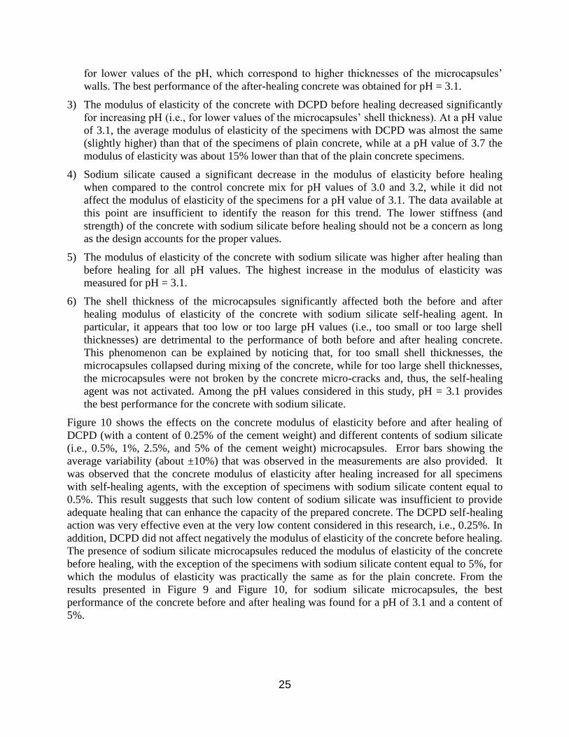
Figure 10 shows the effects on the concrete modulus of elasticity before and after healing of
DCPD (with a content of 0.25% of the cement weight) and different contents of sodium silicate
(i.e., 0.5%, 1%, 2.5%, and 5% of the cement weight) microcapsules. Error bars showing the
average variability (about ±10%) that was observed in the measurements are also provided. It
was observed that the concrete modulus of elasticity after healing increased for all specimens
with self-healing agents, with the exception of specimens with sodium silicate content equal to
0.5%. This result suggests that such low content of sodium silicate was insufficient to provide
adequate healing that can enhance the capacity of the prepared concrete. The DCPD self-healing
action was very effective even at the very low content considered in this research, i.e., 0.25%. In
addition, DCPD did not affect negatively the modulus of elasticity of the concrete before healing.
The presence of sodium silicate microcapsules reduced the modulus of elasticity of the concrete
before healing, with the exception of the specimens with sodium silicate content equal to 5%, for
which the modulus of elasticity was practically the same as for the plain concrete. From the
results presented in Figure 9 and Figure 10, for sodium silicate microcapsules, the best
performance of the concrete before and after healing was found for a pH of 3.1 and a content of
5%.
26
Figure 10: Effect of Amount of Microcapsules (% of Cement Weight) on Concrete Modulus of
Elasticity before and after Healing
Figure 11: Crack Healing after 1-Week Recovery: (a) DCPD before Healing, (b) DCPD after 1-Week Healing, (c) Sodium Silicate (1%) before Healing, and (d) Sodium Silicate (1%) after 1-
Week Healing
0
1000
2000
3000
4000
5000
6000
7000
Control DCPD
0.25%
pH =3.1
SS
0.5%
pH=3.1
SS
1%
pH=3.1
SS
2.5%
pH=3.1
SS
5%
pH=3.1
Modulu
s of
Ela
stic
ity (
ksi
)
Before Healing After Healing
(1 ksi = 6.89 MPa)
(a) DCPD: Before healing (b) DCPD: After 1-week healing
(c) Sodium silicate: Before healing (d) Sodium silicate: After 1-week healing
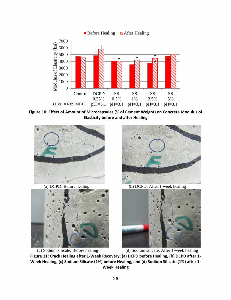
27
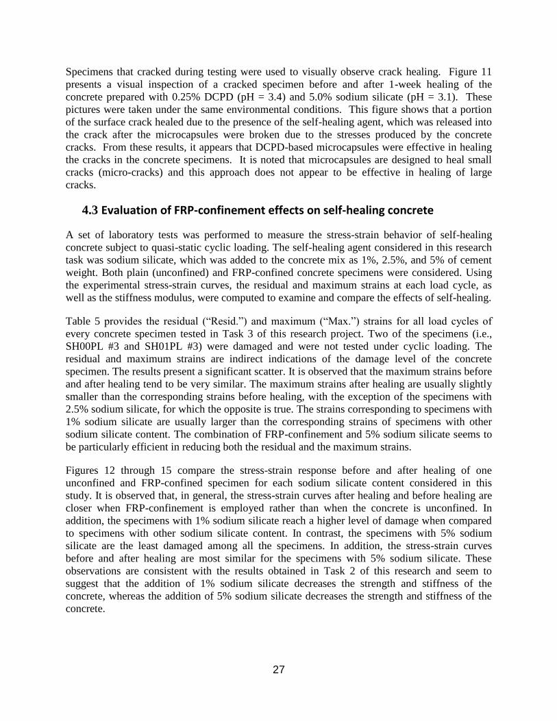
Specimens that cracked during testing were used to visually observe crack healing. Figure 11
presents a visual inspection of a cracked specimen before and after 1-week healing of the
concrete prepared with 0.25% DCPD (pH = 3.4) and 5.0% sodium silicate (pH = 3.1). These
pictures were taken under the same environmental conditions. This figure shows that a portion
of the surface crack healed due to the presence of the self-healing agent, which was released into
the crack after the microcapsules were broken due to the stresses produced by the concrete
cracks. From these results, it appears that DCPD-based microcapsules were effective in healing
the cracks in the concrete specimens. It is noted that microcapsules are designed to heal small
cracks (micro-cracks) and this approach does not appear to be effective in healing of large
cracks.
Evaluation of FRP-confinement effects on self-healing concrete 4.3
A set of laboratory tests was performed to measure the stress-strain behavior of self-healing
concrete subject to quasi-static cyclic loading. The self-healing agent considered in this research
task was sodium silicate, which was added to the concrete mix as 1%, 2.5%, and 5% of cement
weight. Both plain (unconfined) and FRP-confined concrete specimens were considered. Using
the experimental stress-strain curves, the residual and maximum strains at each load cycle, as
well as the stiffness modulus, were computed to examine and compare the effects of self-healing.
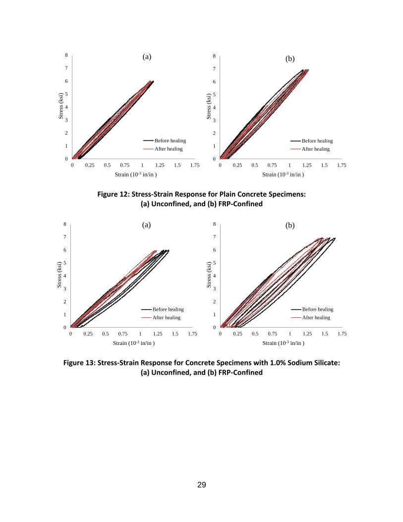
Table 5 provides the residual (“Resid.”) and maximum (“Max.”) strains for all load cycles of
every concrete specimen tested in Task 3 of this research project. Two of the specimens (i.e.,
SH00PL #3 and SH01PL #3) were damaged and were not tested under cyclic loading. The
residual and maximum strains are indirect indications of the damage level of the concrete
specimen. The results present a significant scatter. It is observed that the maximum strains before
and after healing tend to be very similar. The maximum strains after healing are usually slightly
smaller than the corresponding strains before healing, with the exception of the specimens with
2.5% sodium silicate, for which the opposite is true. The strains corresponding to specimens with
1% sodium silicate are usually larger than the corresponding strains of specimens with other
sodium silicate content. The combination of FRP-confinement and 5% sodium silicate seems to
be particularly efficient in reducing both the residual and the maximum strains.
Figures 12 through 15 compare the stress-strain response before and after healing of one
unconfined and FRP-confined specimen for each sodium silicate content considered in this
study. It is observed that, in general, the stress-strain curves after healing and before healing are
closer when FRP-confinement is employed rather than when the concrete is unconfined. In
addition, the specimens with 1% sodium silicate reach a higher level of damage when compared
to specimens with other sodium silicate content. In contrast, the specimens with 5% sodium
silicate are the least damaged among all the specimens. In addition, the stress-strain curves
before and after healing are most similar for the specimens with 5% sodium silicate. These
observations are consistent with the results obtained in Task 2 of this research and seem to
suggest that the addition of 1% sodium silicate decreases the strength and stiffness of the
concrete, whereas the addition of 5% sodium silicate decreases the strength and stiffness of the
concrete.

28
Load cycle #1 (20%) #2 (40%) #3 (75%) #4 (75%) #5 (75%)
ID # Healing Resid. Max. Resid. Max. Resid. Max. Resid. Max. Resid. Max.
(10-3
in/in) (10-3
in/in) (10-3
in/in) (10-3
in/in) (10-3
in/in)
SH00PL
1 Before 0.031 0.260 0.029 0.524 0.084 1.076 0.061 1.088 0.077 1.078
After 0.071 0.290 0.070 0.571 0.083 1.081 0.091 1.090 0.105 1.090
2 Before 0.038 0.260 0.052 0.548 0.117 1.108 0.133 1.156 0.145 1.160
After 0.031 0.286 0.038 0.577 0.064 1.108 0.074 1.125 0.079 1.135
SH00FRP
1 Before 0.039 0.319 0.066 0.696 0.042 1.347 0.265 1.401 0.299 1.467
After 0.044 0.354 0.059 0.718 0.107 1.251 0.129 1.280 0.150 1.322
2 Before 0.032 0.299 0.044 0.640 0.112 1.196 0.133 1.237 0.154 1.267
After 0.035 0.327 0.041 0.684 0.094 1.229 0.110 1.255 0.116 1.277
3 Before 0.042 0.313 0.059 0.654 0.122 0.991 0.105 1.218 0.128 1.254
After 0.030 0.315 0.031 0.658 0.070 1.159 0.081 1.190 0.092 1.220
SH01PL
1 Before 0.033 0.287 0.047 0.601 0.128 1.266 0.148 1.295 0.147 1.311
After 0.035 0.320 0.048 0.652 0.098 1.248 0.117 1.279 0.131 1.299
2 Before 0.037 0.294 0.060 0.630 0.196 1.409 0.097 1.336 0.123 1.372
After 0.033 0.316 0.037 0.634 0.077 1.209 0.076 1.230 0.076 1.240
SH01FRP
1 Before 0.063 0.341 0.095 0.729 0.349 1.599 0.468 1.796 0.570 1.963
After 0.065 0.448 0.105 0.937 0.275 1.735 0.357 1.869 0.351 1.958
2 Before 0.043 0.337 0.079 0.718 0.052 1.511 0.345 1.628 0.280 1.728
After 0.060 0.404 0.102 0.857 0.186 1.503 0.215 1.607 0.260 1.681
3 Before 0.057 0.346 0.089 0.739 0.243 1.464 0.304 1.572 0.351 1.650
After 0.057 0.365 0.084 0.777 0.191 1.424 0.228 1.487 0.261 1.540
SH25PL
1 Before 0.036 0.227 0.047 0.472 0.100 0.951 0.113 0.968 0.129 0.988
After 0.030 0.286 0.039 0.592 0.087 1.235 0.091 1.244 0.104 1.286
2 Before 0.032 0.240 0.047 0.507 0.112 1.032 0.129 1.053 0.137 1.059
After 0.034 0.292 0.044 0.598 0.105 1.266 0.123 1.299 0.118 1.342
3 Before 0.027 0.277 0.035 0.575 0.102 1.176 0.113 1.201 0.115 1.208
After 0.038 0.295 0.056 0.643 0.109 1.243 0.137 1.300 0.153 1.334
SH25FRP
1 Before 0.041 0.327 0.062 0.680 0.135 1.219 0.158 1.281 0.166 1.299
After 0.046 0.350 0.062 0.710 0.098 1.252 0.113 1.284 0.118 1.295
2 Before 0.046 0.334 0.066 0.692 0.127 1.258 0.144 1.297 0.171 1.334
After 0.048 0.371 0.064 0.749 0.109 1.317 0.122 1.345 0.132 1.370
3 Before 0.026 0.306 0.027 0.639 0.085 1.186 0.100 1.205 0.102 1.221
After 0.050 0.353 0.065 0.722 0.097 1.246 0.105 1.276 0.118 1.298
SH50PL
1 Before 0.049 0.267 0.063 0.552 0.121 1.076 0.134 1.090 0.139 1.096
After 0.042 0.285 0.038 0.568 0.050 1.068 0.052 1.073 0.058 1.076
2 Before 0.027 0.253 0.027 0.518 0.059 1.030 0.065 1.038 0.048 1.071
After 0.032 0.267 0.035 0.541 0.053 1.012 0.052 1.013 0.052 1.012
3 Before 0.042 0.256 0.049 0.534 0.082 1.053 0.087 1.058 0.093 1.069
After 0.033 0.286 0.035 0.557 0.057 1.046 0.069 1.061 0.073 1.062
SH50FRP
1 Before 0.055 0.297 0.067 0.628 0.086 1.129 0.098 1.148 0.106 1.164
After 0.047 0.336 0.043 0.665 0.069 1.132 0.075 1.135 0.084 1.149
2 Before 0.076 0.310 0.095 0.676 0.119 1.160 0.088 1.142 0.097 1.153
After 0.046 0.339 0.053 0.682 0.075 1.167 0.086 1.183 0.105 1.206
3 Before 0.062 0.272 0.117 0.590 0.225 1.078 0.287 1.138 0.316 1.157
After 0.043 0.329 0.041 0.649 0.058 1.109 0.067 1.122 0.079 1.140
Table 5: Residual and maximum strains at each load cycle of tested concrete specimens
29
Figure 12: Stress-Strain Response for Plain Concrete Specimens: (a) Unconfined, and (b) FRP-Confined
Figure 13: Stress-Strain Response for Concrete Specimens with 1.0% Sodium Silicate: (a) Unconfined, and (b) FRP-Confined
0
1
2
3
4
5
6
7
8
0 0.25 0.5 0.75 1 1.25 1.5 1.75
Before healing
After healing
Str
ess
(ksi
)
Strain (10-3 in/in )
0
1
2
3
4
5
6
7
8
0 0.25 0.5 0.75 1 1.25 1.5 1.75
Before healing
After healing
Str
ess
(ksi
)
Strain (10-3 in/in )
0
1
2
3
4
5
6
7
8
0 0.25 0.5 0.75 1 1.25 1.5 1.75
Before healing
After healing
Str
ess
(ksi
)
Strain (10-3 in/in )
0
1
2
3
4
5
6
7
8
0 0.25 0.5 0.75 1 1.25 1.5 1.75
Before healing
After healing
Str
ess
(ksi
)
Strain (10-3 in/in )
(a) (b)
(a) (b)

30
Figure 14: Stress-Strain Response for Concrete Specimens with 2.5% Sodium Silicate: (a) Unconfined, and (b) FRP-Confined
Figure 15: Stress-Strain Response for Concrete Specimens with 5.0% Sodium Silicate: (a) Unconfined, and (b) FRP-Confined
Figure 16 shows the values of the stiffness at low levels of stress for the tested concrete
specimens. This stiffness is obtained as the average of the secant stiffnesses computed between
2% and 10% of the estimated peak strength (1) in the first two loading cycles of the loading
procedure before healing (undamaged material), (2) in the last two cycles of the loading
procedure before healing (damaged material), and (3) in the first two cycles of the loading
procedure after healing (material after healing). Also in this case, a non-negligible variability of
the results was observed. In general, the specimens with FRP-confinement were stiffer than the
corresponding unconfined specimens, with the exception of the specimens with 2.5% sodium
silicate. In terms of stiffness recovery, a similar behavior was observed for both unconfined and
0
1
2
3
4
5
6
7
8
0 0.25 0.5 0.75 1 1.25 1.5 1.75
Before healing
After healing
Str
ess
(ksi
)
Strain (10-3 in/in )
0
1
2
3
4
5
6
7
8
0 0.25 0.5 0.75 1 1.25 1.5 1.75
Before healing
After healing
Str
ess
(ksi
)
Strain (10-3 in/in )
0
1
2
3
4
5
6
7
8
0 0.25 0.5 0.75 1 1.25 1.5 1.75
Before healing
After healing
Str
ess
(ksi
)
Strain (10-3 in/in )
0
1
2
3
4
5
6
7
8
0 0.25 0.5 0.75 1 1.25 1.5 1.75
Before healing
After healing
Str
ess
(ksi
)
Strain (10-3 in/in )
(a)
(a)
(b)
(b)
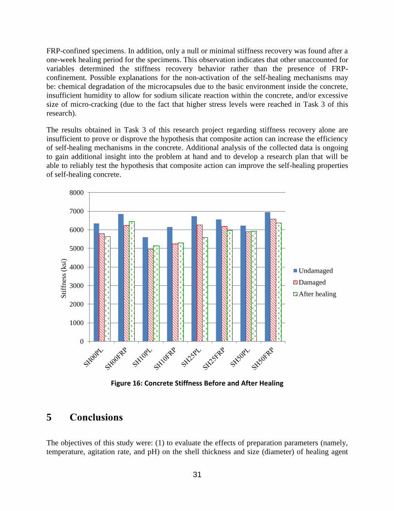
31
FRP-confined specimens. In addition, only a null or minimal stiffness recovery was found after a
one-week healing period for the specimens. This observation indicates that other unaccounted for
variables determined the stiffness recovery behavior rather than the presence of FRP-
confinement. Possible explanations for the non-activation of the self-healing mechanisms may
be: chemical degradation of the microcapsules due to the basic environment inside the concrete,
insufficient humidity to allow for sodium silicate reaction within the concrete, and/or excessive
size of micro-cracking (due to the fact that higher stress levels were reached in Task 3 of this
research).
The results obtained in Task 3 of this research project regarding stiffness recovery alone are
insufficient to prove or disprove the hypothesis that composite action can increase the efficiency
of self-healing mechanisms in the concrete. Additional analysis of the collected data is ongoing
to gain additional insight into the problem at hand and to develop a research plan that will be
able to reliably test the hypothesis that composite action can improve the self-healing properties
of self-healing concrete.
Figure 16: Concrete Stiffness Before and After Healing
5 Conclusions
The objectives of this study were: (1) to evaluate the effects of preparation parameters (namely,
temperature, agitation rate, and pH) on the shell thickness and size (diameter) of healing agent
0
1000
2000
3000
4000
5000
6000
7000
8000
Undamaged
Damaged
After healingSti
ffn
ess
(ksi
)
32
microcapsules for use in self-healing concrete; (2) to evaluate the effects of microcapsules’ shell
thicknesses and size diameters on the concrete self-healing mechanism; and (3) to test the
hypothesis that composite action due to FRP confinement of cylindrical concrete specimens can
improve the self-repairing properties of self-healing concrete materials. Two healing agents were
evaluated for the first two objectives of this study, i.e., DCPD and sodium silicate. The use of
sodium silicate was considered for the third objective of this study. Based on the results of the
experimental program, the following conclusions were made:
1) As the pH was reduced, the shell thickness of the DCPD microcapsules increased. Unlike
DCPD, the shell thickness of sodium silicate microcapsules increased for increasing pH. The
shell thickness of sodium silicate microcapsules was almost twice the shell thickness of
DCPD microcapsules.
2) The more uniform and coherent microcapsules were produced at a temperature of 55°C for
both DCPD and sodium silicate healing agents. For the DCPD microcapsules, the solution
remained an emulsion and no encapsulation took place at 49°C. For sodium silicate, no
microcapsules were formed at 53°C.
3) The increase in agitation rate resulted in a decrease in the average diameter of the
microcapsules for DCPD. By contrast, the diameter of the microcapsules remained almost
constant for sodium silicate microencapsulation as the agitation rate increased.
4) DCPD-based microcapsules were effective in increasing the concrete modulus of elasticity
after healing, even at content as low as 0.25% of cement weight. For sodium silicate, an
optimum pH value and content needs to be identified in order to produce microcapsules that
enhance the concrete modulus of elasticity before and after healing. From the results
presented in this study, a pH value of 3.1 at a sodium silicate content of 5.0% was found to
provide the best performance.
5) The use of FRP confinement generally improved the performance of the concrete. However,
the self-healing effects in terms of stiffness recovery observed in Task 3 of this project were
null or very small for both unconfined and FRP-confined specimens. Thus, the data collected
in this research in terms of stiffness recovery alone are insufficient to prove or disprove the
hypothesis that composite action can increase the efficiency of self-healing mechanisms in
the concrete.
Appendix A: DCPD Microencapsulation Procedure
The adopted procedure was performed by using in-situ polymerization, in an oil-in-water
emulsion. The main steps of this procedure can be summarized as follows:
1) Place 200 ml of Deionized (DI) water in a 1000 ml beaker;
2) Dissolve 50 ml of 2.5 wt.% EMA copolymer using a magnetic stirrer and ultra sound water
bath to develop an aqueous solution;
33
3) Agitate using a IKA RW 20 digital mixer, with a driving 55 mm low shear three-bladed
mixing propeller placed just above the bottom of the beaker;
4) Under agitation, add 5.00 g urea, 0.50 g resorcinol and 0.50 g ammonium chloride in the
solution;
5) Set the pH by using sodium hydroxide (NaOH) and hydrochloric acid (HCl) drop-wise with a
disposable pipet;
6) Add two to three drops of 1-octanol to reduce surface bubbles;
7) Allow the solution to stabilize for approximately 6 – 8 minutes at the appropriate pH and rpm
agitation rate, before 100 ml of DCPD is added at a slow stream rate;
8) Allow the solution to stabilize for 13 – 15 minutes before 12.7 g of 37 wt% aqueous solution
of formaldehyde was added to the emulsion;
9) Wrap and cover the solution with aluminum foil, and slowly heat to the set temperature;
10) Turn off the hot plate after 4 hours of continuous agitation;
11) Once cooled to ambient temperature, the suspension of microcapsules was separated under
vacuum filtration;
12) Rinse microcapsules with DI water three times with 500 ml of DI water, and then allow to air
dry for 48 - 72 h.
Appendix B: Sodium Silicate Microencapsulation Procedure
This procedure was accomplished by using in-situ polymerization, in an oil-in-water emulsion.
The main steps of this procedure can be summarized as follows:
1) Place 200 ml of DI water in a 1000 ml beaker;
2) Dissolve 50 ml of 2.5 wt.% EMA copolymer using a magnetic stirrer and ultra sound water
bath to develop an aqueous solution;
3) Agitate using a IKA RW 20 digital mixer, with a driving 55 mm low shear three-bladed
mixing propeller placed just above the bottom of the beaker;
4) Under agitation, add 5.00 g urea, 0.50 g resorcinol and 0.50 g ammonium chloride;
5) Set the pH by using sodium hydroxide (NaOH) and hydrochloric acid (HCl) drop-wise with a
disposable pipet;
6) Add two to three drops of 1-octanol to reduce surface bubbles;
7) Allow the solution to stabilize for approximately 6 – 8 minutes at the appropriate pH and rpm
agitation rate;
8) Mix 170 ml of DI water with 60 ml of an aqueous sodium silicate and add to the solution;
34
9) Agitate the solution for approximately 5 min. While under agitation, HCL was slowly added
to the solution to form a gel/aqueous solution;
10) Add 100 ml of the gel/aqueous solution to the emulsion while maintaining a pH of 3.0 – 3.5;
11) Allow the solution to stabilize for 13 – 15 minutes before 12.7 g of 37 wt% aqueous solution
of formaldehyde was added to the emulsion;
12) Wrap and cover the solution with aluminum foil, and slowly heat to the set temperature;
13) Turn off the hot plate after 4 hours of continuous agitation;
14) Once cooled to ambient temperature, the suspension of microcapsules was separated under
vacuum filtration;
15) Rinse microcapsules with DI water three times with 500 ml of DI water, and then allow to air
dry for 48 - 72 h.
35
References
Bakis, C.E., Bank, L.C., Brown, V.L., Cosenza, E., Davalos, J.F., Lesko, J.J., Rizkalla, S.H., and
Triantafillou, T.C. (2002). Fiber-reinforced polymer composites for construction-state-of-the-
art review. Journal of Composites for Construction, ASCE, 6(2): 73-78.
Boh, B., and Sumiga, B. (2008). Microencapsulation technology and its applications in building
construction materials, Materials and Geoenvironment, 55(3): 329-344.
Brown, E.N., Kessler, M.R., Sottos, N.R. and White, S.R. (2003). In situ poly(urea-
formaldehyde) microencapsulation of dicyclopentadiene. Journal of Microencapsulation,
20(6): 719-730.
Brown, E.N., White, S.R., and Sottos, N.R. (2005). Retardation and repair of fatigue cracks in a
microcapsule toughened epoxy composite-Part I: Manual infiltration. Composites Science
and Technology, 65(15-16): 2466-2473.
Brown, E.N., White, S.R, and Sottos, N.R. (2005). Retardation and repair of fatigue cracks in a
microcapsule toughened epoxy composite-Part II: In situ self-healing. Composites Science
and Technology, 65(15-16): 2474-2480.
Dietrich, K., Herma, H., Nastke, R., Bonatz, E. and Teige, W., (1989). Amino resin
microcapsules. Acta Polymerica, 40: 243–251.
Einde, L.V.D., Zhao, L., and Seible, F. (2003). Use of FRP composites in civil structural
applications. Construction and Building Materials, 17(6-7): 389-403
Flaga, K. (2000). Advances in materials applied in civil engineering. Journal of Materials
Processing Technology, 106(1): 173-183.
Greenwood, N. and Earnshaw. A. (1997). Chemistry of the Elements (2nd ed.). Butterworth–
Heinemann.
Jonkers, H.M., (2011). Bacteria-based self-Healing concrete, HERON Publications, 56(1).
Kessler, M.R., Sottos, N.R. and White, S.R. (2003). Self-healing structural composite materials.
Composites: Part A, 34: 743-753.
Li, Xiaofang and Hou, Zhaomin. (2005). Scandium-catalyzed copolymerization of ethylene with
dicyclopentadiene and terpolymerization of ethylene, dicyclopentadiene, and styrene.
Macromolecules, 38 (16): 67-67.
Li, V.C., Lim, Y.M., and Chan, Y. (1998). Feasibility study of a passive smart self-healing
cementitious composite. Composites Part B, 29B(6):819-827.
Mertz, D.R., Chajes, M.J., Gillespie, J.W., Kukich, D.S., Sabol, S.A., Hawkins, N.M., Aquino,
W., and Deen, T.B. (2003). Application of fiber reinforced polymer composites to the
highway infrastructure. NCHRP Report 503, Transportation Research Board: Washington,
D.C., USA.
Monti, G. (2003). Seismic upgrade of reinforced concrete columns with FRP. Seminar on:
Seismic Retrofitting of the South Tower, Tehran, Iran.
36
Motavalli, M. and Czaderski, C. (2007). FRP composites for retrofitting of existing civil
structures in Europe: state-of-the art review. Composites and Polycon, American Composites
Manufactures Association, Tampa, FL, USA.
Nanni, A., and Bradford, N.M. (1995). FRP jacked concrete under uniaxial compression.
Construction and Building Materials, 9(2): 115-124.
Nonat, A. (2004). Structure and Stoichiometry of C-S-H. Cement and Concrete Research, 34(9):
1521-1528.
Pantelides, C.P., Gergely, J., Reaveley, L.D., and Volnyy, V.A. (2000). Seismic strengthening of
reinforced concrete bridge pier with FRP composites. Proceedings, 12th
World Conference on
Earthquake Engineering, Auckland, New Zealand.
Reinhardt, H.-W., and Jooss, M. (2003). Permeability and self-healing of cracked concrete as a
function of temperature and crack width. Cement and Concrete Research, 33:981-985.
Sharp, S.R. and Clemena. G.G. (2004). State-of-the-art survey of advanced materials and their
potential application in highway infrastructure. Final Report, Report No. FHWA/VTRC 05-
R9, Virginia Transportation Research Council, Charlottesville, VA.
Tseng, Y.H., Fang, M.H., Tsai, P.S., and Yang, Y.M. (2005). Preparation of microencapsulated
phase-change materials (MCPCMS) by means of interfacial polycondensation. Journal
Microencapsulation. 22(1): 37-46.
White, S.R., Sottos, N.R., Geubelle, P.H., Moore, J.S., Kessler, M.R., Sriram, S.R., Brown, E.N.,
and Viswanathan, S. (2001). Autonomic healing of polymer composites. Nature, 409:794-
797.
Yang, Y, Lepech, M.D., Yang, E.-H., and Li, V.C. (2009). Autogenous healing of engineered
cementitious composites under wet–dry cycles. Cement and Concrete Research, 39:382-390.
Top Related