


Languages
Pages
Legal

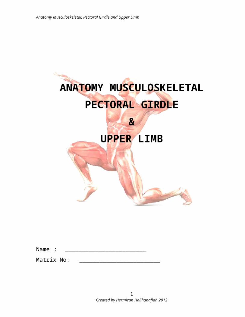
Anatomy Musculoskeletal: Pectoral Girdle and Upper Limb
ANATOMY MUSCULOSKELETAL
PECTORAL GIRDLE
&
UPPER LIMB
Name : ________________________
Matrix No: ________________________
Created by Hermizan Halihanafiah 20121
Anatomy Musculoskeletal: Pectoral Girdle and Upper Limb
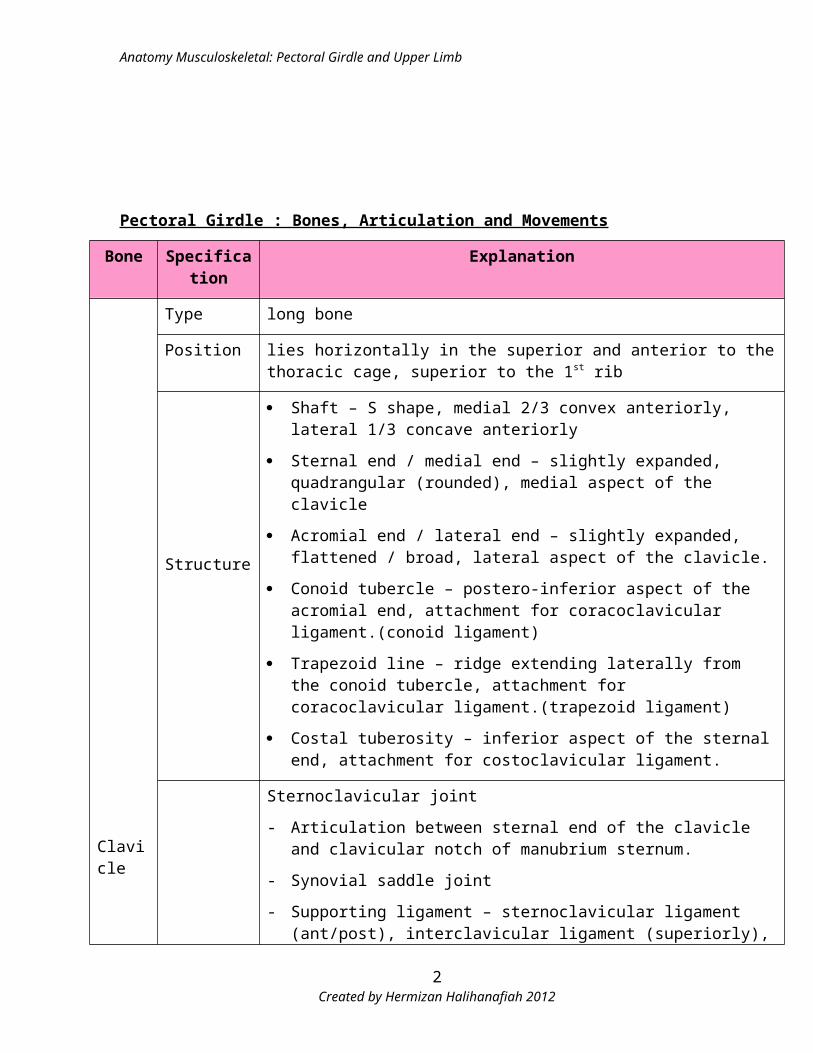
Pectoral Girdle : Bones, Articulation and Movements
Bone Specification Explanation
Clavicle
Type long bone
Position lies horizontally in the superior and anterior to the thoracic cage, superior to the 1st rib
Structure
Shaft – S shape, medial 2/3 convex anteriorly, lateral 1/3 concave anteriorly
Sternal end / medial end – slightly expanded, quadrangular (rounded), medial aspect of the clavicle
Acromial end / lateral end – slightly expanded, flattened / broad, lateral aspect of the clavicle.
Conoid tubercle – postero-inferior aspect of the acromial end, attachment for coracoclavicular ligament.(conoid ligament)
Trapezoid line – ridge extending laterally from the conoid tubercle, attachment for coracoclavicular ligament.(trapezoid ligament)
Costal tuberosity – inferior aspect of the sternal end, attachment for costoclavicular ligament.
Articulation
Sternoclavicular joint
- Articulation between sternal end of the clavicle and clavicular notch of manubrium sternum.
- Synovial saddle joint
- Supporting ligament – sternoclavicular ligament (ant/post), interclavicular ligament (superiorly), costoclavicular ligament (inferiorly).
- Movement – elevation, depression, anterior movement in a horizontal direction.
- Clinical importance – rare
Acromioclavicular joint
- Articulation between acromial end of the clavicle and acromion process of the scapula.
- “ Point of the Shoulder”
- Synovial planar joint
- Supporting ligaments – AC joint is a weak joint, strengthen via
Acromioclavicular ligament – from superior aspect of the acromion process to the superior aspect of the clavicle (acromial end)
Coracoclavicular ligament - anchor clavicle to the coracoid process of the
Created by Hermizan Halihanafiah 20122
Anatomy Musculoskeletal: Pectoral Girdle and Upper Limb
scapula , consist 2 part (conoid lig – from conoid tubercle, trapezoid lig – trapezoid line)
Coracoacromial ligament – attach between coracoid process and acromion process & form the ligamentous arch – protection for the head of humerus (stability for the GH joint).
- Subluxation and dislocation due to fall on the shoulder.
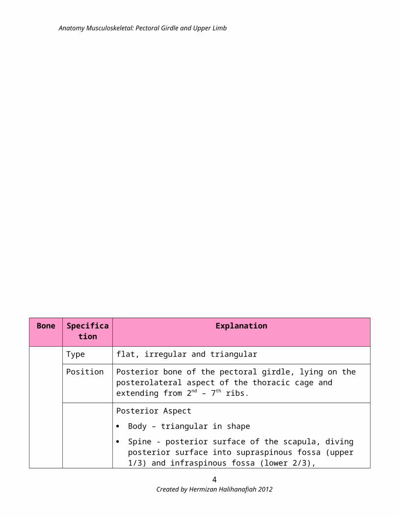
Bone Specification Explanation
Created by Hermizan Halihanafiah 20123
Anatomy Musculoskeletal: Pectoral Girdle and Upper Limb
Scapula
Type flat, irregular and triangular
Position Posterior bone of the pectoral girdle, lying on the posterolateral aspect of the thoracic cage and extending from 2nd – 7th ribs.
Structure
Posterior Aspect
Body – triangular in shape
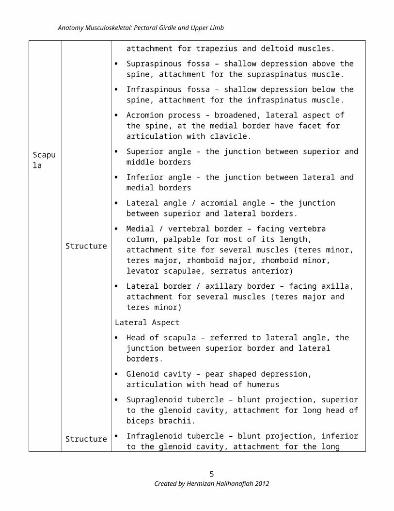
Spine - posterior surface of the scapula, diving posterior surface into supraspinous fossa (upper 1/3) and infraspinous fossa (lower 2/3), attachment for trapezius and deltoid muscles.
Supraspinous fossa – shallow depression above the spine, attachment for the supraspinatus muscle.
Infraspinous fossa – shallow depression below the spine, attachment for the infraspinatus muscle.
Acromion process – broadened, lateral aspect of the spine, at the medial border have facet for articulation with clavicle.
Superior angle – the junction between superior and middle borders
Inferior angle – the junction between lateral and medial borders
Lateral angle / acromial angle – the junction between superior and lateral borders.
Medial / vertebral border – facing vertebra column, palpable for most of its length, attachment site for several muscles (teres minor, teres major, rhomboid major, rhomboid minor, levator scapulae, serratus anterior)
Lateral border / axillary border – facing axilla, attachment for several muscles (teres major and teres minor)
Lateral Aspect
Head of scapula – referred to lateral angle, the junction between superior border and lateral borders.
Glenoid cavity – pear shaped depression, articulation with head of humerus
Supraglenoid tubercle – blunt projection, superior to the glenoid cavity, attachment for long head of biceps brachii.
Infraglenoid tubercle – blunt projection, inferior to the glenoid cavity, attachment for the long head of triceps.
Anterior Aspect
Subscapular fossa – large anteriorly depression, attachment site for the
Created by Hermizan Halihanafiah 20124
Anatomy Musculoskeletal: Pectoral Girdle and Upper Limb
Structure
subscapularis muscle.
Scapular notch – depression at lateral end of the superior border, pathway for the suprascapular nerve.
Coracoid process – anterior projection from the superior border, attachment site for ligament (coracoclavicular ligament) and several muscles (pectoralis minor, coracobrachialis & short head of biceps brachii)
Articulation
Acromioclavicular joint – Articulation between acromial end of clavicle and acromion process of scapula.
Glenohumeral joint / shoulder joint
- Articulation between head of humerus and glenoid cavity of scapula
- Type – synovial ball and socket
- Most mobile, least stable and vulnerable to injury.
- Strengthening structure:
Extracapsular structure
o Ligaments – coracohumeral ligaments (from coracoid process of scapula to the greater tubercle of humerus), glenohumeral ligament (superior, inferior and middle – attach from glenoid cavity of scapula to the lesser tubercle and anatomical neck of humerus), transverse humeral ligament (attach between lesser and greater tubercle, turn the bicipital groove into tunnel, holding tendon of long head of biceps)
o Rotator cuff muscles – supraspinatus, infraspinatus, teres minor and subscapularis (contraction these muscles pulled head of humerus into glenoid cavity).
o Bursae – subscapular bursa, subacromial bursa, subdeltoid bursa, subcoracoid bursa (avoid friction)
Intracapsular structure
o Glenoid labrum/rim – fibrocartilaginous rim surround the edge of the glenoid cavity, deepened the socket
- Movement – flexion / extension, abduction/adduction, medial rotation/lateral rotation, horizontal adduction/horizontal abduction, circumduction.
- Clinical importance – anterior and posterior dislocation, recurrent dislocation, frozen shoulder
- Superior portion – impingement area, this area contain structures that can be damage due to repeated overuse (suprapinatus, long head of biceps, glenoid labrum, coracohumeral ligament, subacromial bursa. The actual impingement occurs in the abducted position with the arm rotated.
Created by Hermizan Halihanafiah 20125
Anatomy Musculoskeletal: Pectoral Girdle and Upper Limb
Scapulothoracic joint / scapulocostal joint
- Physiologic joint - Not a true joint (holding by musculotendinous structure)
- Don’t have any synovial capsule and ligamentous attachment.
- Articulation between anterior surface of the scapula and posterior surface of the 2nd – 7th ribs
- Highly mobile joint
- Stabilization via several muscle – trapezius, rhomboid major, rhomboid minor, serratus anterior
- Movement gliding – abduction / adduction (protraction / retraction), elevation / depression, upward rotation / downward rotation.
- Movement of the scapulothoracic joint – increase the ROM of the GH joint.
Arm : Bones, Articulations and Movements
Created by Hermizan Halihanafiah 20126
Anatomy Musculoskeletal: Pectoral Girdle and Upper Limb
Bone Specification Explanation
Humerus
Type Long bone
Position Largest bone of upper limb, bony part of the arm (brachium)
Structure
Proximal part
Head – rounded in shape, articulate with glenoid cavity of scapula
Anatomical neck – constriction area adjoining the head
Greater tubercle (tuberosity) – blunt projection, posterolaterally, attachment site for several muscles (supraspinatus, infraspinatus, teres minor)
Lesser tubercle (tuberosity) – blunt projection, anteriorly, attachment site for subscapularis muscle.
Intertubercular sulcus / bicipital groove – between greater and lesser tubercle, cover by transverse humeral ligament, contain tendon for long head of biceps.
Surgical neck – constricted area, adjoining site between shaft and proximal part, mostly fracture occur here.
Shaft/body
Long, proximally cylindrical in shape, and gradually become flat and triangular in shape distally.
Deltoid tuberosity – roughen area located anterolaterally, attachment site for the deltoid muscles.
Radial/spiral groove – posterior surface, extend obliquely from medial to lateral site of the shaft, location for the radial nerve
Distal part
Distal end of the lateral border – lateral supracondylar ridge, lateral epicondyle and capitulum.
Lateral supracondylar and lateral epicondyle – attachment for the extensor hand muscles.
Capitulum – rounded in shape, articulate with head of radius
Radial fossa – superior to the capitulum, receive head of radius during elbow flexion
Distal end of the medial border – medial supracondylar ridge, medial epicondyle, trochlear
Medial epicondyle –attachment site for the several ligaments and flexor hand muscles, at the posterior surface – pathway for the ulnar nerve
Trochlea – pulley/spool in shape, articulate with trochlear notch of ulna.
Coronoid fossa – anteriorly, superior to the trochlea, rceive coronoid process
Created by Hermizan Halihanafiah 20127
Anatomy Musculoskeletal: Pectoral Girdle and Upper Limb
of ulna during elbow flexion.
Olecranon fossa – posteriorly, superior to the trochlea, receive olecranon of ulna during elbow extension.
Articulation
Proximally – glenohumeral / shoulder joint
Distally – Elbow joint (include humeroulnar, humeroradial and proximal radioulnar joint) – these joints enclose by single fibrous capsule
Humeroulnar joint (true elbow joint)
- Articulation between trochlea of humerus and trochlear notch of ulna.
- Synovial hinge joint.
- Strengthening structure
Ulnar collateral ligament – on the medial side of the joint, attach from medial epicondyle of humerus to the coronoid process and olecranon process of ulna.
Provide Valgus stability of the elbow
- Movement - flexion / extension
Humeroradial joint
- Articulation between capitulum and head of radius.
- Synovial hinge joint
- Strengthening structure
Radial collateral ligament – on the lateral side of the elbow, attach form the lateral epicondyle of the humerus to the annular ligament.
Provide Varus stability for the elbow.
- Movement - flexion / extension
Proximal radioulnar joint
- Articulation between head of radius and radial notch of ulna.
- Synovial pivot joint.
- Strengthening structure
Annular ligament – surrounds head of radius and attach to the edge of the radial notch of ulna.
Support head of radius.
- Movement – pronation / supination
Forearm : Bones, Articulations and Movements
Bone Specification Explanation
Created by Hermizan Halihanafiah 20128
Anatomy Musculoskeletal: Pectoral Girdle and Upper Limb
Radius
Type Long Bone
Position Lateral bone of forearm
Structure
- Head – rounded, with concave superior surface, articulate with capitulum of humerus and radial notch of ulna.
- Neck – narrow/constricted portion, distal to head
- Radial tuberosity – distal to the neck, on the medial aspect, attachment site for the biceps brachii muscles.
- Interosseous border – on the medial surface of the shaft, attachment for the intersosseous membrane that connects with the shaft of ulna to form middle radioulnar joint.
- Ulnar notch – distal end, located on the medial side, articulates with the head of ulna to form distal radioulnar joint.
- Radial styloid process – distal end, prominent process on the lateral aspect which can be palpated.
- Distal surface articulates with scaphoid, lunate and triquetrum to form radiocarpal joint / wrist joint.
Articulation
Humeroradial joint
- Articulation between head of radius and capitulum of humerus
Proximal radioulnar joint
- Articulation between head of radius and radial notch of ulna
Middle radioulnar joint
- Articulation between shaft of radius and shaft of ulna connect via interosseous membrane.
- Synarthroses / immovable joint
Distal radioulnar joint
- Articulation between head of ulna and ulnar notch of radius
- Synovial pivot joint
- Movement – pronation / supination
- Strengthen via palmar and distal radioulnar ligaments.
Radiocarpal Joint/wrist joint
- Articulation between distal end of radius and radial styloid process with
Created by Hermizan Halihanafiah 20129
Anatomy Musculoskeletal: Pectoral Girdle and Upper Limb
scaphoid, lunate and triquetrum
- Synovial condyloid / ellipsoidal joint
- Strengthen by
Ulnar collateral ligament – medial aspect of the wrist, attached to the ulnar styloid process and the triquetrum and pisiform
Radial collateral ligament – lateral aspect of the wrist – attached to the radial styloid process and the scaphoid
Palmar radiocarpal ligament – anterior aspect of the wrist, attached from the radius to the scaphoid, lunate and the triquetrum.
Dorsal radiocarpal ligament – posterior aspect of the wrist, attached form the radius to the scaphoid, lunate and triquetrum.
Palmar ulnocarpal ligament. – anterior aspect of the wrist, attached from the ulnar styloid process to the lunate and triquetrum.
- Movement – flexion / extension, radial deviation (abduction) /ulnar deviation (adduction).
Bone Specification Explanation
Created by Hermizan Halihanafiah 201210
Anatomy Musculoskeletal: Pectoral Girdle and Upper Limb
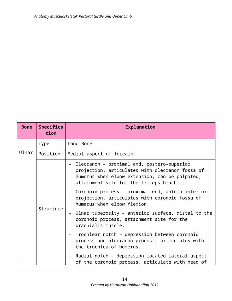
Ulnar
Type Long Bone
Position Medial aspect of forearm
Structure
- Olecranon – proximal end, postero-superior projection, articulates with olecranon fossa of humerus when elbow extension, can be palpated, attachment site for the triceps brachii.
- Coronoid process – proximal end, antero-inferior projection, articulates with coronoid fossa of humerus when elbow flexion.
- Ulnar tuberosity – anterior surface, distal to the coronoid process, attachment site for the brachialis muscle.
- Trochlear notch – depression between coronoid process and olecranon process, articulates with the trochlea of humerus.
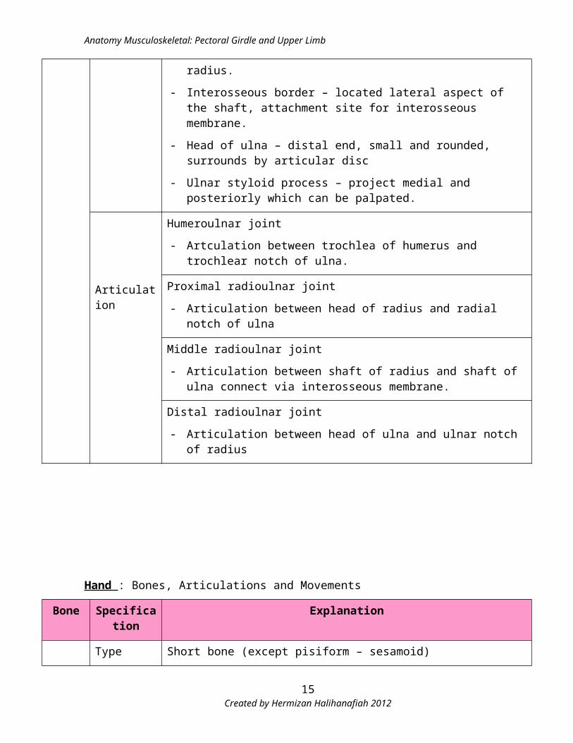
- Radial notch – depression located lateral aspect of the coronoid process, articulate with head of radius.
- Interosseous border – located lateral aspect of the shaft, attachment site for interosseous membrane.
- Head of ulna – distal end, small and rounded, surrounds by articular disc
- Ulnar styloid process – project medial and posteriorly which can be palpated.
Articulation
Humeroulnar joint
- Artculation between trochlea of humerus and trochlear notch of ulna.
Proximal radioulnar joint
- Articulation between head of radius and radial notch of ulna
Middle radioulnar joint
- Articulation between shaft of radius and shaft of ulna connect via interosseous membrane.
Distal radioulnar joint
- Articulation between head of ulna and ulnar notch of radius
Created by Hermizan Halihanafiah 201211
Anatomy Musculoskeletal: Pectoral Girdle and Upper Limb
Hand : Bones, Articulations and Movements
Bone Specification Explanation
Carpal
Type Short bone (except pisiform – sesamoid)
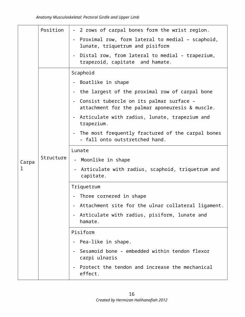
Position - 2 rows of carpal bones form the wrist region.
- Proximal row, form lateral to medial – scaphoid, lunate, triquetrum and pisiform
- Distal row, from lateral to medial – trapezium, trapezoid, capitate and hamate.
Structure
Scaphoid
- Boatlike in shape
- the largest of the proximal row of carpal bone
- Consist tubercle on its palmar surface – attachment for the palmar aponeuresis & muscle.
- Articulate with radius, lunate, trapezium and trapezium.
- The most frequently fractured of the carpal bones – fall onto outstretched hand.
Lunate
- Moonlike in shape
- Articulate with radius, scaphoid, triquetrum and capitate.
Triquetrum
- Three cornered in shape
- Attachment site for the ulnar collateral ligament.
- Articulate with radius, pisiform, lunate and hamate.
Pisiform
- Pea-like in shape.
- Sesamoid bone – embedded within tendon flexor carpi ulnaris
- Protect the tendon and increase the mechanical effect.
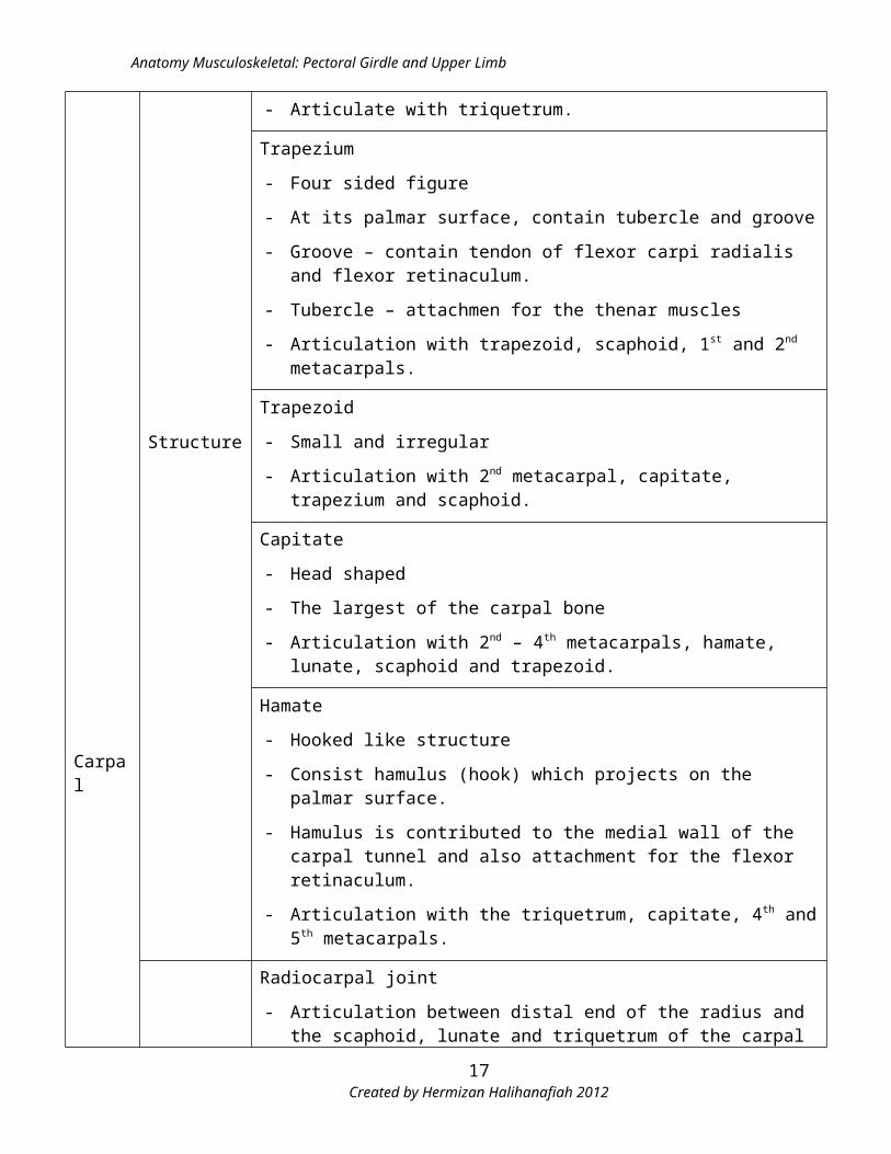
- Articulate with triquetrum.
Trapezium
- Four sided figure
- At its palmar surface, contain tubercle and groove
Created by Hermizan Halihanafiah 201212
Anatomy Musculoskeletal: Pectoral Girdle and Upper Limb
Carpal
Structure
- Groove – contain tendon of flexor carpi radialis and flexor retinaculum.
- Tubercle – attachmen for the thenar muscles
- Articulation with trapezoid, scaphoid, 1st and 2nd metacarpals.
Trapezoid
- Small and irregular
- Articulation with 2nd metacarpal, capitate, trapezium and scaphoid.
Capitate
- Head shaped
- The largest of the carpal bone
- Articulation with 2nd – 4th metacarpals, hamate, lunate, scaphoid and trapezoid.
Hamate
- Hooked like structure
- Consist hamulus (hook) which projects on the palmar surface.
- Hamulus is contributed to the medial wall of the carpal tunnel and also attachment for the flexor retinaculum.
- Articulation with the triquetrum, capitate, 4th and 5th metacarpals.
Articulation
Radiocarpal joint
- Articulation between distal end of the radius and the scaphoid, lunate and triquetrum of the carpal bones.
Intercarpal joint
- Proximal row - Articulation between scaphoid, lunate, triquetrum and pisiform
- Distal row - Articulation between trapezium, trapezoid, capitate and hamate
- Synovial planar joint
- Movement – gliding motion
Midcarpal joint
- Articulation between proximal row and distal row of carpal bone. (Articulation between the scaphoid, lunate and triquetrum (proximally) and trapezium, trapezoid, capitate and hamate (distally).
- Synovial planar joint.
Created by Hermizan Halihanafiah 201213
Anatomy Musculoskeletal: Pectoral Girdle and Upper Limb
Carpal
Articulation
- Movement – gliding motion
Carpometacarpal joint
- Articulation between distal row of carpal bones and metacarpal.
- 5 carpometacarpal joint
- 1st carpometacarpal joint
Modified saddle joint
Articulation between 1st metacarpal (base) and the distal surface of the trapezium.
Supporting ligaments
Lateral ligament - lateral surface of the trapezium to the lateral side of the base of metacarpal.
Palmar ligament - oblique band, from palmar surface of the trapezium to the medial side of the base metacarpal.
Dorsal ligament - oblique band, from dorsal surface of the trapezium to the medial side of the base metacarpal.
Movement – flexion / extension, abduction / adduction, rotation, opposition / reposition
Permitting the ability to hold and manipulate objects (pinch grip, tripod pinch and chuck grip)
- 2nd – 5th carpometacarpal joint
Articulation between distal row of carpal bones and 2nd – 5th base of metacarpal
Synovial ellipsoidal joint
Supporting ligaments
- Dorsal ligament – the strongest ligament, attached from the dorsal surface of the carpal and metacarpal.
- Palmar ligament - attached from the palmar surface of the carpal and metacarpal.
- Interosseous ligament – attached from capitate and hamate to the 3rd and 4th metatarsal.
Movement – gliding motion
Created by Hermizan Halihanafiah 201214
Anatomy Musculoskeletal: Pectoral Girdle and Upper Limb
Bone Specification Explanation
Metacarpal
Type Miniature long bones
Position - 5 metacarpal bones, numbered I – V (lateral to medial) form the palmar of the hand,
- Distal to distal row of carpal bones and proximal to the phalanges.
Structure
- Head – rounded, located distally, and articulates with corresponding phalanx.
- Shaft / body – middle portion, anterior border concave longitudinal.
- Base – expanded, articulate with appropriate carpal bones. The base of 2nd – 5th metacarpal articulates each other.
Articulation
Carpometacarpal joint
Metacarpophalangeal joint
- Articulation between proximal head of metacarpal and base of proximal phalanges.
- Synovial ellipsoidal joint
- Strengthen by – collateral ligament (strong and flank the joints), palmar ligaments and deep transverse metacarpal ligaments.
- Movement –flexion / extension, abduction / adduction
Intermetacarpal joint
- Articulation between based of the 2nd – 5th metacarpal.
- Strengthen by palmar ligament, dorsal ligament and interosseous ligament.
- Movement – slightly gliding.
Created by Hermizan Halihanafiah 201215
Anatomy Musculoskeletal: Pectoral Girdle and Upper Limb
Bone Specification Explanation
Phalanges
Type Miniature long bones
Position Distal to the metacarpal, forming the 14 fingers (14 digits)
Structure
There are 14 phalanges. Every digits consist 3 phalanges (proximal, intermediate and distal), except thumb only have 2 phalanges (proximal and distal)
- Head – distally, expanded, distal phalanges support the tissue of the finger tips
- Shaft / body – intermediate portion, anterior border are concave longitudinally.
- Base – proximally, expanded, articulates with either the phalanges or the metacarpal to it.
Articulation
Metacarpophalangeal joint
Interphalangeal Joint
- Articulation between phalanges bone itself
- Synovial hinge joint
- All digits compose by 2 interphalangeal joints; proximal IP and distal IP, except thumb only have one.
- Strengthen ligaments – palmar ligament / volar plate (floor of the IP) and 2 collateral ligaments (on the lateral and medial side of the IP).
- Movement – flexion / extension
Created by Hermizan Halihanafiah 201216
Anatomy Musculoskeletal: Pectoral Girdle and Upper Limb
Muscles of the Pectoral Girdle
Attachment Site Joint Muscle Origin Insertion Action
Clavicle – humerus GH Anterior fibre of Deltoid Lateral 1/3 (anterior surface)
Deltoid tuberosity Flexion
Clavicle origin of pectoralis major
Medial half (anterior surface)
Bicipital groove (lateral lip) of humerus
Flexion, medial rotation, horizontal adduction
Sternum – humerus GH Sternocostal fibre of pectoralis major
Anterior surface of sternum, costal cartilages of upper 6th or 7th ribs.
Bicipital groove (lateral lip) of humerus
Extension , medial rotation, horizontal adduction
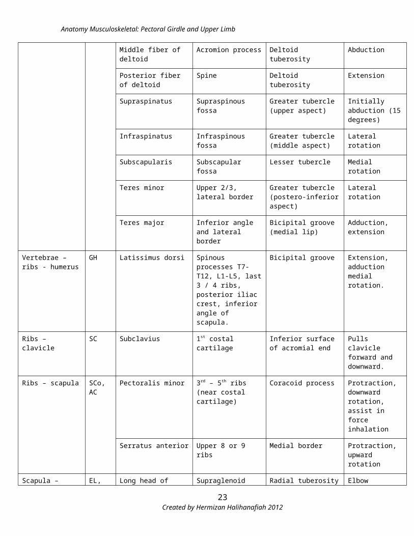
Scapula – humerus GH Coracobrachialis Coracoid process Middle shaft (opposite to deltoid tubesoity)
Flexion and adduction
Middle fiber of deltoid Acromion process Deltoid tuberosity Abduction
Posterior fiber of deltoid Spine Deltoid tuberosity Extension
Supraspinatus Supraspinous fossa Greater tubercle (upper aspect)
Initially abduction (15 degrees)
Infraspinatus Infraspinous fossa Greater tubercle (middle aspect)
Lateral rotation
Subscapularis Subscapular fossa Lesser tubercle Medial rotation
Teres minor Upper 2/3, lateral border
Greater tubercle (postero-inferior aspect)
Lateral rotation
Teres major Inferior angle and lateral border
Bicipital groove (medial lip)
Adduction, extension
Vertebrae – ribs - humerus
GH Latissimus dorsi Spinous processes T7-T12, L1-L5, last 3 / 4 ribs, posterior iliac crest, inferior angle of scapula.
Bicipital groove Extension, adduction medial rotation.
Ribs – clavicle SC Subclavius 1st costal cartilage Inferior surface of acromial end
Pulls clavicle forward and downward.
Ribs – scapula SCo, AC
Pectoralis minor 3rd – 5th ribs (near costal cartilage)
Coracoid process Protraction, downward rotation, assist in force inhalation
Serratus anterior Upper 8 or 9 ribs Medial border Protraction, upward rotation
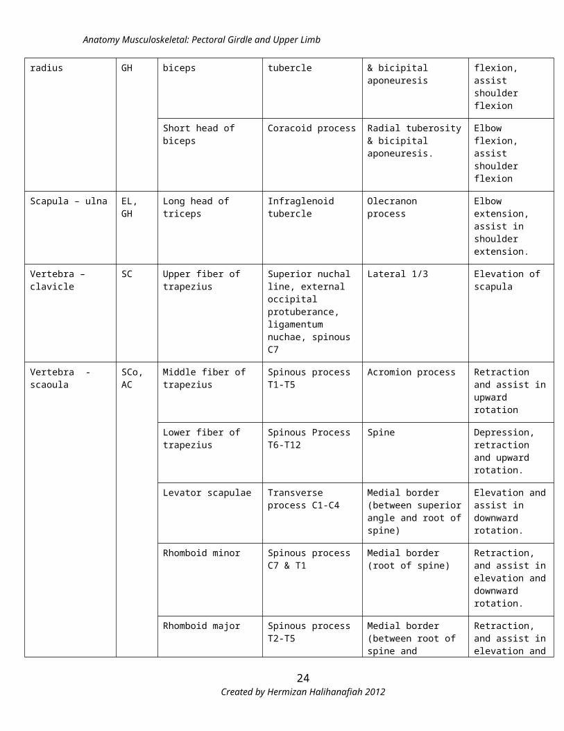
Scapula – radius EL, GH Long head of biceps Supraglenoid tubercle Radial tuberosity & Elbow flexion,
Created by Hermizan Halihanafiah 201217
Anatomy Musculoskeletal: Pectoral Girdle and Upper Limb
bicipital aponeuresis assist shoulder flexion
Short head of biceps Coracoid process Radial tuberosity & bicipital aponeuresis.
Elbow flexion, assist shoulder flexion
Scapula – ulna EL, GH Long head of triceps Infraglenoid tubercle Olecranon process Elbow extension, assist in shoulder extension.
Vertebra – clavicle SC Upper fiber of trapezius Superior nuchal line, external occipital protuberance, ligamentum nuchae, spinous C7
Lateral 1/3 Elevation of scapula
Vertebra - scaoula SCo, AC
Middle fiber of trapezius Spinous process T1-T5 Acromion process Retraction and assist in upward rotation
Lower fiber of trapezius Spinous Process T6-T12
Spine Depression, retraction and upward rotation.
Levator scapulae Transverse process C1-C4
Medial border (between superior angle and root of spine)
Elevation and assist in downward rotation.
Rhomboid minor Spinous process C7 & T1
Medial border (root of spine)
Retraction, and assist in elevation and downward rotation.
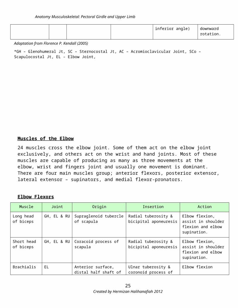
Rhomboid major Spinous process T2-T5 Medial border (between root of spine and inferior angle)
Retraction, and assist in elevation and downward rotation.
Adaptation from Florence P. Kendall (2005)
*GH – Glenohumeral Jt, SC – Sternocostal Jt, AC – Acromioclavicular Joint, SCo – Scapulocostal Jt, EL – Elbow Joint,
Created by Hermizan Halihanafiah 201218
Anatomy Musculoskeletal: Pectoral Girdle and Upper Limb
Muscles of the Elbow
24 muscles cross the elbow joint. Some of them act on the elbow joint exclusively, and others act on the wrist and hand joints. Most of these muscles are capable of producing as many as three movements at the elbow, wrist and fingers joint and usually one movement is dominant. There are four main muscles group; anterior flexors, posterior extensor, lateral extensor – supinators, and medial flexor-pronators.
Elbow Flexors
Muscle Joint Origin Insertion Action
Long head of biceps
GH, EL & RU Supraglenoid tubercle of scapula
Radial tuberosity & bicipital aponeuresis
Elbow flexion, assist in shoulder flexion and elbow supination.
Short head of biceps
GH, EL & RU Coracoid process of scapula Radial tuberosity & bicipital aponeuresis
Elbow flexion, assist in shoulder flexion and elbow supination.
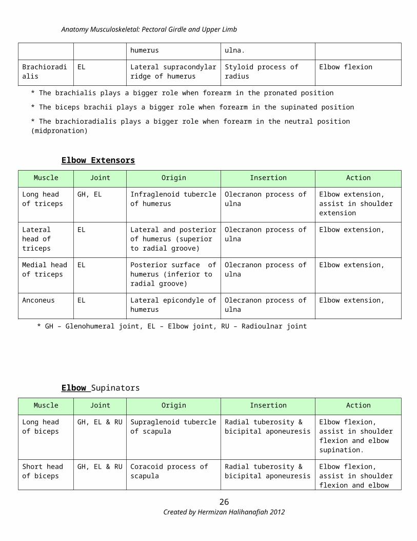
Brachialis EL Anterior surface, distal half shaft of humerus
Ulnar tuberosity & coronoid process of ulna.
Elbow flexion
Brachioradialis EL Lateral supracondylar ridge of humerus
Styloid process of radius Elbow flexion
* The brachialis plays a bigger role when forearm in the pronated position
* The biceps brachii plays a bigger role when forearm in the supinated position
* The brachioradialis plays a bigger role when forearm in the neutral position (midpronation)
Elbow Extensors
Muscle Joint Origin Insertion Action
Long head of triceps
GH, EL Infraglenoid tubercle of humerus
Olecranon process of ulna Elbow extension, assist in shoulder extension
Lateral head of triceps
EL Lateral and posterior of humerus (superior to radial groove)
Olecranon process of ulna Elbow extension,
Medial head of triceps
EL Posterior surface of humerus (inferior to radial groove)
Olecranon process of ulna Elbow extension,
Anconeus EL Lateral epicondyle of humerus Olecranon process of ulna Elbow extension,
* GH – Glenohumeral joint, EL – Elbow joint, RU – Radioulnar joint
Created by Hermizan Halihanafiah 201219
Anatomy Musculoskeletal: Pectoral Girdle and Upper Limb
Elbow Supinators
Muscle Joint Origin Insertion Action
Long head of biceps
GH, EL & RU Supraglenoid tubercle of scapula
Radial tuberosity & bicipital aponeuresis
Elbow flexion, assist in shoulder flexion and elbow supination.
Short head of biceps
GH, EL & RU Coracoid process of scapula Radial tuberosity & bicipital aponeuresis
Elbow flexion, assist in shoulder flexion and elbow supination.
Supinator EL, RU Lateral epicondyle of humerus Lateral surface proximal 1/3 shaft of radius
Elbow supination
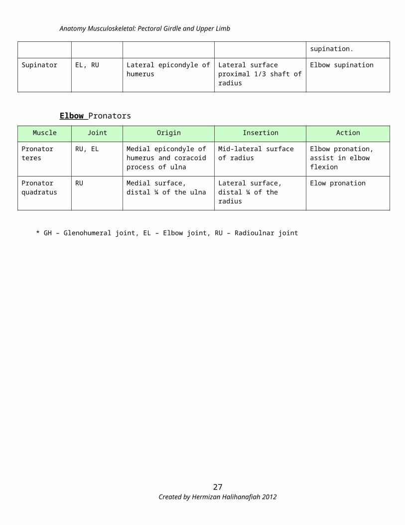
Elbow Pronators
Muscle Joint Origin Insertion Action
Pronator teres RU, EL Medial epicondyle of humerus and coracoid process of ulna
Mid-lateral surface of radius Elbow pronation, assist in elbow flexion
Pronator quadratus
RU Medial surface, distal ¼ of the ulna
Lateral surface, distal ¼ of the radius
Elow pronation
* GH – Glenohumeral joint, EL – Elbow joint, RU – Radioulnar joint
Created by Hermizan Halihanafiah 201220
Anatomy Musculoskeletal: Pectoral Girdle and Upper Limb
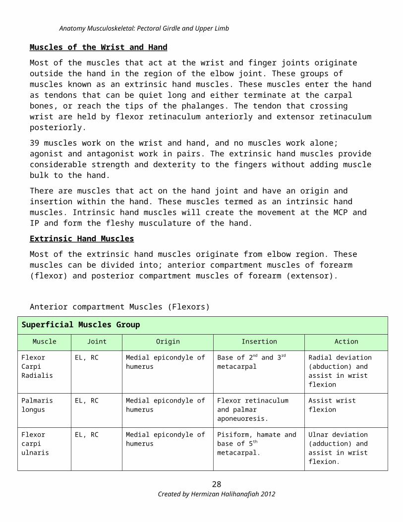
Muscles of the Wrist and Hand
Most of the muscles that act at the wrist and finger joints originate outside the hand in the region of the elbow joint. These groups of muscles known as an extrinsic hand muscles. These muscles enter the hand as tendons that can be quiet long and either terminate at the carpal bones, or reach the tips of the phalanges. The tendon that crossing wrist are held by flexor retinaculum anteriorly and extensor retinaculum posteriorly.
39 muscles work on the wrist and hand, and no muscles work alone; agonist and antagonist work in pairs. The extrinsic hand muscles provide considerable strength and dexterity to the fingers without adding muscle bulk to the hand.
There are muscles that act on the hand joint and have an origin and insertion within the hand. These muscles termed as an intrinsic hand muscles. Intrinsic hand muscles will create the movement at the MCP and IP and form the fleshy musculature of the hand.
Extrinsic Hand Muscles
Most of the extrinsic hand muscles originate from elbow region. These muscles can be divided into; anterior compartment muscles of forearm (flexor) and posterior compartment muscles of forearm (extensor).
Anterior compartment Muscles (Flexors)
Superficial Muscles Group
Muscle Joint Origin Insertion Action
Flexor Carpi Radialis
EL, RC Medial epicondyle of humerus Base of 2nd and 3rd metacarpal Radial deviation (abduction) and assist in wrist flexion
Palmaris longus EL, RC Medial epicondyle of humerus Flexor retinaculum and palmar aponeuoresis.
Assist wrist flexion
Flexor carpi ulnaris
EL, RC Medial epicondyle of humerus Pisiform, hamate and base of 5th metacarpal.
Ulnar deviation (adduction) and assist in wrist flexion.
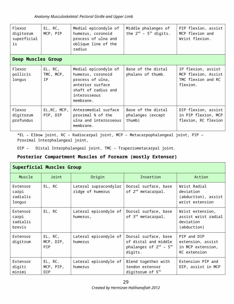
Flexor digitorum superficialis
EL, RC, MCP, PIP
Medial epicondyle of humerus, coronoid process of ulna and oblique line of the radius
Middle phalanges of the 2nd – 5th digits.
PIP flexion, assist MCP flexion and Wrist flexion.
Deep Muscles Group
Flexor pollicis longus
EL, RC, TMC, MCP, IP
Medial epicondyle of humerus, coronoid process of ulna, anterior surface shaft of radius and interosseous membrane.
Base of the distal phalanx of thumb.
IP flexion, assist MCP flexion, Assist TMC flexion and RC flexion.
Flexor digitorum profundus
EL,RC, MCP, PIP, DIP
Anteromedial surface proximal ¾ of the ulna and interosseous membrane.
Base of the distal phalanges (except thumb)
DIP flexion, assist in PIP flexion, MCP flexion, RC flexion
*EL – Elbow joint, RC – Radiocarpal joint, MCP – Metacarpophalangeal joint, PIP – Proximal Interphalangeal joint,
DIP – Distal Interphalangeal joint, TMC – Trapeziometacarpal joint.
Created by Hermizan Halihanafiah 201221

Anatomy Musculoskeletal: Pectoral Girdle and Upper Limb
Posterior Compartment Muscles of Forearm (mostly Extensor)
Superficial Muscles Group
Muscle Joint Origin Insertion Action
Extensor carpi radialis longus
EL, RC Lateral supracondylar ridge of humerus
Dorsal surface, base of 2nd metacarpal.
Wrist Radial deviation (abduction), assist wrist extension
Extensor carpi radialis brevis
EL, RC Lateral epicondyle of humerus, Dorsal surface, base of 3rd metacarpal.
Wrist extension, assist wrist radial deviation (abduction)
Extensor digitrum
EL, RC, MCP, DIP, PIP
Lateral epicondyle of humerus Dorsal surface, base of distal and middle phalanges of 2nd – 5th digits.
PIP and DIP extension, assist in MCP extension, RC extension
Extensor digiti minimi
EL, RC. MCP, PIP, DIP
Lateral epicondyle of humerus Blend together with tendon extensor digitorum of 5th digit.
Extension PIP and DIP, assist in MCP and RC extension.
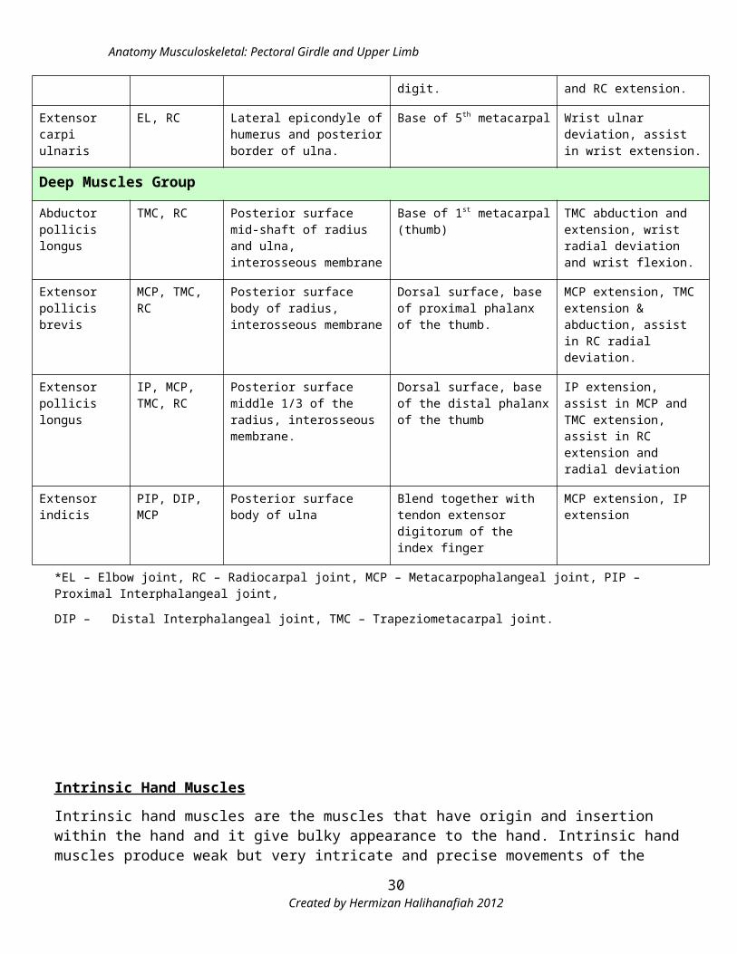
Extensor carpi ulnaris
EL, RC Lateral epicondyle of humerus and posterior border of ulna.
Base of 5th metacarpal Wrist ulnar deviation, assist in wrist extension.
Deep Muscles Group
Abductor pollicis longus
TMC, RC Posterior surface mid-shaft of radius and ulna, interosseous membrane
Base of 1st metacarpal (thumb) TMC abduction and extension, wrist radial deviation and wrist flexion.
Extensor pollicis brevis
MCP, TMC, RC Posterior surface body of radius, interosseous membrane
Dorsal surface, base of proximal phalanx of the thumb.
MCP extension, TMC extension & abduction, assist in RC radial deviation.
Extensor pollicis longus
IP, MCP, TMC, RC
Posterior surface middle 1/3 of the radius, interosseous membrane.
Dorsal surface, base of the distal phalanx of the thumb
IP extension, assist in MCP and TMC extension, assist in RC extension and radial deviation
Extensor indicis PIP, DIP, MCP Posterior surface body of ulna Blend together with tendon extensor digitorum of the index finger
MCP extension, IP extension
*EL – Elbow joint, RC – Radiocarpal joint, MCP – Metacarpophalangeal joint, PIP – Proximal Interphalangeal joint,
DIP – Distal Interphalangeal joint, TMC – Trapeziometacarpal joint.
Created by Hermizan Halihanafiah 201222
Anatomy Musculoskeletal: Pectoral Girdle and Upper Limb
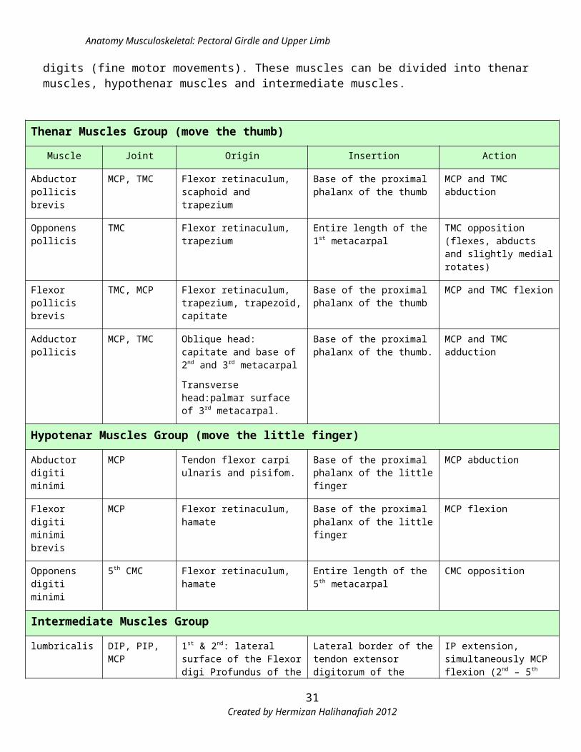
Intrinsic Hand Muscles
Intrinsic hand muscles are the muscles that have origin and insertion within the hand and it give bulky appearance to the hand. Intrinsic hand muscles produce weak but very intricate and precise movements of the digits (fine motor movements). These muscles can be divided into thenar muscles, hypothenar muscles and intermediate muscles.
Thenar Muscles Group (move the thumb)
Muscle Joint Origin Insertion Action
Abductor pollicis brevis
MCP, TMC Flexor retinaculum, scaphoid and trapezium
Base of the proximal phalanx of the thumb
MCP and TMC abduction
Opponens pollicis
TMC Flexor retinaculum, trapezium Entire length of the 1st metacarpal
TMC opposition (flexes, abducts and slightly medial rotates)
Flexor pollicis brevis
TMC, MCP Flexor retinaculum, trapezium, trapezoid, capitate
Base of the proximal phalanx of the thumb
MCP and TMC flexion
Adductor pollicis MCP, TMC Oblique head: capitate and base of 2nd and 3rd metacarpal
Transverse head:palmar surface of 3rd metacarpal.
Base of the proximal phalanx of the thumb.
MCP and TMC adduction
Hypotenar Muscles Group (move the little finger)
Abductor digiti minimi
MCP Tendon flexor carpi ulnaris and pisifom.
Base of the proximal phalanx of the little finger
MCP abduction
Flexor digiti minimi brevis
MCP Flexor retinaculum, hamate Base of the proximal phalanx of the little finger
MCP flexion
Opponens digiti minimi
5th CMC Flexor retinaculum, hamate Entire length of the 5th metacarpal
CMC opposition
Intermediate Muscles Group
lumbricalis DIP, PIP, MCP 1st & 2nd: lateral surface of the Flexor digi Profundus of the index and middle finger
3rd : adjacent side of the FDP of the middle and ring finger
4th:adjacent side of the FDP of the ring and little fingers
Lateral border of the tendon extensor digitorum of the respective fingers.
IP extension, simultaneously MCP flexion (2nd – 5th digits)
Palmar interossei MCP, DIP, PIP 1st : base of 1st metacarpal, medial side
2nd: length of 2nd metacarpal, medial side
1st: base of proximal phalanx (thumb), medial side
2nd: base of proximal phalanx (index finger), medial side.
MCP adduction of 1st, 2nd, 3rd and 4th digits.
Assist in IP extension of 2nd, 3rd and 4th digits.
Created by Hermizan Halihanafiah 201223
Anatomy Musculoskeletal: Pectoral Girdle and Upper Limb
3rd: length of 4th metacarpal, lateral side
4th: length of 5th metacarpal, lateral side
3rd: base of proximal phalanx (ring finger), lateral side.
4th: base of proximal phalanx (little finger), lateral side
Dorsal Interossei MCP, PIP, DIP Adjacent sides of the metacarpals in each interspace (for example: 1st dorsal interossei – adjacent sides between 1st and 2nd metacarpals)
1st: base of proximal phalanx of index finger (lateral side)
2nd: base of proximal phalanx of middle finger (lateral side)
3rd base of proximal phalanx of middle finger (medial side)
4th :base of proximal phalanx of ring finger (medial side)
MCP abduction (2nd, 3rd and 4th digits)
Assist in MCP flexion and PIP and DIP extension.
* MCP – Metacarpophalangeal joint, PIP – Proximal Interphalangeal joint, DIP – Distal Interphalangeal joint, TMC – Trapeziometacarpal joint, CMC – Carpometacarpal joint
Created by Hermizan Halihanafiah 201224
Anatomy Musculoskeletal: Pectoral Girdle and Upper Limb
Nerves Supply of the Shoulder and Upper Limb
The sources of the nerves supply to the shoulder girdle and the entire of upper limb mostly from the networks of the ventral rami of spinal nerves termed as a brachial plexus and also some contribution from the cervical plexus.). Brachial plexus have a root from ventral rami of cervical spinal nerves C5 until C8 and thoracic spinal nerve T1 and give motor and cutaneous distribution to the shoulder and upper limb region. (Please refer to the text books and ppt for further explanation
Source Level Spinal Segment (root) NervesMotor / sensory
Muscles
Cervical plexus
Cervical nerve and cranial nerve (XI)
C(1), C2, C3 & Cranial nerve (XI)
Spinal accessory nerve Motor and
Sensory
Sternocleidomastoid
C2, C3, C4 & Cranial nerve (XI)
Spinal accessory nerve
Fiber of trapezius
Brachial Plexus
Roots
C3, C4, C5 Dorsal scapular nerve
Motor
Levator scapulae
C4, C5 Dorsal scapular nerveRhomboid major and rhomboid
minor
C5, C6, C7, C(8) Long thoracic nerve Serratus anterior
Superior Trunk
(middle & inferior - no nerves arise)
C5, C6 Subclavian nerve
Motor
subclavius
C4, C5, C6 Suprascapular nerve Supraspinatus and infraspinatus
Posterior cord
C5, C6, C7Upper and lower
subscapular nervesMotor Subcapularis and teres major
C6, C7, C8 Thoracodorsal nerve Motor Latissimus dorsi
C5, C6 Axillary nerveMotor and
sensoryDeltoid and teres minor
C5, C6, C7, C8, T1 Radial NerveMotor and
sensory
Triceps, anconeus, supinator, brachioradialis, extrinsic hand
muscles (extensor group)
Lateral cord C5, C6, C7 Lateral pectoral nerve Motor Pectoralis major (upper)
C5, C7Musculocutaneous
nerve Motor and
sensorycoracobrachialis
C5, C6Musculocutaneous
nerveMotor and
sensoryBiceps brachii, brachialis
C5, C6, C7 Lateral root of median nerve
Motor and sensory
Pronator teres and quadratus, All flexor group of extrinsic hand muscles (except FCU, medial
half FDP), 1st & 2nd Lumbricalis, OP, APB, FPB
Created by Hermizan Halihanafiah 201225
Anatomy Musculoskeletal: Pectoral Girdle and Upper Limb
Medial cord
C8, T1 Medial pectoral nerve Motor Pectoralis major (lower),
pectoralis minor
C8, T1Medial root of median
nerveMotor and
sensory
Pronator teres and quadratus, All flexor group of extrinsic hand muscles (except FCU, medial
half FDP), 1st & 2nd Lumbricalis, OP, APB, FPB
C8, T1 Ulnar nerveMotor and
sensory
FCU, medial half FDP, most of the intrinsic hand muscles
(except LOAF innervates by median nerve)
Created by Hermizan Halihanafiah 201226
Anatomy Musculoskeletal: Pectoral Girdle and Upper Limb
Peripheral Nerves of Upper Limb
Five terminal branches arise from the brachial plexus and innervate most of the skin and muscles of the pectoral girdle and upper limb. These five nerves are axillary, musculocutaneous, radial, median and ulnar nerves.
Nerve Root Pathway Motor innervation
Sensory innervation Nerve lesion
Axillary C5, C6
- Posterior cord- Lies anterior to
subscapularis m and posterior to axillary artery.
- Winds posteriorly around the neck of humerus
- Terminates as a anterior and posterior branches
Anterior branch – anterior and lateral fiber of deltoidPosterior branch – posterior fiber of deltoid and teres minor
Posterior branch will terminates as a upper lateral brachial cutaneous nerve – skin lower part of the deltoid
Dislocation of the GH joint, fracture of the humeral neck – weakness of the deltoid (muscle wasting).
Musculocutameous C5 – C7
- Lateral cord- Opposite lower border of the
pectoralis minor- Pierce coracobrachialis m and descends laterally between biceps and brachialis to the lateral side of the arm.- Pierce the deep fascia on the elbow and continue to the forearm as a lateral anterbrachial cutaneous nerve.
Along the pathway, supply coracobrachialis, biceps brachii and brachialis
Antebrachial cutaneous nerve – divide into anterior and posterior branchesAnterior branch – skin of anterolateral surface of forearm as far as ball of the thumbPosterior branch – skin of posterolateral surface of forearm
Fracture of the humerus, patient with neuralgic amyothrophyWeakness of elbow flexion
Radial C5 – T1
-Posterior cord-Descend posterior to the axillary artery-Enter the radial groove at the back of the humerus and enter anterior compartment of the arm.-Continue the jurney between brachialis and brachioradialis-At the distal part of humerus, passes anterior to the lateral epicondyle and enter the forearm-And terminate as superficial and deep terminal branches.
Arm: triceps brachii, anconeus, supinator, brachioradialisForearm: extrinsic hand muscles (extensor group)
ArmPosterior Brachial Cutaneous nerve – skin back on the armInferior lateral brachial cutaneous nerve – skin at the lower lateral aspect of the armForearmPosterior antebrachial cutaneous nerve – skin at the back of the forearmHandSuperficial branch – back of the hand
Fracture of the midshaft of humerus (radial groove) – ‘wrist drop’- the hand can’t be lifted against gravity and the power grip is weak.
Created by Hermizan Halihanafiah 201227
Anatomy Musculoskeletal: Pectoral Girdle and Upper Limb
Ulnar C7 – T1
-Medial cordPasses down the medial side of the arm between biceps and triceps-At the elbow, the nerve lies behind, and contact with the medial epicondyle of humerusAt the forearm give motor branches to FCU and medial half of FDP.-Just above the wrist, the nerve branch off to the superficial and deep branch.
Forearm – FCU and medial half FDPHand – via deep branch supply hypothenar muscles, palmar & dorsal interossei, medial half of lumbricalis and adductor pollicis
Superficial branch supply Skin anterior and posterior of the hand( medial aspect of the hand, ring and little fingers)
Injury to the elbow whether traumatic or entrapment near to the medial epicondyle.Ulnar nerve lesion will give appearance known as a claw hand.Claw hand – little and ring finger curl in flexion deformity and MCP hyperextension – due to paralysis of lumbricalis.
Median C5-T1
-Lateral and medial cords -Passes down the arm with the brachial artery and medial to the MC nerve. At the elbow lies on the brachialis and medial to the tendon of biceps.-Passes anterior to the elbow joint (within cubital fossa) and then down and supply flexor muscles of forearm.-In the hand, passes through the carpal tunnel and then divide into motor and cutaneous branch.
Elbow – pronator teres, FCR, Palmaris Longus, FDSForearm – lateral half FDP, FPL, and pronator quadratusHand – thenar muscles (except adductor pollicis) and lateral half lumbricalis.
Cutaneous branches - skin of the palmar aspect of the thumb and the lateral 2 ½ fingers and the distal ends of the same fingers and skin of distal phalanx on same finger
Carpal tunnel syndrome – compression of median nerve (pain, numbness and tingling) within carpal tunnel.
Created by Hermizan Halihanafiah 201228
Anatomy Musculoskeletal: Pectoral Girdle and Upper Limb
Important Area of the Upper Limb
Area Borders Contents Clinical ImportanceAxillaPyramidal space inferior to GH joint. Provide passageway for vessels and nerves going to and from the upper limb.
Apex: Cervico-axillary canal (space between neck and axilla, posterior to the clavicle)Base: skin, subcutaneous tissue (axillary fossa)Anterior wall: Pectoralis major and minor and form anterior axillary foldPosterior wall: subscapularis, scapula, teres major and latissimus dorsi. Teres major and latissimus dorsi form posterior axillary fold.Medial wall: thoracic wall and underlying serratus anteriorLateral wall: Intertubecular sulcus of humerus
- Axillary artery and its branches
- Axillary veins and its tributaries-Nerves of the
cords and branches of the brachial plexus
- Lymphatic vessels- Several groups of
axillary lymph nodes.
Enlargement of axillary lymph nodes – due to infection on upper limbInjury to axillary vein – profuse bleeding and risk of the air emboli.
Superior view of the axilla region and its related contents
Created by Hermizan Halihanafiah 201229
Anatomy Musculoskeletal: Pectoral Girdle and Upper Limb
Area Borders Contents Clinical ImportanceCubital FossaShallow triangular depression on the anterior surface of elbow.
Superior: imaginary line connection between medial and lateral epicondyle of humerusMedial : pronator teresLateral: brachioradialisFloor: brachialis and supinatorRoof: Bicipital aponeurosis, subcutaneous tissue and skin
- Terminal part of brachial artery and veins
- Biceps brachii tendon
- Median nerve- Radial nerveIn the subcutaneous tissue – medial cubital vein, basilic vein and cephalic vein.
Venous blood drainage from median cubital vein, cephalic vein and basilica vein.Blood pressure measurement – brachial artery
Anterior View of cubital fossa and its related structures
Area Borders Contents Clinical ImportanceCarpal Lateral border: scaphoid and Tendon Flexor Carpal Tunnel
Created by Hermizan Halihanafiah 201230
Anatomy Musculoskeletal: Pectoral Girdle and Upper Limb
TunnelConcave surface located at the palmar surface of the wrist
trapeziumMedial border: pisiform and hamateRoof: Flexor retinaculum (transverse carpal ligament) attach between bones of the medial and lateral borders.
digitorum profundusTendon flexor digitorum superficialisTendon flexor pollicis longusMedian nerve
SyndromeCompression of the median nerve, microvascular insufficiency of the median nerve and prolong vibration of the median nerve.Numbness, pain and tingling of the hand.
Created by Hermizan Halihanafiah 201231
Top Related