Locomotor function of the pectoral girdle 'muscular sling' in trotting ...
Upload
susanna-jacksonCategory
view
232download
0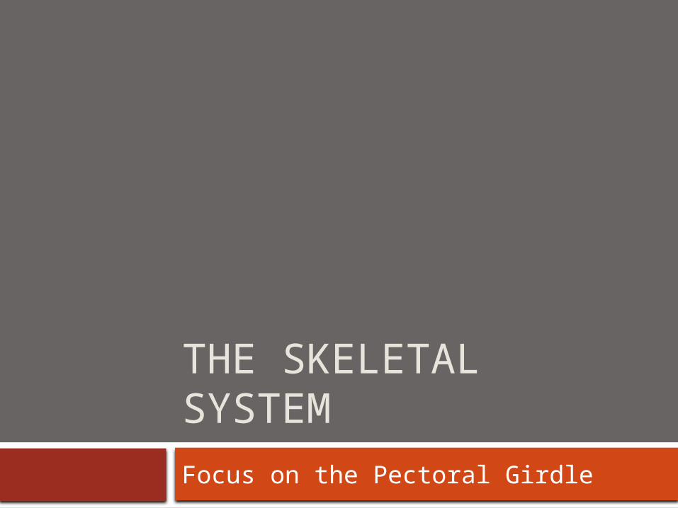
THE SKELETAL SYSTEM
Focus on the Pectoral Girdle
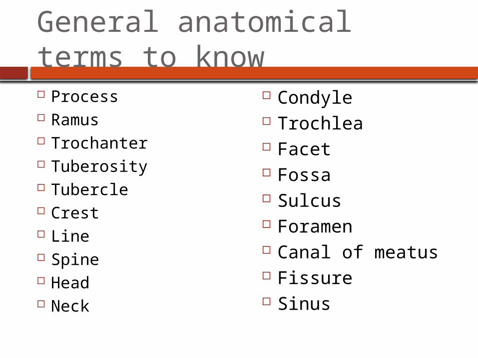
General anatomical terms to know Process Ramus Trochanter Tuberosity Tubercle Crest Line Spine Head Neck
Condyle Trochlea Facet Fossa Sulcus Foramen Canal of meatus Fissure Sinus
Appendicular Skeleton
126 bones Includes
bones of the limbs (appendages) Supporting bones of the pectoral and pelvic
girdles (connect limbs to axial skeleton)
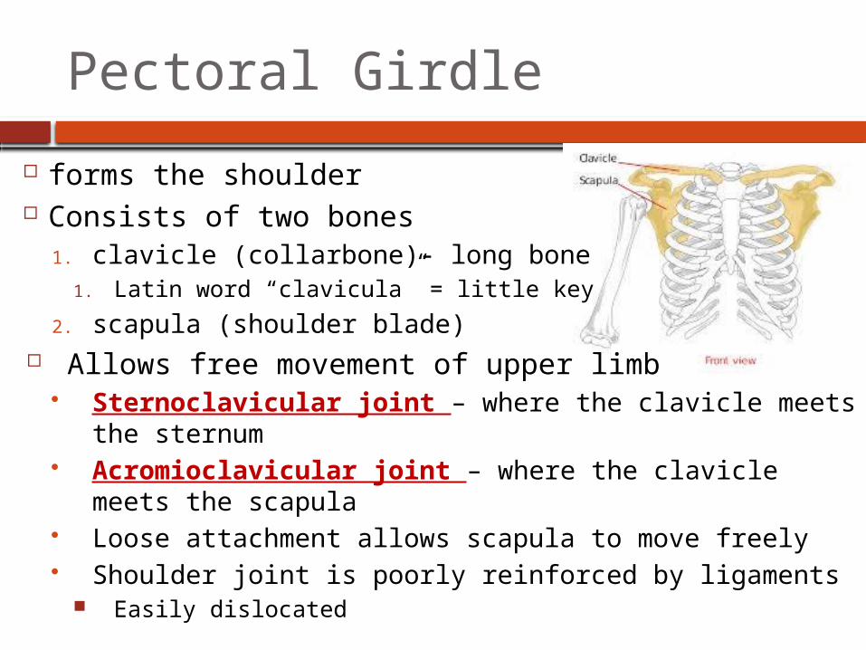
forms the shoulder Consists of two bones
1. clavicle (collarbone)- long bone1. Latin word “clavicula” = little key
2. scapula (shoulder blade) Allows free movement of upper limb
Sternoclavicular joint – where the clavicle meets the sternum
Acromioclavicular joint – where the clavicle meets the scapula
Loose attachment allows scapula to move freely Shoulder joint is poorly reinforced by ligaments
Easily dislocated
Pectoral Girdle
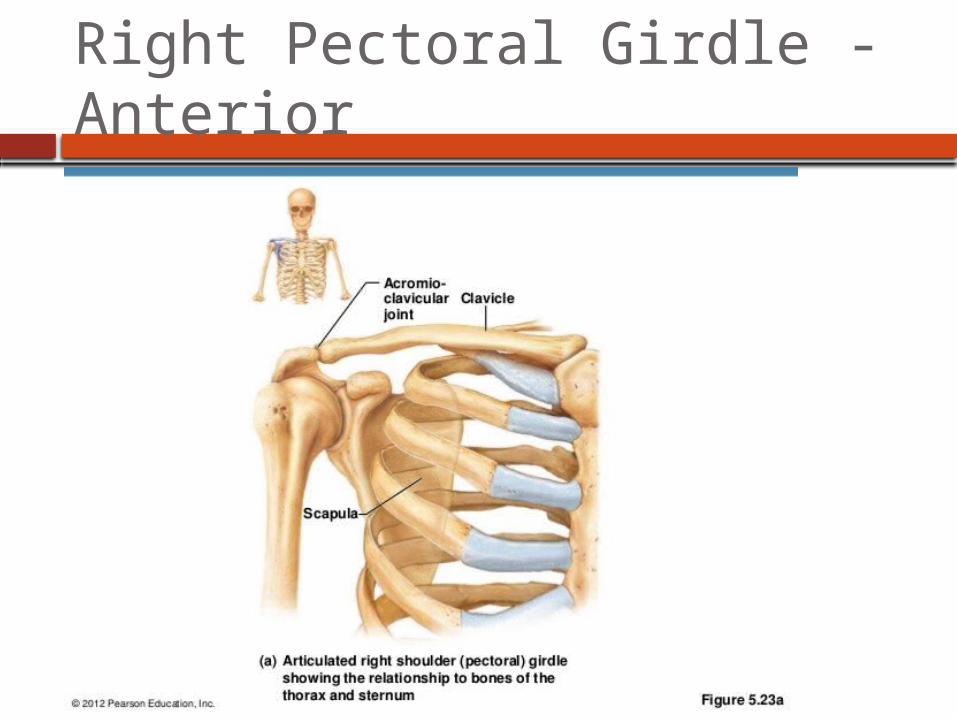
Right Pectoral Girdle - Anterior

Right Pectoral Girdle -Posterior

The Clavicle
Aka Collarbone Slender, doubly curved bone Acts as a brace to hold the
arm away from the thoracic cage
Helps prevent shoulder dislocation
Structures to know: Acromioclavicular joint and
sternoclavicular joint
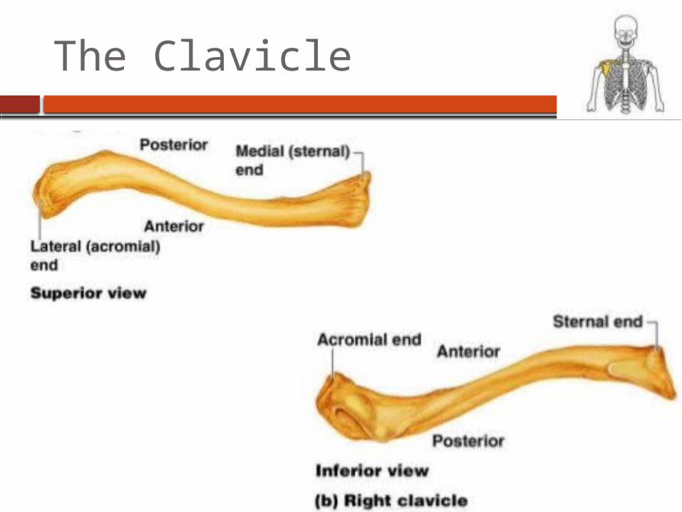
The Clavicle
The Scapula
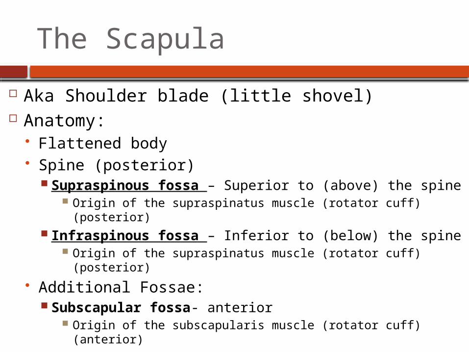
Aka Shoulder blade (little shovel) Anatomy:
Flattened body Spine (posterior)
Supraspinous fossa – Superior to (above) the spine Origin of the supraspinatus muscle (rotator cuff)
(posterior) Infraspinous fossa – Inferior to (below) the spine
Origin of the supraspinatus muscle (rotator cuff) (posterior)
Additional Fossae: Subscapular fossa- anterior
Origin of the subscapularis muscle (rotator cuff) (anterior)
The Scapula
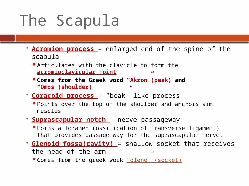
Acromion process = enlarged end of the spine of the scapula Articulates with the clavicle to form the acromioclavicular
joint Comes from the Greek word “Akron”(peak) and
“Omos”(shoulder) Coracoid process = “beak”-like process
Points over the top of the shoulder and anchors arm muscles Suprascapular notch = nerve passageway
Forms a foramen (ossification of transverse ligament) that provides passage way for the suprascapular nerve.
Glenoid fossa(cavity) = shallow socket that receives the head of the arm Comes from the greek work “glene” (socket)
The Scapula
Borders: Lateral (axillary) border Medial (vertebral) border Superior border
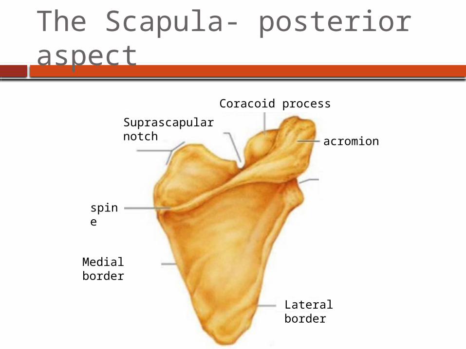
The Scapula- posterior aspect
Medial border
Lateral border
spine
Suprascapular notch
Coracoid process
acromion
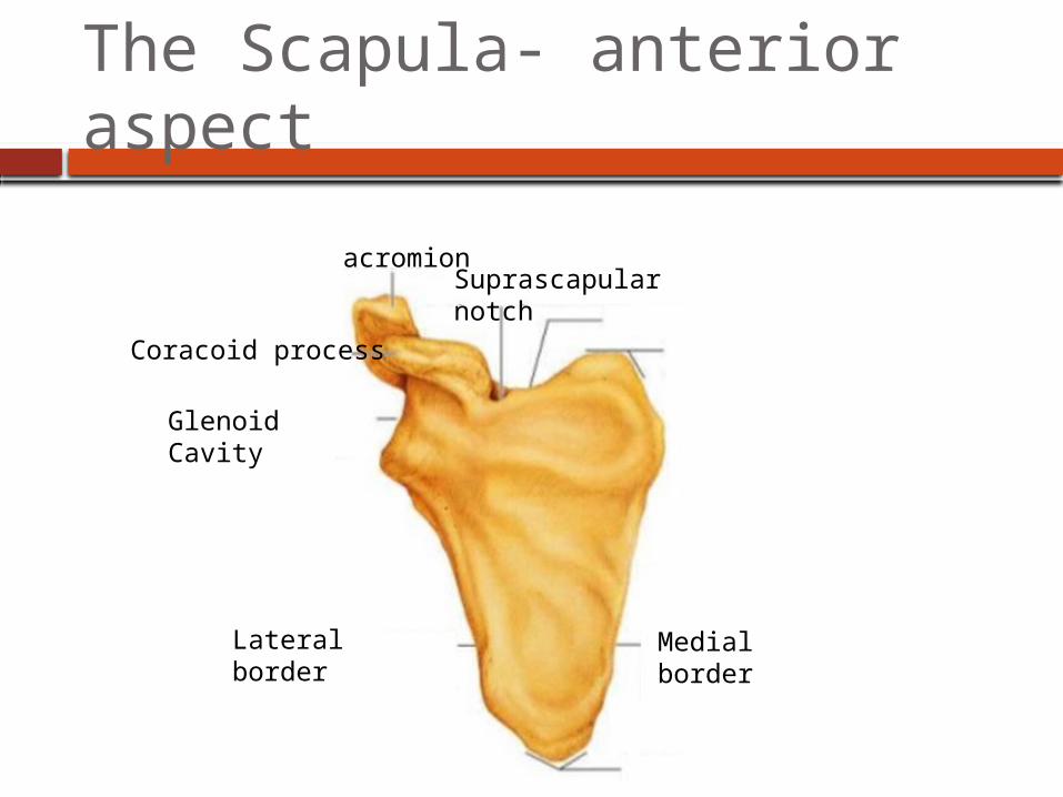
The Scapula- anterior aspect
Medial border
Lateral border
Glenoid Cavity
Suprascapular notch
Coracoid process
acromion
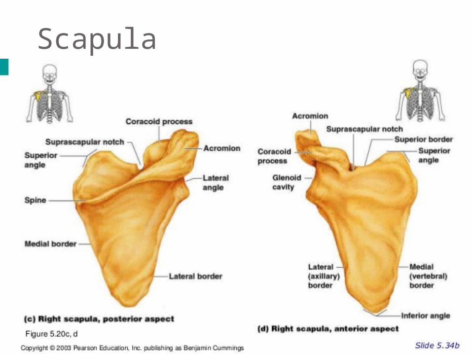
Scapula
THE UPPER LIMB

Bones of the Upper limbs
30 separate bones in each upper limb
Arm, foreharm, hand
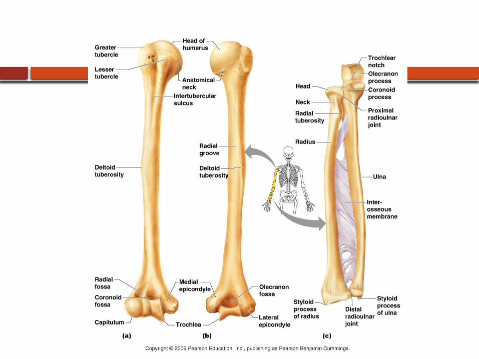
The arm
The humerus – single typical long bone of the upper arm Articulates proximally with scapula and clavicle and distally
with radius and ulna Proximal features:
Head – fits into glenoid cavity of scapula Greater and lesser tubercles – two bony projections lateral
to the head Separated by the intertubercular sulcus Attachment of tendons
Anatomical neck – slight constriction just inferior to the head Surgical neck – most frequently fractured part of the humerus
Features of the diaphysis:
Features of the diaphysis (shaft): Deltoid tuberosity: attachment for the
deltoid(shoulder) muscle Radial groove- marks the course of the
radial nerve

The arm
Distal features: Lateral and medial epicondyles: External and internal condyles: Olecranon fossa: groove that receives the
olecranon process of the ulna upon extension of the arm.
Coronoid fossa: groove that receives the coronoid process of the ulna upon flexion of the arm.
Trochlea (medial) articulates with trochlear notch of the ulna.
Capitulum (lateral) articulates with head of the radius
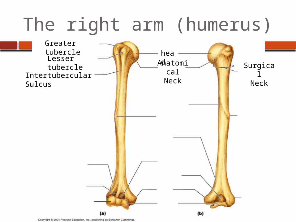
The right arm (humerus)head
Greater tubercleLesser tubercle
Intertubercular Sulcus
Anatomical
Neck
SurgicalNeck
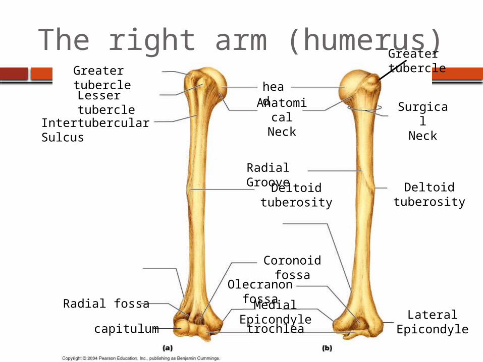
The right arm (humerus)head
Greater tubercleLesser tubercle
Intertubercular Sulcus
Anatomical
Neck
SurgicalNeck
Deltoid tuberosity
Deltoid tuberosity
Radial Groove
trochleacapitulum
Coronoid fossa
Olecranon fossaMedial Epicondyle Lateral
Epicondyle
Radial fossa
Greater tubercle
The forearm
Consists of two bones1. Radius = lateral bone when in anatomical position2. Ulna = medial bone when in anatomical position
Radioulnar joints = sight of articulation of radius and ulna
Two bones are connected along their entire length by interosseous membrane
Structures to know: radial tuberosity, styloid process, coronoid process, olecranon process, trochlear notch
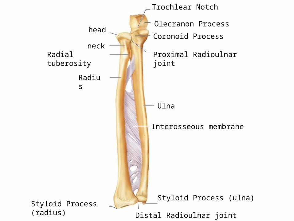
Radius
Ulna
Interosseous membrane
Trochlear Notch
Olecranon Process
Coronoid Process
Proximal Radioulnar joint
Distal Radioulnar joint
Styloid Process (ulna)Styloid Process (radius)
Radial tuberosity
neck
head
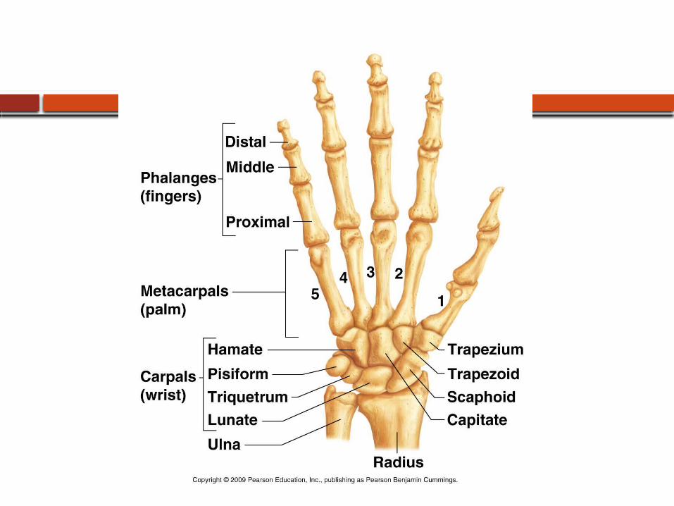
The Hand
Wrist (carpals) = 8 short bones
Palm (metacarpals) = 5 long bones
Fingers (phalanges)= long bones Thumb has 2 phalanges Each finger has 3 phalanges
Proximal phalange Medial phalange Distal phalange
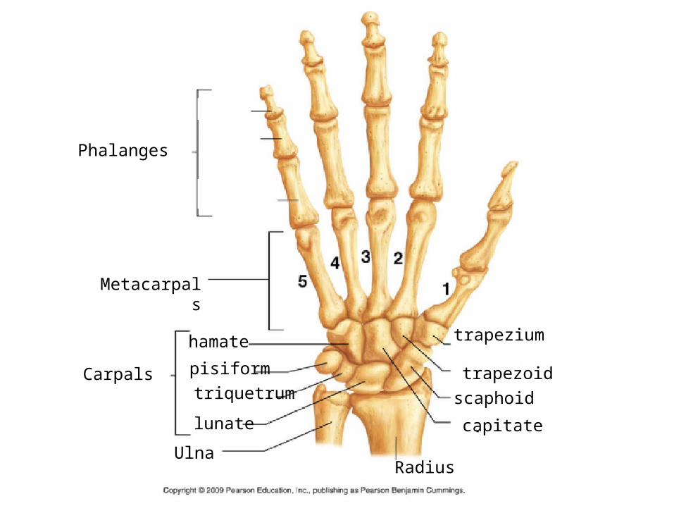
Phalanges
Metacarpals
Carpals
UlnaRadius
trapezium
trapezoid
scaphoid
capitate
hamate
pisiform
triquetrum
lunate
Can you identify the following??1.
8.
3.
4.
5.
6.
2.
7.