WHO Infection Control Guidelines for Transmissible Spongiform
43
WHO/CDS/CSR/APH/2000.3 WHO Infection Control Guidelines for Transmissible Spongiform Encephalopathies Report of a WHO consultation Geneva, Switzerland, 23-26 March 1999 World Health Organization Communicable Disease Surveillance and Control This document has been downloaded from the WHO/EMC Web site. The original cover pages and lists of participants are not included. See http://www.who.int/emc for more information.
Transcript of WHO Infection Control Guidelines for Transmissible Spongiform
WHO/CDS/CSR/APH/2000.3
WHO Infection Control Guidelines for TransmissibleSpongiform Encephalopathies
Report of a WHO consultationGeneva, Switzerland, 23-26 March 1999
World Health OrganizationCommunicable Disease Surveillance and Control
This document has been downloaded from the WHO/EMC Web site. Theoriginal cover pages and lists of participants are not included. Seehttp://www.who.int/emc for more information.
© World Health OrganizationThis document is not a formal publication of the World Health Organization(WHO), and all rights are reserved by the Organization. The document may,however, be freely reviewed, abstracted, reproduced and translated, in part orin whole, but not for sale nor for use in conjunction with commercial purposes.
The views expressed in documents by named authors are solely theresponsibility of those authors. The mention of specific companies or specificmanufacturers' products does no imply that they are endorsed orrecommended by the World Health Organization in preference to others of asimilar nature that are not mentioned.

Contents
SECTION 1. INTRODUCTION........................................................................................................... 1
SECTION 2. GENERAL CONSIDERATIONS .................................................................................. 1
2.1 TRANSMISSIBLE SPONGIFORM ENCEPHALOPATHIES IN HUMANS AND IN ANIMALS......................... 12.2 DIAGNOSIS OF HUMAN TRANSMISSIBLE SPONGIFORM ENCEPHALOPATHIES .................................. 22.3 IATROGENIC TRANSMISSION ........................................................................................................... 22.4 EVALUATING RISK IN HEALTHCARE ENVIRONMENTS...................................................................... 3
SECTION 3. PATIENT CARE .................................................................................................................. 5
3.1 CARE OF PATIENTS IN THE HOME AND HEALTHCARE SETTINGS ...................................................... 53.2 DENTAL PROCEDURES .................................................................................................................... 63.3 DIAGNOSTIC PROCEDURES ............................................................................................................. 63.4 SURGICAL PROCEDURES ................................................................................................................. 73.5 HANDLING OF SURGICAL INSTRUMENTS ........................................................................................ 83.6 ANAESTHESIA................................................................................................................................. 93.7 PREGNANCY AND CHILDBIRTH ....................................................................................................... 9
SECTION 4. OCCUPATIONAL INJURY......................................................................................... 10
4.1 OCCUPATIONAL EXPOSURE .......................................................................................................... 104.2 POST-EXPOSURE MANAGEMENT ................................................................................................... 10
SECTION 5. LABORATORY INVESTIGATIONS........................................................................... 10
5.1 SAFETY IN THE HEALTHCARE LABORATORY................................................................................. 105.2 CLINICAL DIAGNOSTIC LABORATORIES ........................................................................................ 115.3 SURGICAL PATHOLOGY ................................................................................................................ 125.4 TRANSPORT OF SPECIMENS BY AIR ............................................................................................... 13
SECTION 6. DECONTAMINATION PROCEDURES .................................................................... 13
6.1 GENERAL CONSIDERATIONS ......................................................................................................... 136.2 DECONTAMINATION OF INSTRUMENTS ......................................................................................... 146.3 DECONTAMINATION OF WORK SURFACES..................................................................................... 156.4 DECONTAMINATION OF WASTES AND WASTE-CONTAMINATED MATERIALS.................................. 156.5 PERSONAL PROTECTION DURING DECONTAMINATION PROCEDURES............................................. 156.6 DECONTAMINATION RISK CATEGORIES......................................................................................... 16
SECTION 7. WASTE DISPOSAL ..................................................................................................... 16
SECTION 8. AFTER DEATH ........................................................................................................... 17
8.1 PRECAUTIONS FOR HANDLING OF THE DECEASED PATIENT........................................................... 178.2 POST MORTEM EXAMINATION....................................................................................................... 188.3 NATIONAL AND INTERNATIONAL TRANSPORT OF BODIES ............................................................. 198.4 UNDERTAKERS AND EMBALMERS................................................................................................. 208.5 FUNERALS AND CREMATIONS....................................................................................................... 208.6 EXHUMATIONS ............................................................................................................................. 218.7 BODY DONATION FOR TEACHING PURPOSES ................................................................................. 21

ANNEX I LIST OF PARTICIPANTS........................................................................................... 23
ANNEX II LIST OF PRESENTATIONS........................................................................................29
ANNEX III DECONTAMINATION METHODS FOR TRANSMISSIBLE SPONGIFORMENCEPHALOPATHIES.............................................................................................. 29
ANNEX IV MANAGEMENT OF HEALTHY ‘AT RISK’ INDIVIDUALS .................................. 33
ANNEX V MANAGEMENT OF INDIVIDUALS WITH CONFIRMED OR SUSPECTEDVARIANT CREUTZFELDT-JAKOB DISEASE ........................................................ 35

WHO/CDS/CSR/APH/2000.3
WHO Infection Control Guidelines for Transmissible Spongiform Encephalopathies 1
Section 1. INTRODUCTIONTransmissible spongiform encephalopathies (TSEs), also known as prion diseases,
are fatal degenerative brain diseases that occur in humans and certain animal species.They are characterized by microscopic vacuoles and the deposition of amyloid (prion)protein in the grey matter of the brain. All forms of TSE are experimentallytransmissible.
The following guideline on the prevention of iatrogenic and nosocomial exposure toTSE agents was prepared following the WHO Consultation on Caring for Patients andHospital Infection Control in Relation to Human Transmissible SpongiformEncephalopathies, held in Geneva from 24 to 26 March 1999. The meeting was chairedby Dr Paul Brown. Dr Martin Zeidler and Dr Maurizio Pocchiari kindly agreed to beRapporteurs. The full list of participants is given in Annex I. Presentations made at theConsultation are listed in Annex II.
This document provides guidance upon which infection control practitioners,healthcare practitioners, medical officers of health, and those involved in the care ofpersons suffering from TSE can base their care and infection control practices, to preventevents which are either extremely rare (e.g. transmission of TSE through a surgicalprocedure) or hypothetical (e.g. transmission of TSE to a healthcare worker or familymember). Throughout the document there is specific and assumed reference to countryor region-specific guidelines for matters which lie within the legal jurisdiction of thatcountry or region, i.e. International Air Transport Association (IATA) regulations fortransportation of hazardous goods, or bio-safety containment levels for laboratories.Readers should be familiar with such requirements for their own country or region.
Issues on which the consultants could not agree, or where the consultantsdid not feel there was sufficient expertise to render an opinion, have been noted.The consultation recognized that its recommendations to ensure maximum safety to care-givers and the environment may under some circumstances be regarded as impractical.However, they urged personnel involved with TSE patients or tissues to endeavour tocomply as far as possible. There is no reason for a patient with a TSE to be denied anyprocedure, as any associated risks should be reduced to negligible levels by following therecommendations in this document.
Section 2. GENERAL CONSIDERATIONS
2.1 Transmissible Spongiform Encephalopathies in humans and in animalsHuman TSEs occur in sporadic, familial, and acquired forms. The most common
form, sporadic Creutzfeldt-Jakob disease (CJD), has a worldwide death rate of about1 case per million people each year, and typically affects people between 55 and 75 yearsof age. The disease usually begins with a progressive mental deterioration that soonbecomes associated with progressive unsteadiness and clumsiness, visual deterioration,muscle twitching (myoclonus), a variety of other neurological symptoms and signs, andis often associated with a characteristic periodic electroencephalogram. The patient isusually mute and immobile in the terminal stages and in most cases, death occurs withina few months of onset of symptoms. TSEs are invariably fatal and there is no proventreatment or prophylaxis.
WHO/CDS/CSR/APH/2000.3
2 WHO Infection Control Guidelines for Transmissible Spongiform Encephalopathies
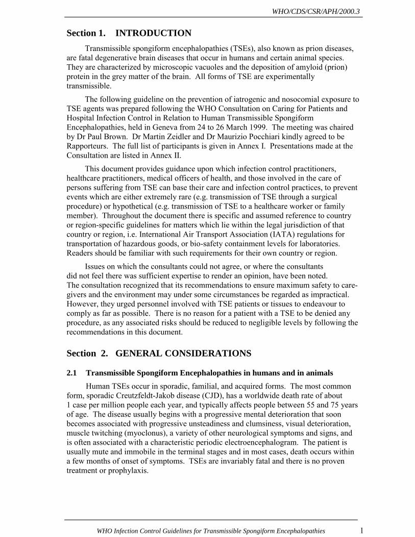
Table 1 Human TSEs
Human TSE First Reported
Creutzfeldt-Jakob Disease (CJD):1 Sporadic (85-90%) Familial (5-10%) Iatrogenic (<5%) Variant (vCJD)
1921192419741996
Gerstmann-Sträussler-Scheinker Syndrome (GSS) 1936
Kuru 1957
Fatal Insomnia Familial Sporadic
19861999
Similar neurodegenerative diseases also occur naturally in some animal species(scrapie in sheep and goats, chronic wasting disease in deer and elk), or as a result ofexposure of susceptible species to infected animal tissues (transmissible minkencephalopathy, bovine spongiform encephalopathy, and spongiform encephalopathy indomestic cats and a variety of captive zoo animals).
TSE agents exhibit an unusual resistance to conventional chemical and physicaldecontamination methods. They are not adequately inactivated by most commondisinfectants, or by most tissue fixatives, and some infectivity may persist under standardhospital or healthcare facility autoclaving conditions (e.g. 121°C for 15 minutes). Theyare also extremely resistant to high doses of ionizing and ultra-violet irradiation and someresidual activity has been shown to survive for long periods in the environment. Theunconventional nature of these agents, together with the appearance in the UnitedKingdom, Republic of Ireland and France of a new variant of CJD (vCJD) since the mid-1990s, has stimulated interest in an updated guidance on safe practices for patient careand infection control.
2.2 Diagnosis of Human Transmissible Spongiform EncephalopathiesThe February 1998 Report of a WHO Consultation the Global Surveillance,
Diagnosis and Therapy of Human Transmissible Spongiform Encephalopathies2,3
provides a guideline for diagnostic criteria of human TSEs. Readers should be aware ofefforts to revise diagnostic criteria for CJD and vCJD due to the introduction of newdiagnostic tests and intense surveillance efforts. Surveillance case definitions (whichmay not be the same as diagnostic criteria) for both forms of the disease may also besubject to change.
2.3 Iatrogenic transmissionTSEs are not known to spread by contact from person to person, but transmission
can occur during invasive medical interventions. Exposure to infectious material throughthe use of human cadaveric-derived pituitary hormones, dural and cornea homografts,and contaminated neurosurgical instruments has caused human TSEs. The Report of a
1 Percentages vary somewhat from country to country.2 All cited WHO reports and consultations are available at the WHO Web site
http://www.who.int/emc/diseases/bse/.3 WHO Consultation on Global Surveillance, Diagnosis and Therapy of Human Transmissible.
Spongiform Encephalopathies. WHO/EMC/ZDI/98.9 Geneva, 9-11 February 1998.

WHO/CDS/CSR/APH/2000.3
WHO Infection Control Guidelines for Transmissible Spongiform Encephalopathies 3
WHO Consultation on Medicinal and other Products in Relation to Human and AnimalTransmissible Spongiform Encephalopathies4 can be consulted for more information andguidance on these issues.
2.4 Evaluating risk in healthcare environmentsWhen considering measures to prevent the transmission of TSE from patients to
other individuals (patients, healthcare workers, or other care providers), it is important tounderstand the basis for stipulating different categories of risk. Risk is dependent uponthree considerations:
- the probability that an individual has or will develop TSE (see Section 2.4.1);- the level of infectivity in tissues or fluids of these individuals (Section 2.4.2);- the nature or route of the exposure to these tissues (Section 2.4.3).
From these considerations it is possible to make decisions about whether anyspecial precautions are needed. Specific TSE decontamination procedures are describedin Section 6. If TSE decontamination is required, the question remains as to howstringent it should be. The specific recommendations are described in sections devoted toPatient Care (Section 3), Occupational Injury (Section 4), Laboratory Investigations(Section 5) and Management After Death (Section 8).
2.4.1 Identification of persons for whom special precautions apply
Persons with confirmed or suspected TSEs are the highest risk patients. They mustbe managed using specific precautions which will be described in this and subsequentsections. All precautions recommended in the body of this document apply to the care ofconfirmed or suspect cases of TSE, or the handling of tissues from such patients, andunless otherwise noted, no distinction will be made between confirmed and suspect cases.
However, the concept of �persons at risk for TSE� is useful in infection control, asit allows for the development of intermediate precautionary measures. The followingpersons have been regarded as �at risk� for developing TSEs. The bracketed numbers arethe number of reported occurrences of CJD transmitted through that route:
• recipients of dura mater (110 cases);• recipients of human cadaver derived pituitary hormones, especially human cadaver
derived growth hormone (130 cases);• recipients of cornea transplants (3 cases - 1 definite, 1 probable, 1 possible);• persons who have undergone neurosurgery (6);• members of families with heritable TSE (5-10% of all cases of TSE are heritable,
but the number of families varies widely from country to country).
The discussion and recommendations for healthy asymptomatic individualsconsidered to be at risk for TSE are described in Annex IV and referred to in Table 9.
The consultants did not extensively discuss the management of persons who haveconfirmed or suspected vCJD, due to the absence of specific data for review and the
4 Report of a WHO Consultation on Medicinal and other Products in Relation to Human and Animal
Transmissible Spongiform Encepalopathies. Geneva, World Health Organization, 1997.WHO/EMC/ZOO/97.3 or WHO/BLG/97.2.
WHO/CDS/CSR/APH/2000.3
4 WHO Infection Control Guidelines for Transmissible Spongiform Encephalopathies
geographical isolation of the current cases. The discussion and their recommendationsare described in Annex V and Table 9.
2.4.2 Tissue infectivity
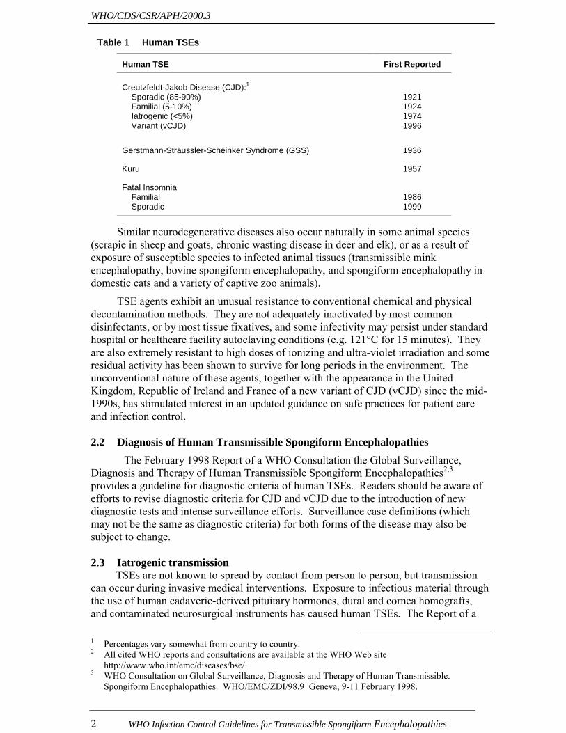
From published and unpublished information, infectivity is found most often and inhighest concentration in the central nervous system (CNS), specifically the brain, spinalcord and eye. This document will refer to these tissues as �high infectivity tissues�.
Infectivity is found less often in the cerebrospinal fluid (CSF) and several organsoutside the CNS (lung, liver, kidney, spleen/lymph nodes, and placenta). This documentwill refer to these tissues as �low infectivity tissues�.
No infectivity has been detected in a wide variety of other tested tissues (heart,skeletal muscle, peripheral nerve, adipose tissue, gingival tissue, intestine, adrenal gland,thyroid, prostate, testis) or in bodily secretions or excretions (urine, faeces, saliva,mucous, semen, milk, tears, sweat, serous exudate). Experimental results investigatingthe infectivity of blood have been conflicting, however even when infectivity has beendetectable, it is present in very low amounts and there are no known transfusiontransmissions of CJD. This document will classify these tissues as having no detectableinfectivity (�no detectable infectivity tissues�) and, for the purposes of infection control,they will be regarded as non-infectious.
Table 2 Distribution of infectivity in the human body5
Infectivity Category Tissues, Secretions, and Excretions
High Infectivity BrainSpinal cordEye
Low Infectivity CSFKidneyLiverLungLymph nodes/spleenPlacenta
No Detectable Infectivity Adipose tissueAdrenal glandGingival tissueHeart muscleIntestinePeripheral nerveProstateSkeletal muscleTestisThyroid gland
Blood6
TearsNasal mucousSalivaSweatSerous exudateMilkSemenUrineFaeces
5 Assignment of different organs and tissues to categories of high and low infectivity is chiefly based
upon the frequency with which infectivity has been detectable, rather than upon quantitative assays ofthe level of infectivity, for which data are incomplete. Experimental data include primates inoculatedwith tissues from human cases of CJD, but have been supplemented in some categories by data obtainedfrom naturally occurring animal TSEs. Actual infectivity titres in the various human tissues other thanthe brain are extremely limited, but data from experimentally-infected animals generally corroborate thegrouping shown in the table.
6 See discussion this Section and Section 5.2.

WHO/CDS/CSR/APH/2000.3
WHO Infection Control Guidelines for Transmissible Spongiform Encephalopathies 5
The consultants agreed that an international effort to identify stored tissues frompersons who later developed CJD or that were collected during the investigation for CJD(sporadic, iatrogenic or familial) should be initiated. These specimens should be tested inorder to clarify the extent and level of infectivity during the pre-clinical phase of disease.Collections of these tissues, which are potentially infective, should be properly labelledas to their source and potential infectivity and appropriately stored to avoid crosscontamination.
2.4.3 Route of exposure
When determining risk, infectivity of a tissue must be considered together with theroute of exposure. Cutaneous exposure of intact skin or mucous membranes (exceptthose of the eye) poses negligible risk; however, it is prudent and highly recommended toavoid such exposure when working with any high infectivity tissue. Transcutaneousexposures, including contact exposures to non-intact skin or mucous membranes,7splashes to the eye,8 and inoculations via needle9,10 or scalpel and other surgicalinstruments11 pose a greater potential risk. Thus, it is prudent to avoid these types ofexposures when working with either low infectivity or high infectivity tissues. CNSexposures (i.e. inoculation of the eye or CNS) with any infectious material poses a veryserious risk, and appropriate precautions must always be taken to avoid these kinds ofexposures.
Section 3. PATIENT CARE3.1 Care of patients in the home and healthcare settings3.1.1 Patient care
Normal social and clinical contact, and non-invasive clinical investigations(e.g. x-ray imaging procedures) with TSE patients do not present a risk to healthcareworkers, relatives, or the community. There is no reason to defer, deny, or in any waydiscourage the admission of a person with a TSE into any healthcare setting. Based oncurrent knowledge, isolation of patients is not necessary; they can be nursed in the openward using Standard Precautions.
As the disease is usually rapidly progressive, the patient will develop highdependency needs and require ongoing assessment. It is essential to address the physical,nutritional, psychological, educational, and social needs of the patient and the associatedneeds of his or her family. Co-ordinated planning is vital in transferring care from oneenvironment to another.
Private room nursing care is not required for infection control, but may beappropriate for compassionate reasons. Patient waste should be handled according tocountry, regional or federal regulations. Contamination by body fluids (categorized as nodetectable infectivity tissues) poses no greater hazard than for any other patient. Nospecial precautions are required for feeding utensils, feeding tubes, suction tubes, bed
7 TSE can be experimentally transmitted to healthy animals by exposing abraded gingival tissue to
infected brain homogenate.8 By analogy with cornea transplants.9 A documented route of transmission in humans, from contaminated human cadaver extracted pituitary
hormones (hGH and gonadotropin).10 Intraperitoneal, intramuscular and intravenous administration of low infectivity tissue extracts can cause
transmission of TSE in experimental animals.11 By analogy with transmissions following neurosurgical procedures.
WHO/CDS/CSR/APH/2000.3
6 WHO Infection Control Guidelines for Transmissible Spongiform Encephalopathies
linens, or items used in skin or bed sore care in the home environment. Section 7provides detailed information on disposal of medical waste.3.1.2 Psychiatric manifestations
Caregivers both in the home and healthcare setting should be made aware andanticipate the possibility of labile psychiatric symptoms e.g. mood swings, hallucinations,or aggressive behavior. For this reason, training and counselling of professional and non-professional caregivers is recommended.
3.1.3 Confidentiality
Current heightened awareness requires special sensitivity to confidentiality ofwritten and verbal communications. Special measures to safeguard the privacy of thepatient and family are essential.
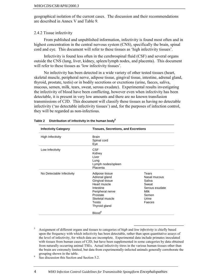
3.2 Dental proceduresAlthough epidemiological investigation has not revealed any evidence that dental
procedures lead to increased risk of iatrogenic transmission of TSEs among humans,experimental studies have demonstrated that animals infected by intraperitonealinoculation develop a significant level of infectivity in gingival and dental pulp tissues,and that TSEs can be transmitted to healthy animals by exposing root canals and gingivalabrasions to infectious brain homogenate. The consultants agreed that the generalinfection control practices recommended by national dental associations are sufficientwhen treating TSE patients during procedures not involving neurovascular tissue. Thecommittee was unable to come to a consensus on the risk of transmission of TSEsthrough major dental procedures; therefore, extra precautions such as those listed inTable 3 have been provided for consideration without recommendation.
Table 3 Optional precautions for major dental work
1.
2.
3.
Use single-use items and equipment e.g. needles and anaesthetic cartridges.
Re-usable dental broaches and burrs that may have become contaminated withneurovascular tissue should either be destroyed after use (by incineration) or alternativelydecontaminated by a method listed in Section 6 (Annex and III).
Schedule procedures involving neurovascular tissue at end of day to permit more extensivecleaning and decontamination.
3.3 Diagnostic proceduresDuring the earlier stages of disease, patients with TSE who develop intercurrent
illnesses may need to undergo the same kinds of diagnostic procedures as any otherhospitalized patient. These could include ophthalmoscopic examinations, various typesof endoscopy, vascular or urinary catheterization, and cardiac or pulmonary functiontests. In general, these procedures may be conducted without any special precautions, asmost tissues with which the instruments come in contact contain no detectable infectivity(see sub-Section 2.4.2). A conservative approach would nevertheless try to schedulesuch patients at the end of the day to allow more strict environmental decontamination(see Section 6.3) and instrument cleaning (see Section 6.2). When there is knownexposure to high or low infectivity tissues, the instruments should be subjected to thestrictest form of decontamination procedure which can be tolerated by the instrument.Instrument decontamination is discussed in more detail in Section 6.2 anddecontamination methods are specifically described in Annex III.

WHO/CDS/CSR/APH/2000.3
WHO Infection Control Guidelines for Transmissible Spongiform Encephalopathies 7
3.4 Surgical proceduresBefore admission to a hospital or healthcare facility, the infection control team
should be informed of the intention to perform a surgical procedure on any person withconfirmed or suspected TSE. Every effort should be made to plan carefully not only theprocedure, but also the practicalities surrounding the procedure, e.g. instrument handling,storage, cleaning and decontamination or disposal. Written protocols are essential. Allstaff directly involved in these procedures or in the subsequent re-processing or disposalof potentially contaminated items, should be aware of the recommended precautions, andbe adequately trained. The staff should be made aware of any such procedures insufficient time to allow them to plan and to obtain suitable instruments and equipment(such as single use items), and it may be useful to schedule the patient at the end of theday�s operating list. Staff must adhere to protocols that identify specifics regarding pre-operative, peri-operative and post-operative management of the patient, disposablematerials, including bandages and sponges, and re-usable materials. Ancillary staff, suchas laboratory and central instrument cleaning personnel, must be informed andappropriate training provided.
Basic protective measures are described in Table 4. Recommendations listed inSection 6 and Annex III for decontamination of equipment and environment, and inSection 7 for disposal of infectious waste should be followed. Supervisors should beresponsible for ensuring that the appropriate procedures are followed and that effectivemanagement systems are in place.
Table 4 Precautions for surgical procedures
Wherever appropriate and possible, the intervention should:
1. be performed in an operating theatre;
2. involve the minimum required number of healthcare personnel;
3. use single-use equipment as follows: i) liquid repellent operating theatre gown, over a plastic apron ii) gloves iii) mask iv) visor or goggles v) linens and covers;
4. mask all non-disposable equipment;
5. maintain one-way flow of instruments;
6. treat all protective clothing, covers, liquid and solid waste by a method listed in Section 6;and Annex III; incineration is preferred
7. mark samples with a “Biohazard” label;
8. clean all surfaces according to recommendations specified in Section 6 and Annex III.
Procedures which are normally carried out at the bedside (e.g. lumbar puncture,bone marrow biopsy) may be performed at the bedside, but care should be taken to ensureease of environmental decontamination should a spillage occur.

WHO/CDS/CSR/APH/2000.3
8 WHO Infection Control Guidelines for Transmissible Spongiform Encephalopathies
3.5 Handling of surgical instruments3.5.1 General measures
Methods for instrument decontamination are fully discussed in Section 6.Determination of which method to use is based upon the infectivity level of the tissue andthe way in which instruments will subsequently be re-used. For example, where surgicalinstruments contact high infectivity tissues, single-use surgical instruments are stronglyrecommended. If single-use instruments are not available, maximum safety is attainedby destruction of re-usable instruments. Where destruction is not practical, re-usableinstruments must be handled as per Table 5 and must be decontaminated as per Section 6and Annex III.
Although CSF is classified as a low infectivity tissue and is less infectious thanhigh infectivity tissues it was felt that instruments contaminated by CSF should behandled in the same manner as those contacting high infectivity tissues. This exceptionreflects the higher risk of transmission to any person on whom the instruments would bere-used for the procedure of lumbar puncture.
Table 5 General measures for cleaning instruments and environment
1. Instruments should be kept moist until cleaned and decontaminated.
2. Instruments should be cleaned as soon as possible after use to minimize drying of tissues,blood and body fluids onto the item.
3. Avoid mixing instruments used on no detectable infectivity tissues with those used on high andlow infectivity tissues.
4. Recycle durable items for re-use only after TSE decontamination by methods found in Section6 and Annex III.
5. Instruments to be cleaned in automated mechanical processors must be decontaminated bymethods described in Section 6 and Annex III before processing through these machines, andthe washers (or other equipment) should be run through an empty cycle before any furtherroutine use.
6. Cover work surfaces with disposable material, which can then be removed and incinerated;otherwise clean and decontaminate underlying surfaces thoroughly using recommendeddecontamination procedures in Section 6 and Annex III.
7. Be familiar with and observe safety guidelines when working with hazardous chemicals suchas sodium hydroxide (NaOH, ‘soda lye’) and sodium hypochlorite (NaOCl, ‘bleach’) (see AnnexIII for definitions).
8. Observe manufacturers’ recommendations regarding care and maintenance of equipment.
Those instruments used for invasive procedures on TSE patients (i.e. used on highor low infectivity tissues) should be securely contained in a robust, leak-proof containerlabelled �Biohazard�. They should be transferred to the sterilization department as soonas possible after use, and treated by a method listed in Annex III, or transferred to theincinerator as per Section 3.5.2. A designated person who is familiar with this guidelineshould be responsible for the transfer and subsequent management.
The consultation did not address the issue of post-exposure notification in the eventthat an instrument used on a high-risk tissue and/or high-risk patient was subsequently re-used without adequate decontamination.
WHO/CDS/CSR/APH/2000.3
WHO Infection Control Guidelines for Transmissible Spongiform Encephalopathies 9
3.5.2 Destruction of surgical instruments
Items for disposal by incineration should be isolated in a rigid clinical wastecontainer, labelled �Hazardous� and transported to the incinerator as soon as practicable,in line with the current disposal of clinical waste guidance described in the Teacher�sGuide: Management of Wastes from Health-care Facilities12 published by WHO. Toavoid unnecessary destruction of instruments, quarantine of instruments whiledetermining the final diagnosis of persons suspected of TSEs may be used.
3.5.3 Quarantine
If a facility can safely quarantine instruments until a diagnosis is confirmed,quarantine can be used to avoid needless destruction of instruments when suspect casesare later found not to have a TSE. Items for quarantine should be cleaned by the bestnon-destructive method as per Section 6 and Annex III, sterilized, packed, date and�Hazard� labelled, and stored in specially marked rigid sealed containers.13 Monitoringand ensuring maintenance of quarantine is essential to avoid accidental re-introduction ofthese instruments into the circulating instrument pool. If TSE is excluded as a diagnosis,the instruments may be returned to circulation after appropriate sterilization.
3.6 Anaesthesia3.6.1 General anaesthesia
TSEs are not transmissible by the respiratory route; however, it is prudent to treatany instruments in direct contact with mouth, pharynx, tonsils and respiratory tract by amethod described in Annex III. Destruction by incineration of non re-usable equipmentis recommended.
3.6.2 Local anaesthesia
Needles should not be re-used, and in particular, needles contacting the CSF(e.g. for saddle blocks and other segmental anaesthetic procedures) must be discarded anddestroyed.
3.7 Pregnancy and childbirthTSE is not known to be transmitted from mother to child during pregnancy or
childbirth; familial disease is inherited as a result of genetic mutations. In the event that aperson with TSE becomes pregnant, no particular precautions need to be taken during thepregnancy, except during invasive procedures as per Section 3.4. Childbirth should bemanaged using standard infection control procedures, except that precautions should betaken to reduce the risk of exposure to placenta and any associated material and fluids.These should be disposed of by incineration. Instruments should be handled as for anyother clinical procedure (Table 5). In home deliveries, the midwife (or any other personsin charge of delivery) should ensure that any contaminated material is removed anddisposed of in accordance with correct procedures for infected clinical waste.
12 Pruess A, Townend WK. Teacher�s Guide: Management of Wastes from Health-care Activities.
Geneva, World Health Organization, 1998. WHO/EOS/98.6.13 Although the intention of quarantine is to avoid destruction of instruments and will permit the
re-introduction of instruments only if TSEs are not diagnosed, the use of a decontamination method forTSEs will confer additional safety should an instrument unintentionally come in contact with staff orpatients.

WHO/CDS/CSR/APH/2000.3
10 WHO Infection Control Guidelines for Transmissible Spongiform Encephalopathies
Section 4. OCCUPATIONAL INJURY4.1 Occupational exposure
Although there have been no confirmed cases of occupational transmission of TSEto humans, cases of CJD in healthcare workers have been reported in which a link tooccupational exposure is suggested. Therefore, it is prudent to take a precautionaryapproach. In the context of occupational exposure, the highest potential risk is fromexposure to high infectivity tissues through needle-stick injuries with inoculation;however exposure to either high or low infectivity tissues through direct inoculation(e.g. needle-sticks, puncture wounds, �sharps� injuries, or contamination of broken skin)must be avoided. Exposure by splashing of the mucous membranes (notably theconjunctiva) or unintentional ingestion may be considered a hypothetical risk and mustalso be avoided. Healthcare personnel who work with patients with confirmed orsuspected TSEs, or with their high or low infectivity tissues, should be appropriatelyinformed about the nature of the hazard, relevant safety procedures, and the high level ofsafety which will be provided by the proposed procedures described throughout thisdocument.
4.2 Post-exposure managementAppropriate counselling should include the fact that no case of human TSE is
known to have occurred through occupational accident or injury. A number of strategiesto minimize the theoretical risk of infection following accidents have been proposed, buttheir usefulness is untested and unknown. For the present the following common-senseactions are recommended:
• Contamination of unbroken skin with internal body fluids or tissues: wash withdetergent and abundant quantities of warm water (avoid scrubbing), rinse, and dry.Brief exposure (l minute, to 0.1N NaOH or a 1: 10 dilution of bleach) can beconsidered for maximum safety.
• Needle sticks or lacerations: gently encourage bleeding; wash (avoid scrubbing)with warm soapy water, rinse, dry and cover with a waterproof dressing. Furthertreatment (e.g., sutures) should be appropriate to the type of injury. Report theinjury according to normal procedures for your hospital or healthcarefacility/laboratory.
• Splashes into the eye or mouth: irrigate with either saline (eye) or tap water(mouth); report according to normal procedures for your hospital or healthcarefacility/laboratory.
• Health and safety guidelines mandate reporting of injuries, and records should bekept for no less than 20 years.
Section 5. LABORATORY INVESTIGATIONS5.1 Safety in the healthcare laboratory
Adherence to the following routine precautions during any diagnostic procedure orlaboratory work will reduce the risk of infection. General protective measures and basicprecautions as outlined in Table 6 are recommended for hospital-based diagnosticlaboratories as well as during decontamination procedures in those laboratories. Detaileddescriptions of these general protective measures can be found in the WHO document:
WHO/CDS/CSR/APH/2000.3
WHO Infection Control Guidelines for Transmissible Spongiform Encephalopathies 11
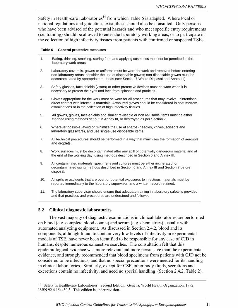
Safety in Health-care Laboratories14 from which Table 6 is adapted. Where local ornational regulations and guidelines exist, these should also be consulted. Only personswho have been advised of the potential hazards and who meet specific entry requirements(i.e. training) should be allowed to enter the laboratory working areas, or to participate inthe collection of high infectivity tissues from patients with confirmed or suspected TSEs.
Table 6 General protective measures
1. Eating, drinking, smoking, storing food and applying cosmetics must not be permitted in thelaboratory work areas.
2. Laboratory coveralls, gowns or uniforms must be worn for work and removed before enteringnon-laboratory areas; consider the use of disposable gowns; non-disposable gowns must bedecontaminated by appropriate methods (see Section 7 Waste Disposal and Annex III).
3. Safety glasses, face shields (visors) or other protective devices must be worn when it isnecessary to protect the eyes and face from splashes and particles.
4. Gloves appropriate for the work must be worn for all procedures that may involve unintentionaldirect contact with infectious materials. Armoured gloves should be considered in post mortemexaminations or in the collection of high infectivity tissues.
5. All gowns, gloves, face-shields and similar re-usable or non re-usable items must be either cleaned using methods set out in Annex III, or destroyed as per Section 7.
6. Wherever possible, avoid or minimize the use of sharps (needles, knives, scissors andlaboratory glassware), and use single-use disposable items.
7. All technical procedures should be performed in a way that minimizes the formation of aerosolsand droplets.
8. Work surfaces must be decontaminated after any spill of potentially dangerous material and atthe end of the working day, using methods described in Section 6 and Annex III.
9. All contaminated materials, specimens and cultures must be either incinerated, ordecontaminated using methods described in Section 6 and Annex III and Section 7 beforedisposal.
10. All spills or accidents that are overt or potential exposures to infectious materials must bereported immediately to the laboratory supervisor, and a written record retained.
11. The laboratory supervisor should ensure that adequate training in laboratory safety is providedand that practices and procedures are understood and followed.
5.2 Clinical diagnostic laboratoriesThe vast majority of diagnostic examinations in clinical laboratories are performed
on blood (e.g. complete blood counts) and serum (e.g. chemistries), usually withautomated analyzing equipment. As discussed in Section 2.4.2, blood and itscomponents, although found to contain very low levels of infectivity in experimentalmodels of TSE, have never been identified to be responsible for any case of CJD inhumans, despite numerous exhaustive searches. The consultation felt that thisepidemiological evidence was more relevant and more persuasive than the experimentalevidence, and strongly recommended that blood specimens from patients with CJD not beconsidered to be infectious, and that no special precautions were needed for its handlingin clinical laboratories. Similarly, except for CSF, other body fluids, secretions andexcretions contain no infectivity, and need no special handling (Section 2.4.2, Table 2).
14 Safety in Health-care Laboratories. Second Edition. Geneva, World Health Organization, 1992.ISBN 92 4 154450 3. This edition is under revision.
WHO/CDS/CSR/APH/2000.3
12 WHO Infection Control Guidelines for Transmissible Spongiform Encephalopathies
CSF may be infectious and must be handled with care. It is recommended thatanalysis not be performed in automated equipment, and any materials coming in contactwith the CSF must either be incinerated or decontaminated according to one of themethods listed in Section 6 and Annex III. There is no reason for a diagnostic test to bedenied if these measures are observed.
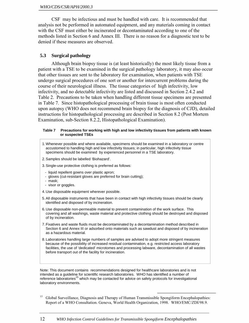
5.3 Surgical pathologyAlthough brain biopsy tissue is (at least historically) the most likely tissue from a
patient with a TSE to be examined in the surgical pathology laboratory, it may also occurthat other tissues are sent to the laboratory for examination, when patients with TSEundergo surgical procedures of one sort or another for intercurrent problems during thecourse of their neurological illness. The tissue categories of high infectivity, lowinfectivity, and no detectable infectivity are listed and discussed in Section 2.4.2 andTable 2. Precautions to be taken when handling different tissue specimens are presentedin Table 7. Since histopathological processing of brain tissue is most often conductedupon autopsy (WHO does not recommend brain biopsy for the diagnosis of CJD), detailedinstructions for histopathological processing are described in Section 8.2 (Post MortemExamination, sub-Section 8.2.2, Histopathological Examination).
Table 7 Precautions for working with high and low infectivity tissues from patients with knownor suspected TSEs
1. Whenever possible and where available, specimens should be examined in a laboratory or centreaccustomed to handling high and low infectivity tissues; in particular, high infectivity tissuespecimens should be examined by experienced personnel in a TSE laboratory.
2. Samples should be labelled ‘Biohazard’.
3. Single-use protective clothing is preferred as follows:
- liquid repellent gowns over plastic apron;- gloves (cut-resistant gloves are preferred for brain cutting);- mask;- visor or goggles.
4. Use disposable equipment wherever possible.
5. All disposable instruments that have been in contact with high infectivity tissues should be clearlyidentified and disposed of by incineration.
6. Use disposable non-permeable material to prevent contamination of the work surface. Thiscovering and all washings, waste material and protective clothing should be destroyed and disposedof by incineration.
7. Fixatives and waste fluids must be decontaminated by a decontamination method described inSection 6 and Annex III or adsorbed onto materials such as sawdust and disposed of by incinerationas a hazardous material.
8. Laboratories handling large numbers of samples are advised to adopt more stringent measuresbecause of the possibility of increased residual contamination, e.g. restricted access laboratoryfacilities, the use of ‘dedicated’ microtomes and processing labware, decontamination of all wastesbefore transport out of the facility for incineration.
Note: This document contains recommendations designed for healthcare laboratories and is notintended as a guideline for scientific research laboratories. WHO has identified a number ofreference laboratories15 which may be contacted for advice on safety protocols for investigationallaboratory environments.
15 Global Surveillance, Diagnosis and Therapy of Human Transmissible Spongiform Encephalopathies:
Report of a WHO Consultation. Geneva, World Health Organization, 1998. WHO/EMC/ZDI/98.9.

WHO/CDS/CSR/APH/2000.3
WHO Infection Control Guidelines for Transmissible Spongiform Encephalopathies 13
5.4 Transport of specimens by airThe transportation of pathology samples by air must comply with the International
Air Transport Association (IATA) Restricted Articles Regulations and any additionalrequirements of the individual carriers. Documentation required by the IATA includesShipper�s Certificate for Restricted Articles, which requires that the content, nature andquantity of infectious material to be disclosed. The WHO Guidelines for the SafeTransport of Infectious Substances and Diagnostic Specimens16 provides moreinformation on the safe transport of material. Where properly packaged according tothese guidelines, there is no danger to the carriers.
Section 6. DECONTAMINATION PROCEDURES6.1 General considerations
TSE agents are unusually resistant to disinfection and sterilization by most of thephysical and chemical methods in common use for decontamination of infectiouspathogens. Table 8 lists a number of commonly used chemicals and processes thatcannot be depended upon for decontamination, as they have been shown to be eitherineffective or only partially effective in destroying TSE infectivity. Variability in theeffectiveness appears to be highly influenced by the nature and physical state of theinfected tissues. For example, infectivity is strongly stabilized by drying or fixation withalcohol, formalin or glutaraldehyde. As a consequence, contaminated materials shouldnot be exposed to fixation reagents, and should be kept wet between the time of use anddisinfection by immersion in chemical disinfectants.
Table 8 Ineffective or sub-optimal disinfectants
Chemicaldisinfectants
Gaseousdisinfectants
Physicalprocesses
Ineffective17
alcoholammoniaß-propiolactoneformalinhydrochloric acidhydrogen peroxideperacetic acidphenolicssodium dodecyl sulfate (SDS) (5%)
Variably or partially effectivechlorine dioxideglutaraldehydeguanidinium thiocyanate (4 M)iodophoressodium dichloro-isocyanuratesodium metaperiodateurea (6 M)
Ineffectiveethylene oxideformaldehyde
Ineffectiveboilingdry heat (<300°C)ionising, UV or microwave radiation
Variably or partially effectiveautoclaving at 121°C for 15 minutesboiling in 3% sodium dodecylsulfate (SDS)
16 Guidelines for the Safe Transport of Infectious Substances and Diagnostic Specimens. Geneva, World
Health Organization, 1997. WHO/EMC/97.3.17 Some of these chemicals may have very small effects on TSE infectivity and are not adequate for
disinfection.
WHO/CDS/CSR/APH/2000.3
14 WHO Infection Control Guidelines for Transmissible Spongiform Encephalopathies
6.2 Decontamination of instrumentsPolicy makers should be guided by the infectivity level of the tissue contaminating
the instrument and by the expectations of how the instrument will be re-used, as perSection 2.4. In this way, the most stringent recommendations are applied to instrumentscontacting high infectivity tissues of a person with a known TSE, which will alsosubsequently be re-used in the CNS or spinal column. Policy makers are encouraged toadopt the highest decontamination methods feasible until studies are published whichclarify the risk of re-using decontaminated instruments.
Annex III lists the decontamination methods recommended by the consultation inorder of decreasing effectiveness. It was emphasized that the safest and mostunambiguous method for ensuring that there is no risk of residual infectivity on surgicalinstruments is to discard and destroy them by incineration. While this strategy should beuniversally applied to those devices and materials that are designed to be disposable, itwas also recognized that this may not be feasible for many devices and materials thatwere not designed for single use. For these situations, the methods recommended inAnnex III appear to remove most and possibly all infectivity under the widest range ofconditions.
Those surgical instruments that are going to be re-used may be mechanicallycleaned in advance of subjecting them to decontamination. Mechanical cleaning willreduce the bio-load and protect the instrument from damage caused by adherent tissues. Ifinstruments are cleaned before decontamination, the cleaning materials must be treated asinfectious waste, and the cleaning station must be decontaminated by one of the methodslisted in Annex III. The instruments are then treated by one of the decontaminationmethods recommended in Annex III before reintroduction into the general instrumentsterilization processes. A minority opinion held that instruments should bedecontaminated before mechanical cleaning, and then handled as per general instrumentsterilization processes.
Annex III recommends that, where possible, two or more different methods ofinactivation be combined in any sterilization procedure for these agents. Procedures thatemploy heat and NaOH (either consecutively or simultaneously) appear to be sterilizingunder worst-case conditions ( e.g., infected brain tissue partly dried on to surfaces).Moreover, hot alkaline hydrolysis reduces biological macromolecules to their constituentsub-units, thereby cleaning as well as inactivating.
The consultation recognized that complex and expensive instruments such asintracardiac monitoring devices, fiberoptic endoscopes, and microscopes cannot bedecontaminated by the harsh procedures specified in Annex III. Instead, to the extentpossible, such instruments should be protected from surface contamination by wrappingor bagging with disposable materials. Those parts of the device that come into contactwith internal tissues of patients should be subjected to the most effective decontaminatingprocedure that can be tolerated by the instrument. All adherent material must be removedand, if at all possible, the exposed surfaces cleaned using a decontamination methodrecommended in Annex III. Some instruments can be partly disassembled (e.g. drills anddrill bits). Removable parts that would not be damaged by autoclaving, NaOH, or bleachshould be dismounted and treated with these agents. In all instances where unfamiliardecontamination methods are attempted, the manufacturer should be consulted. Thesecleaning procedures should be applied even if the instrument has been re-used beforediscovery of its potential contamination.
WHO/CDS/CSR/APH/2000.3
WHO Infection Control Guidelines for Transmissible Spongiform Encephalopathies 15
Contaminated instruments or other contaminated materials should not be cleaned inautomated washers without first having been decontaminated using a methodrecommended in Annex III.
6.3 Decontamination of work surfacesBecause TSE infectivity persists for long periods on work surfaces, it is important
to use disposable cover sheets whenever possible to avoid environmental contamination,even though transmission to humans has never been recognized to have occurred fromenvironmental exposure. It is also important to mechanically clean and disinfectequipment and surfaces that are subject to potential contamination, to preventenvironmental build-ups. Surfaces contaminated by TSE agents can be disinfected byflooding, for one hour, with NaOH or sodium hypochlorite, followed by water rinses (seeAnnex III for detailed instructions). Surfaces that cannot be treated in this manner shouldbe thoroughly cleaned; consider use of a partially effective method as listed in Table 8.Cleaning materials treated as potentially contaminated (see Section 6.4).
6.4 Decontamination of wastes and waste-contaminated materialsDecontamination of waste liquid and solid residues should be conducted with the
same care and precautions recommended for any other exposure to TSE agents. Thework area should be selected for easy containment of contamination and for subsequentdisinfection of exposed surfaces. All waste liquids and solids must be captured andtreated as infectious waste.
Liquids used for cleaning should be decontaminated in situ by addition of NaOH orhypochlorite or any of the procedures listed in Annex III, and may then be disposed of asroutine hospital waste. Absorbents, such as sawdust, may be used to stabilize liquids thatwill be transported to an incinerator; however, this should be added afterdecontamination.
Cleaning tools and methods should be selected to minimize dispersal of thecontamination by splashing, splatters and aerosols. Great care is required in the use ofbrushes and scouring tools. Where possible, cleaning tools such as brushes, towellingand scouring pads, as well as tools used for disassembling contaminated apparatus,should either be disposable or selected for their ability to withstand the disinfectionprocedures listed in Annex III.
Upon completion of the cleaning procedure, all solid wastes including disposablecleaning materials should be collected and decontaminated. Incineration is highlyrecommended. The cleaning station should then itself be decontaminated using one ofthe methods in Annex III.
Automated cleaning equipment must not be used for any instrument or material thathas not previously been thoroughly decontaminated following the recommendations inSection 6.2 and Annex III.
6.5 Personal protection during decontamination proceduresPersons involved in the disinfection and decontamination of instruments or surfaces
exposed to the tissues of persons with TSE should wear single-use protective clothing,gloves, mask and visor or goggles, as noted in Section 5.1, Table 6. Therecommendations found in Table 6 can be adapted to different situations. All individualsinvolved with disinfection and decontamination procedures should be familiar with thesebasic protective measures and precautions. Handling of contaminated instruments duringtransfers and cleaning should be kept to a minimum.

WHO/CDS/CSR/APH/2000.3
16 WHO Infection Control Guidelines for Transmissible Spongiform Encephalopathies
6.6 Decontamination risk categoriesThe recommended levels of decontamination are shown in Table 9 for different
patient and tissue risk categories (including patients at risk of TSE, and patients withvCJD). The table reflects the consensus of the consultation, and should be used inconjunction with Section 2.4.2 (Table 2) which lists specific high and low infectivitytissues, and Annex III, which describes specific decontamination options.
Table 9 Decontamination levels for different risk categories
Patientcategory
Tissuecategory
Decontaminationoptions
High infectivity Annex IIIConfirmed or suspect cases of TSE
Low infectivity Annex III (but note that CSF, andperipheral organs and tissues areregarded as less infectious than the CNS)
High infectivity Annex IIIPersons with known prior exposureto human pituitary derivedhormones, cornea or dura matergrafts
Low Infectivity Routine cleaning and disinfectionprocedures
High Infectivity No consensus was reached.The majority felt that TSE decontaminationmethod should be used, but a minority feltthis was unwarranted.
Members of families with heritableforms of TSE
Low Infectivity Routine cleaning and disinfectionprocedures
All of the above categories No detectableInfectivity
Routine cleaning and disinfectionprocedures
Confirmed or suspect cases ofvCJD
All tissue categories Annex III
Section 7. WASTE DISPOSALInfectious healthcare waste is defined as the discarded materials that have been in
contact with blood and its derivatives, or wastes from infection isolation wards. Theseinclude but are not limited to cultures, tissues, dressings, swabs or other items soakedwith blood, syringe needles, scalpels, diapers, and blood bags. The term �TSE infectioushealthcare waste� applies to high and low infectivity tissues from persons with confirmedor suspected TSEs, or high infectivity tissue from persons with known prior exposure tocornea, dura matter or human growth hormone, and any disposable items that have comein contact with these tissues.
In the absence of a national standard, disposal of biological waste contaminated bya TSE is to be performed in accordance with the best practice that is most consistent withthis document or equivalent standards. Practitioners should review guidelines prescribedunder the laws, procedures, codes of practice or other regulatory provisions in force in therelevant state or territory. All material classified as clinical waste should be placed insecure leak-proof containers and disposed of by incineration at an authorized incinerationsite. Avoid external contamination of the container to ensure safe handling of clinical
WHO/CDS/CSR/APH/2000.3
WHO Infection Control Guidelines for Transmissible Spongiform Encephalopathies 17
waste. The WHO guide, Safe Management of Wastes from Health Care Activities,18
provides recommendations on medical and laboratory waste disposal.
TSE infectious waste should be incinerated or treated by a method that is effectivefor the inactivation of TSE agents (see Annex III). In regions where no incinerationfacilities are available, it is recommended that these wastes be chemically disinfected andthen burnt in pits dedicated to final disposal. Residues should be checked for totalcombustion. Authorities should ensure that waste is adequately managed, as in certainbig cities of the developing world it has been estimated that as much as one half ofinfectious waste is cleaned, re-packaged and sold in the marketplace.
In hospital or healthcare facility environments, drainage equipment, linens or swabscontaminated by high infectivity tissues or CSF should be collected into tough plasticbags or containers labelled �Biohazard� and incinerated. Low infectivity tissues anddrainage from low infectivity tissues19 should be handled cautiously.
For tissues, secretions, or excretions with no detectable infectivity, no specialrequirements beyond Standard Precautions are required for the handling of body fluids orbody-fluid contaminated linen, equipment or environments. Other infectious wastes fromhome care require no special precautions beyond those taken for any other disease. Sharpwaste items (i.e. syringe needles) used during home care of TSE patients should becollected in impermeable containers and returned to the treating physician or healthcareestablishment for disposal.
The use of enamel, heat-stable plastic or disposable trays when working withinfectious specimens will help to confine contamination. If re-usable, they should betreated by a method listed in Annex III. Disposable items should be incinerated after use,although methods listed in Annex III may be used before disposal. Use absorbentmaterial to soak up spills, which can then be contained and incinerated or treated by amethod described in Annex III. Spills of potentially TSE infectious materials in the wardshould be removed using absorbent material and the surface disinfected according toAnnex III.
Use secure leak-proof containers, e.g. double bagging, for the safe handling ofclinical waste. Avoid external contamination of the waste container. Disposable glovesand an apron should be worn when removing such spills and should subsequently bedisposed of by incineration, together with the recovered waste and cleaning materials,although a method described in Annex III may be used.
Section 8. AFTER DEATH8.1 Precautions for handling of the deceased patient
On the death of a patient with confirmed or suspected TSE, the removal of the bodyfrom the ward, community setting, or hospice, should be carried out using normalinfection control measures. It is recommended that the deceased patient be placed in asealed body bag prior to moving, in line with normal procedures for bodies where there isa known infection risk. Where the skull is open or there is CSF leakage, and wheresutures do not completely control this leaking, the bag should be lined with materials toabsorb any fluid, and the body should be moved in a sealed body bag. Refer to
18 A. Prüss, E. Giroult, P. Rushbrook, eds. Safe Management of Wastes from Health Care Activies.
Geneva, World Health Organization, 1999.19 Drainage from low infectivity tissue that has not been specifically tested for infectivity, however, may
retain infectivity.
WHO/CDS/CSR/APH/2000.3
18 WHO Infection Control Guidelines for Transmissible Spongiform Encephalopathies
country-based guidelines and regulations for more information on care and handling of adeceased and infected patient.
8.2 Post mortem examinationPost mortem examinations remain an essential element in confirming the clinical
diagnosis and the cause of death as TSE. Ideally, three people should be present duringthe examination: the pathologist assisted by one technician, and one further person tohandle and label specimen containers. Except for training purposes, observers should beprohibited or kept to a minimum. All personnel should be made aware of the relevanthistory of the patient and fully informed of procedures for such post mortemexaminations.
8.2.1 Conducting the autopsy
To the extent possible, disposable protective clothing should be worn includingsurgical cap and gown, apron, double gloves, and a face visor which completely enclosesthe operator�s head to protect the eyes, nose and mouth. Consideration should be givento the use of hand protection, such as armoured or cut-resistant gloves.
Disposable or dedicated reuseable instruments are recommended in order tominimize the risk of environmental contamination. Manual saws are recommended inorder to avoid the creation of tissue particulates and aerosols and for ease ofdecontamination after use. Electric saws, if used, should be operated inside an aerosol-containing bag unless ventilated helmets with an appropriate filter are worn. Instrumentsand mortuary working surfaces should be decontaminated following the guidance inSection 6 and Annex III.
Restricted post mortem examinations on TSE cases can be undertaken in anymortuary. If examination is limited to the brain, a plastic sheet with absorbent waddingand raised edges is first placed underneath the head to ensure containment of tissue debrisand body fluids (e.g., CSF). The scalp is reflected in the normal way and the cranium isopened. After removal of the brain, replacement of the skullcap and suturing of the skin,the plastic sheet containing all tissue debris and drainage is bagged and sealed and sentfor incineration. A full post mortem examination is discouraged except in dedicatedfacilities, unless special circumstances warrant the added difficulty of infectivitycontainment.
8.2.2 Histopathological examination
Only persons who have been advised of the potential hazards and trained in thespecific methods used for TSE infectious tissues should be permitted to work inlaboratories where high infectivity tissues are being processed. Facilities conducting alarge number of histological examinations on high infectivity tissues should dedicatelaboratory space, processors, instruments, glassware and reagents for this purpose.Guidelines in some countries and regions require Bio-Safety Containment Level 3 forhandling these tissues.
It is important to note that formalin and glutaraldehyde-fixed TSE tissue retainsinfectivity for long periods, if not indefinitely. As a result, they should be handled withthe same precautions as fresh material and be considered infectious throughout the entireprocedure of fixation, embedding, sectioning, staining, and mounting on slides, until orunless treated with formic acid. Treatment with formic acid reduces infectivity tonegligible levels. Although exact procedures may vary, formic acid treatment consists of
WHO/CDS/CSR/APH/2000.3
WHO Infection Control Guidelines for Transmissible Spongiform Encephalopathies 19
placing small pieces of fixed tissue, no more than 4 to 5 mm thick, in 50 to 100 ml of95% formic acid for an hour, and then transferring them to fresh formalin for another twodays before further processing. The entire procedure is conducted using continuous,gentle agitation.
All of the serial steps involved in bringing the blocks from formalin into paraffinand, after sectioning, bringing the mounted paraffin sections back into aqueous stainingsolutions, can be carried out manually, or in an automatic processor dedicated to TSEtissues. Similarly, it would be advisable to dedicate a microtome for sectioning non-formic acid treated tissue blocks, as there is no practical way to disinfect the instrument.Formic acid treated sections can be cut on a standard microtome (if possible, using adisposable knife or dedicated blade) and processed as usual. Processing fluid should bedecontaminated and debris (such as wax shavings) from section cutting should becontained and disposed of by incineration (see Annex III for decontamination methods).Formic acid treated sections tend to be brittle, but show good preservation of histologicmorphology.
Slides made from sections which have been treated with formic acid can beconsidered non-infectious. Slides made from sections that have not been treated withformic acid may also be handled without specific precautions, once the cover slip issealed to the slide and chemically disinfected to ensure external sterility, but should belabelled as a hazardous material. These slides, if damaged, should be treated using amethod described in Annex III, and destroyed.
Containers used for the storage of formalin-fixed tissues should, after secureclosing, be cleaned using a method in Annex III, marked �Hazardous�, and storedseparately (e.g., in sealed plastic bags). When tissue is needed, the container can beremoved from the bag, set upon a water-resistant disposable mat, and manipulation of thetissue confined to the mat. After the tissue is replaced, the area and container are cleanedaccording to methods described in Annex III, and the container put into a new plastic bagfor further storage.
8.2.3 Electron microscopy
Electron microscopic examination of tissue sections is not indicated for diagnosticpurposes, and is not recommended except as an investigational research tool. Preparationof specimens for electron microscopy should be performed with the same precautions asfor histopathology. Electron microscopy of tissue sections poses negligible risk both to themicroscope and the operator due to the very small amount of tissue deposited on a grid.An electron microscope section 0.01 micron thick x 0.1 mm x 0.05 mm containsapproximately 50 pg of tissue. Even the most infectious models of the disease producing1010ID50/g of brain would result in less than 0.5 ID50 immobilized on the grid. Handlingrequires no special precautions except for disposal of such grids as infectious wastethrough incineration.
8.3 National and international transport of bodiesIf there is a need to transport the deceased patient nationally or internationally, it
will be necessary to comply with the International Civil Aviation Organization (ICAO),International Air Transport Association (IATA) Restricted Articles Regulations, and anyadditional requirements of the individual carriers. It should be noted that the IATARegulations require the embalming of the body.
WHO/CDS/CSR/APH/2000.3
20 WHO Infection Control Guidelines for Transmissible Spongiform Encephalopathies
8.4 Undertakers and embalmers8.4.1 General measures
Mortuary procedures may be performed on the bodies of patients who have diedfrom CJD with a minimum of inconvenience to ensure the safety of personnel and avoidcontamination of the workplace. Transportation of the unembalmed body to the mortuaryshould be in an sealable, impermeable plastic pouch. Ordinary contact or handling of anintact, unautopsied body does not pose a risk, and cosmetic work may be undertakenwithout any special precautions. If the body has undergone autopsy, care should be takento limit contamination of the workplace by any leaking bodily fluids (especially from thecranium) when transferring the body from its transport bag to the mortuary table that hasbeen covered with an impermeable sheet. No other precautions are required, except forembalming (see Section 8.4.2).
8.4.2 Embalming
An intact (unautopsied) body can be safely managed with only minor adjustmentsto the usual procedures. The body should be placed on an impermeable sheet or bodypouch to avoid surface contamination from perfusion drain sites, and all drainage fluidsshould be collected into a stainless steel container. Perfusion sites should be closed withcyanoacrylates (super glue) and then wiped with bleach.
Embalming an autopsied or traumatized body is not encouraged, but may be safelyperformed when the following precautions are observed. Disposable masks, gowns, andgloves should be worn, just as is done by pathologists performing an autopsy. The bodyshould be placed on an impermeable sheet or body pouch so that suture site leakage canbe contained, and perfusion drain sites should be similarly arranged to avoid surfacecontamination. All drainage fluids should be collected into a stainless steel container.Perfusion and autopsy incision sites should be closed with cyanoacrylates (super glue).The entire body should be wiped down with bleach, and special care taken to ensurecontact of bleach with perfusion sites and closed autopsy incisions.
At the conclusion of the perfusion procedure, the container of drainage fluidsshould be decontaminated by adding sodium hydroxide pellets at the rate of 40g per litreof fluid. The mixture should be stirred after a few minutes and care should be taken toavoid spillage, as the fluid will be hot. It should then be left undisturbed for at least onehour, after which it can be disposed of as for any other mortuary waste. Plastic sheetsand other disposable items that have come into contact with bodily fluids should beincinerated. Mortuary working surfaces that have accidentally become contaminatedshould be flooded with sodium hydroxide or bleach, left undisturbed for at least one hour,then (using gloves) mopped up with absorbent disposable rags, and the surface swabbedwith water sufficient to remove any residual disinfectant solution.
Non-disposable instruments and tools should be decontaminated using one of themethods recommended in Annex III. At the conclusion of the decontaminationprocedure, the instruments are washed with water to remove residual disinfectant fluidbefore drying and re-use. Sodium hydroxide or bleach can be disposed of as uninfectious(but corrosive) waste fluid.
8.5 Funerals and cremationsRelatives of the deceased may wish to view or have some final contact with the
body. Superficial contact, such as touching or kissing the face, need not be discouraged,
WHO/CDS/CSR/APH/2000.3
WHO Infection Control Guidelines for Transmissible Spongiform Encephalopathies 21
even if an autopsy has been conducted. Interment in closed coffins does not present anysignificant risk of environmental contamination, and cremated remains can be consideredto be sterile, as the infectious agents do not survive incineration-range temperatures(1000°C). Transport and interment are subject to local and national guidelines, andtransport overseas is governed by international regulations.
8.6 ExhumationsStandard procedures are conducted according to local and national guidelines. The
body should be considered as having the same infectivity as at the time of burial and theprecautions used for an autopsy should be followed.
8.7 Body donation for teaching purposesAnatomy departments should not accept, for teaching or research purposes, any
body or organs from persons confirmed, suspected, or at risk for TSE, unless they havespecific training or research programs for TSEs, including access to specializedequipment, procedures, appropriate containment facilities and training for managing TSEcontaminated tissues. Departments should make inquiries of those responsible fordonating the body, and of the medical staff involved in the care of the donor, to insure therigorous adherence to this recommendation.

WHO/CDS/CSR/APH/2000.3
22 WHO Infection Control Guidelines for Transmissible Spongiform Encephalopathies
WHO/CDS/CSR/APH/2000.3
WHO Infection Control Guidelines for Transmissible Spongiform Encephalopathies 23
Annex I List of Participants
Temporary Advisers
Dr Catherine Bergeron, Associate Professor of Pathology, University of Toronto, Centre for Research inNeurodegenerative Diseases, Tanz Neuroscience Building, 6, Queen Park Crescent West, Toronto,Ontario, M5S 1A8, Canada
Dr Sebastian Brandner, Institute of Neuropathology, University Hospital Zurich, Schmelzberstrasse 12,CH 8091 Zurich, Switzerland
Dr Paul Brown, Laboratory of Central Nervous System Studies, National Institute of NeurologicalDisorders and Stroke, National Institutes of Health, Building 36, Room 4 A05, 36 Convent Drive,Bethesda, MD 20892-4122, USA
Dr H. Budka, Austrian Reference Center for Human Prion Diseases and Institute of Neurology,University of Vienna, Postfach 48 Vienna A-1097, Austria
Dr Jennifer L. Cleveland, D.D.S., M.P.H., Dental Officer, Division of Oral Health Centers for DiseaseControl and Prevention, 4770 Buford Highway, MS F-10, Chamblee, GA 30341 USA
Dr Joe Gibbs, Laboratory of Central Nervous System Studies, National Institute of NeurologicalDisorders and Stroke, National Institutes of Health, Building 36, Room 4 A05, 36 Convent Drive,Bethesda, MD 20892-4122, USA
Professor Thiravat Hemachudha, Professor of Medicine and Neurology, Department of Medicine,Neurology Division. Chulalongkorn University Hospital, Rama 4 Road, Patumwan, Bangkok 10330,Thailand
Dr James W. Ironside, CJD Surveillance Unit, Western General Hospital, Edinburgh EH4 2XU, UK
Professor D. J. Jeffries, Head of Medical Microbiology, St Bartholomew's and Royal London School ofMedicine and Dentistry, Department of Virology, 51/53 Bartholomew Close, London EC1A 7BE, UK
Ms Marie Kassai, RN, BSN, MPH, CIC. Representative for CJD Voice. 107, 17th Avenue. ElmwoodPark, New Jersey, USA
Mr George Lamb, Hahnemann University Hospital, Philadelphia, Pennsylvania 19102, USA
Dr Pavel P. Liberski. MD, PhD. Professor & Chief, Laboratory of Electron Microscopy &Neuropathology, Department of Molecular Biology, Medical Academy Lodz, Chair of OncologyPaderewskiego Street 4, PL. 93-509 Lodz, Poland
Dr Juan Martinez-Lage, Servicio regional de Neurocirugía,, Hospital Universitario Virgen de la Arrixaca,E-30120 Murcia
Professor C. Masters, Department of Pathology, The University of Melbourne, Parkville, Victoria, 3052,Australia
Dr Melboucy Tazir Meriem, Chef de Service de Neurologie, CHU Mustapha Alger-Centre, Alger, 1600,Algeria
Dr Eva Mitrova, Institute of Preventive and Clinical Medicine, National Reference Center of Slow VirusNeuroinfections, Limbova 14, 833 01 Bratislava,
Professor I. P. Ndiaye, Chef de Service, Centre Hospitalo-Universitaire de Fann, Clinique Neuroloque,Post 434, Dakar, Senegal
WHO/CDS/CSR/APH/2000.3
24 WHO Infection Control Guidelines for Transmissible Spongiform Encephalopathies
Ms Shirley Paton, Chief, Nosocomial and Occupational Infections, Laboratory Centre for DiseaseControl, Health Canada, PL 0603E1, Tunney's Pasture, Ottawa Ontario, K2A 0L1, Canada
Dr M. Pocchiari, Director of Research, Laboratory of Virology, Istituto Superiore di Sanita', Viale ReginaElena 299, 00161 Rome, Italy
Dr R. G. Rohwer, Veteran Affairs Medical Center, Medical Research Center Medical Research Service151, 10N Green St, 3A-129 Baltimore, Maryland 2120, USA
Dr Lawrence B. Schonberger, M.D., M.P.H., Assistant Director for Public Health, Division of Viral andRickettsial Diseases. CDC, Mailstop A39, Centers for Disease Control and Prevention, Atlanta, Georgia30333, U.S.A
Dr S.K. Shankar, National Institute of Mental Health and Neurosciences, Bangalore 560 029, India
Mr Mike Sinnott, 2 Dove House Cottages, Annables Lane, Kinsbourne Green, Harpenden, HertfordshireAL5 3RR, UK
Professor Peter G. Smith, Head of Department of Infectious and Tropical Diseases, London School ofHygiene & Tropical Medicine Keppel Street, London WC1E 7HT, UK
Ms Blaire Smith-Bathgate,Consultant Nurse, Department of Neurology, Edinburgh Western GeneralInfirmary, Edinburgh, UK
Dr Ana-Lia Taratuto, Head, Department of Neuropathology, Institute for Neurological Research,Montaneses 2325
Dr D. M. Taylor, Institute for Animal Health BBSRC and MRC Neuropathogenesis Unit, King's BuildingCampus, West Mains Rd, Edinburgh EH9 3JF., UK
Dr Burleigh Trevor-Deutsch, 585, Island Park Crescent, Ottawa, Ontario K1Y 3P3, Canada
Ms Gillian Turner, National CJD Co-ordinator, CJD Support Network, Birchwood, Heath Top, AshleyHeath, Market Drayton, Shropshire TF9 4QR, UK
Dr Robert Will, Department of Neurology, Edinburgh Western General Infirmary, Edinburgh
Dr Martin Zeidler, Department of Clinical Neurology, Western General Hospital, Crewe Road, EdinburghEH4 2XU, UK
Other Organizations
Office International des Epizooties (OIE)
Veterinary Laboratory Agency (VLA)
Dr Raymond Bradley, VLA, New Haw, Addlestone, Surrey KT15 3NB, UK
Secretariat
WHO Headquarters
Dr D. L. Heymann, Executive Director, Communicable DiseasesDr L. J. Martinez, Director, Department of Communicable Disease Surveillance and Response (CSR)
WHO/CDS/CSR/APH/2000.3
WHO Infection Control Guidelines for Transmissible Spongiform Encephalopathies 25
Dr F.-X. Meslin (Secretary), Team Coordinator, Animal and Food-Related Public Health Risks (APH),Department of Communicable Disease Surveillance and Response
Dr M. Ricketts (Secretary), Animal and Food-Related Public Health Risks, Department of CommunicableDisease Surveillance and Response (CDS/CSR)
Dr J. Emmanuel, Blood Transfusion Safety, Blood Safety and Clinical Technology, Health Technologyand Pharmaceuticals (HTP/BCT)
Dr G. Vercauteren, Blood Transfusion Safety, Blood Safety and Clinical Technology, Health Technologyand Pharmaceuticals (HTP/BCT)
Dr E. Griffiths, Quality Assurance and Safety for Biologicals, Vaccines and other Biologicals(HTP/VAB)
Dr A. Padilla Marroquin, Quality Assurance and Safety for Biologicals, Blood Transfusion Safety, BloodSafety and Clinical Technology, Health Technology and Pharmaceuticals (HTP/BCT)
Ms A. Pruess, Water, Sanitation and Health, Department of Protection of the Human Environment,(SDE/PHE)
WHO Regional Offices
EMRODr El Fatih El-Samani, WHO Representative, Saudi Arabia
Observers
Dr David M. Asher, Chief, Laboratory of Method Development. Division of Viral Products. Office ofVaccine Research and Review, Center for Biologics Evaluation and Research, United States Food andDrug Administration HFM-470, 1401 Rockville Pike, Rockville MD 20852-1448 USA
Dr Ermias Belay, M.D. Medical Epidemiologist, Office of the Director, Division of Viral and RickettsialDiseases, National Center for Infectious Diseases, CDC, Mailstop A-39, 1600 Clifton Road, NE, Atlanta,GA. 30333, USA
Mr Dave Churchill, Human BSE Foundation, Greenfields, Bath Road, Devizes, Wiltshire SN10 1QG, UK
Mr Clive Evers, Alzheimer�s Disease Society, Gordon House, 10 Greencoat Place, London SW1 P 1PH,UK
Prof Nicolas Kopp, Neuropathologie, Hopital Neurologique, 59, Boulevard Pinel, F-69003 Lyon, France
Ms Michele L. Pearson, M.D. Medical Epidemiologist, Hospital Infections Program, Centers for DiseaseControl and Prevention, Mailstop E-69, 1600 Clifton Road, NE, Atlanta, GA. 30333, UK
Dr Lic.Walter Schuller, Directorate-General XXIV, European Commission, Monitoring & Disseminationof Scientific Opinions � Unit B1, rue de la Loi 200, B-1049 Brussels, Belgium
Dr Paul Vossen, Directorate-General XXIV, European Commission, Rue de la Loi 200,B-1049, Brussels, Belgium
Dr Ailsa Wight. Head, CJD Policy Unit, Department of Health, 510 Skipton House, 80, London Road,London SE1 6LW, UK
WHO/CDS/CSR/APH/2000.3
26 WHO Infection Control Guidelines for Transmissible Spongiform Encephalopathies
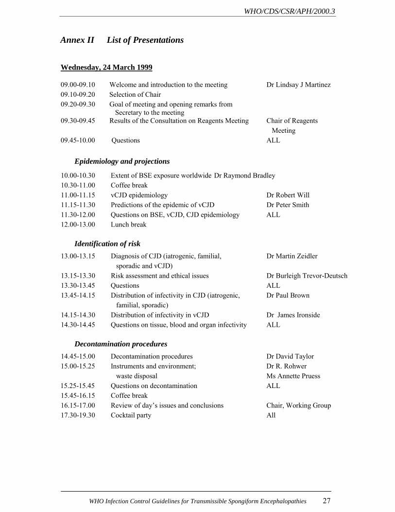
WHO/CDS/CSR/APH/2000.3
WHO Infection Control Guidelines for Transmissible Spongiform Encephalopathies 27
Annex II List of Presentations
Wednesday, 24 March 1999
09.00-09.10 Welcome and introduction to the meeting Dr Lindsay J Martinez09.10-09.20 Selection of Chair09.20-09.30 Goal of meeting and opening remarks from Secretary to the meeting09.30-09.45 Results of the Consultation on Reagents Meeting Chair of Reagents
Meeting09.45-10.00 Questions ALL
Epidemiology and projections
10.00-10.30 Extent of BSE exposure worldwide Dr Raymond Bradley10.30-11.00 Coffee break11.00-11.15 vCJD epidemiology Dr Robert Will11.15-11.30 Predictions of the epidemic of vCJD Dr Peter Smith11.30-12.00 Questions on BSE, vCJD, CJD epidemiology ALL12.00-13.00 Lunch break
Identification of risk13.00-13.15 Diagnosis of CJD (iatrogenic, familial, Dr Martin Zeidler
sporadic and vCJD)13.15-13.30 Risk assessment and ethical issues Dr Burleigh Trevor-Deutsch13.30-13.45 Questions ALL13.45-14.15 Distribution of infectivity in CJD (iatrogenic, Dr Paul Brown
familial, sporadic)14.15-14.30 Distribution of infectivity in vCJD Dr James Ironside14.30-14.45 Questions on tissue, blood and organ infectivity ALL
Decontamination procedures14.45-15.00 Decontamination procedures Dr David Taylor15.00-15.25 Instruments and environment; Dr R. Rohwer
waste disposal Ms Annette Pruess15.25-15.45 Questions on decontamination ALL15.45-16.15 Coffee break16.15-17.00 Review of day�s issues and conclusions Chair, Working Group17.30-19.30 Cocktail party All
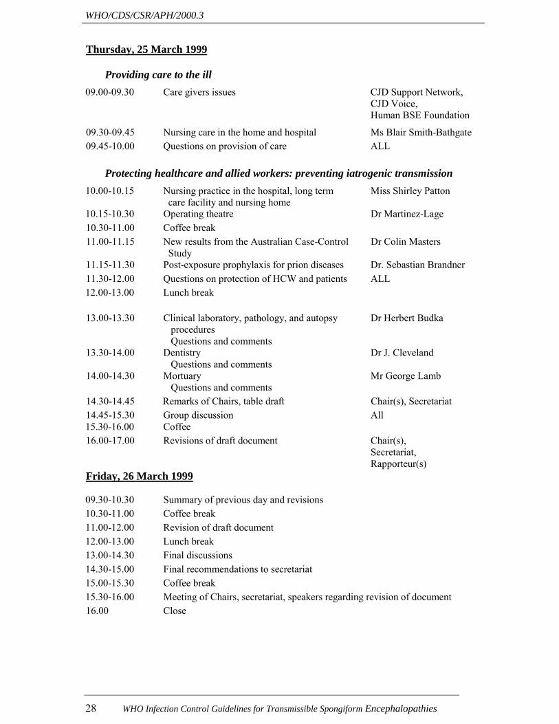
WHO/CDS/CSR/APH/2000.3
28 WHO Infection Control Guidelines for Transmissible Spongiform Encephalopathies
Thursday, 25 March 1999
Providing care to the ill09.00-09.30 Care givers issues CJD Support Network,
CJD Voice,Human BSE Foundation
09.30-09.45 Nursing care in the home and hospital Ms Blair Smith-Bathgate09.45-10.00 Questions on provision of care ALL
Protecting healthcare and allied workers: preventing iatrogenic transmission10.00-10.15 Nursing practice in the hospital, long term Miss Shirley Patton
care facility and nursing home10.15-10.30 Operating theatre Dr Martinez-Lage10.30-11.00 Coffee break11.00-11.15 New results from the Australian Case-Control Dr Colin Masters
Study11.15-11.30 Post-exposure prophylaxis for prion diseases Dr. Sebastian Brandner11.30-12.00 Questions on protection of HCW and patients ALL12.00-13.00 Lunch break
13.00-13.30 Clinical laboratory, pathology, and autopsy Dr Herbert Budka procedures
Questions and comments13.30-14.00 Dentistry Dr J. Cleveland
Questions and comments14.00-14.30 Mortuary Mr George Lamb
Questions and comments14.30-14.45 Remarks of Chairs, table draft Chair(s), Secretariat14.45-15.30 Group discussion All15.30-16.00 Coffee16.00-17.00 Revisions of draft document Chair(s),
Secretariat,Rapporteur(s)
Friday, 26 March 1999
09.30-10.30 Summary of previous day and revisions10.30-11.00 Coffee break11.00-12.00 Revision of draft document12.00-13.00 Lunch break13.00-14.30 Final discussions14.30-15.00 Final recommendations to secretariat15.00-15.30 Coffee break15.30-16.00 Meeting of Chairs, secretariat, speakers regarding revision of document16.00 Close

WHO/CDS/CSR/APH/2000.3
WHO Infection Control Guidelines for Transmissible Spongiform Encephalopathies 29
Annex III Decontamination methods for Transmissible SpongiformEncephalopathies
The safest and most unambiguous method for ensuring that there is no risk ofresidual infectivity on contaminated instruments and other materials is to discard anddestroy them by incineration. In some healthcare situations, as described in theguidance, one of the following less effective methods may be preferred. Whereverpossible, instruments and other materials subject to re-use should be kept moistbetween the time of exposure to infectious materials and subsequent decontaminationand cleaning. If it can be done safely, removal of adherent particles throughmechanical cleaning will enhance the decontamination process.
The following recommendations are based on the best available evidence at thistime and are listed in order of more to less severe treatments. These recommendationsmay require revision if new data become available.
1. Incineration
1. Use for all disposable instruments, materials, and wastes.2. Preferred method for all instruments exposed to high infectivity tissues.
2. Autoclave/chemical methods for heat-resistant instruments
1. Immerse in sodium hydroxide (NaOH)20 and heat in a gravity displacementautoclave at 121°C for 30 min; clean; rinse in water and subject to routinesterilization.
2. Immerse in NaOH or sodium hypochlorite21 for 1 hr; transfer instruments towater; heat in a gravity displacement autoclave at 121°C for 1 hr; clean andsubject to routine sterilization.
3. Immerse in NaOH or sodium hypochlorite for 1 hr.; remove and rinse inwater, then transfer to open pan and heat in a gravity displacement (121°C) orporous load (134°C) autoclave for 1 hr.; clean and subject to routinesterilization.
4. Immerse in NaOH and boil for 10 min at atmospheric pressure; clean, rinse inwater and subject to routine sterilization.
5. Immerse in sodium hypochlorite (preferred) or NaOH (alternative) at ambienttemperature for 1 hr; clean; rinse in water and subject to routine sterilization.
6. Autoclave at 134°C for 18 minutes.22
20 Unless otherwise noted, the recommended concentration is 1N NaOH. 21 Unless otherwise noted, the recommended concentration is 20 000 ppm available chlorine.22 In worse-case scenarios (brain tissue bake-dried on to surfaces) infectivity will be largely but not
completely removed.

WHO/CDS/CSR/APH/2000.3
30 WHO Infection Control Guidelines for Transmissible Spongiform Encephalopathies
3. Chemical methods for surfaces and heat sensitive instruments
1. Flood with 2N NaOH or undiluted sodium hypochlorite; let stand for 1 hr.;mop up and rinse with water.
2. Where surfaces cannot tolerate NaOH or hypochlorite, thorough cleaningwill remove most infectivity by dilution and some additional benefit may bederived from the use of one or another of the partially effective methods listedin Section 5.1 (Table 8).
4. Autoclave/chemical methods for dry goods1. Small dry goods that can withstand either NaOH or sodium hypochlorite
should first be immersed in one or the other solution (as described above) andthen heated in a porous load autoclave at ≥ 121°C for 1 hr.
2. Bulky dry goods or dry goods of any size that cannot withstand exposure toNaOH or sodium hypochlorite should be heated in a porous load autoclaveat 134°C for 1 hr.
5. Notes about autoclaving and chemicalsGravity displacement autoclaves: Air is displaced by steam through a port in the
bottom of the chamber. Gravity displacement autoclaves are designed forgeneral decontamination and sterilization of solutions and instruments.
Porous load autoclaves: Air is exhausted by vacuum and replaced by steam.Porous load autoclaves are optimized for sterilization of clean instruments,gowns, drapes, towelling, and other dry materials required for surgery. Theyare not suitable for liquid sterilization.
Sodium Hydroxide (NaOH, or soda lye): Be familiar with and observe safetyguidelines for working with NaOH. 1N NaOH is a solution of 40 g NaOH in1 litre of water. 1 N NaOH readily reacts with CO2 in air to form carbonatesthat neutralize NaOH and diminish its disinfective properties. 10 N NaOHsolutions do not absorb CO2, therefore, 1N NaOH working solutions should beprepared fresh for each use either from solid NaOH pellets, or by dilution of10 N NaOH stock solutions.
Sodium hypochlorite (NaOCl solution, or bleach): Be familiar with and observesafety guidelines for working with sodium hypochlorite. Household orindustrial strength bleach is sold at different concentrations in differentcountries, so that a standard dilution cannot be specified. Efficacy dependsupon the concentration of available chlorine and should be 20 000 ppmavailable chlorine. One common commercial formulation is 5.25% bleach,which contains 25 000 ppm chlorine. Therefore, undiluted commercial bleachcan be safely used. If solid precursors of hypochloric acid is available, thenstock solution and working solutions can be prepared fresh for each use.
WHO/CDS/CSR/APH/2000.3
WHO Infection Control Guidelines for Transmissible Spongiform Encephalopathies 31
6. Cautions regarding hazardous materials
In all cases, hazardous materials guidelines must be consulted.
1. Personnel
NaOH is caustic but relatively slow acting at room temperature, and can beremoved from skin or clothing by thorough rinsing with water. Hot NaOH isaggressively caustic, and should not be handled until cool. The hazard posed byhot NaOH explains the need to limit boiling to 10 minutes, the shortest timeknown to be effective.Hypochlorite solutions continuously evolve chlorine and so must be kept tightlysealed and away from light. The amount of chlorine released during inactivationmay be sufficient to create a potential respiratory hazard unless the process iscarried out in a well-ventilated or isolated location.
2. Material
In principle, NaOH does not corrode stainless steel, but in practice someformulations of stainless steel can be damaged (including some used for surgicalinstruments). It is advisable to test a sample or consult with the manufacturerbefore dedicating a large number of instruments to decontamination procedures.NaOH is known to be corrosive to glass and aluminum. Hypochlorite does notcorrode glass or aluminum and has also been shown to be an effective sterilizingagent; it is, however, corrosive both to stainless steel and to autoclaves and(unlike NaOH) cannot be used as an instrument bath in the autoclave. Ifhypochlorite is used to clean or soak an instrument, it must be completely rinsedfrom the surfaces before autoclaving. Other decontamination methods may needtesting, or consultation with the manufacturer to verify their effect on theinstrument.

WHO/CDS/CSR/APH/2000.3
32 WHO Infection Control Guidelines for Transmissible Spongiform Encephalopathies

WHO/CDS/CSR/APH/2000.3
WHO Infection Control Guidelines for Transmissible Spongiform Encephalopathies 33
Annex IV Management of healthy ‘at risk’ individuals
Tissue recipientsThe consultation felt that the risk from recipients of dura mater, cornea
transplants and human pituitary hormones, and from persons who have undergoneneurosurgical procedures, is no longer sufficient to warrant classifying this populationas a risk for transmitting TSEs, except under conditions where there could be exposureto their high infectivity tissues (see Section 2.4.2). The consultants considered thatappropriate control measures have immensely reduced or eliminated exposure tocontaminated dura mater and pituitary hormones, and noted that there are only threereports of TSE transmission through cornea transplantation, and six reports (all before1980) of transmission via neurosurgical instruments. In addition, it was recognized thatrecipients of dura mater are largely unaware of the fact, making identification of manyof the dura mater recipients unlikely.
Countries not applying appropriate control measures cannot assume similarlylow levels of current risk among tissue recipients.
Familial Transmissible Spongiform Encephalopathies
Consensus was not reached as to whether asymptomatic persons at risk forfamilial TSE should be classified as �at risk� when determining appropriate infectioncontrol levels. It was argued that the identification of familial risk among asymptomaticpeople would confer a lifetime requirement for high-level infection control for atransmission risk that remains only hypothetical. Discrimination against such personsand legal implications regarding their access to insurance, employment and healthcarewas described by several participants in the consultation, and it was proposed that suchdiscrimination would inevitably lead to a harm which exceeded any evidence of riskposed by them to others.
Others argued that if a familial risk were identified, then more stringent levels ofinfection control could be adopted even in the absence of firm evidence of risk,particularly during procedures involving high infectivity tissues. All consultants agreedthat persons �at risk� for familial TSE should not be denied access to treatment orsurgical procedures, particularly given the range of decontamination options available.Scientific resolution of these issues was impossible due to a lack of precise informationabout tissue infectivity during the pre-clinical phase of human disease, and theconsultants emphasized the need to study any available tissues (including blood) frommutation-positive, but still asymptomatic, members of TSE families.

WHO/CDS/CSR/APH/2000.3
34 WHO Infection Control Guidelines for Transmissible Spongiform Encephalopathies
WHO/CDS/CSR/APH/2000.3
WHO Infection Control Guidelines for Transmissible Spongiform Encephalopathies 35
Annex V Management of individuals with confirmed or suspectedvariant Creutzfeldt-Jakob Disease
The TSE agent causing vCJD has shown certain differences from that of sporadicCJD, including the detection of prion protein (PrP) in a range of lymphoreticular tissues.Patients with vCJD might therefore pose a greater risk of transmitting iatrogenicinfections than sporadic CJD. However, this hypothetical risk has to be balancedagainst the real danger of stigmatizing patients and causing distress and anxiety to thepatient�s relatives by the introduction of rigorous and possibly unnecessary infectioncontrol procedures in general patient care.
On current evidence, the infection control procedures in nursing care settings forsporadic CJD may be applied to cases of vCJD without the need for additionalprecautions, although a more conservative approach may be taken for interventionsinvolving surgical procedures, or when handling tissues and body fluids in thelaboratory. See Section 6.6 (Table 9) for measures that have been recommended forhigh infectivity tissues in patients with other forms of TSE, that could be applied to alltissues of persons with vCJD. It is noted that considerable safety is afforded throughthe measures described in Section 6 and Annex III, and that no person should be denieda diagnostic test given the efficacy of the recommended measures. Comparative tissuerisks for vCJD and a case definition of possible vCJD cases will need to be redefined asfurther research findings emerge. If vCJD is suspected, consultation with personsexpert in this disease, such as The Edinburgh CJD Surveillance Unit, Western GeneralHospital, United Kingdom, is recommended.

WHO/CDS/CSR/APH/2000.3
36 WHO Infection Control Guidelines for Transmissible Spongiform Encephalopathies
WHO Infection Control Guidelines for TransmissibleSpongiform Encephalopathies
This document provides an authoritative guide to procedures and precautions neededto prevent iatrogenic and nosocomial exposure to transmissible spongiformencephalopathies (TSEs) in hospitals, health care facilities, and laboratories. Preparedby an international group of 32 leading experts, the guidelines respond to the unusualresistance of TSE agents to conventional chemical and physical methods ofdecontamination and the corresponding need for special precautions. Areas of patientcare and categories of interventions, tissues, instruments, and wastes that do notrequire special precautions are also clearly indicated.
In issuing these guidelines, WHO aims to help medical officers, specialists ininfection control, care-givers, and laboratory workers reduce the risks of TSEtransmission to negligible levels. With this goal in mind, the guidelines provide alogical framework for determining levels of risk and knowing when departures fromstandard procedures for infection control are required.
Specific recommendations are set out in tables and explanatory text covering patientcare, occupational injury, laboratory investigations, decontamination procedures,waste disposal, and precautions after death. Adherence to these procedures shouldensure a high level of safety. As the document repeatedly emphasizes, no TSE patientshould be denied admission to a health facility, kept in isolation, or deprived of anyprocedure.
Further practical advice is provided in a series of annexes, which give exactinstructions for recommended decontamination methods and discuss the managementof healthy �at risk� individuals and individuals with confirmed or suspected variantCreutzfeldt-Jakob disease.

WHO/CDS/CSR/APH/2000.3
38 WHO Infection Control Guidelines for Transmissible Spongiform Encephalopathies