Infection of Cell Lines with Experimental and Natural ...jvi.asm.org/content/84/5/2444.full.pdf ·...
9
JOURNAL OF VIROLOGY, Mar. 2010, p. 2444–2452 Vol. 84, No. 5 0022-538X/10/$12.00 doi:10.1128/JVI.01855-09 Copyright © 2010, American Society for Microbiology. All Rights Reserved. Infection of Cell Lines with Experimental and Natural Ovine Scrapie Agents Michael H. Neale, 1 * Susan J. Mountjoy, 1 Jane C. Edwards, 1 Didier Vilette, 2 Hubert Laude, 3 Otto Windl, 1 and Ginny C. Saunders 1 Molecular Pathogenesis and Genetics Department, Veterinary Laboratories Agency, New Haw, Addlestone KT15 3NB, United Kingdom 1 ; UMR INRA ENVT 1225 Interactions Ho ˆte-Agent Pathoge `ne, Toulouse, France 2 ; and UR892 INRA, Virologie Immunologie Mole ´culaires, F-78350 Jouy-en-Josas, France 3 Received 2 September 2009/Accepted 9 December 2009 Mouse bioassay remains the gold standard for determining proof of infectivity, strain type, and infectious titer estimation in prion disease research. The development of an approach using ex vivo cell-based assays remains an attractive alternative, both in order to reduce the use of mice and to hasten results. The main limitation of a cell-based approach is the scarcity of cell lines permissive to infection with natural transmissible spongiform encephalopathy strains. This study combines two advances in this area, namely, the standard scrapie cell assay (SSCA) and the Rov9 and MovS6 cell lines, which both express the ovine PrP VRQ allele, to assess to what extent natural and experimental ovine scrapie can be detected ex vivo. Despite the Rov9 and MovS6 cell lines being of different biological origin, they were both permissive and resistant to infection with the same isolates of natural sheep scrapie as detected by SSCA. Rov9 subclones that are 20 times more sensitive than Rov9 to SSBP/1-like scrapie infection were isolated, but all the subclones maintained their resistance to isolates that failed to transmit to the parental line. The most sensitive subclone of the Rov9 cell line was used to estimate the infectious titer of a scrapie brain pool (RBP1) and proved to be more sensitive than the mouse bioassay using wild-type mice. Increasing the sensitivity of the Rov9 cell line to SSBP/1 infection did not correlate with broadening susceptibility, as the specificity of permissiveness and resistance to other scrapie isolates was maintained. Prion diseases are a group of neurodegenerative diseases affecting humans and animals, including scrapie in sheep and goats and bovine spongiform encephalopathy (BSE) in cattle. A feature of prion diseases and, in particular, of scrapie, is the existence of different strains (6) which influence pathology and is most probably related to the conformation of the pathogenic form of the prion protein (PrP Sc ). The susceptibility of sheep to scrapie is determined by the PrP genotype; codons 136, 154, and 171 determine relative resistance and susceptibility, with amino acids valine (V), arginine (R), and glutamine (Q) at these positions (known as VRQ) being considered the sheep PrP allele most susceptible to classical scrapie (3). An array of diagnostic tests exist for prion diseases, aimed at the detection of the disease-associated protease-resistant form of the naturally occurring PrP C protein, termed PrP Sc or PrP res after partial protease digestion. However, the level of detect- able PrP Sc does not quantitatively correlate with prion infec- tivity (2) and the current biochemical analysis of PrP Sc cannot always determine the strain (6, 7). Mouse bioassay remains the gold standard for determining proof of infectivity, strain type, and infectious titer estimate in ruminant transmissible spongiform encephalopathy (TSE) re- search. Conventional mouse bioassays using wild-type mice are generally slow (150 days, and considerably longer, 600, days for obtaining infectious titer information) and require multiple mice to be dosed (typically 6 or more) at each dilution of infectious material. Therefore, the development of an ap- proach using ex vivo cell-based assays remains an ethically and economically desirable alternative. Using cell lines permissive to mouse-passaged scrapie strains, Klo ¨hn et al. have developed a cell-based assay for measuring de novo infection and the titer of mouse-passaged scrapie (18). The main limitation of adopting a cell-based approach is the scarcity of cell lines permissive to infection with natural TSE strains (for a review, see references 31 and 34), as the majority of permissive cell lines can only be infected with rodent- adapted strains of scrapie and BSE (4, 9, 16, 20, 23, 24, 29, 33, 36). While there are currently no cell lines reported to be permissive to bovine BSE or human TSE diseases, there are cell lines which express ovine PrP that have been shown to be permissive to natural scrapie infection (1, 35). There is also one fibroblast-like deer cell line that is able to propagate chronic wasting disease (27). Two of the sheep scrapie-susceptible cell lines are the MovS6 cell line (1), a Schwann cell line derived from the tg301 transgenic mouse, and the Rov9 cell line (35), based on a stably transfected rabbit kidney epithelial cell line (RK13) that does not express endogenous PrP. Both express the VRQ allele of ovine PrP, the latter upon induction with doxycycline (35). These cell lines were found to be permissive to infection with a PrP genotype-matched VRQ homozygous scrapie field case, and de novo PrP Sc maintained its phenotype when used as an inoculum in mouse bioassays (1, 35). Using fluorescence-acti- vated cell sorting, Falanga et al. isolated Rov9 subclones that * Corresponding author. Mailing address: Molecular Pathogene- sis and Genetics Department, Veterinary Laboratories Agency, New Haw, Addlestone, Surrey KT15 3NB, United Kingdom. Phone: 44 (0)1932 357292. Fax: 44 (0)1932 359525. E-mail: m.neale@vla .defra.gsi.gov.uk. Published ahead of print on 23 December 2009. 2444 on September 1, 2018 by guest http://jvi.asm.org/ Downloaded from
Transcript of Infection of Cell Lines with Experimental and Natural ...jvi.asm.org/content/84/5/2444.full.pdf ·...
JOURNAL OF VIROLOGY, Mar. 2010, p. 2444–2452 Vol. 84, No. 50022-538X/10/$12.00 doi:10.1128/JVI.01855-09Copyright © 2010, American Society for Microbiology. All Rights Reserved.
Infection of Cell Lines with Experimental and NaturalOvine Scrapie Agents�
Michael H. Neale,1* Susan J. Mountjoy,1 Jane C. Edwards,1 Didier Vilette,2 Hubert Laude,3Otto Windl,1 and Ginny C. Saunders1
Molecular Pathogenesis and Genetics Department, Veterinary Laboratories Agency, New Haw, Addlestone KT15 3NB,United Kingdom1; UMR INRA ENVT 1225 Interactions Hote-Agent Pathogene, Toulouse, France2; and
UR892 INRA, Virologie Immunologie Moleculaires, F-78350 Jouy-en-Josas, France3
Received 2 September 2009/Accepted 9 December 2009
Mouse bioassay remains the gold standard for determining proof of infectivity, strain type, and infectioustiter estimation in prion disease research. The development of an approach using ex vivo cell-based assaysremains an attractive alternative, both in order to reduce the use of mice and to hasten results. The mainlimitation of a cell-based approach is the scarcity of cell lines permissive to infection with natural transmissiblespongiform encephalopathy strains. This study combines two advances in this area, namely, the standardscrapie cell assay (SSCA) and the Rov9 and MovS6 cell lines, which both express the ovine PrP VRQ allele, toassess to what extent natural and experimental ovine scrapie can be detected ex vivo. Despite the Rov9 andMovS6 cell lines being of different biological origin, they were both permissive and resistant to infection withthe same isolates of natural sheep scrapie as detected by SSCA. Rov9 subclones that are 20 times moresensitive than Rov9 to SSBP/1-like scrapie infection were isolated, but all the subclones maintained theirresistance to isolates that failed to transmit to the parental line. The most sensitive subclone of the Rov9 cellline was used to estimate the infectious titer of a scrapie brain pool (RBP1) and proved to be more sensitivethan the mouse bioassay using wild-type mice. Increasing the sensitivity of the Rov9 cell line to SSBP/1infection did not correlate with broadening susceptibility, as the specificity of permissiveness and resistance toother scrapie isolates was maintained.
Prion diseases are a group of neurodegenerative diseasesaffecting humans and animals, including scrapie in sheep andgoats and bovine spongiform encephalopathy (BSE) in cattle.A feature of prion diseases and, in particular, of scrapie, is theexistence of different strains (6) which influence pathology andis most probably related to the conformation of the pathogenicform of the prion protein (PrPSc). The susceptibility of sheepto scrapie is determined by the PrP genotype; codons 136, 154,and 171 determine relative resistance and susceptibility, withamino acids valine (V), arginine (R), and glutamine (Q) atthese positions (known as VRQ) being considered the sheepPrP allele most susceptible to classical scrapie (3).
An array of diagnostic tests exist for prion diseases, aimed atthe detection of the disease-associated protease-resistant formof the naturally occurring PrPC protein, termed PrPSc or PrPres
after partial protease digestion. However, the level of detect-able PrPSc does not quantitatively correlate with prion infec-tivity (2) and the current biochemical analysis of PrPSc cannotalways determine the strain (6, 7).
Mouse bioassay remains the gold standard for determiningproof of infectivity, strain type, and infectious titer estimate inruminant transmissible spongiform encephalopathy (TSE) re-search. Conventional mouse bioassays using wild-type mice aregenerally slow (�150 days, and considerably longer, �600,
days for obtaining infectious titer information) and requiremultiple mice to be dosed (typically 6 or more) at each dilutionof infectious material. Therefore, the development of an ap-proach using ex vivo cell-based assays remains an ethically andeconomically desirable alternative. Using cell lines permissiveto mouse-passaged scrapie strains, Klohn et al. have developeda cell-based assay for measuring de novo infection and the titerof mouse-passaged scrapie (18).
The main limitation of adopting a cell-based approach is thescarcity of cell lines permissive to infection with natural TSEstrains (for a review, see references 31 and 34), as the majorityof permissive cell lines can only be infected with rodent-adapted strains of scrapie and BSE (4, 9, 16, 20, 23, 24, 29, 33,36). While there are currently no cell lines reported to bepermissive to bovine BSE or human TSE diseases, there arecell lines which express ovine PrP that have been shown to bepermissive to natural scrapie infection (1, 35). There is alsoone fibroblast-like deer cell line that is able to propagatechronic wasting disease (27).
Two of the sheep scrapie-susceptible cell lines are theMovS6 cell line (1), a Schwann cell line derived from the tg301transgenic mouse, and the Rov9 cell line (35), based on a stablytransfected rabbit kidney epithelial cell line (RK13) that doesnot express endogenous PrP. Both express the VRQ allele ofovine PrP, the latter upon induction with doxycycline (35).These cell lines were found to be permissive to infection witha PrP genotype-matched VRQ homozygous scrapie field case,and de novo PrPSc maintained its phenotype when used as aninoculum in mouse bioassays (1, 35). Using fluorescence-acti-vated cell sorting, Falanga et al. isolated Rov9 subclones that
* Corresponding author. Mailing address: Molecular Pathogene-sis and Genetics Department, Veterinary Laboratories Agency,New Haw, Addlestone, Surrey KT15 3NB, United Kingdom. Phone:44 (0)1932 357292. Fax: 44 (0)1932 359525. E-mail: [email protected].
� Published ahead of print on 23 December 2009.
2444
on Septem
ber 1, 2018 by guesthttp://jvi.asm
.org/D
ownloaded from
produce higher levels of PrPC and PrPSc than the parental cellline when infected (11).
The primary objective of this study was to assess the permis-siveness of the Rov9 and MovS6 cell lines to a panel of scrapieisolates from a range of sheep breeds with a range of PrPgenotypes. Second, subcloning of the Rov9 cell line was un-dertaken in an attempt to identify subclones with greater sen-sitivity and more diverse permissibility to ovine scrapie isolates.
MATERIALS AND METHODS
Cell lines. The MovS6 and Rov9 cell lines have been described previously (1,35). The MovS6 cell line was cultured in 75% Dulbecco’s modified Eagle’smedium (DMEM; Sigma) and 25% F-12 Ham medium (Sigma) supplementedwith 10% fetal calf serum and antibiotics. Infections in MovS6 cells were per-formed with Optimem (Gibco) as a replacement for DMEM. Rov9 cells wereroutinely cultured in Eagle’s minimal essential medium (EMEM; Gibco) sup-plemented with 10% fetal calf serum, 2% HEPES, and antibiotics (penicillin,streptomycin, and mycostatin). All Rov9 infections were performed using Opti-mem (Gibco) as a replacement for EMEM. Cells were cultured at 37°C in ahumidified 5% CO2 atmosphere in a cell culture incubator.
SSCA. For the standard scrapie cell assay (SSCA), cells were infected follow-ing the published protocol from Klohn et al. (18), briefly described here. Ap-proximately 20,000 cells per well were plated out in 96-well plates, Rov9 cellswere cultured in the presence of 1 �g/ml doxycycline (Sigma) to induce PrPC
expression for 48 h prior to the addition of infectious brain homogenate, anddoxycycline was added to the medium for subsequent culturing of the Rov9 cellsin order to maintain PrPC expression. Cells were cultured for a further 3 daysbefore being passaged every 3 days at a 1:6 dilution into fresh 96-well plates andthen on the fourth day were transferred to 96-well enzyme-linked immunospotassay (ELISPOT) plates (Multiscreen-IP filter plates; Millipore). The plateswere dried, and a modified SSCA was performed. Alterations to the publishedprotocol included increased proteinase K (PK) concentrations (4 and 10 �g/mlfor MovS6 and Rov9 cells, respectively) and the use of 6H4 (Prionics) as aprimary antibody for detection of PrPres in the plates. Following immunodetec-tion, infected cells were visualized with an alkaline phosphate conjugate sub-strate kit (Bio-Rad). Positive cells (spots) were counted using a Zeiss KS-ELIS-POT imaging system running WellScan software (Imaging Associates).
To accurately assess the total cell number per well, duplicate ELISPOT plateswere prepared with a 1:10 dilution of cells and dried in the same way as describedabove. Cells were stained with Trypan blue solution and counted using the ZeissKS-ELISPOT imaging system. All results are presented as the number of in-fected cells per 20,000 cells for ease of comparison; however, in most cases thenumber of cells assayed per well was �5,000 cells. In some cases, the number ofinfected cells detected by the SSCA with the Rov9-2A3 subclone is an underes-timate: due to the large number of infected cells present, the individual spotsmerge and the ELISPOT plate reader is unable to count the individual infectedcells, resulting in undercounting of infected cells (see Fig. 3). However, at lowerconcentrations of inocula, the number of infected cells is decreased and differ-ences between the sensitivities of the subclones become apparent (see Fig. 5a).In all cases, the wells have been inspected under the microscope to confirminfected cells; this is especially important for samples that are borderline positivewith regard to the negative controls. In some cases, wells appear positive by spotnumbers but, upon microscopic inspection, the spots detected are drying defectswithin the well or the effects of the edge of the well.
To aid comparison of the subclones, the concentration of the inoculum re-quired to infect 1,000 cells out of 20,000 cells plated has been calculated. This isan arbitrary value similar to that used by Mahal et al. (21), but whereas Mahalet al., used 300 infected cells per 20,000, we have chosen 1,000 cells.
Scrapie inocula. Two experimental scrapie isolates, SSBP/1 from a VRQ/VRQsheep and CH1641 from an AHQ/AHQ sheep (14); a brain pool (RBP1) (30);New Zealand-derived uninfected controls; and field case samples of scrapie wereobtained from the Veterinary Laboratories Agency (Table 1). All material wasbrain frontal cortex tissue except RBP1, which is a pool of whole brains from 17sheep of different PrP genotypes. All brain samples were homogenized in sterilephosphate-buffered saline (PBS) (10% wt/vol) and stored at �80°C; they werediluted in cell culture medium to appropriate dilutions prior to being added tocells.
Subcloning by limiting dilution. Rov9 cells were counted and plated out in96-well plates with an average of one cell per 3 wells. After clonal cells were
established, they were transferred to new plates and tested for permissiveness toinfection using the SSCA.
Western blotting. For PrPC analysis, Rov9 cells and subclones were grown in25-cm2 flasks in the presence of 1 �g/ml doxycycline for 48 h. For infected cells,cells were grown in 24-well plates and inoculated with scrapie-infected brain;after 3 days the cells were passaged 1:6 into a fresh 24-well plate. At 3-dayintervals, cells were passaged 1:1 into 6-well plates, transferred to 25-cm2 flasks,and finally transferred to 75-cm2 flasks before collection once confluent. Prior tocollection, 20,000 cells from each infection were plated out for SSCA analysis toconfirm infection. Cells were lysed in 1 ml radioimmunoprecipitation buffer (50mM Tris, pH 8.0, 150 mM NaCl, 0.5% sodium deoxycholate, 0.1% SDS, 1%NP-40, 1 mM EDTA). Lysates were treated with benzonase (25 U/ml of lysates;Sigma) to digest DNA. Cell lysates were clarified by centrifugation, and proteinconcentration determined by bicinchoninic acid (BCA) protein assay (Pierce).For PrPC analysis, 100 �g of protein was precipitated in 1 ml cold methanol at�20°C for 1 h before being washed and resuspended in 50 �l loading buffer andboiled for 5 min. Five microliters (equivalent to 10 �g of total protein) wasloaded onto each lane of a gel. For detecting PrPres in infected cell lysates, 500�g of total protein was digested with PK (20 �g/ml for 60 min at 37°C, Roche)before centrifugation at 47,000 � g for 30 min, and the pellet was resuspendedin 25 �l loading dye (Prionics) and loaded onto a polyacrylamide gel (Novex 12%Bis–Tris gels). After separation by electrophoresis, proteins were transferred topolyvinylidene difluoride (PVDF) membrane (Millipore) and detected by anti-bodies against PrP (6H4; Prionics) or tubulin (Santa Cruz Biotechnology, Inc.).Proteins were visualized by using CDP-Star.
For Western blotting of scrapie inoculum samples, 20 �l of 10% brain ho-mogenate was mixed with an equal volume of homogenization buffer (Prionics).The homogenate was digested with 37.5 �g/ml PK for 1 h at 37°C before theaddition of 2 mM phenylmethylsulfonyl fluoride (PMSF; Sigma), to stop diges-tion, and 40 �l loading buffer. Samples were boiled, 25-�l amounts run on 12%Bis–Tris gels (Novex), and Western blots performed as described above for celllysates.
Deglycosylation of PrPC. For deglycosylating PrPC, lysates were digested withpeptide-N-glycosidase F (PNGase). Proteins in 10 �l of cell lysate were precip-itated with 7 volumes of ice-cold methanol for 2 h at �20°C and collected bycentrifugation at 13,000 rpm for 20 min. The protein pellet was resuspended in2 �l of denaturing buffer (New England Biolabs) and 18 �l water and heated to100°C for 10 min. Two microliters Nonidet P40, 2 �l G7 buffer, and 0.8 �lPNGase (all reagents from New England Biolabs) were added to the resus-pended pellet. Samples were briefly vortexed for mixing and incubated at 37°Covernight. Digested proteins were reprecipitated with 7 volumes of ice-coldmethanol for 2 h at �20°C and collected by centrifugation at 13,000 rpm for 20min. Precipitates were resuspended in loading dye and separated by electro-phoresis as described above. Densitometry measurements were performed usingLabworks software (UVP Bioimaging Systems).
Immunofluorescence confocal microscopy. Uninfected Rov9 cells were grownon coverslips in 24-well plates in the presence or absence of doxycycline for 48 hand then fixed in 4% formaldehyde solution in PBS. Cells were permeabilizedwith 0.1% Triton X-100 in PBS for 2 min before immunofluorescent staining withan antibody targeted against PrP (6H4; Prionics) and an IgG secondary anti-mouse antibody to which Alexa 488 was conjugated (Molecular Probes). Nucleiwere counterstained with Hoechst 33342 (Molecular Probes). Coverslips weremounted in ProLong Gold (Molecular Probes), and images were captured usinga Leica TCS SP2 confocal microscope.
Flow cytometry. Rov9 cells and subclones were grown in 25-cm2 flasks in thepresence or absence of 1 �g/ml doxycycline for 48 h. Cells were detached byincubation with 3 ml Versene (Gibco) for 30 min at 37°C. Detached cells werewashed in PBS and labeled with SAF32 anti-PrP antibody (Spi-Bio) (1 �g/106
cells for 1 h). Cells were washed twice in wash buffer (PBS, 0.5% bovine serumalbumin, 0.1% sodium azide, 0.002% EDTA) and incubated for an hour at roomtemperature with a phycoerythrin-conjugated secondary antibody (BD Phar-minogen). Cells were washed twice and resuspended in 0.5 ml PBS. Cells wereanalyzed using a Beckman Coulter Epics XL Flow Cytometer. Ten thousandevents were recorded for each sample and analyzed using Expo32 ADC software.
Titer estimation with SCEPA. Thirty-two wells for each of 7 dilutions ofhomogenate and 32 wells of New Zealand-derived negative control were plated,and the scrapie cell endpoint assay (SCEPA) performed (22). This differs fromthe standard scrapie cell assay in that after the initial 3-day incubation periodwith the inoculum, 50% of the medium is changed and the cells are cultured fora further 3 days. The cells are then passaged three times at a 1:3 dilution every2 days, followed by three passages at a 1:6 dilution every 3 days before theELISPOT assay is performed as described for the SSCA. Titers were estimatedusing the Reed and Muench 50% endpoint calculation method (28). A well is
VOL. 84, 2010 INFECTION OF CELL LINES WITH SCRAPIE 2445
on Septem
ber 1, 2018 by guesthttp://jvi.asm
.org/D
ownloaded from
deemed to be positively infected if the number of spots detected by SCEPA isgreater than the average of the results for the 32 wells of New Zealand-derivedscrapie-negative isolate plus 3 times the standard deviation.
RESULTS
Transmission of sheep scrapie to Rov9 and MovS6 cells. Tocompare the susceptibilities of the Rov9 and MovS6 cell linesto natural cases of scrapie, both cell lines were challenged witha panel of 30 field isolates of scrapie and de novo infectionassessed with the standard scrapie cell assay (SSCA) (18). Asthe Rov9 and MovS6 cell lines express high levels of PrPC, theproteinase K (PK) concentration was optimized to ensure com-plete digestion of PrPC, ensuring that only PK-resistant PrP(PrPSc)-expressing cells were detected in the SSCA and pre-venting false-positive results (data not shown).
Rov9 and MovS6 cells were challenged with brain homoge-nates from 30 field cases of scrapie, 2 experimental scrapieisolates (SSBP/1 and CH1641) (10, 12), and a New Zealand-derived scrapie-negative case, described in Table 1.
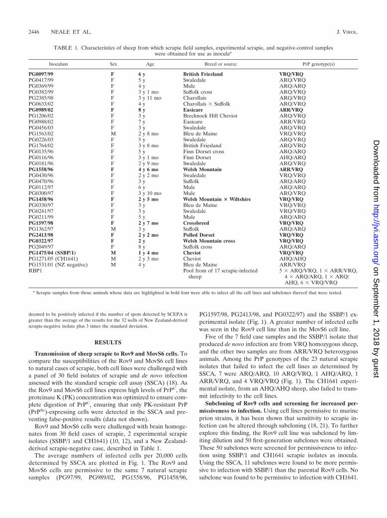
The average numbers of infected cells per 20,000 cellsdetermined by SSCA are plotted in Fig. 1. The Rov9 andMovS6 cells are permissive to the same 7 natural scrapiesamples (PG97/99, PG989/02, PG1558/96, PG1458/96,
PG1597/98, PG2413/98, and PG0322/97) and the SSBP/1 ex-perimental isolate (Fig. 1). A greater number of infected cellswas seen in the Rov9 cell line than in the MovS6 cell line.
Five of the 7 field case samples and the SSBP/1 isolate thatproduced de novo infection are from VRQ homozygous sheep,and the other two samples are from ARR/VRQ heterozygousanimals. Among the PrP genotypes of the 23 natural scrapieisolates that failed to infect the cell lines as determined bySSCA, 7 were ARQ/ARQ, 10 ARQ/VRQ, 1 AHQ/ARQ, 1ARR/VRQ, and 4 VRQ/VRQ (Fig. 1). The CH1641 experi-mental isolate, from an AHQ/AHQ sheep, also failed to trans-mit infectivity to the cell lines.
Subcloning of Rov9 cells and screening for increased per-missiveness to infection. Using cell lines permissive to murineprion strains, it has been shown that sensitivity to scrapie in-fection can be altered through subcloning (18, 21). To furtherexplore this finding, the Rov9 cell line was subcloned by lim-iting dilution and 50 first-generation subclones were obtained.These 50 subclones were screened for permissiveness to infec-tion using SSBP/1 and CH1641 scrapie isolates as inocula.Using the SSCA, 11 subclones were found to be more permis-sive to infection with SSBP/1 than the parental Rov9 cells. Nosubclone was found to be permissive to infection with CH1641.
TABLE 1. Characteristics of sheep from which scrapie field samples, experimental scrapie, and negative-control sampleswere obtained for use as inoculaa
Inoculum Sex Age Breed or source PrP genotype(s)
PG0097/99 F 6 y British Friesland VRQ/VRQPG0417/99 F 5 y Swaledale ARQ/VRQPG0369/99 F 4 y Mule ARQ/ARQPG0382/99 F 3 y 1 mo Suffolk cross ARQ/VRQPG2385/98 F 3 y 11 mo Charollais ARQ/VRQPG0633/02 F 4 y Charollais � Suffolk ARQ/VRQPG0989/02 F 8 y Easicare ARR/VRQPG1206/02 F 3 y Brecknock Hill Cheviot ARQ/VRQPG0988/02 F 7 y Easicare ARR/VRQPG0456/03 F 3 y Swaledale ARQ/VRQPG1563/02 M 2 y 8 mo Bleu de Maine VRQ/VRQPG0226/03 F 5 y Swaledale ARQ/VRQPG1764/02 F 3 y 8 mo British Friesland ARQ/VRQPG0135/96 F 5 y Finn Dorset cross ARQ/ARQPG0116/96 F 3 y 1 mo Finn Dorset AHQ/ARQPG0181/96 F 2 y 9 mo Swaledale ARQ/VRQPG1558/96 F 4 y 6 mo Welsh Mountain ARR/VRQPG0430/96 F 2 y 2 mo Swaledale VRQ/VRQPG0470/96 F 3 y Suffolk ARQ/ARQPG0112/97 F 6 y Mule ARQ/ARQPG0300/97 F 3 y 10 mo Mule ARQ/VRQPG1458/96 F 2 y 5 mo Welsh Mountain � Wiltshire VRQ/VRQPG0330/97 F 3 y Bleu de Maine VRQ/VRQPG0241/97 F 3 y Swaledale VRQ/VRQPG0211/99 F 5 y Mule ARQ/ARQPG1597/98 F 2 y 7 mo Crossbreed VRQ/VRQPG1362/97 M 3 y Suffolk ARQ/ARQPG2413/98 F 2 y 2 mo Polled Dorset VRQ/VRQPG0322/97 F 2 y Welsh Mountain cross VRQ/VRQPG2049/97 F 8 y Suffolk cross ARQ/ARQPG1475/04 (SSBP/1) M 1 y 4 mo Cheviot VRQ/VRQPG1271/05 (CH1641) M 2 y 3 mo Cheviot AHQ/AHQPG1531/01 (NZ negative) M 4 y Bleu de Maine ARR/VRQRBP1 Pool from of 17 scrapie-infected
sheep5 � ARQ/VRQ, 1 � ARR/VRQ,
4 � ARQ/ARQ, 1 � ARQ/AHQ, 6 � VRQ/VRQ
a Scrapie samples from those animals whose data are highlighted in bold font were able to infect all the cell lines and subclones thereof that were tested.
2446 NEALE ET AL. J. VIROL.
on Septem
ber 1, 2018 by guesthttp://jvi.asm
.org/D
ownloaded from
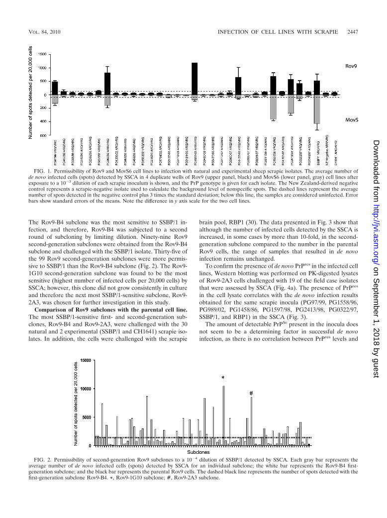
The Rov9-B4 subclone was the most sensitive to SSBP/1 in-fection, and therefore, Rov9-B4 was subjected to a secondround of subcloning by limiting dilution. Ninety-nine Rov9second-generation subclones were obtained from the Rov9-B4subclone and challenged with the SSBP/1 isolate. Thirty-five ofthe 99 Rov9 second-generation subclones were more permis-sive to SSBP/1 than the Rov9-B4 subclone (Fig. 2). The Rov9-1G10 second-generation subclone was found to be the mostsensitive (highest number of infected cells per 20,000 cells) bySSCA; however, this clone did not grow consistently in cultureand therefore the next most SSBP/1-sensitive subclone, Rov9-2A3, was chosen for further investigation in this study.
Comparison of Rov9 subclones with the parental cell line.The most SSBP/1-sensitive first- and second-generation sub-clones, Rov9-B4 and Rov9-2A3, were challenged with the 30natural and 2 experimental (SSBP/1 and CH1641) scrapie iso-lates. In addition, the cells were challenged with the scrapie
brain pool, RBP1 (30). The data presented in Fig. 3 show thatalthough the number of infected cells detected by the SSCA isincreased, in some cases by more than 10-fold, in the second-generation subclone compared to the number in the parentalRov9 cells, the range of samples that resulted in de novoinfection remains unchanged.
To confirm the presence of de novo PrPres in the infected celllines, Western blotting was performed on PK-digested lysatesof Rov9-2A3 cells challenged with 19 of the field case isolatesthat were assessed by SSCA (Fig. 4a). The presence of PrPres
in the cell lysate correlates with the de novo infection resultsobtained for the same scrapie inocula (PG97/99, PG1558/96,PG989/02, PG1458/86, PG1597/98, PG2413/98, PG0322/97,SSBP/1, and RBP1) in the SSCA (Fig. 3).
The amount of detectable PrPSc present in the inocula doesnot seem to be a determining factor in successful de novoinfection, as there is no correlation between PrPres levels and
FIG. 1. Permissibility of Rov9 and MovS6 cell lines to infection with natural and experimental sheep scrapie isolates. The average number ofde novo infected cells (spots) detected by SSCA in 4 duplicate wells of Rov9 (upper panel, black) and MovS6 (lower panel, gray) cell lines afterexposure to a 10�3 dilution of each scrapie inoculum is shown, and the PrP genotype is given for each isolate. The New Zealand-derived negativecontrol represents a scrapie-negative isolate used to calculate the background level of nonspecific spots. The dashed lines represent the averagenumber of spots detected in the negative control plus 3 times the standard deviation; below this line, the samples are considered uninfected. Errorbars show standard errors of the means. Note the difference in y axis scale for the two cell lines.
FIG. 2. Permissibility of second-generation Rov9 subclones to a 10�4 dilution of SSBP/1 detected by SSCA. Each gray bar represents theaverage number of de novo infected cells (spots) detected by SSCA for an individual subclone; the white bar represents the Rov9-B4 first-generation subclone; and the black bar represents the parental Rov9 cells. The dashed black line represents the number of spots detected with thefirst-generation subclone Rov9-B4. *, Rov9-1G10 subclone; #, Rov9-2A3 subclone.
VOL. 84, 2010 INFECTION OF CELL LINES WITH SCRAPIE 2447
on Septem
ber 1, 2018 by guesthttp://jvi.asm
.org/D
ownloaded from
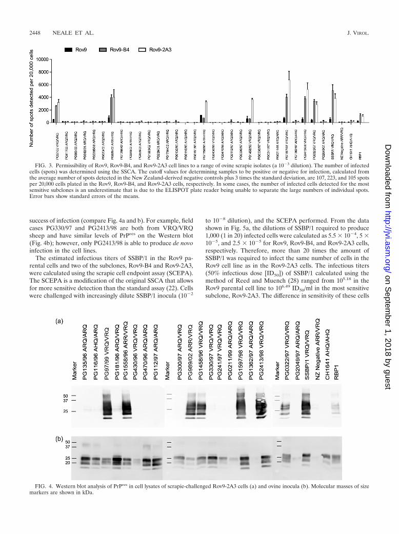
success of infection (compare Fig. 4a and b). For example, fieldcases PG330/97 and PG2413/98 are both from VRQ/VRQsheep and have similar levels of PrPres on the Western blot(Fig. 4b); however, only PG2413/98 is able to produce de novoinfection in the cell lines.
The estimated infectious titers of SSBP/1 in the Rov9 pa-rental cells and two of the subclones, Rov9-B4 and Rov9-2A3,were calculated using the scrapie cell endpoint assay (SCEPA).The SCEPA is a modification of the original SSCA that allowsfor more sensitive detection than the standard assay (22). Cellswere challenged with increasingly dilute SSBP/1 inocula (10�2
to 10�8 dilution), and the SCEPA performed. From the datashown in Fig. 5a, the dilutions of SSBP/1 required to produce1,000 (1 in 20) infected cells were calculated as 5.5 � 10�4, 5 �10�5, and 2.5 � 10�5 for Rov9, Rov9-B4, and Rov9-2A3 cells,respectively. Therefore, more than 20 times the amount ofSSBP/1 was required to infect the same number of cells in theRov9 cell line as in the Rov9-2A3 cells. The infectious titers(50% infectious dose [ID50]) of SSBP/1 calculated using themethod of Reed and Muench (28) ranged from 105.18 in theRov9 parental cell line to 106.49 ID50/ml in the most sensitivesubclone, Rov9-2A3. The difference in sensitivity of these cells
FIG. 3. Permissibility of Rov9, Rov9-B4, and Rov9-2A3 cell lines to a range of ovine scrapie isolates (a 10�3 dilution). The number of infectedcells (spots) was determined using the SSCA. The cutoff values for determining samples to be positive or negative for infection, calculated fromthe average number of spots detected in the New Zealand-derived negative controls plus 3 times the standard deviation, are 107, 223, and 105 spotsper 20,000 cells plated in the Rov9, Rov9-B4, and Rov9-2A3 cells, respectively. In some cases, the number of infected cells detected for the mostsensitive subclones is an underestimate that is due to the ELISPOT plate reader being unable to separate the large numbers of individual spots.Error bars show standard errors of the means.
FIG. 4. Western blot analysis of PrPres in cell lysates of scrapie-challenged Rov9-2A3 cells (a) and ovine inocula (b). Molecular masses of sizemarkers are shown in kDa.
2448 NEALE ET AL. J. VIROL.
on Septem
ber 1, 2018 by guesthttp://jvi.asm
.org/D
ownloaded from
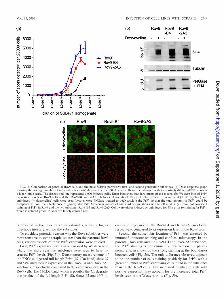
is reflected in the infectious titer estimates, where a higherinfectious titer is given for the subclones.
To elucidate potential reasons why the Rov9 subclones weremore sensitive to some scrapie isolates than the parental Rov9cells, various aspects of their PrPC expression were studied.
First, PrPC expression levels were assessed by Western blot,where the more sensitive subclones were seen to have in-creased PrPC levels (Fig. 5b). Densitometry measurements ofthe PNGase-digested full-length PrPC (27-kDa band) show 37and 54% increases in expression in the Rov9-B4 and Rov9-2A3subclones, respectively, compared to the expression level in theRov9 cells. The 17-kDa band, which is possibly the C1 degrada-tion product of the full-length PrPC (8), shows 62 and 16% in-
creases in expression in the Rov9-B4 and Rov9-2A3 subclones,respectively, compared to its expression level in the Rov9 cells.
Second, the subcellular location of PrPC was assessed byimmunofluorescent staining and confocal microscopy. In theparental Rov9 cells and the Rov9-B4 and Rov9-2A3 subclones,the PrPC staining is predominantly localized on the plasmamembrane, as shown by the strong staining at the boundariesbetween cells (Fig. 5c). The only difference observed appearsto be the number of cells staining positively for PrPC, with agreater number of PrPC-expressing cells found in the subclonesthan in the Rov9 cells. This increased number of cells withpositive expression may account for the increased total PrPC
levels seen in the Western blots (Fig. 5b).
FIG. 5. Comparison of parental Rov9 cells and the most SSBP/1-permissive first- and second-generation subclones. (a) Dose-response graphshowing the average number of infected cells (spots) detected by the SSCA when cells were challenged with increasingly dilute SSBP/1. x axis isa logarithmic scale. The dashed red line represents 1,000 infected cells. Error bars show standard errors of the means. (b) Western blot of PrPC
expression levels in Rov9 cells and the Rov9-B4 and -2A3 subclones. Amounts of 10 �g of total protein from induced (� doxycycline) anduninduced (� doxycycline) cells were used. Lysates were PNGase treated to deglycosylate the PrPC so that the total amount of PrPC could becompared without the interference of glycosylated PrP. Molecular masses of size markers are shown on the left in kDa. (c) Immunofluorescentstaining of PrPC in Rov9 and the two subclones Rov9-B4 and Rov9-2A3. Cells were either induced or uninduced for 48 h prior to staining for PrPC,which is colored green. Nuclei are falsely colored red.
VOL. 84, 2010 INFECTION OF CELL LINES WITH SCRAPIE 2449
on Septem
ber 1, 2018 by guesthttp://jvi.asm
.org/D
ownloaded from

To quantify whether there are indeed more PrPC-expressingcells in the subclones than in the parental cells, the cell surfacePrPC was fluorescently labeled and cells subjected to flow cy-tometry. Approximately 76% of the Rov9 cells stained posi-tively for cell surface PrPC, slightly less than for Rov9-B4, at81%, and Rov9-2A3 cells, at 88% (data not shown). Theseresults confirm the findings seen with the immunofluorescentstaining and confocal microscopy.
SCEPA titer estimation of a well-characterized scrapie sam-ple and comparison to mouse bioassay. The Ripley brain pool1 (RBP1) is a scrapie pool produced from 17 scrapie-infectedbrains from sheep with a range of PrP genotypes. By mousebioassay in RIII mice, the infectious titer of the RBP1 wascalculated to be 103.96 ID50/gram of tissue (30). The infectioustiter of RBP1 in Rov9-2A3 cells, calculated using the 50%endpoint estimation of Reed and Muench (28), was 104.99
ID50/gram of tissue, more than 1 log higher than the wild-typemouse bioassay titer estimate.
DISCUSSION
In this study, we have adapted the SSCA protocol for usewith Rov9 and MovS6 cell lines and subclones thereof, and theassay has been successfully used for the detection of de novoinfectivity and infectious titer estimation of ovine scrapie.
Using this method, the range of sheep scrapie samples ableto infect the cell lines examined in this study was found to belimited. Out of the 30 natural scrapie and 2 experimentalscrapie isolates used to challenge the MovS6 and Rov9 cells,the same 7 natural scrapie isolates and SSBP/1 resulted in denovo PrPres in all the cell lines, while the remaining 23 naturalisolates and CH1641 did not transmit to any of the cell lines.
There is evidence that different cell lines are able to prop-agate different strains of mouse-adapted scrapie (21). Further-more, Mahal et al., have shown that for mouse-passaged TSEstrains, cell lines of different origin have different levels ofsensitivity (21). For example, all four of the cell lines they used(2 subclones of neuroblastoma [N2a-PK1 and R33], a fibro-blast cell line [L929], and a central nervous system-derived cellline [CAD]) were permissive to infection with 22L, but onlythree cell lines were permissive to infection with RML, 2 werepermissive to infection with ME7, and one cell line was per-missive to infection with the 301C strain. In our study, in spiteof using cell lines from different origins, namely, Rov9 cells,derived from a rabbit kidney epithelial cell line, and the MovS6cell line, which is a murine Schwann cell line, no differences inpermissibility to the different isolates were seen. However, theRov9 cell line was consistently more sensitive to all the trans-missible ovine scrapie isolates than the MovS6 cell line.
The amount of PrPres present in the scrapie inocula did notcorrelate with de novo infection results. Western blotting of theoriginal inocula used to challenge the cell lines shows a rangeof PrPres levels; in fact, the isolate with the highest levels ofPrPres is an ARQ/VRQ heterozygous sheep (PG181/96) (Fig.4b) which is unable to infect the cell lines. Of the VRQ/VRQisolates tested, those that are able to infect the cell lines do nothave higher levels of PrPres than those isolates that are unableto infect the cells, and the glycoform profiles are the same (Fig.4a and b).
All of the 7 natural scrapie isolates that were able to infect
the cells (5 VRQ/VRQ and 2 VRQ/ARR) plus the SSBP/1isolate (VRQ/VRQ) were from sheep that carry at least onecopy of the VRQ PrP allele, the same allele expressed by thecell lines. However, not all scrapie isolates from VRQ het-erozygous or homozygous sheep were able to infect the cells.Therefore, although some PrP homology between inoculumand cell line may be required, it is not sufficient to ensure denovo infection.
There was no discernible difference in the ages of the sheepthat had the transmissible and nontransmissible VRQ/VRQ iso-lates (Table 1); however, the four nontransmissible VRQ/VRQisolates are, notably, from the Swaledale and Bleu de Mainebreeds. The significance of this observation cannot be determineddue to the low numbers of samples analyzed. It is possible that thenontransmitting isolates, particularly the VRQ/VRQ isolates, area different scrapie strain than the transmitting isolates of thesame PrP genotype. It is well known that different strains ofnatural sheep scrapie exist, as determined by lesion profiles(13) and incubation periods in mice. There are very limitedamounts of ovine scrapie material of known strains availablefor testing; however, 17 of the scrapie isolates assayed here arecurrently being strain typed by mouse bioassay. Once known,the strain information will be used to elucidate whether thepermissiveness to infection of the cells tested here is deter-mined by strain.
Studies of mice have shown that scrapie strain can affectwhether an isolate is transmissible to a cell line. In a study byMahal et al. (22), the 4 cell lines used in a cell panel assay andthe 4 mouse-adapted scrapie strains used were all from Prnpa
mice, and therefore, permissiveness or resistance to infectionwas thought to be due to strain. The authors considered thatstrains of prions with the same PrP protein could be differen-tiated by glycosylation, the requirement for different cell-spe-cific factors (i.e., small RNAs), structure-specific chaperones,or a cell-specific fibril cleavage activity.
Subcloning of the Rov9 cell line was undertaken to try toimprove the sensitivity of the parental line to infection and inan attempt to increase the range of scrapie isolates that re-sulted in de novo PrPres detection. While the first undertakingwas successful, the second was not. All 50 first-generation and99 second-generation sibling clones were screened with 2experimental scrapie isolates (CH1641 and SSBP/1) whichdiffer in terms of PrP genotypes of inocula, biochemicalcharacteristics of PrPSc, and known transmissibility to sheep(12, 15, 17, 32).
As previously described, SSBP/1 infected the Rov9 andMovS6 cell lines and the Rov9 subclones to various degrees.The CH1641 scrapie isolate did not infect any of the cell linesexamined in this study. The CH1641 isolate has a lower ap-parent PrPres molecular mass than other sheep scrapie isolatesand, in this respect, is more similar to ovine BSE. Like ovineBSE, CH1641 is not easily transmitted to VRQ/VRQ sheep(12); therefore, it is perhaps not surprising that the VRQ-expressing cell lines used in this study were not permissive toinfection with this isolate. However, although rare, naturalcases of CH1641-like scrapie detected in VRQ/VRQ sheephave been reported (19).
The process of selecting the Rov9 subclones evaluated inthis study may have biased the results such that only subcloneswhich are highly permissive to SSBP/1 and SSBP/1-like scrapie
2450 NEALE ET AL. J. VIROL.
on Septem
ber 1, 2018 by guesthttp://jvi.asm
.org/D
ownloaded from
samples were selected. Screening of all the Rov9 subcloneswith other natural scrapie cases, in particular the VRQ/VRQnontransmitting isolates, may result in identifying additionalpermissive subclones.
The finding in our study that sibling Rov9 clones have dif-ferent sensitivities to SSBP/1-like scrapie isolates is not unex-pected, as a similar effect has been noted in other cell lines (4,18, 22). The fraction of cells expressing PrPC may be a con-tributory factor in the increased sensitivity in the subclones,and the PrPC levels in the most sensitive subclones are elevatedcompared to the levels in the parental Rov9 cell line. However,previous studies have shown that cells expressing increasedlevels of PrPC are not necessarily the most sensitive to infec-tion (23), so this may not completely account for the increasedsensitivity seen in the subclones.
The distribution of PrPC throughout the cell does not appearto vary greatly between the subclones and the parental cells,with the gross subcellular location revealed by confocal micros-copy being indistinguishable between the parental cells and thesubclones. The majority of PrPC staining is located on theplasma membrane, and a small fraction in intracellular or-ganelles, most likely the Golgi body. Using flow cytometry, wehave shown that the number of cells expressing cell surfacePrPC increased from 76% in the Rov9 cells to 88% in theRov9-2A3 clone, and this may have an influence on infection,as more cells would be available for infection. A study byPaquet et al. (25) has shown that Rov9 cells need to be ex-pressing PrPC at the time of exposure to prions for infection tobe successful.
Furthermore, PrPC expression is required for efficient cell-to-cell transmission of prions (26). The subclone cell popula-tions in our study are clearly not homogenous in this respect, asnot all cells were expressing PrPC upon induction. The homo-geneity may have been lost during the subsequent culturingthrough some degree of genetic drift. Alternatively, the phaseof the cell cycle may affect inducible expression, resulting innot all cells expressing detectable PrPC at the same time, as hasbeen shown for the cytomegalovirus promoter (5).
Improving the sensitivity of cell lines to scrapie infection isan essential requirement since, if they are to be used to esti-mate infectious titer, the cell-based bioassay needs to be atleast as sensitive as the mouse bioassay. The titer estimation ofthe RBP1 isolate using Rov9-2A3 cells and the SCEPA ap-proach determined in this study demonstrated that the Rov9-2A3 bioassay was 1 log more sensitive than the mouse bioassayusing a wild-type mouse line.
In conclusion, this study has shown that the SSCA sensitivelydetects de novo infection in a subset of natural scrapie cases,including a 10�6 dilution of high-titer brain tissue (estimatedto contain 106.49 ID50/gram of tissue). The SSCA efficientlydetected de novo infection in two cell lines and proved to bemore sensitive in its titer estimation than the mouse bioassay.Subcloning of cell lines was successfully applied to the Rov9cells, and subclones more sensitive to natural scrapie infectionwere isolated. However, increasing the sensitivity of the Rov9cell line to SSBP/1 infection did not correlate with broadeningsusceptibility, as the specificity of permissiveness and resis-tance to other scrapie isolates was maintained. Furthermore,neither high levels of detectable PrPSc nor PrP protein homol-ogy between inoculum and cell line were sufficient to ensure
successful transmission of infectivity, suggesting that other fac-tors contributing to the diversity of isolates, such as the scrapiestrain, influence transmission ex vivo. With further develop-ment of existing cell lines and identification of new cell linespermissive to infection with scrapie, a cell-based approachcould allow for a cheaper, faster, and more ethical way toassess infectivity, estimate titer, and determine the strain ofovine scrapie isolates.
ACKNOWLEDGMENTS
This work was supported by Defra, UK, project no. SE2003.We are grateful to R. Lockey, S. Bellworthy, S. Ryder, and H.
Simmons for the RBP1 tissue and titer estimation by mouse bioassayand to Peter Klohn for advice on the SSCA and SCEPA.
REFERENCES
1. Archer, F., C. Bachelin, O. Andreoletti, N. Besnard, G. Perrot, C. Langevin,A. Le Dur, D. Vilette, A. Baron-Van Evercooren, J. L. Vilotte, and H. Laude.2004. Cultured peripheral neuroglial cells are highly permissive to sheepprion infection. J. Virol. 78:482–490.
2. Barron, R. M., S. L. Campbell, D. King, A. Bellon, K. E. Chapman, R. A.Williamson, and J. C. Manson. 2007. High titers of transmissible spongiformencephalopathy infectivity associated with extremely low levels of PrPSc invivo. J. Biol. Chem. 282:35878–35886.
3. Baylis, M., C. Chihota, E. Stevenson, W. Goldmann, A. Smith, K. Sivam, S.Tongue, and M. B. Gravenor. 2004. Risk of scrapie in British sheep ofdifferent prion protein genotype. J. Gen. Virol. 85:2735–2740.
4. Bosque, P. J., and S. B. Prusiner. 2000. Cultured cell sublines highly sus-ceptible to prion infection. J. Virol. 74:4377–4386.
5. Brightwell, G., V. Poirier, E. Cole, S. Ivins, and K. W. Brown. 1997. Serum-dependent and cell cycle-dependent expression from a cytomegalovirus-based mammalian expression vector. Gene 194:115–123.
6. Bruce, M. E. 1993. Scrapie strain variation and mutation. Br. Med. Bull.49:822–838.
7. Bruce, M. E., A. Boyle, S. Cousens, I. McConnell, J. Foster, W. Goldmann,and H. Fraser. 2002. Strain characterization of natural sheep scrapie andcomparison with BSE. J. Gen. Virol. 83:695–704.
8. Chen, S. G., D. B. Teplow, P. Parchi, J. K. Teller, P. Gambetti, and L.Autilio-Gambetti. 1995. Truncated forms of the human prion protein innormal brain and in prion diseases. J. Biol. Chem. 270:19173–19180.
9. Courageot, M. P., N. Daude, R. Nonno, S. Paquet, M. A. Di Bari, A. Le Dur,J. Chapuis, A. F. Hill, U. Agrimi, H. Laude, and D. Vilette. 2008. A cell lineinfectible by prion strains from different species. J. Gen. Virol. 89:341–347.
10. Dickinson, A. G. 1976. Scrapie in sheep and goats. Front. Biol. 44:209–241.11. Falanga, P. B., M. C. Blom-Potar, P. Bittoun, M. E. Goldberg, and M.
Hontebeyrie. 2006. Selection of ovine PrP high-producer subclones from atransfected epithelial cell line. Biochem. Biophys. Res. Commun. 340:309–317.
12. Foster, J. D., and A. G. Dickinson. 1988. The unusual properties of CH1641,a sheep-passaged isolate of scrapie. Vet. Rec. 123:5–8.
13. Fraser, H., and A. G. Dickinson. 1968. The sequential development of thebrain lesion of scrapie in three strains of mice. J. Comp. Pathol. 78:301–311.
14. Gielbert, A., L. A. Davis, A. R. Sayers, J. Hope, A. C. Gill, and M. J. Sauer.2009. High-resolution differentiation of transmissible spongiform encepha-lopathy strains by quantitative N-terminal amino acid profiling (N-TAAP) ofPK-digested abnormal prion protein. J. Mass Spectrom. 44:384–396.
15. Goldmann, W., N. Hunter, G. Smith, J. Foster, and J. Hope. 1994. PrPgenotype and agent effects in scrapie: change in allelic interaction withdifferent isolates of agent in sheep, a natural host of scrapie. J. Gen. Virol.75(Pt. 5):989–995.
16. Iwamaru, Y., T. Takenouchi, K. Ogihara, M. Hoshino, M. Takata, M.Imamura, Y. Tagawa, H. Hayashi-Kato, Y. Ushiki-Kaku, Y. Shimizu, H.Okada, M. Shinagawa, H. Kitani, and T. Yokoyama. 2007. Microglial cellline established from prion protein-overexpressing mice is susceptible tovarious murine prion strains. J. Virol. 81:1524–1527.
17. Jeffrey, M., L. Gonzalez, A. Chong, J. Foster, W. Goldmann, N. Hunter, andS. Martin. 2006. Ovine infection with the agents of scrapie (CH1641 isolate)and bovine spongiform encephalopathy: immunochemical similarities can beresolved by immunohistochemistry. J. Comp. Pathol. 134:17–29.
18. Klohn, P. C., L. Stoltze, E. Flechsig, M. Enari, and C. Weissmann. 2003. Aquantitative, highly sensitive cell-based infectivity assay for mouse scrapieprions. Proc. Natl. Acad. Sci. U. S. A. 100:11666–11671.
19. Lezmi, S., S. Martin, S. Simon, E. Comoy, A. Bencsik, J. P. Deslys, J. Grassi,M. Jeffrey, and T. Baron. 2004. Comparative molecular analysis of theabnormal prion protein in field scrapie cases and experimental bovine spon-giform encephalopathy in sheep by use of Western blotting and immunohis-tochemical methods. J. Virol. 78:3654–3662.
VOL. 84, 2010 INFECTION OF CELL LINES WITH SCRAPIE 2451
on Septem
ber 1, 2018 by guesthttp://jvi.asm
.org/D
ownloaded from
20. Maas, E., M. Geissen, M. H. Groschup, R. Rost, T. Onodera, H. Schatzl, andI. M. Vorberg. 2007. Scrapie infection of prion protein-deficient cell lineupon ectopic expression of mutant prion proteins. J. Biol. Chem. 282:18702–18710.
21. Mahal, S. P., C. A. Baker, C. A. Demczyk, E. W. Smith, C. Julius, and C.Weissmann. 2007. Prion strain discrimination in cell culture: the cell panelassay. Proc. Natl. Acad. Sci. U. S. A. 104:20908–20913.
22. Mahal, S. P., C. A. Demczyk, E. W. Smith, Jr., P. C. Klohn, and C. Weiss-mann. 2008. Assaying prions in cell culture: the standard scrapie cell assay(SSCA) and the scrapie cell assay in end point format (SCEPA). MethodsMol. Biol. 459:49–68.
23. Nishida, N., D. A. Harris, D. Vilette, H. Laude, Y. Frobert, J. Grassi, D.Casanova, O. Milhavet, and S. Lehmann. 2000. Successful transmission ofthree mouse-adapted scrapie strains to murine neuroblastoma cell linesoverexpressing wild-type mouse prion protein. J. Virol. 74:320–325.
24. Nishida, N., S. Katamine, and L. Manuelidis. 2005. Reciprocal interferencebetween specific CJD and scrapie agents in neural cell cultures. Science310:493–496.
25. Paquet, S., N. Daude, M. P. Courageot, J. Chapuis, H. Laude, and D. Vilette.2007. PrPc does not mediate internalization of PrPSc but is required at anearly stage for de novo prion infection of Rov cells. J. Virol. 81:10786–10791.
26. Paquet, S., C. Langevin, J. Chapuis, G. S. Jackson, H. Laude, and D. Vilette.2007. Efficient dissemination of prions through preferential transmission tonearby cells. J. Gen. Virol. 88:706–713.
27. Raymond, G. J., E. A. Olsen, K. S. Lee, L. D. Raymond, P. K. Bryant III,G. S. Baron, W. S. Caughey, D. A. Kocisko, L. E. McHolland, C. Favara, J. P.Langeveld, F. G. van Zijderveld, R. T. Mayer, M. W. Miller, E. S. Williams,and B. Caughey. 2006. Inhibition of protease-resistant prion protein forma-
tion in a transformed deer cell line infected with chronic wasting disease.J. Virol. 80:596–604.
28. Reed, L. J., and H. Muench. 1938. A simple method of estimating fiftypercent endpoints. Am. J. Hygiene 27:493–497.
29. Rubenstein, R., R. I. Carp, and S. M. Callahan. 1984. In vitro replication ofscrapie agent in a neuronal model: infection of PC12 cells. J. Gen. Virol.65(Pt. 12):2191–2198.
30. Ryder, S. J., G. E. Dexter, L. Heasman, R. Warner, and S. J. Moore. 2009.Accumulation and dissemination of prion protein in experimental sheepscrapie in the natural host. BMC Vet. Res. 5:9.
31. Solassol, J., C. Crozet, and S. Lehmann. 2003. Prion propagation in culturedcells. Br. Med. Bull. 66:87–97.
32. Stack, M. J., M. J. Chaplin, and J. Clark. 2002. Differentiation of prionprotein glycoforms from naturally occurring sheep scrapie, sheep-passagedscrapie strains (CH1641 and SSBP1), bovine spongiform encephalopathy(BSE) cases and Romney and Cheviot breed sheep experimentally inocu-lated with BSE using two monoclonal antibodies. Acta Neuropathol. 104:279–286.
33. Vella, L. J., R. A. Sharples, V. A. Lawson, C. L. Masters, R. Cappai, and A. F.Hill. 2007. Packaging of prions into exosomes is associated with a novelpathway of PrP processing. J. Pathol. 211:582–590.
34. Vilette, D. 2008. Cell models of prion infection. Vet. Res. 39:10.35. Vilette, D., O. Andreoletti, F. Archer, M. F. Madelaine, J. L. Vilotte, S.
Lehmann, and H. Laude. 2001. Ex vivo propagation of infectious sheepscrapie agent in heterologous epithelial cells expressing ovine prion protein.Proc. Natl. Acad. Sci. U. S. A. 98:4055–4059.
36. Vorberg, I., A. Raines, B. Story, and S. A. Priola. 2004. Susceptibility ofcommon fibroblast cell lines to transmissible spongiform encephalopathyagents. J. Infect. Dis. 189:431–439.
2452 NEALE ET AL. J. VIROL.
on Septem
ber 1, 2018 by guesthttp://jvi.asm
.org/D
ownloaded from