Wallerian degeneration: an emerging axon death pathway ...
36
1 Wallerian degeneration: an emerging axon death pathway linking injury and disease *Laura Conforti 1 , *Jonathan Gilley 2 and Michael P. Coleman 2 1 School of Biomedical Sciences, University of Nottingham, Medical School, Queen’s Medical Centre, Nottingham NG7 2UH, UK. 2 The Babraham Institute, Babraham Research Campus, Babraham, Cambridge CB22 3AT, UK. Correspondence to: [email protected] *Equal contribution
Transcript of Wallerian degeneration: an emerging axon death pathway ...

1
Wallerian degeneration: an emerging axon death
pathway linking injury and disease
*Laura Conforti1, *Jonathan Gilley2 and Michael P. Coleman2
1School of Biomedical Sciences, University of Nottingham, Medical School, Queen’s Medical
Centre, Nottingham NG7 2UH, UK.
2The Babraham Institute, Babraham Research Campus, Babraham, Cambridge CB22 3AT, UK.
Correspondence to: [email protected]
*Equal contribution

2
Preface
Axon degeneration is a prominent, early feature of most neurodegenerative disorders and can also
be induced directly by nerve injury in a process known as Wallerian degeneration. The discovery of
genetic mutations that delay Wallerian degeneration has provided insight into mechanisms
underlying axon degeneration in disease. Rapid Wallerian degeneration requires the pro-
degenerative molecules SARM1 (sterile and HEAT/Armadillo motif containing protein 1) and
PHR1 (PAM-Highwire-Rpm-1 ubiquitin ligase). Nicotinamide mononucleotide adenylyltransferase 2
(NMNAT2) is essential for axon growth and survival. Its loss from injured axons may activate
Wallerian degeneration, explaining why NMNAT overexpression also rescues them. Here, we
discuss the roles of these and other proposed regulators of Wallerian degeneration, new
opportunities for understanding disease mechanisms and intriguing links between Wallerian
degeneration, innate immunity, synaptic growth and cell death.
Introduction
Axons are lost before neuronal cell bodies in many neurodegenerative disorders, including
amyotrophic lateral sclerosis (ALS), Alzheimer’s disease, Parkinson’s disease (PD), Huntington’s
disease and glaucoma. It is the primary pathology in hereditary spastic paraplegia and many
peripheral neuropathies and underlies progressive decline in multiple sclerosis (MS). The atrophy
of many axonal arbors during normal ageing1 exacerbates the effects of age-related disorders.
Thus, it is essential to understand and devise means to target the mechanisms underlying axon
degeneration.
The capacity of experimental models of these diseases to shed light on the mechanisms of axonal
loss is often limited by a gradual and heterogeneous onset for individual axons within a population
and by uncertainty over where and how pathogenesis begins. By contrast, Wallerian degeneration
is a process in which axonal injury at a defined site and time affects all axons simultaneously (FIG.
1). These features have enabled significant progress to be made towards understanding the
mechanisms underlying Wallerian degeneration.
During Wallerian degeneration, the distal portions of injured axons fragment after a predictable
latent phase, which lasts 36-44h in mouse sciatic nerve2. A number of genetic mutations delay
Wallerian axon degeneration for up to two weeks3-5. For example, NMNATs (BOX1), including the
slow Wallerian degeneration protein (WLDS) which contains NMNAT1 (FIG 2), delay Wallerian
degeneration up to tenfold when overexpressed within axons3,6-9. Research focussed on such gain-

3
of-function effects for several years, but new loss-of-function studies have begun to ask ‘how is
Wallerian degeneration initiated?’ and ‘which proteins are required for its completion?’. For
example, one endogenous isoform, NMNAT2, is present in axons (FIG 3) and its removal causes
WLDS-sensitive axon degeneration10-12. Conversely, deletion of SARM1 delays Wallerian
degeneration as strongly as WLDS, suggesting that it functions in axon death signalling4. Loss of
PHR1 (also known as MYCBP2) has a protective effect in vivo that approaches this level5. Current
evidence suggests that a range of other modifiers influence Wallerian degeneration more modestly
(or their full efficacy in vivo remains to be established). The in vivo effects of NMNATs, SARM1
and PHR1 are closely reproduced both in primary cultures of mammalian neurons and by their
Drosophila melanogaster orthologs dNmnat, dSarm and Highwire4,13-15. This has facilitated studies
of exogenous agents and genetic manipulations that influence Wallerian degeneration.
Thus, experimental Wallerian degeneration is an excellent model system that can be used to
understand one pathway of axon degeneration. Testing whether disease models are influenced by
these mutations can then relate this mechanism back to those taking place in neurodegenerative
diseases. Many different types of stress initiate axon loss. For example, inflammatory damage
mediates MS, perturbations of mRNA metabolism are implicated in several types of motor neuron
disease, poor mitochondrial quality control may be important in some PD cases and failure of glial
support or disruptions of axonal transport are causative factors in some peripheral neuropathies16.
Despite this diversity of causes, there is evidence of some convergence onto a common pathway.
Overexpression of WLDS delays axon degeneration in some peripheral neuropathy, PD and motor
neuron disease models, suggesting that there is at least one shared step in the axon degeneration
pathways in these disorders17-21. Even in models that are not influenced by WLDS, such as some
SOD1 transgenic ALS models22, this approach helps to categorize degenerative mechanisms.
Here, we review recent studies that have increased our understanding of the relationship between
Wallerian degeneration and axon loss in disease (TABLE 1) and how newly identified regulators of
Wallerian degeneration should extend this. We begin by discussing progress towards a full
understanding of the Wallerian degeneration pathway. Ordering events in this pathway, and
defining the putative initiation and execution phases, is a key aim to help identify the best steps to
target therapeutically. Finally, we ask whether the roles of SARM1 in innate immunity and cell
death, and of PHR1 in synaptic growth, indicate mechanistic overlap between Wallerian
degeneration and other important biological processes.
Wallerian-like and WLDS-sensitive degeneration

4
Injury-induced axon degeneration, now known as Wallerian degeneration, was first described in
1850 by Augustus Waller, who predicted “important…applications…in relation to nervous diseases” (23). The term ‘Wallerian-like degeneration’ traditionally describes morphologically similar axon
degeneration seen in peripheral neuropathies, ALS and other disorders, exhibiting granular
disintegration of the cytoskeleton, ovoids of degenerating myelin, fragmentation of distal axons and,
in the CNS, large axonal swellings. Morphological similarity does not necessarily imply a related
molecular mechanism but the serendipitous discovery of the WldS spontaneous mutant mouse24,
139 years after Waller described injury-induced axon degeneration, finally made it possible to
confirm that axon loss in some disease models is mechanistically related to Wallerian
degeneration (TABLE 1)17,20,25,26. WLDS protects axons after injury and in some (but not all) models
that fit the classical definition of Wallerian-like degeneration. Thus, in this review we use the term
‘WLDS-sensitive degeneration’ to refer to conditions in which WLDS (or related NMNAT enzymes)
delays the process.
Towards a Wallerian degeneration pathway
Injured axons in WldS mice show prolonged survival, challenging the earlier assumption that axon
degeneration is a passive consequence of separation from cell bodies. Instead, this suggests that
Wallerian degeneration and other types of WLDS-sensitive axon degeneration are active processes
involving an autonomous self-destruction pathway.
Although Wallerian degeneration appears to be largely distinct from apoptosis in molecular terms27-
29, there are some clear parallels. Both are conserved processes (BOX 2), each with a latent phase
during which initiating events occur without morphological change, and an execution phase
involving frank breakdown of the cell or axon. Apoptosis also features a particular stage after which
a cell is committed to die but before which cell death can be prevented by removing the triggering
insult30. Axon transection is irreversible, but there is evidence from a non-transecting, reversible
model that WLDS-sensitive degeneration can also be prevented before a commitment point is
reached. The degeneration of uninjured superior cervical ganglion (SCG) neurites after blocking
protein synthesis is only slightly slower than the degeneration seen after injury and is strongly
delayed by WLDS (10). However, even wild-type neurites remain healthy if the reversible translation
inhibitor cycloheximide is removed within 4-6 hours of addition. Later removal from overtly healthy
cultures fails to prevent their subsequent degeneration.
The number of known modulators of Wallerian degeneration has grown significantly over the past
few years, but attempts to integrate these into a cohesive pathway have been limited. Determining
the sequence of events is crucial for understanding related disease mechanisms and for identifying

5
the most appropriate targets for therapeutic manipulation. Here, we propose that these events can
be ordered into a conserved signalling pathway consisting of initiation and execution phases
triggered by injury or other stresses (FIG. 4). We hypothesise that a series of core events receive a
variety of modifying inputs and have putatively assigned molecules to these different groups based
on the type and relative strength of reported phenotypes. Specifically, molecules involved in core
events are defined here as those for which a gain- or loss-of-function either confers protection
against Wallerian degeneration comparable to the archetypical tenfold delay provided by WLDS
expression, or induces WLDS-sensitive degeneration of otherwise undamaged axons. Core events
would thus include those influenced by WLDS, SARM1/dSarm and PHR1/Highwire. Modifiers of
the pathway are defined as those having more modest but significant effects. In some cases the
full phenotype of animals with a gain- or loss-of-function of the proposed pathway modifier has not
yet been tested in vivo so future data may suggest they are core components. Thus, our
categorisations and relative positioning of different events should not be considered definitive;
rather we hope they will act as a stimulus for future studies.
The initiation phase
The question of whether Wallerian degeneration is initiated by the absence of a survival signal
from cell bodies or a pro-degenerative signal originating from the injury site has long been
discussed31. The initiation of WLDS-sensitive degeneration by a wide range of non-injury stresses
now shows that an injury-derived signal such as calcium influx through the cut site is not necessary
to initiate such a pathway. For example, genetic or toxic disruption of axonal transport, protein
synthesis inhibition, NMNAT2 knockdown, and mutation of Schwann cell proteins all induce WLDS-
sensitive degeneration without physical injury10,20,32,33.
NMNATs and PHR1/Highwire:
The robust delay of Wallerian and Wallerian-like axon degeneration conferred by ectopic
expression of WLDS, or ectopic overexpression or engineered mislocalisation of any of the three
mammalian NMNATs, is highly reproducible both in vivo in varied organisms and in primary
culture3,7,8,13,34-36. Together with the early depletion of NAD+ in injured axons, which is prevented by
WLDS (37), this suggests that loss of a critical NMNAT function is a core event in the Wallerian
degeneration pathway. As a number of different physical or toxic initiating stresses can trigger
WLDS-sensitive degeneration, we suggest that these pathways converge on or before this NMNAT
step.
Data from our own work and that of others suggest that loss of endogenous NMNAT2 is a good
candidate for the putative core NMNAT-reversible event. NMNAT2 is present in axons and

6
continuously trafficked by a population of Golgi-derived vesicles10,38. Like other endogenous
NMNAT isoforms and WLDS, exogenous overexpression of NMNAT2 delays Wallerian
degeneration. However, because NMNAT2 is much more labile than the other NMNATs (its half-
life is under an hour), the wild-type protein protects only when expressed at high levels10,38. In vivo,
constitutive genetic depletion of NMNAT2 during development leads to stalled axon outgrowth
resulting in axon truncation, failed peripheral innervation and perinatal lethality, consistent with a
critical axonal function for this protein11,12. The levels of highly labile NMNAT2 decline rapidly after
neurite transection in primary culture prior to degeneration, which led us to propose that early loss
of this essential axon maintenance factor is a critical initiating event for subsequent degeneration10.
Indeed, in primary culture, siRNA-mediated NMNAT2 knockdown is sufficient to initiate robust
axon degeneration that is independent of cell death and not prevented by the presence of other
endogenous NMNAT isoforms10. Consistent with our model, WLDS rescues the NMNAT2 depletion
phenotype both in primary culture and in vivo, probably by directly substituting its NMNAT function
in axons10,11. Interestingly, WLDS is far more stable than NMNAT210. Our suggestion that this
explains the prolonged survival of injured axons10 is now supported by several studies in which the
stabilities of WLDS and NMNAT2 were altered and axon survival varied accordingly7,38,39.
A key involvement of NMNAT2 loss in axon degeneration is consistent with recent data from
multiple laboratories and experimental systems5,11,12,15,40. First, depletion of the single Drosophila
NMNAT (dNmnat), like mammalian NMNAT2, causes spontaneous axon degeneration that is
rescued by expression of WLDS or mammalian or Drosophila NMNATs40. Second, depletion or
loss-of-function of the orthologous E3 ubiquitin ligases Drosophila Highwire (Hiw) and murine
PHR1, which influence turnover of dNmnat and NMNAT2, delays Wallerian degeneration5,15. In the
case of the Hiw loss-of-function, the effect on axon protection is broadly equivalent to that of WLDS
(15). In mammalian neurons, especially in primary culture, the effects of PHR1 depletion on axon
survival and NMNAT2 stabilisation are significant but somewhat weaker5. This may reflect greater
complexity in the regulation of NMNAT2 turnover in mammals, perhaps specifically in injured
axons, although incomplete Cre-mediated deletion of PHR1 in the neuronal population may also
have contributed. Finally, slowing the ubiquitination and turnover of NMNAT2 by disrupting its
palmitoylation-dependent localization to Golgi-derived vesicles strongly enhances its protective
capacity both in primary culture and in vivo7,38. Together these studies support the hypothesis that
regulation of NMNAT2 turnover or activity could represent a critical, early node in the Wallerian
degeneration pathway (FIG 4).
Although the ‘NMNAT2 depletion model’ has not been directly challenged by contradictory data,
some important questions remain. Foremost is how this unstable protein reaches the ends of
longer axons in sufficient quantities to ensure viability. Local protein translation is one possibility.
Global suppression of protein synthesis does not discernibly alter Wallerian degeneration in injured
primary culture neurites10 but further investigation is needed in vivo. Alternatively, distinct pools of

7
NMNAT2 with different half-lives may exist. The ability of a cytoplasmic NMNAT1 variant to protect
cut DRG neurites when virally transduced 4 hours after injury41 (after substantial NMNAT2 loss has
occurred10,38), may reflect slow progression of downstream events or suggest a second NMNAT-
modifiable event later in the pathway. This may fit with some alternative models of axon
degeneration (see below). Finally, WLDS and a cytoplasmic variant of NMNAT1 confer strong
protection against axon degeneration induced by NGF-withdrawal27,42, but this in vitro model of
axon pruning proceeds via activation of a seemingly different pathway that culminates in caspase
activation42-45. It remains unclear whether NMNAT2 loss is also involved here or whether NMNAT
activity impacts another event following NGF withdrawal.
There is still no absolute consensus regarding how NMNATs maintain axon health, although most
reports (with exceptions36) suggest that enzymatic activity is critical for delaying Wallerian
degeneration8,46-51. NAD+, the product of the NMNAT activity, is thus a candidate effector,
especially as it declines in the distal stump of injured wild-type neurites37. However, no other
consistent correlation between neuronal NAD+ levels, NAD+-dependent events and axon survival
has emerged. WLDS and NMNAT overexpression does not increase basal NAD+ levels3,46. NAD+
applied exogenously has been reported to preserve neurites but very high and variable, non-
physiological concentrations have been applied and some groups find no protection37,46,52-54.
Exogenous NAD+ may also enter cells not as NAD+ but only after being converted to its precursor
nicotinamide riboside (NR)55. Crucially, NR requires NMNAT to generate NAD+, so as this enzyme
is depleted in injured axons10, it is unclear whether exogenous NAD+ would actually increase NAD+
inside them. Neither the NAD+ dependent deacetylase SIRT1, nor the single Drosophila sirtuin,
Sir2, is required for WLDS to preserve axons37,48 and increasing endogenous NAD+ in mice by
removing the NAD+-consuming enzymes CD38 (ADP-ribosyl cyclase 1) and PARP1 (Poly [ADP
ribose] polymerase 1) confers no protection50.
Remarkably, pharmacological inhibition of nicotinamide phosphoribosyltransferase (NAMPT), the
rate-limiting enzyme in the NAD+ salvage pathway (BOX1), lowers NAD+ but confers moderate
protection against neurite degeneration induced by injury or energy deprivation50,56, rather than
reversing the axon protection by NMNATs as might have been expected. WLDS expression and
NAMPT inhibition have both been shown to counter an early decline in ATP levels after energy
deprivation, so it has been suggested that NMNATs might catalyze the reverse reaction to produce
ATP and NMN from NAD+ (56). However, whether ATP loss is a cause or consequence of injury-
induced axon degeneration remains unclear. Interestingly, stimulation of the NAD+ biosynthetic
pathway at multiple points, including NAMPT (NmPRT) overexpression prior to axotomy, also
appears to have protective effects that are comparable to the effects of NAMPT inhibition by
FK866 after axotomy50,52, suggesting complex interactions of the different steps. Further studies of
NAD+ related metabolites, particularly NMN (FIG 3), should help to resolve these questions.

8
Intriguingly, enzymatically inactive NMNATs have been reported to delay non-injury and
developmental axon degeneration in some models through an independent chaperone function,
potentially underlying amelioration of pathology in some models of protein aggregation disorders57-
63. It remains to be determined whether axon and synapse loss in these situations is
mechanistically related to Wallerian degeneration or secondary to the effects of protein aggregates
on cell bodies. Establishing which aspect of NMNAT function is important in any particular context
will be critical when devising appropriate therapeutic strategies. Importantly, enzymatic activity-
independent neuroprotection has so far only been demonstrated using overexpression of enzyme-
dead NMNAT mutants or other chaperones, such as heat shock protein 26 (Hsp26) and Hsp7062. It
will be important to test whether this alters enzymatic activity or stability of endogenous NMNATs.
SARM1/dSarm:
An absence of the Toll-like receptor (TLR) adapter SARM1 provides protection against Wallerian
degeneration comparable in strength to WLDS. Its involvement emerged from genetic screening for
loss-of-function mutations delaying axon degeneration following antennal ablation in Drosophila4
(BOX 2). Three nonsense alleles of the dSarm gene, the Drosophila ortholog of murine Sarm1,
conferred robust axon protection and SARM1 was shown to be required for rapid Wallerian
degeneration in mice4. Like WldS axons, Sarm1-/- axons survive for over two weeks after nerve
injury and this striking protective effect is replicated in primary culture4 demonstrating that both
proteins influence axon degeneration cell-autonomously. Sarm1-/- DRG neurites also appear to be
protected against axon degeneration induced by NGF-withdrawal64, although this may depend on
the culture time4 and is more transient than protection conferred by WLDS or cytoplasmic variant
NMNAT1 in the same situation27,42. This might indicate that parallel SARM1-dependent and -
independent degeneration pathways are activated after NGF withdrawal, each containing an
NMNAT-regulated step.
The robust effect of SARM1 (or dSarm) loss-of-function after axotomy indicates a core involvement
for this protein in the Wallerian degeneration pathway according to our suggested criteria. Its
position in this pathway appears to be upstream of calpastatin degradation and calpain activation
(see below)65 but whether SARM1 has a role in initiation or execution remains unclear (FIG 4).
Thus, determining its relationship to the proposed NMNAT-regulated step, as well as to
mitochondrial and other events (below), should be revealing. In one recent report, overexpression
of wild-type SARM1 did not cause axon degeneration in the absence of injury, suggesting that it
requires an additional signal after injury to promote degeneration64. It is tempting to speculate that
NMNAT2 loss or other events described below help generate this signal. Initial structure-function
studies suggest that SARM1, which is a modular protein (FIG 2B), dimerizes through its SAM
domains and this promotes interaction of the associated TIR signalling domains64. It will be
important to refine these observations, to establish whether and how SARM1 functions within

9
axons themselves, and how its axonal role relates to its functions in promoting neuronal death after
oxygen-glucose deprivation (OGD)66 and cytokine regulation67.
Other potential modulators of initiation:
Loss of superior cervical ganglion 10 (SCG10), a labile protein like NMNAT2, occurs early during
the latent phase of Wallerian degeneration in DRG cultures68. Maintaining sufficient SCG10 in
injured neurites confers moderate protection against degeneration and SCG10 depletion
accelerates degeneration after injury, possibly by altering microtubule stability68. However, unlike
NMNAT2, depletion of SCG10 alone is not sufficient to trigger degeneration. Thus, SCG10 loss
appears to be a permissive signal for degeneration rather than causative68. Importantly, SCG10
turnover is c-Jun amino(N)-terminal kinase (JNK)-dependent, linking it to a JNK signalling cascade
involving dual leucine zipper kinase (DLK; or its Drosophila ortholog Wallenda). Inhibiting this
cascade, either genetically in vivo or pharmacologically in DRG cultures, also delays Wallerian
degeneration, although protection is modest relative to that conferred by WLDS or loss of
SARM169. Importantly, pharmacological inhibition is only effective if started early during the latent
phase, supporting an initiation role69. Interestingly SCG10, like NMNAT2, at least partially localises
to the Golgi apparatus and Golgi-derived vesicles via palmitoylation70,71. It will be interesting to test
for any functional interaction between them.
Other signalling cascades may also have a modifying influence on Wallerian degeneration
according to our criteria. IκB kinase (IKK) and a cascade involving glycogen synthase kinase 3
(GSK3) promote axon degeneration induced by injury or non-injury stresses, including NGF
withdrawal, in DRG cultures72,73. However, protection following GSK3 or IKK inhibition is modest
compared to WLDS(72) and pro-degenerative manipulation of GSK3 signalling has only limited
effects on uninjured neurites73. As with JNK signalling (above), inhibitors of these kinases delay
degeneration only when applied around the time of injury72, suggesting that both impact early
steps. Furthermore, the GSK3 cascade appears to act downstream of NMNAT as various
manipulations can revert WLDS-mediated protection of cut axons73.
Finally, inhibiting serine proteases delays neurite degeneration in several situations72,74. TLCK, a
trypsin-like serine protease inhibitor delays degeneration induced by injury, microtubule disruption
or NGF withdrawal in SCG cultures74. Thus, like the NMNAT-regulated step, activation of these
proteases appears to be a common event during degeneration triggered by multiple different
stresses. A chymotrypsin-like serine protease inhibitor, TPCK, can also delay injury-induced
neurite degeneration in DRG (although not SCG) cultures72,74. TPCK only delays degeneration
when added around the time of injury, again suggesting a role in the initiation phase for serine
proteases72. However, although there was no direct comparison, protection by both inhibitors

10
appears to be significantly weaker than that conferred by WLDS suggesting a modifying influence
based on these criteria.
The execution phase
It will be critically important to identify the event that defines the start of the execution phase of
Wallerian degeneration, as this is likely to be the point at which axons become committed to
degenerate. In apoptosis, mitochondria regulate an analogous step but in Wallerian degeneration
their functions in initiation or execution have yet to be resolved (see below).
By contrast, increased intra-axonal Ca2+ and calpains (calcium-dependent cysteine proteases)
activation are well-established events in the execution phase of Wallerian degeneration28,65,72,75-78.
Recent support for calpain involvement comes from a study showing that endogenous inhibition of
calpain by calpastatin influences axon degeneration in primary culture and in vivo65. Interestingly,
calpastatin degradation occurs both after injury and following NGF withdrawal in DRG neurites,
suggesting it could be a potential convergence point between the Wallerian degeneration and axon
pruning pathways. However, its degradation seems to be primarily calpain- and caspase-mediated
respectively in these pathways65.
The same study also confirmed that increased intra-axonal calcium and calpain activation occur
downstream of the proposed NMNAT-regulated step during Wallerian degeneration65. In injured
DRG neurites, WLDS expression significantly delays the increase in intra-axonal calcium during the
latent phase of Wallerian degeneration, and exogenous expression of a cytoplasmic NMNAT1
mutant delays calpastatin degradation65. Regulation of neuronal excitability through changes in
sodium and potassium channel conductance also appears to occur upstream of the changes in
calcium levels79. The fact that the Ca2+ channel blocker nifedipine can delay Wallerian
degeneration when added to DRG neurites 2 hours after injury further supports a relatively late role
for calcium influx72. Given that a virally-produced cytoplasmic NMNAT1 mutant is protective when
added to primary cultures 4 hours after injury41, it appears likely that much later nifedipine addition,
or indeed Ca2+ chelation, would also be effective.
Although pharmacological inhibition of calpains blocks cytoskeletal changes during the execution
phase in injured SCG and DRG neurites, it does not prevent blebbing28,72,76. This probably reflects
a failure to prevent microtubule destabilisation. Crucially, calpain inhibition is significantly less
protective than calcium chelation76. This has led to the suggestion of other, as yet unidentified
calcium-dependent events76.

11
Interestingly, membrane occupation and recognition nexus (MORN) family member retinophilin,
identified in a Drosophila RNAi screen for modifiers of Taxol-induced neuropathy, also appears to
promote a later step during the execution phase of Wallerian degeneration, as depletion of the
mouse ortholog MORN4 in DRG cultures also prevents fragmentation but not blebbing after
injury80. Therefore, MORN4 may act downstream of intracellular Ca2+ but on the same pathway
branch as calpain activation.
Mitochondrial events: early or late?
Mitochondria have been proposed to play one or more key roles in Wallerian degeneration but
whether this relates to initiation, execution or both remains undetermined. Mutations in
mitochondrial proteins cause neurodegenerative diseases with prominent axon degeneration,
including PD, Charcot-Marie-Tooth disease (CMT) and Friedrich’s ataxia81. Mitochondrial
malfunction lowers ATP, generates reactive oxygen species (ROS) and impairs Ca2+ buffering,
leading to cellular homeostasis imbalance, mitochondrial permeability transition pore (mPTP)
activation, release of proapoptotic signals and other cell death mechanisms81. In a non-axotomy
model, ATP depletion initiates axon degeneration and is attenuated by WLDS(56), consistent with an
early and causative contribution to axon degeneration by mitochondria.
Early during Wallerian degeneration mitochondria swell, fragment, accumulate at paranodes and
lose their membrane potential82. However, whether these changes are a cause or consequence of
degeneration, or general markers of compromised axons remains unclear. WLDS partially
colocalizes with mitochondria8,9,83 (BOX 1; FIG 3) and overexpression of the mitochondrial NMNAT
isoform NMNAT3 (sufficient to increase total NMNAT activity at least fivefold) protects injured
axons8. These observations led to suggestions that NMNATs act within mitochondria to confer
axon protection. One proposal, based on observations in Drosophila, is that WLDS increases
mitochondrial motility and calcium buffering capacity, reducing calcium influx immediately after
injury84. However, in zebrafish embryos WLDS had only small effects on mitochondrial transport
and its arrest after injury and a PTEN-induced putative kinase 1 (PINK1) mutation that increases
mitochondrial transport does not delay Wallerian degeneration85. As injury is not necessary to
induce WLDS-sensitive axon degeneration (see above), calcium influx cannot be the only way to
activate the pathway.
Importantly, the exact sites of action of WLDS and overexpressed NMNAT3 in axons remain
undetermined and both proteins clearly localize also outside mitochondria8,83. Endogenous
NMNAT3 is present at low levels in brain despite the abundance of mitochondria86,87 and cannot
substitute for NMNAT210. A recent study questions whether endogenous NMNAT3 is even present

12
in mitochondria88, and targeting a cytosolic variant of NMNAT2 to mitochondria actually abolished
its protective capability38. Depletion of mitochondria from axons has produced conflicting results.
Dominant negative mutations of mitochondrial Rho GTPase (Miro), which decrease mitochondrial
transport in Drosophila larvae, reduce the protective capacity of WLDS(84) but in loss-of-function
mutants for Milton, a mitochondrial transport adapter, axons lacking mitochondria were slightly
protected from Wallerian degeneration and mitochondria were dispensable for WLDS protection36.
Axons lacking functional Milton gradually degenerate spontaneously and neither WLDS nor
NMNAT3 overexpression can rescue them40, suggesting they act either upstream of the
mitochondrial defect or on a separate pathway consistent with the gradual atrophy after Milton
downregulation compared to the rapid fragmentation after dNmnat depletion40. The recent finding
that ATP from glycolysis can drive axonal transport89 could explain how dNmnat continues to reach
axons in the absence of mitochondria and indicates that sufficient ATP can be generated in the
absence of mitochondria to support active axonal processes for some time. If the mitochondrial
model is correct, these observations will need to be explained.
Whether SARM1 localizes to, and acts at, mitochondria is also unclear. Ectopically expressed
SARM1-GFP localizes to mitochondria, where it recruits JNK3 from cytosol66. Based on this finding
an N-terminal mitochondrial binding domain in SARM1 was identified90. However, there are
conflicting reports regarding whether this domain is required for the protein’s prodegenerative
function64,91. A partial colocalization of endogenous SARM1 and mitochondria has been reported64
although in comparison to overexpressed, tagged SARM1, the endogenous protein remains largely
independent of mitochondria4,64. Intriguingly, SARM1 does relocalise to mitochondria in response
to the encephalitis pathogen Bunyavirus, leading to mitochondrial damage and neuronal death. It
will be important to determine whether similar relocalization occurs during Wallerian degeneration92.
Mitochondria may, however, play a role in Wallerian degeneration downstream of the WLDS-
sensitive step. The mPTP may function during execution, as its pharmacological or genetic
inhibition protects degenerating axons93. An increase in ROS before degeneration, potentially
originating from mitochondria or cytoplasm, could lie upstream or downstream of mPTP activation
in Wallerian degeneration94-96. ROS scavengers delay Wallerian degeneration and axon pathology
associated with neurodegenerative disorders94,97,98 99, and WLDS reduces ROS accumulation85.
Abnormalities in a variety of mitochondrial, peroxisomal or cytosolic enzymes or of intracellular
antioxidant defenses can trigger ROS increases94,100, and consequently the source of the ROS
increase in Wallerian degeneration remains unidentified.
In summary, although a role for mitochondria appears likely in the execution phase, and
mitochondrial changes are certainly a marker of degeneration, it remains unclear whether they also

13
help initiate Wallerian degeneration and whether mitochondrial roles of NMNATs or SARM1 are
involved.
Relationship to disease mechanisms
It has been known for more than ten years that WLDS prolongs axon survival in some motor
neuron disease and peripheral neuropathy models, suggesting there is some pathway
convergence33,101. However, as some disorders are unaffected by WLDS, both the extent of the
convergence and the molecular explanation for these differences are important to understand21.
Many recent studies have investigated the relationship between Wallerian-like mechanisms and
neurodegenerative disease. Here, we have grouped the neurodegenerative models studied into
several categories: those in which WLDS confers strong protection (defined as axon preservation at
or near control levels that clearly alleviates symptoms), those where protection is moderate
(defined as axon preservation with only a marginal or no improvement in symptoms), and those
that are insensitive to WLDS or NMNAT (TABLE 1). Most published studies report alleviation of
axon degeneration in the presence of WLDS or overexpressed NMNAT, although some negative
results could remain unpublished. Prominent among the modifiable in vivo models are axon
degeneration induced by neurotoxins17-19, defective microtubule assembly or axonal transport
impairment17,20,25,26, and models of CMT33,101, PD18,19,102, and retinal disorders25,26,103,104. Neurites in
WLDS or NMNAT overexpressing primary cultures are resistant to vincristine, vinblastine, MPP+
and rotenone32,95,96, mirroring some of these themes.
Regarding disorders that are insensitive to WLDS (TABLE 1), we previously noted that these were
mostly disorders with a late onset and suggested that a 2-3 week delay in a slowly evolving
disorder could be hard to detect21. However, recent examples of early onset disorders that are not
rescued, such as the glycyl-tRNA synthase (Gars) model of CMT2D105 and early onset models of
spinal muscular atrophy106,107, instead suggest there are distinct pathways to axon degeneration
that can be distinguished by their sensitivity to WLDS. However, it is important to note that WLDS
rescue of axons can be heavily dose dependent11 and not all studies have tested its effects in
homozygotes.
Axonal and nuclear effects of WLDS in disease
These findings have led researchers to consider how WLDS preserves axons in disease. Its effect
is most easily understood in severe axonal transport disorders in which the delivery of cargoes
from cell bodies is restricted (as it is after axotomy). In particular, we have proposed that the failure

14
to deliver NMNAT2 is a key determinant of Wallerian degeneration (see above). However, this
appears less likely to explain why other models, such as CMT33,101 and the wabbler-lethal mouse108,
are WLDS-sensitive. Thus, we speculate that NMNAT2 or its enzyme activity could also be
depleted by other mechanisms, such as changes in transcription, RNA processing, translation,
Golgi processing, post-translational modification or turnover.
Some interesting developments suggest that Wallerian degeneration has a wider relevance to
disease than previously expected. It now appears that WLDS-sensitive axon degeneration can be
caused not only by stresses to the axon itself but also by impairment of the soma. WLDS blocks
axon degeneration caused by excitotoxic damage to the retina104 or by inhibition of protein
synthesis in cell bodies in compartmentalized cultures10. In both cases an additional direct effect of
the stressor on proximal axons cannot be ruled out; however, as knockdown of a single gene,
Nmnat2, also causes WLDS-sensitive axon degeneration10 it seems likely that impairment of
processes within the soma, and not only within the axon itself, can initiate a pathway related to
Wallerian degeneration.
Surprisingly, WLDS also preserves glucose tolerance in models of Type I and Type II diabetes109,110.
Its preservation of pancreatic beta cells after streptozotocin treatment probably involves a
mechanism distinct from axonal protection, where WLDS clearly acts within the axon itself9,39 (BOX
1; FIG 3). As this cell type has no axon, the abundance of WLDS in nuclei appears likely to underlie
its effect. Specifically, WLDS may boost the capacity for nuclear NAD+ synthesis, reducing NAD+
depletion upon steptozotocin-induced DNA damage. Consistent with this, nuclear SIRT1 is
required for the glucose homeostatic effect of WLDS (110) but is dispensable for axon protection37,48.
The effect of WLDS on glucose tolerance makes it difficult to interpret whether WLDS additionally
preserves axons in diabetic peripheral neuropathy. Nevertheless, when diabetes was induced in
WldS mice, neuropathy was less severe than in wild-type controls109.
Similarly, nuclear WLDS could sometimes preserve neuronal cell bodies in disease through a
mechanism distinct from blocking Wallerian degeneration. In some disease models only axons are
preserved29,104, or neuronal cell bodies may be protected as a result of axon survival which
ensures the continued delivery of neurotrophic factors20,111. However, some studies suggest a
more direct effect of WLDS in the attenuation of cell body death in ischemic injuries103,112,113 or
primary culture toxicity114. As in models of diabetes, WLDS within neuronal nuclei could influence
nuclear NAD+ synthesis and thereby enzymes such as SIRT1 and PARP1 in addition to its axonal
function. Moreover, NMNAT1 is known to influence SIRT1 and PARP1 through a direct
interaction115,116. It will thus be important to investigate the nuclear and axonal roles of NMNAT and
WLDS, a process that may be aided by the use of NMNAT1 overexpressing54, null and floxed
mice86.

15
It is important to extend these studies to the newly identified modifiers of Wallerian degeneration
(see above). The outcomes may differ according to which step in the pathway is blocked,
especially for any disease mechanisms converging downstream of the WLDS-modifiable step.
Moreover, whereas WLDS may additionally prevent disruption of nuclear NAD+ metabolism (see
above) other modifiers of an axon-specific pathway may not. In this regard it is interesting to note
that SARM1 has already been reported to modify ischemia-induced damage66 and its genomic
locus is associated with ALS117.
In summary, the homogeneity and predictability of axon degeneration after injury (FIG 1C) has led
to substantial advances in understanding this mechanism (below). This in turn sheds light on the
more difficult challenge of understanding axon degeneration in disease, where onset is more
heterogeneous, dispersed and multifactorial (FIG 1A, B). There is evidence for both WLDS-
sensitive and independent mechanisms, and perhaps for separate axonal and non-axonal actions
of WLDS. Novel modifiers of Wallerian degeneration are likely to help clarify these pathways.
Links to wider biological mechanisms
The recently identified regulators of Wallerian degeneration suggest links to other mechanisms not
apparent from WLDS studies. Apart from specific roles in cell and axon survival, WLDS has little
other notable effect. It influences neither synapse development118 nor axonal pruning14, although it
may alter pruning of specific Drosophila dendrites45. Apoptosis is unaffected by the presence of
WLDS both in primary culture and in vivo27,29, and retinal ganglion cells continue to die in some
glaucoma and excitotoxic retinal injury models26,119.
Against this background, the identification of the innate immunity signalling molecule SARM14,120
as a regulator of Wallerian degeneration was surprising. SARM1 is one of five mammalian TLR
adapters. It appears not to induce NF-B signalling like other TLR adapters120, but does alter
susceptibility to several specific viruses92,121,122. The most intensively studied role of innate
immunity is the recognition of pathogens and induction of an inflammatory response to remove
them, or of cell death to minimise their spread. However, in addition to pathogens, some host
molecules also signal through TLRs to induce cell death: for example, the microRNA let-7 interacts
with TLR7, providing a possible mechanism of cell damage signalling123. Thus, one could
speculate that SARM1 is similarly activated by an intra-axonal injury signal. The nature of such a
signal and its putative receptor remain unclear but as SARM1 requires its TIR signalling domain to
induce axon degeneration, a TLR signalling mechanism appears likely64.

16
SARM1 also influences virally-induced death in cortical neuron primary cultures92. An important
question is whether Wallerian degeneration and some types of cell death are initiated differently
but share a common, downstream mechanism, perhaps with SARM1 lying beyond the
convergence point.
Finally, there are emerging links to neuronal development. First, NMNAT2 is required for axons to
grow beyond a threshold length, as well as for survival of established axons11,12. Intriguingly,
NMNAT2 levels also increase proximal to an axon lesion where NMNAT2 could function in
regrowth10. Second, in primary hippocampal neurons, SARM1 knockdown strongly reduces
dendritic branching, alters spine morphology and reduces axon growth124 and SARM1 depletion in
vivo reduces brain weight and dendritic branching124. Third, a loss-of-function mutation in Highwire
results in synaptic overgrowth in Drosophila125. However, a different Highwire ubiquitination
substrate, Wnd/DLK, mediates this developmental phenotype, as well as an increase in axon
sprouting after injury and a modest effect on axon degeneration15. Thus, there may be some
similarities between regulation of axon and dendrite growth and Wallerian degeneration, but some
effects may reflect multiple functions for these proteins.
Evolution of Wallerian degeneration
Recent developments also demand that we rethink how Wallerian degeneration evolved. As rapid
Wallerian degeneration is a necessary step towards axon regeneration in peripheral nerves, it is
often assumed that this is its evolutionary advantage126. However, it is hard to see this as a major
driver of evolution as free-living animals are unlikely to survive long enough after nerve injury for
regeneration. Furthermore, it now appears that Wallerian degeneration evolved in a common
ancestor of mammals and flies so its advantage must be considered in this context. As most
invertebrates lack adaptive immunity, innate immunity confers a powerful survival advantage.
Many viruses spread around the nervous system using axonal transport127,128, so an intriguing
possibility is that rapid axon degeneration originally arose as a mechanism to limit this spread and
was only later adopted as a response to injury.
Although difficult to prove, there are interesting implications for neuropathology in humans and
other mammals. West Nile Virus requires intact peripheral nerves to enter the CNS129 and removal
of SARM1 enhances its replication in the brainstem122, consistent with a role for intact axons in
virus replication. Thus, axon degeneration could limit the spread of this virus, a notion proposed
previously for Theiler’s murine encephalomyelitis virus130. SARM1 expression also responds to
viral infections, rising during an infection with La Crosse Virus, where it may contribute to neuronal

17
death in paediatric viral encephalitis92, and falling after infection with porcine reproductive and
respiratory syndrome virus121.
Other viral disorders are associated with axon damage, notably human immunodeficiency virus,
which causes distal sensory neuropathy in the periphery and axonal transport disruption in the
CNS131,132. Viral-induced axon degeneration could contribute to some cases of MS, potentially
underlying the so-called ‘inside-out’ model, which proposes that demyelination and the
autoimmune response are secondary events133. The potential roles of MHC expression in ALS and
chemokine secretion in CMT could also indicate an innate immune role in axon survival134,135. It will
be particularly important to determine whether SARM1, and degenerative pathways related to
Wallerian degeneration, are required for many of these pathologies.
Conclusions and perspective
Until recently, expression of WLDS or related NMNATs was the only known way to preserve
severed axons for weeks in vivo and days in primary culture. In parallel with an improved
understanding of their actions, several additional modulators with an equally robust effect have
now emerged. We propose that they lie on the same putative core pathway, whereas proteins
influencing Wallerian degeneration less strongly modify this pathway. Alternatively, there could be
other equally important, but still unknown pathways in which some of these factors act.
Morphologically, Wallerian degeneration involves a latent phase followed by abrupt fragmentation,
likely with a commitment point prior to fragmentation. The updated list of disease models
influenced by WLDS and NMNATs (TABLE 1) shows clusters of some disease types but as yet no
underlying principle. The additional functions of recently-identified modifiers dSarm/SARM1 and
Highwire/PHR1 in innate immunity and synapse growth suggests possible links between the
Wallerian degeneration mechanism and other important areas of neuroscience.
This is an exciting time for the field as this progress opens many new questions. Foremost among
them is whether, as we propose, SARM1 and PHR1 lie on the same pathway as NMNAT and, if so,
which steps are upstream and which downstream. Ongoing genetic screens in Drosophila and
functional studies of existing modulators should reveal additional steps in the putative pathway.
The involvement of mitochondria needs further clarification and the molecular identity of the
commitment point is critically important. Any successful therapy will need to target a step
preceding this point. It is important to determine what activates the prodegenerative action of
SARM1 and whether this relates to its roles in innate immunity and cell death.

18
From a therapeutic viewpoint, the key questions are how, and in which disorders, to target the
emerging pathway. Temporary periods of axon stress, such as chemotherapy-induced peripheral
neuropathy, MS relapses or stroke, would appear to be better initial goals than chronic disease, as
permanently compromised axons are protected only for limited periods. Inhibition of NAMPT, JNK,
GSK-3 and other proteins can preserve injured axons in primary culture, albeit more modestly
than genetic mutations, so it is essential to understand their modes of action pursue these studies
in vivo. In parallel with attempts to mimic SARM1 and PHR1 loss-of-function pharmacologically,
genetic studies to test which disease models they influence are needed. Unlike WLDS, the SARM1
and PHR1 null phenotypes do not require the delivery of a protein into axons, suggesting that they
may provide a more long-lasting protection in transport disorders. Actions at different points on the
pathway could lead to different therapeutic profiles. For example, if disease pathways converge,
blocking a step downstream of the convergence but before the commitment to degenerate should
have a wider protective effect. Finally, it will be important to translate this knowledge into strategies
for targeting human disease, especially as WLDS preserves human axons36 and NMNAT1
deficiency causes neurodegeneration in man136-139. The ability of SARM1 and WLDS to influence
cell body survival in some circumstances suggests the feasibility of combinatorial therapies to
protect both cellular compartments. The genetic association of axonal disorders, or relative
resistance to them, with haplotypes of NMNAT 1, 2 or 3, SARM1, PHR1 and other modulators is
an important area for human studies, as is the identification of biomarkers that provide mechanistic
insight and diagnostic capability.
Figure Legends
Figure 1. Systems for studying axon degeneration
Schematic representation of the causes and features of axon degeneration in different diseases
and model systems. a,b| Early loss of axons is common in neurodegenerative disorders but the
mechanisms can be difficult to study directly in disease models. In ALS (a) for example, a number
of different causative factors have been proposed to lead to axonal degeneration in a population of
vulnerable spinal motorneurons. This leads to disruption of the connectivity between the muscles
and spinal cord and, eventually, to the death of the affected neurons. A population of ‘resistant’
motor axons, however, do not undergo degeneration in response to these stressors. Similarly, in
Alzheimer’s disease (b) the effects of amyloid beta peptide, tau pathology and a combination of
other causative factors result in axonal dystrophy in vulnerable neurons. Other axons may
encounter less of these pathogenic factors and do not degenerate. Progress towards
understanding mechanisms of axon degeneration in these disorders is therefore limited by their
multifactorial causes, by heterogeneity in the timing and sites of key pathogenic events, and by the

19
differential responses of vulnerable or resistant axons. c| By contrast, in Wallerian degeneration
(injury-induced axon degeneration) the location and timing of the initial lesion is well understood
and the process involves sudden degeneration of all axons in a nerve within a short time window
following nerve injury2. Consequently, progress towards understanding this mechanism can be
made relatively quickly.
Figure 2. Structure-activity relationships of WLDS and SARM1.
a|. WldS is a chimeric gene that results from a tandem triplication11 and encodes full length
NMNAT1 (BOX 1) fused to 70 N-terminal amino acids (N70) from the ubiquitin ligase UBE4B
(ubiquitin conjugation factor E4 B). N70 does not contain UBE4B catalytic activity, but retains a
small motif within the N-terminal 16 amino acids that binds the ubiquitous cytoplasmic protein
valosin-containing protein (VCP, also called p97). This sequence is important for the localisation of
WldS in the cytoplasm and axoplasm48,49 (BOX 1; FIG. 3). A variant WLDS protein lacking this motif
shows a predominant nuclear location and fails to confer axon protection48,49. An 18 amino acid-
linker sequence (18AA) originates from the 5’UTR of Nmnat1 and is unique to the WLDS protein.
Full length NMNAT1 sequence retains NAD+-synthesis activity. This sequence and its catalytic
activity are required for WLDS axon protective activity48,49.
b|. SARM1 is a modular protein comprising an N-terminal Heat/Armadillo (ARM) domain, two
central sterile motif (SAM) domains and a C-terminal Toll-interleukin-1 receptor (TIR) domain in
common with other TLR signaling molecules. Several studies indicate a mitochondrial interacting
sequence at the extreme N-terminus64,66,90 but this remains to be confirmed for the endogenous
protein (see text) and appears to be unnecessary for axon degeneration64. The SAM and TIR
sequences are both essential for rapid axon degeneration, probably as sites for homodimerization
and signaling respectively64. The N-terminal ARM domain appears to inhibit the prodegenerative
function, as its removal appears to generate a constitutively active protein64.
Figure 3. NAD+ metabolism and compartmentalization in neurons
a|. The key steps of NAD+ biosynthesis from nicotinamide (Nam) and nicotinic acid (Na). The
NMNAT-catalyzed step is common to all the NAD biosynthetic pathways. FK866 and CHS-828
potently inhibit NAMPT, the rate-limiting enzyme. NaMN = nicotinic acid mononucleotide; NMN =
nicotinamide mononucleotide; NaAD = nicotinic acid adenine dinucleotide.
b|. The compartmentalization of NAD+ biosynthetic enzymes in neurons. NMNATs 1, 2, 3 (N1, N2,
N3) are endogenous proteins that are localised to the nucleus, cytoplasm and mitochondria
respectively, a distribution that is paralleled by that of NAD-metabolizing enzymes such as SIRTs
and PARPs. NAMPT, another endogenous protein, is widely distributed throughout the cytoplasm
and nucleus. WLDS, which is naturally present only in WldS mice, is found in axons but with a much
higher concentration in nuclei. Variant forms of WLDS and NMNATs (not shown for reasons of
clarity) have also been generated for structure-function studies. CytNMNAT1, Ax-NMNAT1 and

20
deltaNLS-WLDS all increased the amount of WLDS or NMNAT1 in axons6,9,83, and NMNAT2Ex6
disrupted the tethering of NMNAT2 to Golgi-derived vesicles7. Each of these modifications
increased the protective activity relative to NMNAT1, WLDS and NMNAT2 respectively. ATX-WLDS,
replaced the VCP/p97 binding site with a motif derived from the protein ATX3, restoring the
protective capacity of the N-terminally truncated WLDS (49).
Figure 4. An emerging molecular pathway of Wallerian and WLDS-sensitive axon
degeneration.
We have assembled a speculative pathway of events that occur during Wallerian and WLDS-
sensitive axon degeneration in mammals based on our current assessment of the literature (see
main text). Many events appear to be conserved in lower organisms. The variety of injury and non-
injury stresses that are presumed to activate this putative pathway are listed to the left (NGF
withdrawal additionally activates a presumed parallel pathway involving death receptor signaling
and caspase activation that is required for axon pruning43,44). The pathway shown relates to events
that occur in the main body of the axon and not to acute axon degeneration that occurs close to a
site of physical injury. We propose that a core pathway, consisting of an initiation phase and an
execution phase, receives additional input from other modifying signals. Many molecules and
signals can be assigned to initiation or execution phases with some confidence (although their
relative ordering remains open in many cases) but it is difficult to assign some participants,
including SARM1 (thought to contribute to a core event), mitochondria and reactive oxygen
species (ROS), to either phase based on current experimental data. As such they are shown as
provisionally influencing steps in both phases although they likely act at only one point (question
marks indicate alternative options). There are also likely to be several gaps in the pathway where
important modulators have not yet been identified. We propose that regulation of NMNAT2
turnover and activity represents an early node in the pathway and the point at which WLDS /
NMNAT isoform overexpression or loss of PHR1 is likely to act to delay degeneration. Endogenous
NMNAT2 is predicted to inhibit the pathway in healthy axons and rapid loss of this very labile
protein could act as a trigger for pathway activation in the situations in which its delivery to axons
fails. However, it remains possible that WLDS / NMNAT overexpression could modulate other
events in the pathway downstream of NMNAT2.
Table 1: The effect of WLDS and overexpressed NMNAT proteins on animal models of
disease. Strong protection is here defined as significant preservation of axons at or near control
levels, accompanied by clear improvements in symptoms and/ or electrophysiological measures.
Moderate axon protection is defined as preservation of axons with only marginal or no
improvement of symptoms; in other disease models there was no effect. An additional small group
showed some cellular preservation but axons were not studied.
21
Acknowledgements
This work was supported by the Biotechnology and Biological Sciences Research Council
(BBSRC) (MC & JG) and a Faculty of Medicine and Health Sciences, University of Nottingham,
nonclinical senior fellowship (LC). We are grateful to Dr. Massimo Hilliard for permission to cite
unpublished data.
References
1. Samuel, M. A., Zhang, Y., Meister, M. & Sanes, J. R. Age-related alterations in neurons of the mouse retina. J Neurosci 31, 16033–16044 (2011).
2. Beirowski, B. et al. The progressive nature of Wallerian degeneration in wild-type and slow Wallerian degeneration (WldS) nerves. BMC Neurosci 6, 6 (2005).
3. Mack, T. G. et al. Wallerian degeneration of injured axons and synapses is delayed by a Ube4b/Nmnat chimeric gene. Nat Neurosci 4, 1199–1206 (2001).
4. Osterloh, J. M. et al. dSarm/Sarm1 is required for activation of an injury-induced axon death pathway. Science 337, 481–484 (2012).
5. Babetto, E., Beirowski, B., Russler, E. V., Milbrandt, J. & Diantonio, A. The Phr1 Ubiquitin Ligase Promotes Injury-Induced Axon Self-Destruction. CellReports 1–8 (2013). doi:10.1016/j.celrep.2013.04.013
6. Sasaki, Y., Vohra, B., Baloh, R. & Milbrandt, J. Transgenic mice expressing the Nmnat1 protein manifest robust delay in axonal degeneration in vivo. Journal of Neuroscience 29, 6526 (2009).
7. Milde, S., Fox, A. N., Freeman, M. R. & Coleman, M. P. Deletions within its subcellular targeting domain enhance the axon protective capacity of Nmnat2 in vivo. Sci Rep 3, 2567 (2013).
8. Yahata, N., Yuasa, S. & Araki, T. Nicotinamide mononucleotide adenylyltransferase expression in mitochondrial matrix delays Wallerian degeneration. Journal of Neuroscience 29, 6276 (2009).
9. Babetto, E. et al. Targeting NMNAT1 to Axons and Synapses Transforms Its Neuroprotective Potency In Vivo. Journal of Neuroscience 30, 13291 (2010).
10. Gilley, J. & Coleman, M. P. Endogenous Nmnat2 is an essential survival factor for maintenance of healthy axons. PLoS Biol 8, e1000300 (2010).
11. Gilley, J., Adalbert, R., Yu, G. & Coleman, M. P. Rescue of Peripheral and CNS Axon Defects in Mice Lacking NMNAT2. Journal of Neuroscience 33, 13410–13424 (2013).
12. Hicks, A. N. et al. Nicotinamide Mononucleotide Adenylyltransferase 2 (Nmnat2) Regulates Axon Integrity in the Mouse Embryo. PLoS ONE 7, e47869 (2012).
13. MacDonald, J. M. et al. The Drosophila cell corpse engulfment receptor Draper mediates glial clearance of severed axons. Neuron 50, 869–881 (2006).
14. Hoopfer, E. D. et al. Wlds protection distinguishes axon degeneration following injury from naturally occurring developmental pruning. Neuron 50, 883–895 (2006).
15. Xiong, X. et al. The highwire ubiquitin ligase promotes axonal degeneration by tuning levels of nmnat protein. PLoS Biol 10, e1001440 (2012).
16. Coleman, M. P. The challenges of axon survival: Introduction to the special issue on axonal degeneration. Exp Neurol (2013). doi:10.1016/j.expneurol.2013.06.007
17. Wang, M. S., Davis, A. A., Culver, D. G. & Glass, J. D. WldS mice are resistant to paclitaxel (taxol) neuropathy. Ann Neurol 52, 442–447 (2002).
18. Sajadi, A., Schneider, B. L. & Aebischer, P. Wlds-mediated protection of dopaminergic fibers in an animal model of Parkinson disease. Curr Biol 14, 326–330 (2004).
19. Hasbani, D. M. & O'Malley, K. L. Wld(S) mice are protected against the Parkinsonian mimetic MPTP. Exp Neurol 202, 93–99 (2006).
20. Ferri, A., Sanes, J. R., Coleman, M. P., Cunningham, J. M. & Kato, A. C. Inhibiting Axon Degeneration and Synapse Loss Attenuates Apoptosis and Disease Progression in a

22
Mouse Model of Motoneuron Disease. Current Biology 13, 669–673 (2003). 21. Coleman, M. Axon degeneration mechanisms: commonality amid diversity. Nat Rev
Neurosci 6, 889–898 (2005). 22. Vande Velde, C., Garcia, M. L., Yin, X., Trapp, B. D. & Cleveland, D. W. The
neuroprotective factor Wlds does not attenuate mutant SOD1-mediated motor neuron disease. Neuromolecular Med 5, 193–203 (2004).
23. Waller, A. Experiments on the section of the glossopharyngeal and hypoglossal nerves of the frog, and observations of the alterations produced thereby in the structure of their …. … Transactions of the Royal Society of … (1850).
24. Lunn, E. R., Perry, V. H., Brown, M. C., Rosen, H. & Gordon, S. Absence of Wallerian Degeneration does not Hinder Regeneration in Peripheral Nerve. Eur J Neurosci 1, 27–33 (1989).
25. Howell, G. R. et al. Axons of retinal ganglion cells are insulted in the optic nerve early in DBA/2J glaucoma. J Cell Biol 179, 1523–1537 (2007).
26. Beirowski, B., Babetto, E., Coleman, M. P. & Martin, K. R. The WldS gene delays axonal but not somatic degeneration in a rat glaucoma model. Eur J Neurosci 28, 1166–1179 (2008).
27. Deckwerth, T. L. & Johnson, E. M. Neurites can remain viable after destruction of the neuronal soma by programmed cell death (apoptosis). Dev Biol 165, 63–72 (1994).
28. Finn, J. T. et al. Evidence that Wallerian degeneration and localized axon degeneration induced by local neurotrophin deprivation do not involve caspases. J Neurosci 20, 1333–1341 (2000).
29. Adalbert, R., Nógrádi, A., Szabó, A. & Coleman, M. P. The slow Wallerian degeneration gene in vivo protects motor axons but not their cell bodies after avulsion and neonatal axotomy. Eur J Neurosci 24, 2163–2168 (2006).
30. Elmore, S. Apoptosis: a review of programmed cell death. Toxicologic Pathology 35, 495–516 (2007).
31. Lubińska, L. Early course of Wallerian degeneration in myelinated fibres of the rat phrenic nerve. Brain Res 130, 47–63 (1977).
32. Wang, M., Wu, Y., Culver, D. G. & Glass, J. D. The gene for slow Wallerian degeneration (Wld(s)) is also protective against vincristine neuropathy. Neurobiol Dis 8, 155–161 (2001).
33. Samsam, M. et al. The Wlds mutation delays robust loss of motor and sensory axons in a genetic model for myelin-related axonopathy. J Neurosci 23, 2833–2839 (2003).
34. Adalbert, R. et al. A rat model of slow Wallerian degeneration (WldS) with improved preservation of neuromuscular synapses. Eur J Neurosci 21, 271–277 (2005).
35. Martin, S. M., O'Brien, G. S., Portera-Cailliau, C. & Sagasti, A. Wallerian degeneration of zebrafish trigeminal axons in the skin is required for regeneration and developmental pruning. Development 137, 3985–3994 (2010).
36. Kitay, B. M., McCormack, R., Wang, Y., Tsoulfas, P. & Zhai, R. G. Mislocalization of neuronal mitochondria reveals regulation of Wallerian degeneration and NMNAT/WldS mediated axon protection independent of axonal mitochondria. Hum Mol Genet 1–41 (2013).
37. Wang, J. et al. A local mechanism mediates NAD-dependent protection of axon degeneration. J Cell Biol 170, 349–355 (2005).
38. Milde, S., Gilley, J. & Coleman, M. P. Subcellular localization determines the stability and axon protective capacity of axon survival factor nmnat2. PLoS Biol 11, e1001539 (2013).
39. Cohen, M. S., Ghosh, A. K., Kim, H. J., Jeon, N. L. & Jaffrey, S. R. Chemical genetic-mediated spatial regulation of protein expression in neurons reveals an axonal function for wld(s). Chem Biol 19, 179–187 (2012).
40. Fang, Y., Soares, L., Teng, X., Geary, M. & Bonini, N. M. A novel Drosophila model of nerve injury reveals an essential role of Nmnat in maintaining axonal integrity. Curr Biol 22, 590–595 (2012).
41. Sasaki, Y. & Milbrandt, J. Axonal degeneration is blocked by nicotinamide mononucleotide adenylyltransferase (Nmnat) protein transduction into transected axons. Journal of Biological Chemistry 285, 41211–41215 (2010).
42. Vohra, B. P. S. et al. Amyloid precursor protein cleavage-dependent and -independent axonal degeneration programs share a common nicotinamide mononucleotide

23
adenylyltransferase 1-sensitive pathway. Journal of Neuroscience 30, 13729–13738 (2010).
43. Nikolaev, A., McLaughlin, T., O'Leary, D. D. M. & Tessier-Lavigne, M. APP binds DR6 to trigger axon pruning and neuron death via distinct caspases. Nature 457, 981–989 (2009).
44. Simon, D. J. et al. A caspase cascade regulating developmental axon degeneration. Journal of Neuroscience 32, 17540–17553 (2012).
45. Schoenmann, Z. et al. Axonal degeneration is regulated by the apoptotic machinery or a NAD+-sensitive pathway in insects and mammals. Journal of Neuroscience 30, 6375–6386 (2010).
46. Araki, T., Sasaki, Y. & Milbrandt, J. Increased nuclear NAD biosynthesis and SIRT1 activation prevent axonal degeneration. Science 305, 1010 (2004).
47. Jia, H. et al. Identification of a critical site in Wld(s): essential for Nmnat enzyme activity and axon-protective function. Neurosci Lett 413, 46–51 (2007).
48. Avery, M. A., Sheehan, A. E., Kerr, K. S., Wang, J. & Freeman, M. R. Wld S requires Nmnat1 enzymatic activity and N16-VCP interactions to suppress Wallerian degeneration. J Cell Biol 184, 501–513 (2009).
49. Conforti, L. et al. Wld S protein requires Nmnat activity and a short N-terminal sequence to protect axons in mice. J Cell Biol 184, 491–500 (2009).
50. Sasaki, Y., Vohra, B. P. S., Lund, F. E. & Milbrandt, J. Nicotinamide mononucleotide adenylyl transferase-mediated axonal protection requires enzymatic activity but not increased levels of neuronal nicotinamide adenine dinucleotide. Journal of Neuroscience 29, 5525–5535 (2009).
51. Yan, T. et al. Nmnat2 delays axon degeneration in superior cervical ganglia dependent on its NAD synthesis activity. Neurochem Int 56, 101–106 (2010).
52. Sasaki, Y., Araki, T. & Milbrandt, J. Stimulation of nicotinamide adenine dinucleotide biosynthetic pathways delays axonal degeneration after axotomy. Journal of Neuroscience 26, 8484–8491 (2006).
53. Magnifico, S. et al. NAD+ acts on mitochondrial SirT3 to prevent axonal caspase activation and axonal degeneration. FASEB J (2013). doi:10.1096/fj.13-229781
54. Conforti, L. et al. NAD(+) and axon degeneration revisited: Nmnat1 cannot substitute for Wld(S) to delay Wallerian degeneration. Cell Death Differ 14, 116–127 (2007).
55. Nikiforov, A., Dölle, C., Niere, M. & Ziegler, M. Pathways and subcellular compartmentation of NAD biosynthesis in human cells: from entry of extracellular precursors to mitochondrial NAD generation. Journal of Biological Chemistry 286, 21767–21778 (2011).
56. Shen, H., Hyrc, K. L. & Goldberg, M. P. Maintaining energy homeostasis is an essential component of WldS-mediated axon protection. Neurobiol Dis 1–11 (2013). doi:10.1016/j.nbd.2013.07.007
57. Zhai, R. G. et al. Drosophila NMNAT maintains neural integrity independent of its NAD synthesis activity. PLoS Biol 4, e416 (2006).
58. Zhai, R. G. et al. NAD synthase NMNAT acts as a chaperone to protect against neurodegeneration. Nature 452, 887–891 (2008).
59. Ali, Y. O., Ruan, K. & Zhai, R. G. NMNAT suppresses tau-induced neurodegeneration by promoting clearance of hyperphosphorylated tau oligomers in a Drosophila model of tauopathy. Hum Mol Genet 21, 237–250 (2012).
60. Ali, Y. O., McCormack, R., Darr, A. & Zhai, R. G. Nicotinamide mononucleotide adenylyltransferase is a stress response protein regulated by the heat shock factor/hypoxia-inducible factor 1alpha pathway. Journal of Biological Chemistry 286, 19089–19099 (2011).
61. Ljungberg, M. C. et al. CREB-activity and nmnat2 transcription are down-regulated prior to neurodegeneration, while NMNAT2 over-expression is neuroprotective, in a mouse model of human tauopathy. Hum Mol Genet 21, 251–267 (2012).
62. Rallis, A. et al. Molecular chaperones protect against JNK- and Nmnat-regulated axon degeneration in Drosophila. J Cell Sci 1–40 (2012). doi:10.1242/jcs.117259
63. Zang, S., Ali, Y. O., Ruan, K. & Zhai, R. G. Nicotinamide mononucleotide adenylyltransferase maintains active zone structure by stabilizing Bruchpilot. EMBO Rep 14, 87–94 (2013).

24
64. Gerdts, J., Summers, D. W., Sasaki, Y., DiAntonio, A. & Milbrandt, J. Sarm1-Mediated Axon Degeneration Requires Both SAM and TIR Interactions. Journal of Neuroscience 33, 13569–13580 (2013).
65. Yang, J. et al. Regulation of Axon Degeneration after Injury and in Development by the Endogenous Calpain Inhibitor Calpastatin. Neuron 1–15 (2013). doi:10.1016/j.neuron.2013.08.034
66. Kim, Y. et al. MyD88-5 links mitochondria, microtubules, and JNK3 in neurons and regulates neuronal survival. J Exp Med 204, 2063–2074 (2007).
67. Lin, C.-W., Liu, H. Y., Chen, C.-Y. & Hsueh, Y.-P. Neuronally-expressed Sarm1 regulates expression of inflammatory and antiviral cytokines in brains. Innate Immunity (2013). doi:10.1177/1753425913485877
68. Shin, J. E. et al. SCG10 is a JNK target in the axonal degeneration pathway. Proceedings of the National Academy of Sciences (2012). doi:10.1073/pnas.1216204109
69. Miller, B. R. et al. A dual leucine kinase-dependent axon self-destruction program promotes Wallerian degeneration. Nat Neurosci 12, 387–389 (2009).
70. Di Paolo, G. et al. Differential distribution of stathmin and SCG10 in developing neurons in culture. J Neurosci Res 50, 1000–1009 (1997).
71. Lutjens, R. et al. Localization and targeting of SCG10 to the trans-Golgi apparatus and growth cone vesicles. Eur J Neurosci 12, 2224–2234 (2000).
72. Gerdts, J., Sasaki, Y., Vohra, B., Marasa, J. & Milbrandt, J. Image-based screening identifies novel roles for IKK and GSK3 in axonal degeneration. J Biol Chem (2011). doi:10.1074/jbc.M111.250472
73. Wakatsuki, S., Saitoh, F. & Araki, T. ZNRF1 promotes Wallerian degeneration by degrading AKT to induce GSK3B-dependent CRMP2 phosphorylation. Nat Cell Biol (2011). doi:10.1038/ncb2373
74. Ikegami, K., Kato, S. & Koike, T. N-alpha-p-tosyl-L-lysine chloromethyl ketone (TLCK) suppresses neuritic degeneration caused by different experimental paradigms including in vitro Wallerian degeneration. Brain Res 1030, 81–93 (2004).
75. George, E. B., Glass, J. D. & Griffin, J. W. Axotomy-induced axonal degeneration is mediated by calcium influx through ion-specific channels. J Neurosci 15, 6445–6452 (1995).
76. Zhai, Q. et al. Involvement of the ubiquitin-proteasome system in the early stages of wallerian degeneration. Neuron 39, 217–225 (2003).
77. Adalbert, R. et al. Intra-axonal calcium changes after axotomy in wild-type and slow Wallerian degeneration axons. Neuroscience 225, 44–54 (2012).
78. Ma, M. et al. Calpains mediate axonal cytoskeleton disintegration during Wallerian degeneration. Neurobiol Dis 56C, 34–46 (2013).
79. Mishra, B., Carson, R., Hume, R. I. & Collins, C. A. Sodium and potassium currents influence wallerian degeneration of injured Drosophila axons. Journal of Neuroscience 33, 18728–18739 (2013).
80. Bhattacharya, M. R. C. et al. A model of toxic neuropathy in Drosophila reveals a role for MORN4 in promoting axonal degeneration. Journal of Neuroscience 32, 5054–5061 (2012).
81. Court, F. A. & Coleman, M. P. Mitochondria as a central sensor for axonal degenerative stimuli. Trends Neurosci 35, 364–372 (2012).
82. Sievers, C., Platt, N., Perry, V. H., Coleman, M. P. & Conforti, L. Neurites undergoing Wallerian degeneration show an apoptotic-like process with Annexin V positive staining and loss of mitochondrial membrane potential. Neurosci Res 46, 161–169 (2003).
83. Beirowski, B. et al. Non-nuclear WldS determines its neuroprotective efficacy for axons and synapses in vivo. Journal of Neuroscience 29, 653 (2009).
84. Avery, M. A. et al. WldS prevents axon degeneration through increased mitochondrial flux and enhanced mitochondrial Ca2+ buffering. Curr Biol 22, 596–600 (2012).
85. O'Donnell, K. C., Vargas, M. E. & Sagasti, A. WldS and PGC-1 Regulate Mitochondrial Transport and Oxidation State after Axonal Injury. Journal of Neuroscience 33, 14778–14790 (2013).
86. Conforti, L. et al. Reducing expression of NAD+ synthesizing enzyme NMNAT1 does not affect the rate of Wallerian degeneration. FEBS J 278, 2666–2679 (2011).

25
87. Orsomando, G. et al. Simultaneous Single-Sample Determination of NMNAT Isozyme Activities in Mouse Tissues. PLoS ONE 7, e53271 (2012).
88. Felici, R., Lapucci, A., Ramazzotti, M. & Chiarugi, A. Insight into Molecular and Functional Properties of NMNAT3 Reveals New Hints of NAD Homeostasis within Human Mitochondria. PLoS ONE 8, e76938 (2013).
89. Zala, D. et al. Vesicular glycolysis provides on-board energy for fast axonal transport. Cell 152, 479–491 (2013).
90. Panneerselvam, P., Singh, L. P., Ho, B., Chen, J. & Ding, J. L. Targeting of pro-apoptotic TLR adaptor SARM to mitochondria: definition of the critical region and residues in the signal sequence. Biochemical Journal 442, 263–271 (2012).
91. Panneerselvam, P. et al. T-cell death following immune activation is mediated by mitochondria-localized SARM. Cell Death Differ 20, 478–489 (2013).
92. Mukherjee, P., Woods, T. A., Moore, R. A. & Peterson, K. E. Activation of the Innate Signaling Molecule MAVS by Bunyavirus Infection Upregulates the Adaptor Protein SARM1, Leading to Neuronal Death. Immunity (2013). doi:10.1016/j.immuni.2013.02.013
93. Barrientos, S. A. et al. Axonal degeneration is mediated by the mitochondrial permeability transition pore. Journal of Neuroscience 31, 966–978 (2011).
94. Nikić, I. et al. A reversible form of axon damage in experimental autoimmune encephalomyelitis and multiple sclerosis. Nat Med 17, 495–499 (2011).
95. Antenor-Dorsey, J. A. V. & O'Malley, K. L. WldS but not Nmnat1 protects dopaminergic neurites from MPP+ neurotoxicity. Mol Neurodegener 7, 5 (2012).
96. Press, C. & Milbrandt, J. Nmnat delays axonal degeneration caused by mitochondrial and oxidative stress. Journal of Neuroscience 28, 4861–4871 (2008).
97. Wagner, R., Heckman, H. M. & Myers, R. R. Wallerian degeneration and hyperalgesia after peripheral nerve injury are glutathione-dependent. Pain 77, 173–179 (1998).
98. Calixto, A., Jara, J. S. & Court, F. A. Diapause Formation and Downregulation of Insulin-Like Signaling via DAF-16/FOXO Delays Axonal Degeneration and Neuronal Loss. PLoS Genet 8, e1003141 (2012).
99. López-Erauskin, J. et al. Antioxidants halt axonal degeneration in a mouse model of X-adrenoleukodystrophy. Ann Neurol 70, 84–92 (2011).
100. Finkel, T. & Holbrook, N. J. Oxidants, oxidative stress and the biology of ageing. Nature 408, 239–247 (2000).
101. Horste, G. M. Z. et al. The Wlds transgene reduces axon loss in a Charcot-Marie-Tooth disease 1A rat model and nicotinamide delays post-traumatic axonal degeneration. Neurobiol Dis 42, 1–8 (2010).
102. Cheng, H.-C. & Burke, R. E. The Wld(S) mutation delays anterograde, but not retrograde, axonal degeneration of the dopaminergic nigro-striatal pathway in vivo. J Neurochem 113, 683–691 (2010).
103. Zhu, Y., Zhang, L., Sasaki, Y., Milbrandt, J. & Gidday, J. M. Protection of mouse retinal ganglion cell axons and soma from glaucomatous and ischemic injury by cytoplasmic overexpression of nmnat1. Invest Ophthalmol Vis Sci 54, 25–36 (2013).
104. Bull, N. D., Chidlow, G., Wood, J. P. M., Martin, K. R. & Casson, R. J. The mechanism of axonal degeneration after perikaryal excitotoxic injury to the retina. Exp Neurol 236, 34–45 (2012).
105. Stum, M. et al. An assessment of mechanisms underlying peripheral axonal degeneration caused by aminoacyl-tRNA synthetase mutations. Mol Cell Neurosci 46, 432–443 (2011).
106. Kariya, S., Mauricio, R., Dai, Y. & Monani, U. R. The neuroprotective factor Wld(s) fails to mitigate distal axonal and neuromuscular junction (NMJ) defects in mouse models of spinal muscular atrophy. Neurosci Lett 449, 246–251 (2009).
107. Rose, F. F. et al. The Wallerian degeneration slow (Wld(s)) gene does not attenuate disease in a mouse model of spinal muscular atrophy. Biochem Biophys Res Commun 375, 119–123 (2008).
108. Zhu, X. et al. Mutations in a P-type ATPase gene cause axonal degeneration. PLoS Genet 8, e1002853 (2012).
109. Zhu, S. S. et al. Wld(S) protects against peripheral neuropathy and retinopathy in an experimental model of diabetes in mice. Diabetologia 54, 2440–2450 (2011).
110. Wu, J. et al. WldS enhances insulin transcription and secretion via a SIRT1-dependent

26
pathway and improves glucose homeostasis. Diabetes 60, 3197–3207 (2011). 111. Wang, C.-H. et al. Protective role of Wallerian degeneration slow (Wld(s)) gene against
retinal ganglion cell body damage in a Wallerian degeneration model. Exp Ther Med 5, 621–625 (2013).
112. Verghese, P. B. et al. Nicotinamide mononucleotide adenylyl transferase 1 protects against acute neurodegeneration in developing CNS by inhibiting excitotoxic-necrotic cell death. Proceedings of the National Academy of Sciences 108, 19054–19059 (2011).
113. Gillingwater, T. H., Haley, J. E., Ribchester, R. R. & Horsburgh, K. Neuroprotection after transient global cerebral ischemia in Wld(s) mutant mice. J Cereb Blood Flow Metab 24, 62–66 (2004).
114. Tokunaga, S. & Araki, T. Wallerian degeneration slow mouse neurons are protected against cell death caused by mechanisms involving mitochondrial electron transport dysfunction. J Neurosci Res 90, 664–671 (2012).
115. Zhang, T. et al. Enzymes in the NAD+ salvage pathway regulate SIRT1 activity at target gene promoters. J Biol Chem 284, 20408–20417 (2009).
116. Berger, F., Lau, C. & Ziegler, M. Regulation of poly (ADP-ribose) polymerase 1 activity by the phosphorylation state of the nuclear NAD biosynthetic enzyme NMN adenylyl transferase 1. Proceedings of the National Academy of Sciences 104, 3765 (2007).
117. Fogh, I. et al. A genome-wide association meta-analysis identifies a novel locus at 17q11.2 associated with sporadic amyotrophic lateral sclerosis. Hum Mol Genet (2013). doi:10.1093/hmg/ddt587
118. Parson, S. H. et al. Axotomy-dependent and -independent synapse elimination in organ cultures of Wld(s) mutant mouse skeletal muscle. J Neurosci Res 76, 64–75 (2004).
119. Bull, N. D., Guidi, A., Goedert, M., Martin, K. R. & Spillantini, M. G. Reduced axonal transport and increased excitotoxic retinal ganglion cell degeneration in mice transgenic for human mutant P301S tau. PLoS ONE 7, e34724 (2012).
120. Carty, M. et al. The human adaptor SARM negatively regulates adaptor protein TRIF-dependent Toll-like receptor signaling. Nat Immunol 7, 1074–1081 (2006).
121. Zhou, X. et al. Molecular characterization of porcine SARM1 and its role in regulating TLRs signaling during highly pathogenic porcine reproductive and respiratory syndrome virus infection in vivo. Dev. Comp. Immunol. 39, 117–126 (2013).
122. Szretter, K. J. et al. The immune adaptor molecule SARM modulates tumor necrosis factor alpha production and microglia activation in the brainstem and restricts West Nile Virus pathogenesis. J Virol 83, 9329–9338 (2009).
123. Lehmann, S. M. et al. An unconventional role for miRNA: let-7 activates Toll-like receptor 7 and causes neurodegeneration. Nat Neurosci 15, 827–835 (2012).
124. Chen, C.-Y., Lin, C.-W., Chang, C.-Y., Jiang, S.-T. & Hsueh, Y.-P. Sarm1, a negative regulator of innate immunity, interacts with syndecan-2 and regulates neuronal morphology. J Cell Biol 193, 769–784 (2011).
125. DiAntonio, A. et al. Ubiquitination-dependent mechanisms regulate synaptic growth and function. Nature 412, 449–452 (2001).
126. Brown, M., Perry, V., Hunt, S. & Lapper, S. Further studies on motor and sensory nerve regeneration in mice with delayed Wallerian degeneration. European Journal of Neuroscience 6, 420–428 (1994).
127. Koyuncu, O. O., Perlman, D. H. & Enquist, L. W. Efficient Retrograde Transport of Pseudorabies Virus within Neurons Requires Local Protein Synthesis in Axons. Cell Host & Microbe 13, 54–66 (2013).
128. Salegio, E. A. et al. Axonal transport of adeno-associated viral vectors is serotype-dependent. Gene Ther 20, 348–352 (2013).
129. Samuel, M. A., Wang, H., Siddharthan, V., Morrey, J. D. & Diamond, M. S. Axonal transport mediates West Nile virus entry into the central nervous system and induces acute flaccid paralysis. Proc Natl Acad Sci USA 104, 17140–17145 (2007).
130. Tsunoda, I. Axonal degeneration as a self-destructive defense mechanism against neurotropic virus infection. Future Virol 3, 579–593 (2008).
131. Hoke, A. Animal models of peripheral neuropathies. Neurotherapeutics 9, 262–269 (2012). 132. Medana, I. M. & Esiri, M. M. Axonal damage: a key predictor of outcome in human CNS
diseases. Brain 126, 515–530 (2003).

27
133. Stys, P. K., Zamponi, G. W., van Minnen, J. & Geurts, J. J. G. Will the real multiple sclerosis please stand up? Nat Rev Neurosci 13, 507–514 (2012).
134. Nardo, G. et al. Transcriptomic indices of fast and slow disease progression in two mouse models of amyotrophic lateral sclerosis. Brain (2013). doi:10.1093/brain/awt250
135. Groh, J. et al. Colony-stimulating factor-1 mediates macrophage-related neural damage in a model for Charcot-Marie-Tooth disease type 1X. Brain 135, 88–104 (2012).
136. Koenekoop, R. K. et al. Mutations in NMNAT1 cause Leber congenital amaurosis and identify a new disease pathway for retinal degeneration. Nat Genet 44, 1035–1039 (2012).
137. Falk, M. J. et al. NMNAT1 mutations cause Leber congenital amaurosis. Nat Genet 44, 1040–1045 (2012).
138. Chiang, P.-W. et al. Exome sequencing identifies NMNAT1 mutations as a cause of Leber congenital amaurosis. Nat Genet 44, 972–974 (2012).
139. Perrault, I. et al. Mutations in NMNAT1 cause Leber congenital amaurosis with early-onset severe macular and optic atrophy. Nat Genet 44, 975–977 (2012).
140. Magni, G. et al. Enzymology of NAD+ homeostasis in man. Cell Mol Life Sci 61, 19–34 (2004).
141. Di Stefano, M. & Conforti, L. Diversification of NAD biological role : the importance of location. FEBS J 280, 4711–4728 (2013).
142. Hicks, A. N. et al. Nicotinamide mononucleotide adenylyltransferase 2 (Nmnat2) regulates axon integrity in the mouse embryo. PLoS ONE 7, e47869 (2012).
143. Berger, F., Lau, C., Dahlmann, M. & Ziegler, M. Subcellular compartmentation and differential catalytic properties of the three human nicotinamide mononucleotide adenylyltransferase isoforms. Journal of Biological Chemistry 280, 36334 (2005).
144. Esposito, E. et al. The NAMPT inhibitor FK866 reverts the damage in spinal cord injury. Journal of Neuroinflammation 9, 66 (2012).
145. Meyer, H., Bug, M. & Bremer, S. Emerging functions of the VCP/p97 AAA-ATPase in the ubiquitin system. Nat Cell Biol 14, 117–123 (2012).
146. Lorber, B., Tassoni, A., Bull, N. D., Moschos, M. M. & Martin, K. R. Retinal ganglion cell survival and axon regeneration in WldS transgenic rats after optic nerve crush and lens injury. BMC Neurosci 13, 56 (2012).
147. Carneiro, J. et al. The Evolutionary Portrait of Metazoan NAD Salvage. PLoS ONE 8, e64674 (2013).
148. Meyer zu Hörste, G. et al. The Wlds transgene reduces axon loss in a Charcot-Marie-Tooth disease 1A rat model and nicotinamide delays post-traumatic axonal degeneration. Neurobiol Dis 42, 1–8 (2011).
149. Mi, W. et al. The slow Wallerian degeneration gene, WldS, inhibits axonal spheroid pathology in gracile axonal dystrophy mice. Brain 128, 405–416 (2005).
150. Kaneko, S. et al. Protecting axonal degeneration by increasing nicotinamide adenine dinucleotide levels in experimental autoimmune encephalomyelitis models. Journal of Neuroscience 26, 9794 (2006).
151. Fischer, L. R. et al. The WldS gene modestly prolongs survival in the SOD1G93A fALS mouse. Neurobiol Dis 19, 293–300 (2005).
152. Edgar, J. M. et al. Oligodendroglial modulation of fast axonal transport in a mouse model of hereditary spastic paraplegia. J Cell Biol 166, 121–131 (2004).
153. Gültner, S., Laue, M., Riemer, C., Heise, I. & Baier, M. Prion disease development in slow Wallerian degeneration (Wld(S)) mice. Neurosci Lett 456, 93–98 (2009).
154. Edgar, J. M. et al. Early ultrastructural defects of axons and axon-glia junctions in mice lacking expression of Cnp1. Glia 57, 1815–1824 (2009).

1
Table 1 The effects of WLDS and overexpressed NMNAT isoforms on animal models of disease.
Disease modelled Species Insult or mutation
Onset age of degeneration or time after insult
NMNAT added
Level of axon protection Effect on progression and symptoms References
Studies showing strong axon protection
Peripheral nerve injury Mouse / Rat
Transection / Crush Acute WLDS 2-3 week delay N/A 24
Defective axon extension
Mouse Nmnat2 gene trap <E12.5 WLDS > P1 / implied at 3 months Improved survival to > 3 months 11
Optic nerve injury Mouse Crush Acute WLDS ~ 50% protected after 2 weeks N/A 26,111
Toxic neuropathy Mouse Taxol Acute WLDS > 2 week delay Rescue of Rotarod performance 17
Glaucoma Mouse DBA/2J strain 6-16 months
WLDS at 12 months Reduced 25
Glaucoma Rat Experimentally raised IOP
Acute WLDS > 2 week delay (no protection at 4 weeks)
N/A 26
Glaucoma Mouse Experimentally raised IOP
Acute cytNMNAT1 after 3 weeks N/A 103
Excitotoxic injury to retina
Rat NMDA injection Acute WLDS > 2 week delay N/A 104
Retinal ischemia Mouse Experimentally raised IOP (transient)
Acute cytNMNAT1 after 4 days N/A 103
Hypoxic-ischemic injury
Mouse Carotid artery ligation / hypoxia chamber
Acute cytNMNAT1 implied from tissue damage / loss at 1-7 days after insult
N/A 112
Parkinson's disease Mouse 6-hydroxydopamine Acute WLDS > 11 days delay for some N/A 18
Parkinson's disease Mouse 6-hydroxydopamine Acute WLDS 6 day delay against anterograde degeneration (modest after 9 days)
N/A 102
Parkinson's disease Mouse MPTP Acute WLDS after 7 days Enhanced mouse survival, but likely independent of axon protection
19
Wabbler-lethal (wl) Mouse Atp8a2 mutation 12 days WLDS at 2 months No effect 108
Progressive motor neuronopathy
Mouse Tbce mutation < 2 weeks WLDS 2-3 week delay Delayed by > 1-3 weeks 20
CMT (type 1B) Mouse P0 (MPZ) null < 6 weeks WLDS at 3 months (but none at 5.5 months) Modest improvements in conduction and muscle strength
33

2
Studies showing moderate axon protection
CMT (type 1A) Rat Pmp22 transgene ~ 2 months WLDS at 13 weeks Reduced electrophysiological abnormalities and increased strength gain
148
Gracile axonal dystrophy
Mouse Uchl1 mutation 6 weeks WLDS reduction in axonal swelling at 4 months No improvement 149
EAE Mouse Myelin oligodendrocyte glycoprotein
Acute WLDS up to 8 weeks Modest delay of behavioral defects 150
Studies showing neuronal protection; in which axons were not studied
Global cerebral ischemia
Mouse Transient carotid artery occlusion
Acute WLDS No direct measure of axon health Substantial reduction in neuronal damage 113
Diabetes Mouse Streptozotocin Acute WLDS No direct measure of axon health Improved lifespan and reduced neuropathy and retinopathy; possibly secondary to rescue of pancreatic beta cells
109
Tauopathy Mouse P301L tau transgene (rTg4510)
2.5 months NMNAT2 No direct measure of axon health Reduced neurodegeneration at 5 months 61
No effect
CMT (type 2D) Mouse Gars (glycyl-tRNA synthetase) mutation
~ 2 weeks WLDS None at 4 weeks Not reported 105
Spinal muscular atrophy
Mouse SMNΔ7;SMN2;Smn−/− / SMN2;Smn−/−
< P5 / before birth
WLDS no effect on distal axon & NMJ defects at P5
No 106
Spinal muscular atrophy
Mouse SMNΔ7;SMN2;Smn−/−
< P5 WLDS no axon loss in mutant at P12 No 107
ALS Mouse SOD1 G93A transgene
> 11 weeks WLDS None from 80 days Modest extension of lifespan 151
ALS Mouse SOD1 G37R transgene
4-5 months WLDS None at 5-6 months No 22
ALS Mouse SOD1 G85R transgene
9-10 months
WLDS None at 1 year No 22
Hereditary spastic paraplegia
Mouse Plp null 8-18 months
WLDS None at 18 months Not reported 152
Prion disease Mouse scrapie infection > 140 days post infection
WLDS None at terminal stage >180 days post infection
No 153
Defective axon support Mouse Cnp1 null from P5 WLDS None at P20 No 154

3

Box 2 Conservation of the NMNAT survival role
Remarkably, overexpression of WLDS confers similarly strong axon protective phenotypes in rats,
flies and zebrafish, as does ectopic expression of any mammalian NMNAT isoform in flies13,14,34,35.
WLDS and dNmnat also protect lesioned human neurites in primary cultures36. Whether this
evolutionary conservation of function extends to C. elegans is currently unclear. Overexpression of
NMNAT does preserve C. elegans axons and cell bodies in the presence of constitutively open
mechanosensory channels (degenerins), which cause axon degeneration and neuronal death by
raising intracellular calcium98. However, neither WLDS nor overexpression of the two endogenous
NMNATs preserves C. elegans transected axons (A. Nichols & M. Hilliard, personal
communication).
Conservation of the Wallerian degeneration pathway from flies to mammals (including man)
validates Drosophila and zebrafish as systems to study its mechanisms. This brings a number of
crucial experimental advantages. Importantly, these organisms are already widely used to model
diseases and are highly amenable to genetic manipulation. For example, Drosophila genetic loss-
of function screens, coupled in one case with MARCM (Mosaic analysis with a repressing cell
marker) to avoid lethality4, have identified novel modulators of Wallerian degeneration, such as
SARM1/dSarm and MORN4/retinophilin4,80. Drosophila larvae, adult Drosophila wings and
zebrafish embryos are all transparent, allowing time lapse fluorescent imaging to be performed in
vivo35,40. Thus, live imaging in zebrafish of Wallerian degeneration and its consequences during
development confirmed the sudden switch to axon fragmentation following a latent period35 that
was previously inferred from analysis of fixed mouse nerves2. Zebrafish also provide a platform for
aqueous drug screening and manipulating axon degeneration in flies or zebrafish disease models
is a promising avenue for mechanistic and therapeutic studies. Finally, the generation of WldS rats
has widened the scope for studying neurodegenerative models26,101,146 and provided a second
mammalian system to test mechanistic hypotheses.
Nevertheless, there are important differences between species. C. elegans and Drosophila
NMNATs are also required for the maintenance of neuronal cell bodies57, possibly reflecting the
fact that there are fewer isoforms in these species. The single Drosophila Nmnat probably fulfills
the functions of all three mammalian homologs, preserving axons and cell bodies through its
enzymatic activity and chaperone function respectively58. Mammalian evolution appears to have
favored NMNAT expression from distinct genes and tight compartmentalization of each isoform
(BOX 1). Thus, mammalian NMNAT2 is required for axon maintenance but NMNAT1 may be more
instrumental in neuronal cell body survival: indeed, loss-of-function mutations in NMNAT1 cause
retinal neurodegeneration136-139. Interestingly, other NAD+ biosynthetic enzymes and their
precursors also differ in some invertebrates147. These differences may be related to specialized
NAD+ metabolic reactions at specific locations141 and smaller cellular dimensions.
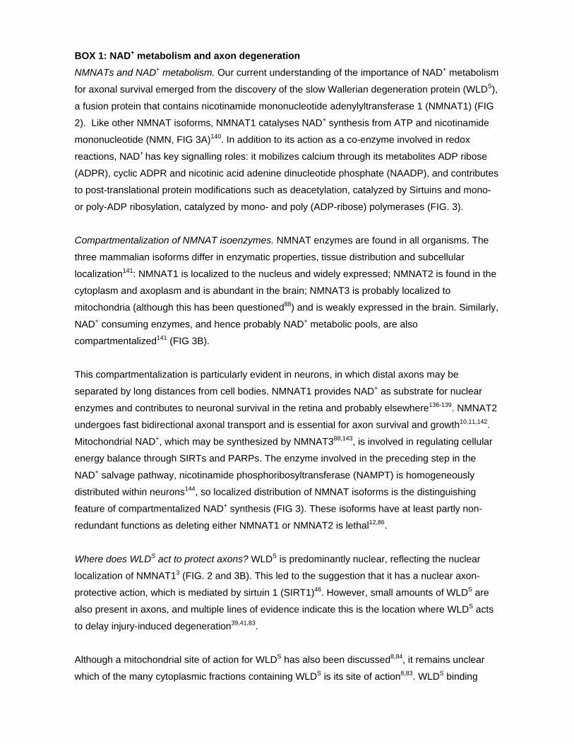
BOX 1: NAD+ metabolism and axon degeneration
NMNATs and NAD+ metabolism. Our current understanding of the importance of NAD+ metabolism
for axonal survival emerged from the discovery of the slow Wallerian degeneration protein (WLDS),
a fusion protein that contains nicotinamide mononucleotide adenylyltransferase 1 (NMNAT1) (FIG
2). Like other NMNAT isoforms, NMNAT1 catalyses NAD+ synthesis from ATP and nicotinamide
mononucleotide (NMN, FIG 3A)140. In addition to its action as a co-enzyme involved in redox
reactions, NAD+ has key signalling roles: it mobilizes calcium through its metabolites ADP ribose
(ADPR), cyclic ADPR and nicotinic acid adenine dinucleotide phosphate (NAADP), and contributes
to post-translational protein modifications such as deacetylation, catalyzed by Sirtuins and mono-
or poly-ADP ribosylation, catalyzed by mono- and poly (ADP-ribose) polymerases (FIG. 3).
Compartmentalization of NMNAT isoenzymes. NMNAT enzymes are found in all organisms. The
three mammalian isoforms differ in enzymatic properties, tissue distribution and subcellular
localization141: NMNAT1 is localized to the nucleus and widely expressed; NMNAT2 is found in the
cytoplasm and axoplasm and is abundant in the brain; NMNAT3 is probably localized to
mitochondria (although this has been questioned88) and is weakly expressed in the brain. Similarly,
NAD+ consuming enzymes, and hence probably NAD+ metabolic pools, are also
compartmentalized141 (FIG 3B).
This compartmentalization is particularly evident in neurons, in which distal axons may be
separated by long distances from cell bodies. NMNAT1 provides NAD+ as substrate for nuclear
enzymes and contributes to neuronal survival in the retina and probably elsewhere136-139. NMNAT2
undergoes fast bidirectional axonal transport and is essential for axon survival and growth10,11,142.
Mitochondrial NAD+, which may be synthesized by NMNAT388,143, is involved in regulating cellular
energy balance through SIRTs and PARPs. The enzyme involved in the preceding step in the
NAD+ salvage pathway, nicotinamide phosphoribosyltransferase (NAMPT) is homogeneously
distributed within neurons144, so localized distribution of NMNAT isoforms is the distinguishing
feature of compartmentalized NAD+ synthesis (FIG 3). These isoforms have at least partly non-
redundant functions as deleting either NMNAT1 or NMNAT2 is lethal12,86.
Where does WLDS act to protect axons? WLDS is predominantly nuclear, reflecting the nuclear
localization of NMNAT13 (FIG. 2 and 3B). This led to the suggestion that it has a nuclear axon-
protective action, which is mediated by sirtuin 1 (SIRT1)46. However, small amounts of WLDS are
also present in axons, and multiple lines of evidence indicate this is the location where WLDS acts
to delay injury-induced degeneration39,41,83.
Although a mitochondrial site of action for WLDS has also been discussed8,84, it remains unclear
which of the many cytoplasmic fractions containing WLDS is its site of action8,83. WLDS binding

partner VCP (FIGS. 2 and 3B) is widely distributed but is most abundant at the surface of
cytoplasmic organelles145. A highly protective WLDS variant in which nuclear localization is
disrupted partially localizes to mitochondria and endoplasmic reticulum83. Other protective
constructs, in which NMNAT1 is targeted to axons or cytoplasm are distributed among all
subcellular and cytosolic compartments9 or present with a diffuse extranuclear appearance6,
respectively. Lentivirally-transduced NMNAT1 does enter axons and strongly overexpressed
NMNAT3 is observed in the cytosol and cytoplasm in addition to mitochondria8. Thus,
overexpressed NMNAT1 and NMNAT3 may act in one of these ectopic locations to delay
Wallerian degeneration rather than in nuclei or mitochondria. Interestingly, NMNAT2 is axonally
transported on Golgi-derived vesicles, but its protective efficacy is greatly increased (to a level
comparable with WLDS) if it is detached from these vesicles38. This suggests a non-vesicular site of
action for NMNAT enzyme activity.

Nature Reviews | Neuroscience
Muscle
a Amyotrophic lateral sclerosis
Spinal cord
• Protein aggregation• Ageing• Toxins• ER stress
• Genetics• Oxidative stress• Poor glial support• Defective RNA processing
DRG
Vulnerable motor axon dies back
Resistantmotor axonsurvives
Sensory axon
b Alzheimer’s disease Cortex
Diffuse Aβ42
Dystrophic axon
Amyloid plaque
Degenerating axon
Surviving axon
• Aβ42• Ageing• Head injury• ApoE• Tau• Metabolic problems• Vascular pathology
c Wallerian degenerationKnown lesion with precise site and timing
Mixed nerve: sensory and motor axons, myelinated and unmyelinated axons
All axons degenerate after injury with broadly similar timing
Figure 1
Nature Reviews | Neuroscience
CNMNAT1
373
N70 18AA
1N
a WLDs
N
b SARM1
CSAM1
7041
ARM domain SAM2 TIR
VCP (p97) binding motif
Unknown function
NAD+ synthesis
Mitochondrial-localization
Dimerization SignallingAuto-inhibition?
Figure 2

Figure 3
N
O–
O
N
NH2
O
N+
O
OHOH
OP–O
O–
OO–
O
N+
O
OHOH
OP–O
O–
ONH2
ON+
O
OHOH
OP–O
O
ONH2
O
NO
OHOH
OP–O
O
N
N
N
NH2
N+
O
OHOH
OP–O
O
OO–
O
NO
OHOH
OP–O
O
N
N
N
NH2
Na NaMN NaAD+
Nam NMN NAD+
PRPP
Nature Reviews | Neuroscience
PPi
NaPRT
PRPP PPi
NAMPT
• FK866• CHS-828
ATP PPi
NMNAT
NADS
ATP
PPi
AMP
ATP PPi
NMNAT
a
b
Nucleus
Golgi
Rough ERMicrotubule
NMNAT2
Mitochondrion
WLDs
Ribosome
NMNAT1 NMNAT3
Kinesin Axonal smooth ER
Chromatin
NAMPT

Nature Reviews | Neuroscience
• Transection or crush• Vincrisitine• Vinblastine• MPP+
• Rotenone• Translation inhibitors• Energy deprivation• NGF withdrawal
Palmitoylation
NMNAT2PHR1
Init
iati
onEx
ecut
ion
NMN NAD+
SCG10
JNK
DLK
SARM1
Serine protease CRMP2
Akt
GSK3β
ZNRF1
IKK
ROS
mPTP
Mitochondrion
Ca2+
MORN4
Calpains
Calpastatin
Axon degeneration
Pathway activators
Putative core events
Putative modifying signalsDegradation orturnover
Core event, exact site in pathway unknown
Figure 4