Novel Stenotic Microchannels to Study Thrombus Formation ...
CARDIOVASCULAR ULTRASOUND
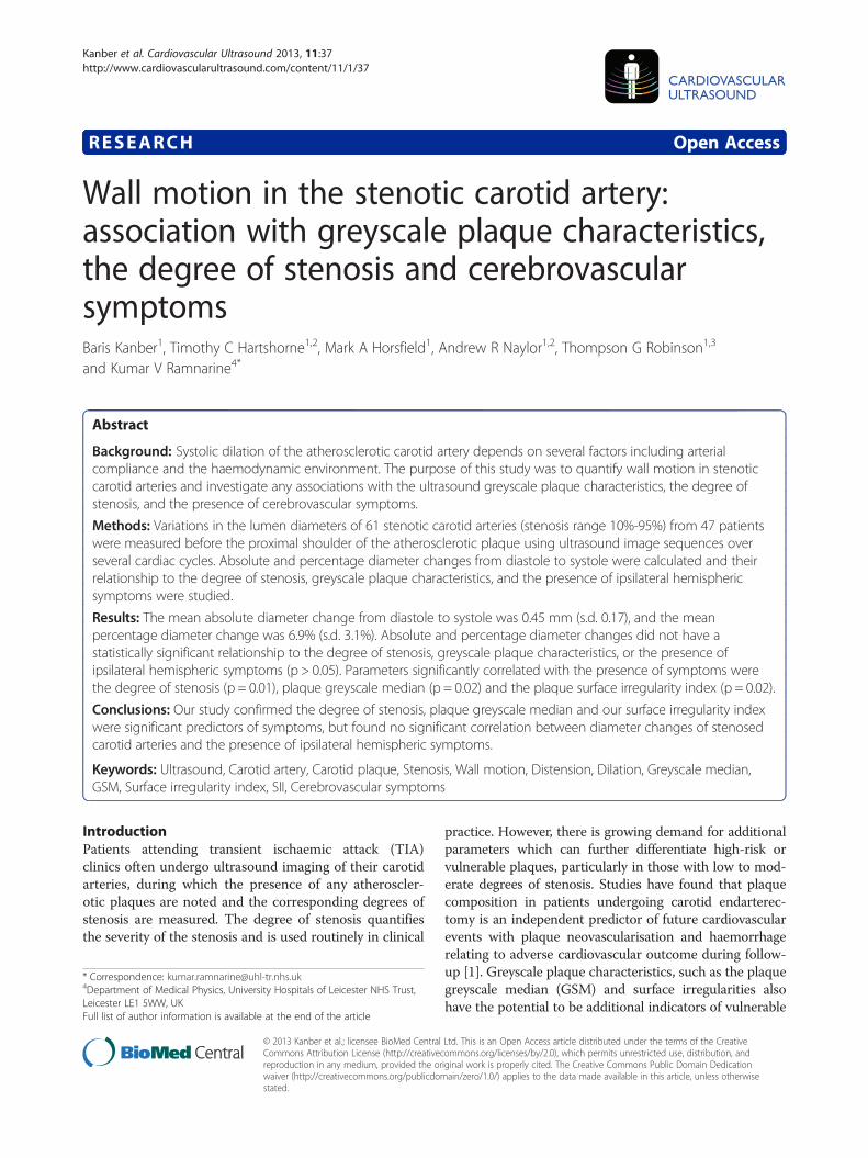
Kanber et al. Cardiovascular Ultrasound 2013, 11:37http://www.cardiovascularultrasound.com/content/11/1/37
RESEARCH Open Access
Wall motion in the stenotic carotid artery:association with greyscale plaque characteristics,the degree of stenosis and cerebrovascularsymptomsBaris Kanber1, Timothy C Hartshorne1,2, Mark A Horsfield1, Andrew R Naylor1,2, Thompson G Robinson1,3
and Kumar V Ramnarine4*
Abstract
Background: Systolic dilation of the atherosclerotic carotid artery depends on several factors including arterialcompliance and the haemodynamic environment. The purpose of this study was to quantify wall motion in stenoticcarotid arteries and investigate any associations with the ultrasound greyscale plaque characteristics, the degree ofstenosis, and the presence of cerebrovascular symptoms.
Methods: Variations in the lumen diameters of 61 stenotic carotid arteries (stenosis range 10%-95%) from 47 patientswere measured before the proximal shoulder of the atherosclerotic plaque using ultrasound image sequences overseveral cardiac cycles. Absolute and percentage diameter changes from diastole to systole were calculated and theirrelationship to the degree of stenosis, greyscale plaque characteristics, and the presence of ipsilateral hemisphericsymptoms were studied.
Results: The mean absolute diameter change from diastole to systole was 0.45 mm (s.d. 0.17), and the meanpercentage diameter change was 6.9% (s.d. 3.1%). Absolute and percentage diameter changes did not have astatistically significant relationship to the degree of stenosis, greyscale plaque characteristics, or the presence ofipsilateral hemispheric symptoms (p > 0.05). Parameters significantly correlated with the presence of symptoms werethe degree of stenosis (p = 0.01), plaque greyscale median (p = 0.02) and the plaque surface irregularity index (p = 0.02).
Conclusions: Our study confirmed the degree of stenosis, plaque greyscale median and our surface irregularity indexwere significant predictors of symptoms, but found no significant correlation between diameter changes of stenosedcarotid arteries and the presence of ipsilateral hemispheric symptoms.
Keywords: Ultrasound, Carotid artery, Carotid plaque, Stenosis, Wall motion, Distension, Dilation, Greyscale median,GSM, Surface irregularity index, SII, Cerebrovascular symptoms
IntroductionPatients attending transient ischaemic attack (TIA)clinics often undergo ultrasound imaging of their carotidarteries, during which the presence of any atheroscler-otic plaques are noted and the corresponding degrees ofstenosis are measured. The degree of stenosis quantifiesthe severity of the stenosis and is used routinely in clinical
* Correspondence: [email protected] of Medical Physics, University Hospitals of Leicester NHS Trust,Leicester LE1 5WW, UKFull list of author information is available at the end of the article
© 2013 Kanber et al.; licensee BioMed CentralCommons Attribution License (http://creativecreproduction in any medium, provided the orwaiver (http://creativecommons.org/publicdomstated.
practice. However, there is growing demand for additionalparameters which can further differentiate high-risk orvulnerable plaques, particularly in those with low to mod-erate degrees of stenosis. Studies have found that plaquecomposition in patients undergoing carotid endarterec-tomy is an independent predictor of future cardiovascularevents with plaque neovascularisation and haemorrhagerelating to adverse cardiovascular outcome during follow-up [1]. Greyscale plaque characteristics, such as the plaquegreyscale median (GSM) and surface irregularities alsohave the potential to be additional indicators of vulnerable
Ltd. This is an Open Access article distributed under the terms of the Creativeommons.org/licenses/by/2.0), which permits unrestricted use, distribution, andiginal work is properly cited. The Creative Commons Public Domain Dedicationain/zero/1.0/) applies to the data made available in this article, unless otherwise

Kanber et al. Cardiovascular Ultrasound 2013, 11:37 Page 2 of 11http://www.cardiovascularultrasound.com/content/11/1/37
plaques [2-10]. Ultrasound assessment of the stiffness ofthe carotid plaque using shearwave elastography is anemerging technique that may also provide additionalbenefit [11]. Another potentially useful parameter whichcan be easily measured from ultrasound scans performedin the TIA clinic is the systolic dilation or distension ofthe artery with the atherosclerotic plaque. The dilation ofthe carotid artery from diastole to systole depends on sev-eral factors including arterial stiffness, and previous stud-ies have shown that stiffer arteries are associated withatherosclerosis and are risk factors for stroke and othercardiovascular diseases [12-15]. The amount of arterialdilation is a physical parameter that may also affect thestability of the plaque, since greater arterial motion mayincrease the mechanical stress on the plaque and promoteinstability [16-20].The presence of the atherosclerotic plaque can have a
significant effect on arterial wall motion [21,22]. Onestudy found that arterial distensibility was not only sig-nificantly lower in the internal carotid artery where therewas a plaque, but it was also lower in the common ca-rotid artery of the affected side in comparison with thecontralateral common carotid artery, providing evidencethat the effect of a plaque on arterial mechanical proper-ties is not limited to the actual plaque site but rather ex-tends to a considerable degree in a proximal direction[21]. Computational models, on the other hand, showedthat the non-uniform thickness of the diseased arterialwall can restrict wall motion and re-distribute stress, giv-ing rise to increased stress concentrations at the plaqueshoulders [22]. Therefore, an assessment of the wall mo-tion characteristics of the stenosed carotid artery may pro-vide useful indicators that may correlate with the risk ofplaque rupture and the prevalence of symptoms.A previous study found that patients with acute
symptomatic carotid stenosis had impaired brachial flowmediated dilation (FMD) compared to patients withasymptomatic carotid stenosis in a patient populationwith greater than 50% reduction in the diameter of thecarotid artery [23]. That study showed that impairedbrachial FMD was an independent predictor of cerebralischaemic symptoms. However, not many studies haveconsidered whether the physiological dilation of thestenosed carotid artery itself might have any correlationto cerebrovascular symptoms, addressing the question ofwhether patients presenting with ipsilateral hemisphericsymptoms have distinctly different carotid artery dilationscompared to patients that do not. A study by Ramnarineet al. [24] looked at the physiological dilation of athero-sclerotic carotid arteries and correlated results with thedegree of stenosis, but any relationships to the presence ofpatient symptoms were not investigated. Another studyexamined the dilation characteristics of the carotid arteryat the level of the plaque and compared this with the
adjacent common carotid artery leading to a longitudinalstrain gradient estimation, but again, any relationships tothe presence of patient symptoms were not studied [25].More recently, Beaussier et al. [26] studied the longitu-dinal distension gradient between the plaque and the adja-cent common carotid artery with respect to the presenceof ipsilateral hemispheric symptoms and found no sta-tistically significant differences. However, their resultsdo not appear to indicate whether there were any sig-nificant differences in degree of arterial dilation at theadjacent carotid segment between the symptomatic andasymptomatic groups. That particular study also in-volved only a small number of carotid arteries with ipsi-lateral symptoms (n = 9).It is plausible that wall motion in the stenotic carotid
artery may affect the stability of the carotid plaque andconsequently, relate to the presence of ipsilateral hemi-spheric symptoms. Wall motion data along with the de-gree of stenosis and greyscale plaque characteristics mayhelp identify the vulnerable plaque. The purpose of thisstudy was, therefore, to test the hypothesis that the sys-tolic dilation of the stenosed carotid artery is related tothe presence of ipsilateral hemispheric symptoms andcan be used to differentiate between symptomatic andasymptomatic patients. Arterial wall motion was mea-sured before the proximal shoulder of the plaque as thisis an ideal, upstream location close to the plaque wherea well defined segment of the artery can often be found.The latter is important because arterial wall motionmeasurements across the plaque can suffer from highvariability [24]. Our investigation measured the absoluteand percentage dilation of stenotic carotid arteries, fromend diastole to peak systole, and explored whether theseparameters had any statistically significant associations tothe degree of stenosis, greyscale plaque characteristics,and the presence of ipsilateral hemispheric symptoms.
MethodsForty seven patients who attended the University Hospi-tals of Leicester NHS Trust’s Rapid Access TransientIschaemic Attack clinic were recruited. Variations in thelumen diameters of 61 stenotic carotid arteries (stenosisrange 10%-95%) were measured. The study was ap-proved by the National Research Ethics Service (NRES)Committee East Midlands - Northampton (reference 11/EM/0249) and followed institutional guidelines. Each pa-tient gave informed consent before participating in thestudy. Patients who did not have carotid artery stenosiswere excluded from the study. Carotid arteries for whichthe ultrasound image quality was poor were excludedfrom the wall motion analysis. Image sequences whichwere considered to be of poor quality included thosewith substantial image noise in the vessel lumen andthose with poorly defined vessel wall segments. In the

Kanber et al. Cardiovascular Ultrasound 2013, 11:37 Page 3 of 11http://www.cardiovascularultrasound.com/content/11/1/37
case of patients with stenosed left/right carotid arteries,each side was included and analyzed separately. In total,lumen diameter variations of 45 stenosed carotid arterieswere included in the final wall motion analysis. Carotid ar-teries with atherosclerotic plaque were classified as eitherhaving ipsilateral hemispheric cerebrovascular symptoms(i.e. symptomatic) or asymptomatic following specialistmedical review. Symptoms included aphasia, transientmonocular blindness and hemimotor/sensory symptomsconsistent with transient ischaemic attack or stroke.
Data acquisitionLongitudinal cross-sections of the carotid artery andplaque were imaged by experienced sonographers usinga Philips iU22 ultrasound scanner (Philips Healthcare,Eindhoven, The Netherlands) and an L9-3 probe. Acquisi-tions included B-Mode (i.e. greyscale) and Colour Dopplerimage sequences. B-Mode image sequences were acquiredusing the vascular carotid preset on the scanner (Vasc Carpreset, persistence low, XRES and SONOCT on) and wererecorded in DICOM format over an average of 6 cardiaccycles (mean frame rate was 32 frames per second). Gainwas optimized by the experienced sonographers. In thecase of B-Mode acquisitions, the greyscale transfer curvewas set to Gray Map 2, as this was reported to be the mostlinear transfer curve on this scanner [27]. Colour Dopplercine-loops were used as a qualitative aid to identifying thelocation and extent of carotid plaques, while the B-Mode data were used for the quantitative analyses in-cluding that of arterial wall motion and greyscale plaquecharacteristics.
AnalysisQuantitative analyses were carried out using MATLABversion 7.14, release 2012a (MathWorks, Natick,Massachusetts, USA) and employed an image processingalgorithm to track and measure arterial lumen diametersover time. This algorithm was based on a probabilisticapproach to vessel lumen segmentation [28]. In this ap-proach, given a point B with probability Pb of being in thearterial lumen of interest, the probability Pa that aneighbouring point A was also part of the same lumenwas proportional to Pb with a Gaussian fall in probabilitywith increasing greyscale contrast between the two points(Equation 1). Here Gb and Ga were the greyscale inten-sities of points B and A, and the constant ζ was deter-mined by considering the amount of greyscale contrast(Gth) required to reduce Pa to 1/2 that of Pb.
Pa ¼ Pb exp − Gb−Gað Þ2=ζ� �whereζ ¼ Gth
2=log 2
ð1Þ
The arterial lumen detection technique based on thisalgorithm was previously found to have good arterial
wall tracking performance, comparable to that of TissueDoppler Imaging [29].Arterial diameter variation waveforms (Figure 1) were
obtained before the proximal shoulder of the plaque, butas close to it as possible, and averaged over a region ap-proximately 3 mm long for each image frame (Figure 2).The measurements were made without prior knowledgeof the patient’s symptomatic status. The peaks of thediameter variation waveforms (Figure 1) were taken tobe the (peak) systolic values and the troughs as the (end)diastolic. The absolute value of the systolic arterial dila-tion was calculated as the increase in the arterial lumendiameter from diastole to systole and percentage systolicdilation as the same figure divided by the diastolic diam-eter. The same calculations were carried out for all thecardiac cycles observed on the arterial diameter variationwaveforms and averages were taken. Normalized andun-normalized plaque GSM and surface irregularity in-dices (SII) were obtained using previously describedmethods [8,10] while the degree of stenosis of the corre-sponding arteries were measured using criteria consist-ent with the NASCET method utilizing blood flowvelocities in conjunction with the B-Mode and colourflow imaging [30-32].
Statistical analysisStatistical analyses were carried out using SPSS version20 (IBM Corporation, Armonk, New York, USA). Thenon-parametric Wilcoxon-Mann–Whitney test was usedto determine whether quantitative measurements suchas the absolute and percentage diameter changes, degreeof stenosis, and greyscale plaque characteristics differedsignificantly between patient groups (e.g. symptomatic/asymptomatic, hypertensive/normotensive, etc.). A furtherANCOVA test was carried out between the percentagesystolic dilation of the artery and symptomatic status, con-trolling for the effects of the diastolic arterial diameter.Pearson’s correlation was used to determine whether theabsolute and percentage diameter changes had any statis-tical relationship to the age of the patient, the degree ofstenosis, and the greyscale plaque characteristics. Partialcorrelations, controlling for the effects of the baseline dia-stolic diameters, were also carried out for the percentagediameter changes. Finally, logistic regression was carriedout to investigate which parameters significantly corre-lated with the presence of ipsilateral hemispheric symp-toms. Two-tailed values of significance were used andvalues less than 0.05 were considered to be statisticallysignificant.
ReproducibilityIn order to assess the reproducibility of the arterial wallmotion detection technique used, we investigated the intra-observer coefficients of variation for the measurement of

Figure 1 Example of an arterial dilation waveform showing lumen diameter variations of a carotid artery throughout severalcardiac cycles.
Figure 2 A carotid bifurcation plaque and illustration of the location of the diameter measurements. In this case, the plaque appears onthe carotid bulb, and diameter measurements are taken in the distal common carotid artery immediately before the proximal shoulder ofthe plaque.
Kanber et al. Cardiovascular Ultrasound 2013, 11:37 Page 4 of 11http://www.cardiovascularultrasound.com/content/11/1/37

Figure 3 Box and whisker plots showing the distribution, versus the presence of ipsilateral hemispheric symptoms, of the absoluteand percentage arterial diameter changes, degree of stenosis, normalized and un-normalized plaque GSM, and the surface irregularityindex (SII).
Kanber et al. Cardiovascular Ultrasound 2013, 11:37 Page 5 of 11http://www.cardiovascularultrasound.com/content/11/1/37
the systolic/diastolic diameters, and absolute/percentagediameter changes for 10 arteries. This subset of arterieswere selected from the available dataset to give a widerange of stenosis severity, plaque echogenicity and arterialdiameters for reproducibility analysis. The measurementswere made by the same operator and in sequential order.The same ultrasound acquisition sequences were used foreach artery respectively.
Table 1 Non-parametric Wilcoxon-Mann–Whitneyassociations between the absolute and percentagesystolic diameter changes before the proximal shoulderof the atherosclerotic plaque and patient characteristics
Patient characteristic Significance (p-value)
Absolute diameterchange
Percentage diameterchange
Age 0.44 0.33
Sex 0.96 0.68
Hypertension 0.45 0.34
Hypercholesterolaemia 0.54 0.41
Ischaemic heart disease 0.94 0.90
Diabetes mellitus 0.31 0.28
Previous TIA/stroke 0.16 0.32
Smoking 0.34 0.49
Alcohol 0.29 0.47
Family history of stroke 0.35 0.41
Age was dichotomized using the median of the dataset as a cut-off value.
Comparison against manual measurementsArterial diameter measurements made using our methodwere compared against diameter measurements mademanually by placing cursors on the ultrasound imagesand measuring the distances between the near and farwalls of the arteries. This was done using a computerprogram with a graphical user interface written inMATLAB version 7.14, release 2012a (MathWorks,Natick, Massachusetts, USA). Using the same arteriesselected for our reproducibility analysis, we manuallymeasured arterial diameters at the same location and onmatching image frames as for the automated technique.Approximately 30 diameter measurements per arteryspread over the cardiac cycles were compared betweenthe two techniques. Arterial diameters obtained usingthe manual method were compared with those obtainedusing the automated technique using Bland-Altman andlinear regression analysis.
ResultsMean age was 77.3 years (range 58–95 years); 19 female.The prevalence of cerebrovascular risk factors was: 76%hypertension, 53% hypercholesterolaemia, 33% ischaemicheart disease, 29% diabetes mellitus, 47% previous TIA/stroke, 64% smoking, 44% alcohol consumption and 33%family history of stroke. Thirty one of the 61 arteries

Kanber et al. Cardiovascular Ultrasound 2013, 11:37 Page 6 of 11http://www.cardiovascularultrasound.com/content/11/1/37
studied were found to be associated with ipsilateralhemispheric symptoms, while the remaining 30 werefound to be asymptomatic following expert specialiststroke physician assessment.The mean percentage systolic dilation of the symptom-
atic arteries (6.6%) was lower than that of the asymptom-atic arteries (7.2%), but this difference was not statisticallysignificant (p = 0.16, Figure 3). ANCOVA, controlling forthe effects of the diastolic diameters, also found the samedifference to be not statistically significant (p = 0.14). Arter-ies with ipsilateral hemispheric symptoms also had lowerabsolute diameter changes on average (0.42 mm) thanasymptomatic arteries (0.47 mm) but this difference wasalso not significant (p = 0.17, Figure 3). The degree of sten-osis (p < 0.01), normalized plaque GSM (p = 0.021) and theplaque surface irregularity index (p = 0.016) differed signifi-cantly between the symptomatic and asymptomatic groupswhile the un-normalized plaque GSM (p = 0.14) did not.
Figure 4 Box and whisker plots showing the distribution of the perce
Patient characteristics age, sex, hypertension, high chol-esterol, ischaemic heart disease, diabetes mellitus, previ-ous TIA/stroke, smoking, alcohol consumption, andfamily history of stroke did not show significant differ-ences in the percentage and absolute systolic dilation ofthe arteries (Table 1, Figures 4 and 5). There were nostatistically significant correlations between the percentagesystolic dilation of arteries and the degree of stenosis(p = 0.82), patient age (p = 0.14), un-normalized plaqueGSM (p = 0.29), normalized plaque GSM (p = 0.34) or theplaque surface irregularity index (p = 0.54, Figure 6). Par-tial correlations, adjusting for the effects of the baselinediastolic diameters, also found no statistically significantrelationship between the percentage systolic dilation ofthe arteries and the degree of stenosis, patient age, or thegreyscale plaque characteristics (p > 0.05 for all). Absolutediameter changes were also not significantly correlatedwith the degrees of stenosis (p = 0.70), patient age
ntage systolic diameter changes versus patient characteristics.

Figure 5 Box and whiskers plots showing the distribution of the absolute systolic diameter changes versus patient characteristics.
Figure 6 Scatter plots of the absolute and percentage systolic diameter changes versus patient age, degree of stenosis, un-normalizedand normalized plaque GSM, and the plaque surface irregularity index (SII), illustrating a lack of association between the absolute andpercentage systolic dilation and any of these parameters.
Kanber et al. Cardiovascular Ultrasound 2013, 11:37 Page 7 of 11http://www.cardiovascularultrasound.com/content/11/1/37
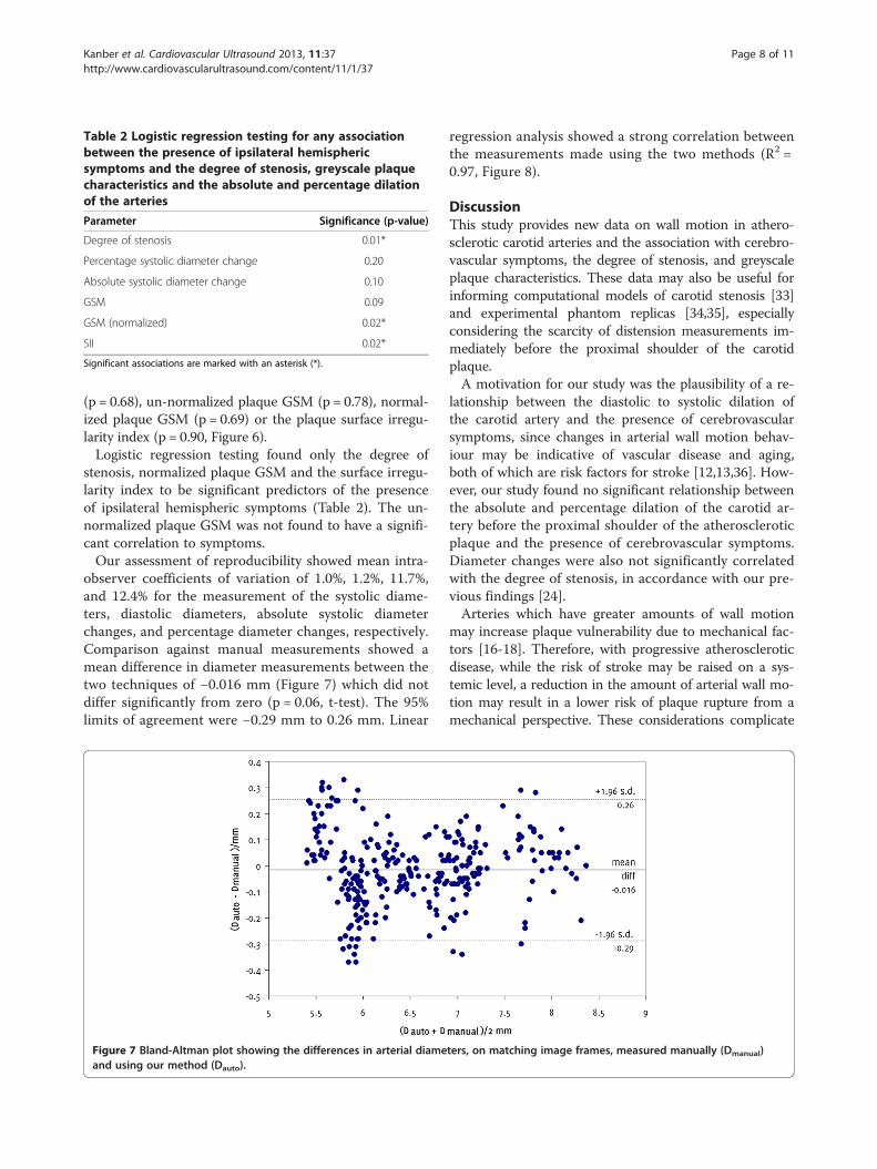
Table 2 Logistic regression testing for any associationbetween the presence of ipsilateral hemisphericsymptoms and the degree of stenosis, greyscale plaquecharacteristics and the absolute and percentage dilationof the arteries
Parameter Significance (p-value)
Degree of stenosis 0.01*
Percentage systolic diameter change 0.20
Absolute systolic diameter change 0.10
GSM 0.09
GSM (normalized) 0.02*
SII 0.02*
Significant associations are marked with an asterisk (*).
Kanber et al. Cardiovascular Ultrasound 2013, 11:37 Page 8 of 11http://www.cardiovascularultrasound.com/content/11/1/37
(p = 0.68), un-normalized plaque GSM (p = 0.78), normal-ized plaque GSM (p = 0.69) or the plaque surface irregu-larity index (p = 0.90, Figure 6).Logistic regression testing found only the degree of
stenosis, normalized plaque GSM and the surface irregu-larity index to be significant predictors of the presenceof ipsilateral hemispheric symptoms (Table 2). The un-normalized plaque GSM was not found to have a signifi-cant correlation to symptoms.Our assessment of reproducibility showed mean intra-
observer coefficients of variation of 1.0%, 1.2%, 11.7%,and 12.4% for the measurement of the systolic diame-ters, diastolic diameters, absolute systolic diameterchanges, and percentage diameter changes, respectively.Comparison against manual measurements showed amean difference in diameter measurements between thetwo techniques of −0.016 mm (Figure 7) which did notdiffer significantly from zero (p = 0.06, t-test). The 95%limits of agreement were −0.29 mm to 0.26 mm. Linear
Figure 7 Bland-Altman plot showing the differences in arterial diameand using our method (Dauto).
regression analysis showed a strong correlation betweenthe measurements made using the two methods (R2 =0.97, Figure 8).
DiscussionThis study provides new data on wall motion in athero-sclerotic carotid arteries and the association with cerebro-vascular symptoms, the degree of stenosis, and greyscaleplaque characteristics. These data may also be useful forinforming computational models of carotid stenosis [33]and experimental phantom replicas [34,35], especiallyconsidering the scarcity of distension measurements im-mediately before the proximal shoulder of the carotidplaque.A motivation for our study was the plausibility of a re-
lationship between the diastolic to systolic dilation ofthe carotid artery and the presence of cerebrovascularsymptoms, since changes in arterial wall motion behav-iour may be indicative of vascular disease and aging,both of which are risk factors for stroke [12,13,36]. How-ever, our study found no significant relationship betweenthe absolute and percentage dilation of the carotid ar-tery before the proximal shoulder of the atheroscleroticplaque and the presence of cerebrovascular symptoms.Diameter changes were also not significantly correlatedwith the degree of stenosis, in accordance with our pre-vious findings [24].Arteries which have greater amounts of wall motion
may increase plaque vulnerability due to mechanical fac-tors [16-18]. Therefore, with progressive atheroscleroticdisease, while the risk of stroke may be raised on a sys-temic level, a reduction in the amount of arterial wall mo-tion may result in a lower risk of plaque rupture from amechanical perspective. These considerations complicate
ters, on matching image frames, measured manually (Dmanual)

Figure 8 Scatter plot showing a strong linear relationship between arterial diameters measured manually (Dmanual) and using ourmethod (Dauto).
Kanber et al. Cardiovascular Ultrasound 2013, 11:37 Page 9 of 11http://www.cardiovascularultrasound.com/content/11/1/37
the relationship between the dilation characteristics of thecarotid artery before the proximal shoulder of the ath-erosclerotic plaque and the presence of cerebrovascularsymptoms, and are likely to be factors that contribute tothe absence of a difference in the carotid artery dilationsof the symptomatic and asymptomatic patients found inthis study.Arterial lumen diameters measured using our tech-
nique were found to be comparable to those measuredusing a manual method. Our study found good reprodu-cibility for the measurement of the diastolic and systolicdiameters but lower reproducibility for the measurementof the absolute and percentage diameter changes. Theseresults are in accordance with previous studies whichfound derived parameters combining the systolic anddiastolic arterial diameters to be considerably less repro-ducible than to the diameter readings on their own[37,38]. It has been reported that even a small variance inarterial diameter measurements may cause a considerablevariance in the derived metrics of carotid distension,therefore, limiting its potential usability in the clinicalsetting [38]. Godia et al. attributed the different and some-times conflicting results reported on the association be-tween carotid distension and cardiovascular outcomes tothis variability [38]. In the present study, the greater vari-abilities associated with absolute and percentage diameterchanges may be additional factors contributing to the ab-sence of a difference found in the carotid artery dilationsof symptomatic and asymptomatic patients. Studies in-corporating larger datasets or more precise methods maybe able to find such a difference.
The statistically significant relationship between thepresence of ipsilateral hemispheric symptoms and boththe normalized plaque GSM and the surface irregularityindex confirm our previous findings [8,10]. Interestingly,the un-normalized plaque GSM was not found to be asignificant predictor of symptoms. This may be indicativeof variations in overall image brightness, due to differ-ences in ultrasound gain settings or tissue attenuation,and highlights the importance of the normalization pro-cedure for GSM measurements.A limitation of this study is that we did not have pulse
pressure measurements which would have allowed us toquantify arterial distensibility. However, this study fo-cussed on motion aspects rather than stiffness and ispart of our broader research aim to develop and define aplaque risk index based on ultrasound measurements.Previously, we have quantified greyscale plaque charac-teristics such as the plaque GSM and surface irregular-ities [8,10] as possible indicators of vulnerable plaquesand the present study was conducted to investigate thepotential of dilation characteristics before the proximalplaque shoulder as an additional parameter to include in aprospective vulnerable plaque-stroke risk model. In thisstudy we did not perform measurements across the plaqueto assess any differential wall motion between the plaqueand the proximal carotid segment. Our previous studyusing Tissue Doppler Imaging demonstrated a variety ofpertinent wall motion features across the plaque site thatmay be related to the biophysics of arterial disease. How-ever, high variability demonstrated the limitations of arterialwall motion measurements across the plaque, in contrast

Kanber et al. Cardiovascular Ultrasound 2013, 11:37 Page 10 of 11http://www.cardiovascularultrasound.com/content/11/1/37
to more robust measurements that can be performed onwell defined segments of vessels [24].
ConclusionsThis study investigated the systolic dilation of stenosedcarotid arteries measured before the proximal shoulderof the atherosclerotic plaque. Absolute and percentagediameter changes were lower for the arteries of patientswith ipsilateral hemispheric symptoms, but these differ-ences were not statistically significant. Normalized plaqueGSM and our novel surface irregularity index were foundto be significant predictors of symptoms.
Competing interestsThe authors declare that they have no competing interests.
Authors’ contributionsThe study was conceived by KVR and BK. Ultrasound data were collected byTCH while the analyses were carried out by BK. All authors contributed tothe interpretation and presentation of the results and, read and approvedthe final manuscript.
AcknowledgementsThe research was funded by and took place at the National Institute forHealth Research (NIHR) Collaboration for Leadership in Applied HealthResearch and Care based at the University Hospitals of Leicester NHS Trust.The views expressed are those of the authors and not necessarily those ofthe NHS, the NIHR or the Department of Health.
Author details1Department of Cardiovascular Sciences, University of Leicester, Leicester, UK.2Department of Surgery, University Hospitals of Leicester NHS Trust, Leicester,UK. 3NIHR Biomedical Research Unit for Cardiovascular Sciences, University ofLeicester, Leicester, UK. 4Department of Medical Physics, University Hospitalsof Leicester NHS Trust, Leicester LE1 5WW, UK.
Received: 5 September 2013 Accepted: 4 October 2013Published: 20 October 2013
References1. Hellings WE, Peeters W, Moll FL, Piers SRD, Van Setten J, Van Spek D, Peter J,
De Vries JPM, Seldenrijk KA, De Bruin PC, Vink A, Velema E, De Kleijn DPV,Pasterkamp G: Composition of carotid atherosclerotic plaque isassociated with cardiovascular outcome: a prognostic study.Circulation 2010, 121(17):1941–1950.
2. Elatrozy T, Nicolaides A, Tegos T, Griffin M: The objective characterisationof ultrasonic carotid plaque features. Eur J Vasc Endovasc Surg 1998,16(3):223–230.
3. Tegos TJ, Stavropoulos P, Sabetai MM, Khodabakhsh P, Sassano A,Nicolaides AN: Determinants of carotid plaque instability: echoicity versusheterogeneity. Eur J Vasc Endovasc Surg 2001, 22(1):22–30.
4. Biasi GM, Sampaolo A, Mingazzini P, De Amicis P, El-barghouty N, NicolaidesAN: Computer analysis of ultrasonic plaque echolucency in identifyinghigh risk carotid bifurcation lesions. Eur J Vasc Endovasc Surg 1999,17(6):476–479.
5. Salem MK, Sayers RD, Bown MJ, West K, Moore D, Nicolaides A, RobinsonTG, Naylor AR: Patients with recurrent ischaemic events from carotidartery disease have a large lipid core and low GSM. Eur J Vasc EndovascSurg 2012, 43(2):147–153.
6. Grønholdt ML, Nordestgaard BG, Schroeder TV, Vorstrup S, Sillesen H:Ultrasonic echolucent carotid plaques predict future strokes. Circulation2001, 104(1):68–73.
7. Polak JF, Shemanski L, Leary DH, Lefkowitz D, Price TR, Savage PJ, Brant WE,Reid C: Hypoechoic plaque at US of the carotid artery: an independentrisk factor for incident stroke in adults aged 65 years or older.Cardiovascular Health Study. Radiology 1998, 208(3):649–654.
8. Kanber B, Hartshorne TC, Horsfield MA, Naylor AR, Robinson TG, RamnarineKV: Dynamic variations in the ultrasound greyscale median of carotidartery plaques. Cardiovasc Ultrasound 2013, 11(1):21.
9. Prabhakaran S, Rundek T, Ramas R, Elkind MSV, Paik MC, Boden-albala B,Sacco RL: Carotid plaque surface irregularity predicts ischemic stroke: thenorthern Manhattan study. Stroke 2006, 37(11):2696–2701.
10. Kanber B, Hartshorne TC, Horsfield MA, Naylor AR, Robinson TG, Ramnarine KV:Quantitative Assessment of Carotid Plaque Surface Irregularities andCorrelation to Cerebrovascular Symptoms. Cardiovasc Ultrasound. in press.
11. Ramnarine KV, Garrard JW, Dexter K, Nduwayo S, Panerai RB, Robinson TG:Shear wave elastography assessment of carotid plaque stiffness: in-vitroreproducibility study. Ultrasound Med Biol 2013. in press.
12. Dijk JM, Van Der Graaf Y, Grobbee DE, Banga JD, Bots ML: Increased arterialstiffness is independently related to cerebrovascular disease andaneurysms of the abdominal aorta: the second manifestations of arterialdisease (SMART) study. Stroke 2004, 35(7):1642–1646.
13. Leone N, Ducimetière P, Gariépy J, Courbon D, Tzourio C, Dartigues J,Ritchie K, Alpérovitch A, Amouyel P, Safar ME, Zureik M: Distension of thecarotid artery and risk of coronary events: the three-city study.Arterioscler Thromb Vasc Biol 2008, 28(7):1392–1397.
14. Agabiti-rosei E, Muiesan ML: Carotid atherosclerosis, arterial stiffness andstroke events. Adv Cardiol 2006, 44:173–186.
15. Rothwell PM: Carotid artery disease and the risk of ischaemic stroke andcoronary vascular events. Cerebrovasc Dis 2000, 10(Suppl 5):21–33.
16. Lenzi GL, Vicenzini E: The ruler is dead: an analysis of carotid plaquemotion. Cerebrovasc Dis 2006, 23(2–3):121–125.
17. Kashiwazaki D, Yoshimoto T, Mikami T, Muraki M, Fujimoto S, Abiko K,Kaneko S: Identification of high-risk carotid artery stenosis: motion ofintraplaque contents detected using B-mode ultrasonography.J Neurosurg 2013, 117(3):574–578.
18. Kume S, Hama S, Yamane K, Wada S, Nishida T, Kurisu K: Vulnerable carotidarterial plaque causing repeated ischemic stroke can be detected withB-mode ultrasonography as a mobile component: jellyfish sign.Neurosurg Rev 2011, 33(4):419–430.
19. Meairs S, Hennerici M: Four-dimensional ultrasonographiccharacterization of plaque surface motion in patients with symptomaticand asymptomatic carotid artery stenosis. Stroke 1999, 30(9):1807–1813.
20. Bang J, Dahl T, Bruinsma A, Kaspersen JH, Nagelhus Hernes TA, Myhre HO: Anew method for analysis of motion of carotid plaques from RFultrasound images. Ultrasound Med Biol 2003, 29(7):967–976.
21. Giannattasio C, Failla M, Emanuelli G, Grappiolo A, Boffi L, Corsi D, Mancia G:Local effects of atherosclerotic plaque on arterial distensibility.Hypertension 2001, 38(5):1177–1180.
22. Lee KW, Wood NB, Xu XY: Ultrasound image-based computer model of acommon carotid artery with a plaque. Med Eng Phys 2004, 26(10):823–840.
23. Hsu H, Chen Y, Sheu WH, Sheng W, Chao A: Comparison of brachial arteryflow-mediated vasodilatation in symptomatic and asymptomaticpatients with carotid arterial stenosis. Am J Cardiol 2002, 90(7):814–816.
24. Ramnarine KV, Hartshorne T, Sensier Y, Naylor M, Walker J, Naylor AR,Panerai RB, Evans DH: Tissue Doppler imaging of carotid plaque wallmotion: a pilot study. Cardiovasc Ultrasound 2003, 1:17.
25. Paini A, Boutouyrie P, Calvet D, Zidi M, Agabiti-rosei E, Laurent S: Multiaxialmechanical characteristics of carotid plaque: analysis by multiarrayechotracking system. Stroke 2006, 38(1):117–123.
26. Beaussier H, Naggara O, Calvet D, Joannides R, Guegan-massardier E, GerardinE, Iacob M, Laloux B, Bozec E, Bellien J, Touze E, Masson I, Thuillez C,Oppenheim C, Boutouyrie P, Laurent S: Mechanical and structuralcharacteristics of carotid plaques by combined analysis with echotrackingsystem and MR imaging. JACC Cardiovasc Imaging 2011, 4(5):468–477.
27. Jansen M, Van Alfen N, Nijhuis Van Der Sanden MWG, Van Dijk JP, Pillen S,De Groot IJM: Quantitative muscle ultrasound is a promising longitudinalfollow-up tool in Duchenne muscular dystrophy. Neuromuscul Disord2012, 22(4):306–317.
28. Kanber B, Ramnarine KV: A Probabilistic Approach to Computerized Trackingof Arterial Walls in Ultrasound Image Sequences. ISRN Signal Processing; 2012.
29. Ramnarine KV, Kanber B, Panerai RB: Assessing the performance of vesselwall tracking algorithms: the importance of the test phantom. J PhysConf Ser 2004, 1:199–204.
30. North American Symptomatic Carotid Endarterectomy Trial Collaborators:Beneficial effect of carotid endarterectomy in symptomatic patients withhigh-grade carotid stenosis. N Engl J Med 1991, 325(7):445–453.

Kanber et al. Cardiovascular Ultrasound 2013, 11:37 Page 11 of 11http://www.cardiovascularultrasound.com/content/11/1/37
31. Grant EG, Benson CB, Moneta GL, Alexandrov AV, Baker JD, Bluth EI, CarrollBA, Eliasziw M, Gocke J, Hertzberg BS, Katanick S, Needleman L, Pellerito J,Polak JF, Rholl KS, Wooster DL, Zierler RE: Carotid artery stenosis: gray-scale and Doppler US diagnosis–society of radiologists in ultrasoundconsensus conference. Radiology 2003, 229(2):340–346.
32. Oates CP, Naylor AR, Hartshorne T, Charles SM, Fail T, Humphries K, AslamM, Khodabakhsh P: Joint recommendations for reporting carotidultrasound investigations in the United Kingdom. Eur J Vasc EndovascSurg 2008, 37(3):251–261.
33. Long Q, Xu XY, Ramnarine KV, Hoskins P: Numerical investigation ofphysiologically realistic pulsatile flow through arterial stenosis. J Biomech2001, 34(10):1229–1242.
34. Meagher S, Poepping TL, Ramnarine KV, Black RA, Hoskins PR: Anatomical flowphantoms of the nonplanar carotid bifurcation, part II: experimentalvalidation with Doppler ultrasound. Ultrasound Med Biol 2007, 33(2):303–310.
35. Watts DM, Sutcliffe CJ, Morgan RH, Meagher S, Wardlaw J, Connell M, BastinME, Marshall I, Ramnarine KV, Hoskins PR, Black RA: Anatomical flowphantoms of the nonplanar carotid bifurcation, part I: computer-aideddesign and fabrication. Ultrasound Med Biol 2007, 33(2):296–302.
36. Schmidt-trucksäss A, Grathwohl D, Schmid A, Boragk R, Upmeier C, Keul J,Huonker M: Structural, functional, and hemodynamic changes of thecommon carotid artery with age in male subjects. Arterioscler ThrombVasc Biol 1999, 19(4):1091–1097.
37. Kanters SD, Elgersma OE, Banga JD, Van Leeuwen MS, Algra A:Reproducibility of measurements of intima-media thickness anddistensibility in the common carotid artery. Eur J Vasc Endovasc Surg 1998,16(1):28–35.
38. Godia EC, Madhok R, Pittman J, Trocio S, Ramas R, Cabral D, Sacco RL,Rundek T: Carotid artery distensibility: a reliability study. J Ultrasound Med2007, 26(9):1157–1165.
doi:10.1186/1476-7120-11-37Cite this article as: Kanber et al.: Wall motion in the stenotic carotidartery: association with greyscale plaque characteristics, the degree ofstenosis and cerebrovascular symptoms. Cardiovascular Ultrasound2013 11:37.
Submit your next manuscript to BioMed Centraland take full advantage of:
• Convenient online submission
• Thorough peer review
• No space constraints or color figure charges
• Immediate publication on acceptance
• Inclusion in PubMed, CAS, Scopus and Google Scholar
• Research which is freely available for redistribution
Submit your manuscript at www.biomedcentral.com/submit