VPMC-8140B Whats New in Vitrea 4.0 - Allen...
46
©2007 Vital Images, Inc. VPMC-8140B Version 4.0
-
Upload
duongkhanh -
Category
Documents
-
view
214 -
download
1
Transcript of VPMC-8140B Whats New in Vitrea 4.0 - Allen...

©2007 Vital Images, Inc. VPMC-8140B
Version 4.0

©2007 Vital Images, Inc. VPMC-8140B
For Help Using Vitrea
Click the Vitrea Help tab, which contains the Vitrea 2 User GuidesOR
Contact Vital Images Customer Support:1.800.208.3005, M-F, 7 a.m. to 7 p.m. (Central time zone)
Vitrea Safety and Regulatory ConsiderationsFor general Vitrea Safety and Regulatory Considerations, refer to the Safety and Regulatory
Considerations section of the Basic Vitrea® 2 manual.
Customer Education at ViTAL UFor more information, or to register, contact our training coordinator:
1.952.487.9559 or [email protected]
©2007 Vital Images, Inc. VPMC-8140B
Quick Links
Click any link below to jump to a specific section of this document.
Basic VitreaReporting
Cardiovascular•EP Planning•Vessel Probe
•Coronary Artery Analysis•SUREPlaque
CT Brain PerfusionColon

©2007 Vital Images, Inc. VPMC-8140B
Basic Vitrea
• Right-click Menu
- Right-click in any view to display
- Protocol specific
• Re-designed Tool Panels
- Buttons re-arranged
- Undo button removed in tool panel
Use Right-click Menu, CTRL-Z, or F11 to undo.

©2007 Vital Images, Inc. VPMC-8140B
Basic Vitrea (continued)
• ALT-3D-TOOL alternative
- Click and drag immediately in 3D view to rotate volume
- Click, hold briefly, and drag in 3D view to apply active tool
• Pre-defined annotations
- Activate the Label tool and click in view to display a dictionary of pre-defined terms
- Select a term from the dictionary
- Save new terms to the dictionary

©2007 Vital Images, Inc. VPMC-8140B
Basic Vitrea (continued)
- In-viewer MPR Rendering and Thickness settings
Slider bar - thickness
Dropdown - MPR rendering

©2007 Vital Images, Inc. VPMC-8140B
Basic Vitrea (continued)
• New presets on Gallery window
Many protocols have updated presets
Note: Clear the Use Modified Presets checkbox to see new presets.

©2007 Vital Images, Inc. VPMC-8140B
Reporting
Report Window Enhancements• New layout
• Filterable list of findings (snapshots, batches, movies)
• Multi-selection of snapshots
• Quick Preview of snapshots, batches and movies
• Direct DICOM export of snapshots and batches
• Large Snapshot tray
• A set of new report templates including text pages with selectable/editable text
• A new comprehensive cardiac template with auto-population of findings
©2007 Vital Images, Inc. VPMC-8140B
Report Window Enhancements (continued)
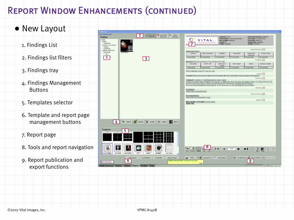
•New Layout
1. Findings List
2. Findings list filters
3. Findings tray
4. Findings Management Buttons
5. Templates selector
6. Template and report page management buttons
7. Report page
8. Tools and report navigation
9. Report publication and export functions
1
2
3
4
5
6
7
8
9
©2007 Vital Images, Inc. VPMC-8140B
Report Window Enhancements (Continued)
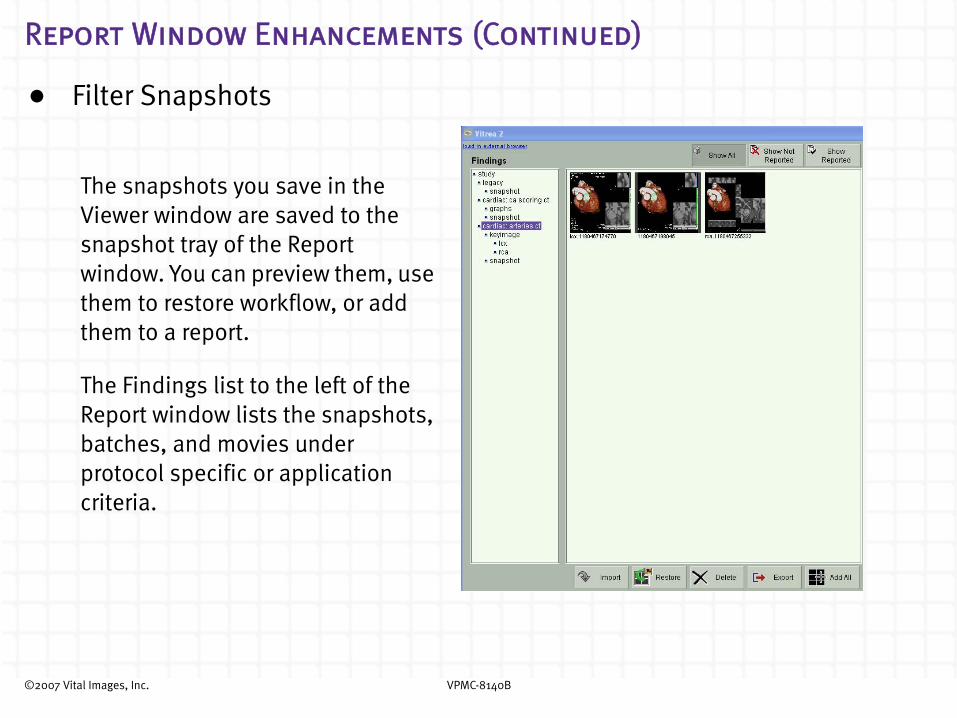
• Filter Snapshots
The snapshots you save in the Viewer window are saved to the snapshot tray of the Report window. You can preview them, use them to restore workflow, or add them to a report.
The Findings list to the left of the Report window lists the snapshots, batches, and movies under protocol specific or application criteria.
©2007 Vital Images, Inc. VPMC-8140B
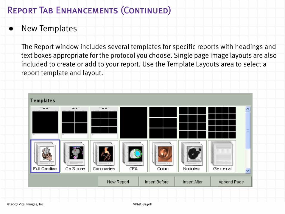
Report Tab Enhancements (Continued)
• New Templates
The Report window includes several templates for specific reports with headings and text boxes appropriate for the protocol you choose. Single page image layouts are also included to create or add to your report. Use the Template Layouts area to select a report template and layout.

©2007 Vital Images, Inc. VPMC-8140B
Report Window Enhancements (continued)
• Multi-selection SnapshotsClick Add All to add all snapshots that currently display in the snapshot tray to the Report Page.
CTRL + Click Press CTRL and click all snapshots that you want to drag to the Report Page. The selected snapshots have a blue border around them.
©2007 Vital Images, Inc. VPMC-8140B
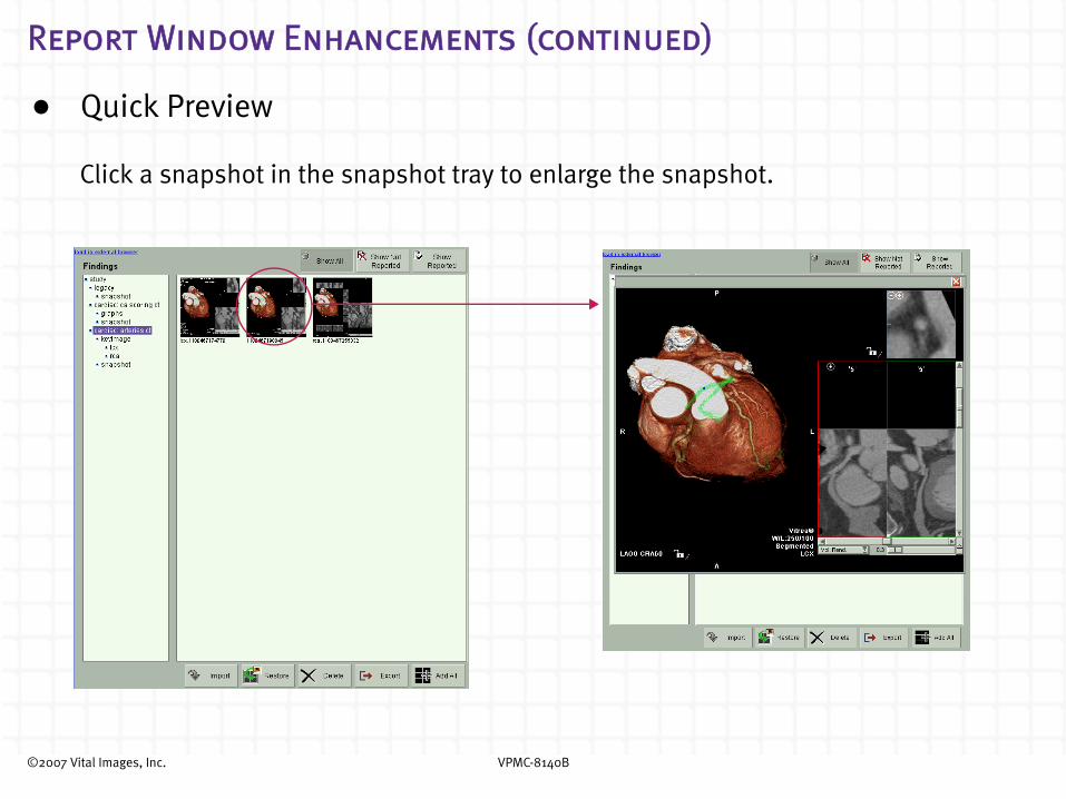
Report Window Enhancements (continued)
• Quick Preview
Click a snapshot in the snapshot tray to enlarge the snapshot.

©2007 Vital Images, Inc. VPMC-8140B
Report Window Enhancements (continued)
• Direct DICOM Export of Snapshots
Select your snapshots and click to export the selected snapshots and batch
to PACS.
• Preview Movies in the Report

©2007 Vital Images, Inc. VPMC-8140B
Cardiovascular
EP Planning
• New Separately-licensed EP Planning option for VitreaCardiac EP Planning is a post processing advanced visualization application that is intended to be used for the analysis and assessment of the heart including the atria, pulmonary veins, and coronary sinus. The application provides analysis tools which include a number of display, quantitative measurement and 3D model export capabilities for use with the St. Jude Ensite® System. The application can be used to aid trained physicians in the visualization and assessment of cardiac anatomy.
©2007 Vital Images, Inc. VPMC-8140B
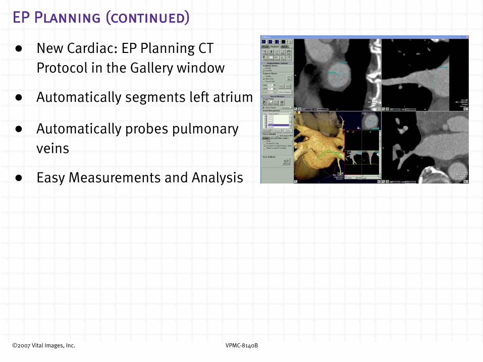
EP Planning (continued)
• New Cardiac: EP Planning CT Protocol in the Gallery window
• Automatically segments left atrium
• Automatically probes pulmonary veins
• Easy Measurements and Analysis

©2007 Vital Images, Inc. VPMC-8140B
EP Planning (continued)
• Show Esophagus - Window level in the esophagus and view in Heart mode
• View in Fly Through mode
• Save the 3D model, burn it to a blank CD, and send it to the EP lab

©2007 Vital Images, Inc. VPMC-8140B
Vessel Probe
• New vessel tracking algorithm for carotid, renal, and peripheral vessels
• New visualization modes and layouts
- Larger images for detailed diagnosis
- Comparative cross-sectional display modes

©2007 Vital Images, Inc. VPMC-8140B
Vessel Probe (continued)
Montage Layout
This is the default layout in the 1-up format. This view contains several 1mm cross-sectional views of the vessel.

©2007 Vital Images, Inc. VPMC-8140B
Vessel Probe (continued)
Large Cross-sectional Layout
By default, the center cross-sectional inset displays the plane of the intersection with the middle of the curved view. This view contains three larger cross-sectional views.

©2007 Vital Images, Inc. VPMC-8140B
Vessel Probe (continued)
CathView display mode (Angiographic overview image)
Cath view and 3D view project in the same plane.

©2007 Vital Images, Inc. VPMC-8140B
Vessel Probe (continued)
• Image manipulation tools
- Automatic centering of 3D images
- Independent zooming for curved views
©2007 Vital Images, Inc. VPMC-8140B
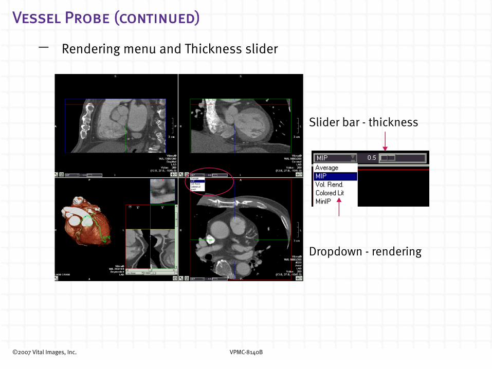
Vessel Probe (continued)
- Rendering menu and Thickness slider
Slider bar - thickness
Dropdown - rendering

©2007 Vital Images, Inc. VPMC-8140B
Vessel Probe (continued)
- Rotating Curved MPRs
Left click/drag with crosshair on to rotate

©2007 Vital Images, Inc. VPMC-8140B
Vessel Probe (continued)
- Auto-oblique Lock-Unlock
Scroll CPR views independent of oblique MPRs
Option to lock together (auto-oblique)
Button: Description:
Locked mode - Scrolling the insets will cause the global crosshair and oblique/curved planes to update. Unlocked mode - Unlocking resets the oblique planes to an orthogonal orientation. The lock will reset when a new patient is loaded.
©2007 Vital Images, Inc. VPMC-8140B
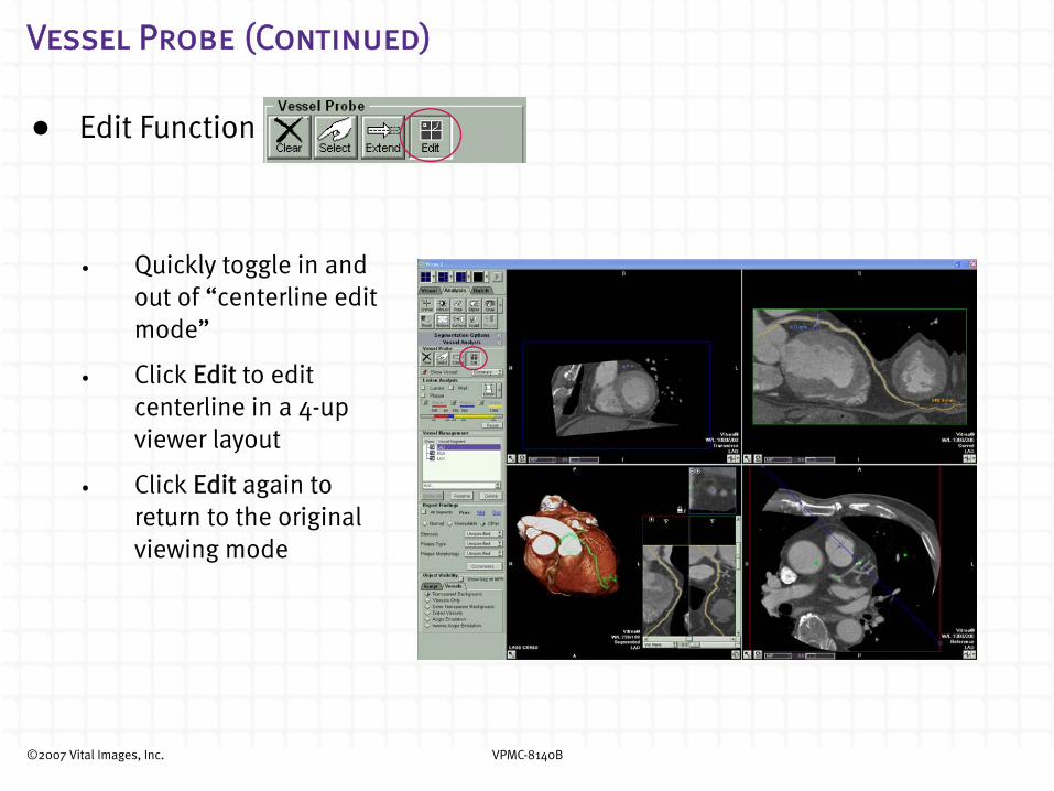
Vessel Probe (Continued)
• Edit Function
• Quickly toggle in and out of “centerline edit mode”
• Click Edit to edit centerline in a 4-up viewer layout
• Click Edit again to return to the original viewing mode

©2007 Vital Images, Inc. VPMC-8140B
Vessel Probe (continued)
• Editing Tools
- Centerline editing in 3D and curved MPR
©2007 Vital Images, Inc. VPMC-8140B
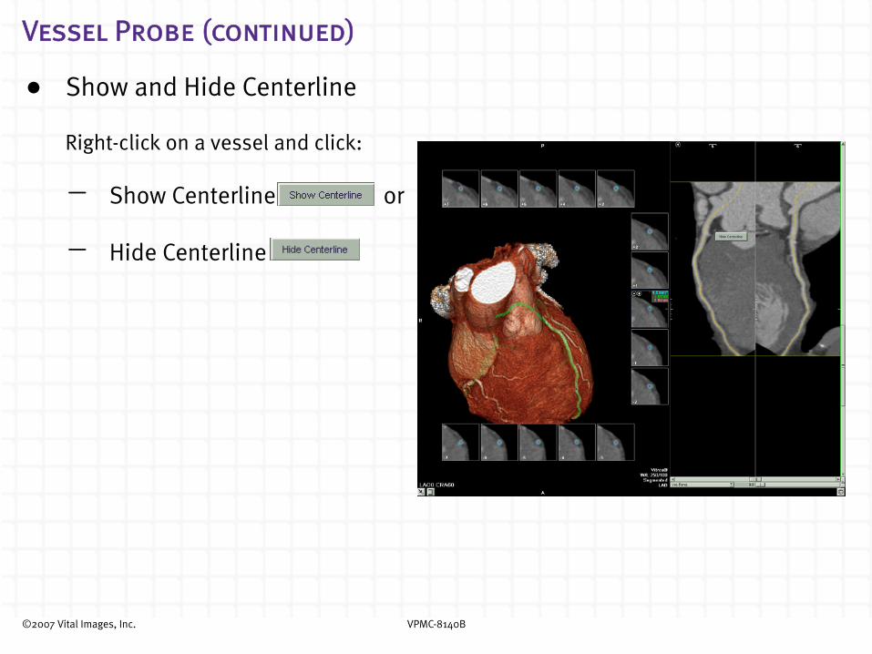
Vessel Probe (continued)
• Show and Hide Centerline
Right-click on a vessel and click:
- Show Centerline or
- Hide Centerline
©2007 Vital Images, Inc. VPMC-8140B
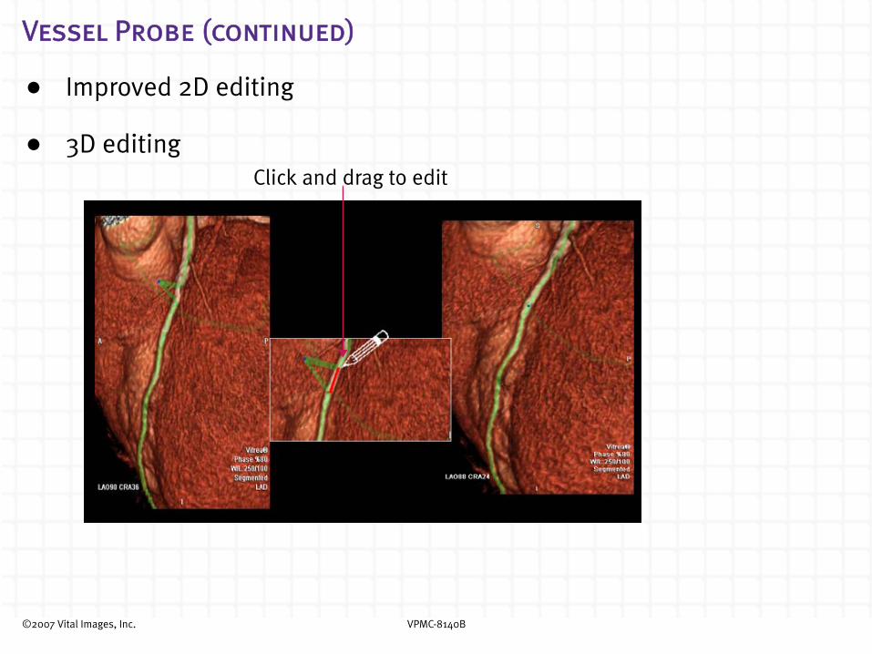
Vessel Probe (continued)
• Improved 2D editing
• 3D editingClick and drag to edit
©2007 Vital Images, Inc. VPMC-8140B
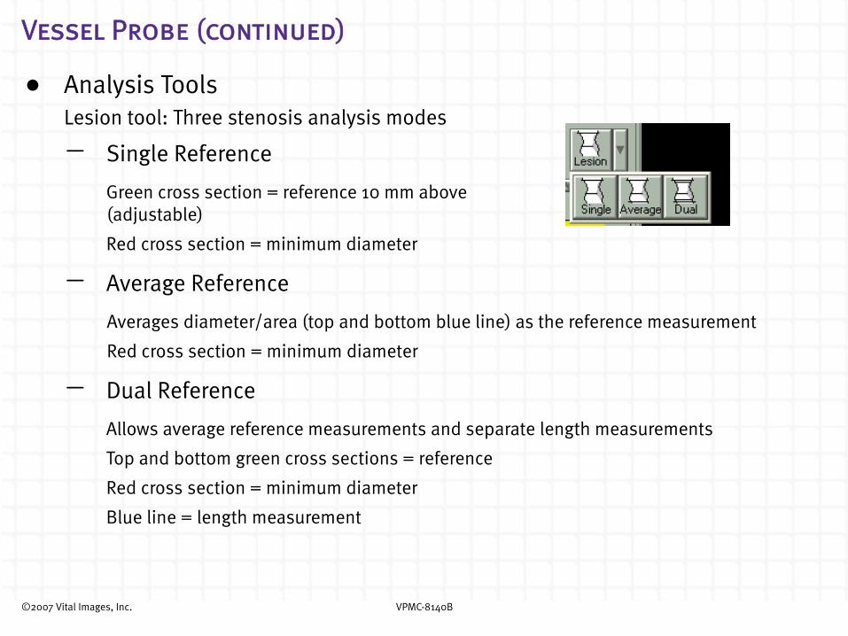
Vessel Probe (continued)
• Analysis ToolsLesion tool: Three stenosis analysis modes
- Single Reference
Green cross section = reference 10 mm above (adjustable)
Red cross section = minimum diameter
- Average Reference
Averages diameter/area (top and bottom blue line) as the reference measurement
Red cross section = minimum diameter
- Dual Reference
Allows average reference measurements and separate length measurements
Top and bottom green cross sections = reference
Red cross section = minimum diameter
Blue line = length measurement

©2007 Vital Images, Inc. VPMC-8140B
Vessel Probe (continued)
• Lesion Measurements
The percentage area stenosis is calculated using the new formula:
% area stenosis = (1 - Stenosed lumen area / Reference lumen area) × 100%
The percentage diameter stenosis is calculated using the new formula:
% diameter stenosis = (1 - Stenosed min lumen diameter / Reference min lumen diameter) × 100%
©2007 Vital Images, Inc. VPMC-8140B
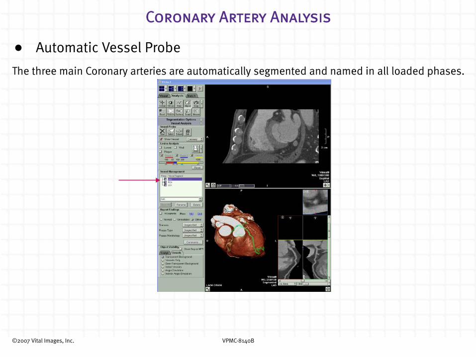
Coronary Artery Analysis
• Automatic Vessel Probe
The three main Coronary arteries are automatically segmented and named in all loaded phases.

©2007 Vital Images, Inc. VPMC-8140B
Coronary Artery Analysis (continued)
• Vessel Management
- Add dropdown box - Select the dropdown to add new vessels, grafts, or stents to probe or report on. You can also enter a name in the Add box.
- Rename vessels and lesions - Click Rename or double-click the vessel name
or lesion name to rename the lesion.
Rename vessels and lesions
Add dropdown box
©2007 Vital Images, Inc. VPMC-8140B
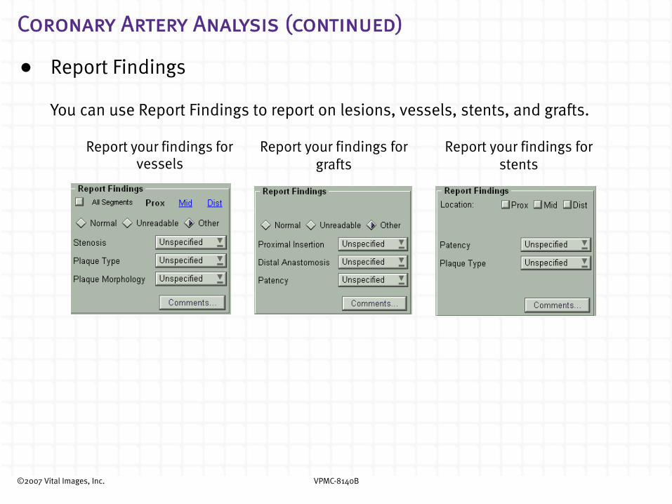
Coronary Artery Analysis (continued)
• Report Findings
You can use Report Findings to report on lesions, vessels, stents, and grafts.
Report your findings for vessels
Report your findings for grafts
Report your findings for stents

©2007 Vital Images, Inc. VPMC-8140B
Coronary Artery Analysis (continued)
Use the Key Image icon to capture key images you want to display in the Findings section of the Report tab.
©2007 Vital Images, Inc. VPMC-8140B
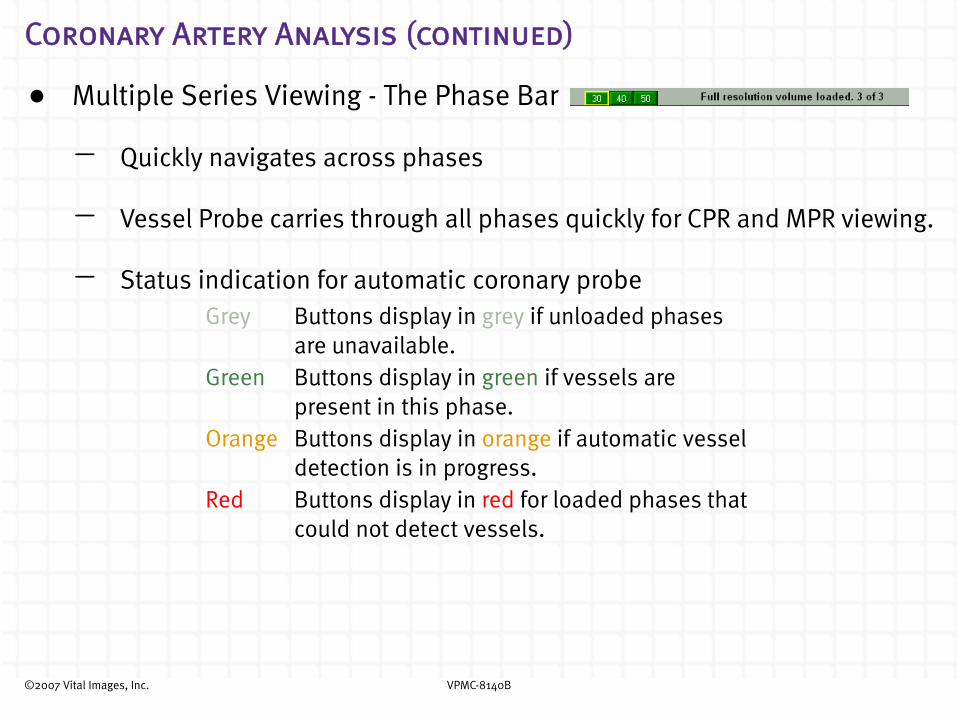
Coronary Artery Analysis (continued)
• Multiple Series Viewing - The Phase Bar
- Quickly navigates across phases
- Vessel Probe carries through all phases quickly for CPR and MPR viewing.
- Status indication for automatic coronary probeGrey Buttons display in grey if unloaded phases
are unavailable.Green Buttons display in green if vessels are
present in this phase.Orange Buttons display in orange if automatic vessel
detection is in progress.Red Buttons display in red for loaded phases that
could not detect vessels.
©2007 Vital Images, Inc. VPMC-8140B
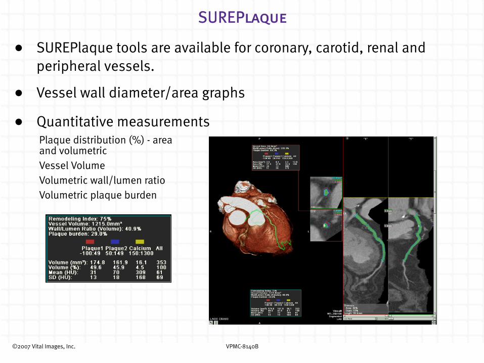
SUREPlaque
• SUREPlaque tools are available for coronary, carotid, renal and peripheral vessels.
• Vessel wall diameter/area graphs
• Quantitative measurementsPlaque distribution (%) - area and volumetricVessel VolumeVolumetric wall/lumen ratioVolumetric plaque burden

©2007 Vital Images, Inc. VPMC-8140B
CT Brain Perfusion
• Improved Algorithm
Singular value decomposition (SVD)
• Multi-series capability
Option to load some Toshiba multi-slice datasets into a single volume
• Automatically selected artery & vein locations
Manual editing available if automatic selections need editing

©2007 Vital Images, Inc. VPMC-8140B
CT Brain Perfusion (continued)
• Motion Correction
If necessary, correct for in-plane head motion.
• Viewer window formats
Select from four different Viewer window layouts
1-up 4-up 4-up Runoff 6-up
2D axial 2D axialrCBVrCBFMTT
2D axial (large pane)rCBVrCBFMTT
2D axialrCBVTTPrCBFMTT
Delay
Apply Motion Correction
Undo Motion Correction

©2007 Vital Images, Inc. VPMC-8140B
CT Brain Perfusion (continued)
• Time-to-Peak (TTP) and Delay maps
Displayed in 6-up Viewer window format
2D Axial
rCBF
MTT
TTP
Delay
rCBV
©2007 Vital Images, Inc. VPMC-8140B
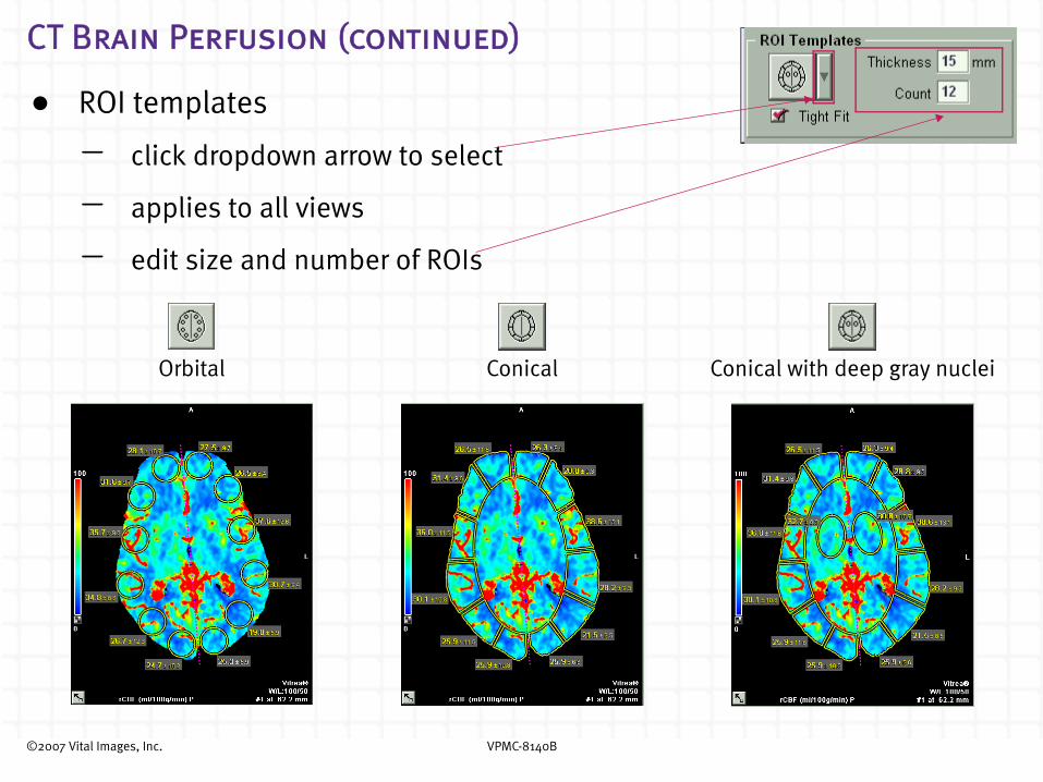
CT Brain Perfusion (continued)
• ROI templates
- click dropdown arrow to select
- applies to all views
- edit size and number of ROIs
Orbital Conical Conical with deep gray nuclei

©2007 Vital Images, Inc. VPMC-8140B
CT Brain Perfusion (continued)
• Color Tables
- Click the color scale to choose from four color tables
- Click Invert Color Scale to invert the color scale
• Vessels/Noise
- Check Vessels to display pixels belonging to vessels
- Check Noise to display noisy pixels
Spectrum
3-Color
ASIST - Japan
Grayscale
©2007 Vital Images, Inc. VPMC-8140B
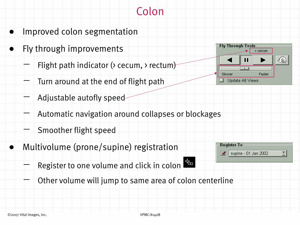
Colon
• Improved colon segmentation
• Fly through improvements
- Flight path indicator (> cecum, > rectum)
- Turn around at the end of flight path
- Adjustable autofly speed
- Automatic navigation around collapses or blockages
- Smoother flight speed
• Multivolume (prone/supine) registration
- Register to one volume and click in colon
- Other volume will jump to same area of colon centerline

©2007 Vital Images, Inc. VPMC-8140B
Colon (continued)
• Polyp Probe
- Probe polyps
• Click Select, then click polyp
• Measurements displayed
• Distance to rectum displayed
- Edit polyp contour
• Click Add
• Drag edge of contour to edit
- Manage Polyps
• Rename, Delete, Match
• All findings, including arrows, listed
• Sorted by distance to rectum
• Select finding in the list to navigate to that area in the colon
• Press SPACEBAR to navigate through the list
©2007 Vital Images, Inc. VPMC-8140B
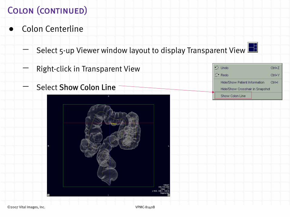
Colon (continued)
• Colon Centerline
- Select 5-up Viewer window layout to display Transparent View
- Right-click in Transparent View
- Select Show Colon Line

©2007 Vital Images, Inc. VPMC-8140B
© Vital Images, Inc. 2007
Protected by U.S. Patents 5,986,662; 6,130,671; 6,219,059; 7,031,504; Other Patents Pending in the U.S. and other countries.
VPMC-8140B What’s New in Vitrea® 2? Version 4.0
No part of this work may be reproduced, stored in a retrieval system, or transmitted in any form or by any means, electronic or mechanical, including photocopying and recording, or by any information storage or retrieval system without permission in writing from Vital Images.
TrademarksVitrea is a registered trademark of Vital Images, Inc.
Aquilion(TM) and SUREPlaque are trademarks of Toshiba Medical Systems Corp.
Restricted Rights LegendIf this software or documentation is delivered to the Department of Defense (DOD) of the U.S. Government, it is delivered with Restricted Rights as follows:
Use, duplication or disclosure of the software by the U.S. Government is subject to restrictions as set forth in subparagraph (c)(l)(ii) of the Rights in Technical Data and Computer Software clause at DFARS 252.227-7013.
If this software or documentation is delivered to any unit or agency of the U.S. Government other than DOD, it is delivered with Restricted Rights and use, duplication or disclosure by the U.S. Government is subject to the restrictions as set forth in FAR 52.227-19 (c)(2). If the software or documentation is delivered to NASA, it is delivered with Restricted Rights subject to the restrictions set forth in 18-52.227-86(d) of the NASA FAR Supplement.
Limits of Liability and Disclaimer of WarrantyVITAL IMAGES SHALL HAVE NO LIABILITY OF ANY KIND FOR ANY DIRECT, INCIDENTAL OR CONSEQUENTIAL DAMAGES BASED ON ANY DEFECT, FAILURE OR MALFUNCTION OF THE SOFTWARE,OR USE OF ANY VITAL IMAGES DOCUMENTATION, WHETHER THE CLAIM IS BASED UPON WARRANTY, CONTRACT, TORT OR OTHERWISE. VITAL IMAGES MAKES NO WARRANTY, EXPRESS OR IMPLIED, INCLUDING BUT NOT LIMITED TO, ANY WARRANTY OF MERCHANTABILITY OR FITNESS FOR A PARTICULAR PURPOSE WHETHER ARISING FROM STATUTE, COMMON LAW, CUSTOM OR OTHERWISE.
Notice of ConfidentialityThis software and the information in this software including, but not limited to, the ideas, concepts and know-how are proprietary, confidential and trade secret to Vital Images, and the information contained therein shall be maintained as proprietary, confidential and trade secret to Vital Images and shall not be copied or reproduced in any form whatsoever. This software and any information contained therein shall not be disclosed to anyone other than authorized representatives of the user's employer, who is contractually obligated not to disclose same without the express written consent of Vital Images. The user of this software and any information contained therein shall not attempt to discern Vital Images' confidential and trade secret information and shall not reverse compile, disassemble, or otherwise reverse engineer this software or any information contained therein.
Software License NoticeThis software is a licensed product of and is distributed by Vital Images, and may only be used according to the terms of that license on the system identified in the License Agreement. In the event of any conflict between these terms and the terms of any written agreement or agreement assented to through electronic means with Vital Images, the terms of such written or assented agreement shall control.
MediMark® Europe11 rue Emile ZOLA, BP 2332, 38033GRENOBLE CEDEX 2, FranceTel: +33 476 86 43 22Fax:+33 476 17 19 82email: [email protected]
MediMark® Europe is an authorized representative in the European Community and acts on behalf of Vital Images, Inc. in the communication of safety-related incidents and regulatory matters with Competent Authorities in the European Community. Distributors are still the first line of communication with their customers regarding service and complaints.