Volume 2 • Issue 1 • 2020
78
www.myjo.org Volume 2 • Issue 1 • 2020 KUGLER PUBLICATIONS www.kuglerpublications.com
Transcript of Volume 2 • Issue 1 • 2020

www.myjo.org
Volume 2 • Issue 1 • 2020
KUGLER PUBLICATIONSwww.kuglerpublications.com

www.myjo.org
ABOUT THE COVER IMAGEThe Pregnant Eye by Dr. Shahidatul Adha Mohamad, Registrar of the Department of Ophthalmology, Universiti Sains Malaysia, was the winner of the Photography Contest held during the 9th Conjoint Ophthalmology Scientific Conference in Kelantan, Malaysia.

The Malaysian Journal of Ophthalmology (MyJO) is the official journal for the Malaysian Society of Ophthalmol-ogy (MSO), College of Ophthalmologists Malaysia, and Malaysian Universities Conjoint Committee in Ophthal-mology (MUCCO). MUCCO is the national board responsible for training ophthalmologists in Malaysia, comprising the Universiti Kebangsaan Malaysia, Universiti Malaya, and Universiti Sains Malaysia, as well as the Ministry of Health.
MyJO aims to provide a platform for ophthalmologists, clinicians, researchers, trainees, students, optome-trists, and eye care providers to publish their work and to promote knowledge enhancement among ophthalmolo-gists and eye care providers in Malaysia.
I: https://myjo.orgE: [email protected]
CopyrightAuthors who publish in MyJO agree to the following terms:a. Authors retain copyright and grant the journal MyJO
right of first publication, with the work twelve (12) months after publication simultaneously licensed under a Creative Commons Attribution License that allows others to share the work with an acknowl-edgement of the work’s authorship and initial publication in MyJO.
b. After 12 months from the date of publication, authors are able to enter into separate, additional contractual arrangements for the non-exclusive dis-tribution of MyJO’s published version of the work, with an acknowledgement of its initial publication in MyJO.
Chief editorLiza Sharmini Ahmad Tajudin
Deputy editorNorlina Ramli
Editorial boardAmir Samsudin Bariah Mohd Ali Gangadhara Sundar Goh Pik Pin Jay Kumar ChhablaniJemaima Che Hamzah Kenneth Choong-Sian FongKhairidzan Mohd Kamal Lee Mun Wai Mae-Lynn Catherine BastionMohd Aziz Husni Mohtar Ibrahim Norfariza NgahNorshamsiah Md Din Nurliza Khaliddin Sabong Srivannaboon Satoshi Kashii Shatriah Ismail Tengku Ain KamaldenWan Hazabbah Wan Hitam
Advisory board Aung TinFang Seng KheongPeng KhawStephanie Watson Timothy YY Lai
Bahasa Malaysia Translator: Azhany YaakubTuan Jamil Tuan Muhammad

Open access policyMyJO provides immediate open access to its content aft er (free) registration, on the principle that making research freely available to the public supports a greater global exchange of knowledge. There are no fees required to publish in the journal.
Article referencingMyJO is primarily an electronic journal that will occasion-ally publish print issues. Articles may be referenced by appending the article ID included in the first page header of each article (i.e., © Malaysian Journal of Ophthalmology 2019; #articleID) using the following url:https://www.MyJO.com/index.php/myjo/article/view/#ar-ticleID.
DisclaimersAll published articles, including editorials and letters, represent the opinions of the authors and do not reflect the off icial policy of MyJO, its sponsors, the publisher or the institution with which the author is aff iliated, unless this is clearly specified. Although every eff ort has been made to ensure the technical accuracy of the contents of MyJO, no responsibility for errors or omissions is accepted. MyJO and the publisher do not endorse or guarantee, directly or indirectly, the quality or eff icacy of any product or service described the advertisements or other material that is commercial in nature in any issue. All advertising is expected to conform to ethical and medical standards. No responsibility is assumed by MyJO or the publisher for any injury and/or damage to persons or property as a matter of products liability, negligence or otherwise, or from any use or operation of any methods, products, instructions, or ideas contained in the material herein. Because of rapid advances in the medical sciences, independent verification of diagnoses and drug dosages should be made.
ISSNOnline: 2665-9565Print: 2665-9557
MalaysiaOnline: 2716-5329Print: 2716-5248
PublisherMalaysian Society ofOphthalmologyUnit #UG33, PJ Midtown,Jalan Kemajuan, Seksyen13, 46200 Petaling Jaya,[email protected]
Kugler PublicationsP.O. Box 205381001 NM AmsterdamThe [email protected]
Manuscript submissionsAuthor guidelines and templates are available via the website, through which all manuscripts should be submitted. For inquiries please contact us via e-mail.
Publication frequencyMyJO is published four issues per year (quarterly) electronically.
Advertising inquiriesMyJO off ers online and in print sponsorship and advertising opportunities. Please contact Kugler Publications for inquiries.

Sponsors

Table of contents
Editorial
From my laptop 6Liza-Sharmini Ahmad Tajudin
Endogenous endophthalmitis: experience from the south of peninsular Malaysia 16Norfariza Ngah
Original articles
Retina & ocular inflammationA five-year retrospective hospital-based study of endogenous endophthalmitis in south Malaysia 18Hayatulrizal Muhd, Ling Kiet Phang, Francesca Martina Vendargon
GlaucomaEvaluation of spontaneous retinal venous pulsation in primary open-angle and primary angle-closure glaucoma patients 27Sylves Patrick, Chan Hui Tze, Rasdi Abdul Rashid, Liza Sharmini Ahmad Tajudin
Brief reports
Retina & ocular inflammationUse of intracameral tenecteplase for rapid resolution of intraocular fibrin in acute anterior uveitis 42Lee Wen Yee, Teh Wee Min, Ling Kiet Phang, Norlina Mohd Ramli, Haslina Mohd Ali
Case reports
Orbit & oculoplastyA case of panophthalmitis with orbital cellulitis related to Erysipelothrix rhusiopathiae infection: a rare ocular infection 48Aasiah Ahmad Sharifuddin, Krishnalatha Buandasan
Neurophthalmology & ocular problems in systemic diseasesOcular complications in pre-eclampsia 55Dian Nadia Abu Talib, Wahidah Wagimon, Ainal Adlin Naffi, Rona Asnida Nasaruddin, Jemaima Che-Hamzah, Mohd Hashim Omar, Norshamsiah Md Din
Isolated abducens nerve palsy secondary to lung metastases 62Hayatulrizal Muhd, Hayati Abdul Aziz, Francesca Martina Vendargon, Jasmi Ramlan, Yeoh Aik Guan
Retina & ocular inflammationCases of cytomegalovirus retinitis in patients treated for cancer 69Marium Jamaluddin Ahmad, Tengku Ain Kamalden, Nurliza Khaliddin, Tajunisah Iqbal

© Malaysian Journal of Ophthalmology 2020; 2:6Editorial comment
From my laptopProfessor Dr. Liza-Sharmini Ahmad Tajudin
Chief Editor
I have been contemplating writing this special note for the past year. It is the best time to write, after a successful first-year run of Malaysian Journal of Ophthalmol-ogy (MyJO). MyJO has been a shared dream among many in the ophthalmology fraternity in Malaysia for years; its birth is proof of the cohesiveness of ophthalmolo-gists in Malaysia represented by the Malaysian Society of Ophthalmology (MSO) and the College of Ophthalmologists and Malaysian Universities Conjoint Committee in Ophthalmology (MUCCO). It has been made possible with the support of Kugler Pub-lications’ dedicated team. A big THANK YOU to all!
MyJO has published a total of 4 issues with 28 interesting articles from Malaysia and abroad. The first volume of MyJO was made possible by the contribution of the authors (please keep it coming), tireless effort by the reviewers to ensure the good quality of the articles, and strong support on the part of our editorial board members. Big thank you to the enthusiastic budding ophthalmic photographers for the beautiful pictures gracing our covers: The volcano is erupting, Blooming web, The desert path, and Blue-eyed Melanau boy.
There were many roadblocks and bumps throughout our journey this past year, a learning process for many of us. I hope 2020 will bring us more success even in the midst of the COVID-19 pandemic. In this issue, we have included guidelines for ophthalmologists during the COVID-19 pandemic in Malaysia. This is a joint effort between the College of Ophthalmologists and MSO. MyJO is offering rapid publication for articles on the impact of the COVID-19 pandemic on ophthalmologi-cal practice. Let’s work together to flatten the curve.
Stay safe. Stay healthy. Stay productive and innovative.

A JOINT STATEMENT BY THE COLLEGE OF OPHTHALMOLOGISTS, AMM AND THE MALAYSIAN SOCIETY OF OPHTHALMOLOGISTSThe COVID-19 pandemic has forced us as ophthalmologists to live and practice in a different and highly risky medical reality. If unchecked this pandemic has the potential to kill tens to hundreds of thousands in Malaysia and tens of millions around the world. It presents a major challenge to our brave frontline health care workers throughout Malaysia and indeed around the globe.
During these challenging times ophthalmologists will need to continue to provide urgent and emergency care to patients to prevent vision loss. This despite the risk to themselves as eye examinations and treatment need close proximity to patients putting them and their entire team at particularly high risk of contracting COVID-19.
As ophthalmologists we need to also play a role from a greater public health perspective and in the present context three things need to be done on an urgent basis.
First, we must reduce the risk of the COVID-19 transmission from human to human and thus reduce the rate of new case development. Only then can a “flattening of the curve” be achieved so as not to overwhelm our very limited supply of hospital beds, ICU beds, and ventilators nationwide. Our window to modify the spread of disease is a narrow and closing one.
Second, we as Malaysian ophthalmologists, must conserve desperately needed disposable medical supplies in order that they can be shared or given to the hospitals and frontline health care workers where they are needed most.
Thirdly, in order to support the above two steps, both the College of Oph-thalmologists and the Malaysian Society of Ophthalmologists now very strongly recommend that it is essential that all ophthalmologists cease with immediate effect, providing any treatment other than urgent or emergency care. This includes both clinic-based and surgical care.
Urgency of care is to be decided upon by individual ophthalmologist’s judgment and must always take into account an individual patient’s medical and social circumstances. However, it must be emphasised that we have a societal

responsibility to not function as a vector of a potentially fatal disease and also to avoid a situation where our patients may also become vectors.
It is recognised that this will involve huge sacrifices from many ophthalmol-ogists and those who work with them. We are all however facing an unprece-dented threat to humankind and all other factors including business and finance become secondary to the onslaught that we face. This is a crisis that threatens our very existence. We as ophthalmologists must support our courageous colleagues who will continue working tirelessly in the days and weeks ahead. As ophthalmologists and responsible human beings our role in reducing the virus transmission and enhancing our colleague’s ability to care for those desperately ill is essential.
Take care and stay safe everyone.

GUIDELINES TO OPHTHALMOLOGISTS DURING THE COVID-19 PANDEMICThe following essential guidelines and recommendations are adapted from various online sources recognised by the College and Society to be reliable and largely applicable to us in Malaysia. However, with the spread of the pandemic, guidelines and recommendations may be subject to constant change and updates. Please keep yourself current by also accessing the international websites listed immediately below.
These are subject to the stand of the College of Ophthalmologists and the Malaysian Society of Ophthalmologists recommendation that it is essential that all ophthalmologists cease with immediate effect, providing any treatment other than urgent or emergency care.
The Royal College of Ophthalmologists, UK Note that the RCO UK has an escalation policy as well as risk stratification in their guidelines. This divides conditions which are high risk, medium risk, and low risk according to the various subspecialties and advises on how management can be carried out in these various categories.
https://rcophth.ac.uk/2020/03/covid-19-update-and-resources-for-ophthalmolo-gists/
The American Academy of Ophthalmologyhttps://www.aao.org/coronavirus
The Canadian Ophthalmological Societyhttps://www.cosprc.ca/resource/guidelines-for-ophthalmic-care/
COVID-19 AND CONJUNCTIVITIS
Several reports suggest that patients with COVID-19 infection may present to the ophthalmologist with conjunctivitis.1,2 This increases the possibility of the oph-thalmologist to be infected by the COVID-19 virus if unprotected at the time of examination.

PREVENTING SPREAD OF COVID-19
Measures must be taken to ensure patient and staff safety during the clinic visit.
I. Screening of patientsOphthalmologists/staff should screen patients and/or accompanying persons at the counter/entrance by asking a few basic but important questions to identify patients with possible exposure to COVID-19. There should be a distance of 1.5 to 2 meters maintained with the patient during verbal screening.
Patients should be asked the following questions: 1. Do you have fever or respiratory symptoms such as sore throat or cough and
shortness of breath? (If possible, take the forehead temperature of patients.)2. Have you or your family members travelled recently (within 14 days)
especially to areas with known outbreaks (China, Iran, Italy, Spain, South Korea, United States – this list will constantly need updating. Please stay current.)
3. Have you or your members attended any mass gatherings or had any close contact with positive COVID1-19 patients?
It is recommended that the patient’s temperature should also be taken at the counter if above risk factors are absent. A raised temperature should be followed by extra vigilance on the part of the staff and doctor.
Health care providers encountering at-risk patients meeting these criteria should notify and refer the patient to the nearest COVID-19 screening facility for further investigation.
II. Protecting the ophthalmologistOphthalmologists are advised to wear protection for the mouth, nose, and eyes when caring for patients as all patients can be potentially infected with COVID-19. The following gear is recommended:
1. Eye shield2. Face mask3. Slit lamp/laser shields4. Shield for Binocular Indirect Ophthalmoscope (BIO)5. Do not touch your face, nose or eyes6. Wash hands with soap and water (duration of 20 secs)
II. Preventing spread of COVID-19 Ο Triage all patients at a safe distance prior to registration
• Refer to screening centre if positive.• Limit clinic visits to only urgent/emergency cases (see Table 1.
Guidelines for triage of ophthalmology patients).

Ο Reduce the number of persons within the clinic at any one time• Limit entry to only the patient and/or one accompanying person.• Ensure social distancing within the clinic (at least 1.5 to 2 meters).• Ensure safe distance between the patients and the clinic staff - set up
barriers.• Appointments should be spaced to avoid crowding.
Ο Reduce the duration of time spent with the patient on the slit lamp• Avoid talking on the slit lamp.
Ο Frequent cleaning of surfaces within the clinic and door handles• Provide hand sanitiser.
Ο Tonometer• The virus causing COVID-19 is an enveloped virus, unlike adenovi-
ruses that are much more resistant to alcohol. The tonometer tip should be cleaned with alcohol and allowed to dry in room air as 70% alcohol solutions are effective at disinfecting tonometer tips from SARS-CoV-2.
• Use single-use, disposable tonometer tips if available. • Avoid non-contact tonometry (air-puff tonometry). This is because
virus DNA was found in patients with COVID-19-associated conjuncti-vitis and air-puff tonometry in such patients may produce a significant amount of virus-loaded aerosol in the local area, thus effectively spreading the virus.

Table 1. Guidelines for triage of ophthalmology patients (based on the AAO guidelines)
Clinical status Recommendation
A. Routine/scheduled appointments
• Routine problems and previously scheduled appointments should be cancelled.
• New appointments should be rescheduled to a later date or once situation is back to normal.
• Reorder all necessary medications.
B. Urgent/emergency ophthalmology appointment:no risk of COVID-19NO: fever, respiratory symptoms (cough, sore throat, or shortness of breath) and recent travel to high risk country/mass gathering
• Standard precautions.* • Avoid speaking during slit-lamp biomicroscopic
examinations.• Surgical mask is highly recommended for the
ophthalmologist and slit lamp shield protection is highly advised
C. Ophthalmic surgery • Elective ophthalmic cases such as cataract surgery, squint surgery, pterygium surgery, cosmetic surgery, LASIK and other non-urgent procedures should be deferred to a later date.
• Urgent and emergency cases can be performed with precautions, taking into consideration the patient’s status and risk of COVID -19 infection.
• Non-urgent cases in COVID-19 positive cases should not be done for any reason
D. Urgent ophthalmic problem in a patient with respiratory illness symptoms NO: fever or other COVID-19 risk factors
• The patient can be seen in the eye clinic if stable.• The patient should be asked to wear a surgical
mask. • The treating ophthalmologist and health care
personnel require surgical masks.• Proper gowns, gloves and eye protection
are recommended (PPE: personal protective equipment) if a procedure is planned.
• The examining room must be cleaned after examination.
Clinical status Recommendation
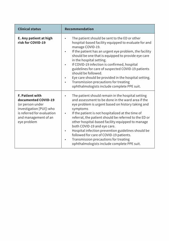
E. Any patient at high risk for COVID-19
• The patient should be sent to the ED or other hospital-based facility equipped to evaluate for and manage COVID-19.
• If the patient has an urgent eye problem, the facility should be one that is equipped to provide eye care in the hospital setting.
• If COVID-19 infection is confirmed, hospital guidelines for care of suspected COVID-19 patients should be followed.
• Eye care should be provided in the hospital setting. • Transmission precautions for treating
ophthalmologists include complete PPE suit.
F. Patient with documented COVID-19 (or person under investigation [PUI]) who is referred for evaluation and management of an eye problem
• The patient should remain in the hospital setting and assessment to be done in the ward area if the eye problem is urgent based on history taking and symptoms
• If the patient is not hospitalized at the time of referral, the patient should be referred to the ED or other hospital-based facility equipped to manage both COVID-19 and eye care.
• Hospital infection prevention guidelines should be followed for care of COVID-19 patients.
• Transmission precautions for treating ophthalmologists include complete PPE suit.

COVID-19 REQUIRING EMERGENCY SURGERY
Staff• Paramedics and surgeons must be in full PPE and well-fitted N95 mask.• Full PPE includes well-fitted N95 mask, goggles or face shield, splash resistant
gown, and foot covers.• Powered air purifying respirator (PAPR) if available or indicated.• Ensure your staff are trained in “donning and doffing” of PPE. • Universal precautions to be adhered to.• Reduce the number of staff in operating room.
Facility• Designated hospital to have a designated operating theatre to operate PUI
and COVID-19 cases.
Anaesthesia• Regional is preferred. If regional technique is chosen, the patient should
wear a surgical face mask at all times.• General anaesthesia if required, the induction and reversal should be
preferably done in a negative pressure room. • Staff participating in aerosol-generating procedures can wear PAPR
equipment.
Table 2. Pros and cons of powered air purifying respirators (PAPR)
Pros Cons
Higher protective factor than N95 respirators
No definitive evidence that PAPR reduces likelihood of viral transmission for potential airborne infections
Provides eye protection (hooded models only)
Inability to auscultate for heart and lung sounds (for hooded PAPR)
More comfortable to wear than N95 respirator Challenges in communication
Can be used if user has facial hair (not possible with N95 respirator)
Patient apprehension (especially among pediatric patients)

Pros Cons
Hooded models do not require fit-testing (unlike N95 respirator)
Training on use, doffing, and care of PAPR is needed to prevent contamination
Eliminates unexpected poor N95 respirator fit Requires decontamination after use
Less likely to be dislodged when managing an agitated patient More expensive than N95 respirator
PAPRs with hood may provide additional protection against contamination compared with typical gear worn with N95 mask
Inability to re-use disposable filters between patients (need large supply of filters)
Need to train staff repeatedly to maintain competency if not frequently used
Risk of battery failure and inadvertent exposure
References
1. https://onlinelibrary.wiley.com/doi/abs/10.1002/jmv.25725 2. https://www.nejm.org/doi/pdf/10.1056/nejmoa2002032.3. https://www.aao.org/headline/alert-important-coronavirus-context.4. https://www.eurotimes.org/guidelines-for-italian-ophthalmologists-on-covid-19/5. Wong J, Goh QY, Tan Z, et al. Preparing for a COVID-19 pandemic: a review of operating room outbreak
response measures in a large tertiary hospital in Singapore. Can J Anaesth. 2020. doi:10.1007/s12630-020-01620-9.

© Malaysian Journal of Ophthalmology 2020; 2:16-17Editorial
Endogenous endophthalmitis: experience from the south of peninsular MalaysiaNorfariza Ngah1,2
1Ophthalmology Service, Ministry of Health, Kuala Lumpur, Malaysia; 2Department of Ophthalmology, Hospital Shah Alam, Persiaran Kayangan, Shah Alam, Selangor, Malaysia
Although endogenous endophthalmitis is a relatively rare intraocular infection, a higher incidence has been reported in high-risk patients. This potentially blinding ocular infection results from hematogenous spread of organisms from a remote primary source. Chronically ill or debilitated patients as well as those who underwent any invasive procedures are especially at risk. Ophthalmologists and medical prac-titioners must have a high index of suspicion in high-risk cases to allow for prompt diagnosis and treatment.
Many etiologic organisms, namely gram-positive, gram-negative, and fungal have been reported to cause endogenous endophthalmitis. Different parts of the world show different types of common organisms, with Staphylococcus aureus and Streptococcal pneumoniae being more common in Europe and North America and Klebsiella pneumoniae more common in East Asia. Candida albicans, on the other hand, is the most common yeast and Aspergillus sp. is the most common mold.1
Endogenous endophthalmitis has no age or sexual predilection. The right eye is involved twice as often as the left eye because of the more proximal and direct blood flow to the right carotid artery. Bilateral involvement occurs in up to 25% of cases.2
Prompt administration of antibiotic therapy is key in the acute management of endogenous endophthalmitis. Surgical intervention is generally recommended for patients infected with more virulent organisms, visual acuity of 3/60 or less, or severe vitreous involvement. The outcome of posterior diffuse endophthalmitis or panophthalmitis is frequently blindness, regardless of treatment measures.3
In the case series reported in this issue by Hayatulrizal et al.,4 negative microbial culture was seen in 11 eyes (57.9%) and were treated with empirical systemic and intravitreal antibiotics. Despite aggressive treatment, the visual outcomes were
Correspondence: Norfariza Ngah, Head of Department, Department of Ophthalmology, Hospital Shah Alam, Persiaran Kayangan, Shah Alam, Selangor, Malaysia. E-mail: [email protected]
17
rather poor, with nearly 75% of patients showing no improvement or worsening of vision. Vitrectomy was performed only in eight eyes.
The outcome of endogenous endophthalmitis is often disappointing. The three main factors that contribute to poor prognosis include more virulent organisms, compromised host conditions, and delayed diagnosis. Therefore, a high degree of suspicion is necessary to make an early diagnosis of endogenous endophthalmitis.5
References
1. Lingappan A, Wykoff CC, Albini TA, et al. Endogenous fungal endophthalmitis: causative organisms, management strategies, and visual acuity outcomes. Am J Ophthalmol. 2012:253(1):162-166.
2. Greenwald MJ, Wohl LG, Sell CH. Metastatic bacterial endophthalmitis: a contemporary reappraisal. Surv Ophthalmol. 1986;31:81-101.
3. Chee SP, Jap A. Endogenous endophthalmitis. Curr Opin Ophthalmol. 2001;12:464–470.4. Hayatulrizal M, LK Phang, FM Vendargon. A five-year retrospective hospital-based study of endoge-
nous endophthalmitis in south Malaysia. Malaysian Journal of Ophthalmology. 2020;2(1):18-26.5. Okada AA, Johnson RP, Liles WC, D’Amico DJ, Baker AS. Endogenous bacterial endophthalmitis.
Report of a ten-year retrospective study. Ophthalmology. 1994;101:832–838.

© Malaysian Journal of Ophthalmology 2020; 2:18-26Original article
A five-year retrospective hospital-based study of endogenous endophthalmitis in south MalaysiaHayatulrizal Muhd, Ling Kiet Phang, Francesca Martina Vendargon
Department of Ophthalmology, Hospital Sultanah Aminah, Johor Bahru, Johor, Malaysia
Abstract
Purpose: To analyse the predisposing factors, microbial profiles, source of infection, and visual outcomes of endogenous endophthalmitis seen in Hospital Sultanah Aminah Johor Bahru (Johor, Malaysia). Study design: Retrospective review. Methods: The medical records of 15 patients, of which 19 eyes were diagnosed with endogenous endophthalmitis, admitted from January 2014 to December 2018 were retrospectively reviewed.Results: The mean age was 55.9 ± 12.7 years (range: 31-78 years of age). There were four patients (26.7%) with bilateral involvement. Diabetes mellitus was the commonest risk factor in this study (odds ratio: 16; 95% confidence interval: 1.09-234.26). The most common source of infection was urosepsis (n = 3, 20%) followed by liver abscesses (n = 2, 13.3%), Klebsiella pneumoniae being the most common micro-organism isolated (n = 4, 44.4%). Only 10.5% of eyes (n = 2) had a final Snellen visual acuity better than 6/60, while 47.4% of eyes (n = 9) had vision of no light perception. Conclusion: In this study, Klebsiella pneumoniae was the most common organism. Overall, endogenous endophthalmitis is associated with poor visual outcomes.
Keywords: diabetes mellitus, endogenous endophthalmitis, Klebsiella pneumoniae, vitrectomy
Correspondence: Dr. Hayatulrizal Muhammad, Ophthalmology Department, Hospital Sultanah Aminah, Johor Bahru, Johor,Malaysia. E-mail : [email protected]
Endogenous endophthalmitis in south Malaysia 19
Kajian retrospektif berasaskan hospital selama lima tahun terhadap endoftalmitis endogenus di selatan Semenanjung Malaysia
AbstrakTujuan: Untuk menganalisa faktor risiko, profil mikrob, sumber jangkitan, dan kesan penglihatan endoftalmitis endogenus yang dilihat di Hospital Sultanah Aminah Johor Bahru (Johor, Malaysia).Reka bentuk kajian: Kajian retrospektif.Kaedah: Rekod perubatan dari 15 pesakit, yang mana 19 mata didiagnosa sebagai endoftalmitis endogenus dari Januari 2014 hingga Disember 2018 telah dikaji secara retrospektif.Keputusan: Purata umur adalah 55.9 ± 12.7 tahun (julat: 31-78 tahun). Terdapat empat pesakit (26.7%) dengan penglibatan dua mata. Diabetes mellitus adalah faktor risiko paling kerap dalam kajian ini (nisbah odds: 16; 95% selang keyakinan: 1.09-234.26). Sumber jangkitan yang paling kerap ialah urosepsis (n = 3, 20%) diikuti oleh abses hati (n = 2, 13.3%). Klebsiella pneumoniae menjadi mikroorganisma yang paling kerap ditemui (n = 4, 44.4%). Hanya 10.5% (n = 2 mata) mempunyai ketajaman visual Snellen terakhir yang lebih baik daripada 6/60, manakala 47.4% (n = 9 mata) mengalami kebutaan iaitu kehilangan penglihatan walau kepada cahaya.Kesimpulan: Dalam kajian ini, Klebsiella pneumoniae adalah organisma penyebab yang paling kerap ditemui. Secara keseluruhan, endopftalmitis endogenus dikaitkan dengan penglihatan yang kurang baik.
Kata kunci: diabetes mellitus, endoftalmitis endogenus, Klebsiella pneumoniae, vitrektomi
Introduction
Endogenous endophthalmitis (EE) is rare and accounts for 2-8% of all cases of endophthalmitis.1,2 It is caused by the hematogenous spread of organisms from a remote infective source to the eyes, resulting in severe visual loss. EE is most often associated with several medical conditions such as diabetes mellitus, renal failure, malignancy, acquired immunodeficiency syndrome, in-dwelling catheters, and intravenous drug abuse.3 The spectrum of the causative agent is broad and includes gram-negative bacteria, gram-positive bacteria, and fungi. However, studies show considerable differences in the frequency of these pathogens in relation to geographical areas.4,5 The visual outcomes following EE are typically

H. Muhd, L.K. Phang and F.M. Vendargon20
poor, particularly when a gram-negative organism is identified as the causative agent.3,6 Hospital Sultanah Aminah is a tertiary referral hospital that provides service to the entire Johor state, which is located in southern West Malaysia. We aim to identify the epidemiology of EE in the southern part of Malaysia.
Materials and methods
A retrospective review of all patients diagnosed with EE and managed at Hospital Sultanah Aminah Johor Bahru (HSAJB) from January 2014 to December 2018 was conducted.
Inclusion criteria was patients with clinical diagnosis of EE made by an ophthal-mologist. Relevant microbial investigations were taken both from blood and vitreous samples via vitreous tap, before the antibiotic injection. The collected samples were inoculated directly on blood agar, Sabouraud, and chocolate agar. Patients with incomplete laboratory data or lost to follow-up in less than one month were excluded.
Demographic characteristics, microbial profiles, management, and initial and final visual acuity (taken at one-month follow-up) were obtained from the patient’s medical records, and data were analysed using the IBM SPSS Statistics for Windows, Version 22.0 (IBM Corp., Armonk, NY, USA). Data with numerical variables were described as mean and standard deviation, while categorical data were expressed by frequency (N) and percentage.
Results
A retrospective review of medical records of 19 eyes of 15 patients diagnosed with EE, including 11 patients (73.3%) with unilateral involvement and 4 (26.7%) with bilateral involvement was conducted. The demographic profiles of the patients are summarized in Table 1.
The 15 patients included 10 males (66.7%) and 5 females (33.3%). The mean age at presentation was 55.9 ± 12.7 years (range: 31-78 years of age). The laterality of the right eye (57.9%) was more common than the left eye (42.1%).
Ten patients had diabetes mellitus (66.7%). Eleven patients (73.3%) had an iden-tifiable source of infection, with urosepsis being the most common (n = 3, 20%). Other sources of infection included liver abscesses (n = 2, 13%) and pneumonia, neck carbuncle, infected femur implant, scrotal abscess, gingivitis, and infective endocarditis identified in one patient each (6.7%). However, sources of infection could not be identified in three patients (20%). The microorganism was success-fully isolated from blood or vitreous samples in nine patients (11 eyes, 60%) with 6 (66.6%) yielding gram-negative organisms, 2 (22.2%) gram-positive organisms, and 1 (11.1%) fungal organisms (Table 2).

Table. 1. Clinical characteristics of patients (N = 15)Patient Gender Age Eye Medical
comorbiditiesSystemic infection
Isolate Vitrectomy Initial VA
Final VA
1
M 48 RE DM, HPT, ESRF Unknown
Gram -ve bacteria No CF NPL
2
M 48 RE DMScrotal abscess
Gram -ve bacteria No CF NPL
3
M 52 RE DMNeck carbuncle
Gram -ve bacteria No HM Evisceration
4
F 65 BE NILInfective endocarditis
Gram +ve bacteria Yes
RE: CF LE: CF
RE: 2/60 LE: 6/36
5
F 78 RE DM, HPT Urosepsis
Gram +ve bacteria HM HM
6
F 40 RE DM Urosepsis
Gram -ve bacteria No 6/36 6/6
7
M 53 BE DM, HPT Cholecystitis
Gram -ve bacteria No
RE: HM LE: 6/6
RE: NPL LE: HM
8M 68 RE DM, HPT Pneumonia
Gram -ve bacteria
Yes HM PL
9F 64 LE
DM, HPT, dyslipidemia Unknown Fungal No PL NPL
10F 73 LE
DM, HPT, IHD, ESRF Unknown
No growth No NPL NPL
11M 46 RE NIL Unknown
No growth No HM NPL
12M 63 LE HPT
Urosepsis, pneumonia
No growth Yes HM PL
13
M 53 BE NIL GingivitisNo growth Yes
RE: PL LE: HM
RE: CF LE: 6/60
14
M 57 BE DM, HPTLiver abscess
No growth Yes
RE :HM LE : PL
RE : CF LE : NPL
15
M 31 LE NIL
Infected femur implant
No growth No HM Evisceration
F: female; M: male; LE: left eye; RE: right eye; BE: both eyes; DM: diabetes mellitus; HPT: hypertension; IHD:ischemic heart disease; ESRF: end-stage renal failure; NPL: non-perceptive to light; PL: perceptiveto light; HM: hand movement; CF: counting fingers; VA: visual acuity: Gram -ve, gram-negative: Gram +ve: gram-positive
Endogenous endophthalmitis in south Malaysia 21

H. Muhd, L.K. Phang and F.M. Vendargon22
At presentation, 17 out of 19 eyes had visual acuity worse than 6/60. Four eyes (21%) were counting fingers, 9 eyes ( 47.4%) could perceive hand movement, 3 (15.8%) were perceptive to light, and 1 eye ( 5.3%) was no light perception. Two eyes had a relatively good vision of 6/36 and 6/6.
Final visual acuity at least at one-month follow-up showed improvement in 6 eyes (26.3%). One eye (5.3%) showed no improvement and 12 eyes (52.6%) worsened. Eight eyes (42.1%) underwent vitrectomy.
Discussion
EE is a vision-threatening disease that occurs mainly in older patients with underlying debilitating systemic disease. The mean age of presentation in our study was 55.9 ± 12.7 years. This finding is consistent with the study by Jackson et al., which reported that the incidence peaks at about 50 years of age.3 The increased risk with age might be explained by reduced natural immunity in this advanced age group; however, the increased risk with age was only true for very advanced ages (≥ 90 years). The right eye in our study was affected twice as much as the left eye (46.7 vs 26.7), probably due to more proximal and direct arterial blood flow to the right carotid artery.7 Studies done by Jackson et al. and Leibovitch et al., however, found the carotid artery anatomy has a lesser effect on the spread of EE.3,8 A case series by Leibovitch et al. observed a male preponderance, similar to our study.8
A case series by Jackson et al. found that 56% of the patients had an underlying medical condition that predisposed to infection, the commonest being diabetes mellitus.3 Binder et al. reported all of their patients had at least one underlying chronic disease, including diabetes mellitus, prosthetic cardiac valves, cancer, chronic obstructive pulmonary disease, permanent pacemaker, rheumatoid arthritis, and end-stage renal failure.9
Although our study suggests that gram-negative organisms were the commonest cause of EE, there was considerable variation based on geographical location. In the case series published by Wong et al., the incidence of EE in the Western hospital was caused mostly by gram-positive organisms, while the East Asian hospital was burdened by gram-negative organisms, particularly Klebsiella sp.5 These findings are consistent with reports from Singapore and Taiwan, which reported 70% of the cases of EE were caused by gram-negative (approximate 60% were Klebsiella pneumoniae) organisms, with liver abscess being the major source of infection.5,10
In our study, urosepsis was the common source of infection. We postulate that this is partly due to the fact that our centre is among the only 13 government centres which provide specialized urology services for the whole of Malaysia.11 With a population of 3.31 million, most Johoreans with urosepsis have a higher probability of being treated in our centre, thus contributing to the higher cases of EE secondary to urosepsis.12 Although our study was conducted on a small sample

Table 2. Microbial isolates from vitreous samples (N = 15)
Organism N (%)Culture positive 9 (60)Gram-positive organisms 2 (22.2)
Streptococcus pneumoniae 1
Group B streptococcus 1
Gram-negative organisms 6 (66.7)
Pseudomonas aeruginosa 1
Klebsiella pneumoniae 4
Escherichia coli 1
Fungal 1 (11.1)
Penicillium sp. 1
Culture negative 6 (40)
Table 3. Reported prevalence of EE in Asian and non-Asian countries
StudyDate range and location
Number of EE cases
Commonest medical comorbidities (%)
Commonest organisms (%)
Asian studies
Wong et al.5 1994 to 1997; Singapore
32 DM (40.7) K. pneumoniae (60)
Michael et al.19 2012 to 2016; Malaysia
18 DM (88.2) K. Pneumoniae, P. aeruginosa (17.6)
Lee et al.6 1996 to 2010; South Korea
97 DM (42.5) K. pneumoniae (48.4)
Non-Asian studies
Jackson et al.3 1984 to 2001: England
21 DM (42.1) E. coli (21.1)
Okada et al.4 1980 to 2990; USA
32 DM (39.3) Staphylococcus aureus (25)
Binder et al.9 1982 to 2000: USA
34 DM and cancer (33)
Candida albicans (37)
DM: diabetes mellitus
Endogenous endophthalmitis in south Malaysia 23
H. Muhd, L.K. Phang and F.M. Vendargon24
population, it did show epidemiologic trends comparable to other studies (Table 3).Visual outcomes in EE have always been poor, especially in bacterial EE, with
gram-negative isolates having a poorer prognosis than gram-positive isolates.3,6 This is consistent with other studies reporting poor visual outcomes for patients with bacterial EE. In a systemic review by Schiedler et al., 50% of patients had visual acuity worse than 6/60, while Okada et al. reported 78% of patients had visual acuity worse than 6/60.4,13
Poor prognosis in cases of gram-negative organisms is due to an array of virulence factors that enable the bacteria to escape host immune systems and replicate in distant organs. The capsule of gram-negative organisms is known to avert fulminant activation of the immune response by decreasing reactive oxygen species. Ryuzo et al. found C reactive protein and interleukin-6 blood levels were significantly higher in gram-negative bacteremia than in gram-positive bacteremia.14,15
The ability of the siderophore to acquire iron in the iron-poor environment during the infection also allows K. pneumoniae to colonize and disseminate inside the vitreous cavity.15
In our study, one of the patients with Klebsiella-associated EE (Table 1, number 7) had a final visual acuity of hand movement despite presenting early with visual acuity of 6/6, showing the virulence of Klebsiella sp., which is rapidly destructive despite early treatment.4 In most of our patients, the initial systemic antibiotics were selected by the infectious disease physician and were aimed at the source of infection and presumed causative organisms. However, in this particular case, it was initially diagnosed by an ophthalmologist and the initial treatment was ceftazidime and vancomycin according to the European Vitrectomy Study (EVS) protocol. This is likely inappropriate for patients with EE, as the protocol was designed for postoperative endophthalmitis.16
Because the causative organisms differ in this condition, systemic antibiotics should be selected differently. We suggest that initial empirical antibiotics for patients suspected of having EE should include ceftriaxone to cover K. pneumoniae and vancomycin to cover gram-positive organisms.
This, however, differs from one of the patients shown in Table 1 (number 6), who attained functional visual acuity of 6/6 from initial visual acuity 6/36. This patient was given intravitreal antibiotics immediately on the same day of blurred vision in the right eye after being referred from the urology department. In our cases, we postulate that a good initial visual acuity is associated with good final visual acuity, in line with case series from Nishida et al. and Binder et al., which described that a good presenting visual acuity was significantly associated with good final visual acuity.9,17
Moreover, Chang et al. found that patients with Klebsiella-related liver abscesses have a 3% risk of developing EE.18 Hence, the physician should be made aware of EE and educate patients about EE, including its symptoms, especially those with Kleb-
Endogenous endophthalmitis in south Malaysia 25
siella-related liver abscesses. This in turn will aid ophthalmologists to diagnose and treat EE early.
The data found in this study provided an overview of the varied causative organisms and sources of infection involved in EE. In addition, diabetes mellitus was one of the main risk factors for EE. This indicates that diabetic control must be addressed at multiple levels in the health care system, including improved detection, adherence to the treatment, and systemic health care monitoring and program evaluation.
Conclusion
EE is a rare but often devastating ocular and systemic disorder. It carries a poor prog-nosis in most patients, especially where gram-negative organisms are involved. Dia-betes mellitus remains one of the major risk factors for EE.
References
1. Chee SP, Jap A . Endogenous endophthalmitis. Curr Opin Ophthalmol. 2001;12:464-470.2. Shrader SK, Band JD, Lauter CB, Murphy P. The clinical spectrum of endophthalmitis: incidence, pre-
disposing factors, and features influencing outcome. J Infect Dis. 1990;162:115-120.3. Jackson TL, Eykyn SJ, Graham EM, Stanford MR. Endogenous bacterial endophthalmitis: a 17-year
prospective series and review of 267 reported cases. Surv. Ophthalmol. 2003;48:403-423.4. Okada AA, Johnson RP, Liles WC, D’Amico DJ, Baker AS. Endogenous bacterial endophthalmitis:
report of a 10-year retrospective study. Ophthalmology. 1994;101:832–838.5. Wong JS, Chan TK, Lee HM, Chee SP. Endogenous bacterial endophthalmitis: An East Asian experi-
ence and a reappraisal of a severe ocular affliction. Ophthalmology. 2000;107:1483-1491.6. Lee S, Um T, Joe SG, et al. Changes in the clinical features and prognostic factors of endogenous
endophthalmitis: fifteen years of clinical experience in Korea. Retina. 2012;32:977-984.7. Greenwald MJ, Wohl LG, Sell CH. Metastatic bacterial endophthalmitis: a contemporary reappraisal.
Surv Ophthalmol. 1986;31:81-101.8. Leibovitch I, Lai T, Raymond G, Zadeh R, Nathan F, Selva D. Endogenous endophthalmitis: a 13-year
review at a tertiary hospital in South Australia. Scand J Infect Dis. 2005;37(3):184-189.9. Binder MI, Chua J, Kaiser PK, Procop GW, Isada CM: Endogenous endophthalmitis: an 18-year review
of culture-positive cases at a tertiary care center. Medicine. 2003;82:97-105.10. Chen YJ, Kuo HK, Wu PC, et al. : A 10-year comparison of endogenous endophthalmitis outcomes: an
east Asian experience with Klebsiella pneumonia infection. Retina 2004;24:383-90.11. Lip HTC, Lip HTC, Lip HTC, et al. Clinical audit of urosepsis in a Malaysian surgery unit. J Coll Physi-
cians Surg Pak. 2019;29(2):185-186.12. Department of Statistics Malaysia. Current Population Estimates, Malaysia, 2018-2019. Available
from: www.dosm.gov.my.
H. Muhd, L.K. Phang and F.M. Vendargon26
13. Schiedler V, Scott IU, Flynn HW, Davis JL, Benz MS, Miller D. Culture-proven endogenous endophthal-mitis: clinical features and visual acuity outcomes. Am J Ophthalmol. 2004;137(4);725-731.
14. Paczosa MK, Mecsas J. Klebsiella pneumoniae: Going on the offense with a strong defense. Microbiol Mol Biol Rev. 2016;80(3):629-661.
15. Abe R, Oda S, Sadahiro T, et al. Gram-negative bacteremia induces greater magnitude of inflamma-tory response than gram-positive bacteremia. Critical Care. 2010;14:R27.
16. The Endophthalmitis Vitrectomy Study Group. Results of the Endophthalmitis Vitrectomy Study: A randomized trial of intravenous antibiotics for the treatment of post-operative bacterial endophthal-mitis. Arch Ophthalmol. 1995;113(12):1479-1496.
17. Nishida T, Ishida K, Niwa Y et al. An eleven-year retrospective study of endogenous bacterial endoph-thalmitis. J Ophthalmol. 2015:2015:1-11.
18. Chang FY, Chou MY, Fan RL, Shaoi MF. A clinical study of Klebsiella liver abscess. J Formos Med Assoc. 1988;87:282–287.
19. Michael N, Gunaseelan S, Norina T, Noordin Z, Adil H. Endogenous endophthalmitis: A five-year review of cases at the Raja Perempuan Zainab II Hospital, Kelantan, Malaysia. Cureus. 2018;10:1-8.

© Malaysian Journal of Ophthalmology 2020; 2:27-41Original article
Evaluation of spontaneous retinal venous pulsation in primary open-angle and primary angle-closure glaucoma patientsSylves Patrick1,2, Chan Hui Tze1, Rasdi Abdul Rashid1, Liza Sharmini Ahmad Tajudin1
1Department of Ophthalmology, School of Medical Sciences, Universiti Sains Malaysia Health Campus, Kota Bharu, Kelantan, Malaysia; 2Faculty of Medicine and Health Sciences, Universiti Malaysia Sabah, Jalan, Kota Kinabalu, Sabah, Malaysia
Abstract
Introduction: Spontaneous retinal venous pulsation (SRVP) is a rhythmic variation in the calibre of one or more retinal veins. The incidence of SRVP was reduced in glaucoma patients. It was also reduced in people with raised intracranial pressure compared to a healthy population.Purpose: The main objective was to report the frequency and rate of SRVP in primary open-angle glaucoma (POAG) and primary angle-closure glaucoma (PACG) patients and to associate these with the severity of glaucoma in Malay patients.Design of study: A comparative cross-sectional study.Materials and methods: A comparative cross-sectional study involving primary glaucoma patients attending the eye clinic at Hospital Universiti Sains Malaysia (HUSM), Kelantan, Malaysia, was performed between December 2015 and June 2017. The main outcomes measured were the presence and rate of SRVP using a confocal scanning laser ophthalmoscope (Spectralis High-Resolution Optical Coherence Tomography Angiography, Heidelberg Engineering GmbH, Heidelberg, Germany). In the presence of SRVP, the rate of SRVP in one minute was counted manually based on the real-time fundus movie recorded using the confocal scanning laser ophthal-moscope.
Correspondence: Dr. Liza Sharmini Ahmad Tajudin, Professor of Ophthalmology and Senior Consultant Ophthalmologist (Glaucoma), Department of Ophthalmology, School of Medical Sciences, Universiti Sains Malaysia, Health Campus, 16150 Kota Bharu, Kelantan, Malaysia. E-mail: [email protected]

S. Patrick et al.28
Results: Thirty-eight POAG, 14 PACG, and 51 control group subjects were included. There was a significantly lower incidence of SRVP in primary glaucoma patients than in the control group (p = 0.003). The presence of SRVP was significantly lower in POAG than PACG (p = 0.04). There was no significant difference in the rate of SRVP between primary glaucoma patients and the control group (p = 0.873) or between the POAG group and PACG group (p = 0.511). There was no association of incidence (p = 0.574) and rate (p = 0.167) of SRVP according to the severity of glaucoma. Systolic blood pressure (95% CI: 0.95–1.00, p = 0.038) and retinal nerve fibre layer thickness (95% CI: 1.01–1.09, p = 0.008) showed a significant association with the presence of SRVP. Conclusions: SRVP is a potential predictive factor for detection of primary glaucoma. The role of SRVP in the severity of glaucoma is still unclear. The role of SRVP in PACG patients warrants further studies in the future.
Keywords: primary angle-closure glaucoma, primary open-angle glaucoma, retinal venous pulsation, spontaneous retinal venous pulsation
Penilaian denyutan spontan salur darah vena retina pada pesakit glaukoma bersudut terbuka primer dan bersudut tertutup primer
AbstrakPengenalan: Denyutan spontan salur darah vena retina (SRVP) adalah variasi berirama satu atau lebih kaliber salur darah vena retina. Didapati SRVP ini adalah berkurangan dalam pesakit glaukoma. Ia juga berkurangan pada pesakit yang mengalami tekanan intracranial yang tinggi berbanding orang yang sihat.Tujuan: Untuk melaporkan kekerapan dan kadar SRVP pada pesakit glaukoma bersudut terbuka primer(POAG) dan bersudut tertutup primer (PACG) dan mengaitkannya dengan keterukkam penyakit glaukoma di kalangan pesakit Melayu.Reka bentuk kajian: Kajian rentas keratan perbandingan.Bahan dan kaedah: Kajian rentas keratan perbandingan yang melibatkan pesakit glaukoma primer yang menghadiri klinik mata di Hospital Universiti Sains Malaysia (HUSM), Kelantan, Malaysia telah dilaksanakan antara Disember 2015 dan Jun 2017. Hasil utama yang diukur adalah kehadiran dan kadar SRVP menggunakan ophthalmoscope laser pengimbasan confocal (Spectralis High-Resolution Optical Coherence Tomography Angiography, Heidelberg Engineering GmbH, Heidelberg, Jerman). Sekiranya terdapat kehadiran SRVP,
SRVP in Malay primary glaucoma patients 29
kadar SRVP dalam satu minit dikira secara manual berdasarkan filem fundus masa nyata yang direkodkan menggunakan ophthalmoscope laser pengimbasan confocal.Keputusan: Seramai 38 POAG, 14 PACG, dan 51 mata dari subjek dalam kumpulan kawalan di rekrutkan dalam kajian ini. Kekerapan SRVP adalah rendah secara signifikan dalam pesakit glaukoma primer berbanding kumpulan kawalan (p = 0.003). Kekerapan SRVP jauh lebih rendah dalam pesakit POAG daripada PACG (p = 0.04). Tetapi tiada perbezaan yang signifikan dalam kadar kekerapan SRVP di antara pesakit glaukoma primer dan kumpulan kawalan (p = 0.873) atau antara kumpulan POAG dan kumpulan PACG (p = 0.511). Tiada perkaitan didapati antara kekerapan (p = 0.574) dan kadar (p = 0.167) SRVP mengikut keterukkan penyakit glaukoma. Tekanan darah sistolik (95% CI: 0.95-1.00, p = 0.038) dan ketebalan lapisan serat saraf retina (95% CI: 1.01-1.09, p = 0.008) menunjukkan perkaitan yang signifikan dengan kekekerapan SRVP.Kesimpulan: SRVP mempunyai potensi sebagai faktor ramalan untuk pengesanan glaukoma primer. Peranan SRVP dalam keterukan glaukoma masih tidak jelas. Peranan SRVP dalam pesakit PACG memerlukan kajian lanjut pada masa akan datang.
Kata kunci: denyutan vena retina, denyutan vena retina spontan, glaukoma sudut terbuka primer, glaukoma sudut tertutup primer
Introduction
The concept of spontaneous retinal venous pulsation (SRVP) was first described by Coccius in 1853 after the invention of the direct ophthalmoscope.1 SRVP is a rhythmic variation in the calibre of one or more retinal veins.2 SRVP is commonly seen on the retina or optic disc surface near to where the veins exit the eye.3 There is no established pathogenesis of SRVP, and it is thought to be complex. The most acceptable explanation is due to changes in the retinal venous pressure along the vein between two compartments: intraocular pressure (IOP) and retrobulbar compartments. The IOP compartment is the space within the eyeball containing the peripheral retinal veins up to and including the prelaminar portion of the central retinal vein. The retrobulbar compartment is the space outside the eyeball containing the retrolaminar and intraneural portions of the central retinal vein. The pressure change arises when the retinal vein exits the eye through the lamina cribrosa towards the retrobulbar region. Changes in IOP are believed to affect the IOP compartment. External pressure, mainly from the cerebrospinal (CSF) pressure and tissue pressure surrounding the optic nerve, affects the retrobulbar compartment.2,3 The imbalance between these two compartments may affect the SRVP.

S. Patrick et al.30
In a healthy population, SRVP has been reported to occur between 74.4% and 98%.4-10 However, the incidence of SRVP was reduced in glaucoma patients and patients with raised intracranial pressure.5,6,8-11 The reason for the absence of SRVP in glaucoma patients is still not fully understood. The most likely explanation, thus far, is due to an increase in central retinal vein (CRV) resistance.8 An increase in CRV resistance is due to intrinsic and extrinsic alterations. As glaucoma is a disease of ageing, systemic comorbidities such as systemic hypertension are not uncommon.12-20 The changes in the endothelium and vessel wall that are intrinsic to the ageing process or the consequence of hemodynamic stress are also believed to be responsible for CRV resistance in glaucoma patients.21 On the other hand, extrinsic compressive connective tissue remodelling response in the lamina cribrosa may also play a role for the absence of SRVP in glaucoma patients.22 Absence of SRVP was also found in patients with increased intracranial pressure (ICP).6,11 On the contrary, glaucoma patients are believed to have low ICP.23-25 Indeed, not all glaucoma patients showed absence of SRVP.
On the other hand, there is evidence suggesting a potential role of SRVP in the severity of primary open-angle glaucoma (POAG). This is based on the finding of higher frequency of SRVP in early glaucoma compared to moderate and advanced glaucoma among POAG patients who achieved target pressure.9 The frequency of SRVP also differs according to the types of glaucoma: lower frequency of SRVP in POAG compared to normotensive glaucoma (NTG).5 To the best of our knowledge, there is no data reported on SRVP in primary angle-closure glaucoma (PACG). The main objective of this study was to report the frequency and rate of SRVP in POAG and PACG patients, and to associate these with the severity of glaucoma in Malay patients.
Materials and methods
This was a comparative cross-sectional study involving primary glaucoma patients: POAG and PACG. Purposive sampling was conducted on primary glaucoma patients attending the eye clinic, Hospital Universiti Sains Malaysia (HUSM), Kelantan, Malaysia, between December 2015 and June 2017. Control subjects were recruited from hospital staff and HUSM students. This study received ethical approval from the Ethics Committee Board of Universiti Sains Malaysia and was conducted in accordance with the Declaration of Helsinki. Written informed consent was obtained from all the subjects prior to their participation in this research.
Patient recruitmentThe inclusion criteria were Malay subjects. “Malay” was defined as a person who ascribed to the religion of Islam, habitually spoke the Malay language, conformed to Malay customs, and whose ancestors were Malay.26 Those with confirmed cases

SRVP in Malay primary glaucoma patients 31
of POAG and PACG who were able to produce two consecutive reliable and repro-ducible visual field tests were included. POAG was defined as a glaucomatous optic neuropathy associated with visual field loss in the absence of other ocular diseases or congenital anomalies, with no evidence of angle closure.27 PACG was defined as a glaucomatous optic neuropathy with the presence of iridotrabecu-lar contact (ITC) associated with visual field loss in the absence of other ocular diseases or congenital anomalies.27 The reliability of the Humphrey visual field (HVF) test was defined as a fixation loss of less than 20%, a false positive response of less than 33%, and a false negative response of less than 33%.28 Control subjects who were healthy subjects also underwent an HVF test. Only those who were able to produce two consecutive reliable and reproducible visual field tests were included.
The exclusion criteria were subjects under 18 years old, with a history of ocular trauma and uveitis, and a history of previous intraocular surgery other than uncomplicated cataract surgery. Those with ocular and systemic diseases affecting the visual fields — diabetic retinopathy, retinal vein occlusion, ischemic optic neuropathy and stroke — were excluded. In addition, poor quality image of CRV and inadequate real-time fundus movies were also excluded.
All the eligible subjects underwent visual acuity testing using the Snellen visual acuity chart for distance (Ametek Reichert Technologies, NY, USA), anterior segment and dilated fundus examinations using a slit lamp biomicroscope (Haag-Streit UK, England). Gonioscopic examination and IOP measurement using the Goldmann applanation tonometer were also conducted. Optic nerve head (ONH) parameters and retinal nerve fibre layer (RNFL) thickness were evaluated using optical coherence tomography (OCT) (Carl Zeiss Meditec Inc., Dublin, CA, USA). Visual field analysis was conducted using the Swedish interactive threshold algorithm (SITA) standard of 24-2 by Humphrey automated perimetry (Carl Zeiss Meditec Inc., Dublin, CA, USA). Blood pressure (BP) was also measured using a sphygmomanometer and pulse rate (PR) was recorded.
SRVP image acquisitionThe SRVP image was obtained using a confocal scanning laser ophthalmoscope (Spectralis High-Resolution OCT Angiography, Heidelberg Engineering GmbH, Heidelberg, Germany) in near-infrared mode (820 nm) from a dilated pupil. The real-time fundus movie was recorded for one minute and centred on the ONH. The observation for SRVP on the optic disc was conducted by two independent masked observers (SP and CHT) at two different times. The presence of SRVP was based on an agreement between the two independent observers. If there was a disagreement, a third observer (RAR) was asked to evaluate. In the presence of SRVP, the rate of the SRVP in one minute was counted manually. If both eyes were eligible, only the right eye was selected. The SRVP image acquisitions were made by the same qualified and trained personnel.

S. Patrick et al.32
Glaucoma severity scoreTwo reliable visual fields were obtained from primary glaucoma patients within three months of the recruitment period. Patients were excluded after three failed attempts in producing two reliable visual fields. Glaucoma severity was scored using a modified Advanced Glaucoma Intervention Study (AGIS) scoring system.29 AGIS scoring was conducted by a masked glaucoma consultant (LS).
All relevant data were analysed using Statistical Package for Social Sciences (SPSS) for Windows, Version 22.0 (IBM Corp., Armonk, NY, USA). Double entry of data was practised to avoid wrong or missing entry of information. The categorical variables, including the SRVP frequency, were described using frequency and percentage. The numerical variables were described based on the distribution of the data. Mean and standard deviation were used for normally distributed data, median and interquartile range for skewed data. The comparison between groups was analysed using a Pearson chi-squared test, Fisher exact test, Mann-Whitney U test, independent t-test, and one-way analysis of variance (ANOVA). Simple and multiple logistic regressions were performed to determine the factors associated with SRVP. The potential factors included in the analysis were age, gender, systolic BP, diastolic BP, PR, IOP, HVF parameters, and ONH parameters. The statistical sig-nificance level was set at p < 0.05.
Results
A total of 52 primary glaucoma patients were included in this study; 38 had POAG and 14 had PACG. A total of 33 primary glaucoma patients were excluded because of failure to produce reliable visual fields. Patients with primary glaucoma (POAG and PACG) were significantly older. Higher frequency of systemic comorbidities and higher systolic BP were identified among primary glaucoma patients (Table 1). The majority of the primary glaucoma patients were on monotherapy with mean IOP of 15.4 ± 3.2 mmHg (Table 2). There was a significant difference in the incidence of SRVP between primary glaucoma patients and the control group (p = 0.003) (Table 3). The presence of SRVP in POAG was 13 (34.2%) and PACG was 11 (78.6%), with a statisti-cally significant difference (p = 0.04).
However, there was no difference in the rate of SRVP between primary glaucoma patients and the control group (p = 0.873) (Table 3). The SRVP rates in the POAG group (73.9 ± 10.5 beats per minute) and PACG group (71.2 ± 8.7 beats per minute) were almost the same, and was not statistically significant (p = 0.511). There were a total of 24 (46.2%) mild, 9 (17.3%) moderate, and 19 (36.5%) severe glaucoma patients included in the study. There was no significant difference in the incidence of SRVP (p = 0.574) and the rate of SRVP (P = 0.167) according to severity of primary glaucoma (Table 4). In multiple logistic regression using the backward method, the systolic BP and RNFL thickness were significant factors affecting SRVP. A reduction
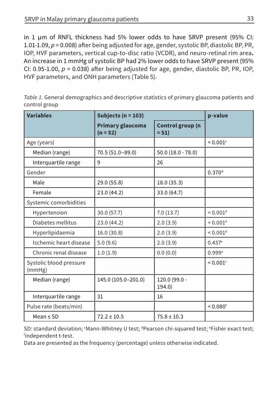
Table 1. General demographics and descriptive statistics of primary glaucoma patients and control group
Variables Subjects (n = 103) p-value
Primary glaucoma (n = 52)
Control group (n = 51)
Age (years) < 0.001c
Median (range) 70.5 (51.0–89.0) 50.0 (18.0 - 78.0)
Interquartile range 9 26
Gender 0.370d
Male 29.0 (55.8) 18.0 (35.3)
Female 23.0 (44.2) 33.0 (64.7)
Systemic comorbidities
Hypertension 30.0 (57.7) 7.0 (13.7) < 0.001d
Diabetes mellitus 23.0 (44.2) 2.0 (3.9) < 0.001d
Hyperlipidaemia 16.0 (30.8) 2.0 (3.9) < 0.001d
Ischemic heart disease 5.0 (9.6) 2.0 (3.9) 0.437e
Chronic renal disease 1.0 (1.9) 0.0 (0.0) 0.999e
Systolic blood pressure (mmHg)
< 0.001c
Median (range) 145.0 (105.0–201.0) 120.0 (99.0 - 194.0)
Interquartile range 31 16
Pulse rate (beats/min) < 0.080f
Mean ± SD 72.2 ± 10.5 75.8 ± 10.3
SD: standard deviation; cMann-Whitney U test; dPearson chi-squared test; eFisher exact test; findependent t-test.Data are presented as the frequency (percentage) unless otherwise indicated.
SRVP in Malay primary glaucoma patients 33
in 1 µm of RNFL thickness had 5% lower odds to have SRVP present (95% CI: 1.01-1.09, p = 0.008) after being adjusted for age, gender, systolic BP, diastolic BP, PR, IOP, HVF parameters, vertical cup-to-disc ratio (VCDR), and neuro-retinal rim area. An increase in 1 mmHg of systolic BP had 2% lower odds to have SRVP present (95% CI: 0.95-1.00, p = 0.038) after being adjusted for age, gender, diastolic BP, PR, IOP, HVF parameters, and ONH parameters (Table 5).
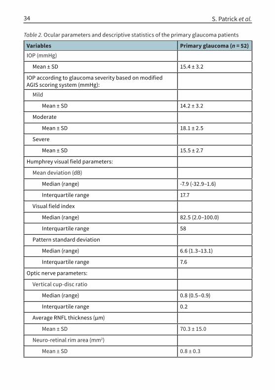
Table 2. Ocular parameters and descriptive statistics of the primary glaucoma patients
Variables Primary glaucoma (n = 52)IOP (mmHg)
Mean ± SD 15.4 ± 3.2
IOP according to glaucoma severity based on modified AGIS scoring system (mmHg):
Mild
Mean ± SD 14.2 ± 3.2
Moderate
Mean ± SD 18.1 ± 2.5
Severe
Mean ± SD 15.5 ± 2.7
Humphrey visual field parameters:
Mean deviation (dB)
Median (range) -7.9 (-32.9–1.6)
Interquartile range 17.7
Visual field index
Median (range) 82.5 (2.0–100.0)
Interquartile range 58
Pattern standard deviation
Median (range) 6.6 (1.3–13.1)
Interquartile range 7.6
Optic nerve parameters:
Vertical cup-disc ratio
Median (range) 0.8 (0.5–0.9)
Interquartile range 0.2
Average RNFL thickness (µm)
Mean ± SD 70.3 ± 15.0
Neuro-retinal rim area (mm2)
Mean ± SD 0.8 ± 0.3
S. Patrick et al.34

Variables Primary glaucoma (n = 52)Management [n (%)]
Monotherapy 17.0 (32.7)
Dual therapy 14.0 (26.9)
Triple therapy 12.0 (23.1)
Quadruple therapy 9.0 (17.3)
IOP: intraocular pressure; AGIS: Advanced Glaucoma Intervention Study: SD: standard deviation: RNFL: retinal nerve fibre layer. Data are presented as the frequency (percentage) unless otherwise indicated.
Table 3. The incidence and rate of SRVP between primary glaucoma patients and controls
Variables Subjects (n = 103) p-value
Primary glaucoma (n = 52)
Control group (n = 51)
SRVP 24.0 (46.2) 38.0 (74.5) 0.003d
Rate of SRVP (beats/min) 0.873c
Median (range) 71.0 (59.0–90.0) 72.0 (56.0–97.0)
Interquartile range 17 14
SRVP: spontaneous retinal venous pulsation; dPearson chi-squared test; cMann-Whitney U test. Data are presented as the frequency (percentage) unless otherwise indicated.
Table 4. The incidence and rate of SRVP according to primary glaucoma severity
Variables Glaucoma severity p-valueMild (n = 24)
Moderate (n = 9)
Severe (n = 19)
SRVP 13.0 (54.2) 4.0 (16.7) 7.0 (29.2) 0.574e
Rate of SRVP (beats/min) 0.167g
Mean ± SD 75.9 ± 10.6 66.8 ± 6.9 69.9 ± 7.1
SD: standard deviation; SRVP: spontaneous retinal venous pulsation; eFisher exact test; gOne-way analysis of variance (ANOVA). Data are presented as the frequency (percentage) unless otherwise indicated.
SRVP in Malay primary glaucoma patients 35

Table 5. Multiple logistic regression on factor affecting SRVP.
VariablesSimple logistic regression Multiple logistic regression
Ba Crude OR (95% CI) p-value Bb Adjusted OR
(95% CI) p-value
Age (years) -0.02 0.98 (0.96, 1.01) 0.136 0.46 1.05 (1.00, 1.09) 0.310
Systolic blood pressure (mmHg)
-0.02 0.98 (0.96, 1.00) 0.017 -0.03 0.98 (0.95, 1.00) 0.038
Optic nerve parameters:
Vertical cup-disc ratio -4.78 0.01 (0.00, 0.14) 0.001 -3.19 0.04 (0.00, 1.09) 0.056
Average RNFL thickness (µm)
0.04 7.90 (2.45, 25.47) < 0.001 0.05 1.05 (1.01, 1.09) 0.008
aCrude regression coefficient, badjusted regression coefficient, OR: odds ratio, CI: confidence interval, B: regression coefficient, RNFL: retinal nerve fibre layer. Backward multiple logistic regression method was applied. There was no multicollinearity detected and no interaction amongst independent variables. Hosmer Lemeshow test, p-value = 0.947. Classification table 69.9% correctly classified. The area under the receiver operating characteristics (ROC) curve was 72.2%.
S. Patrick et al.36
Discussion
There was a significant reduction in the incidence of SRVP in primary glaucoma patients in this study. The findings from the current study are almost similar to several findings in Caucasians. However, in population-based studies, Asians were reported to have a significantly wider mean retinal arteriolar and venular calibres compared to Caucasians.30,31 There is minimal knowledge on SRVP among Asians. Seo et al. reported a significantly lower incidence of SRVP in POAG patients (53.3%) compared to glaucoma suspects (86.3%) among the Korean population.32 In their study, glaucoma suspects were used as a comparison. Glaucoma suspects may sometimes be considered early-stage POAG and this may affect the accuracy of the study.33,34 In addition, SRVP evaluation in previous studies was performed using a slit-lamp examination with a 60 D or 78 D ophthalmoscopic lens, fundus camera, and confocal scanning laser ophthalmoscope (Spectralis HRA) in near-infrared mode (820 nm) installed with an “eye movement correction tool”. 5,8,10,32,35,36
Most studies reported SRVP in POAG and NTG patients but none in PACG.5,9,10,32,36 Since PACG is responsible for more blindness than POAG, the detection of SRVP may be important in the screening and management of PACG.37 However, the number
SRVP in Malay primary glaucoma patients 37
of PACG included in the present study was too small. SRVP was observed in 11 of 14 (78.6%) PACG patients. There is a potential higher incidence of SRVP in PACG compared to POAG that is yet to be proven, although this may not be in agreement with the two-compartment theory.2,3
In our study, the SRVP rate in primary glaucoma patients and controls was almost similar. Even between patients with different severity of primary glaucoma, the SRVP rate was almost the same. One study found that SRVP collapses in phases with IOP at systole and expansion occurs during IOP at diastole.38 On the contrary, another study found that SRVP collapsed in phase with ocular diastole and expansion occurred during ocular systole.39 The cardiac cycle drives IOP oscillation, with peak IOP occuring during systole.38,39 The heart rate reflects the number of cardiac cycles per unit of time, while the pulse rate is close or equal to the heart rate in people with normal and healthy hearts. Based on this knowledge, we have assumed that the SRVP rate measurement could be done by measuring the heart rate or the pulse rate.
Based on the two-compartment theory, SRVP is related to IOP and ICP. Glaucoma severity is closely related to IOP.33,40,41 High IOP may lead to an acceleration of optic neuropathy.41 Thus, absence of SRVP may be related to glaucoma severity. A lower incidence of SRVP was observed in severe glaucoma cases.9,32 Higher incidence of SRVP was observed in mild glaucoma patients compared to severe glaucoma.32 Seo et al. found that the incidence of SRVP reduces as severity increases, with mild cases having an SRVP incidence of 63.4%, moderate cases an incidence of 42.1%, and severe cases an incidence of 26.7%.32 However, in the present study there was no significant association between glaucoma severity and incidence of SRVP. In fact, we found a lower incidence of SRVP in moderate glaucoma. This is mainly due to the small number of patients causing an unequal distribution of patients according to severity. In the future, it would be best to base recruitment on glaucoma severity.
There is evidence supporting the postulation that better IOP control reduces the incidence of SRVP at any stage of the disease.32,36 Since the mean IOP was 15.4 ± 3.2 mmHg, this may partially explain the negative association between SRVP and severity of primary glaucoma. However, target IOP was not considered in our study. Our study used mean IOP, which is not ideal compared to appropriate target IOP. Target IOP differs according to severity of glaucoma42 and is dynamic, especially when there is evidence of progression.27,43 On the contrary, there was a higher frequency of SRVP in control subjects with lower mean IOP (14.6 ± 2.4 mmHg).
Ideally, the comparison should be made at the initial presentation before initiation of treatment and prospective follow-up. As the present study was a cross-sectional study, stopping IOP-lowering drugs was not only unethical but may have also increased the risk of progression. IOP-lowering drugs, such as topical beta blockers, may reduce the incidence of SRVP at any glaucoma stage, as has been reported in a few studies.32,36 Moreover, systemic comorbidities such
S. Patrick et al.38
as hypertension and diabetes mellitus are not uncommon in glaucoma patients. There was a higher number of hypertensive patients among primary glaucoma patients and systemic hypertensive drugs were not discontinued; this limitation may have affected our findings due to the effect of systemic drugs on the vessels. Beta blockers reduce the heart rate44 and systemic beta blockers may exert a more significant effect on SRVP compared to topical beta blockers. As SRVP is related to the phase of the cardiac cycle, a reduction in the heart rate may directly decrease the SRVP rate.
In the present study, the control subjects were not age-matched to glaucoma patients. Ageing causes structural changes in the vessels which subsequently affect function.45 The incidence of glaucoma increases with age. Thus, our primary glaucoma patients were older than the control subjects. The incidence of SRVP was higher in the older age group.46 In this study, age may play a role in the incidence of SRVP in primary glaucoma patients.
A confocal scanning laser ophthalmoscope was used in this study, but “eye movement correction tools” for image movement adjustment were not included. The absence of this special software programme may have affected the accuracy of SRVP detection, in line with a false low incidence of SRVP reported elsewhere.32 Therefore, the incidence of SRVP in our study may have been potentially under-re-ported.
To the best of our knowledge, this is the first study to report the incidence of SRVP in PACG patients. However, the number of PACG patients was too small for any further comparative analysis. There was an even smaller number of patients for analysis according to severity of primary glaucoma, especially moderate glaucoma. This may not provide the actual representation of the association of SRVP and severity. In the future, a larger sample size and prospective cohort study design may provide a clearer understanding of the relationship between SRVP and glaucoma severity.
In conclusion, SRVP is a potential predictive factor for detecting primary glaucoma. The role of SRVP in glaucoma severity remains unclear. A thinner RNFL is associated with lower SRVP incidence. The role of SRVP in PACG patients warrants further studies in the future.
References
1. Coccius EA. The use of the ophthalmoscope and the indication of a new instrument. Leipzig: Immanuel Muller Publisher; 1853.
2. Jacks AS, Miller NR. Spontaneous retinal venous pulsation: aetiology and significance. J Neurol Neu-rosurg Psychiatry. 2003;74(1):7.
3. Morgan WH, Hazelton ML, Yu DY. Retinal venous pulsation: Expanding our understanding and use of this enigmatic phenomenon. Prog Retin Eye Res. 2016;55:82-107.

SRVP in Malay primary glaucoma patients 39
4. Legler U, Jonas JB. Assessment of the spontaneous pulsations of the central retinal vein in daily ophthalmic practice. Clin Exp Ophthalmol. 2007;35(9):870-871.
5. Abegao Pinto L, Vandewalle E, De Clerck E, Marques-Neves C, Stalmans I. Lack of spontaneous venous pulsation: possible risk indicator in normal tension glaucoma? Acta Ophthalmol. 2013;91(6):514-520.
6. Levin BE. The clinical significance of spontaneous pulsations of the retinal vein. Arch Neurol. 1978;35(1):37-40.
7. Harder B, Jonas JB. Frequency of spontaneous pulsations of the central retinal vein. Br J Ophthal-mol. 2007;91(3):401.
8. Morgan WH, Hazelton ML, Azar SL, et al. Retinal venous pulsation in glaucoma and glaucoma suspects. Ophthalmology. 2004;111(8):1489-1494.
9. Pillunat KR, Ventzke S, Spoerl E, Furashova O, Stodtmeister R, Pillunat LE. Central retinal venous pulsation pressure in different stages of primary open-angle glaucoma. Br J Ophthalmol. 2014;98(10):1374-1378.
10. Legler U, Jonas JB. Frequency of spontaneous pulsations of the central retinal vein in glaucoma. J Glaucoma. 2009;18(3):210-212.
11. Walsh TJ, Garden JW, Gallagher B. Obliteration of retinal venous pulsations. Am J Ophthalmol. 1969;67(6):954-6.
12. Leske MC, Connell AM, Wu S-Y, et al. Incidence of open-angle glaucoma: the Barbados Eye Studies. Arch Ophthalmol. 2001;119(1):89-95.
13. Gordon MO, Beiser JA, Brandt JD, et al. The Ocular Hypertension Treatment Study: Baseline factors that predict the onset of primary open-angle glaucoma. Arch Ophthalmol. 2002;120(6):714-720.
14. Leske MC, Heijl A, Hussein M, Bengtsson B, Hyman L, Komaroff E. Factors for glaucoma progression and the effect of treatment: The Early Manifest Glaucoma Trial. Arch Ophthalmol. 2003;121(1):48-56.
15. Nouri-Mahdavi K, Hoffman D, Coleman AL, et al. Predictive factors for glaucomatous visual field pro-gression in the Advanced Glaucoma Intervention Study. Ophthalmology. 2004;111(9):1627-1635.
16. Orzalesi N, Rossetti L, Omboni S, Group OS, Conproso. Vascular risk factors in glaucoma: the results of a national survey. Graefes Arch Clin Exp Ophthalmol. 2007;245(6):795-802.
17. European Glaucoma Prevention Study G, Miglior S, Pfeiffer N, et al. Predictive factors for open-angle glaucoma among patients with ocular hypertension in the European Glaucoma Prevention Study. Ophthalmology. 2007;114(1):3-9.
18. Senthil S, Garudadri C, Khanna RC, Sannapaneni K. Angle closure in the Andhra Pradesh Eye Disease Study. Ophthalmology. 2010;117(9):1729-1735.
19. Mitchell P, Smith W, Attebo K, Healey PR. Prevalence of open-angle glaucoma in Australia. The Blue Mountains Eye Study. Ophthalmology. 1996;103(10):1661-1669.
20. Leske MC, Connell AMS, Wu S-Y, Hyman LG, Schachat AP. Risk Factors for open-angle glaucoma: The Barbados Eye Study. Arch Ophthalmol. 1995;113(7):918-924.
21. Yu D-Y, Yu PK, Cringle SJ, Kang MH, Su E-N. Functional and morphological characteristics of the retinal and choroidal vasculature. Prog Retin Eye Res. 2014;40:53-93.
22. Roberts MD, Grau V, Grimm J, et al. Remodeling of the Connective Tissue Microarchitecture of the Lamina Cribrosa in Early Experimental Glaucoma. Invest Ophthalmol Vis Sci. 2009;50(2):681-690.
23. Berdahl JP, Allingham RR, Johnson DH. Cerebrospinal fluid pressure is decreased in primary open-angle glaucoma. Ophthalmology. 2008;115(5):763-768.
S. Patrick et al.40
24. Ren R, Jonas JB, Tian G, et al. Cerebrospinal fluid pressure in glaucoma: a prospective study. Oph-thalmology. 2010;117(2):259-266.
25. Wang N, Xie X, Yang D, Xian J, Li Y, Ren R, et al. Orbital cerebrospinal fluid space in glaucoma: the Beijing Intracranial And Intraocular Pressure (iCOP) study. Ophthalmology. 2012;119(10):2065-2073. e1.
26. Article 160 of the Federal Constitution of Malaysia. Fifteenth Reprint. Kuala Lumpur: National Malaysia Berhad; 2010.
27. Augusto AB, Luca B, Alessandro B, et al. Terminology and Guidelines for Glaucoma. 4th edition. European Union: Publicomm; 2014.
28. Birt CM, Shin DH, Samudrala V, Hughes BA, Kim C, Lee D. Analysis of reliability indices from Humphrey Visual Field Tests in an urban glaucoma population. Ophthalmology. 1997;104(7):1126-1130.
29. Douglas EG, Fred E, Kenneth S, Joseph C, Marshall NC. Advanced Glaucoma Intervention Study: 2. Visual Field Test Scoring and Reliability. Ophthalmology. 1994;101(8):1445-1455.
30. Rochtchina E, Wang JJ, Taylor B, Wong TY, Mitchell P. Ethnic variability in retinal vessel caliber: a po-tential source of measurement error from ocular pigmentation?—the Sydney Childhood Eye Study. Invest Ophthalmol Vis Sci. 2008;49(4):1362-1366.
31. Wong TY, Islam FA, Klein R, et al. Retinal vascular caliber, cardiovascular risk factors, and inflamma-tion: the multi-ethnic study of atherosclerosis (MESA). Invest Ophthalmol Vis Sci. 2006;47(6):2341-2350.
32. Seo JH, Kim TW, Weinreb RN, Kim YA, Kim M. Relationship of intraocular pressure and frequen-cy of spontaneous retinal venous pulsation in primary open-angle glaucoma. Ophthalmology. 2012;119(11):2254-2260.
33. Quigley HA. New paradigms in the mechanisms and management of glaucoma. Eye (Lond). 2005;19(12):1241-1248.
34. Prum JB, Lim MC, Mansberger SL, Stein JD, Moroi SE, Gedde SJ, et al. Primary Open-Angle Glaucoma Suspect Preferred Practice Pattern (®) Guidelines. Ophthalmology. 2016;123(1):P112-51.
35. Golzan SM, Morgan WH, Georgevsky D, Graham SL. Correlation of retinal nerve fibre layer thick-ness and spontaneous retinal venous pulsations in glaucoma and normal controls. PLoS One. 2015;10(6):e0128433.
36. Lee E, Kim T-W, Kim J-A, Kim JA, Kim H. spontaneous retinal venous pulsation in unilateral primary open-angle glaucoma with low intraocular pressure. J Glaucoma. 2017;26(10):896-901.
37. Foster PJ. Glaucoma in China: how big is the problem? Br J Ophthalmol. 2001;85(11):1277-1282.38. Levine DN. Spontaneous pulsation of the retinal veins. Microvasc Res. 1998;56(3):154-165.39. Kain S, Morgan WH, Yu DY. New observations concerning the nature of central retinal vein pulsation.
Br J Ophthalmol. 2010;94(7):854-7.40. Davanger M, Ringvold A, Blika S. The probability of having glaucoma at different IOP levels. Acta
Ophthalmologica. 1991;69(5):565-568.41. Sommer A. Intraocular pressure and glaucoma. Am J Ophthalmol. 1989;107(2):186-188.42. Paul ER, Halifax, Yvonn MB, et al. Canadian Ophthalmological Society evidence-based clinical practice
guidelines for the management of glaucoma in the adult eye. Can J Ophthalmol. 2009;44:S7-S54.43. Singh K, Spaeth G, Zimmerman T, Minckler D. Target pressure--glaucomatologists’ holey grail. Oph-
thalmology. 2000;107(4):629-630.

SRVP in Malay primary glaucoma patients 41
44. Ko DT, Hebert PR, Coffey CS, et al. Adverse effects of β-blocker therapy for patients with heart failure: A quantitative overview of randomized trials. Arch Intern Med. 2004;164(13):1389-1394.
45. Gates PE, Strain WD, Shore AC. Human endothelial function and microvascular ageing. Exp Physiol. 2009;94(3):311-316.
46. Lorentzen SE. Incidence of spontaneous venous pulsation in the retina. Acta Ophthalmologica. 1970;48(4):765-770.

© Malaysian Journal of Ophthalmology 2020; 2:42-47Brief report
Use of intracameral tenecteplase for rapid resolution of intraocular fibrin in acute anterior uveitisLee Wen Yee1,2, Teh Wee Min2,3, Ling Kiet Phang2,4, Norlina Mohd Ramli1, Haslina Mohd Ali2
1Department of Ophthalmology, University of Malaya, Petaling Jaya, Selangor, Malaysia; 2Department of Ophthalmology, Hospital Sultanah Bahiyah, Alor Setar, Kedah, Malaysia; 3Department of Ophthalmology, Hospital Selayang, Selangor, Malaysia; 4Department of Ophthalmology, Hospital Sultanah Aminah, Johor Bahru, Johor, Malaysia
Abstract
Two patients presenting with acute anterior uveitis (AAU) and severe fibrinous reaction in the anterior chamber were given an injection of intracameral tenect-eplase (50 microgram/0.1 ml). In both patients with AAU, complete rapid fibrin resolution and synechiolysis occurred within three days after intracameral tenect-eplase injection. Significant visual acuity improvement was also seen in both patients and there were no complications observed throughout the follow-up duration of up to six months after administration of treatment. As tenecteplase has many superior properties compared to other medications in its class and can also be diluted and stored for up to a year under specific conditions, it may be an efficacious, safe and cost-effective means of treating severe AAU with fibrin formation.
Keywords: anterior uveitis, intraocular fibrin, tenecteplase, tissue plasminogen activator
Correspondence: Lee Wen Yee, MD, Department of Ophthalmology, University of Malaya, Petaling Jaya, Selangor, Malaysia. E-mail: [email protected]

Intracameral tenecteplase for resolution of intraocular fibrin in uveitis 43
Penggunaan tenecteplase intrakameral untuk pengurangan pantas fibrin intraokular dalam uveitis anterior akut
Abstrak Abstrak Uveitis anterior akut (AAU) dan reaksi fibrin yang teruk di dalam ruang anterior dilihat pada dua pesakit. Mereka diberi suntikan tenecteplase secara intrakameral (50 mikrogram / 0.1 ml). Kedua-dua pesakit dengan AAU ini menunjukkan pengurangan fibrin yang pantas dan penguraian sinaeke berlaku dalam tempoh tiga hari selepas suntikan tenecteplase intrakameral. Peningkatan ketajaman visual yang ketara juga dilihat pada kedua-dua pesakit dan tidak ada komplikasi yang diamati sepanjang tempoh tindakan dilanjutkan sehingga enam bulan selepas rawatan. Oleh kerana tenecteplase mempunyai banyak sifat unggul berbanding dengan ubat-ubatan lain di dalam kelasnya dan juga boleh dicairkan dan disimpan sehingga satu tahun di bawah keadaan tertentu, ia mungkin merupakan cara yang berkesan, selamat dan kos efektif untuk merawat AAU yang teruk dengan pembentukan fibrin.
Kata kunci: aktivator plasminogen tisu, fibrin intraocular, tenecteplase, uveitis anterior
Introduction
Anterior uveitis (AU) is the commonest form of intraocular inflammation. The formation of a massive fibrin clot in the anterior chamber can cause adverse functional and structural changes in the integrity of the eye.
Tissue plasminogen activator (t-PA) is safe and effective in rapid resolution and clearance of intraocular fibrin in humans.1 Tenecteplase (known commercially as TNK or Metalyse, Boehringer Ingelheim, Sydney, Australia) is a clot-specific fibrino-lytic agent that can be produced by recombinant DNA technology, and its activity increases significantly in the presence of fibrin.2 Herein we report our experience with two patients who received intracameral TNK for severe fibrin formation in acute anterior uveitis (AAU).

Fig 1. Severe fibrinous exudation with clumping inferiorly and almost 360° of posterior synechiae obscuring the pupil.
Fig 2. Complete fibrin resolution as well as synechiolysis within three days of injecting one dose of intracameral TNK (50 micro-grams/0.1ml).
W.Y. Lee et al.44
Materials and methods
Approval for the off-label intraocular use of TNK was obtained from the Pharma-ceutical Services Division, Ministry of Health Malaysia in May 2017. Written informed consent was obtained from each patient before intraocular injection of TNK.
TNK was prepared for intraocular use by the in-house pharmacist in the following steps. One vial of powder contained 10,000 units (50 mg) of TNK. The powder was reconstituted with 10 ml of water for injection from a pre-filled syringe under aseptic technique. The reconstituted solution contained 1,000 units (5 mg) of TNK per ml. One milliliter of TNK solution was then diluted with another 9 ml of balanced salt solution to obtain a final concentration of 50 micrograms/0.1 ml. Each vial of TNK can be reconstituted into at least 300 units of 1 cc syringes, with each syringe containing 0.3 ml of TNK. To retain the fibrinolytic activity, aliquots can be kept for up to four weeks if kept at a temperature of -20°C (or up to a year if kept at -80°C) and thawed just before use. Once liquefied, the solution had to be used within 24 hours.
Intracameral injections of TNK were performed in the following manner: The eye was anaesthetized with topical proparacaine. The skin was cleaned with 10% povidone-iodine solution and draped. The cornea and conjunctiva were irrigated with 5% povidone-iodine solution. A 30-gauge needle on a tuberculin syringe was passed through the clear cornea at the limbus and 0.1 ml of TNK solution (50 micrograms) was injected into the anterior chamber. The patients were examined at the slit lamp from day one post-injection until the fibrinous membrane had resolved and post-injection photographs were obtained.

Fig. 4. At day one after injection of intracam-eral TNK (50 microgram/0.1ml), the pupillary membrane had dissolved and posterior synechiae completely broken off, leaving behind an imprint of iris pigments on the anterior lens capsule.
Fig. 3. Fibrinous membrane formation seen at the pupillary margin of the left eye, with almost complete 360° posterior synechiae.
Intracameral tenecteplase for resolution of intraocular fibrin in uveitis 45
Results
Case 1A 28-year-old male presented with left eye redness, pain, and decreased vision for ten days. Further history noted he also experienced on and off right-sided lower backache for the past three years. Visual acuity was hand motion. There was severe fibrinous exudation with almost 360° of posterior synechiae (Fig. 1). B-scan showed clear vitreous without scleral thickening. Musculoskeletal examination revealed tenderness at the right sacroiliac area with positive FABER test. Diagnosis of left eye severe AAU with sacroiliitis was made. He was initially treated with frequent steroid eye drops as well as subconjunctival mydricaine without much improvement. He then received one dose of intracameral TNK (50 micrograms/0.1 ml), which resulted in complete fibrin resolution and synechiolysis within three days (Fig. 2). No adverse events occurred after the injection. Refracted vision at six months was 6/6.
Case 2A 55-year-old female had left eye pain and redness for one week and blurred vision for two days. Visual acuity was counting fingers. There was fibrin in the anterior chamber with extensive posterior synechiae (Fig. 3). B-scan showed no vitreous opacity. Diagnosis of left eye AAU with seclusio pupillae was made. Patient was started on intensive topical steroids and cycloplegic agent followed by intracameral TNK (50 micrograms/0.1 ml), which resulted in complete fibrin resolution as well as synechiolysis within one day (Fig. 4). No adverse effects were observed.
At three months follow-up, her vision improved to 6/9. There was mild cataract noted.

W.Y. Lee et al.46
Discussion
Anterior uveitis is the most common form of intraocular inflammation. Fibrin may be present in the anterior chamber in severe forms of AU, and it may take weeks for the fibrin to completely resolve. The treatment of intraocular fibrin usually involves topical and systemic steroids that function to suppress inflammation; cycloplegics to decrease the risk of posterior synechiae formation and to relieve the painful ciliary spasm; and non-steroidal anti-inflammatory drugs (NSAIDs) to establish the blood ocular barrier by its anti-prostaglandin effect.3
The effects of conventional treatment for fibrinous membranes in AU mentioned above may only be apparent after at least one week of therapy. Thus, other treatment options for rapid resolution of fibrinous membrane are indicated before long-term complications develop. t-PA is an option for intraocular fibrinolysis and it is a 70000-dalton protein that plays an important role in the fibrinolytic cascade by converting plasminogen to plasmin in the presence of fibrin.3 It has been shown to be safe and efficacious for the resolution of intraocular fibrin after vitrectomy, cataract surgery, glaucoma filtering surgery, and penetrating keratoplasty.1,4
Tenecteplase is a new thrombolytic agent which has a slower systemic clearance compared to other t-PAs. Its other properties include:
1. a longer half-life (20 minutes) and greater fibrin specificity; 2. more resistant to inactivation by plasminogen activator inhibitor 1 (PAI-1); 3. contains less than one-third the L-arginine content (implicated in retinal
toxicity) of alteplase; and 4. less bleeding complications.5
One vial of TNK (10,000 U / 50 mg) can be reconstituted into about 300 aliquots of 1 cc syringes, with each syringe containing 0.3 ml of TNK. To retain the fibrinolytic activity, aliquots can be kept for up to a year at a temperature of –80°C and thawed just before use.6 Once thawed, TNK retained 83-100% bioactivity even after 24 hours.
Our two cases demonstrated that TNK rapidly cleared fibrin from the anterior chamber and was also effective in synechiolysis in severe AAU within one to three days of its administration.
A review of available literature shows that the safe and effective dose of TNK ranged from 10 micrograms to 50 micrograms, with no evidence of retinal toxicity when 50 micrograms was injected subretinally.7 Fifty micrograms in 0.1 ml of tenect-eplase was given intracamerally in our cases uneventfully.
Haemorrhage is a major complication which occurs in up to 30% of patients treated with intravenous t-PA. However, only small doses of t-PA are used in intraocular injection; thus, systemic bleeding is rare. However, there is still a small risk of hyphaema and vitreous haemorrhage. Other adverse events like band keratopathy and intraocular lens opacification following intracameral application of t-PA have been reported.8 Our patients were followed up for six months and three months, respectively, with no adverse event was observed throughout.
Intracameral tenecteplase for resolution of intraocular fibrin in uveitis 47
Conclusion
Intracameral tenecteplase is effective for rapid resolution of fibrin and synechiolysis in AAU. However, in view of the lack of clinical trials with adequate sample size, we recommend intracameral tenecteplase be reserved for eyes with AAU and severe fibrin formation unresponsive to conventional therapy.
Acknowledgements
We would like to acknowledge the assistance and feedback given to us by the Department of Pharmacy, Hospital Sultanah Bahiyah, Alor Setar, Kedah, Malaysia.
References
1. Erol N, Özer A, Topbas S, Yildirim N, Yurdakul S. Treatment of intracameral fibrinous membranes with tissue plasminogen activator. Ophthalmic Surg Lasers Imaging Retina. 20031;34(6):451-456. doi:10.3928/1542-8877-20031101-04.
2. Pennica D, Holmes WE, Kohr WJ, Harkins RN, Vehar GA, Ward CA, Bennett WF, Yelverton E, Seeburg PH, Heyneker HL, Goeddel DV. Cloning and expression of human tissue-type plasminogen activator cDNA in E. coli. Nature. 1983;301(5897):214.
3. Dotan A, Kaiserman I, Kremer I, Ehrlich R, Bahar I. Intracameral recombinant tissue plasminogen ac-tivator (r-tPA) for refractory toxic anterior segment syndrome. Br J Ophthalmol. 2014;98(2):252-255.
4. Jaffe GJ, Abrams GW, Williams GA, Han DP. Tissue plasminogen activator for postvitrectomy fibrin formation. Ophthalmology. 1990;97(2):184-189. doi:10.1016/S0161-6420(90)32618-0
5. Kwon YH, Lim SJ, Jeung WJ, You YS, Kim SH, Kwon OW. Subretinal tenecteplase injection in a sub-macular hemorrhage from polypoidal choroidal vasculopathy: a case report. Retin Cases Brief Rep. 2012;6(4):400-405.
6. Ortega-García MP, Blasco-Segura P. Elaboration of preloaded syringes of tenecteplase for treatment of central renal vein occlusion. Hospital Pharmacy Europe Issue 34 September/October 2007. Avail-able at: http://www.hospitalpharmacyeurope.com/featured-articles/elaboration-preloaded-syring-es-tenecteplase-treatment-central-renal-vein-occlusion. Accessed June 23, 2019.
7. McAllister IL, Vijayasekaran S, Khong CH, Yu DY. Investigation of the safety of tenecteplase to the outer retina. Clin Exp Ophthalmol. 2006;34(8):787-793. doi:10.1111/j.1442-9071.2006.01369.x.
8. Fung SS, Sykakis E, Islam NM, et al. Intraocular lens opacification following intracameral injection of recombinant tissue plasminogen activator to treat inflammatory membranes after cataract surgery. J Ophthalmol. 2015;2015:975075. doi:10.1155/2015/975075.
© Malaysian Journal of Ophthalmology 2020; 2:48-54Case report/Case series
A case of panophthalmitis with orbital cellulitis related to Erysipelothrix rhusiopathiae infection: a rare ocular infection Aasiah Ahmad Sharifuddin1, Krishnalatha Buandasan2
1Universiti Malaya, Department of Ophthalmology, Hospital Selayang, Malaysia; 2Universiti Sains Malaysia, Department of Ophthalmology, Hospital Sultanah Nora Ismail, Batu Pahat, Johor, Malaysia
Abstract
Erysipelothrix rhusiopathiae is a zoonotic pathogen that rarely causes infection in humans. Human infection occurs as a result of either contact with animals, their meat products, or waste. The septicaemic form of Erysipelothrix rhusiopathiae infection can subsequently lead to complications which include endocarditis, intra-cranial abscess, liver abscess, and in this case, panophthalmitis. The incidence of Erysipelothrix rhusiopathiae infection however, may be under-diagnosed due the resemblance it bears to other bacteria. Here we report a case of panophthalmitis in a 57-year-old Malay woman with no previous medical illness caused by Erysipelo-thrix rhusiopathiae, which rarely causes ocular infection.
Keywords: Erysipelothrix rhusiopathiae, orbital cellulitis, panophthalmitis, zoonotic pathogen
Correspondence: Aasiah Ahmad Sharifuddin, Hospital Selayang, Lebuhraya Selayang Kepong 68100 Batu Caves, Selangor E-mail: [email protected]

Panophthalmitis with orbital cellulitis related to Erysipelothrix rhusiopathiae 49
Kes panoftalmitis berserta selulitis orbit akibat jangkitan Erysipelothrix rhusiopathiae: jangkitan okular yang jarang ditemui
AbstrakErysipelothrix rhusiopathiae adalah patogen zoonotik yang jarang sekali menyebabkan jangkitan pada manusia. Jangkitan kepada manusia boleh berlaku akibat daripada kontak dengan haiwan, produk daging, atau bahan buangan. Jangkitan septikemik akibat Erysipelothrix rhusiopathiae boleh menyebabkan komplikasi seperti endokarditis, abses intrakranial, abses hati, dan dalam kes ini, panoftalmitis. Insiden jangkitan Erysipelothrix rhusiopathiae, bagaimanapun, mungkin kurang didiagnos akibat kemiripan gejalanya dengan bakteria lain. Di sini kami melaporkan kes panoftalmitis akibat daripada jangkitan Erysipelothrix rhusiopathiae yang jarang menyebabkan jangkitan ocular, pada seorang wanita Melayu berusia 57 tahun yang tidak mengalami sebarang penyakit sebelumnya.,
Kata kunci: Erysipelothrix rhusiopathiae, selulitis orbit, panofthalmitis, patogen zoonotik
IntroductionErysipelothrix rhusiopathiae is a non-sporulating, gram-positive bacillus, first described in 1886 as a causative agent of swine erysipelas.1 Human infection, although rare, can originate from animal or environmental sources.1 Human infection is classified into three forms: a localized cutaneous form (erysipeloid), a generalized cutaneous form, and a septicaemic form. The septiceamic type of infection is associated with a high incidence of infective endocarditis. It was reported that 90% of E. rhusiopathiae septicaemia results in infective endocarditis.2 Other complications from the septicaemic form that have been published include diffuse glomerular nephritis, meningitis, intracranial abscess, liver abscess, endo-carditis, septic arthritis, and endophthalmitis.3 We describe a case of panophthal-mitis caused by Erysipelothrix rhusiopathiae, which is a zoonotic pathogen, rarely causing ocular infection.
Case report
A 57-year old Malay woman with no previous medical illness presented to the eye casualty with a 3-day history of painful loss of vision affecting her right eye. Visual

Fig. 2. Right eye hyperaemia, extensive chemosis, and anterior chamber full of hypopyon before treatment.
Fig. 1. Right eye proptosis and erythematous lid swelling causing complete ptosis before treatment.
A.A. Sharifuddin and K. Buandasan50
acuity for her right eye was no light perception, with 6/9 for her left eye. There was relative afferent pupillary defect on her right eye. There was limitation of movement on all gazes (frozen eye), with proptosis and erythematous lid swelling causing complete ptosis in her right eye (Fig. 1). The conjunctiva was markedly injected and chemotic with a scleral abscess forming infero-temporally (Figs. 2 and 3). The anterior chamber was full of hypopyon and there was a lot of eye discharge, but no corneal infiltrate seen.
Intraocular pressure in the right eye was 50 mmHg. The left eye was normal. She had no fever or any symptoms suggestive of infection prior to the onset of eye symptoms. She is a housewife who handles fish and meat for daily cooking. There was no history of farming or consuming uncooked meat. In the ward, she was afebrile with no skin rash or lesions.

Fig. 4. The CECT brain and orbit showed findings that are suggestive of inflammatory process of right orbit (panophthalmitis).
Fig. 3. Scleral abscess at the infero-temporal area before treatment.
Panophthalmitis with orbital cellulitis related to Erysipelothrix rhusiopathiae 51
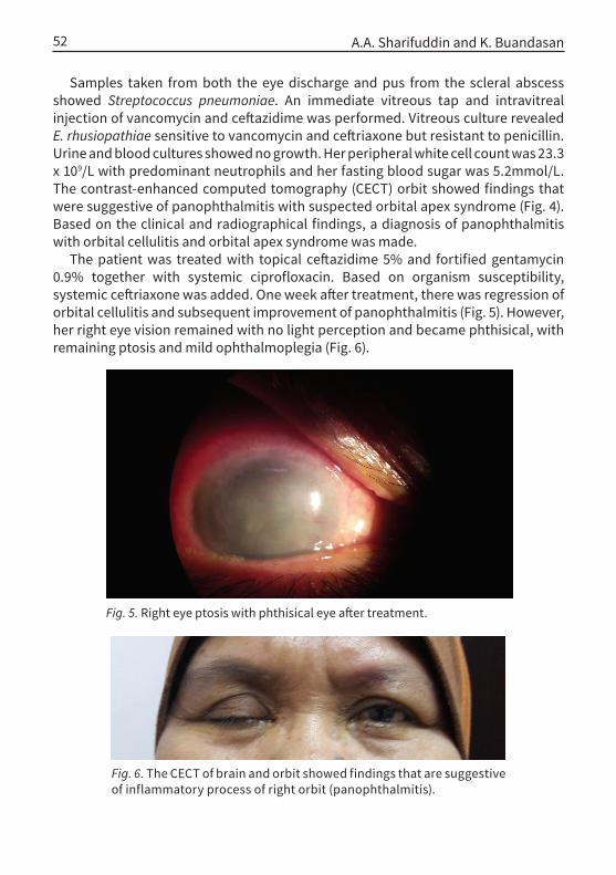
Fig. 5. Right eye ptosis with phthisical eye after treatment.
Fig. 6. The CECT of brain and orbit showed findings that are suggestive of inflammatory process of right orbit (panophthalmitis).
A.A. Sharifuddin and K. Buandasan52
Samples taken from both the eye discharge and pus from the scleral abscess showed Streptococcus pneumoniae. An immediate vitreous tap and intravitreal injection of vancomycin and ceftazidime was performed. Vitreous culture revealed E. rhusiopathiae sensitive to vancomycin and ceftriaxone but resistant to penicillin. Urine and blood cultures showed no growth. Her peripheral white cell count was 23.3 x 10⁹/L with predominant neutrophils and her fasting blood sugar was 5.2mmol/L. The contrast-enhanced computed tomography (CECT) orbit showed findings that were suggestive of panophthalmitis with suspected orbital apex syndrome (Fig. 4). Based on the clinical and radiographical findings, a diagnosis of panophthalmitis with orbital cellulitis and orbital apex syndrome was made.
The patient was treated with topical ceftazidime 5% and fortified gentamycin 0.9% together with systemic ciprofloxacin. Based on organism susceptibility, systemic ceftriaxone was added. One week after treatment, there was regression of orbital cellulitis and subsequent improvement of panophthalmitis (Fig. 5). However, her right eye vision remained with no light perception and became phthisical, with remaining ptosis and mild ophthalmoplegia (Fig. 6).

Panophthalmitis with orbital cellulitis related to Erysipelothrix rhusiopathiae 53
Discussion
Ocular infection caused by E. rhusiopathiae is extremely rare. So far, there is only one reported case of E. rhusiopathiae causing ocular infection in 2008. Elvy and colleagues reported a case of bilateral endogenous endophthalmitis caused by E. rhusiopathiae infection where the patient had underlying ulcerative colitis and had been on regular oral prednisolone.4 The source of infection was from ingestion of undercooked meat that subsequently caused liver abscess formation and hae-matogenous spread to the eyes. The patient responded well to systemic penicillin G.
The route of entry for E. rhusiopathiae is usually though a skin abrasion when handling infected meat, fish, or soil. However, there are two reported cases from Elvy et al.4 and Kichloo et al.5 that recorded septicaemia due to E. rhusiopathiae infection following ingestion of infected undercooked meat. Additionally, immuno-suppression and chronic alcohol abuse are also frequently found in reported cases of E. rhusiopathiae infections.6 This patient is a housewife who regularly handles raw meat and fish for cooking. However, she had no skin lesions or bacteraemia. An alternative explanation to the source of infection may be from direct exposure of E. rhusiopathiae to the eye from touching her eyes with her unwashed hands. However, the conjunctival swab and pus from the scleral abscess grew a different pathogen, Streptococcus pneumoniae. There is a possibility that the cultures from the conjunctiva and pus were contaminated samples.
The organism on blood agar culture showed a non-motile, non-sporulating gram-positive rod which was further identified as E. rhusiopathiae by the Vitek system. E. rhusiopathiae is usually highly susceptible to penicillin and cephalo-sporin, but resistant to vancomycin.4 However, in this case, the vitreous culture and sensitivity showed resistance to penicillin and ampicillin, but sensitivity to vancomycin and ceftriaxone. The patient responded well to systemic ceftriaxone and ciprofloxacin together with topical ceftazidime and fortified gentamicin. This case is unique as it adds to the scarce literature on ocular E. rhusiopathiae infection causing endophthalmitis without septicaemia. However, E. rhusiopathiae has an intrinsic resistance to vancomycin; thus, for this case, there is a possibility of tran-scription error of the bacterial sensitivity during reporting or error in bacterial identification.
Acknowledgements
The authors would like to acknowledge the contributions of Dr. Yanti Muslikhan and Dr. Sai De Zhao of the Ophthalmology Department, Hospital Sultanah Nora Ismail (Batu Pahat, Johor, Malaysia) in preparing this case report.

A.A. Sharifuddin and K. Buandasan54
References
1. Brooke CJ, Riley TV. Erysipelothrix rhusiopathiae: bacteriology, epidemiology and clinical manifesta-tions of an occupational pathogen. J Med Microbial. 1999;48:789-799.
2. Gorby GL, Peacock JE Jr. Erysipelothrix rhusiopathiae endocarditis: microbiologic, epidemiologic and clinical features of an occupational disease. Rev Infect Dis. 1988;10:317-325
3. Luigi P, Silvia B, Carola M, Silvia T, Beatrice P, Francesco L. Erysipelothrix rhusiopathiae bacteremia without endocarditis. Infect Dis. 2016;8:6368
4. Elvy J, Hanspal I, Simcock P. A case of Erysipelothrix rhusiopathiae causing bilateral endogenous en-dophthalmitis. J Clin Pathol. 2008;61:1223-1224.
5. Kichloo AA, Hallac A, Mousavi B, Hirekhan O. Nonspecific Erysipelothrix rhusiopathiae bacteremia in a patient with subclinical alcoholic liver disease. Case Rep Infect Dis. 2013;2013:474593
6. Reboli AC, Farrar WE. Erysipelothrix rhusiopathiae: an occupational pathogen. Clin Microbial Rev. 1989;2:354-359.

© Malaysian Journal of Ophthalmology 2020; 2:55-61Case report/Case series
Ocular complications in pre-eclampsiaDian Nadia Abu Talib1, Wahidah Wagimon2, Ainal Adlin Naffi1, Rona Asnida Nasaruddin1, Jemaima Che-Hamzah1, Mohd Hashim Omar2, Norshamsiah Md Din1
1Department of Ophthalmology, Universiti Kebangsaan Malaysia Medical Centre, Cheras, Kuala Lumpur, Malaysia; 2Department of Obstetrics and Gynecology, Universiti Kebangsaan Malaysia Medical Centre, Jalan Yaacob Latif, 56000 Cheras, Kuala Lumpur, Malaysia.
Abstract
Exudative retinal detachment (ERD) is a rare complication of pre-eclampsia in pregnancy. The pathophysiology is uncertain but it is thought to be due to micro-vasculopathy involving the choroidal circulation. We report a case of a 36-year-old woman with underlying essential hypertension complicated with impending eclampsia in her third trimester of pregnancy. She developed bilateral bullous ERD at 34 weeks of gestation. Following emergency caesarean section, her blood pressure normalised after 10 days and the ERD partially resolved spontaneous-ly after 30 days. Her best-corrected visual acuity improved from hand movement (HM) in the right eye and counting finger (CF) in the left eye to 6/24 bilaterally after 30 days postpartum. Pre-eclampsia-induced ERD is usually managed conserva-tively and the prognosis is usually good.
Keywords: exudative retinal detachment, impending eclampsia, pregnancy, hyper-tension
Correspondence: Ainal Adlin Naffi, Department of Ophthalmology, Universiti Kebangsaan Malaysia Medical Centre, Jalan Yaacob Latif, 56000 Cheras, Kuala Lumpur, Malaysia. E-mail: [email protected]

D.N.A. Talib et al.56
Komplikasi okular pada pra-eklampsia
AbstrakLekang retina eksudatif (ERD) adalah komplikasi pra-eklampsia yang jarang berlaku semasa kehamilan. Patofisiologi tidak begitu jelas tetapi dianggap disebabkan oleh mikrovaskulopati yang terjadi kepada salurdarah jaringan koroidal. Kami melaporkan kes seorang wanita berusia 36 tahun yang berlatarbelakangkan penyakit tekanan darah tinggi yang kemudiannya dirumitkan dengan komplikasi eklampsia yang berlaku dalam trimester ketiga kehamilannya. Beliau mengalami ERD bullous pada kedua belah mata pada minggu 34 kehamilan. Selepas pembedahan secara caesarean tekanan darahnya turun ketahap normal selepas 10 hari dan ERD sembuh secara spontan selepas 30 hari. Ketajaman penglihatannya berubah dari dari pergerakan tangan (HM) sahaja di mata kanan dan mengira jari (CF) di mata kiri kepada 6/24 pada kedua-dua belah mata pada hari ke 30 selepas bersalin. ERD yang disebabkan oleh pra-eklampsia biasanya dirawat secara konservatif dan prognosis biasanya baik.
Kata kunci: hipertensi, kehamilan, lekang retina eksudatif, pra-eklampsia
Introduction
Hypertensive disorder is one of the commonest medical complications of pregnancy. In Southeast Asia, hypertensive disorders accounted for 14.5 % of maternal deaths, whereas in the Caribbean and Latin America, the figure was over 22.1%.1
Systemic hypertension is related to a variety of ocular presentations. This includes the four grades of hypertensive retinopathy, retinal vein occlusions, and ischaemic optic neuropathies. The occurrence of ERD with uncontrolled hyperten-sion in pregnancy from pre-eclampsia poses additional complications unique to this situation, possibly due to microvasculopathy affecting the choroidal vasculature, similar to microvascular complications in the kidney resulting in albuminuria. We present an uncommon case of hypertensive chorioretinopathy with bilateral ERD secondary to impending eclampsia in pregnancy.
Case report
A 38-year-old gravida 3 para 2 woman was diagnosed with essential hypertension at 11 weeks of gestation during her routine antenatal checkup. She was started on T. methyldopa 200 mg TDS to optimize her blood pressure (BP) at 31 weeks’ gestation.

Ocular complications in pre-eclampsia 57
At 34 weeks of gestation, she was admitted for impending eclampsia with a BP of 170/110 mmHg and proteinuria. She presented with headache and generalized blurring of vision bilaterally for one day prior to admission but there were no flashes of lights, floaters, or visual field defect. However, visual acuity was not measured during admission. Biochemistry markers did not suggest HELLP syndrome (haemolysis, elevated liver enzymes, and low platelet level). Intravenous magnesium sulphate and intravenous labetalol were administered, and she underwent an emergency caesarean section on the same day for impending eclampsia.
At postnatal day 1, her BP was better controlled, but her visual acuity worsened to hand movement (HM) in the right eye (OD) and counting finger (CF) at one foot in the left eye (OS). The relative afferent pupillary defect was absent. Anterior segment examination of the eyes was normal with no evidence of inflammation. Fundus examination revealed bilateral bullous ERD inferiorly involving the macula with widespread choroidopathy and scattered retinal haemorrhages at the supero-nasal and supero-temporal quadrants of the optic disc and at the macula in OS. (Fig. 1A-D). The shifting fluid test was positive, confirming the suspicion of ERD. There was no retinal break found clinically. The patient’s BP normalized over ten days, her visual acuity gradually improved over one month, and serial fundus examination showed slow resorption of subretinal fluid (SRF). Her vision at one-month postpartum was 6/24 bilaterally with some residual SRF (Figs. 2 and 3). At 3 months follow-up, her best-corrected vision acuity was 6/12 OD and 6/9 OS, with total resorption of SRF and resolving retinal haemorrhages with irregularities over retinal pigment epithelium (RPE) and inner segment and outer segment (IS/OS) disruptions. Her BP was well controlled only by oral amlodipine 5 mg once daily.
Discussion
Accelerated systemic hypertension may result in various complications to the eye causing considerable amount of visual loss. This includes non-ocular complica-tions such as cerebral infarction or haemorrhage, and complications pertaining to the vasculature in the eye, such as ischemic optic neuropathy and retinal vascular occlusions.2 A more uncommon cause of visual loss as demonstrated in our case is ERD.
ERD is a rare complication of pre-eclampsia, which has only been reported in 1-2% of preeclampsia cases.3 Typically, it is completely reversible depending on the degree of RPE ischemia seen in severe pre-eclampsia. Subretinal fibrosis or epiretinal membrane may follow as a later complication.
The pathophysiology of ERD in pre-eclampsia is not well understood and con-troversial. The passive and active transport mechanism within RPE and ocular forces within the eye are important to maintain a unidirectional flow from vitreous to the choroid. Passive transport mechanisms include intraocular pressure and

Fig. 1. Fundus photographs of both eyes. (A) Widespread whitening of the choroidal area indicating choroidopathy with flame-shaped haemorrhages seen at the major vascular arcades in OD. (B) Inferior bullous retinal detachment extending to the inferior vascular arcades and the macula involving the OD. (C) Similar changes of choroidal whitening at the posterior pole in the OS. The macula is also shallowly detached, indicated by striations on the macula. (D) ERD seen mainly inferior to the vascular arcades in OS.
A B
C D
Fig. 3. OS OCT at one month showing similar features as OD.
Fig. 2. OD OCT at one month revealed residual hyper-reflective, fibrinous SRF.
D.N.A. Talib et al.58
Ocular complications in pre-eclampsia 59
osmotic pressure from the choroid. Disruption in one of these two mechanisms will result in increased fluid in the subretinal space.4
ERD is thought to occur from microvascular occlusion of the choroidal vasculature. Intense vasoconstriction of the choroidal vessels leads to fibrinoid necrosis or thrombosis of the choroidal vessels. This results in choroidal ischemia and subsequently increased vascular permeability as well as reduced ability to accommodate extra fluid5 as the hypoxic drive releases vascular endothelial growth factor (VEGF), leading to increased permeability and disruption of endothelial junctions.6 The ensuing ischemia of the overlying RPE disrupts RPE function and its tight junctions, which form the blood-retinal barrier.5 The subsequent accumu-lation of fluid into the potential space between the neurosensory retina and the RPE results in ERD and areas of choroidal hypo- and non-perfusion manifested as Elschnig spots.7
As an alternative to the vasospasm theory, it is suggested that hyperperfu-sion and breakthrough in autoregulation of orbital vessels will further increase the permeability of retinal and choroidal arterioles, causing retinal oedema and serous detachment.8
Choroidal blood flow depends on the vascular resistance between the arterial input and venous output and on the arterial and venous pressure difference. The mean choroidal blood flow (ChBFm) is as stated below in Formula 1,
ChB F m = (MOAP − IOP) __________ R m =
P P m ____ R m Formula 1
where MOAP is the mean ophthalmic artery blood, which for a sitting subject is approximately 2/3(BPdiastolic + 1/3 (BPsystolic - BPdiastolic). Rm is the average resistance during the heart cycle. PPm is commonly defined as the mean ocular perfusion pressure.9 With reference to Formula 1, it is postulated that relative hypotension episodes that occur immediately post-delivery will further compromise the choroidal blood flow and worsen accumulation of fluid in the subretinal space, thus explaining the postpartum worsening of the patient’s vision.
Management of hypertensive ERD mainly involves stabilization of BP. Nor-malization of BP postpartum results in the return of the RPE blood supply and function. With resorption of SRF and reattachment of the RPE and neurosenso-ry retina, ERD usually resolves spontaneously within a few weeks with the return of normal vascular and metabolic support for the photoreceptors.10 The visual prognosis is usually good, with vision returning in a few weeks. However, in our patient there was some residual visual acuity impairment likely explained by her optical coherence tomography (OCT) result, which showed segmented area of RPE irregularities with IS/OS disruptions (Figs. 4 and 5).
In this anti-VEGF era, more studies are looking into its role as an adjunct treatment in ERD due to elevated ocular VEGF levels. Kim et al. suggested intravit-real bevacizumab injection might be an effective therapeutic modality for treating

Fig. 5. OS OCT at three months revealed RPE irregularities with less severe IS/OS disruptions compared to OD.
Fig. 4. OD OCT at three months revealed RPE irregularities with IS/OS disruptions.
D.N.A. Talib et al.60
exudative hypertensive retinopathy.11 The study is limited by the small number of patients and the lack of a control group.
However, administering medication during pregnancy or breastfeeding remains a challenge due to the paucity of published information on intravitreal injections during pregnancy and breastfeeding. One study reported undetectable levels of bevacizumab in the breast milk of nursing mothers who were treated with monthly injections after delivery.12 However, there are no studies on the penetration of ranibizumab or aflibercept into breast milk.
Treatment should only be administered following a thorough discussion with the patient and informed consent, as well as consultation with an obstetrician. Of the currently available anti-VEGF agents, ranibizumab may be the safest choice as it has been shown to have the most rapid clearance from systemic circulation and weakest effect on plasma VEGF levels.13 Breastfeeding is likely not a contraindi-cation to anti-VEGF therapy. It should be noted that these recommendations are based on the sparse literature that explores this issue and decisions should only be made after taking into consideration risks and benefits for both mother and baby. Furthermore, in hypertensive choroidoretinopathy, the causes of elevated VEGF are transient and reversible after normalization of BP. Thus, in most cases, a more conservative approach is taken.
Conclusion
Most ERD secondary to hypertensive retinopathy resolves spontaneously. Visual prognosis is good unless there is irreversible ischemic RPE in severe eclampsia. It is therefore important to identify those at risk and to have good BP control and timely delivery for the best outcome of both mother and baby.

Ocular complications in pre-eclampsia 61
References
1. Say L, Chou D, Gemmill A, et al. Global causes of maternal death: A WHO systematic analysis. Lancet Glob Health. 2014;2(6):e323-333.
2. Villalba-Pinto L, Hernández-Ortega MÁ, Lavid De Los Mozos FJ, et al. Massive bilateral serous retinal detachment in a case of hypertensive chorioretinopathy. Case Rep Ophthalmol. 2014;5(2):190–194.
3. Ober RR. Pregnancy-induced hypertension (pre-clampsia eclampsia). In: Ryan SJ, editor. Retina, St Louis: CV Mosby;1994. p . 1393–1403.
4. Besharse J, Bok D. The retina and its disorders. 1st ed. Vol. 9, Breakdown of RPE blood –retinal barrier. Academic Press; 2011. p . 59-67.
5. Younis MT, McKibbin M, Wright A. Bilateral exudative retinal detachment causing blindness in severe pre-eclampsia. J Obstet Gynaecol. 2009;27(8):847–848.
6. Lena CW. Vascular permeability- the essentials. Ups J Med Sci. 2015;120(3):135-143.7. Schultz KL, Birnbaum AD, Goldstein DA. Ocular disease in pregnancy. Curr Opin Ophthalmol.
2005;16(5): 308-314. 8. Roos NM, Wiegman MJ, Jansonius NM, et al. Visual disturbances in (pre)eclampsia. Obstet Gynecol
Surv. 2012;67(4):242–250.9. Riva CE, Titze P, Hero M, et al. Effect of acute decreases of perfusion pressure on choroidal blood flow
in humans. Invest Ophthalmol Vis Sci. 1997;38(9):1752-1760.10. Khawla A. The eye and the visual system in preeclampsia /eclampsia: What to expect? Saudi J Oph-
thalmol. 2013;27(1):51-53.11. Kim EY, Lew HM, Song JH. Effect of intravitreal bevacizumab (Avastin®) therapy in malignant hyper-
tensive retinopathy: A report of two cases. J Ocul Pharmacol Ther. 2012;28(3):318-322.12. McFarland TJ, Rhoads AD, Hartzell M, et al. Bevacizumab levels in breast milk after long-term intrav-
itreal injections. Retina. 2015;35(8):1670–1673.13. Grzybowski A, Told R, Sacu S, Bandello F, et al. Update on intravitreal injections: Euretina expert
consensus recommendations. Ophthalmologica. 2018;239(4):181–193.

© Malaysian Journal of Ophthalmology 2020; 2:62-68Case report/Case series
Isolated abducens nerve palsy secondary to lung metastases Hayatulrizal Muhd1, Hayati Abdul Aziz1, Francesca Martina Vendargon1, Jasmi Ramlan2, Yeoh Aik Guan3
1Department of Ophthalmology, Hospital Sultanah Aminah, Johor Bahru, Johor, Malaysia; 2Department of Pathology, Hospital Sultanah Aminah, Johor Bahru, Johor, Malaysia; 3Department of Radiology, Hospital Sultanah Aminah, Johor Bahru, Johor, Malaysia
Abstract
We present a rare case of metastatic lung adenocarcinoma which presented as isolated abducens nerve palsy. A 65-year-old healthy woman presented with horizontal diplopia of three months duration. Her best-corrected visual acuity was 6/18 bilaterally with no relative afferent pupillary defect. Ocular examination showed limited abduction of the right eye. Anterior and posterior segment examination was normal in both eyes. However, she had a non-tender enlarged lymph node at the left supraclavicular area. The systemic review was unremarkable. C-reactive protein, erythrocyte sedimentation rate, and serum alpha-fetoprotein were markedly raised. A chest X-ray revealed right upper lobe opacity with a satellite nodule. Computed tomography of the brain, orbit, thorax, abdomen, and pelvis exhibited a heterogeneously enhancing mass at the right upper lobe of the lung with metastasis to the left frontal, right occipital, and left parietal lobe as well as multiple bone metastases. With histopathology confirmation of lung tissue biopsy, she was diagnosed as isolated right abducens nerve palsy secondary to metastatic lung adenocarcinoma. She was referred to the respiratory, neuromedical, and oncology units for further management. In conclusion, although this appeared to be an isolated case, lung metastasis should be considered in the differential diagnosis of isolated abducens nerve palsy. Hence, lung examination should be performed when encountering such cases.
Keywords: abducens nerve palsy, adenocarcinoma, lung adenocarcinoma, neu-ro-ophthalmology, sixth nerve palsy Correspondence: Dr. Muhammad Hayatulrizal, Ophthalmology Department, Hospital Sultanah Aminah Johor Bahru, Johor, Malaysia. E-mail: [email protected]

Isolated abducens nerve palsy secondary to lung metastases 63
Palsi saraf abdusen akibat metastasis dari paru-paru
AbstrakKami membentangkan satu kes yang jarang berlaku tentang metastasis adenokarsinoma paru-paru yang bermula dengan gejala terpencil palsi saraf abdusen. Seorang wanita yang sihat berusia 65 tahun mengadu mengalami diplopia selama tiga bulan. Ketajaman penglihatannya yang terbaik adalah 6/18 pada kedua-dua belah mata tanpa kecacatan relatif aferen anak mata. Peperiksaan okular menunjukkan pergerakan mata yang terhad. Pemeriksaan segmen anterior dan posterior adalah normal di kedua-dua mata. Walau bagaimanapun, dia mempunyai nod limfa yang tidak menyakitkan di bahagian supraklavikular kiri. Pemeriksaan klinikal pada bahagian sistemik adalah normal. Terdapat peningkatan ketara protein C-reaktif, kadar pemendapan eritrosit, dan alpha-fetoprotein serum. Ujian X-ray dada menunjukkan opasiti lobus kanan atas dengan nodul satelit. Tomografi komputasi otak, orbit, toraks, abdomen, dan pelvis mempamerkan kehadiran ketumbuhan yang bersifat heterogenus pada lobus kanan paru-paru dengan metastasis ke frontal kiri, osipital kanan, dan lobus parietal kiri bahagian otak serta beberapa kesan metastasis pada tulang. Dengan pengesahan histopatologi biopsi tisu paru-paru, dia akhirnya didiagnos sebagai palsi saraf abducens kanan terpencil akibat dari kesan metastasis adenorkarsinoma paru -paru. Dia dirujuk ke unit respiratori, neurologi perubatan, dan onkologi untuk rawatan lanjut. Sebagai kesimpulan, walaupun kes ini kelihatan sebagai kes terpencil, metastasis paru-paru perlu dipertimbangkan sebagai salah satu penyebab palsi saraf abduscen terpencil Oleh itu, pemeriksaan paru-paru perlu dilakukan apabila menghadapi kes-kes seperti ini.
Kata kunci: adenokarsinoma, adenokarsinoma paru-paru, neuro-optalmologi, palsi saraf abdusen, palsi saraf keenam
Introduction
Most isolated abducens nerve palsies are ischemic in nature.1 However, there are other life-threatening causes of abducens nerve palsy. Some require prompt diagnostic and therapeutic management, as delayed treatment may lead to increased morbidity and/or mortality. In this report, we present a rare case of metastatic lung adenocarcinoma that presented as isolated abducens nerve palsy.

Fig. 1. Coronal plane of TAP CT (left) showing heterogeneously enhancing mass at the posterior segment of the right upper lobe of the lung. Axial image of thorax CT (right) showing enhancing mass at the posterior segment of the right upper lobe.
H. Muhd et al.64
Case report
A 65-year-old woman and passive smoker for 30 years presented with a three-month history of binocular horizontal diplopia and significant weight loss. The diplopia was persistent throughout the day and improved slightly on tilting her head to the right. However, there was no associated pain, red eye, headache, vomiting, seizures, or body weakness. She denied any concurrent fever, nasal, or gastro-uri-nary symptoms. There was no high-risk behaviour. Her best-corrected visual acuity was 6/18 bilaterally with no relative afferent pupillary defect. On examination, there was limited abduction of the right eye associated with a right-sided head tilt. Anterior segment was normal in both eyes. Dilated fundus examination of both eyes revealed no optic disc swelling. Examination of other cranial nerves was normal. A non-tender enlarged lymph node was detected in the left supraclavicular area. Systemic review and examination of the chest, abdomen, and breasts were normal.
Blood investigations revealed markedly raised C-reactive protein ( 144 mg/ dl), erythrocyte sedimentation rate (60 mm/hr), and serum alpha-fetoprotein. A chest X-ray revealed a right upper lobe opacity with a satellite nodule. Computed tomography (CT) of the brain, orbit, thorax, abdomen, and pelvis (TAP) exhibited a heterogeneously enhancing mass at the posterior segment of right lung upper lobe (Fig. 1) with metastases to the left frontal, right occipital, and left parietal lobes (Fig. 2, left). There were sclerotic lesions at the body, lamina, and pedicles of T4-T5 as well as right iliac bone, proximal left femoral head, and right tenth rib in keeping with bony metastases.
The patient underwent CT-guided, fine-needle aspiration biopsy of the lung. Histopathology examination (HPE) of lung tissue biopsy revealed adenocarcinoma

Fig. 2. Axial image of brain CT (left) showing metastases to left frontal and right occipital. HPE of lung tissue (right) showing tumour forming acinar pattern, no nuclear moulding, nesting, or rosette formation.
Isolated abducens nerve palsy secondary to lung metastases 65
with features of malignant cells forming an acinar pattern with no nuclear moulding, nesting, or rosette formation (Fig. 2, right). The tumour cells were positive towards thyroid transcription factor-1 (TTF-1), napsin A, but were negative for CD56, synap-tophysin and chromogranin immunostains.
A diagnosis of isolated right abducens nerve palsy secondary to metastatic lung adenocarcinoma was made. The patient was referred to the oncology team and started on 20 Gy whole-brain radiotherapy given in 5 fractions. Subsequently, she received four cycles of palliative double-agent chemotherapy including plati-num-based cisplatin and gemcitabine.
Her general medical condition deteriorated with development of a new patho-logical fracture of the right humerus and femur despite post-chemotherapy imaging showing the radiological and objective clinical response. She was then managed by palliative and supportive care to provide comfort and quality of life.
Discussion
Sixth nerve palsies occurring in the middle-aged group arise from a variety of causes, with the two most common being vascular (18-37%) and idiopathic (21-30%).1 The abducens nerve is prone to trauma and metastatic lesion due to its long intracranial course. Metastasis from lung, breast, liver, colon, and prostate have been previously reported in the literature to cause isolated and multiple cranial nerve palsies.2
The prevalence of brain metastasis in the presence of systemic malignancy ranges from 8-15%.3-5 In Malaysia, lung cancer accounts for 10.2% of brain metastases and 16.2% of patients with lung cancer will develop brain metastasis within five years

H. Muhd et al.66
of diagnosis.5,6 However, the prevalence of a brainstem metastasis is relatively rare, accounting for approximately 5-7%.3,7,8 Patients with brainstem lesions involving the abducens nerve classically present with binocular horizontal diplopia, as observed in this patient.
Despite vascular disorders being the commonest cause of abducens nerve palsy in middle-aged individuals, any lesion at the level of cerebellopontine angle, par-ticularly that involving the abducens nerve, can present with abducens nerve palsy. As observed in this patient, malignancy was the main differential diagnosis in the setting of her being a chronic passive smoker and in the presence of constitutional symptoms.
Other tumours which can affect the cerebellopontine area include nasopharyn-geal carcinoma and acoustic neuroma.9 Classically, the cranial nerves in this area (facial and vestibular nerves) may also be affected, producing added signs and symptoms of decreased hearing and /or facial paralysis. In this case, the patient did not show any signs of other cranial nerve involvement.
Due to clinical suspicion of malignancy, contrast-enhanced computed tomography (CECT) was performed urgently, revealing multiple lesions involving the lung, brain, and bones. Metastases into the brainstem may be too small to be detected by CT scan, and magnetic resonance imaging (MRI) of the brain may be necessary.10 However, there was no MRI evidence to suggest a metastatic lesion. Gaze palsy resulting from the metastatic lesion in the facial colliculus of lower pons detected on MRI has been reported by Han et al.11 A case by Reyes et al. also reported a metastasized brainstem lesion causing abducens nerve palsy as evidenced on MRI imaging.12
Based on the above literature review, there is high a likelihood that the metastasis causing isolated nerve palsy may be located at the abducens nucleus at the pontine region.
As the histopathological examination of the lung tissue biopsy yielded a result compatible with lung adenocarcinoma metastasized to the brain and bones with immunohistochemistry analysis suggesting the lung as an organ of origin, radiation therapy followed by palliative chemotherapy was recommended.
The prognosis of lung carcinoma in the presence of distant metastases is generally poor.13 Despite that, palliative treatment including radiotherapy, chemotherapy, and hormonal treatment was started. Radiotherapy is still the mainstay treatment for orbital and brain metastases, with response rates up to 79% and 80% which may restore vision may induce cataract formation and radiation retinopathy.14 A study by Ramesh et al. found a response rate in 88% of patients who completed 30 Gy dose in 10 fractions over one month.15
Chemotherapy has high response rates up to 35% with a survival rate of 15% in 2 years.13 Double-agent regimens such as the platinum- gemcitabine combination used in our case has been proven to be superior to single-agent regimens.16 Hormonal therapy, such as gefitinib, can be given in the presence of EGFR tyrosine

Isolated abducens nerve palsy secondary to lung metastases 67
kinase mutation and disease progression. It has been proven to exhibit objective clinical response and shows significant anti-tumour activity as a single agent when started in relapsed non-small-cell lung carcinoma patient.13
The most challenging aspect to diagnose this case was its rarity and limited knowledge about it. We believe our case increases the knowledge of metastatic patterns of lung cancer and assists ophthalmologists in identifying and managing patients presenting with isolated abducens nerve palsy.
Conclusion
This case highlights several important points. The aetiology of abducens nerve palsy is diverse, ranging from benign to life-threatening. Metastatic lung carcinoma, although rare, should be considered when encountering isolated abducens nerve palsy with a suggestive history of passive smoking and constitutional symptoms. The diagnosis was made on a high index of suspicion of the varied symptoms. Hence, a thorough lung examination with radiological and pathological analysis is required to establish the diagnosis, which can be life-saving.
References
1. Rush JA, Younge BR, Paralysis of cranial nerves III, IV and VI. Cause and prognosis in 1,000 cases. Arch Ophthalmol. 1981;99:76-79.
2. Chandrashekhar MN, Kishore K, Lakshmaiah V, Nagesha CK, Kumar BN. Clivus metastasis presenting as isolated abducens nerve palsy- A case report. J Evol Med Dent Sci. 2013;43:8383-8385.
3. Kased N, Huang K, Nakamura JL, et al. Gamma Knife radiosurgery for brainste metastases: the UCSF experience. J Neurooncol. 2008;86:195-205.
4. Posner JB, Chernik NL. Intracranial metastasis from systemic cancer. Adv Neurol. 1978;19:579-5925. Schouten LJ, Rutten J, Huveneers HA, Twijnstra A. Incidence of brain metastases in a cohort of
patients with carcinoma of breast, colon, kidney, and lung and melanoma. Cancer. 2002;94:2698-2705.
6. Azizah Ab M, Nor Saleha IT, Noor Hashimah A, Asmah ZA, Mastulu W. Malaysian National Cancer Registry Report 2007-2011. National Cancer Registry; 2015.
7. Barnholtz-Sloan JS, Sloan AE, Davis FG, et al. Incidence proportions of brain metastases in patients diagnosed (1973 to 2001) in the Metropolitan Detroit Cancer Surveillance System. J Clin Oncol. 2004;22:2865-2872.
8. Chason JL, Walker FB, Landers JW. Metastatic carcinoma in central nervous systema nd dorsal root ganglia. A prospective autopsy study. Cancer. 1963;16:781-787.
9. Park KA, Oh SY. Nasopharyngeal carcinoma presenting with rapidly progressive severe binocular optic neuropathy and periocular pain in a young man. J Neuroophthalmol. 2010;30:150-152.
H. Muhd et al.68
10. Pusateri TJ, Sedwick LA, Margo CE. Isolated inferior rectus muscle palsy from a solitary metastasis to the oculomotor nucleus. Arch Ophthalmol. 1987;105:675-677.
11. Han SB, Kim JH, Hwang JM. Presumed metastasis of breast cancer to the abducens nucleus present-ing as gaze palsy. Korean J Ophthalmol. 2010;24:186-188.
12. Reyes KB, Lee HY, Ng I, Goh KY. Abducens (sixth) nerve palsy presenting as a rare case of isolated brainstem metastasis from a primary breast carcinoma. Singapore Med J. 2011;52:e220-222.
13. Claire Saadeh, Pharm. D., BCOP. Pharmacotherapy self-assessment program, 6th edition. PSAP. 57-71.14. Liangchao Sun, Yali Qi, Xindong Sun, Jinming Yu, Xue Meng. Orbital Metastases as the initial presen-
tation of lung adenocarcinoma: A case report. Onco Targets Ther. 2016;9:2643-2648.15. Ramesh S Bilimagga, S Nirmala, Karthik S, et al. Role of palliative radiotherapy in brain metastasis.
Indian J Palliat Care. 2009;15(1):71–75.16. Socinski MA, Evans T, Gettinger S, et al. Treatment of stage IV non-small cell lung cancer: diagno-
sis and management of lung cancer, 3rd ed: American College of Chest Physicians evidence-based clinical practice guidelines. Chest. 2013;143(5Suppl):e341S-e368S.
© Malaysian Journal of Ophthalmology 2020; 2:69-74Case report/Case series
Cases of cytomegalovirus retinitis in patients treated for cancerMarium Jamaluddin Ahmad, Tengku Ain Kamalden, Nurliza Khaliddin, Tajunisah Iqbal
UM Eye Research Centre, Department of Ophthalmology, University of Malaya, Kuala Lumpur, Malaysia
Abstract
Cytomegalovirus (CMV) retinitis is an opportunistic infection affecting immu-nocompromised patients. This case series describes 2 patients aged 19 and 10 years who were undergoing cancer treatment and were referred for complaints of worsening blurred vision. The retinal findings in the first patient showed multiple yellowish subretinal areas with haemorrhages, while in the second patient there was presence of frosted branch angiitis. Confirmation of diagnosis was done using PCR of vitreous samples and blood CMV levels. Both tested negative for HIV. Both received intravitreal and systemic ganciclovir. It is important for ophthalmolo-gists to consider CMV retinitis in all patients with any form of immunosuppression.
Keywords: cancer treatment, cytomegalovirus retinitis, retina
Correspondence: Dr. Marium Binti Jamaluddin Ahmad, MBBS, UM Eye Research Centre, Department of Ophthalmology, University of Malaya Medical Centre, Lembah Pantai 59100, Kuala Lumpur, Malaysia. E-mail: [email protected]

M.J. Ahmad, N. Khaliddin and T. Iqbal70
Kes-kes cytomegalovirus retinitis pada pesakit yang dirawat untuk kanser
AbstrakRetinitis Cytomegalovirus (CMV) adalah jangkitan oportunistik yang memberi jangkitan kepada pesakit kurang pertahanan imuniti badan. Siri kes ini merujuk kepada 2 pesakit berusia 19 dan 10 tahun yang menjalani rawatan kanser dan dirujuk dengan aduan penglihatan yang semakin kabur. Pemeriksaan pada pesakit pertama menunjukkan beberapa kawasan subretinal yang kekuningan dengan pendarahan. Sementara pada pesakit kedua terdapat kehadiran radang dan penyumbatan salur darah halus. Pengesahan diagnosis dilakukan menggunakan kaedah reaksi rangkaian polymerase (PCR) pada sampel vitreous dan tahap kehadiran CMV dalam darah. Kedua-dua kesdiuji negatif untuk kehadiran Human Immunodeficiency Virus (HIV). Kedua-duanya menerima rawatan ganciclovir secara intravitreal dan sistemik. Adalah penting bagi pakar oftalmologi untuk mempertimbangkan retinitis CMV dalam semua pesakit yang mengalami keadan pertahanan imuniti badan yang lemah.
Kata kunci: kurang pertahanan badan, sitomegalovirus, retinitis
Introduction
Cytomegalovirus (CMV) can cause severe disease in those who are immunocom-promised. Infection with CMV is on the rise with the increase in organ transplant, immunosuppressive therapies, and human immunodeficiency virus (HIV)-infected patients. It is also one of the leading causes of birth defects.1 In this case series, we describe two cases of CMV retinitis in two young patients undergoing treatment for cancer.
Case 1
A 19-year-old Chinese man presented with left facial swelling in June 2017 and biopsy was done to confirm the diagnosis. He was diagnosed to have locally advanced and metastatic left maxillary rhabdomyosarcoma with cervical node, right pleural, and splenic node metastasis involving left orbit, gingiva, clivus, and left cheek. The patient had completed a course of VAID (ifosfamide, vincristine, actinomycin D, doxorubicin) regime chemotherapy, was undergoing radiotherapy, and had a planned cycle of vincristine and cyclophosphamide. He presented to the eye clinic
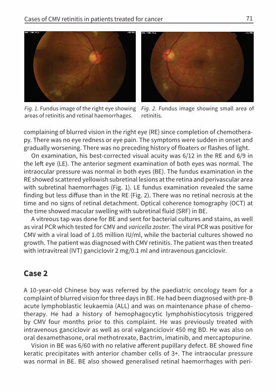
Fig. 2. Fundus image showing small area of retinitis.
Fig. 1. Fundus image of the right eye showing areas of retinitis and retinal haemorrhages.
Cases of CMV retinitis in patients treated for cancer 71
complaining of blurred vision in the right eye (RE) since completion of chemothera-py. There was no eye redness or eye pain. The symptoms were sudden in onset and gradually worsening. There was no preceding history of floaters or flashes of light.
On examination, his best-corrected visual acuity was 6/12 in the RE and 6/9 in the left eye (LE). The anterior segment examination of both eyes was normal. The intraocular pressure was normal in both eyes (BE). The fundus examination in the RE showed scattered yellowish subretinal lesions at the retina and perivascular area with subretinal haemorrhages (Fig. 1). LE fundus examination revealed the same finding but less diffuse than in the RE (Fig. 2). There was no retinal necrosis at the time and no signs of retinal detachment. Optical coherence tomography (OCT) at the time showed macular swelling with subretinal fluid (SRF) in BE.
A vitreous tap was done for BE and sent for bacterial cultures and stains, as well as viral PCR which tested for CMV and varicella zoster. The viral PCR was positive for CMV with a viral load of 1.05 million IU/ml, while the bacterial cultures showed no growth. The patient was diagnosed with CMV retinitis. The patient was then treated with intravitreal (IVT) ganciclovir 2 mg/0.1 ml and intravenous ganciclovir.
Case 2
A 10-year-old Chinese boy was referred by the paediatric oncology team for a complaint of blurred vision for three days in BE. He had been diagnosed with pre-B acute lymphoblastic leukaemia (ALL) and was on maintenance phase of chemo-therapy. He had a history of hemophagocytic lymphohistiocytosis triggered by CMV four months prior to this complaint. He was previously treated with intravenous ganciclovir as well as oral valganciclovir 450 mg BD. He was also on oral dexamethasone, oral methotrexate, Bactrim, imatinib, and mercaptopurine.
Vision in BE was 6/60 with no relative afferent pupillary defect. BE showed fine keratic precipitates with anterior chamber cells of 3+. The intraocular pressure was normal in BE. BE also showed generalised retinal haemorrhages with peri-
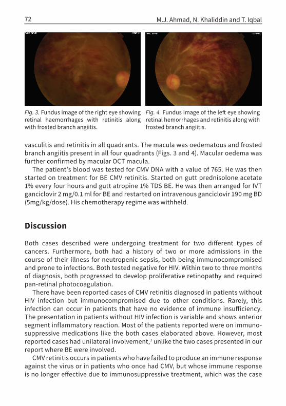
Fig. 4. Fundus image of the left eye showing retinal hemorrhages and retinitis along with frosted branch angiitis.
Fig. 3. Fundus image of the right eye showing retinal haemorrhages with retinitis along with frosted branch angiitis.
M.J. Ahmad, N. Khaliddin and T. Iqbal72
vasculitis and retinitis in all quadrants. The macula was oedematous and frosted branch angiitis present in all four quadrants (Figs. 3 and 4). Macular oedema was further confirmed by macular OCT macula.
The patient’s blood was tested for CMV DNA with a value of 765. He was then started on treatment for BE CMV retinitis. Started on gutt prednisolone acetate 1% every four hours and gutt atropine 1% TDS BE. He was then arranged for IVT ganciclovir 2 mg/0.1 ml for BE and restarted on intravenous ganciclovir 190 mg BD (5mg/kg/dose). His chemotherapy regime was withheld.
Discussion
Both cases described were undergoing treatment for two different types of cancers. Furthermore, both had a history of two or more admissions in the course of their illness for neutropenic sepsis, both being immunocompromised and prone to infections. Both tested negative for HIV. Within two to three months of diagnosis, both progressed to develop proliferative retinopathy and required pan-retinal photocoagulation.
There have been reported cases of CMV retinitis diagnosed in patients without HIV infection but immunocompromised due to other conditions. Rarely, this infection can occur in patients that have no evidence of immune insufficiency. The presentation in patients without HIV infection is variable and shows anterior segment inflammatory reaction. Most of the patients reported were on immuno-suppressive medications like the both cases elaborated above. However, most reported cases had unilateral involvement,2 unlike the two cases presented in our report where BE were involved.
CMV retinitis occurs in patients who have failed to produce an immune response against the virus or in patients who once had CMV, but whose immune response is no longer effective due to immunosuppressive treatment, which was the case

Cases of CMV retinitis in patients treated for cancer 73
for both our patients undergoing chemotherapy for cancer. CMV retinitis produces direct cytopathic effects, giving rise to the pale necrotic retina with focal areas of haemorrhage that spread centrifugally along vascular arcades. Visual loss is either by direct involvement of the retinitis to the macula or optic disc, or due to retinal detachment.3
CMV retinitis is usually seen in advanced HIV infection and is rarely found in other immunocompromised patients. There has been emerging data on non-HIV related CMV retinitis associated with systemic steroid use.4 CMV is a known infection in allogeneic transplantation recipients or those with T-cell deficiencies. Typically, CMV infection after stem cell transplantation gives rise to pneumonitis or enteritis, with retinitis being more common in those with HIV.5
CMV retinitis has been reported in a patient with long-standing rheumatoid arthritis who was being treated with immunosuppressive drugs, some of which included methotrexate, sulfasalazine, corticosteroids, cyclophosphamide, aza-thioprine, and finally anti-tumour necrosis factor-α antibodies (infliximab).6 CMV retinitis has also been found in patients with limited immune dysfunction such as advanced aged, diabetes mellitus, and noncytotoxic immunotherapy, which would present with variable clinical features of intraocular inflammation and most also developed neovascularisation due to extensive retinal ischemia.7
Conclusion
CMV retinitis is a visually disabling disease and should be promptly treated to avoid visual loss. This infection is closely related to the patient’s immune status and presentation may be variable.
References
1. Landolfo S, Gariglio M, Gribaudo G, Lembo D. The human cytomegalovirus. Pharmacol Ther. 2003;98(3):269-297.
2. Pathanapitoon K, Tesavibul N, Choopong P, et al. Clinical manifestations of cytomegalovirus-associ-ated posterior uveitis and panuveitis in patients without human immunodeficiency virus infection. JAMA Ophthalmol. 2013;131(5):638-645.
3. Carmichael A. Cytomegalovirus and the eye. Eye. 2012;26(2):237.4. Sloan D, Taegtmeyer M, Pearce I, Hart I, Miller A, Beeching N. Cytomegalovirus retinitis in the absence
of HIV or immunosuppression. SAGE Publications Sage UK: London, England; 2008.5. Goldberg SL, Pecora AL, Alter RS, et al. Unusual viral infections (progressive multifocal leukoenceph-
alopathy and cytomegalovirus disease) after high-dose chemotherapy with autologous blood stem cell rescue and peritransplantation rituximab. Blood. 2002;99(4):1486-1488.

M.J. Ahmad, N. Khaliddin and T. Iqbal74
6. Haerter G, Manfras BJ, de Jong-Hesse Y, et al. Cytomegalovirus retinitis in a patient treated with anti—tumor necrosis factor alpha antibody therapy for rheumatoid arthritis. Clin Infect Dis. 2004;39(9):e88-e94.
7. Schneider EW, Elner SG, van Kuijk FJ, et al. Chronic retinal necrosis: cytomegalovirus necrotizing retinitis associated with panretinal vasculopathy in non-HIV patients. Retina. 2013;33(9):1791-1799.

www.myjo.org

www.myjo.org