Vitamin D Expert Panel Meeting - Centers for Disease Control and
42
Vitamin D Expert Panel Meeting October 11 - 12, 2001 Atlanta, Georgia Final Report Kelley S. Scanlon, PhD, RD, editor Background On October 11 and 12, 2001, scientists, health practitioners, and policy makers from the Centers for Disease Control and Prevention (CDC), the American Academy of Pediatrics (AAP), academic and professional institutions, and government agencies met in Atlanta, Georgia to discuss vitamin D supplementation of infants. CDC convened the meeting to examine scientific issues and policy implications regarding vitamin D supplementation and to identify current research needs. Experts presented information on the incidence of rickets in the United States; the role of sunlight in preventing vitamin D deficiency and in the occurrence of skin cancer; and the risks and benefits of vitamin D supplementation, including its impact on breastfeeding; alternatives to supplementing infants with vitamin D; and development of a communication strategy to promote a new policy on vitamin D. Following is a summary of the presentations given at the meeting and the panel discussions that followed each presentation. The statements in this document reflect the views of the expert panel members and guests and should not be interpreted as CDC policy. The Expert Panel Stephanie A. Atkinson, PhD Professor, Department of Pediatrics, McMaster University Medical Center Hamilton, Ontario Ursuline Beal-Singleton, MPH, RD Nutritionist Coordinator for Lactation Support United States Department of Agriculture, Food and Nutrition Service Alexandria, Virginia Karin D. Cadwell, PhD Executive Director, Healthy Children Sandwich, Massachusetts 1
Transcript of Vitamin D Expert Panel Meeting - Centers for Disease Control and
Vitamin D Expert Panel MeetingOctober 11 - 12, 2001
Atlanta, Georgia
Final Report
Kelley S. Scanlon, PhD, RD, editor
Background
On October 11 and 12, 2001, scientists, health practitioners, and policy makers from the Centers for Disease Control and Prevention (CDC), the American Academy of Pediatrics (AAP), academic and professional institutions, and government agencies met in Atlanta, Georgia to discuss vitamin D supplementation of infants. CDC convened the meeting to examine scientific issues and policy implications regarding vitamin D supplementation and to identify current research needs. Experts presented information on the incidence of rickets in the United States; the role of sunlight in preventing vitamin D deficiency and in the occurrence of skin cancer; and the risks and benefits of vitamin D supplementation, including its impact on breastfeeding; alternatives to supplementing infants with vitamin D; and development of a communication strategy to promote a new policy on vitamin D. Following is a summary of the presentations given at the meeting and the panel discussions that followed each presentation. The statements in this document reflect the views of the expert panel members and guests and should not be interpreted as CDC policy.
The Expert Panel
Stephanie A. Atkinson, PhDProfessor, Department of Pediatrics,McMaster University Medical CenterHamilton, Ontario
Ursuline Beal-Singleton, MPH, RDNutritionist Coordinator for Lactation SupportUnited States Department of Agriculture, Food and Nutrition ServiceAlexandria, Virginia
Karin D. Cadwell, PhDExecutive Director, Healthy ChildrenSandwich, Massachusetts
1
Ali Calikoglu, MDAssistant Professor of PediatricsDepartment of Pediatric EndocrinologyUniversity of North Carolina at Chapel Hill
Mary E. Cogswell, Dr.PH, RNEpidemiologist, Maternal Child Nutrition BranchDivision of Nutrition and Physical ActivityCenters for Disease Control and PreventionAtlanta, Georgia
William H. Dietz, MD, PhDDirector, Division of Nutrition and Physical ActivityCenters for Disease Control and PreventionAtlanta, Georgia
Beverly Gaines, MDNational Medical AssociationWashington, DC
Lawrence Gartner, MDChair, Work Group on Breast-Feeding,American Academy of PediatricsElk Grove Village, Illinois
Frank R. Greer, MDProfessor of Pediatrics/Nutritional SciencesPerinatal Center, Meriter Hospital Madison, Wisconsin
Laurence Grummer-Strawn, PhDChief, Maternal Child Nutrition BranchDivision of Nutrition and Physical ActivityCenters for Disease Control and PreventionAtlanta, Georgia
Michael F. Holick, MD, PhD Professor, Boston University School of MedicineBoston, Massachusetts
Bruce Hollis, PhDProfessor of Pediatrics, Biochemistry, and Molecular Biology, Department of Pediatrics,
2
Medical University of South CarolinaCharleston, South Carolina
Rhonda R. Kane, MS, RDConsumer Safety Officer, Division of Standards and Labeling RegulationsOffice of Nutritional Products, Labeling and Dietary SupplementsCenter for Food Safety and Applied NutritionFood and Drug AdministrationWashington, DC
Nancy F. Krebs, MDChair, Committee on Nutrition American Academy of PediatricsElk Grove Village, Illinois
Ruth A. Lawrence, MDDepartment of PediatricsUniversity of Rochester Medical CenterRochester, New York
Ruowei (Rosie) Li, MD, PhDMedical Epidemiologist, Maternal Child Nutrition BranchDivision of Nutrition and Physical ActivityCenters for Disease Control and PreventionAtlanta, Georgia
Gladys Mason, MS, RD, IBCLCNutrition Consultant, North Carolina Department of Health & Human ServicesRaleigh, North Carolina
Claudia Parvanta, PhDDirector, Division of Health Communication, Office of the DirectorCenters for Disease Control and PreventionAtlanta, Georgia
Roy M. Pitkin, MDEditor EmeritusObstetrics and GynecologyWashington, DC
Dan Raiten, PhD
3
Health Science Administrator, Office of Prevention Research and International ProgramsNational Institute of Child Health and DevelopmentNational Institutes of HealthBethesda, Maryland
Mona Saraiya, MDMedical Officer, Division of Cancer Prevention and Control, MS K-55Centers for Disease Control and PreventionAtlanta, Georgia
Kelley S. Scanlon, PhD, RDEpidemiologist, Maternal Child Nutrition BranchDivision of Nutrition and Physical ActivityCenters for Disease Control and PreventionAtlanta, Georgia
Bonny L. Specker, PhDProfessor, Ethel Austin Martin Program in Human NutritionSouth Dakota State UniversityBrookings, South Dakota
Kay Tomashek, MD, MPHMedical Officer, Division of Reproductive HealthCenters for Disease Control and PreventionAtlanta, Georgia
Invited Guests
Sandra Benton-Davis, BS, RDPublic Health Nutritionist, Maternal Child Nutrition BranchDivision of Nutrition and Physical ActivityCenters for Disease Control and PreventionAtlanta, Georgia
Norman F. Carvalho, MBChBPediatrician, Scottish Rite Pediatric and Adolescent ConsultantsChildrens Healthcare of Atlanta at Scottish RiteAtlanta, Georgia
Ralph J. Coates, PhD Associate Director for Science, Division of Cancer Prevention and ControlCenters for Disease Control and Prevention
4
Atlanta, Georgia
Suzanne G. Haynes, PhDSenior Science AdvisorDepartment of Health and Human Services - Office on Women's HealthWashington, D.C.
Marsha L. Houston, BSDirector , Healthy Mothers: Babies Best Start, Inc.Atlanta, Georgia
Judith McDivitt. PhDTeam Leader, Nutrition and Physical Activity Communications TeamDivision of Nutrition and Physical ActivityCenters for Disease Control and PreventionAtlanta, Georgia
Paulette Murphy, AB, MLLSActing Deputy Branch Chief, Maternal Child Health BranchDivision of Nutrition and Physical ActivityCenters for Disease Control and PreventionAtlanta, Georgia
Ibrahim (Abe) Parvanta, MSChief, International Activities, Maternal Child Nutrition BranchDivision of Nutrition and Physical ActivityCenters for Disease Control and PreventionAtlanta, Georgia
Mary K. Serdula, MD, MPHChief, Chronic Disease and Nutrition BranchDivision of Nutrition and Physical ActivityCenters for Disease Control and PreventionAtlanta, Georgia
Pamela Weisberg, RDEmory student / CDC internMaternal Child Nutrition BranchDivision of Nutrition and Physical ActivityCenters for Disease Control and PreventionAtlanta, Georgia
5
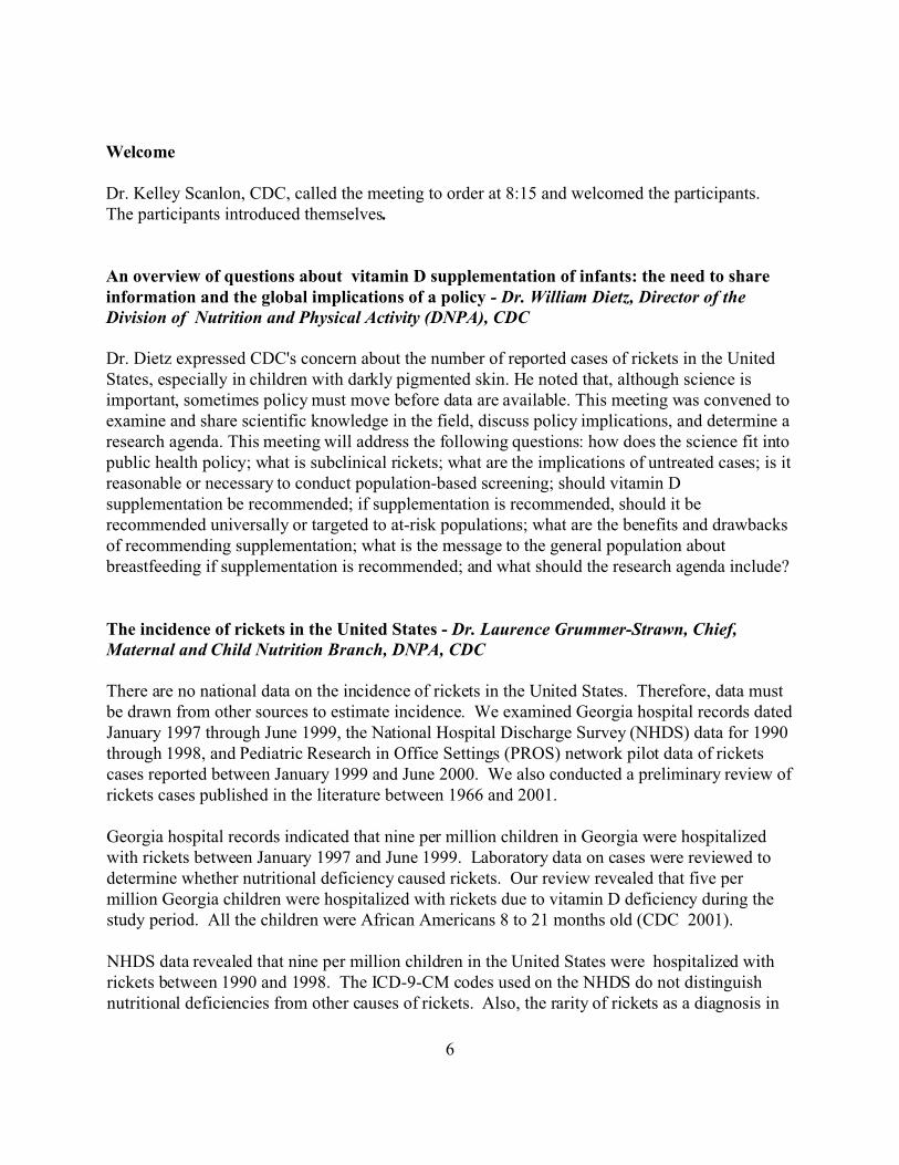
Welcome
Dr. Kelley Scanlon, CDC, called the meeting to order at 8:15 and welcomed the participants. The participants introduced themselves.
An overview of questions about vitamin D supplementation of infants: the need to share information and the global implications of a policy - Dr. William Dietz, Director of the Division of Nutrition and Physical Activity (DNPA), CDC
Dr. Dietz expressed CDC's concern about the number of reported cases of rickets in the United States, especially in children with darkly pigmented skin. He noted that, although science is important, sometimes policy must move before data are available. This meeting was convened to examine and share scientific knowledge in the field, discuss policy implications, and determine a research agenda. This meeting will address the following questions: how does the science fit into public health policy; what is subclinical rickets; what are the implications of untreated cases; is it reasonable or necessary to conduct population-based screening; should vitamin D supplementation be recommended; if supplementation is recommended, should it be recommended universally or targeted to at-risk populations; what are the benefits and drawbacks of recommending supplementation; what is the message to the general population about breastfeeding if supplementation is recommended; and what should the research agenda include?
The incidence of rickets in the United States - Dr. Laurence Grummer-Strawn, Chief, Maternal and Child Nutrition Branch, DNPA, CDC
There are no national data on the incidence of rickets in the United States. Therefore, data must be drawn from other sources to estimate incidence. We examined Georgia hospital records dated January 1997 through June 1999, the National Hospital Discharge Survey (NHDS) data for 1990 through 1998, and Pediatric Research in Office Settings (PROS) network pilot data of rickets cases reported between January 1999 and June 2000. We also conducted a preliminary review of rickets cases published in the literature between 1966 and 2001.
Georgia hospital records indicated that nine per million children in Georgia were hospitalized with rickets between January 1997 and June 1999. Laboratory data on cases were reviewed to determine whether nutritional deficiency caused rickets. Our review revealed that five per million Georgia children were hospitalized with rickets due to vitamin D deficiency during the study period. All the children were African Americans 8 to 21 months old (CDC 2001).
NHDS data revealed that nine per million children in the United States were hospitalized with rickets between 1990 and 1998. The ICD-9-CM codes used on the NHDS do not distinguish nutritional deficiencies from other causes of rickets. Also, the rarity of rickets as a diagnosis in
6
the NHDS results in an unstable estimate and therefore should be interpreted with caution. Seventy-five percent of rickets cases detected through NHDS data occurred in African American children, 20% in Caucasia American children, and 5% in Asian American children. All cases occurred in children less than 4 years old; 50% were infants less than 12 months old (K. S. Scanlon and L. Grummer-Stawn, unpublished data, 2001).
The PROS pilot survey detected 23 to 32 ambulatory cases of rickets due to vitamin D deficiency per million children served between January 1999 and June 2000. All were African American children. Age data were incomplete. A limitation of the pilot survey of pediatric practices was a low response rate (26%) (K. S. Scanlon and L. Grummer-Stawn, unpublished data, 2001).
A preliminary review of published reports between 1966 and 2001 revealed 13 reports of 122 cases of rickets due to vitamin D deficiency in 12 U.S. states (P. Weisberg, K. S. Scanlon, M. E. Cogswell, and R. Li, unpublished data, 2001). The racial/ethnic distribution of cases was as follows: 87% African American, 3% Caucasia American, 1% Asian American, and 9% other or unknown. The children with rickets ranged from 4 to 58 months of age at diagnosis.
Discussion of Dr. Grummer-Strawn’s presentation by expert panel members and guests
Dr. Grummer-Strawn said case reports were distributed throughout the United States. All cases of rickets from the Georgia and PROS data occurred in full-term infants. All cases also occurred in breastfed children. Vitamin D status of the mother was usually unknown. According to the published literature, some mothers were Muslim and were covered all the time, as were their children. Other mothers reported their children did not receive sunlight exposure because of a lack of sidewalks in their neighborhoods or because the neighborhood was too dangerous for children to walk or play outdoors.
Additional contributory factors include the following:
� Vitamin D status of a woman during pregnancy directly affects the vitamin D status of her infants at birth.
� Low milk consumption by African Americans because of confirmed or perceived lactose intolerance may affect vitamin D status.
� African Americans may have chronic vitamin D deficiency. Parathyroid hormone (PTH) concentrations are elevated when serum 25-hydroxyvitamin D [25(OH)D] is decreased. (Vitamin D deficiency results in hypocalcemia, which in turn raises PTH concentration. PTH stimulates bone resorption to mobilize bone calcium to maintain plasma calcium concentrations). Fasting phosphorus concentration may be low or normal in vitamin D deficiency and therefore is not a good indicator.
7

� It was the opinion of experts at the meeting that we should be concerned about secondary hyperparathyroidism in adults when serum 25(OH)D is � 37.5 nmol/L (15 ng/mL). Studies in older white populations have shown that PTH concentration rises when 25(OH)D concentration falls below 37.5 nmol/L (15 ng/mL), providing support for this cutoff in determining vitamin D adequacy in adults (Gloth et al. 1995, Lips et al. 1988, Thomas et al. 1998, Webb et al. 1990). Some studies suggest that the increase in PTH may start at an even higher 25(OH)D concentration (Chapuy et al. 1997, Malabanan et al. 1998). There is currently no study in children to determine the serum 25(OH)D concentration at which PTH increases. As a result, there is no definitive cutoff value to determine vitamin D deficiency in children; however, a serum 25(OH)D < 27.5 nmol/L (11 ng/mL) is considered to be consistent with vitamin D deficiency in infants, neonates, and young children and is used as an indicator for establishing the vitamin D reference value (IOM 1997, Specker et al. 1992).
� Rickets was a common childhood disease in the United States until vitamin D was added to milk.
� North Carolina reported 30 cases of rickets in a recent publication (Kreiter et al. 2000). Since then, 16 additional cases have been diagnosed at the same two medical centers; four were Muslim children.
� An ongoing study in England may provide an opportunity for studying determinants of vitamin D deficiency in children. An upcoming longitudinal study in the United States may also provide this opportunity.
Sunlight exposure to protect against rickets in infancy: the impact of sunscreens and indirect sunlight - Dr. Bonny Specker, South Dakota State University
Dr. Specker reviewed five studies.
Study I (Lichtenstein et al. 1986) This study looked at 192 term infants < 18 months of age (128 < 6 months of age; 64 > 6 months of age). Infant serum 25(OH)D was lower in winter than in summer and lower in breastfed than in formula-fed babies. In this study, no racial differences were noted in 25(OH)D concentrations.
Paradoxically, serum 1,25-dihydroxyvitamin D [1,25(OH)2D] concentrations were higher in winter than in summer and higher in black than white infants. Seasonal differences were greater in breastfed infants. Higher serum 1,25(OH)2D concentrations in black infants may be due to lower serum phosphorus concentrations observed in black than in white infants and toddlers in this population. The higher serum 1,25(OH)2D and lower serum phosphorus concentrations in black than in white infants and toddlers, despite similar vitamin D status, suggests racial differences in calcium and vitamin D metabolism.
8

Studies II and III (Specker et al. 1985a, Specker et al. 1985b) The sources of vitamin D in breastfed infants are placental transfer, human milk, and sunexposure.
In the first study (Specker et al. 1985a), mothers recorded sunshine exposure and clothingcoverage of their breastfed infants < 6 months of age for 1 week before blood was drawn. Theyalso kept 3-day diet diaries of their own food and vitamin intake. Breast milk and blood samplesfrom both mother and infant were taken on exam day.
Placental transfer (Specker et al. 1985a) Maternal and infant serum 25(OH)D concentrations were correlated, especially in infants < 8weeks of age. After 8 weeks, maternal 25(OH)D concentrations are not as important to the child's25(OH)D concentrations as sunlight exposure [n = 61 mother-infant pairs (51 white and 10black); n = 48 pairs with all data]. It was not clear from the study if the correlation betweeninfant and maternal concentrations is the result of placental transfer, human milk transfer, orsimilar exposure to the sun.
Human milk (Specker et al. 1985b)In a second study, maternal milk vitamin D correlated with maternal vitamin D intake, andmaternal milk vitamin D correlated with maternal serum 25(OH)D. However, infant serum25(OH)D did not correlate with maternal milk vitamin D. Breast milk vitamin D correlates withmaternal vitamin D intake in black and white women; black mothers have lower vitamin Dintake and lower milk vitamin D.
Sun exposure (Specker et al. 1985a)In the first study, infant serum 25(OH)D concentration was correlated with exposure to sunshine independent of maternal 25(OH)D. An additional study investigating the role of sunscreen onvitamin D synthesis in breast fed infants received Institutional Review Board (IRB) approval, butdoctors did not cooperate, because they thought it was unethical to expose children to sunlightwithout sunscreen.
To achieve a serum 25(OH)D concentration > 27.5 nmol/L (11 ng/mL) in the infant, Specker andcolleagues (1985a) estimate that the infants of mothers with low serum 25(OH)D [< 87.5 nmol/L(35 ng/mL)] would need to be in the sunlight 10 to 30 minutes per week wearing only a diaper or30 minutes to 2 hours per week fully clothed with no hat. Infants of mothers with a high serum25(OH)D [> 87.5 nmol/L (35 ng/mL)] would require < 10 minutes per week wearing only adiaper or 30 minutes per week fully clothed with no hat. Adopting conservative estimates, theauthors suggest that infants would need to be outdoors for either 30 minutes per week (diaperonly) or 2 hours per week (fully clothed, no hat). All the black infants in this study had nosunshine exposure and their mothers had low vitamin D status. Therefore, it was not possible todetermine the amount of sunshine exposure needed to maintain adequate vitamin D for black
9
infants. A study by Brazerol and colleagues (1988) indicated that black adults increase serum 25(OH)D in response to UV exposure similar to white adults.
Study IV (Specker and Tsang 1987) This study looked at the time an infant spends outside daily based on season of birth. All babies received more sun exposure in summer than in winter, regardless of the season when they were born. Seasonal changes in serum 25(OH)D parallel sun exposure.
Study V (Specker et al. 1992) This study looked at vitamin D supplementation in infants fed human milk at two sites in northern China and two sites in southern China. Serum 25(OH)D concentrations increased with increasing vitamin D dose and were lower in infants in northern than in southern China. Among infants in northern China supplemented daily with 2.5 or 5 µg (100 IU or 200 IU) of vitamin D, 36% and 30%, respectively, had serum 25(OH)D concentrations below the lower limit of normal [27.5 nmol/L (11 ng/mL)]. These data suggest that these doses may be too low for infants in northern China. No cases of rickets were observed in the 280 infants who completed the 6 month study. Serum concentrations increased as vitamin D dose increased in infants from northern China, but there was no dose effect among infants in southern China.
Conclusions Both maternal vitamin D status and sunlight exposure affect the vitamin D status of breastfed infants. Sunlight exposure appears to have the greatest impact. Maternal vitamin D status appears to be more important before infants are 8 weeks of age, possibly through placental transfer. The contribution of vitamin D from maternal milk is insignificant relative to the contribution from sunlight exposure.
Discussion of Dr. Specker’s presentation by expert panel members and guests
The difficulty in measuring sun exposure with a dosimeter was noted because dosimeter readings do not take into account the amount of skin exposed to sunlight. It was also pointed out that ozone absorbs UVB rays used in the synthesis of vitamin D. Dr. Specker said cases of rickets appear to be increasing in Mexico City, perhaps as a result of the high concentration of ozone in the city. A study was planned, but a volcano erupted, and it was not possible to interpret the results.
Ultraviolet radiation exposure during childhood and subsequent risk of skin cancer - Dr. Mona Saraiya, Division of Cancer Prevention and Control, CDC
Sunlight exposure is necessary for vitamin D synthesis. A few studies identified UV light exposure as playing a positive role in treating seasonal affective disorder and breast and prostate cancer. UV exposure has been identified as a definite major risk factor in causing basal and squamous cell carcinoma, melanoma, and potentially cataracts and immunosuppression. The population attributable risk of UV exposure for melanoma is between 65% and 90%; for the nonmelanoma skin cancers (basal and squamous cell carcinoma), it is about 60%.
10
Skin cancer is the most common cancer in the United States. Both the incidence of and mortality from melanoma have increased since the early 1970s (from 6/100,000 in 1973 to 14/100,000 in 1998). In 1998, there were 51,400 new cases and 7800 deaths from melanoma in the United States. The incidence of melanoma is higher in whites than in blacks. Among blacks, incidence is less than 2/100,000 and mortality is less than 1/100,000. Family history, personal history, and the presence of large and/or numerous moles affect cancer risk. Sun exposure in childhood is an important risk factor for melanoma in adults, and sunburn is a risk marker.
Ambient exposure studies (studies that measure UV exposure based on latitude) show that when a person moves from an area of low sun exposure to one of high sun exposure, the risk of developing melanoma is increased if the person arrives earlier in life. Alternatively, the later in life the person arrives, the lower the risk of developing melanoma. Reverse migration studies show that, when moving from an area of high exposure to an area of low exposure, the duration of residence in the lower exposure area affects the risk of cancer later in life (Autier et al. 1997). These studies indicate that exposure early in life is more important than exposure later in life. Personal exposure studies show sunburns in childhood lead to a greater cancer risk, but the association is not strong (odds ratio = 1.91). However, the association is consistent, as this is a pooled odds ratio from 29 case control studies (Elwood and Jopson 1997).
Melanocytic nevi are potential mediators of melanoma. If these nevi develop during childhood, reach their maximum density on the skin at about 10 years of age, and are caused by sun exposure, they correlate with a high risk of melanoma.
The International Agency for Research on Cancer (IARC) has deemed solar UV radiation a carcinogen and artificial UV radiation a probable carcinogen. UV radiation damages DNA, and sunlight has both early and late effects on the development of melanoma. A study by Noonan et al. (2001) showed that transgenic mice given a sunburn-inducing dose of UV light at 3.5 days of age began to develop melanomas around 6 months of age. By the time they were 1 year old, half the mice had cancer. Six-week-old mice given the same dose of UV light did not develop tumors.
Sun-safe behaviors include minimizing UV exposure during peak periods, wearing sun-protective clothes, seeking shade, wearing sunscreen of SPF 15 that protects for both UVA and UVB rays, and avoiding artificial tanning devices.
Topical use of sunscreen reduces the risk of sunburns. When used mainly during unintentional sun exposure, sunscreens probably prevent squamous cell carcinoma of the skin. No conclusion can be drawn about the cancer-preventive activity of the topical use of sunscreens against basal cell carcinoma and cutaneous melanoma. The use of sunscreen may extend the duration of intentional sun exposure, which may increase the risk of cutaneous melanoma.
11
Most people do not use sunscreen properly; one quarter of a bottle per use is needed to get the SPF protection. Also, some sunscreens do not block UVA rays (IARC 2001).
Discussion of Dr. Saraiya’s presentation by expert panel members and guests
There was debate about whether decreased sunlight exposure contributes to the development of
other types of cancer, such as breast, colon, and prostate cancers. Dr. Saraiya responded that there
is insufficient evidence; other members of the panel disagreed. One participant added that
vitamin D and sunlight provide protection against tuberculosis. If a patient is vitamin D deficient,
the infection may be more aggressive.
Other comments included the following:
� It was asked whether there was a dosage of sunlight exposure that would make vitamin D,
but not encourage cancer. The opinion of one panel member was 25% of the minimal
erythermal dose (MED) twice a week.
� Approximately 25 µg (1000 IU) of vitamin D can be synthesized when 6% of the body is
exposed to one MED for 5 minutes two or three times per week (Holick 1999). This is
based on the observation that a healthy individual whose whole body is exposed to one
MED of simulated sunlight will have circulating vitamin D concentrations comparable to
those from ingesting 250 µg to 625 µg (10,000 IU to 25,000 IU) of vitamin D.
� One expert added that blacks need up to ten times the sun exposure of whites to
synthesize the same amount of vitamin D (Clemens et al. 1982). These results vary from
those of Brazerol et al. (1988) discussed by Dr. Specker.
� Proper use of sunscreen reduces vitamin D synthesis by 97.3%.
� It was asked whether vitamin D can be synthesized from indirect sunlight. The response
was that it depends on what reflects the light. Sun reflection from sand produces some
synthesis of vitamin D. There is no vitamin D synthesis when an individual is in total
shade. Indirect light reflected from asphalt does not produce vitamin D, whereas
reflection from light cement may produce some vitamin D.
� It was asked whether infants are less efficient in synthesizing vitamin D than adults and
the response was no.
12
Supplementing breastfed infants with vitamin D: a historical perspective and current
discussions on age, dosage, preparation, and targeting - Dr. Frank Greer, Meriter Hospital,
Wisconsin
History of vitamin D supplementation of infants
In the first half of the 20th century, vitamin D deficiency was a big problem, with many cases of
rickets reported in the United States. Eventually, cow's milk was fortified with vitamin D and
nutritional rickets became almost extinct. In 1963, the AAP Committee on Nutrition
recommended that all infants receive 10 µg (400 IU) of vitamin D per day starting in the first 2
weeks of life (AAP 1963). More than 10 µg (400 IU) does not improve the effect of rickets
prevention. The committee did not comment on the vitamin D levels in human milk but did
comment on the increase in vitamin D deficiency in African Americans and that sunlight
exposure cannot be relied on to improve vitamin D status.
The 1974 Textbook of Infant Nutrition (Fomon 1974) estimated the daily requirement of
vitamin D for infants to be 2.5 to 5 µg (100 to 200 IU), citing the 1963 AAP recommendation.
The 1963 AAP recommendation actually recommended twice the estimated requirement, or 10
µg (400 IU) of vitamin D daily. Fomon also recommended ignoring the contribution of sunlight
during the first weeks of life. Fomon’s text noted that human milk contains 0.55 µg (22 IU) of
vitamin D per liter.
A 1978 breastfeeding statement from the AAP Committee on Nutrition in Pediatrics (AAP 1978)
recommended vitamin D as a possible supplement for breastfed infants. In 1982, the committee's
statement said that arguments could be made for supplementing breastfed infants with both
fluoride and vitamin D during the first 6 months of life, but there was disagreement about
whether all breastfed infants needed such supplementation (AAP 1982).
The 1998 Pediatric Nutrition Handbook, 4th edition (AAP 1998), as in earlier editions,
recommends 10 µg (400 IU) of vitamin D per day for breastfed infants in the chapter on
vitamins. However, the chapter on breastfeeding states that only dark-skinned infants need
vitamin D supplementation. The 5th edition (currently in production) will provide a consistent
recommendation throughout the text.
In 1997, the National Academy of Sciences Food and Nutrition Board recommended 5 µg (200
IU) per day as the adequate intake (AI) of vitamin D for all infants (IOM 1997).
In the 1997 breastfeeding policy statement from the work group on breastfeeding, AAP states
that vitamin D and iron may need to be given to selected infants before they are 6 months of age,
13
if the mothers are vitamin D deficient or if the infants are not exposed to adequate sunlight (AAP
1997). Those who have low iron stores or anemia would need iron.
Current discussions on vitamin D supplementation of infants
AAP has decided to develop a policy statement on vitamin D supplementation for the following
reasons: the results of the North Carolina study (Kreiter et al. 2000) on nutritional rickets in
breastfed African American infants; the Healthy People 2010 goal of having 75% of infants
breastfed for the first 6 months of life (U.S. Department of Health and Human Services 2000);
the uncertainty about how much sun exposure is required for each infant as well as how air
pollution and latitude affect sun exposure; the growing concern about UV exposure in childhood
and its relationship to skin cancer in later years; the uncertainty about the amount of sunlight
exposure needed for people with darker skin pigmentation; and the fact that vitamin D is not a
nutrient but a precursor of steroid hormones that is not naturally present in any infant food,
including human milk, in quantities that will meet individual needs.
Considerations for vitamin D supplementation include the following: 1) prevention of rickets in
all infants, including those with chronic diseases that may decrease vitamin D absorption; 2) a
minimum dosage of 5 µg (200 IU) a day to meet the AI recommended by the National Academy
of Sciences and Food and Nutrition Board (IOM 1997); 3) no ethnic or minority group should
be singled out for supplementation, given the increasing difficulty in defining those groups; 4)
supplementation should be started at birth and continued until vitamin D is adequately derived
from food; 5) infants who are fed formula exclusively do not need supplementation; 6) vitamin D
supplementation must not cause any harm; 7) supplementation should not interfere with
breastfeeding; 8) vitamin D supplements should be safe to have in the home; 9) the ideal
preparation should contain vitamin D only; 10) to maximize compliance, vitamin D
supplementation should become part of the daily routine; 11) the vitamin D status of the
pregnant/lactating mother should be taken into consideration by providers in the population; and
12) supplementation should be cost-effective, including treating overdoses and missed cases that
need further care.
Discussion of Dr. Greer’s presentation by expert panel members and guests
There was much discussion of whether the recommendation to supplement infants with
vitamin D should target infants with dark skin. The major arguments on both sides are as
follows:
Not targeting
� Defining dark skinned is difficult.
14

� Dark-skinned populations are not the only ones at risk. Any breastfed infant is at risk if
not exposed to sunlight. Therefore, the recommendation must be applicable for all
groups.
� Muslim infants who are covered are at risk, regardless of race.
For targeting
� The parents of African American infants, for example, need to know about their infants’
increased risk of vitamin D deficiency and need for supplementation, and so do the
physicians who care for those infants.
� It appears paternalistic to say "if it is good for one group, it is good for all".
� Because most minorities are treated by nonminority doctors, the recommendation may be
missed if African Americans are not singled out in the policy.
� Because race is a sensitive issue, we sometimes make poor recommendations.
Panel members concluded the discussion in agreeing that supplementation should be universal,
but education efforts should be targeted to infants at highest risk.
Other comments included the following:
� If supplementation at birth is recommended, it could interfere with breastfeeding.
� Formula manufacturers will use the recommendation to market formula. The packaging
of an infant supplement of vitamin D from Canada was displayed to show how it was
used to advertise formula.
� In Bulgaria, it is recommended that infants be supplemented with 20 µg (800 IU) of
vitamin D per day.
� In Romania, it is recommended that infants be supplemented with 10 µg (400 IU) of
vitamin D per day.
� In Canada, it is recommended that breast fed infants be supplemented with 10 µg (400
15
IU) of vitamin D per day and 20 µg (800 IU) per day in the winter. Canada has had this
recommendation for 10 years.
Risks associated with vitamin D supplementation during infancy - Dr. Ali Calikoglu,
University of North Carolina at Chapel Hill
Vitamin D supplementation during infancy may cause hypervitaminosis D as well as an excess of
other vitamins and minerals when vitamin D is given in a multivitamin form. Hypervitaminosis
D is rarely fatal but it can cause significant morbidity due to the complications of hypercalcemia
and hypercalciuria. Complications include general symptoms, such as muscle weakness and
fatigue; central nervous system symptoms; gastrointestinal problems; cardiac rhythm
abnormalities; urinary system complications such as kidney stones and nephrocalcinosis; and soft
tissue calcifications.
The risks associated with vitamin D supplementation depend on the following factors: the type of
vitamin D given, the dosage and duration of supplementation, and the vitamin preparation.
Type
Vitamins D2 and D3 are similar in terms of toxicity, although vitamin D2 has poorer stability and
purity. Supplementation is mainly with D2 in the United States and D3 in Europe. The half-lives
of vitamins D2 and D3 are 20 days to months. The half-life of 25(OH)D is about 15 days.
Hypercalcemia secondary to hypervitaminosis D can last as long as 18 months. In contrast,
hypercalcemia secondary to high doses of 1,25(OH)2D, which cannot be stored in the body and
has a short half-life, is brief.
Dose, scheduling, and duration
High doses of vitamin D can cause hypercalcemia, hypercalciuria and their consequences.
Intermittent high-dose studies
Markestad et al. (1987), Germany - 43 infants were given 15 mg (600,000 IU) of ergocalciferol.
Two weeks later, all had high 25(OH)D levels, and 14 infants had hypercalcemia. Hypercalciuria
was not assessed.
Zeghoud et al. (1994), France - 15 mg (600,000 IU) of ergocalciferol was given to 30 infants.
Two weeks after ingestion, all but two had high 25(OH)D levels. Six months after the first dose,
50% of all infants still had high 25(OH)D levels. In the other arms of the study, 58% of the
infants who received 5 mg (200,000 IU) of ergocalciferol and 23% of infants who received 2.5
16
mg (100,000 IU) of ergocalciferol had high 25(OH)D levels 2 weeks after ingestion. There was
no hypercalcemia; hypercalciuria was not assessed.
Ronnefarth et al. (2000), Germany - A retrospective survey of children with nephrocalcinosis was
conducted. Thirteen of 152 patients with nephrocalcinosis received bolus vitamin D prophylaxis
in infancy [15 mg (600,000 IU) of ergocalciferol every 3 months].
Intermittent high-dose vitamin D treatment is effective. However, high doses [15 mg (600,000
IU) and 5 mg (200,000 IU ] carry significant risk for hypervitaminosis D. 2.5 mg (100,000 IU)
appears to be relatively safe.
Single high-dose study
Oliveri et al. (1996) - 79 healthy children (5 to 11 years of age) received 3.75 mg (150,000 IU) of
ergocalciferol at the beginning of autumn. The dose was sufficient to maintain normal 25(OH)D
levels, however, the winter increment of PTH was not inhibited. There was no hypercalcemia and
no hypercalciuria.
Low-dose daily vitamin D
No data are available demonstrating that 10 µg (400 IU) or less of vitamin D daily is harmful.
The current myth that hypervitaminosis D can occur with 50 µg (2000 IU) or less of daily
vitamin D supplementation is based on very limited data and clinical observations.
I. Experience in childhood
Jeans and Sterns (1938) - Vitamin D in doses ranging from 45 to 90 µg (1800 –3600 IU) was
given to nine healthy infants for 9 to 12 months. Their linear growth decelerated after 6 months
of normal growth and recovered after the doses of vitamin D were reduced. This observation led
to the conclusion that vitamin D doses > 45 µg (1800 IU) can suppress growth. However, this
study was small and poorly designed. There was only one child per dose, no control group, and
no statistical analysis.
British experience - In the 1950s, milk and cereals were enriched with vitamin D [45 to 50 µg
(1800 to 2000 IU)] in the United Kingdom. An epidemic of hypercalcemia was observed in the
following years. The number of children with hypercalcemia declined when the vitamin D
enrichment was lowered to 10 to 15 µg (400 to 600 IU). This observation led to the conclusion
that even a modest amount of vitamin D can cause vitamin D intoxication. Of the thousands of
children who ingested 100 µg (4000 IU) or more of vitamin D per day, a few hundred developed
hypervitaminosis (Bransby et al. 1964). Most of the affected children had distinct phenotypic
features, therefore, their hypersensitivity to vitamin D might be attributed to other causes, such as
Williams syndrome.
17
Fomon et al. (1966) - Three groups of infants were given daily vitamin D2 doses of 7.5, 8.75 to
13.75, or 34.5 to 54.25 µg (300, 350 to 550 or 1380 to 2170 IU). The groups had no differences
in length, weight, or serum calcium concentrations. Neither 25(OH)D concentration nor
hypercalciuria was assessed.
Pittard et al. (1991) - Sixteen low-birth-weight (LBW) or full-term infants were randomly
assigned to receive either 10 µg (400 IU) or 20 µg (800 IU) of vitamin D daily from birth to 16
weeks of age. These doses were sufficient to maintain normal calcium, phosphorus, PTH, and
25(OH)D concentrations. No hypercalcemia was observed, and hypercalciuria was not assessed.
II. Experience in adults
The AI of vitamin D for adults is 5 µg (200 IU) until 50 years of age, 10 µg (400 IU) for ages 50
to 70, and 15 µg (600 IU) over age 70 (IOM 1997). There are no data to support the myth that a
toxic dose of vitamin D can be as low as five times the AI.
Vieth et al. (2001) - 61 healthy adults were randomly assigned to receive either 25 µg (1000 IU)
or 100 µg (4000 IU) of vitamin D3 for 2 to 5 months. The maximum plateau serum 25(OH)D
concentrations were 100 and 125 nmol/L (40 and 50 ng/mL), respectively. Serum calcium and
urinary calcium excretion did not change from baseline at either dosage. The conclusion was that
100 µg (4000 IU) of vitamin D3 daily caused no harm in adults.
Preparation
Multivitamin solutions for infants contain several vitamins plus iron. The amounts of the various
vitamins vary greatly, thus increasing the probability of vitamin toxicity.
Vitamin A toxicity includes skin lesions, alopecia, pseudotumor cerebri, hepatotoxicity, and
death. Adverse effects have been observed at 25,000 IU daily. There is no evidence that vitamin
A supplements < 10,000 IU daily are harmful.
Vitamin C toxicity includes oxalate kidney stones, excessive iron absorption, and gastrointestinal
distress. Adverse effects are observed at 1 to 1.5 g daily. There is no evidence that vitamin C
supplements < 250 mg daily are harmful.
Vitamin E has no apparent toxicity after 100 to 800 IU daily for 3 years.
Thiamin, riboflavin, niacin, vitamin B6, and vitamin B12 have no toxicity.
18
Conclusion
Vitamin D supplementation at 10 µg (400 IU) or less daily appears to be safe for infants. Studies
are needed to determine the lowest dose of vitamin D supplementation for infants that is both
safe and effective.
Discussion of Dr. Calikoglu’s presentation by expert panel members and guests
It was noted that vitamin A in as low a dose as 10,000 IU in pregnant women may be teratogenic.
Recommending vitamin D supplements for U. S. breastfed infants: does vitamin D interfere
with the physiologic aspects of breastfeeding? - Dr. Ruth Lawrence, University of Rochester
Medical Center
This question is not answered in the medical literature. The many articles on vitamin D and
vitamin D supplementation do not address the impact on the breastfeeding infant or the
breastfeeding mother. It is a matter of risk/benefit ratio. What is the risk to the infant’s
physiologic state of taking the vitamin D supplement, and what is the psychologic impact on the
mother's ability to continue breastfeeding versus the benefits of giving vitamin D across the
board to babies who do not have a deficiency.
If we look first at the infant, there are no physiologic changes examined or reported in the
literature, but the following questions we need to pose: (1) Is there a change in the pH of the gut
with vitamin D supplementation? (2) Keeping in mind that a change in gut flora may change the
pH and precipitate a cascade of events, do gut flora change? (3) If there is a change in gut flora,
would it affect the infection rate for this infant? (4) Would oral vitamin D change the absorption
of calcium and phosphorus from milk?
Although we have no data on vitamin D, we have some experience with iron supplementation.
For example, iron binds with lactoferrin, which is a prime infection protection constituent of
human milk. An overload of iron inactivates lactoferrin and leaves excess iron in the gut.
Escherichia coli depends on iron for growth. Lactoferrin therefore suppresses the growth of
Escherichia coli in the gut in the breastfed infants. Increased growth of Escherichia coli
suppresses lactobacillus, which is the normal flora of newborn and young infants. This
consequence has the potential for increasing the number of infections, particularly infections that
result in diarrhea and malabsorption.
19
We know also that iron is better absorbed from human milk than from formula. This work was
done in adult volunteers who were provided tagged iron either in human milk or in formula.
Does human milk enhance absorption of vitamin D?
Would there be an effect on growth of infants who are given vitamin D when they are not
vitamin D deficient? Domellof et al. (2001) studied iron absorption of Swedish infants and
noted that linear growth was impaired in healthy infants who did not need the iron, especially in
the first 6 months. Whether a similar response occurs with vitamin D is not known.
The need to evaluate pH, intestinal flora, infection rate, and linear growth in infants
supplemented when they are not deficient remains crucial. It is interesting to note as well that
Coutsoudis et al. (1999) showed that partially breastfed infants (mixed feed of formula and breast
milk) of HIV-positive mothers had a greater incidence of HIV infection than exclusively
breastfed infants of HIV-positive mothers.
The persistent issue of compliance needs to be addressed as well because other studies have
shown that 75% of mothers are noncompliant with iron supplements.
Another risk to the infant receiving a supplement is aspiration. Breastfed babies often do not
tolerate non-breast milk added to their diet in the early weeks. Years ago, when cod liver oil was
given, many cases of lipoid pneumonia resulted. The risk of excessive vitamin D that is
associated with hyperostosis needs to be investigated. Hyperostosis was a common disease in the
1950s and 1960s not only in England but also in this country where vitamin D supplementation
of many foods was made available. Hyperostosis was diagnosed on physical exam, confirmed by
x-ray, and associated with hyperpyrexia and loss of hair.
Vitamin D supplementation may have a 2-fold effect on breastfeeding. (1) Does supplementation
destroy the mother's confidence so she never starts or (2) does supplementation affect the
duration of breastfeeding? Dennis and Faux (1999) studied the self-efficacy framework. They
point out that perception of abilities is much more important than the mother's true abilities in
choosing a behavior and performing and maintaining it. Therefore, if the mother perceives a
deficiency in her breast milk because she has to supplement, it could theoretically interfere with
her ability to breastfeed successfully. Other factors affect one's self-efficacy or confidence
including previous experience, verbal persuasion, and performance accomplishments.
Confidence has always been an important factor in successful breastfeeding. As Derrick Jelliffe
said, "Breastfeeding is a confidence game". For a mother who has not breastfed previously,
infant vitamin D supplements may make her worry about her ability to breastfeed, she may be
less likely to breastfeed, or she may be afraid to breastfeed. Confidence can influence women's
judgments about their ability to initiate, persist, and continue breastfeeding.
20
Survey at the University of Rochester
A brief survey was performed with forced-answer format recording age, parity, and previous
breastfeeding experience to examine maternal perceptions about vitamin D supplementation of
their breastfed infants. Young mothers who had just given birth to their first child thought
perhaps something was wrong with their milk and would give the vitamin D. Multiparous
mothers who had breastfed before were very confident with their breastfeeding, and most would
not give the supplement. Several mothers said they would supplement if their doctor said to do
so. When professionals were interviewed, they expressed the belief that vitamin D should be
given only to babies who are deficient. A solution in Germany is to put an oral vitamin D
preparation on the mother's nipple once a day before the baby breastfeeds. This technique avoids
feeding the baby some other substance with an artificial device that is not mother's milk.
Conclusions
� If all breastfed infants are to receive vitamin D regardless of risk of deficiency, the change
in the physiologic state of the infant should be evaluated, including change in flora in the
gut and the change in infection rate.
� The duration of breastfeeding with or without vitamin D should also be examined,
particularly targeting primiparas women.
Discussion of Dr. Lawrence’s presentation by expert panel members and guests
Some participants thought supplementation from birth would interrupt breastfeeding and should
not be introduced until the child has been breastfed for 6 months. Another participant countered
that, in his experience with babies with breast milk jaundice, breastfeeding was interrupted from
1 to 5 days; then all mothers went back to breastfeeding at the end of that period. The biggest
factor in resumption of breastfeeding seems to be how the doctor addresses the topic with the
mother. A third participant commented that a hospital study in Canada showed no change in
breastfeeding practices when supplementation was introduced in the hospital.
Others comments included the following:
� There is no evidence that 5 µg (200 IU) of vitamin D will change gut flora per se, but it
has not been proven unequivocally.
� Some doctors and other health care professionals recommend no sunscreen for the first
year of life, whereas the AAP says not for the first 6 months (AAP 1999).
21

� One participant suggested that it might be easier to recommend that parents take children
outside in the sunlight for 30 minutes per week than to recommend giving the infant a
vitamin D supplement. Concerns about sunlight exposure and skin cancer were repeated.
Vitamin D supplementation of breastfed infants in North Carolina: cost, compliance,
effectiveness, and impact on breastfeeding - Ms. Gladys Mason, North Carolina Department
of Health and Human Services
North Carolina had a growing number of documented cases of rickets secondary to vitamin D
deficiency. Two medical centers reported 30 cases between 1990 and 1999, half the cases
occurred between 1998 and the first half of 1999 (Kreiter et al. 2000). All cases were in
breastfed African American children who had not been given vitamin D supplements. This
increase in cases was attributed to the increase in breastfeeding among African American
mothers, the decline in the number of physicians prescribing vitamin D for breastfed infants, and
a decrease in exposure to sunlight. Data show that breastfeeding rates in North Carolina have
risen among low-income women of all races. Between 1988 and 1999 breastfeeding rates among
African Americans increased from 5.2% to 34.7%.
Major problems in providing vitamin D supplements were as follows: 1) uncertainty and lack of
awareness of who is at risk for rickets; and 2) Medicaid and some insurance plans were not
reimbursing for vitamin drops even though rickets is a diagnosed condition.
At the request of the North Carolina Pediatric Association, the Women's and Children's Section
of the Division of Public Health implemented the Vitamin D Initiative, using Maternal Child
Health block grant funding to provide free vitamin D supplements to all breastfed infants,
regardless of income. Tri-Vi-Sol vitamin A, D and C drops were purchased through the
Minnesota multistate contract at a cost of $1.50 per month per child and shipped to the 87 local
Special Supplemental Nutrition Program for Women, Infants, and Children (WIC) agencies in
December 1999. Local agencies were instructed to begin distribution as soon as they developed
procedures and standing orders signed by their medical director. A 3-month supply of vitamin
drops could be issued to each breastfed child beginning at 6 weeks of age, provided the child was
not receiving any infant formula or vitamin D-fortified milk daily.
There was some initial resistance and confusion about issuing vitamin D supplements to breast-
fed infants. Breastfeeding has been widely promoted in North Carolina, but vitamin D
supplementation has not. The American Academy of Pediatrics policy statement "Breastfeeding
and the Use of Human Milk" has done little to endorse vitamin D supplementation, whereas it
strongly supports breastfeeding for at least 12 months (AAP 1997). A survey completed by 400
22
North Carolina physicians looking at their prescribing habits showed that only 42% prescribe
vitamin D to breastfed infants (unpublished data).
Physicians, nutritionists, lactation consultants and nurses challenged the rationale for issuing
vitamin drops to all infants who receive breast milk as their milk source. It was necessary to
clarify that nutritionists could legally issue vitamins. Lactation consultants were very concerned
that vitamin D would be "one more reason to stop breastfeeding". Some mothers refused the
vitamin drops, and parents who had children diagnosed with rickets were very upset because no
one told them about the consequences of vitamin D deficiency. One lawsuit resulted from parents
who were never told that rickets could occur in their child.
As part of the initiative, health professionals and parents were instructed about vitamin D
deficiency rickets, the essential role of sunlight and vitamin D supplementation for bone growth,
and the history and prevalence of rickets before supplementation began in the United States
before 1925. Fact sheets were developed and continue to be available on the Nutrition Services
web site (www.ncnutrition.com). Presentations were made at grand rounds and professional
meetings. Health professionals continue to counsel parents of breastfed infants whenever the
vitamin supplements are issued.
Concerns prevail about the formulation of over-the-counter vitamin drops that contain vitamin D.
It is unclear why a vitamin D only preparation is not available in the United States. Tri-Vi-Sol
vitamin A, C and D drops is an approved and affordable product that provides 1500 IU of
vitamin A, 35 mg of vitamin C, and 10 µg (400 IU) of vitamin D and is the product issued in
North Carolina. Tri-Vi-Sol vitamin A, C and D drops were not found in local pharmacies that
were surveyed, but several other formulations were available without prescription that included
additional vitamins and minerals, such as iron, zinc, vitamin E, and B vitamins. A 5 g/mL (200
IU/mL) drop preparation is not currently available (the current recommendation according to
IOM's dietary reference intake level for this age group (IOM 1997). Furthermore, the supplement
packaging is associated with formula recognition, which violates the World Health Code.
Another concern some nutritionists expressed is that vitamin A is palmitate rather than �-
carotene.
The North Carolina Initiative had a slower start than anticipated. There are approximately
100,000 births per year in North Carolina. Over a 16-month period only 1500 infants received
vitamin drops, whereas 2810 bottles were issued. Eight months after the initiative began, 96% of
local agencies had standing orders and distribution procedures in place, and all were in place
when the agencies were surveyed 15 months after the initiative began. Most of the vitamin drops
were issued to WIC participants. Some doctors prefer to distribute the drops from their offices
because of the inconvenience of sending parents to the WIC clinics. Fewer infants have been
eligible for the vitamin drops than originally estimated because fewer than expected consume
23
breast milk as their sole milk source. There is growing concern that the definition of “eligible”
for vitamin D supplementation is too conservative and probably needs to allow up to 16 ounces
of infant formula per day in addition to breast milk.
After nearly 2 years of experience, the North Carolina WIC Program remains the only source of
free vitamin D supplements for breastfed infants. However, this may not be the best delivery
system because it does not provide seamless service. Only a few private non-WIC clinic patients
receive the free vitamin drops even though physicians are aware of the initiative and many say
they refer their patients to WIC. Private clinic patients do not readily come to WIC clinics. Local
agency commitment to this initiative varies. Parental compliance has not been evaluated, but
there is documentation of some refusal to accept the vitamin drops. Increasing knowledge among
health professionals and parents about the essential role of vitamin D and vitamin D-deficiency
rickets will continue to be important.
Discussion of Ms. Mason’s presentation by expert panel members and guests
There was a lengthy discussion of the economic implications and cost of the program. If the cost
is compared with the cost of treatment and hospitalization of rickets cases, the program may not
be cost-effective. A quick calculation of the hypothetical cost for the United States, using an
estimated cost of Tri-Vi-Sol vitamins to the consumer ($5.69 per child per month) and our rough
estimates of incidence (5 hospitalized cases per million children and 25 ambulatory cases per
million children), produced the following estimates: cost per hospital case averted
$4,794,614.28; cost per case averted $958,922.86 (L. Grummer-Stawn, unpublished data, 2001).
Even if the cost were reduced to $1.50 per child per month, the cost per case averted would be
$252,614. However, the cost of untreated vitamin D deficiency is unknown. A cost-effectiveness
analysis is difficult, because the real incidence is unknown.
It was asked whether recommending sunlight is out of the question.
Alternatives to daily vitamin D supplementation: supplementation of pregnant and
lactating women - Dr. Bruce Hollis, Medical University of South Carolina
UV radiation promotes vitamin D3 synthesis in the skin. The UVB rays are absorbed by the skin
and induce conversion of 7-dehydrocholesterol into previtamin D3. After a few hours, vitamin D3
is generated and is carried in the bloodstream to the liver and kidneys, where it is activated.
A study by Matsuoka, et al. (1989) determined that the increase in serum 25(OH)D in the blood
occurs after whole body exposure to UVB radiances equal to or above 18 mJ/cm2.
24
Sun exposure and racial differences in vitamin D
A study by Matsuoka, et al. (1991) exposed 31 subjects to the equivalent of less than 10 minutes
of noonday sun. Because peak serum 25(OH)D3 occurs 24 hours after acute UVB exposure,
serum levels were measured 24 hours later and compared with baseline measures. Subjects
represented four racial/ethnic groups: eight African Americans, eight Caucasian Americans,
seven South Asians, and eight East Asians. Basal serum levels were similar in all groups. After
UVB irradiation, significant racial differences were apparent. The post-UVB serum 25(OH)D
concentrations in Caucasian Americans and East Asians were significantly higher than in African
Americans and South Asians. Intragroup serum 25(OH)D increases were significant for all
groups except African Americans.
Another study looked at the effects of oral vitamin D2 supplementation in adult males. Subjects
were given 2.5 mg (100,000 IU) of vitamin D2 for 4 days. Unpublished data from this study
revealed that serum 25(OH)D concentrations among Caucasian Americans increased from 65.0
nmol/L (26 ng/mL) to 167.5 nmol/L (67 ng/mL), and among African Americans, they increased
from 20.0 nmol/L (8 ng/mL) to 75.0 nmol/L (30 ng/mL), which indicates a difference in the
metabolic handling of vitamin D, aside from just skin pigmentation. Also, 24-hydroxylation
(inactivation step) of 25(OH)D is more active in South Asians and African Americans than in
Caucasian Americans (B. Hollis, personal communication, 2001).
Older data showed that serum 25(OH)D3 concentrations tended to be much lower in African
Americans than in Caucasian Americans. Examination of cord blood indicates that 25(OH)D is
transferred across the placenta; the neonates' levels are half that of their mothers. In a study by
Hollis and Pittard (1984), 25(OH)D3 concentrations among African American mothers and
neonates were 16.5 ± 8.0 nmol/L (6.6 ± 3.2 ng/mL) and 8.8 ± 3.0 nmol/L (3.5 ± 1.2 ng/mL),
respectively, compared with concentrations of 29.5 ± 13.3 nmol/L (11.8 ± 5.3 ng/mL) and 15.5 ±
7.5 nmol/L (6.2 ± 3.0 ng/mL) among Caucasian American mothers and neonates.
Vitamin D in human milk
Vitamin D levels in human milk from healthy mothers who are not vitamin D deficient, range
between 0.5 and 1.5 µg (20 and 60 IU) (Greer et al. 1984). Vitamin D (the parent compound)
translocates to human milk at a much greater rate than does 25(OH)D.
Several studies have shown that the vitamin D content of breast milk can be raised by
supplementing the mothers' vitamin D levels, but sufficient vitamin D levels cannot be achieved
in human milk without doses exceeding the current AIs for mothers. Maternal UVB exposure
greatly alters the vitamin D content of human milk, but the concentrations quickly fall without
further exposure.
25
A Finnish study by Ala-Houhala et al. (1986) looked at three groups of healthy, well nourished
mothers:
Group 1: 17 mothers given 50 µg (2,000 IU) of vitamin D3 a day, infants not
supplemented.
Group 2 : 16 mothers given 25 µg (1,000 IU) of vitamin D3 a day, infants not
supplemented.
Group 3 : 16 mothers not supplemented, breastfed infants given 10 µg (400 IU) of
vitamin D2 a day.
At birth, serum concentrations of all three vitamin D metabolites were comparable in all groups.
At 8 weeks, the 25(OH)D concentrations in infants in groups 1 and 3 were similar, but in group 2
they were significantly lower, with three infants at or below the risk limit for rickets. At 15
weeks, the infant 25(OH)D concentrations were significantly lower for group 2 than for groups 1
and 3. The study concluded the following: 1) breast milk does not have enough antirachitic
activity by itself or when mothers are supplemented with 25 µg (1000 IU) of vitamin D per day;
2) vitamin D supplementation of 10 µg (400 IU) per day to breastfed infants is adequate to
prevent rickets during winter in northern latitudes; 3) an adequate supply of vitamin D to
breastfed infants is achieved only by increasing maternal supplementation to 50 µg (2000 IU) per
day, the safety of which is unknown.
Dr. Hollis wants to study supplementation of lactating mothers with 100 µg (4000 IU) of vitamin
D per day and to look across racial lines and in various climates. He estimates that this dose will
increase milk vitamin D to 5 to 7.5 µg/L (200 to 300 IU/L). Dr. Hollis also commented that
chronic exposure of the mother to the sun has been shown to increase her breast milk vitamin D
to 2.5 to 3.75 µg/L (100 to 150 IU/L), but these studies are in white women.
Discussion of Dr. Hollis’s presentation by expert panel members and guests
According to a study published in the American Journal of Nutrition, 2001, vitamin D does not
reach toxic levels until supplementation exceeds 250 µg (10,000 IU) per day (Veith et al. 2001).
Alternatives to daily vitamin D supplementation: less frequent, high dose supplementation
of infants - Dr. Mike Holick, Boston University School of Medicine
After the movie Jurassic Park became popular, many children wanted their own dinosaur. The
closest they could come to one was an iguana. Unfortunately, these animals are vertebrates like
humans and need a source of calcium and vitamin D. However, they were put in terrariums
where they received no sunlight and were often fed lettuce, which contains little calcium and no
vitamin D. They developed rickets and osteoporosis. With the addition of a sun lamp and an
26
adequate source of calcium and vitamin D, the iguanas were healthy.
Case study of rickets
A case study was cited in which a 13-month-old African American child, who was solely
breastfed and did not receive vitamin D supplementation, experienced cramping of the hands and
feet. The child was developing normally, had no known medical problems, and was up to date on
immunizations. He was solely breastfed, received 3 to 10 ounces of juice per day, no whole
milk, and limited dairy products. His serum calcium was 5.9 (normal 8 to 10.5); ionized calcium
was 3.4 (normal 4.5 to 5.3). Magnesium and phosphorus were normal. His serum PTH and
1,25(OH)2D concentrations were checked [but not his serum 25(OH)D, the best indicator of
vitamin D status]. Hypocalcemia was initially treated with intravenous calcium. Treatment
progressed to oral calcium carbonate and 25 µg (1000 IU) of vitamin D twice per day. The child
was eventually treated with 125 µg (5000 IU) of vitamin D four times per day and recovered.
Chronic low phosphorus, not calcium, causes bone disease in vitamin D deficiency.
Vitamin D: assessment, function, source, and deficiency
Vitamin D is hydroxylated to 25(OH)D in the liver and 25(OH)D is further hydroxylated to
1,25(OH)2D in the kidney. Although 1,25(OH)2D is the active form of vitamin D, it has a short
half-life and is not a marker of vitamin D status. The appropriate test is a serum 25(OH)D assay.
The normal range of serum 25(OH)D is the mean serum 25(OH)D ± 2 standard deviations from a
sample of healthy individuals. The lower limit of the normal range is as low as 20 nmol/L (8
ng/mL) or as high as 37.5 nmol/L (15 ng/mL), depending on the geographic location (IOM
1997). In general, a serum 25(OH)D concentration below 27.5 nmol/L (11 ng/mL) is considered
deficient for children (IOM 1997). For optimal calcium and bone metabolism, studies in adults
suggest that serum 25(OH)D concentrations should be �37.5 nmol/L (15 ng/mL) (Gloth et al.
1995, Lips et al. 1988, Thomas et al. 1998, Webb et al. 1990) , �50 nmol/L (20 ng/mL)
Malabanan et al. 1998), or even higher (Chapuy et al. 1997).
Vitamin D is essential for bone health. Vitamin D regulates intestinal absorption of dietary
calcium; without vitamin D the absorption rate of calcium is only 10% to 15%. With vitamin D,
the intestine absorbs 30% to 80% of dietary calcium. Thus, calcium supplements are most
effective when there is an adequate supply of vitamin D.
Vitamin D is rare in foods. Ice cream, yogurt, and cheese do not have vitamin D. Vitamin D-
fortified milk should have 10 µg (400 IU) of vitamin D per quart, but only about 29% of milk
samples tested contain between 8 and 12 µg (320 and 480 IU) of vitamin D per quart (Holick et
al. 1992). Approximately 21% of skim milk samples had undetectable vitamin D (Holick et al.
1992). Vitamin D is also available in fatty fish and fish liver oils. A rich source of vitamin D is
salmon. If eaten two or three times per week, it provides all the vitamin D a healthy person
needs.
27

UVB rays make vitamin D when skin is exposed to sunlight. However, little vitamin D is made
in the winter months, particularly at higher latitudes. Sunscreen, if used correctly, reduces the
UVB rays absorbed by the skin. SPF 8 reduces vitamin D production by 97.5%; SPF 15 reduces
vitamin D production by 99%.
Exposure of an individual’s whole body to one MED of sunlight is equivalent to ingesting about
250 µg (10,000 IU) of vitamin D (Holick 1999). Therefore, exposure to 1 MED of sunlight is 17
to 50 times the recommended AI for vitamin D from dietary sources [5 to 15 µg (200 to 600 IU)]
(IOM 1997). Therefore, for an older woman to obtain the equivalent of 15 µg (600 IU) of
vitamin D per day (AI for women > 70 years of age), she would need to expose 6% of her body
surface to sunlight for 15 to 30 minutes two or three times a week. In Britain, UVB lamps have
been used as ambient lighting in nursing homes, and residents have maintained their vitamin D
concentrations all year long. There have been reductions in fractures and depression in these
facilities as a result of the lights. The average Caucasian in Boston needs to expose hands, face,
and arms to 5 to 15 minutes of sunlight two or three times per week in the summer, before
applying sunscreen. The average African American may need up to ten times as much sunlight as
Caucasians to produce the same amount of vitamin D (Clemens et al. 1982). Muslims who are
covered from head to toe present a major problem in terms of vitamin D synthesis. Their vitamin
D production is almost 0, and all will need vitamin D supplementation.
The industrial revolution increased air pollution. Tall buildings blocked sunlight, so people were
not synthesizing vitamin D. As a result, rickets became a common disease. Cesarean section
became a common method of delivery in Great Britain because women had malformed pelvises
from rickets and were unable to deliver their babies vaginally.
In 1822, Sniadecki noted that “strong and obvious is the influence of sun on the cure of rickets
and the frequent occurrence of the disease in densely populated towns where the streets are
narrow and poorly lit.” Sunlight, a source of vitamin D, was used to treat disease throughout the
last century. In 1905, phototherapy was used to treat tuberculosis, and rickets was first cured with
sunlight in 1921. By 1939, phototherapy was a popular treatment for many diseases, including
lupus.
Vitamin D treatment of adults aged 49 to 83 years resulted in a 109% increase in serum 25(OH)D
concentrations and a 22% decrease in PTH concentrations (Malabanan et al. 1998). The decline
in PTH was 55% among study subjects with an initial serum 25(OH)D concentration between 25
and 37.25 nmol/L (10 and 14.9 ng/mL) and 35% among those with a serum 25(OH)D between
37.5 and 49.75 nmol/L (15 and 19.9 ng/mL). There was no decline in PTH among people with a
serum 25(OH)D concentration between 50 and 62.5 nmol/L (20 and 25 ng/mL).
28

Twenty-five micrograms (1000 IU) of vitamin D daily for adults in winter reduced PTH levels by
33%.
In the elderly, vitamin D deficiency is treated with 1.25 mg (50,000 IU) per week for 8 weeks.
Vitamin D deficiency can lead to osteoporosis. Many cases of fibromyalgia are often caused by
vitamin D deficiency. Up to 50% of women over 50 years of age are vitamin D deficient
(Malabanan et al. 1998). In those over age 65, sunlight deprivation is the leading cause of
vitamin D deficiency.
Vitamin D and cancer There may be a relationship between vitamin D and cancer. As early as 1941, it was discovered
that there were higher cancer rates at higher latitudes (Apperly 1941). Breast (Garland et al.
1990), colon (Garland et al. 1990), and prostate (Hanchette and Schwartz 1992) cancer rates are
all higher in northern than in southern latitudes. 1,25(OH)2D is the active form of vitamin D.
1,25(OH)2D3 inhibits proliferation of benign and malignant prostate cancer cells (T.C. Chen,
G.G. Schwartz, and M.F. Holick, unpublished data, 1998). But increased vitamin D intake or
sunlight exposure does not increase 1,25(OH)2D. It is possible that prostate cells make
1,25(OH)2D to maintain normal cell growth activity by increasing 1�-OHase activity. Research
by Schwartz et al, (1998) suggests that benign and malignant prostate cancer cells metabolize
25(OH)D to 1,25(OH)2D to control cell growth.
Vitamin D supplementation of breastfed infants Vitamin D deficiency does not need to be a consequence of breastfeeding. To have adequate
levels of vitamin D in breast milk, mothers would need 100 µg (4000 IU) of vitamin D daily.
However, the tolerable upper intake levels recommended for individuals 1 to 90 years old is 50
µg/day (2000 IU/day). Neonates should not leave the hospital vitamin D deficient. It was the
speaker’s opinion that all neonates should receive a 25 to 125 µg (1000 to 5000 IU) single dose
of vitamin D before leaving the hospital.
Discussion of Dr. Holick’s presentation by expert panel members and guests
One participant suggested that mothers, rather than infants, receive vitamin D supplements. The
response was that the literature does not support that suggestion. Because the amount of vitamin
D passed to the child through breast milk is so small, mothers would have to take large doses of
vitamin D to provide an adequate supply to the infant. The dose necessary to increase breast
milk vitamin D sufficiency is above the current tolerable upper intake level recommended by the
Institute of Medicine (IOM 1997).
29
There was concern that the AI of 5 µg (200 IU) was based on infants receiving sun exposure,
when, in fact, sun is no longer being recommended for infants under 6 months of age. Also, this
amount does not take into account skin pigmentation, latitude, and pollution.
Bolus administration of vitamin D at birth was recommended as a way to avoid vitamin D
deficiency. The problems with this approach are that rickets is a rare event, and supplementing
infants at that level amounts to an uncontrolled population-based experiment. Toxicity is also a
concern. There is no surveillance system that would catch problems with this strategy.
Other comments include the following:
� A reasonable dose of sunlight, free of sunscreen, that promotes vitamin D synthesis but
does not increase skin cancer risk needs to be calculated.
� Infants in Europe are supplemented with vitamin D, and diabetes rates are low. Vitamin
D may protect against diabetes.
Rickets in low-birth-weight infants: approaches to detecting subclinical disease -
Dr. Stephanie Atkinson, McMaster University Medical Center
Protecting infants against osteopenia/rickets in early life may reduce their risk of osteoporosis as
adults (Cooper et al. 1997). Before 1990, 15% of infants with a birth weight between 1000 and
1500 g and 73% of infants with a birth weight below 800 g developed rickets (Koo et al. 1989).
Multiple etiologic factors contribute to the development of osteopenia and rickets in LBW
infants. Fetal accretion of bone may be negatively influenced by maternal smoking and thinness,
and size at birth and duration of gestation are strong determinants of bone mineral content
(BMC) later in life (Godfrey et al. 2001). Because 80% of the calcium and phosphorus deposited
in the fetus occurs during the last trimester of pregnancy, infants born prematurely begin life with
very hypomineralized bones compared with full-term infants. Although they are born with
normal circulating concentrations of 25(OH)D, it is unlikely that stores of vitamin D or 25(OH)D
in prematurely born infants are extensive because of their shortened gestation and lack of adipose
tissue for storage.
In early postnatal life of premature infants, calcium and phosphorus deposition as it would have
occurred in utero may not be achievable for the following reasons: 1) prolonged total parenteral
nutrition or oral feeds with inadequate calcium/phosphorus/vitamin D; 2) lack of cutaneous de
novo synthesis of vitamin D when hospitalized for weeks to months after birth and lack of
exposure to the outdoors and sunlight for an extended period after discharge; and 3)
administration of steroid and/or diuretic drugs that interfere with vitamin D or calcium
30
metabolism. A tapering course of dexamethasone is used clinically in early life in very LBW
infants because it improves lung function and allows earlier weaning from the ventilator.
However, the negative side effects of dexamethasone therapy include lower weight and length
growth and lower bone mineral mass (Weiler et al. 1997).
Measurement of whole BMC content of various subpopulations of LBW infants determined that,
by term-adjusted age (equivalent to 40 weeks of gestation), LBW infants lag behind term infants
in BMC (Atkinson et al 2000). All infants studied received at least 10 µg (400 IU) of vitamin D
per day (as a supplement of D.Vi. Sol, Mead Johnson, Canada) while in the hospital. Thus, the
deficit in bone mineral accrued during early life is likely due to a deficiency of calcium and
phosphorus intake rather than of vitamin D.
After premature infants reach their expected term age (and usually are no longer in the hospital),
the recommended intakes for vitamin D, calcium, and phosphorus are the same as for term
infants. No data yet support recommendations for special nutrient needs. There is emerging
evidence that higher intakes of calcium and phosphorus, and at least 5 to 10 µg (200 to 400 IU)
of vitamin D, may be necessary for LBW infants to attain catch-up of BMC quickly. In
extremely LBW infants, a randomized intervention with a high protein and mineral formula for 4
months after hospital discharge increased whole body BMC by 3 months corrected age among
infants receiving the high mineral and protein formula (Brunton et al 1998). Follow-up of this
population of extremely LBW infants to 12 months corrected age demonstrated a lack of catch-
up in BMC compared with term infants fed standard formula despite intakes of calcium and
vitamin D that met or exceeded current recommendations [intake from food and supplements
averaged 8 to 10 µg (320 to 400 IU)/day] (Brunton et al. 1997).
In a descriptive study, LBW infants who were fed primarily mother’s milk (defined as receiving
over 60% of enteral fluid intake as breast milk) had lower intakes of calcium and phosphorus up
to 6 months and significantly lower BMC at 6 months than infants fed standard infant formula
(Wauben et al. 1998). These findings were supported by a subsequent longitudinal follow-up to
12 months corrected age of a larger number of LBW infants (Randall-Simpson et al. 2000).
LBW infants receiving 50% or more of fluid intake as breast milk to 6 months corrected age
were initially lighter and shorter than were formula-fed infants. BMC was lower in breastfed
than in formula-fed infants at 6 and 12 months of age, even when expressed as a function of body
weight. Calcium and phosphorus intakes met or exceeded the current recommendations for term
infants. From these studies, one could hypothesize that such LBW infants have higher mineral
and vitamin D needs than currently recommended or another nutrient is limiting to bone mineral
growth. This raises the question "are these infants vitamin D deficient?"
A challenge for clinicians caring for LBW infants is to determine what biochemical and
functional measures can serve as sensitive and specific indicators of abnormal bone status,
31

especially before the onset of severe osteopenia or fulminant rickets. Biochemical markers for
abnormal bone status include low serum 25(OH)D, low serum phosphorus (P), high serum PTH,
high urinary calcium, and high alkaline phosphatase (ALP). However, there are problems in
interpreting these markers. Although 25(OH)D is the best early measure, the lower limit of
normal that reflects interruption of the process of bone mineralization is not known for the LBW
infant population. Also, the assay kits are costly and the lab must be appropriately equipped to
analyze the sample. In addition, serum Pi is affected by PTH level, increased PTH and ALP are
not early indicators of hypomineralized bone, and increased urinary calcium suggests only
increased bone turnover.
Several studies indicate that ALP is an insensitive marker of subclinical bone disease. In term
infants, grouped by values for plasma 25(OH)D of < 15 nmol/L (6 ng/mL) , 16 to 30 nmol/L (6.4
to 12 ng/mL) or > 30 nmol/L (12 ng/mL), ALP values were similar (101 to 107 U/L); thus, the
enzyme was not a sensitive biochemical indicator of vitamin D deficiency, as noted in the group
with the lowest 25(OH)D value (Zeghoud et al. 1997). Glass et al. (1982) found that only very
high ALP was associated with overt rickets or fracture. Ryan et al. (1993) reported that plasma
ALP explains only 4% to 6% of the variation in radial BMC, and Pittard et al. (1992) reported no
association between ALP activity and change in BMC. In LBW infants, increased ALP may
reflect slow linear growth (Lucas et al. 1989, James et al. 1985) and not an acute index of bone
mineralization. Therefore, plasma ALP will not likely identify LBW infants at greatest risk of
bone mineral deficits until those deficits can be detected radiologically as osteopenia or rickets.
Additional measures of abnormal bone status in very LBW infants include BMC by dual-energy
x-ray absorptiometry (DXA)/ultrasound, radiology, and osteocalcin or cross-linked C-telopeptide
of type 1 collagen. The problems associated with DXA/ultrasound, osteocalcin, and pyridinium
cross-links of collagen include expense of the tests, the need for special software or labs, and
lack of reference data for young infants. A limitation of radiology is its insensitivity to mild bone
loss.
The first report of rickets in infants was published in 1651. Given the recent reports of this
clinical entity, it appears that we have regressed with time. LBW infants are a population at high
risk of osteopenia and rickets. Whereas osteopenia/rickets in early life may be primarily related
to calcium/phosphorus deficiency, vitamin D deficiency may be an important factor in the
restricted catch-up of bone mass in the first year or two, especially in LBW infants who are breastfed.
Dr. Atkinson concluded, "breast is best" just add vitamin D and, for preterm infants,
supplemental calcium and phosphorus in addition to what is found in breast milk or standard
infant formula may be warranted.
Discussion of Dr. Atkinson’s presentation by expert panel members and guests
32
� It was stressed that not enough is known about preterm infants, and more research is
needed.
� In Canada, D.Vi.Sol (Mead Johnson Canada) is sold in a vitamin D only formula that is
not available in the United States.
Recommending vitamin D supplements for U.S. breastfed infants: issues in developing a
communication strategy - Dr. Claudia Parvanta, CDC
There are several levels to pass through between formulating a scientific policy and translating it
into “messages” going out to the public. Some of the larger steps include wording of the policy,
formulation of intervention objectives, and development of a communication strategy to achieve
some of these objectives. A systematic approach to health communication planning at CDC
includes six steps: 1) problem definition and description, 2) problem analysis, 3) audience
identification and profiling, 4) communication strategy and tactics, 5) evaluation, and 6)
feedback. Formulation of the policy overlaps with steps 1 and 2 in this model.
Formulation of the policy
During the problem definition stage, questions such as “what is occurring in contrast to what
should be occurring” need to be asked. Analytical epidemiology is used to understand who is
most affected; to what extent or how severe; where or when the problem is occurring; and
whether there are behavioral factors. Potential solutions need to be worked through with the
intended users (sometimes called “target audiences”). The potential solutions are the basis of the
new public health policy. An optimal public health policy, if implemented on a population level,
will result in the vast majority of individuals being healthier. To avoid unintended consequences,
it is important to consider whether anyone will be less healthy if they follow the recommendation
exactly as intended. At this stage, interpretation or misinterpretation of the policy is not the
issue, but literal adherence to a scientific recommendation is.
Wording the policy
AAP and other scientists working on wording a new vitamin D policy share a paradigm, or world
view, and the words selected for the policy all mean the same thing to everyone in the room.
However, while trying to formulate a policy that is scientifically valid, virtually every item in it is
subject to a cultural interpretation as well - an interpretation using different paradigms than the
ones intended by the framers. Hence, a scientific policy could be misunderstood on a number of
levels, which could lead to unintended effects.
33

Some alternative interpretations to be mindful of include framing vitamin D deficiency as a
“health disparity” experienced in a greater proportion by racial minority infants. There is
considerable debate about the value of making such a distinction, and whether it would cause
more harm than good to call this issue to the attention of minority mothers in contrast to the
general population.
There is debate about the risks and benefits of sunlight exposure and whether any quantity can be
safely recommended. There is concern that women who hear that infants fed formula do not
need supplements will conclude that breast milk is inferior, or they might cut short their
breastfeeding when they learn a supplement is necessary. Although these interpretations need to
be considered in developing the policy, they all stem from a scientific paradigm and can be easily
accounted for in the policy itself.
There is still the possibility of what are called “incommensurate” interpretations. This occurs
when people with diametrically opposed world views attempt to interpret the same information.
An important example is the global health community’s regard for exclusive breastfeeding as a
sine qua non for child survival. There is the possibility that a U.S. policy to supplement
breastfed infants will be judged through fears that it will further erode exclusive breastfeeding in
developing countries. In addition, there is an enormous cost to mounting supplement programs
in developing countries. Therefore, supporters of international breastfeeding efforts are hopeful
that wording of the policy will prevent wide-scale international emulation.
Formulation of intervention objectives
Intervention objectives may aim to affect any or all of the following: policies; products; delivery
mechanisms; and beliefs, attitudes, and practices. There are many recipients of belief, attitude.
and practice messages, including health care providers, managed care payers, and women
(currently breastfeeding, currently pregnant, of childbearing age). Once these large groups are
identified, a precise behavioral or attitudinal goal must be developed for each group. This is the
beginning of the communication-planning process.
Communication program planning
The large groups noted above are further refined based on demographic, psychological, and
behavioral factors; all other communication variables are considered at this point such as
possible settings, channels, activities, and materials. Formative research is used to develop
message content as well as to identify appropriate spokespersons, tone, and other message
variables. For example, focusing only on women, the vitamin D message strategy needs to be
researched and developed so that messages to add supplements do not result in a decrease in
breastfeeding or too early introduction of complementary foods. Timing is a critical factor, as
most women have made their breastfeeding decision by the second trimester of pregnancy. The
literature suggests that this decision is easily undermined. The supplement message is possibly
34

one that is better delivered during postpartum visits, once breastfeeding is established.
Discussion of Dr. Parvanta’s presentation by expert panel members and guests
There was discussion of the Canadian experience. In Canada, vitamin D supplementation is
recommended for all babies because of the latitude of the country, and breastfeeding has
increased, rather than decreased. Most mothers give vitamin D to their children in the winter. In
the United States and Canada, infants fed with commercial formula, all of which is supplemented
with 10 µg (400 IU) of vitamin D per liter, receive extra vitamin D beyond that obtained through
sun exposure. Because the amounts are not large enough to cause harm, it is not considered a
problem. The message should not be that breast milk is inadequate but that formula-fed babies
are getting something breastfed babies are not.
Concern was expressed about the international impact the AAP recommendation would have.
Expert panel members from AAP responded that its recommendations were for its constituents
i.e., North America, not the entire world.
It was stressed that, for people to go along with the recommendation, it must be consistent with
those made by other entities. Panel members from AAP responded that it communicated with
many groups, all of whom have the opportunity to review and comment on the recommendation
before it is published. It was pointed out that the breastfeeding handbook was inconsistent with
the current AAP recommendations. This will not be the case with the next published handbook.
Panel members from AAP were urged by other panel members to meet with formula
manufacturers to stress that AAP's recommendation could not be used as a vehicle for selling
formula. AAP panel members responded that they would do so but added that they had no
authority to forbid use of their recommendation for commercial purposes.
Concluding remarks - Dr. William Dietz, CDC
� It is clear that the public health perspective must include vitamin D deficiency and not
just rickets. Because little is known about the consequences for children who are vitamin
D deficient, population-based longitudinal assessments are needed.
� Although no consensus was achieved, many panel members think universal
supplementation is warranted. Still others think targeted supplementation is a better idea.
Even if universal supplementation is recommended, targeted marketing campaigns may
be essential.
� It would be useful to look at functional outcomes of vitamin D deficiency, including any
35

outcomes after treatment, to determine the implications of subclinical disease.
� Research is needed on whether high-dose supplementation of mothers, infants, or both
will work to prevent vitamin D deficiency in infants and for which groups it is feasible. A
single high dose is appealing, but not enough is known about safety.
� Other unresolved issues include the following: when to start supplementation, the impact
of supplementation, how supplementation should be delivered, the taste of the
supplement, and how to market supplementation.
� The risk of formula manufacturers trying to push formula over breastfeeding in response
to the occurrence of rickets among breastfed infants and the recommendation to
supplement vitamin D is of great concern.
� The AAP and any CDC recommendations will have worldwide implications that must be
considered.
� The research agenda in the field has not been adequately addressed. Funding for such
research deserves higher priority.
Final discussion points raised by expert panel members and guests
It was suggested that CDC put together a slide show on the topic that could be used by others to
gain support for research and action. North Carolina already has a slide show available, and AAP
has a teaching set on breastfeeding under review to which the vitamin D aspect can be added.
The meeting adjourned at noon.
36

References
Ala-Houhala M, Koskinen T, Terho A, Koivula T, Visakorpi J. Maternal compared with infant
vitamin D supplementation. Arch Dis Child 1986;61:1159-1163.
American Academy of Pediatrics, Committee on Environmental Health. Ultraviolet light: a
hazard to children (RE9913). American Academy of Pediatrics Policy Statement 1999;104:328-
333. http://www.aap.org/policy/RE9913.htm.
American Academy of Pediatrics, Committee on Nutrition. The prophylactic requirement and
toxicity of vitamin D. Pediatrics 1963;31:512-525.
American Academy of Pediatrics, Committee on Nutrition. Kleinman RE, ed. Pediatric Nutrition
Handbook, Fourth edition. Elk Grove Village, IL: American Academy of Pediatrics, 1998.
American Academy of Pediatrics, Nutrition Committee of the Canadian Paediatric Society and
the Committee on Nutrition of the American Academy of Pediatrics. Breast-feeding. A
commentary in celebration of the International Year of the Child, 1979. Pediatrics 1978;62:591-
601.
American Academy of Pediatrics. The promotion of breast-feeding. Pediatrics 1982;69:654-661.
American Academy of Pediatrics, Work Group on Breast-Feeding. Breast-feeding and the use of
human milk. Pediatrics 1997;100;1035-1039.
Apperly FL. The relation of solar radiation to cancer mortality in North America. Cancer Res
1941;1:191-195.
Atkinson SA, Randall-Simpson J. Factors influencing body composition of premature infants at
term-adjusted age. In: Annals of the New York Academy of Sciences. S. Yasumura ed. 5th
International Symposium on Vivo Body Composition Studies, New York. 2000;904:393-400.
Autier P, Dore JF, Gefeller O, et al. Melanoma risk and residence in sunny areas. EORTC
Melanoma Co-operative Group. European Organization for Research and Treatment of Cancer.
Br J Cancer 1997;76:1521-1524.
Bransby ER, Berry WTC, Taylor DM. Study of the vitamin-D intakes of infants in 1960. Br Med
J 1964;1:1661-1663.
37
Brazerol WF, McPhee AJ, Mimouni F, Specker BL, Tsang RC. Serial ultraviolet B exposure and
serum 25-hydroxyvitamin D response in young adult blacks and whites: No racial differences. J
Am Coll Nutr 1988;7:111-118.
Brunton JA, Saigal S, Atkinson SA. Nutrient intake similar to recommended values does not
result in catch-up growth by 12 mo of age in very low birth weight infant (VLBW) with
bronchopulmonary dysplasia (BPD). Am J Clin Nutr 1997;55; Abst, 102.
Brunton JA, Saigal S, Atkinson SA. Growth and body composition in infants with
bronchopulmonary dysplasia up to 3 months corrected age: a randomized trial of a high-energy
nutrient-enriched formula fed after hospital discharge. J Pediatr 1998;133:340-345.
CDC. Severe malnutrition among young children - Georgia, January 1997-June 1999. MMWR
2001;50:224-227.
Chapuy MC, Preziosi P, Maamer M, et al. Prevalence of vitamin D insufficiency in an adult
normal population. Osteoporos Int 1997;7:439-443.
Clemens TL, Adams JS, Henderson SL, Holick MF. Increased skin pigment reduces the capacity
of the skin to synthesize vitamin D. Lancet 1982;1:74-76.
Cooper C Fall C, Egger P, Hobbs R, Eastell R, Barker D. Growth in infancy and bone mass in
later life. Ann Rheum Dis 1997;56:17-21.
Coutsoudis A, Pillay K, Spooner E, Kuhn L, Coovadia HM. Influence of infant-feeding patterns
on early mother-to-child transmission of HIV-1 in Durban, South Africa: a prospective cohort
study. Lancet 1999;354:471-476.
Dennis C-L, Faux S. Development and psychometric testing of the Breastfeeding Self-Efficacy
Scale. Res Nursing Health 1999;22:399-409.
Domellof M, Cohen RJ, Dewey KG, Hernell O, Rivera LL, Lonnerdal B. Iron supplementation
of breast-fed Honduran and Swedish infants from 4 to 9 months of age. J Pediatr 2001;138:679-
687.
Elwood JM, Jopson J. Melanoma and sun exposure: an overview of published studies. Int J
Cancer 1997;73:198-203.
Fomon SJ. Infant Nutrition, second edition, Philadelphia, PA: WB Saunders, 1974
38

Fomon SJ, Younoszai MK, Thomas LN. Influence of vitamin D on linear growth of normal full-
term infants. J Nutr 1966;88:345-350.
Garland FC, Garland CF, Gorham ED, Young JF. Geographic variation in breast cancer mortality
in the United States: a hypothesis involving exposure to solar radiation. Prevent Med
1990;19:614-622.
Glass EJ, Hume R, Hendry GMA, Strange RC, Forfar JO. Plasma alkaline phosphatase activity
in rickets of prematurity. Arch Dis Child 1982;57:373-376.
Godfrey K, Walker-Bone K, Robinson S, et al. Neonatal bone mass: Influence of parental
birthweight, maternal smoking, body composition, and activity during pregnancy. J Bone Min
Res 2001;16(9):1694-1703.
Gloth FM, Gundberg CM, Hollis BW, Haddad JG, Tobin JD. Vitamin D deficiency in
homebound elderly persons. J Am Med Assoc 1995;274(21):1683-1686.
Greer FR, Hollis BW, Cripps DJ, Tsang RC. Effects of maternal ultraviolet B irradiation on
vitamin D content of human milk. J Pediatr 1984;105:431-433.
Hanchette CL and Schwartz GG. Geographic patters of prostate cancer mortality. Evidence for a
protective effect of ultraviolet radiation. Cancer 1992;70:2861-2869.
Holick MF, Shao Q, Wei Liu W, Chen TC. The vitamin D content of fortified milk and infant
formula. N Eng J Med 1992;326:1178-1181.
Holick MF. Vitamin D. In: Shills ME, Olson JA, Shike M, Ross CA, eds. Modern Nutrition in
Health and Disease. 9th edition. Baltimore: Williams & Williams, 1999: 329-345.
Hollis BW and Pittard WB 3rd. Evaluation of the total fetomaternal vitamin D relationships at
term: evidence for racial differences. J Clin Endocrinol Metab 1984;59:652-657.
International Agency for Research on Cancer (IARC) Working Group on the Evaluation of
Cancer Preventive Agents. Sunscreens. IARC Handbooks of Cancer Prevention. Lyon, France,
2001.
James JA, Mayne PD, Barnes IC, Kovar IZ. Growth velocity and plasma alkaline phosphatase
activity in the preterm infant. Early Hum Dev 1985;11:27-32.
39
Jeans PC, Stearns G. The effect of vitamin D on linear growth in infancy. II. The effect of intakes
above 1,800 USP units daily. J Pediatr 1938;13;730-740.
Koo WWK, Sherman R, Succop P, et al. Fracture and rickets in very low birth weight infants:
Conservative management and outcome. J Pediatr Orthoped 1989,9:326-330.
Kreiter SR. Schwartz RP. Kirkman HN Jr. Charlton PA. Calikoglu AS. Davenport ML.
Nutritional rickets in African American breast-fed infants. J Pediatr 2000;137:153-157.
Lichtenstein P, Specker BL, Tsang RC, Mimouni F, Gormley C. Calcium regulating hormones
and minerals from birth to 18 months: A cross sectional study I. Effects of sex, race, age, season
and diet on vitamin D status. Pediatrics 1986;77:883-890.
Lips P, Wiersinga A, VanGinkle FC, et al. The effect of vitamin D supplementation on vitamin D
status and parathyroid function in elderly subjects. J Clin Endocrinol Meta 1988;67(4):644-650.
Lucus A, Brooke OGM, Baker BA, Bishop N, Morley R. High alkaline phosphatase activity and
growth in preterm neonates. Arch Dis Child 1989;64:902-909.
Malabanan A, Veronikis IE, Holick MF. Redefining vitamin D insufficiency. Lancet
1998;351:805-806.
Markestad T, Hesse V, Siebenhuner M, et al. Intermittent high-dose vitamin D prophylaxis
during infancy: effect on vitamin D metabolites, calcium, and phosphorus. Am J Clin Nutr
1987;46:652-658.
Matsuoka LY, Wortsman J, Haddad JG, Hollis BW. In vivo threshold for cutaneous synthesis of
vitamin D3. J Lab Clin Med 1989;114:301-305.
Matsuoka LY, Wortsman J, Haddad JG, Kolm P, Hollis BW. Racial pigmentation and the
cutaneous synthesis of vitamin D. Arch Dermatol 1991;127:536-538.
Noonan FP, Recio JA, Takayama H, et al. Neonatal sunburn and melanoma in mice. Nature
2001; 413:271-272.
Oliveri B, Cassinelli H, Mautalen C, Ayala M. Vitamin D prophylaxis in children with a single
dose of 150000 IU of vitamin D. Eur J Clin Nutr 1996;50:807-810.
40

Pittard WB 3rd, Geddes KM, Halsey TC, Hollis BW. Osteocalcin, skeletal alkaline phosphatase
and bone mineral content in very low birth weight infants: a longitudinal assessment. Pediatr
Res 1992;31:181-185.
Pittard WB 3rd, Geddes KM, Hulsey TC. Hollis BW. How much vitamin D for neonates? Am J
Dis Child 1991;145:1147-1149.
Randall Simpson J, Pinelli J, Saigal S, Atkinson S. Feeding preterm infants mother’s milk
compared to standard formula influences bone mass accretion and growth to one year corrected
age. J Bone Min Res 2000;15(S1):S582.
Rönnefarth G, Misselwitz J, and Members of the Arbeitsgemeinschaft für pädiatrische
Nephrologie : Nephrocalcinosis in children: a retrospective survey
Pediatr Nephrol 2000;14:1016-1021.
Ryan SW, Truscott J, Simpson M, James J. Phosphate, alkaline phosphatase and bone
mineralization in preterm neonates. Acta Paediatr 1993;82:518-521.
Schwartz GG, Whitlatch LW, Chen TC, Lokeshwar BL, Holick MF. Human prostate cells
synthesize 1,25-dihydroxyvitamin D3 from 25-hydroxyvitamin D3. Cancer Epidem, Biomarkers
& Prevention. 1998;7:391-395.
Specker BL, Valanis B, HertzbergV, Edwards N, Tsang RC. Sunshine exposure and serum 25-
hydroxyvitamin D concentration in exclusively breast-fed infants. J Pediatr. 1985a;107: 372-
376.
Specker BL, Tsang RC. Cyclical serum 25-hydroxyvitamin D concentrations paralleling
sunshine exposure in exclusively breast-fed infants. J Pediatr. 1987;110:744-747.
Specker BL, Ho ML, Oestreich A, et al. A prospective study of vitamin D supplementation and
rickets in China. J Pediatr. 1992;120:733-739.
Specker BL, Tsang RC, Hollis BW. Effect of race and diet on human-milk vitamin D and 25-
hydroxyvitamin D. Am J Dis Child 1985b;139;1134-1137.
Standing Committee on the Scientific Evaluation of Dietary Reference Intakes, Food and
Nutrition Board, Institute of Medicine (IOM). Dietary Reference Intakes for Calcium,
Phosphorus, Magnesium, Vitamin D, and Fluoride. Washington, DC: National Academy
Press;1997
41

Thomas MK, Lloyd-Jones DM, Thadhani RI, et al. Hypovitaminosis D in medical inpatients. N
Engl J Med 1998;338(12):777-783.
U. S. Department of Health and Human Services. Healthy People 2010 (Conference Edition, in
Two Volumes). Washington DC: January 2000.
Vieth R. Chan PC. MacFarlane GD. Efficacy and safety of vitamin D3 intake exceeding the
lowest observed adverse effect level. Am J Clin Nutr 2001;73:288-294.
Wauben IPM, Atkinson SA, Shah JK, Paes B. Growth and body composition of preterm infants:
influence of nutrient fortification of mother’s milk in hospital and breastfeeding post-hospital
discharge. Acta Paediatr 1998;87:780-785.
Webb AR, Pilbeam C, Hanafin N, Holick MF. An evaluation of the relative contributions of
exposure to sunlight and of diet to the circulating concentrations of 25- hydroxyvitamin D in an
elderly nursing home population in Boston. Am J Clin Nutr 1990;51:1075-1081.
Weiler HA, Paes B, Shah JK, Atkinson SA. Longitudinal assessment of growth and bone mineral
accretion in prematurely born infants treated for chronic lung disease with dexamethasone. J
Early Hum Dev 1997;47:271-286.
Zeghoud F, Ben-Mekhbi H, Djeghri N, Garabedian M. Vitamin D prophylaxis during infancy:
comparison of the long-term effects of three intermittent doses (15, 5, or 2.5 mg) on 25-
hydroxyvitamin D concentrations. Am J Clin Nutr 1994;60:393-396.
Zegoud F, Vervel C, Guillozo H, Walrant-Debray O, Boutignon H, Garabedian M. Subclinical
vitamin D deficiency in neonates: definition and response to vitamin D supplements. Am J Clin
Nutr 1997;65:771-778.
42


![Vitamin B6 [PDF - 399 KB] - Centers for Disease Control and](https://static.fdocuments.us/doc/165x107/6203933dda24ad121e4b0169/vitamin-b6-pdf-399-kb-centers-for-disease-control-and.jpg)