Visualizing Conformational Changes in Crystal Structures by a ...
12
Acetylcholinesterase in Motion: Visualizing Conformational Changes in Crystal Structures by a Morphing Procedure T. Zeev-Ben-Mordehai 1 I. Silman 2 J. L. Sussman 1 1 Department of Structural Biology, Weizmann Institute of Science, Rehovot 76100, Israel 2 Department of Neurobiology, Weizmann Institute of Science, Rehovot 76100, Israel Received 7 August 2002; accepted 15 August 2002 Abstract: In order to visualize and appreciate conformational changes between homologous three-dimensional (3D) protein structures or protein/inhibitor complexes, we have developed a user-friendly morphing procedure. It enabled us to detect coordinated conformational changes not easily discernible by analytic methods or by comparison of static images. This procedure was applied to comparison of native Torpedo californica acetylcholinesterase and of complexes with reversible inhibitors and conjugates with covalent inhibitors. It was likewise shown to be valuable for the visualization of conformational differences between acetylcholinesterases from different species. The procedure involves generation, in Cartesian space, of 25 interpolated intermediate structures between the initial and final 3D structures, which then serve as the individual frames in a QuickTime movie. © 2003 Wiley Periodicals, Inc. Biopolymers 68: 395– 406, 2003 Keywords: acetylcholinesterase; fasciculin; morphing; conformational change; organophosphate INTRODUCTION Acetylcholinesterase (AChE) is responsible for the terminating impulse transmission at cholinergic syn- apses by rapid hydrolysis of the neurotransmitter ace- tylcholine (ACh). 1 Its key role makes it the target of nerve gases, pesticides, snake venom toxins, and anti- Alzheimer drugs. 1–6 The three-dimensional (3D) structure of the en- zyme from Torpedo californica (TcAChE) was deter- mined by x-ray crystallography in 1991. 7 The crystal structure reveals a single-domain protein, containing Correspondence to: J. L. Sussman; email: Joel.Sussman@ weizmann.ac.il Contract grant sponsor: U.S. Army Medical and Material Com- mand (USAMMC), the EC 5th Framework Program on the Quality of Life and Management of Living Resources, the Kimmelman Center for Biomolecular Structure and Assembly, and the Benoziyo Center for Neuroscience (Rehovot, Israel) Contract grant number: DAMD17-97-2-7022 Biopolymers, Vol. 68, 395– 406 (2003) © 2003 Wiley Periodicals, Inc. 395
Transcript of Visualizing Conformational Changes in Crystal Structures by a ...

Acetylcholinesterase inMotion: VisualizingConformational Changes inCrystal Structures by aMorphing Procedure
T. Zeev-Ben-Mordehai1
I. Silman2
J. L. Sussman1
1Department of StructuralBiology,
Weizmann Institute ofScience,
Rehovot 76100, Israel
2Department of Neurobiology,Weizmann Institute of
Science,Rehovot 76100, Israel
Received 7 August 2002;accepted 15 August 2002
Abstract: In order to visualize and appreciate conformational changes between homologousthree-dimensional (3D) protein structures or protein/inhibitor complexes, we have developed auser-friendly morphing procedure. It enabled us to detect coordinated conformational changes noteasily discernible by analytic methods or by comparison of static images. This procedure wasapplied to comparison of native Torpedo californica acetylcholinesterase and of complexes withreversible inhibitors and conjugates with covalent inhibitors. It was likewise shown to be valuablefor the visualization of conformational differences between acetylcholinesterases from differentspecies. The procedure involves generation, in Cartesian space, of 25 interpolated intermediatestructures between the initial and final 3D structures, which then serve as the individual frames ina QuickTime movie. © 2003 Wiley Periodicals, Inc. Biopolymers 68: 395–406, 2003
Keywords: acetylcholinesterase; fasciculin; morphing; conformational change; organophosphate
INTRODUCTION
Acetylcholinesterase (AChE) is responsible for theterminating impulse transmission at cholinergic syn-apses by rapid hydrolysis of the neurotransmitter ace-tylcholine (ACh).1 Its key role makes it the target of
nerve gases, pesticides, snake venom toxins, and anti-Alzheimer drugs.1–6
The three-dimensional (3D) structure of the en-zyme from Torpedo californica (TcAChE) was deter-mined by x-ray crystallography in 1991.7 The crystalstructure reveals a single-domain protein, containing
Correspondence to: J. L. Sussman; email: [email protected]
Contract grant sponsor: U.S. Army Medical and Material Com-mand (USAMMC), the EC 5th Framework Program on the Qualityof Life and Management of Living Resources, the KimmelmanCenter for Biomolecular Structure and Assembly, and the BenoziyoCenter for Neuroscience (Rehovot, Israel)
Contract grant number: DAMD17-97-2-7022Biopolymers, Vol. 68, 395–406 (2003)© 2003 Wiley Periodicals, Inc.
395
537 amino acids, with an overall �/� hydrolase fold.8
TcAChE is a serine hydrolase. Its active site lies nearthe bottom of a deep and narrow cavity, named theactive-site gorge, which is about 15 Å deep. Thestructure can be further divided into two subdo-mains, each comprising one contiguous segment ofthe polypeptide: domain 1, residues 1–305, anddomain 2, residues 306 –537 (Figure 1). The active-site gorge is lined by residues derived from bothsubdomains.9
Since solution of the crystal structure of TcAChE,those of AChEs from three other species have beensolved: those of mouse AChE10,11 (mAChE), humanAChE12 (hAChE), and Drosophila AChE13 (DmAChE).In addition, a repertoire of structures, primarily ofTcAChE, either complexed with reversible ligands, orconjugated with covalent inhibitors, have been re-ported (for literature, see Refs. 14 and 15). Currentlyca. 30 such structures have been deposited in theProtein Databank (PDB). All four native structureshave a similar overall fold, but DmAChE, whichshares only 36% sequence identity with the otherthree, differs substantially in some of its outer loopsand in the dimensions and other features of the active-site gorge.13 In general, the protein backbones of thecomplexes and conjugates do not differ substantiallyfrom their native counterparts; but as the number ofstructures solved has increased, such differences are
becoming increasingly apparent, though often quitesubtle.16–18
Morphing, a computer graphics technique that hasbeen used for more than two decades, permits smoothtransition from one structure or pattern to another.Tom Brigham used a form of morphing in experimen-tal art at New York Institute of Technology in theearly 1980s. Industrial Light and Magic used mor-phing for cinematic special effects in such movies asIndiana Jones and the Last Crusade as reviewed byWolberg.19 It is increasingly being applied to patternrecognition in general and for analyzing macromolec-ular motions in particular.20 We have developed auser-friendly, straightforward procedure that per-mits us to readily morph from one protein structureto another, and thus to perceive subtle conforma-tional differences and/or transitions that might notbe detected or fully appreciated either by visualcomparison of discrete structures or by computa-tional procedures.
METHODS
Structural Alignment
Coordinates of structures employed were retrieved from thePDB. The native TcAChE structure (PDB code 1ea5) was
FIGURE 1 C� trace of the 3D structure of TcAChE. Residues 4–305 are colored blue, andresidues 306–535 are colored red, highlighting the division of the molecule into two subdomains.
396 Zeev-Ben-Mordehai, Silman, and Sussman

taken as the reference structure. All others were structurallyaligned to native TcAChE using the LSQMAN package.21
The alignment was accomplished in two steps: (1) explicitleast-squares superposition; (2) improvement of the matrixrelating the two structures by omitting the most divergentparts. This was done by employing a 3.5 Å distancecutoff and requiring a minimal fragment length of 5contiguous residues. The rotation/translation operator ob-tained was applied to the second structure. This would beeither the structure of a TcAChE complex or conjugatewith an inhibitor, or the structure of an AChE from anotherspecies.
InterpolationThe putative conformational transition from one structure toanother was modeled by a series of interpolated intermedi-ate model structures calculated by use of the morphingoption in the LSQMAN package.21 All transitions weremorphed in Cartesian space since, in all cases examined,there are one or more large torsion-angle changes betweenthe initial and final structures. It has been observed byKleywegt22 that “If one of the torsions changes a lot, thismeans that all residues C-terminal of it will swing with it,which may lead to very strange effects . . . .” When mor-phing in Cartesian space, every atom moves in a straightline from its initial to its final position. Morphing wasimplemented on each atom independently for the entireinitial structure. For the cross-species transition, the inputresidue range was fragmented to ensure that the equivalentresidues in the two sequences (structures) were matchedcorrectly. For all morphing procedures, 25 interpolated in-termediate structures were calculated. The output consistedof 25 PDB files containing the coordinates of the models, inwhich the B factor for each residue was replaced by thedistance between the positions of its C� in the initial andfinal models. The correctness of the intermediate modelswas validated visually using the O program.23
AnimationThe PDB files were converted to RGB images, via themolecular graphics program RasMol,24 with each image fileserving as a single frame in the movie. Since this procedureresults in freezing the image displayed, the coloring scheme,the style of display, and the orientation for visualizationwere all specified before writing the images into files. Inorder to compose the movie, all frames (images) werecollated into one QuickTime format file by the MediaCon-vert program25 on a Silicon Graphics (SGI) workstation.The resulting file is viewable via the QuickTime program26
(with “.mov” extension) on virtually all computer platforms,e.g., PC, Mac, Unix.
RESULTS
In principle, conformational differences between na-tive proteins and their conjugates with ligands, or
between the same protein from different species, canbe assessed by a variety of analytical procedures. Inpractice, however, the size of an individual proteinstructure, and the complex nature of its motifs andfolds, often makes such conformational differencesdifficult to detect and assess. The morphing procedureutilized in the present study serves as a powerfulmethod for visualizing significant, though often sub-tle, conformational changes, as will be apparent fromthe examples presented and discussed below.
Morphing from Native TcAChE toOrganophosphoryl Conjugates
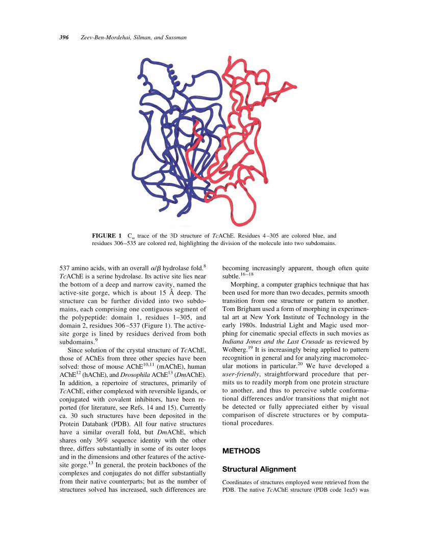
Organophosphorus (OP) agents act as powerful inhib-itors of AChE by interacting covalently with its ac-tive-site serine.27,28 This is the basis for their toxicityand thus for their potency as nerve agents and insec-ticides.2,29 Some OPs, subsequent to this covalentmodification, undergo a reaction called “aging,”which involves dealkylation of the bound OP moi-ety.16,30 One such reagent is diisopropylfluorophos-phate (DFP) that, subsequent to phosphorylation and“aging,” yields the monoisopropylphosphoryl (MIP)/TcAChE conjugate whose 3D structure was solved.30
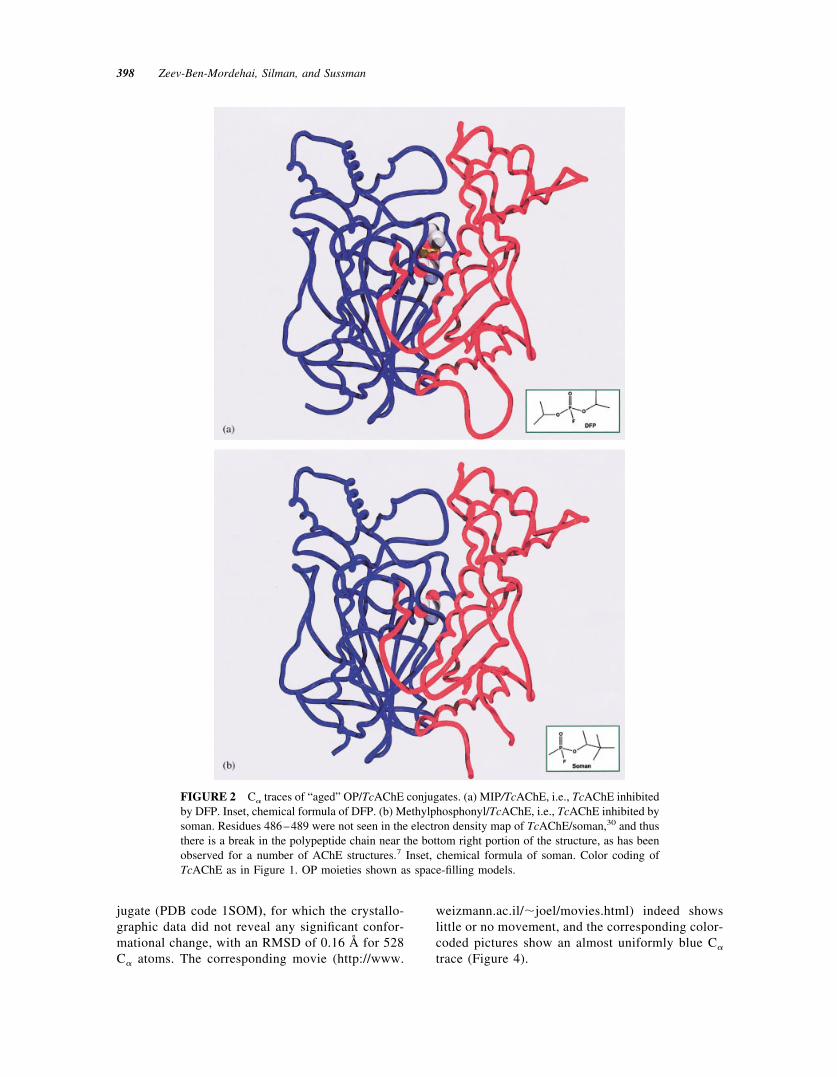
The crystal structure revealed that the bulky isopropylgroup distorts the acyl-binding pocket, which nor-mally recognizes the acetyl group of the substrate,ACh (Figure 2). The two structures have a root meansquare deviation (RMSD) of 0.36 Å for 530 C� atoms.The transition from native TcAChE (PDB code 1ea5)to the MIP/TcAChE conjugate (PDB code 2dfp) ismorphed in a movie (http://www.weizmann.ac.il/�joel/moveis.html) of which three superimposedframes, the initial, the final, and one intermediateframe, are displayed in Figure 3.
The movie shows that phosphorylation by DFPcauses a movement of the main chain in the conservedloop (residues 279–291), which includes the acylpocket residues, F288 and F290, as reported by Mil-lard and co-workers.30 However, this major move-ment of residues in the acyl pocket also affects part ofthe peripheral site, which is on the same loop (viz.W279); this, in turn, causes substantial movement ofresidues in the second subdomain, across the active-site gorge, with which the 279–291 loop is in con-tact.9 This is also shown by color coding the move-ments between native TcAChE and the MIP conjugateon a scale from blue to red, going from the smallest tothe largest differences (Figure 3).
For comparison, we show the transition fromnative TcAChE (PDB code 1ea5) to the TcAChE/soman conjugate, viz. the methylphosphonyl con-
Morphing Conformational Changes in AChE 397
jugate (PDB code 1SOM), for which the crystallo-graphic data did not reveal any significant confor-mational change, with an RMSD of 0.16 Å for 528C� atoms. The corresponding movie (http://www.
weizmann.ac.il/�joel/movies.html) indeed showslittle or no movement, and the corresponding color-coded pictures show an almost uniformly blue C�
trace (Figure 4).
FIGURE 2 C� traces of “aged” OP/TcAChE conjugates. (a) MIP/TcAChE, i.e., TcAChE inhibitedby DFP. Inset, chemical formula of DFP. (b) Methylphosphonyl/TcAChE, i.e., TcAChE inhibited bysoman. Residues 486–489 were not seen in the electron density map of TcAChE/soman,30 and thusthere is a break in the polypeptide chain near the bottom right portion of the structure, as has beenobserved for a number of AChE structures.7 Inset, chemical formula of soman. Color coding ofTcAChE as in Figure 1. OP moieties shown as space-filling models.
398 Zeev-Ben-Mordehai, Silman, and Sussman
Morphing from Native TcAChE to ItsComplex with Fasciculin-II
Fasciculin-II (FAS-II) is a member of the “three-fingered” polypeptide toxin family,31,32 found in thevenom of the green mamba, which inhibits vertebrateAChEs by binding tightly to the entrance of the ac-tive-site gorge.32 Its inhibitory action is ascribed pri-marily to steric hindrance10,12,33–36 but an allostericcomponent has also been invoked.10,34,37–41 Wethought it worthwhile, therefore, to utilize the mor-phing technique to visualize putative conformationalchanges occurring in the enzyme upon binding of thetoxin.
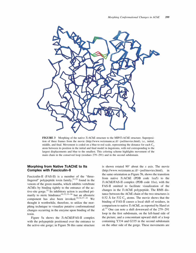
Figure 5a shows the TcAChE/FAS-II complexwith the polypeptide positioned over the entrance tothe active-site gorge; in Figure 5b this same structure
is shown rotated 90° about the x axis. The movie(http://www.weizmann.ac.il/�joel/movies.html), inthe same orientation as Figure 5b, shows the transitionfrom native TcAChE (PDB code 1ea5) to theTcAChE/FAS-II complex (PDB code 1fss), with theFAS-II omitted to facilitate visualization of thechanges in the TcAChE polypeptide. The RMS dis-tance between the AChE chain of the two structures is0.52 Å for 532 C� atoms. The movie shows that thebinding of FAS-II causes a local shift of residues, incomparison to native TcAChE, as reported by Harel etal.33 One can note a shift downward of the 279–291loop in the first subdomain, on the left-hand side ofthe picture, and a concomitant upward shift of a loopcontaining Y334 and G335 in the second subdomainon the other side of the gorge. These movements are
FIGURE 3 Morphing of the native TcAChE structure to the MIP/TcAChE structure. Superposi-tion of three frames from the movie (http://www.weizmann.ac.il/�joel/movies.html), i.e., initial,middle, and final. Movement is coded on a blue-to-red scale, representing the distance for each C�
atom between its position in the initial and final model in ångstroms, with red corresponding to thelargest displacements and blue to the smallest. This coloring scheme highlights movement of themain chain in the conserved loop (residues 279–291) and in the second subdomain.
Morphing Conformational Changes in AChE 399

presumably driven by insertion of loop 2 of FAS-IIinto the mouth of the gorge. Inspection of Table 3 inHarel et al.33 shows that both these loops, indeed,interact with FAS-II. In Figure 5b, the contact resi-dues of FAS-II loop 2 are color coded in cyan and thecontact residues on the complementary loops in thegorge of AChE are color coded in yellow. One can seethe good correspondence of these color coded loopsand the color code for movement in Figure 6 and inthe corresponding movie. It is worth noting that inFigure 6 one can see also substantial movement of theomega loop in agreement with previous sugges-tions.10,34,38–40 The color coding also suggests aslight movement of Ser200. It was earlier suggestedon the basis of kinetic data that FAS-II acts predom-inantly by altering the conformation of the active sitein the ternary complex, so that the steps involvingproton transfer during enzyme acetylation are
slowed.37 Molecular dynamics simulation also sug-gested a disruption of the catalytic triad in the AChE/FAS-II complex.41 A subtle movement of the triadresidues would obviously suffice to affect catalyticactivity profoundly.
Morphing from the TcAChE/FAS-IIComplex to the hAChE/FAS-II Complex
hAChE displays 53% sequence identity to TcAChE,suggesting that their 3D structures should be verysimilar. This was indeed born out when the 3D struc-ture of hAChE, complexed with FAS-II, was solved.12
The RMSD between the TcAChE/FAS-II complex(1fss, PDB code) and the hAChE/FAS-II complex(1b41, PDB code) is 0.88 Å for 521 C� atoms12
However, comparison of the two structures revealed asignificant difference which involves flipping of the
FIGURE 4 Morphing of the native TcAChE structure to the methylphosphonyl/TcAChE struc-ture. Superposition of three frames from the movie (http://www.weizmann.ac.il/�joel/movies.html),i.e., initial, middle, and final. The almost uniformly blue C� trace indicates little or no differencebetween the two structures. Color coding as in Figure 3. Residues 486–489 were not seen in thisstructure (see Figure 2b).
400 Zeev-Ben-Mordehai, Silman, and Sussman

FIGURE 5 C� trace of the 3D structure of the TcAChE/FAS-II complex. (a) Entrance toactive-site gorge at the top; (b) rotation �90° about the x axis relative to (a). The TcAChEsubdomains are represented as in Figure 1, and FAS-II is color coded in green. Interacting residuesof FAS-II loop 2 are colored cyan and the complementary interacting residues in TcAChE arecolored yellow.
Morphing Conformational Changes in AChE 401

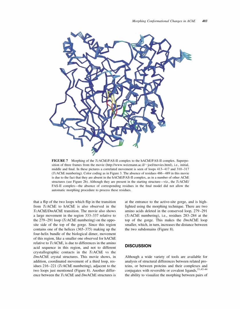
310–317 loop (TcAChE numbering).12 This was notdue to crystal contacts, as was established by com-parison with the native mAChE structure,11 and withthat of the mouse AChE/FAS-II complex10, both ofwhich showed the same flip relative to the TcAChE/FAS-II structure. In the movie (http://www.weizmann.ac.il/�joel/movies.html) of the transition fromTcAChE/FAS-II to hAChE/FAS-II, this flip is clearlyvisualized. However, the movie also highlights a cor-related movement of an adjacent but noncontiguousloop, residues 413–417 (TcAChE numbering), notreported previously (Figure 7).
Morphing from TcAChE to DmAChE
DmAChE displays 36% sequence identity toTcAChE. This homology is substantially lower than
that between TcAChE and hAChE; a similar fold was,nevertheless, to be expected, as was born out by the3D structure of DmAChE,13 even though the similar-ity of the structures was not high enough to permitsolution of the structure by molecular replacementbased on the TcAChE structure. The RMSD betweenthe C� atoms of TcAChE (1ea5, PDB code) andDmAChE (1qo9, PDB code), using 481 residues, is1.28 Å. Although the geometry of the catalytic appa-ratus of the two enzymes is almost isomorphous,substantial differences were observed in the volumeand geometry of the active-site gorge, as well as largedifferences in some of the distal loops.13 In the movie(http://www.weizmann.ac.il/�joel/movies.html) ofthe transition from TcAChE to DmAChE one clearlysees, not surprisingly, much larger overall movementsthan in the previous movies. It is interesting, however,
FIGURE 6 Morphing of the native TcAChE structure to the TcAChE/FAS-II complex. Super-position of three frames from the movie (http://www.weizmann.ac.il/�joel/movies.html), i.e.,initial, middle, and final. One sees good correspondence of the color coding, indicating substantialmovement of the conserved 279–291 loop, and of the loop containing Y334 and G335, across theactive-site gorge, in the second subdomain, with the residues coded yellow in Figure 5. The apparentchange in conformation of residues 486–489 is most likely a consequence of the known flexibilityof this external loop, which often, indeed, is not seen in the electron density map (see Figure 2b).
402 Zeev-Ben-Mordehai, Silman, and Sussman
that a flip of the two loops which flip in the transitionfrom TcAChE to hAChE is also observed in theTcAChE/DmAChE transition. The movie also showsa large movement in the region 333–337 relative tothe 279–291 loop (TcAChE numbering) on the oppo-site side of the top of the gorge. Since this regioncontains one of the helices (365–375) making up thefour-helix bundle of the biological dimer, movementof this region, like a smaller one observed for hAChErelative to TcAChE, is due to differences in the aminoacid sequence in this region, and not to differentcrystallographic contacts in the TcAChE vs theDmAChE crystal structures. This movie shows, inaddition, coordinated movement of a third loop, res-idues 216–221 (TcAChE numbering), adjacent to thetwo loops just mentioned (Figure 8). Another differ-ence between the TcAChE and DmAChE structures is
at the entrance to the active-site gorge, and is high-lighted using the morphing technique. There are twoamino acids deleted in the conserved loop, 279–291(TcAChE numbering), i.e., residues 283–284 at thetop of the gorge. This makes the DmAChE loopsmaller, which, in turn, increases the distance betweenthe two subdomains (Figure 8).
DISCUSSION
Although a wide variety of tools are available foranalysis of structural differences between related pro-teins, or between proteins and their complexes andconjugates with reversible or covalent ligands,21,42-44
the ability to visualize the morphing between pairs of
FIGURE 7 Morphing of the TcAChE/FAS-II complex to the hAChE/FAS-II complex. Superpo-sition of three frames from the movie (http://www.weizmann.ac.il/�joel/movies.html), i.e., initial,middle and final. In these pictures a correlated movement is seen of loops 413–417 and 310–317(TcAChE numbering). Color coding as in Figure 3. The absence of residues 486–489 in this movieis due to the fact that they are absent in the hAChE/FAS-II complex, as in a number of other AChEstructures (see Figure 2b). Although they are present in the starting structure—viz., the TcAChE/FAS-II complex—the absence of corresponding residues in the final model did not allow theautomatic morphing procedure to process these residues.
Morphing Conformational Changes in AChE 403

structures often reveals differences not otherwisereadily perceived.
One of the most illustrative examples of the use ofmorphing was in the case of the four different crystalstructures of adenyl kinase and of the movie producedto show the “motions” between these differentforms45 (see also: http://bio.chemie.uni-freiburg.de/ak_movie/). More recent progress in this area hasbeen reviewed by Krebs and Gerstein20 (see alsohttp://bioinfo.mbb.yale).
In this study, we have developed a “user-friendly”tool to automate transitions between any pair ofclosely related protein structures. As described underMethods, our procedure utilizes Cartesian coordi-nates, and is not intended to faithfully reflect the“chemical” trajectory of a conformational change.However, it does make it possible to readily perceiveand evaluate coordinated changes in structure that
may occur when a ligand/protein complex is formed,or to evaluate conformational differences betweenclosely related proteins from different species.
For example, our morphing technique clearly re-veals the coordinated movements occurring in the twosubdomains across the active-site gorge from eachother upon distortion of the acyl pocket by covalentmodification with DFP, or upon insertion of loop 2 ofFAS-II into the mouth of the gorge. It also shows thatthese changes do not occur in all cases of ligandbinding, e.g., for the TcAChE/soman conjugate. Thetechnique also highlights interspecies differences.Thus, comparison of the TcAChE structure with thoseof both hAChE and DmAChE shows a coordinatedchange in structure of several loops that would not beeasily perceived from inspection of static structures.
The morphing movies can also reveal the nature ofthe conformational changes between structures—e.g.,
FIGURE 8 Morphing of the native TcAChE structure to the native DmAChE structure. Super-position of three frames from the movie (http://www.weizmann.ac.il/�joel/movies.html), i.e.,initial, middle, and final. These pictures clearly show a similar flipping of loops as observed in thetransition from TcAChE to hAChE (residues 310–317, 413–417; TcAChE numbering). In addition,a large movement is observed in subdomain 2 across from the conserved loop 279–291 insubdomain 1.
404 Zeev-Ben-Mordehai, Silman, and Sussman
the apparent “spiral” movement of the two helicescontributing to the four-helix bundle of the dimer inboth TcAChE and DmAChE (viz. helices �F�3 and �H
in TcAChE7).When examining the apparent conformational
changes between any pair of structures, care must beexercised to ascertain that the apparent differences arenot a trivial consequence of different crystallographicpacking. For the native vs TcAChE conjugates, sincethe same crystal form was employed, this was not aproblem. However, for native TcAChE vs theTcAChE/FAS-II complex, as well as for the cross-species comparisons, it was indeed a potential prob-lem. For the case of the FAS-II complex, the FAS-IIbinds at virtually the same site as another TcAChEbinds in the native enzyme; thus in both cases the topof the gorge is making contacts, in fact, with anothermolecule, albeit in one case with another copy ofTcAChE and in the other case with a FAS-II mole-cule. For both the hAChE and DmAChE structures,the major movements seen relative to the TcAChEstructure are found in the region of the two helicesthat form the four-helix bundle of the biologicaldimer. As this four-helix bundle is very similar in allthree structures—i.e., TcAChE, hAChE, DmAChE—the large changes in structures seen in the morphingmovies likely reflect real differences in conformationbetween these structures, reflecting the differences inamino acid sequences of the residues in the four-helixbundle and in adjacent parts of the structures.
The method we have developed is a “user-friendly”approach based on tying together a series of preexist-ing tools, and taking advantage of the capacity of thehuman visual system for subtle pattern recognition.What we have done here, using morphing techniques,is in the spirit of a study carried out in collaborationwith Shneior Lifson in the cross-species analysis ofelectrostatic properties of AChE.46 We feel thatShneior would have been delighted to see our currentmovies, and probably would have been able to per-ceive more than our eyes did.
This work was supported by the U.S. Army Medical andMaterial Command under Contract No. DAMD17-97-2-7022, the EC 5th Framework Program on the Quality of Lifeand Management of Living Resources, the KimmelmanCenter for Biomolecular Structure and Assembly and theBenoziyo Center for Neuroscience (Rehovot, Israel). IS isthe Bernstein-Mason Professor of Neurochemistry.
REFERENCES
1. Taylor, P. The Pharmacological Basis of Therapeutics,9th Ed.; Hardman, J. G., Limbird, L. E., Molinoff,
P. B., Ruddon, R. W., Gilman, A. G., Eds.; McGraw-Hill: New York, 1996; pp 161–176.
2. Millard, C. B.; Broomfield, C. A. J Neurochem 1995,64, 1909–1918.
3. Davis, K. L.; Powchik, P. Lancet 1995, 345, 625–630.4. Nightingale, S. L. JAMA 1997, 277, 10.5. Casida, J. E.; Quistad, G. B. Ann Rev Entomol 1998,
43, 1–16.6. Martin, R. J. Vet J 1997, 154, 11–34.7. Sussman, J. L.; Harel, M.; Frolow, F.; Oefner, C.;
Goldman, A.; Toker, L.; Silman, I. Science 1991, 253,872–879.
8. Ollis, D. L.; Cheah, E.; Cygler, M.; Dijkstra, B.;Frolow, F.; Franken, S. M.; Harel, M.; Remington, S. J.;Silman, I.; Schrag, J.; Sussman, J. L.; Verschueren,K. H. G.; Goldman, A. Protein Eng 1992, 5, 197–211.
9. Morel, N.; Bon, S.; Greenblatt, H.; Wodak, S.; Suss-man, J. L.; Massoulie, J.; Silman, I. Mol Pharmacol1999, 55, 982–992.
10. Bourne, Y.; Taylor, P.; Marchot, P. Cell 1995, 83,503–512.
11. Bourne, Y.; Taylor, P.; Bougis, P. E.; Marchot, P. J BiolChem 1999, 274, 2963–2970.
12. Kryger, G.; Harel, M.; Giles, K.; Toker, L.; Velan, B.;Lazar, A.; Kronman, C.; Barak, D.; Ariel, N.; Shaffer-man, A.; Silman, I.; Sussman, J. L. Acta Cryst 2000,D56, 1385–1394.
13. Harel, M.; Kryger, G.; Rosenberry, T. L.; Mallender,W. D.; Lewis, T.; Fletcher, R. J.; Guss, J. M.; Silman,I.; Sussman, J. L. Protein Sci 2000, 9, 1063–1072.
14. Greenblatt, H. M.; Silman, I.; Sussman, J. L. DrugDevelop Res 2000, 50, 573–583.
15. Silman, I.; Sussman, J. L. Cholinesterases and Cho-linesterase Inhibitors; Giacobini, E., Ed.; Martin Dun-itz: London, 2000; pp 9–25.
16. Millard, C. B.; Koellner, G.; Ordentlich, A.; Shaffer-man, A.; Silman, I.; Sussman, J. L. J Am Chem Soc1999, 121, 9883–9884.
17. Bar-On, P.; Millard, C. B.; Harel, M.; Dvir, H.; Enz, A.;Sussman, J. L.; Silman, I. Biochemistry 2002, 41,3555–3564.
18. Dvir, H.; Wong, D. M.; Harel, M.; Barril, X.; Orozco,M.; Luque, F. J.; Munoz-Torrero, D.; Camps, P.;Rosenberry, T. L.; Silman, I.; Sussman, J. L. Biochem-istry 2002, 41, 2970–2981.
19. Wolberg, G. Digital Image Warping; Wiley-IEEEPress, New York, 1990.
20. Krebs, W. G.; Gerstein, M. Nucleic Acids Res 2000,28, 1665–1675.
21. Kleywegt, G. J. Acta Cryst 1996, D52, 842–857.22. Kleywegt, G. J. Uppsala Software Factory—LSQMAN
Manual; Uppsala Software Factory, 2002; http://xray.bmc.uu.se/�gerard/manuals/lsqman_man.html#S71.
23. Jones, T. A.; Zou, J.-Y.; Cowan, S. W.; Kjeldgaard, M.Acta Cryst 1991, A47, 110–119.
24. Sayle, R. A.; Milner-White, E. J. TIBS 1995, 20, 374–376.
25. Mediaconvert; SGI, 2002; http://www.sgi.com.
Morphing Conformational Changes in AChE 405

26. QuickTime; Apple Computer, 2002; http://www.apple.com/quicktime/.
27. Quinn, D. M. Chem Rev 1987, 87, 955–975.28. Aldridge, W. N.; Reiner, E. Enzyme Inhibitors as Sub-
strates: Interactions of Esterases with Esters of Organ-ophosphorus and Carbamic Acids; North-Holland Pub-lishing: Amsterdam, 1972; Vol 26.
29. Koelle, G. B. Cholinesterases and AnticholinesteraseAgents; Springer-Verlag: Heidelberg, 1963; Vol 15.
30. Millard, C. B.; Kryger, G.; Ordentlich, A.; Harel, M.;Raves, M.; Greenblatt, H. M.; Segall, Y.; Barak, D.;Shafferman, A.; Silman, I.; Sussman, J. L. Biochemis-try 1999, 38, 7032–7039.
31. Giles, K.; Raves, M. L.; Silman, I.; Sussman, J. L.Theoretical and Computational Methods in GenomeResearch; Suhai, S., Ed.; Plenum Press: New York,1997; pp 303–315.
32. Karlsson, E.; Harvey, A. L.; Cervenansky, C.; Kley-wegt, G. J.; Harel, M.; Silman, I.; Sussman, J. L.Enzymes from Snake Venoms; Bailey, G. S., Ed.;Alaken: Fort Collins, CO, 1998; pp 633–688.
33. Harel, M.; Kleywegt, G. J.; Ravelli, R. B. G.; Silman,I.; Sussman, J. L. Structure 1995, 3, 1355–1366.
34. Radic, Z.; Quinn, D. M.; Vellom, D. C.; Camp, S.;Taylor, P. J Biol Chem 1995, 270, 20391–20399.
35. Marchot, P.; Bourne, Y.; Prowse, C. N.; Bougis, P. E.;Taylor, P. Toxicon 1998, 36, 1613–1622.
36. De Ferrari, G. V.; Mallender, W. D.; Inestrosa, N. C.;Rosenberry, T. L. J Biol Chem 2001, 276, 23282–23287.
37. Eastman, J.; Wilson, E. J.; Cervenansky, C.; Rosen-berry, T. L. J Biol Chem 1995, 270, 19694–19701.
38. Szegletes, T.; Mallender, W. D.; Thomas, P. J.; Rosen-berry, T. L. Biochemistry 1999, 38, 122–133.
39. Mallender, W. D.; Szegletes, T.; Rosenberry, T. L.Biochemistry 2000, 39, 7753–7763.
40. Shi, J.; Boyd, A. E.; Radic, Z.; Taylor, P. J Biol Chem2001, 276, 42196–42204.
41. Tai, K.; Shen, T.; Henchman, R. H.; Bourne, Y.; Mar-chot, P.; McCammon, J. A. J Am Chem Soc 2002, 124,6153–6161.
42. Jacobs, D. J.; Rader, A. J.; Kuhn, L. A.; Thorpe, M. F.Proteins 2001, 44, 150–165.
43. Ming, D.; Kong, Y.; Lambert, M. A.; Huang, Z.; Ma, J.Proc Natl Acad Sci USA 2002, 99, 8620–8625.
44. Godzik, A. Protein Sci 1996, 5, 1325–1338.45. Vonrhein, C.; Schlauderer, G. J.; Schulz, G. E. Struc-
ture 1995, 3, 483–490.46. Felder, C. E.; Botti, S. A.; Lifson, S.; Silman, I.; Suss-
man, J. L. J Mol Graphics Model 1997, 15, 318–327.
406 Zeev-Ben-Mordehai, Silman, and Sussman