New Orleans Academy of Ophthalmology (NOAO) 63rd Annual Symposium

Visual Field Defects1
Here is a representation of the VF for each eye. Which is OD, and which OS?
? ?

Visual Field Defects2
Here is a representation of the VF for each eye. Which is OD, and which OS?Remember, VFs are not drawn as if the pt is looking at you; they’re drawn as if you are the pt!
OS OD

Visual Field Defects3
Measured in degrees from fixation, how far does the normal VF extend superiorly, inferiorly, nasally and temporally?
OS OD
? ?? ?
? ?
? ?

Visual Field Defects4
Measured in degrees from fixation, how far does the normal VF extend superiorly, inferiorly, nasally and temporally?(Don’t get too fixated on these specific numbers--different sources will give slightly different values.)
OS OD
100o 100o60o 60o
60o 60o
70o 70o
Visual Field Defects5
OS OD
? ?
Measured in degrees from fixation, how much of the VF is assessed via the automated perimetry machines found in most ophthalmology practices?

Visual Field Defects6
OS ODMeasured in degrees from fixation, how much of the VF is assessed via the automated perimetry machines found in most ophthalmology practices?The central 24 degrees
24o24o

Visual Field Defects7
How far in degrees from fixation is the blind spot?
OS OD
? ?

Visual Field Defects8
How far in degrees from fixation is the blind spot?About 15 (again, don’t get too hung up on that specific number.)
OS OD
15o 15o
Visual Field Defects
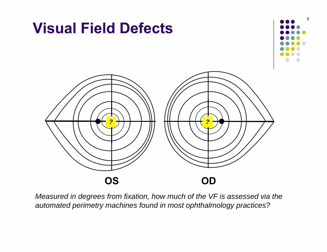
Retina
Anatomic locations forlesions producing VF defects
most anteriorlocation
9

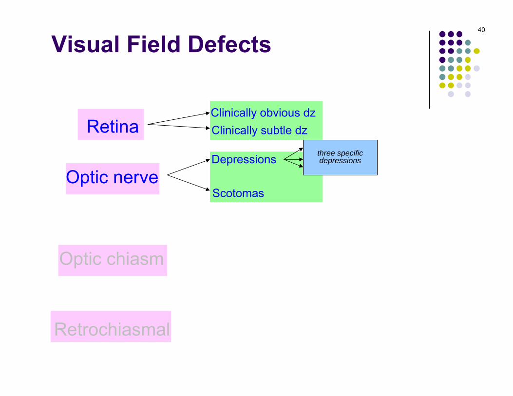
Visual Field Defects
Retina
Optic nerve
Anatomic locations forlesions producing VF defects
next location
10

Visual Field Defects
Retina
Optic nerve
Optic chiasm
Anatomic locations forlesions producing VF defects
next location
11

Visual Field Defects
Retina
Optic nerve
Optic chiasm
Anatomic locations forlesions producing VF defects
general term for all locationsposterior to the previous one
12

Visual Field Defects
Retina
Optic nerve
Optic chiasm
Anatomic locations forlesions producing VF defects
Retrochiasmal
13

Retina
Optic nerve
Optic chiasm
Retrochiasmal
Clinically obvious dzClinically subtle dz
two very generalcategories of retinal dz
Visual Field Defects14

Retina
Optic nerve
Optic chiasm
Retrochiasmal
Clinically obvious dzClinically subtle dz
Visual Field Defects15

Retina
Optic nerve
Optic chiasm
Retrochiasmal
Clinically obvious dzClinically subtle dz
Visual Field Defects16
What is meant by clinically obvious vs clinically subtle retinal dz?

Retina
Optic nerve
Optic chiasm
Retrochiasmal
Clinically obvious dzClinically subtle dz
Visual Field Defects17
What is meant by clinically obvious vs clinically subtle retinal dz?In clinically obvious disease, the retina will appear abnormal on DFE, whereas in clinically subtle disease it will look normal

Retina
Optic nerve
Optic chiasm
Retrochiasmal
Clinically obvious dz (eg…?)Clinically subtle dz
Visual Field Defects18
What is meant by clinically obvious vs clinically subtle retinal dz?In clinically obvious disease, the retina will appear abnormal on DFE, whereas in clinically subtle disease it will look normal
What is an example of……clinically obvious disease?

Retina
Optic nerve
Optic chiasm
Retrochiasmal
Clinically obvious dz (eg…RP)
Visual Field Defects19
What is meant by clinically obvious vs clinically subtle retinal dz?In clinically obvious disease, the retina will appear abnormal on DFE, whereas in clinically subtle disease it will look normal
What is an example of……clinically obvious disease? ‘Typical’ retinitis pigmentosa
Clinically subtle dz

Retina
Optic nerve
Optic chiasm
Retrochiasmal
Clinically obvious dz (eg…RP)Clinically subtle dz (eg…?)
Visual Field Defects20
What is meant by clinically obvious vs clinically subtle retinal dz?In clinically obvious disease, the retina will appear abnormal on DFE, whereas in clinically subtle disease it will look normal
What is an example of……clinically obvious disease? ‘Typical’ retinitis pigmentosa---clinically subtle disease?

Retina
Optic nerve
Optic chiasm
Retrochiasmal
Clinically obvious dz (eg…RP)Clinically subtle dz (eg…CAR)
Visual Field Defects21
What is meant by clinically obvious vs clinically subtle retinal dz?In clinically obvious disease, the retina will appear abnormal on DFE, whereas in clinically subtle disease it will look normal
What is an example of……clinically obvious disease? ‘Typical’ retinitis pigmentosa---clinically subtle disease? Cancer-associated retinopathy

Retina
Optic nerve
Optic chiasm
Retrochiasmal
Clinically obvious dzClinically subtle dz
Visual Field Defects22
Let’s take a brief aside to cover optic nerve fundamentals before we address optic nerve VF defects
23
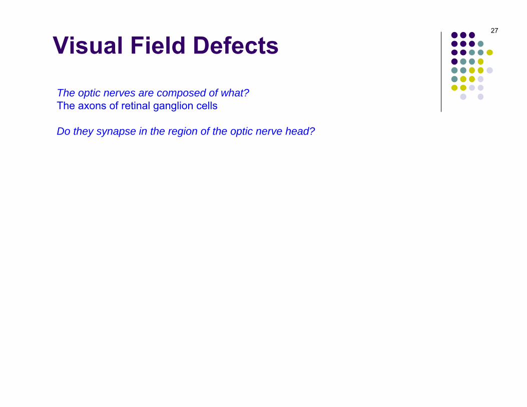
The optic nerves are composed of what?
Visual Field Defects

24
The optic nerves are composed of what?The axons of retinal ganglion cells
Visual Field Defects

25
The optic nerves are composed of what?The axons of retinal ganglion cells
How many fibers (axons) comprise an optic nerve?
Visual Field Defects

26
The optic nerves are composed of what?The axons of retinal ganglion cells
How many fibers (axons) comprise an optic nerve?Depends upon which book you ask, but the answer 1.2M works
Glaucoma book: 1.2-1.5MNeuro: 1-1.2MFundamentals: “more than a million”
Visual Field Defects
27
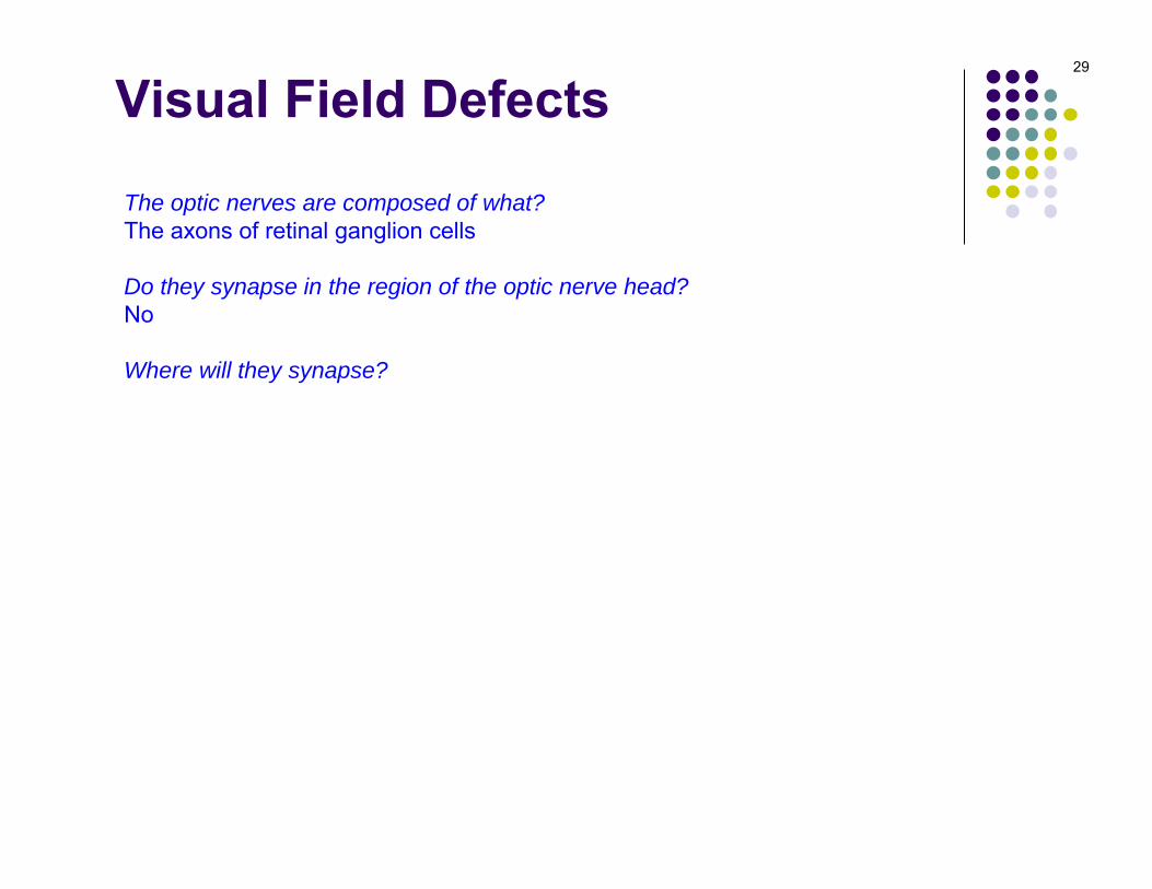
The optic nerves are composed of what?The axons of retinal ganglion cells
Do they synapse in the region of the optic nerve head?
Visual Field Defects

28
The optic nerves are composed of what?The axons of retinal ganglion cells
Do they synapse in the region of the optic nerve head?No
Visual Field Defects
29
The optic nerves are composed of what?The axons of retinal ganglion cells
Do they synapse in the region of the optic nerve head?No
Where will they synapse?
Visual Field Defects

30
The optic nerves are composed of what?The axons of retinal ganglion cells
Do they synapse in the region of the optic nerve head?No
Where will they synapse?Most will synapse in the lateral geniculate nucleus (LGN)
Visual Field Defects

31
The optic nerves are composed of what?The axons of retinal ganglion cells
Do they synapse in the region of the optic nerve head?No
Where will they synapse?Most will synapse in the lateral geniculate nucleus (LGN)
Most? Where will the others synapse, and what are they responsible for?
Visual Field Defects

32
The optic nerves are composed of what?The axons of retinal ganglion cells
Do they synapse in the region of the optic nerve head?No
Where will they synapse?Most will synapse in the lateral geniculate nucleus (LGN)
Most? Where will the others synapse, and what are they responsible for?Most of the others are involved in the pupillary light reflex; they peel off just prior to reaching the LGN, heading instead to the pretectum of the dorsal midbrain to synapse in the pretectal nuclei
Visual Field Defects
33
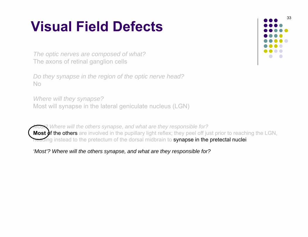
The optic nerves are composed of what?The axons of retinal ganglion cells
Do they synapse in the region of the optic nerve head?No
Where will they synapse?Most will synapse in the lateral geniculate nucleus (LGN)
‘Most’? Where will the others synapse, and what are they responsible for?
Most? Where will the others synapse, and what are they responsible for?Most of the others are involved in the pupillary light reflex; they peel off just prior to reaching the LGN, heading instead to the pretectum of the dorsal midbrain to synapse in the pretectal nuclei
Visual Field Defects

34
The optic nerves are composed of what?The axons of retinal ganglion cells
Do they synapse in the region of the optic nerve head?No
Where will they synapse?Most will synapse in the lateral geniculate nucleus (LGN)
‘Most’? Where will the others synapse, and what are they responsible for?The hypothalamus, where they are involved in modulating circadian responses
Most? Where will the others synapse, and what are they responsible for?Most of the others are involved in the pupillary light reflex; they peel off just prior to reaching the LGN, heading instead to the pretectum of the dorsal midbrain to synapse in the pretectal nuclei
Visual Field Defects

35
The optic nerves are composed of what?The axons of retinal ganglion cells
Do they synapse in the region of the optic nerve head?No
Where will they synapse?Most will synapse in the lateral geniculate nucleus (LGN)
‘Most’? Where will the others synapse, and what are they responsible for?The hypothalamus, where they are involved in modulating circadian responses
Most? Where will the others synapse, and what are they responsible for?Most of the others are involved in the pupillary light reflex; they peel off just prior to reaching the LGN, heading instead to the pretectum of the dorsal midbrain to synapse in the pretectal nuclei
For a more in-depth look at the optic nerve, see slide-set FELT6
Visual Field Defects

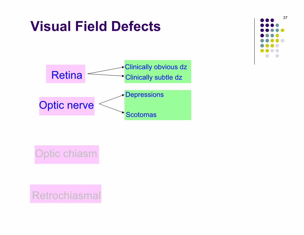
Retina
Optic nerve
Optic chiasm
Retrochiasmal
Depressions
Scotomas
Clinically obvious dzClinically subtle dz
two generalcategories of ON
VF defects
Visual Field Defects36
Retina
Optic nerve
Optic chiasm
Retrochiasmal
Depressions
Scotomas
Clinically obvious dzClinically subtle dz
Visual Field Defects37

Retina
Optic nerve
Optic chiasm
Retrochiasmal
Depressions
Scotomas
Clinically obvious dzClinically subtle dz
Visual Field Defects38
What’s the difference between a depression and a scotoma?A depression is an inward shifting of the outer limit of the visual field, whereas a scotoma is an area of field loss surrounded on all sides by areas of normal sensitivity.

Retina
Optic nerve
Optic chiasm
Retrochiasmal
Depressions
Scotomas
Clinically obvious dzClinically subtle dz
Visual Field Defects39
What’s the difference between a depression and a scotoma?A depression is an inward shifting of the outer limit of the visual field, whereas a scotoma is an area of field loss surrounded on all sides by areas of normal sensitivity.
Retina
Optic nerve
Optic chiasm
Retrochiasmal
Depressions
Scotomas
Clinically obvious dzClinically subtle dz
Visual Field Defects
Nasal stepAltitudinalTemporal wedge
three specificdepressions
40

Retina
Optic nerve
Optic chiasm
Retrochiasmal
Depressions
Scotomas
Clinically obvious dzClinically subtle dz
Visual Field Defects
Nasal stepAltitudinalTemporal wedge
41

Retina
Optic nerve
Optic chiasm
Retrochiasmal
Depressions
Scotomas
Clinically obvious dzClinically subtle dz
Visual Field Defects
Nasal stepAltitudinalTemporal wedge
42
Superior nasal step
Inferior nasal step

Retina
Optic nerve
Optic chiasm
Retrochiasmal
Depressions
Scotomas
Clinically obvious dzClinically subtle dz
Visual Field Defects
Nasal stepAltitudinalTemporal wedge
43
Superior altitudinal
Inferior altitudinal

Retina
Optic nerve
Optic chiasm
Retrochiasmal
Depressions
Scotomas
Clinically obvious dzClinically subtle dz
Visual Field Defects
Nasal stepAltitudinalTemporal wedge
44

Retina
Optic nerve
Optic chiasm
Retrochiasmal
Depressions
Scotomas
Clinically obvious dzClinically subtle dz
Visual Field Defects
Nasal stepAltitudinalTemporal wedge
CentralCeco-central
three specificscotomas
45

Retina
Optic nerve
Optic chiasm
Retrochiasmal
Depressions
Scotomas
Clinically obvious dzClinically subtle dz
Visual Field Defects
ArcuateCentralCeco-central
Nasal stepAltitudinalTemporal wedge
46

Retina
Optic nerve
Optic chiasm
Retrochiasmal
Depressions
Scotomas
Clinically obvious dzClinically subtle dz
Visual Field Defects
ArcuateCentralCeco-central
Nasal stepAltitudinalTemporal wedge
47
Superior arcuate
Inferior arcuate

Retina
Optic nerve
Optic chiasm
Retrochiasmal
Depressions
Scotomas
Clinically obvious dzClinically subtle dz
Visual Field Defects
ArcuateCentralCeco-central
Nasal stepAltitudinalTemporal wedge
48
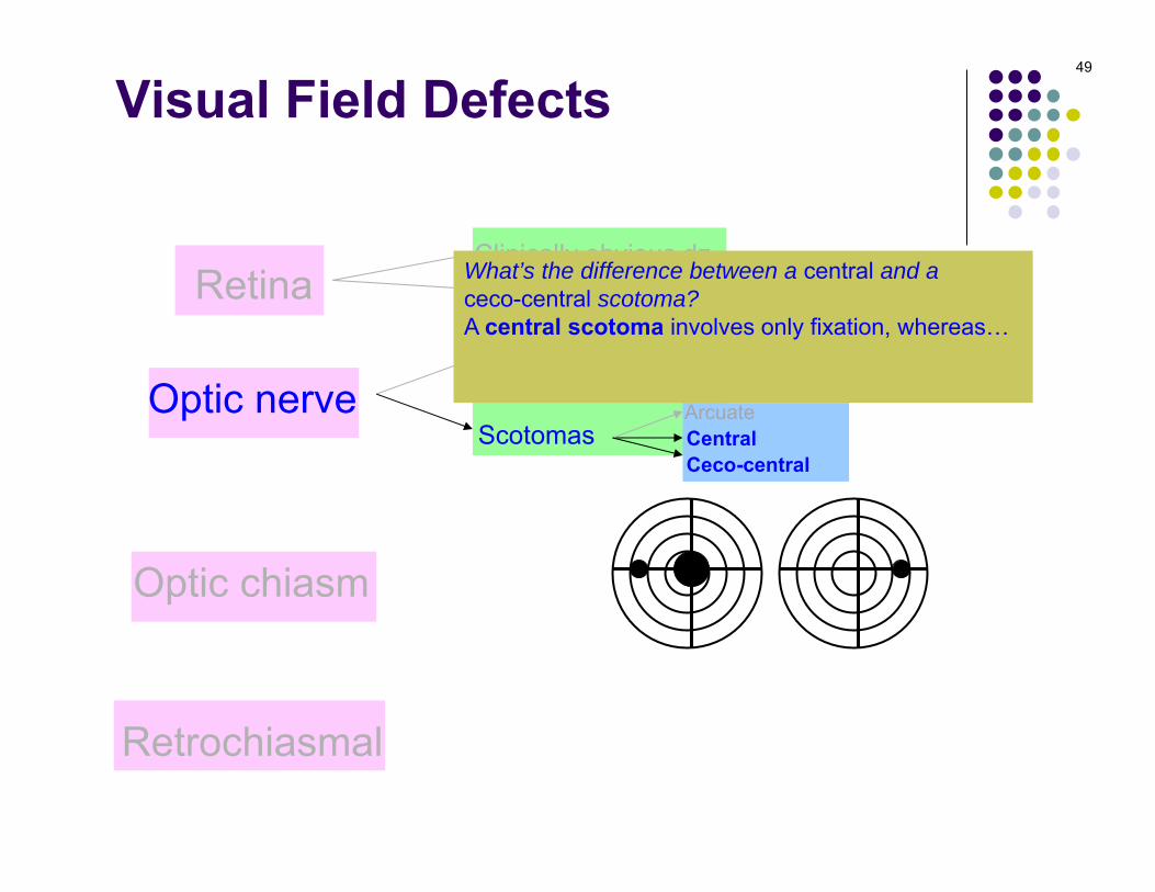
What’s the difference between a central and a ceco-central scotoma?A central scotoma involves only fixation, whereas… a ceco-central scotoma involves fixation andextends all the way to the blind spot.
Retina
Optic nerve
Optic chiasm
Retrochiasmal
Depressions
Scotomas
Clinically obvious dzClinically subtle dz
Visual Field Defects
ArcuateCentralCeco-central
Nasal stepAltitudinalTemporal wedge
49
What’s the difference between a central and a ceco-central scotoma?A central scotoma involves only fixation, whereas… a ceco-central scotoma involves fixation andextends all the way to the blind spot.
Retina
Optic nerve
Optic chiasm
Retrochiasmal
Depressions
Scotomas
Clinically obvious dzClinically subtle dz
Visual Field Defects
ArcuateCentralCeco-central
Nasal stepAltitudinalTemporal wedge
50
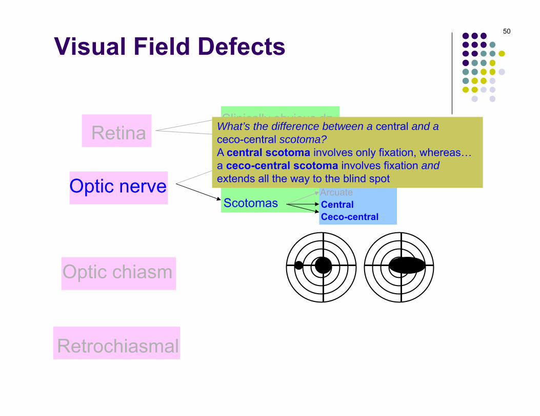
What’s the difference between a central and a ceco-central scotoma?A central scotoma involves only fixation, whereas… a ceco-central scotoma involves fixation andextends all the way to the blind spot

Retina
Optic nerve
Optic chiasm
Retrochiasmal
Depressions
Scotomas
Clinically obvious dzClinically subtle dz
Visual Field Defects
ArcuateCentralCeco-central
Nasal stepAltitudinalTemporal wedge
51
What’s the difference between a central and a ceco-central scotoma?A central scotoma involves only fixation, whereas… a ceco-central scotoma involves fixation andextends all the way to the blind spot
(Take note: Bilateral ceco-central scotomas could be mistaken for bitemporal VF loss!)

Retina
Optic nervehead
Optic chiasm
Retrochiasmal
Nasal fibers
Clinically obvious dzClinically subtle dz
Visual Field Defects52
Arcuate fibers#1?#2?#3?
1
Another way to think about the optic nerve is with respect to its topography at the optic nerve head. Specifically, the retinal nerve fibers composing the optic nerve can be divided into three groups:

Retina
Optic nervehead
Optic chiasm
Retrochiasmal
Papillomacular bundle
Nasal fibers
Clinically obvious dzClinically subtle dz
Visual Field Defects53
Arcuate fibers#2?#3?
Another way to think about the optic nerve is with respect to its topography at the optic nerve head. Specifically, the retinal nerve fibers composing the optic nerve can be divided into three groups:

Retina
Optic nervehead
Optic chiasm
Retrochiasmal
Papillomacular bundle
Nasal fibers
Clinically obvious dzClinically subtle dz
Visual Field Defects54
Arcuate fibers#2?#3?
2
2
Another way to think about the optic nerve is with respect to its topography at the optic nerve head. Specifically, the retinal nerve fibers composing the optic nerve can be divided into three groups:

Retina
Optic nervehead
Optic chiasm
Retrochiasmal
Papillomacular bundle
Nasal fibers
Clinically obvious dzClinically subtle dz
Visual Field Defects55
Arcuate fibers#3?
Another way to think about the optic nerve is with respect to its topography at the optic nerve head. Specifically, the retinal nerve fibers composing the optic nerve can be divided into three groups:

Retina
Optic nervehead
Optic chiasm
Retrochiasmal
Papillomacular bundle
Nasal fibers
Clinically obvious dzClinically subtle dz
Visual Field Defects56
Arcuate fibers#3?
3
Another way to think about the optic nerve is with respect to its topography at the optic nerve head. Specifically, the retinal nerve fibers composing the optic nerve can be divided into three groups:

Retina
Optic nervehead
Optic chiasm
Retrochiasmal
Papillomacular bundle
Nasal radiating fibers
Clinically obvious dzClinically subtle dz
Visual Field Defects57
Arcuate fibers
Another way to think about the optic nerve is with respect to its topography at the optic nerve head. Specifically, the retinal nerve fibers composing the optic nerve can be divided into three groups:

Retina
Optic nervehead
Optic chiasm
Retrochiasmal
Papillomacular bundle
Nasal radiating fibers
Clinically obvious dzClinically subtle dz
Visual Field Defects58
Arcuate fibers
Another way to think about the optic nerve is with respect to its topography at the optic nerve head. Specifically, the retinal nerve fibers composing the optic nerve can be divided into three groups:
The basic topography of the RNFL looks a
lot like a fish!

Retina
Optic nervehead
Optic chiasm
Retrochiasmal
Papillomacular bundle
Nasal radiating fibers
Clinically obvious dzClinically subtle dz
Visual Field Defects59
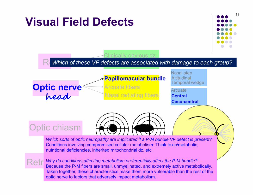
Arcuate fibers
Which of these VF defects are associated with damage to each group?
ArcuateCentralCeco-central
Nasal stepAltitudinalTemporal wedge ?
?

Retina
Optic nervehead
Optic chiasm
Retrochiasmal
Papillomacular bundle
Nasal radiating fibers
Clinically obvious dzClinically subtle dz
Visual Field Defects60
Arcuate fibers ArcuateCentralCeco-central
Nasal stepAltitudinalTemporal wedge
Which of these VF defects are associated with damage to each group?

Retina
Optic nervehead
Optic chiasm
Retrochiasmal
Papillomacular bundle
Nasal radiating fibers
Clinically obvious dzClinically subtle dz
Visual Field Defects61
Arcuate fibers ArcuateCentralCeco-central
Nasal stepAltitudinalTemporal wedge
Which of these VF defects are associated with damage to each group?
Which sorts of optic neuropathy are implicated if a P-M bundle VF defect is present? Conditions involving compromised cellular metabolism: Think toxic/metabolic, nutritional deficiencies, inherited mitochondrial dz, etc
Why do conditions affecting metabolism preferentially affect the P-M bundle?Because the P-M fibers are small, unmyelinated, and extremely active metabolically. Taken together, these characteristics make them more vulnerable than the rest of the optic nerve to factors that adversely impact metabolism.

Retina
Optic nervehead
Optic chiasm
Retrochiasmal
Papillomacular bundle
Nasal radiating fibers
Clinically obvious dzClinically subtle dz
Visual Field Defects62
Arcuate fibers ArcuateCentralCeco-central
Nasal stepAltitudinalTemporal wedge
Which of these VF defects are associated with damage to each group?
Which sorts of optic neuropathy are implicated if a P-M bundle VF defect is present? Conditions involving compromised cellular metabolism: Think toxic/metabolic, nutritional deficiencies, inherited mitochondrial dz, etc
Why do conditions affecting metabolism preferentially affect the P-M bundle?Because the P-M fibers are small, unmyelinated, and extremely active metabolically. Taken together, these characteristics make them more vulnerable than the rest of the optic nerve to factors that adversely impact metabolism.

Retina
Optic nervehead
Optic chiasm
Retrochiasmal
Papillomacular bundle
Nasal radiating fibers
Clinically obvious dzClinically subtle dz
Visual Field Defects63
Arcuate fibers ArcuateCentralCeco-central
Nasal stepAltitudinalTemporal wedge
Which of these VF defects are associated with damage to each group?
Which sorts of optic neuropathy are implicated if a P-M bundle VF defect is present? Conditions involving compromised cellular metabolism: Think toxic/metabolic, nutritional deficiencies, inherited mitochondrial dz, etc
Why do conditions affecting metabolism preferentially affect the P-M bundle?Because the P-M fibers are small, unmyelinated, and extremely active metabolically. Taken together, these characteristics make them more vulnerable than the rest of the optic nerve to factors that adversely impact metabolism.
Retina
Optic nervehead
Optic chiasm
Retrochiasmal
Papillomacular bundle
Nasal radiating fibers
Clinically obvious dzClinically subtle dz
Visual Field Defects64
Arcuate fibers ArcuateCentralCeco-central
Nasal stepAltitudinalTemporal wedge
Which of these VF defects are associated with damage to each group?
Which sorts of optic neuropathy are implicated if a P-M bundle VF defect is present? Conditions involving compromised cellular metabolism: Think toxic/metabolic, nutritional deficiencies, inherited mitochondrial dz, etc
Why do conditions affecting metabolism preferentially affect the P-M bundle?Because the P-M fibers are small, unmyelinated, and extremely active metabolically. Taken together, these characteristics make them more vulnerable than the rest of the optic nerve to factors that adversely impact metabolism.

Retina
Optic nervehead
Optic chiasm
Retrochiasmal
Papillomacular bundle
Nasal radiating fibers
Clinically obvious dzClinically subtle dz
Visual Field Defects65
Arcuate fibers ArcuateCentralCeco-central
Nasal stepAltitudinalTemporal wedge
Which of these VF defects are associated with damage to each group?
Which sorts of optic neuropathy are implicated if a P-M bundle VF defect is present? Conditions involving compromised cellular metabolism: Think toxic/metabolic, nutritional deficiencies, inherited mitochondrial dz, etc
Why do conditions affecting metabolism preferentially affect the P-M bundle?Because the P-M fibers are small, unmyelinated, and extremely active metabolically. Taken together, these characteristics make them more vulnerable than the rest of the optic nerve to factors that adversely impact metabolism.
Toxins that shouldn’t be ingested at all:--------(many others)Toxins that shouldn’t be ingested in large quantities for prolonged periods:----Toxins you were told to ingest by a doc:----------Nutrients that weren’t ingested in sufficient quantity:------Inherited mitochondrial diseases:----

Retina
Optic nervehead
Optic chiasm
Retrochiasmal
Papillomacular bundle
Nasal radiating fibers
Clinically obvious dzClinically subtle dz
Visual Field Defects66
Arcuate fibers ArcuateCentralCeco-central
Nasal stepAltitudinalTemporal wedge
Which of these VF defects are associated with damage to each group?
Which sorts of optic neuropathy are implicated if a P-M bundle VF defect is present? Conditions involving compromised cellular metabolism: Think toxic/metabolic, nutritional deficiencies, inherited mitochondrial dz, etc
Why do conditions affecting metabolism preferentially affect the P-M bundle?Because the P-M fibers are small, unmyelinated, and extremely active metabolically. Taken together, these characteristics make them more vulnerable than the rest of the optic nerve to factors that adversely impact metabolism.
Toxins that shouldn’t be ingested at all:--Methanol--Ethylene glycol--Lead (in children)--(many others)Toxins that shouldn’t be ingested in large quantities for prolonged periods:----Toxins you were told to ingest by a doc:----------Nutrients that weren’t ingested in sufficient quantity:------Inherited mitochondrial diseases:----

Retina
Optic nervehead
Optic chiasm
Retrochiasmal
Papillomacular bundle
Nasal radiating fibers
Clinically obvious dzClinically subtle dz
Visual Field Defects67
Arcuate fibers ArcuateCentralCeco-central
Nasal stepAltitudinalTemporal wedge
Which of these VF defects are associated with damage to each group?
Which sorts of optic neuropathy are implicated if a P-M bundle VF defect is present? Conditions involving compromised cellular metabolism: Think toxic/metabolic, nutritional deficiencies, inherited mitochondrial dz, etc
Why do conditions affecting metabolism preferentially affect the P-M bundle?Because the P-M fibers are small, unmyelinated, and extremely active metabolically. Taken together, these characteristics make them more vulnerable than the rest of the optic nerve to factors that adversely impact metabolism.
Toxins that shouldn’t be ingested at all:--Methanol--Ethylene glycol--Lead (in children)--(many others)Toxins that shouldn’t be ingested in large quantities for prolonged periods:----Toxins you were told to ingest by a doc:----------Nutrients that weren’t ingested in sufficient quantity:------Inherited mitochondrial diseases:----

Retina
Optic nervehead
Optic chiasm
Retrochiasmal
Papillomacular bundle
Nasal radiating fibers
Clinically obvious dzClinically subtle dz
Visual Field Defects68
Arcuate fibers ArcuateCentralCeco-central
Nasal stepAltitudinalTemporal wedge
Which of these VF defects are associated with damage to each group?
Which sorts of optic neuropathy are implicated if a P-M bundle VF defect is present? Conditions involving compromised cellular metabolism: Think toxic/metabolic, nutritional deficiencies, inherited mitochondrial dz, etc
Why do conditions affecting metabolism preferentially affect the P-M bundle?Because the P-M fibers are small, unmyelinated, and extremely active metabolically. Taken together, these characteristics make them more vulnerable than the rest of the optic nerve to factors that adversely impact metabolism.
Toxins that shouldn’t be ingested at all:--Methanol--Ethylene glycol--Lead (in children)--(many others)Toxins that shouldn’t be ingested in large quantities for prolonged periods:--Ethanol--TobaccoToxins you were told to ingest by a doc:----------Nutrients that weren’t ingested in sufficient quantity:------Inherited mitochondrial diseases:----
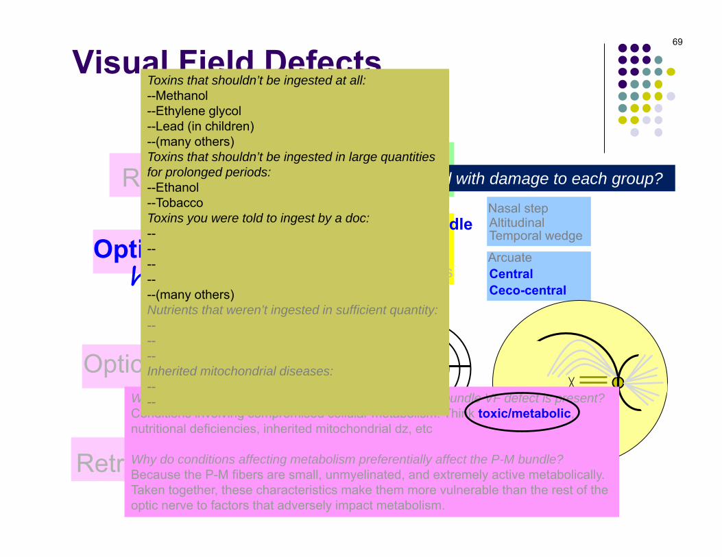
Retina
Optic nervehead
Optic chiasm
Retrochiasmal
Papillomacular bundle
Nasal radiating fibers
Clinically obvious dzClinically subtle dz
Visual Field Defects69
Arcuate fibers ArcuateCentralCeco-central
Nasal stepAltitudinalTemporal wedge
Which of these VF defects are associated with damage to each group?
Which sorts of optic neuropathy are implicated if a P-M bundle VF defect is present? Conditions involving compromised cellular metabolism: Think toxic/metabolic, nutritional deficiencies, inherited mitochondrial dz, etc
Why do conditions affecting metabolism preferentially affect the P-M bundle?Because the P-M fibers are small, unmyelinated, and extremely active metabolically. Taken together, these characteristics make them more vulnerable than the rest of the optic nerve to factors that adversely impact metabolism.
Toxins that shouldn’t be ingested at all:--Methanol--Ethylene glycol--Lead (in children)--(many others)Toxins that shouldn’t be ingested in large quantities for prolonged periods:--Ethanol--TobaccoToxins you were told to ingest by a doc:----------(many others)Nutrients that weren’t ingested in sufficient quantity:------Inherited mitochondrial diseases:----

Retina
Optic nervehead
Optic chiasm
Retrochiasmal
Papillomacular bundle
Nasal radiating fibers
Clinically obvious dzClinically subtle dz
Visual Field Defects70
Arcuate fibers ArcuateCentralCeco-central
Nasal stepAltitudinalTemporal wedge
Which of these VF defects are associated with damage to each group?
Which sorts of optic neuropathy are implicated if a P-M bundle VF defect is present? Conditions involving compromised cellular metabolism: Think toxic/metabolic, nutritional deficiencies, inherited mitochondrial dz, etc
Why do conditions affecting metabolism preferentially affect the P-M bundle?Because the P-M fibers are small, unmyelinated, and extremely active metabolically. Taken together, these characteristics make them more vulnerable than the rest of the optic nerve to factors that adversely impact metabolism.
Toxins that shouldn’t be ingested at all:--Methanol--Ethylene glycol--Lead (in children)--(many others)Toxins that shouldn’t be ingested in large quantities for prolonged periods:--Ethanol--TobaccoToxins you were told to ingest by a doc:--Amiodarone--Ethambutol--Isoniazid--Linezolid--(many others)Nutrients that weren’t ingested in sufficient quantity:------Inherited mitochondrial diseases:----

Retina
Optic nervehead
Optic chiasm
Retrochiasmal
Papillomacular bundle
Nasal radiating fibers
Clinically obvious dzClinically subtle dz
Visual Field Defects71
Arcuate fibers ArcuateCentralCeco-central
Nasal stepAltitudinalTemporal wedge
Which of these VF defects are associated with damage to each group?
Which sorts of optic neuropathy are implicated if a P-M bundle VF defect is present? Conditions involving compromised cellular metabolism: Think toxic/metabolic, nutritional deficiencies, inherited mitochondrial dz, etc
Why do conditions affecting metabolism preferentially affect the P-M bundle?Because the P-M fibers are small, unmyelinated, and extremely active metabolically. Taken together, these characteristics make them more vulnerable than the rest of the optic nerve to factors that adversely impact metabolism.
Toxins that shouldn’t be ingested at all:--Methanol--Ethylene glycol--Lead (in children)--(many others)Toxins that shouldn’t be ingested in large quantities for prolonged periods:--Ethanol--TobaccoToxins you were told to ingest by a doc:--Amiodarone--Ethambutol--Isoniazid--Linezolid--(many others)Nutrients that weren’t ingested in sufficient quantity:------Inherited mitochondrial diseases:----

Retina
Optic nervehead
Optic chiasm
Retrochiasmal
Papillomacular bundle
Nasal radiating fibers
Clinically obvious dzClinically subtle dz
Visual Field Defects72
Arcuate fibers ArcuateCentralCeco-central
Nasal stepAltitudinalTemporal wedge
Which of these VF defects are associated with damage to each group?
Which sorts of optic neuropathy are implicated if a P-M bundle VF defect is present? Conditions involving compromised cellular metabolism: Think toxic/metabolic, nutritional deficiencies, inherited mitochondrial dz, etc
Why do conditions affecting metabolism preferentially affect the P-M bundle?Because the P-M fibers are small, unmyelinated, and extremely active metabolically. Taken together, these characteristics make them more vulnerable than the rest of the optic nerve to factors that adversely impact metabolism.
Toxins that shouldn’t be ingested at all:--Methanol--Ethylene glycol--Lead (in children)--(many others)Toxins that shouldn’t be ingested in large quantities for prolonged periods:--Ethanol--TobaccoToxins you were told to ingest by a doc:--Amiodarone--Ethambutol--Isoniazid--Linezolid--(many others)Nutrients that weren’t ingested in sufficient quantity:--Vitamin B12--Folate--ThiamineInherited mitochondrial diseases:----

Retina
Optic nervehead
Optic chiasm
Retrochiasmal
Papillomacular bundle
Nasal radiating fibers
Clinically obvious dzClinically subtle dz
Visual Field Defects73
Arcuate fibers ArcuateCentralCeco-central
Nasal stepAltitudinalTemporal wedge
Which of these VF defects are associated with damage to each group?
Which sorts of optic neuropathy are implicated if a P-M bundle VF defect is present? Conditions involving compromised cellular metabolism: Think toxic/metabolic, nutritional deficiencies, inherited mitochondrial dz, etc
Why do conditions affecting metabolism preferentially affect the P-M bundle?Because the P-M fibers are small, unmyelinated, and extremely active metabolically. Taken together, these characteristics make them more vulnerable than the rest of the optic nerve to factors that adversely impact metabolism.
Toxins that shouldn’t be ingested at all:--Methanol--Ethylene glycol--Lead (in children)--(many others)Toxins that shouldn’t be ingested in large quantities for prolonged periods:--Ethanol--TobaccoToxins you were told to ingest by a doc:--Amiodarone--Ethambutol--Isoniazid--Linezolid--(many others)Nutrients that weren’t ingested in sufficient quantity:--Vitamin B12--Folate--ThiamineInherited mitochondrial diseases:----

Retina
Optic nervehead
Optic chiasm
Retrochiasmal
Papillomacular bundle
Nasal radiating fibers
Clinically obvious dzClinically subtle dz
Visual Field Defects74
Arcuate fibers ArcuateCentralCeco-central
Nasal stepAltitudinalTemporal wedge
Which of these VF defects are associated with damage to each group?
Which sorts of optic neuropathy are implicated if a P-M bundle VF defect is present? Conditions involving compromised cellular metabolism: Think toxic/metabolic, nutritional deficiencies, inherited mitochondrial dz, etc
Why do conditions affecting metabolism preferentially affect the P-M bundle?Because the P-M fibers are small, unmyelinated, and extremely active metabolically. Taken together, these characteristics make them more vulnerable than the rest of the optic nerve to factors that adversely impact metabolism.
Toxins that shouldn’t be ingested at all:--Methanol--Ethylene glycol--Lead (in children)--(many others)Toxins that shouldn’t be ingested in large quantities for prolonged periods:--Ethanol--TobaccoToxins you were told to ingest by a doc:--Amiodarone--Ethambutol--Isoniazid--Linezolid--(many others)Nutrients that weren’t ingested in sufficient quantity:--Vitamin B12--Folate--ThiamineInherited mitochondrial diseases:--Leber’s hereditary optic neuropathy--Autosomal dominant optic atrophy

Retina
Optic nervehead
Optic chiasm
Retrochiasmal
Papillomacular bundle
Nasal radiating fibers
Clinically obvious dzClinically subtle dz
Visual Field Defects75
Arcuate fibers ArcuateCentralCeco-central
Nasal stepAltitudinalTemporal wedge
Which of these VF defects are associated with damage to each group?
Which sorts of optic neuropathy are implicated if a P-M bundle VF defect is present? Conditions involving compromised cellular metabolism: Think toxic/metabolic, nutritional deficiencies, inherited mitochondrial dz, etc
Why do conditions affecting metabolism preferentially affect the P-M bundle?Because the P-M fibers are small, unmyelinated, and extremely active metabolically. Taken together, these characteristics make them more vulnerable than the rest of the optic nerve to factors that adversely impact metabolism.
In addition to central/ceco-central VF defects, what other aspects of visual function are invariably degraded by pathology affecting the P-M bundle?----

Retina
Optic nervehead
Optic chiasm
Retrochiasmal
Papillomacular bundle
Nasal radiating fibers
Clinically obvious dzClinically subtle dz
Visual Field Defects76
Arcuate fibers ArcuateCentralCeco-central
Nasal stepAltitudinalTemporal wedge
Which of these VF defects are associated with damage to each group?
Which sorts of optic neuropathy are implicated if a P-M bundle VF defect is present? Conditions involving compromised cellular metabolism: Think toxic/metabolic, nutritional deficiencies, inherited mitochondrial dz, etc
Why do conditions affecting metabolism preferentially affect the P-M bundle?Because the P-M fibers are small, unmyelinated, and extremely active metabolically. Taken together, these characteristics make them more vulnerable than the rest of the optic nerve to factors that adversely impact metabolism.
In addition to central/ceco-central VF defects, what other aspects of visual function are invariably degraded by pathology affecting the P-M bundle?--Visual acuity*--Color vision *Which makes sense—after all, a central VF defect is present

Retina
Optic nervehead
Optic chiasm
Retrochiasmal
Papillomacular bundle
Nasal radiating fibers
Clinically obvious dzClinically subtle dz
Visual Field Defects77
Arcuate fibers ArcuateCentralCeco-central
Nasal stepAltitudinalTemporal wedge
Which of these VF defects are associated with damage to each group?
Which sorts of optic neuropathy are implicated if a P-M bundle VF defect is present? Conditions involving compromised cellular metabolism: Think toxic/metabolic, nutritional deficiencies, inherited mitochondrial dz, etc
Why do conditions affecting metabolism preferentially affect the P-M bundle?Because the P-M fibers are small, unmyelinated, and extremely active metabolically. Taken together, these characteristics make them more vulnerable than the rest of the optic nerve to factors that adversely impact metabolism.
In addition to central/ceco-central VF defects, what other aspects of visual function are invariably degraded by pathology affecting the P-M bundle?--Visual acuity*--Color vision *Which makes sense—after all, a central VF defect is present
For more on PMB-related optic neuropathy, see slide-set N9

Retina
Optic nervehead
Optic chiasm
Retrochiasmal
Papillomacular bundle
Nasal radiating fibers
Clinically obvious dzClinically subtle dz
Visual Field Defects78
Arcuate fibers ArcuateCentralCeco-central
Nasal stepAltitudinalTemporal wedge ?
?
Which of these VF defects are associated with damage to each group?
Retina
Optic nervehead
Optic chiasm
Retrochiasmal
Papillomacular bundle
Nasal radiating fibers
Clinically obvious dzClinically subtle dz
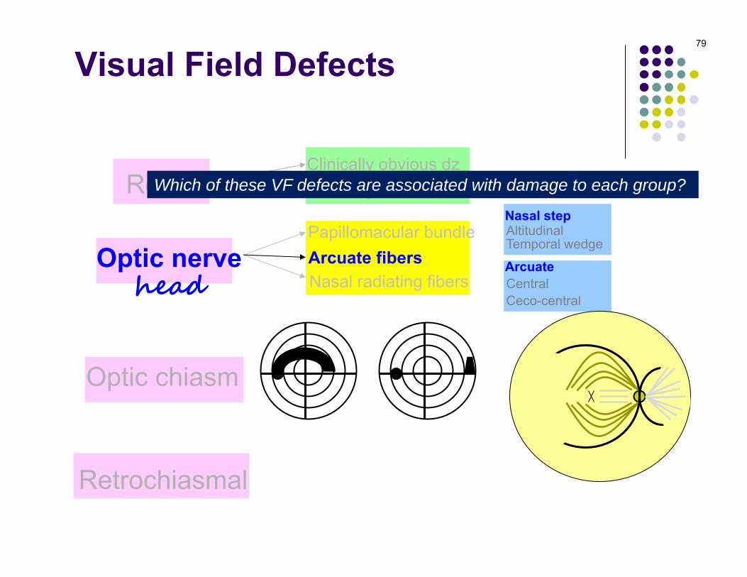
Visual Field Defects79
Arcuate fibers ArcuateCentralCeco-central
Nasal stepAltitudinalTemporal wedge
Which of these VF defects are associated with damage to each group?
Retina
Optic nervehead
Optic chiasm
Retrochiasmal
Papillomacular bundle
Nasal radiating fibers
Clinically obvious dzClinically subtle dz
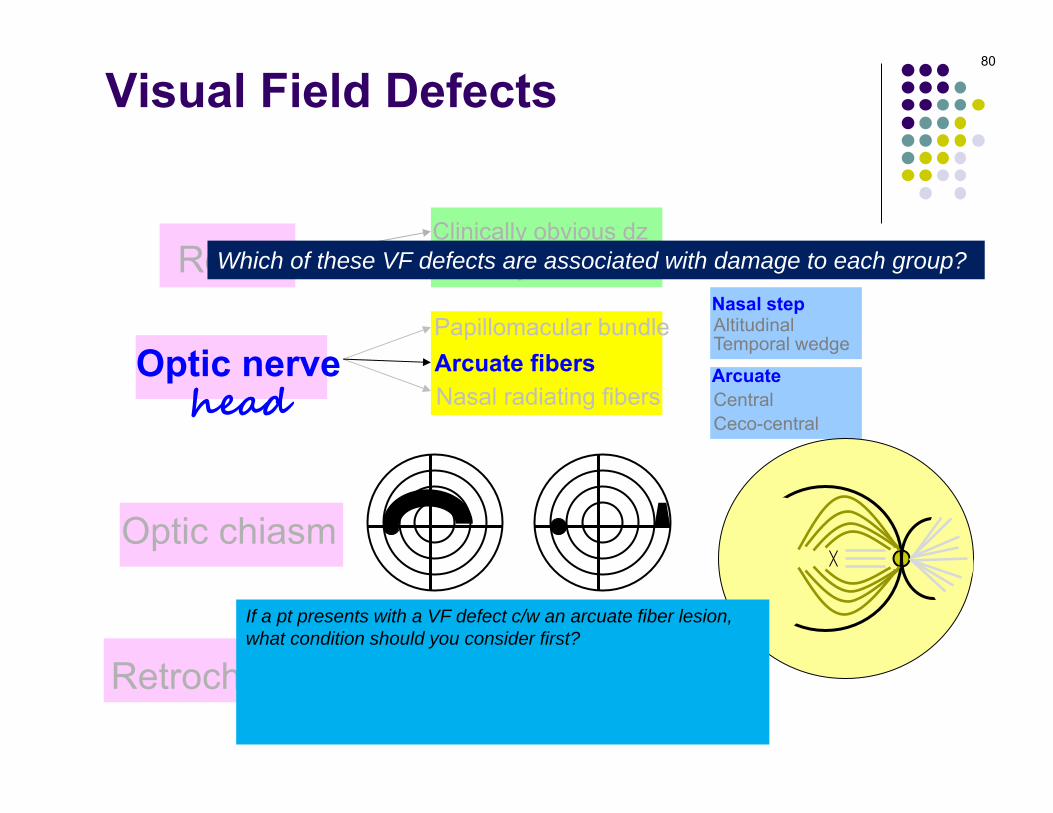
Visual Field Defects80
Arcuate fibers ArcuateCentralCeco-central
Nasal stepAltitudinalTemporal wedge
Which of these VF defects are associated with damage to each group?
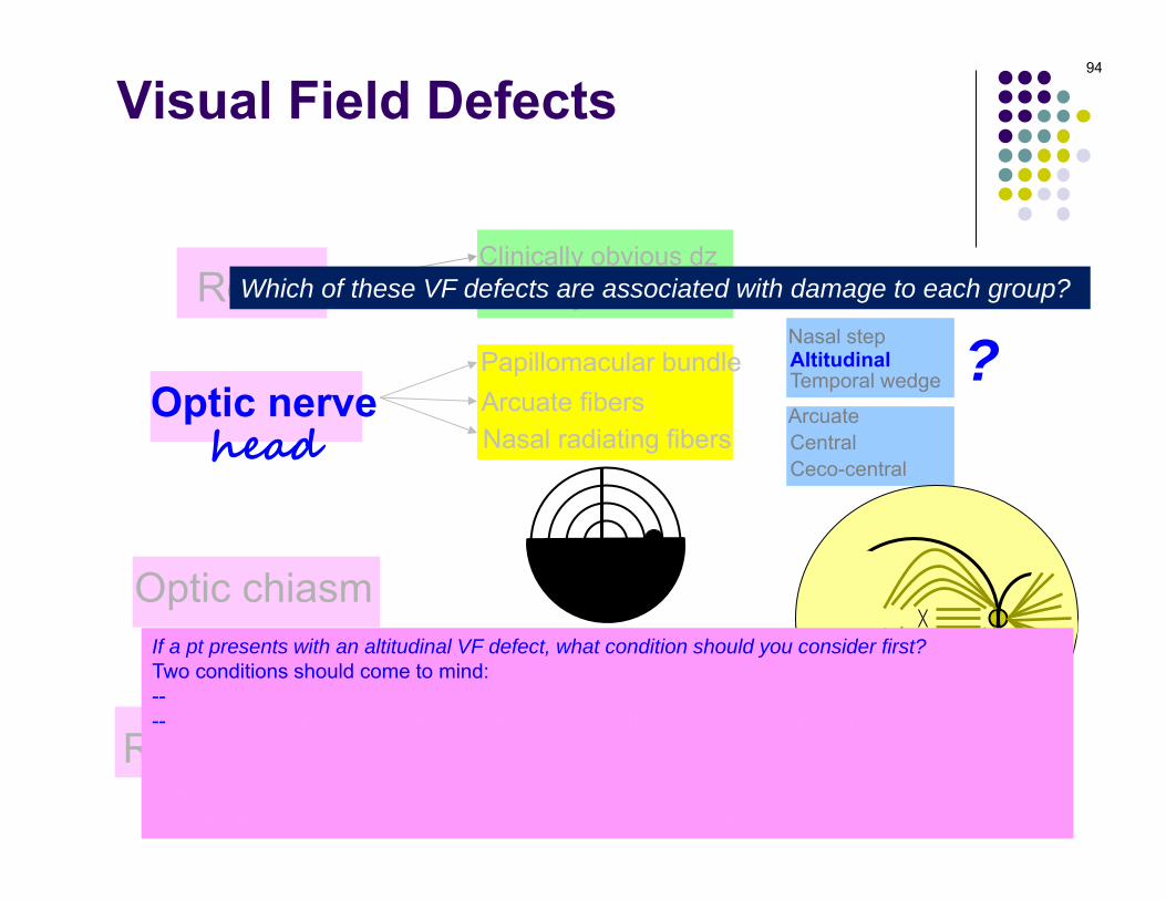
If a pt presents with a VF defect c/w an arcuate fiber lesion, what condition should you consider first? Glaucoma
Why does glaucoma preferentially damage arcuate fibers?It’s unclear at this time

Retina
Optic nervehead
Optic chiasm
Retrochiasmal
Papillomacular bundle
Nasal radiating fibers
Clinically obvious dzClinically subtle dz
Visual Field Defects81
Arcuate fibers ArcuateCentralCeco-central
Nasal stepAltitudinalTemporal wedge
Which of these VF defects are associated with damage to each group?
If a pt presents with a VF defect c/w an arcuate fiber lesion, what condition should you consider first? Glaucoma
Why does glaucoma preferentially damage arcuate fibers?It’s unclear at this time

Retina
Optic nervehead
Optic chiasm
Retrochiasmal
Papillomacular bundle
Nasal radiating fibers
Clinically obvious dzClinically subtle dz
Visual Field Defects82
Arcuate fibers ArcuateCentralCeco-central
Nasal stepAltitudinalTemporal wedge
Which of these VF defects are associated with damage to each group?
If a pt presents with a VF defect c/w an arcuate fiber lesion, what condition should you consider first? Glaucoma
Why does glaucoma preferentially damage arcuate fibers?It’s unclear at this time

Retina
Optic nervehead
Optic chiasm
Retrochiasmal
Papillomacular bundle
Nasal radiating fibers
Clinically obvious dzClinically subtle dz
Visual Field Defects83
Arcuate fibers ArcuateCentralCeco-central
Nasal stepAltitudinalTemporal wedge
Which of these VF defects are associated with damage to each group?
If a pt presents with a VF defect c/w an arcuate fiber lesion, what condition should you consider first? Glaucoma
Why does glaucoma preferentially damage arcuate fibers?It’s unclear at this time

Retina
Optic nervehead
Optic chiasm
Retrochiasmal
Papillomacular bundle
Nasal radiating fibers
Clinically obvious dzClinically subtle dz
Visual Field Defects84
Arcuate fibers ArcuateCentralCeco-central
Nasal stepAltitudinalTemporal wedge
Which of these VF defects are associated with damage to each group?
Compare the distribution of arcuate-fiber defects with those associated with a P-M bundle dysfunction. What important difference do you see?Unlike P-M defects, arcuate fiber bundle defects do not cross (ie, they ‘respect’) the horizontal midline
Why not?Because fibers on the temporal side of the ONH approach, but do not cross, the horizontal midline. The arcuate fibers arc around the P-M bundle, and meet along a horizontal demarcation line. Thus, damage to these fibers always result in VF defects that are limited to either the superior or the inferior portion of the field.
What is this ‘horizontal demarcation line’ called?The horizontal raphe

Retina
Optic nervehead
Optic chiasm
Retrochiasmal
Papillomacular bundle
Nasal radiating fibers
Clinically obvious dzClinically subtle dz
Visual Field Defects85
Arcuate fibers ArcuateCentralCeco-central
Nasal stepAltitudinalTemporal wedge
Which of these VF defects are associated with damage to each group?
Compare the distribution of arcuate-fiber defects with those associated with a P-M bundle dysfunction. What important difference do you see?Unlike P-M defects, arcuate fiber bundle defects do not cross (ie, they ‘respect’) the horizontal midline
Why not?Because fibers on the temporal side of the ONH approach, but do not cross, the horizontal midline. The arcuate fibers arc around the P-M bundle, and meet along a horizontal demarcation line. Thus, damage to these fibers always result in VF defects that are limited to either the superior or the inferior portion of the field.
What is this ‘horizontal demarcation line’ called?The horizontal raphe

Retina
Optic nervehead
Optic chiasm
Retrochiasmal
Papillomacular bundle
Nasal radiating fibers
Clinically obvious dzClinically subtle dz
Visual Field Defects86
Arcuate fibers ArcuateCentralCeco-central
Nasal stepAltitudinalTemporal wedge
Which of these VF defects are associated with damage to each group?
Compare the distribution of arcuate-fiber defects with those associated with a P-M bundle dysfunction. What important difference do you see?Unlike P-M defects, arcuate fiber bundle defects do not cross (ie, they ‘respect’) the horizontal midline
Why not?Because fibers on the temporal side of the ONH approach, but do not cross, the horizontal midline. The arcuate fibers arc around the P-M bundle, and meet along a horizontal demarcation line. Thus, damage to these fibers always result in VF defects that are limited to either the superior or the inferior portion of the field.
What is this ‘horizontal demarcation line’ called?The horizontal raphe

Retina
Optic nervehead
Optic chiasm
Retrochiasmal
Papillomacular bundle
Nasal radiating fibers
Clinically obvious dzClinically subtle dz
Visual Field Defects87
Arcuate fibers ArcuateCentralCeco-central
Nasal stepAltitudinalTemporal wedge
Which of these VF defects are associated with damage to each group?
Compare the distribution of arcuate-fiber defects with those associated with a P-M bundle dysfunction. What important difference do you see?Unlike P-M defects, arcuate fiber bundle defects do not cross (ie, they ‘respect’) the horizontal midline
Why not?Because fibers on the temporal side of the ONH approach, but do not cross, the horizontal midline. The arcuate fibers arc around the P-M bundle, and meet along a horizontal demarcation line. Thus, damage to these fibers always result in VF defects that are limited to either the superior or the inferior portion of the field.
What is this ‘horizontal demarcation line’ called?The horizontal raphe

Retina
Optic nervehead
Optic chiasm
Retrochiasmal
Papillomacular bundle
Nasal radiating fibers
Clinically obvious dzClinically subtle dz
Visual Field Defects88
Arcuate fibers ArcuateCentralCeco-central
Nasal stepAltitudinalTemporal wedge
Which of these VF defects are associated with damage to each group?
Compare the distribution of arcuate-fiber defects with those associated with a P-M bundle dysfunction. What important difference do you see?Unlike P-M defects, arcuate fiber bundle defects do not cross (ie, they ‘respect’) the horizontal midline
Why not?Because fibers on the temporal side of the ONH approach, but do not cross, the horizontal midline. The arcuate fibers arc around the P-M bundle, and meet along a horizontal demarcation line. Thus, damage to these fibers always result in VF defects that are limited to either the superior or the inferior portion of the field.
What is this ‘horizontal demarcation line’ called?The horizontal raphe
Retina
Optic nervehead
Optic chiasm
Retrochiasmal
Papillomacular bundle
Nasal radiating fibers
Clinically obvious dzClinically subtle dz
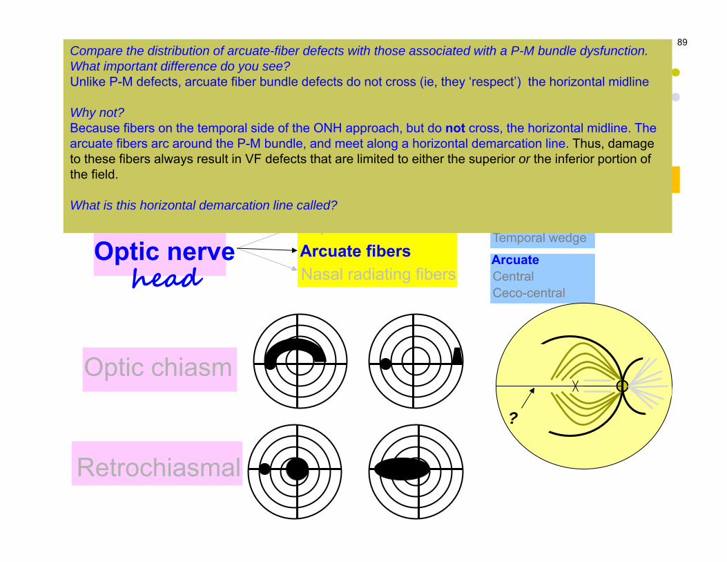
Visual Field Defects89
Arcuate fibers ArcuateCentralCeco-central
Nasal stepAltitudinalTemporal wedge
Which of these VF defects are associated with damage to each group?
?
Compare the distribution of arcuate-fiber defects with those associated with a P-M bundle dysfunction. What important difference do you see?Unlike P-M defects, arcuate fiber bundle defects do not cross (ie, they ‘respect’) the horizontal midline
Why not?Because fibers on the temporal side of the ONH approach, but do not cross, the horizontal midline. The arcuate fibers arc around the P-M bundle, and meet along a horizontal demarcation line. Thus, damage to these fibers always result in VF defects that are limited to either the superior or the inferior portion of the field.
What is this horizontal demarcation line called?The horizontal raphe

Retina
Optic nervehead
Optic chiasm
Retrochiasmal
Papillomacular bundle
Nasal radiating fibers
Clinically obvious dzClinically subtle dz
Visual Field Defects90
Arcuate fibers ArcuateCentralCeco-central
Nasal stepAltitudinalTemporal wedge
Which of these VF defects are associated with damage to each group?
Compare the distribution of arcuate-fiber defects with those associated with a P-M bundle dysfunction. What important difference do you see?Unlike P-M defects, arcuate fiber bundle defects do not cross (ie, they ‘respect’) the horizontal midline
Why not?Because fibers on the temporal side of the ONH approach, but do not cross, the horizontal midline. The arcuate fibers arc around the P-M bundle, and meet along a horizontal demarcation line. Thus, damage to these fibers always result in VF defects that are limited to either the superior or the inferior portion of the field.
What is this horizontal demarcation line called?The horizontal raphe
‘Horizontal raphe’

Retina
Optic nervehead
Optic chiasm
Retrochiasmal
Papillomacular bundle
Nasal radiating fibers
Clinically obvious dzClinically subtle dz
Visual Field Defects91
Arcuate fibers ArcuateCentralCeco-central
Nasal stepAltitudinalTemporal wedge ?
?
Which of these VF defects are associated with damage to each group?

Retina
Optic nervehead
Optic chiasm
Retrochiasmal
Papillomacular bundle
Nasal radiating fibers
Clinically obvious dzClinically subtle dz
Visual Field Defects92
Arcuate fibers ArcuateCentralCeco-central
Nasal stepAltitudinalTemporal wedge
Which of these VF defects are associated with damage to each group?

Retina
Optic nervehead
Optic chiasm
Retrochiasmal
Papillomacular bundle
Nasal radiating fibers
Clinically obvious dzClinically subtle dz
Visual Field Defects93
Arcuate fibers ArcuateCentralCeco-central
Nasal stepAltitudinalTemporal wedge
Which of these VF defects are associated with damage to each group?
?
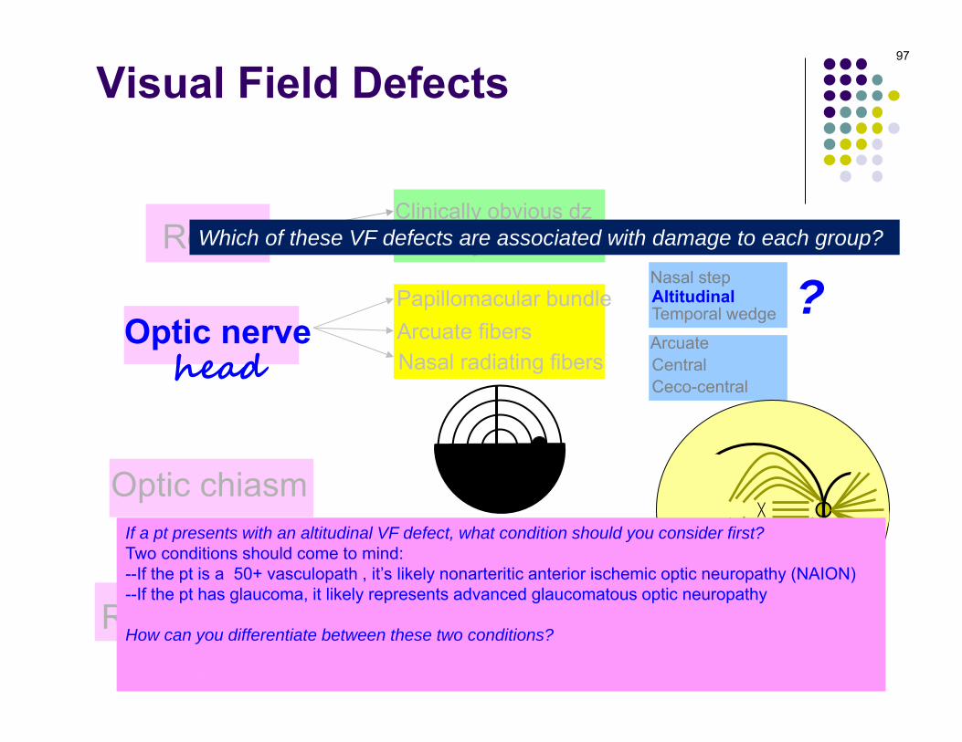
If a pt presents with an altitudinal VF defect, what condition should you consider first? Two conditions should come to mind:--If the pt is a 50+ vasculopath , it’s likely nonarteritic anterior ischemic optic neuropathy (NAION)--If the pt has glaucoma, it likely represents advanced glaucomatous optic neuropathy
How can you differentiate between these two conditions?There are a number of ways, but the most straightforward would be to inspect the ONH, which will be edematous in NAION, and severely cupped in advanced glaucoma
Retina
Optic nervehead
Optic chiasm
Retrochiasmal
Papillomacular bundle
Nasal radiating fibers
Clinically obvious dzClinically subtle dz
Visual Field Defects94
Arcuate fibers ArcuateCentralCeco-central
Nasal stepAltitudinalTemporal wedge
Which of these VF defects are associated with damage to each group?
?
If a pt presents with an altitudinal VF defect, what condition should you consider first? Two conditions should come to mind:----If the pt has glaucoma, it likely represents advanced glaucomatous optic neuropathy
How can you differentiate between these two conditions?There are a number of ways, but the most straightforward would be to inspect the ONH, which will be edematous in NAION, and severely cupped in advanced glaucoma

Retina
Optic nervehead
Optic chiasm
Retrochiasmal
Papillomacular bundle
Nasal radiating fibers
Clinically obvious dzClinically subtle dz
Visual Field Defects95
Arcuate fibers ArcuateCentralCeco-central
Nasal stepAltitudinalTemporal wedge
Which of these VF defects are associated with damage to each group?
?
If a pt presents with an altitudinal VF defect, what condition should you consider first? Two conditions should come to mind:--If the pt is a 50+ vasculopath , it’s likely nonarteritic anterior ischemic optic neuropathy (NAION)--If the pt has glaucoma, it likely represents advanced glaucomatous optic neuropathy
How can you differentiate between these two conditions?There are a number of ways, but the most straightforward would be to inspect the ONH, which will be edematous in NAION, and severely cupped in advanced glaucoma
age and condition

Retina
Optic nervehead
Optic chiasm
Retrochiasmal
Papillomacular bundle
Nasal radiating fibers
Clinically obvious dzClinically subtle dz
Visual Field Defects96
Arcuate fibers ArcuateCentralCeco-central
Nasal stepAltitudinalTemporal wedge
Which of these VF defects are associated with damage to each group?
?
If a pt presents with an altitudinal VF defect, what condition should you consider first? Two conditions should come to mind:--If the pt is a 50+ vasculopath , it’s likely nonarteritic anterior ischemic optic neuropathy (NAION)--If the pt has glaucoma, it likely represents advanced glaucomatous optic neuropathy
How can you differentiate between these two conditions?There are a number of ways, but the most straightforward would be to inspect the ONH, which will be edematous in NAION, and severely cupped in advanced glaucoma
Retina
Optic nervehead
Optic chiasm
Retrochiasmal
Papillomacular bundle
Nasal radiating fibers
Clinically obvious dzClinically subtle dz
Visual Field Defects97
Arcuate fibers ArcuateCentralCeco-central
Nasal stepAltitudinalTemporal wedge
Which of these VF defects are associated with damage to each group?
?
If a pt presents with an altitudinal VF defect, what condition should you consider first? Two conditions should come to mind:--If the pt is a 50+ vasculopath , it’s likely nonarteritic anterior ischemic optic neuropathy (NAION)--If the pt has glaucoma, it likely represents advanced glaucomatous optic neuropathy
How can you differentiate between these two conditions?There are a number of ways, but the most straightforward would be to inspect the ONH, which will be edematous in NAION, and severely cupped in advanced glaucoma

Retina
Optic nervehead
Optic chiasm
Retrochiasmal
Papillomacular bundle
Nasal radiating fibers
Clinically obvious dzClinically subtle dz
Visual Field Defects98
Arcuate fibers ArcuateCentralCeco-central
Nasal stepAltitudinalTemporal wedge
Which of these VF defects are associated with damage to each group?
?
If a pt presents with an altitudinal VF defect, what condition should you consider first? Two conditions should come to mind:--If the pt is a 50+ vasculopath , it’s likely nonarteritic anterior ischemic optic neuropathy (NAION)--If the pt has glaucoma, it likely represents advanced glaucomatous optic neuropathy
How can you differentiate between these two conditions?There are a number of ways, but the most straightforward would be to inspect the ONH, which will be edematous in NAION, and severely cupped in advanced glaucomaone word two words

Retina
Optic nervehead
Optic chiasm
Retrochiasmal
Papillomacular bundle
Nasal radiating fibers
Clinically obvious dzClinically subtle dz
Visual Field Defects99
Arcuate fibers ArcuateCentralCeco-central
Nasal stepAltitudinalTemporal wedge
Which of these VF defects are associated with damage to each group?
If a pt presents with an altitudinal VF defect, what condition should you consider first? Two conditions should come to mind:--If the pt is a 50+ vasculopath , it’s likely nonarteritic anterior ischemic optic neuropathy (NAION)--If the pt has glaucoma, it likely represents advanced glaucomatous optic neuropathy
How can you differentiate between these two conditions?There are a number of ways, but the most straightforward would be to inspect the ONH, which will be edematous in NAION, and severely cupped in advanced glaucoma
?

Retina
Optic nerve
Optic chiasm
Retrochiasmal
Depressions
Scotomas
Clinically obvious dzClinically subtle dz
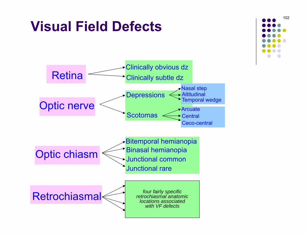
Visual Field Defects
ArcuateCentralCeco-central
Nasal stepAltitudinalTemporal wedge
Bitemporal hemianopiaBinasal hemianopiaJunctional commonJunctional rare
four very specifictypes of chiasmal
VF defects
100

Retina
Optic nerve
Optic chiasm
Retrochiasmal
Depressions
Scotomas
Clinically obvious dzClinically subtle dz
Visual Field Defects
ArcuateCentralCeco-central
Nasal stepAltitudinalTemporal wedge
Bitemporal hemianopiaBinasal hemianopiaJunctional commonJunctional rare
101
Retina
Optic nerve
Optic chiasm
Retrochiasmal
Depressions
Scotomas
Clinically obvious dzClinically subtle dz
Visual Field Defects
ArcuateCentralCeco-central
Nasal stepAltitudinalTemporal wedge
Bitemporal hemianopiaBinasal hemianopiaJunctional commonJunctional rare
Optic tractLGNOptic radiationsOccipital cortex
four fairly specificretrochiasmal anatomic
locations associatedwith VF defects
102

Retina
Optic nerve
Optic chiasm
Retrochiasmal
Depressions
Scotomas
Clinically obvious dzClinically subtle dz
Visual Field Defects
ArcuateCentralCeco-central
Nasal stepAltitudinalTemporal wedge
Bitemporal hemianopiaBinasal hemianopiaJunctional commonJunctional rare
Optic tractLGNOptic radiationsOccipital cortex
103

Forget all of these specific VF findingsfor just a minute…In the most generalof terms, what can we say about VF defects associated with lesions ineach of these locations?
Visual Field Defects
Retina
Optic nerve
Optic chiasm
Retrochiasmal
Bitemporal hemianopiaBinasal hemianopiaJunctional commonJunctional rare
Optic tractLGNOptic radiationsOccipital cortex
ArcuateCentralCeco-central
Nasal stepAltitudinalTemporal wedge
Depressions
Scotomas
Clinically obvious dzClinically subtle dz
Forget all of these specific VF findingsfor just a minute…In the most generalof terms, what can we say about VF defects associated with lesions ineach of these locations?
104

Forget all of these specific VF findingsfor just a minute…In the most generalof terms, what can we say about VF defects associated with lesions ineach of these locations?
Visual Field Defects
Retina
Optic nerve
Optic chiasm
Retrochiasmal
Bitemporal hemianopiaBinasal hemianopiaJunctional commonJunctional rare
Optic tractLGNOptic radiationsOccipital cortex
ArcuateCentralCeco-central
Nasal stepAltitudinalTemporal wedge
Depressions
Scotomas
Clinically obvious dzClinically subtle dz
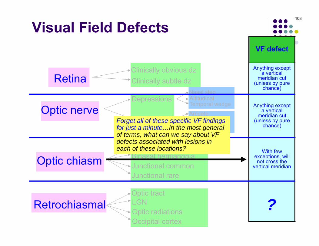
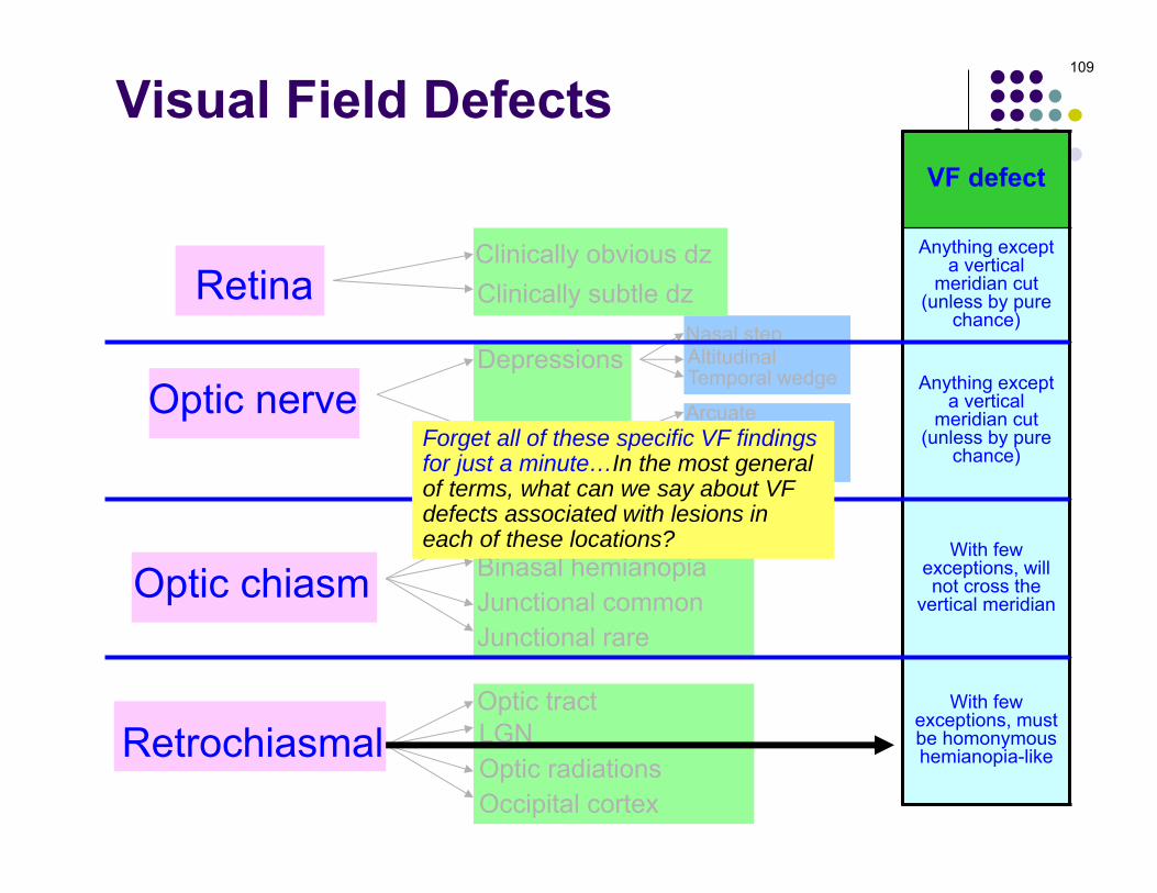
VF defect
Forget all of these specific VF findingsfor just a minute…In the most generalof terms, what can we say about VF defects associated with lesions ineach of these locations?
?
?
?
?
105
(Start here)
Visual Field Defects
Retina
Optic nerve
Optic chiasm
Retrochiasmal
Bitemporal hemianopiaBinasal hemianopiaJunctional commonJunctional rare
Optic tractLGNOptic radiationsOccipital cortex
ArcuateCentralCeco-central
Nasal stepAltitudinalTemporal wedge
Depressions
Scotomas
Clinically obvious dzClinically subtle dz
VF defect
Anything except a vertical
meridian cut (unless by pure
chance)
Anything except a vertical
meridian cut (unless by pure
chance)
With few exceptions, will not cross the
vertical meridian
With few exceptions, must be homonymous hemianopia-like
Forget all of these specific VF findingsfor just a minute…In the most generalof terms, what can we say about VF defects associated with lesions ineach of these locations?
?
?
?
106

Visual Field Defects
Retina
Optic nerve
Optic chiasm
Retrochiasmal
Bitemporal hemianopiaBinasal hemianopiaJunctional commonJunctional rare
Optic tractLGNOptic radiationsOccipital cortex
ArcuateCentralCeco-central
Nasal stepAltitudinalTemporal wedge
Depressions
Scotomas
Clinically obvious dzClinically subtle dz
VF defect
Anything except a vertical
meridian cut (unless by pure
chance)
Anything except a vertical
meridian cut (unless by pure
chance)
With few exceptions, will not cross the
vertical meridian
With few exceptions, must be homonymous hemianopia-like
Forget all of these specific VF findingsfor just a minute…In the most generalof terms, what can we say about VF defects associated with lesions ineach of these locations?
?
?
107
Visual Field Defects
Retina
Optic nerve
Optic chiasm
Retrochiasmal
Bitemporal hemianopiaBinasal hemianopiaJunctional commonJunctional rare
Optic tractLGNOptic radiationsOccipital cortex
ArcuateCentralCeco-central
Nasal stepAltitudinalTemporal wedge
Depressions
Scotomas
Clinically obvious dzClinically subtle dz
VF defect
Anything except a vertical
meridian cut (unless by pure
chance)
Anything except a vertical
meridian cut (unless by pure
chance)
With few exceptions, will not cross the
vertical meridian
With few exceptions, must be homonymous hemianopia-like
Forget all of these specific VF findingsfor just a minute…In the most generalof terms, what can we say about VF defects associated with lesions ineach of these locations?
?
108
Visual Field Defects
Retina
Optic nerve
Optic chiasm
Retrochiasmal
Bitemporal hemianopiaBinasal hemianopiaJunctional commonJunctional rare
Optic tractLGNOptic radiationsOccipital cortex
ArcuateCentralCeco-central
Nasal stepAltitudinalTemporal wedge
Depressions
Scotomas
Clinically obvious dzClinically subtle dz
VF defect
Anything except a vertical
meridian cut (unless by pure
chance)
Anything except a vertical
meridian cut (unless by pure
chance)
With few exceptions, will not cross the
vertical meridian
With few exceptions, must be homonymous hemianopia-like
Forget all of these specific VF findingsfor just a minute…In the most generalof terms, what can we say about VF defects associated with lesions ineach of these locations?
109

Visual Field Defects
Retina
Optic nerve
Optic chiasm
Retrochiasmal
Bitemporal hemianopiaBinasal hemianopiaJunctional commonJunctional rare
Optic tractLGNOptic radiationsOccipital cortex
ArcuateCentralCeco-central
Nasal stepAltitudinalTemporal wedge
Depressions
Scotomas
Clinically obvious dzClinically subtle dz
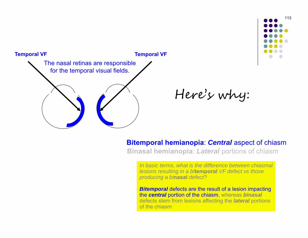
In basic terms, what is the difference between chiasmallesions resulting in a bitemporal VF defect vs thoseproducing a binasal defect?
Bitemporal defects are the result of a lesion impactingthe central portion of the chiasm, whereas binasaldefects stem from lesions affecting the lateral portionsof the chiasm
110

Visual Field Defects
Retina
Optic nerve
Optic chiasm
Retrochiasmal
Bitemporal hemianopia: Central aspect of chiasm
Junctional commonJunctional rare
Optic tractLGNOptic radiationsOccipital cortex
ArcuateCentralCeco-central
Nasal stepAltitudinalTemporal wedge
Depressions
Scotomas
Clinically obvious dzClinically subtle dz
In basic terms, what is the difference between chiasmallesions resulting in a bitemporal VF defect vs thoseproducing a binasal defect?
Bitemporal defects are the result of a lesion impactingthe central portion of the chiasm, whereas binasaldefects stem from lesions affecting the lateral portionsof the chiasm
Binasal hemianopia: Lateral portions of chiasm
111

112
In basic terms, what is the difference between chiasmallesions resulting in a bitemporal VF defect vs thoseproducing a binasal defect?
Bitemporal defects are the result of a lesion impactingthe central portion of the chiasm, whereas binasaldefects stem from lesions affecting the lateral portionsof the chiasm
Bitemporal hemianopia: Central aspect of chiasmBinasal hemianopia: Lateral portions of chiasm
Here’s why:
113
The nasal retinas are responsible for the temporal visual fields.
In basic terms, what is the difference between chiasmallesions resulting in a bitemporal VF defect vs thoseproducing a binasal defect?
Bitemporal defects are the result of a lesion impactingthe central portion of the chiasm, whereas binasaldefects stem from lesions affecting the lateral portionsof the chiasm
Bitemporal hemianopia: Central aspect of chiasmBinasal hemianopia: Lateral portions of chiasm
Temporal VF
Here’s why:
Temporal VF

114
Optic
Chiasm
Fibers originating in the nasal retinas cross at the chiasm.
The nasal retinas are responsible for the temporal visual fields.
In basic terms, what is the difference between chiasmallesions resulting in a bitemporal VF defect vs thoseproducing a binasal defect?
Bitemporal defects are the result of a lesion impactingthe central portion of the chiasm, whereas binasaldefects stem from lesions affecting the lateral portionsof the chiasm
Bitemporal hemianopia: Central aspect of chiasmBinasal hemianopia: Lateral portions of chiasm
Here’s why:
Temporal VF Temporal VF

115
Optic
Chiasm
Fibers originating in the nasal retinas cross at the chiasm.
The nasal retinas are responsible for the temporal visual fields.
In basic terms, what is the difference between chiasmallesions resulting in a bitemporal VF defect vs thoseproducing a binasal defect?
Bitemporal defects are the result of a lesion impactingthe central portion of the chiasm, whereas binasaldefects stem from lesions affecting the lateral portionsof the chiasm
Bitemporal hemianopia: Central aspect of chiasmBinasal hemianopia: Lateral portions of chiasm
Here’s why:So a lesion of the central chiasm will bag these fibers, and thus tend to cause bitemporal defects
Temporal VF Temporal VF

116
In basic terms, what is the difference between chiasmallesions resulting in a bitemporal VF defect vs thoseproducing a binasal defect?
Bitemporal defects are the result of a lesion impactingthe central portion of the chiasm, whereas binasaldefects stem from lesions affecting the lateral portionsof the chiasm
Bitemporal hemianopia: Central aspect of chiasmBinasal hemianopia: Lateral portions of chiasm
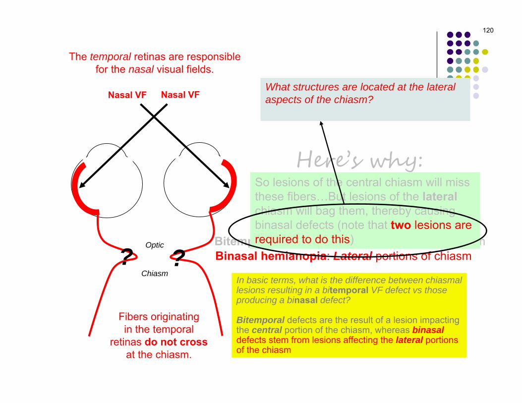
The temporal retinas are responsible for the nasal visual fields.
Nasal VF
Here’s why:
Nasal VF

117
In basic terms, what is the difference between chiasmallesions resulting in a bitemporal VF defect vs thoseproducing a binasal defect?
Bitemporal defects are the result of a lesion impactingthe central portion of the chiasm, whereas binasaldefects stem from lesions affecting the lateral portionsof the chiasm
Bitemporal hemianopia: Central aspect of chiasmBinasal hemianopia: Lateral portions of chiasm
The temporal retinas are responsible for the nasal visual fields.
Optic
Chiasm
Fibers originating in the temporal
retinas do not cross at the chiasm.
Here’s why:
Nasal VF Nasal VF

118
In basic terms, what is the difference between chiasmallesions resulting in a bitemporal VF defect vs thoseproducing a binasal defect?
Bitemporal defects are the result of a lesion impactingthe central portion of the chiasm, whereas binasaldefects stem from lesions affecting the lateral portionsof the chiasm
Bitemporal hemianopia: Central aspect of chiasmBinasal hemianopia: Lateral portions of chiasm
The temporal retinas are responsible for the nasal visual fields.
Optic
Chiasm
Fibers originating in the temporal
retinas do not cross at the chiasm.
Here’s why:
Nasal VF Nasal VF
So lesions of the central chiasm will miss these fibers… lesions of the lateralchiasm will bag them, thereby causing binasal defects (note that two lesions are required to do this)

119
In basic terms, what is the difference between chiasmallesions resulting in a bitemporal VF defect vs thoseproducing a binasal defect?
Bitemporal defects are the result of a lesion impactingthe central portion of the chiasm, whereas binasaldefects stem from lesions affecting the lateral portionsof the chiasm
Bitemporal hemianopia: Central aspect of chiasmBinasal hemianopia: Lateral portions of chiasm
The temporal retinas are responsible for the nasal visual fields.
Optic
Chiasm
Fibers originating in the temporal
retinas do not cross at the chiasm.
Here’s why:
Nasal VF Nasal VF
So lesions of the central chiasm will miss these fibers…But lesions of the lateralchiasm will bag them, thereby causing binasal defects (note that two lesions are required to do this)
120
In basic terms, what is the difference between chiasmallesions resulting in a bitemporal VF defect vs thoseproducing a binasal defect?
Bitemporal defects are the result of a lesion impactingthe central portion of the chiasm, whereas binasaldefects stem from lesions affecting the lateral portionsof the chiasm
Bitemporal hemianopia: Central aspect of chiasmBinasal hemianopia: Lateral portions of chiasm
The temporal retinas are responsible for the nasal visual fields.
Optic
Chiasm
Fibers originating in the temporal
retinas do not cross at the chiasm.
Here’s why:
Nasal VF Nasal VFWhat structures are located at the lateral aspects of the chiasm?The internal carotid arteries
So lesions of the central chiasm will miss these fibers…But lesions of the lateralchiasm will bag them, thereby causing binasal defects (note that two lesions are required to do this)
? ?

121
In basic terms, what is the difference between chiasmallesions resulting in a bitemporal VF defect vs thoseproducing a binasal defect?
Bitemporal defects are the result of a lesion impactingthe central portion of the chiasm, whereas binasaldefects stem from lesions affecting the lateral portionsof the chiasm
Bitemporal hemianopia: Central aspect of chiasmBinasal hemianopia: Lateral portions of chiasm
The temporal retinas are responsible for the nasal visual fields.
Optic
Chiasm
Fibers originating in the temporal
retinas do not cross at the chiasm.
Here’s why:
Nasal VF Nasal VFWhat structures are located at the lateral aspects of the chiasm?The internal carotid arteries
So lesions of the central chiasm will miss these fibers…But lesions of the lateralchiasm will bag them, thereby causing binasal defects (note that two lesions are required to do this)
Visual Field Defects
Retina
Optic nerve
Optic chiasm
Retrochiasmal
Bitemporal hemianopiaBinasal hemianopiaJunctional commonJunctional rare
Optic tractLGNOptic radiationsOccipital cortex
ArcuateCentralCeco-central
Nasal stepAltitudinalTemporal wedge
Depressions
Scotomas
Clinically obvious dzClinically subtle dz
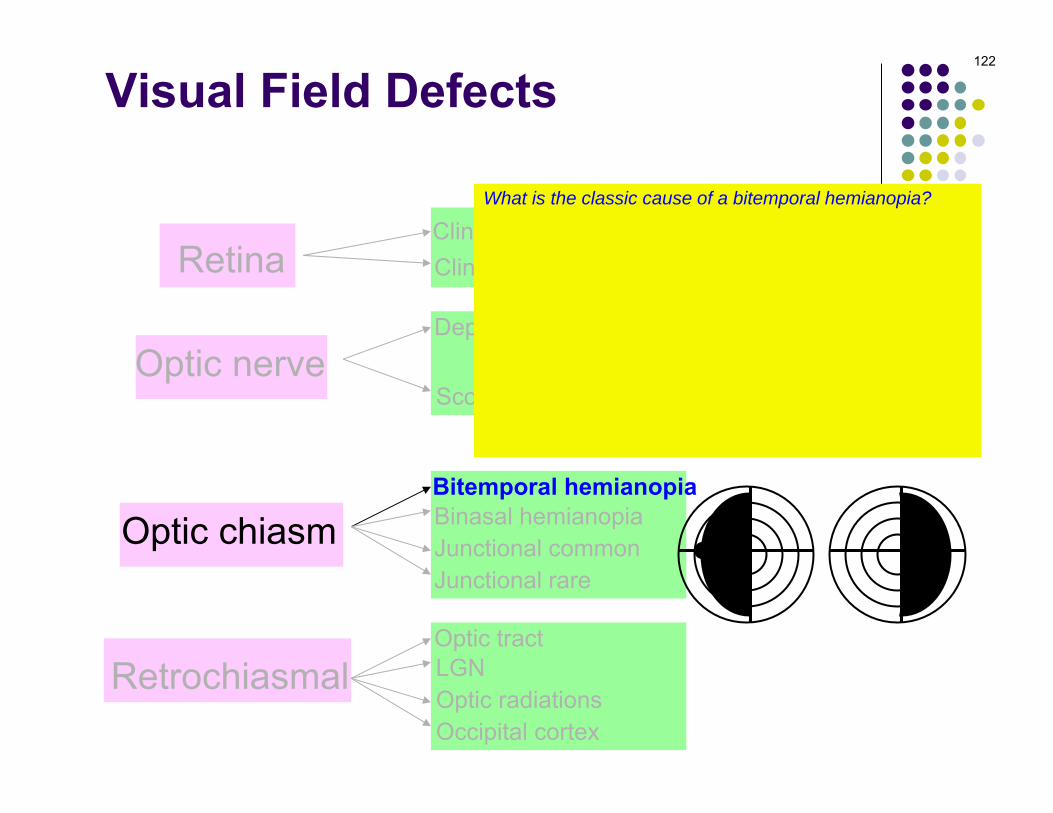
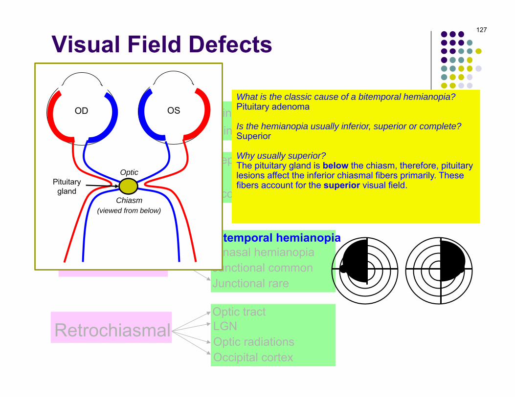
What is the classic cause of a bitemporal hemianopia?Pituitary adenoma
Is the hemianopia usually inferior, superior or complete?Superior
Why usually superior?The pituitary gland is below the chiasm, therefore, pituitarylesions affect the inferior chiasmal fibers primarily. Thesefibers account for the superior VF.
Is it usually congruous or incongruous?Incongruous
122

Visual Field Defects
Retina
Optic nerve
Optic chiasm
Retrochiasmal
Bitemporal hemianopiaBinasal hemianopiaJunctional commonJunctional rare
Optic tractLGNOptic radiationsOccipital cortex
ArcuateCentralCeco-central
Nasal stepAltitudinalTemporal wedge
Depressions
Scotomas
Clinically obvious dzClinically subtle dz
What is the classic cause of a bitemporal hemianopia?Pituitary adenoma
Is the hemianopia usually inferior, superior or complete?Superior
Why usually superior?The pituitary gland is below the chiasm, therefore, pituitarylesions affect the inferior chiasmal fibers primarily. Thesefibers account for the superior VF.
Is it usually congruous or incongruous?Incongruous
123

Visual Field Defects
Retina
Optic nerve
Optic chiasm
Retrochiasmal
Bitemporal hemianopiaBinasal hemianopiaJunctional commonJunctional rare
Optic tractLGNOptic radiationsOccipital cortex
ArcuateCentralCeco-central
Nasal stepAltitudinalTemporal wedge
Depressions
Scotomas
Clinically obvious dzClinically subtle dz
What is the classic cause of a bitemporal hemianopia?Pituitary adenoma
Is the hemianopia usually inferior, superior or complete?Superior
Why usually superior?The pituitary gland is below the chiasm, therefore, pituitarylesions affect the inferior chiasmal fibers primarily. Thesefibers account for the superior VF.
Is it usually congruous or incongruous?Incongruous
124

Visual Field Defects
Retina
Optic nerve
Optic chiasm
Retrochiasmal
Bitemporal hemianopiaBinasal hemianopiaJunctional commonJunctional rare
Optic tractLGNOptic radiationsOccipital cortex
ArcuateCentralCeco-central
Nasal stepAltitudinalTemporal wedge
Depressions
Scotomas
Clinically obvious dzClinically subtle dz
What is the classic cause of a bitemporal hemianopia?Pituitary adenoma
Is the hemianopia usually inferior, superior or complete?Superior
Why usually superior?The pituitary gland is below the chiasm, therefore, pituitarylesions affect the inferior chiasmal fibers primarily. Thesefibers account for the superior VF.
Is it usually congruous or incongruous?Incongruous
125

Visual Field Defects
Retina
Optic nerve
Optic chiasm
Retrochiasmal
Bitemporal hemianopiaBinasal hemianopiaJunctional commonJunctional rare
Optic tractLGNOptic radiationsOccipital cortex
ArcuateCentralCeco-central
Nasal stepAltitudinalTemporal wedge
Depressions
Scotomas
Clinically obvious dzClinically subtle dz
What is the classic cause of a bitemporal hemianopia?Pituitary adenoma
Is the hemianopia usually inferior, superior or complete?Superior
Why usually superior?The pituitary gland is below the chiasm, therefore, pituitarylesions affect the inferior chiasmal fibers primarily. Thesefibers account for the superior VF.
Is it usually congruous or incongruous?Incongruous
126
Visual Field Defects
Retina
Optic nerve
Optic chiasm
Retrochiasmal
Bitemporal hemianopiaBinasal hemianopiaJunctional commonJunctional rare
Optic tractLGNOptic radiationsOccipital cortex
ArcuateCentralCeco-central
Nasal stepAltitudinalTemporal wedge
Depressions
Scotomas
Clinically obvious dzClinically subtle dz
What is the classic cause of a bitemporal hemianopia?Pituitary adenoma
Is the hemianopia usually inferior, superior or complete?Superior
Why usually superior?The pituitary gland is below the chiasm, therefore, pituitarylesions affect the inferior chiasmal fibers primarily. Thesefibers account for the superior visual field.
Is it usually congruous or incongruous?Incongruous
127
Optic
Chiasm
OD OS
(viewed from below)
Pituitarygland

Visual Field Defects
Retina
Optic nerve
Optic chiasm
Retrochiasmal
Bitemporal hemianopiaBinasal hemianopiaJunctional commonJunctional rare
Optic tractLGNOptic radiationsOccipital cortex
ArcuateCentralCeco-central
Nasal stepAltitudinalTemporal wedge
Depressions
Scotomas
Clinically obvious dzClinically subtle dz
What is the classic cause of a bitemporal hemianopia?Pituitary adenoma
Is the hemianopia usually inferior, superior or complete?Superior
Why usually superior?The pituitary gland is below the chiasm, therefore, pituitarylesions affect the inferior chiasmal fibers primarily. Thesefibers account for the superior visual field.
Is it usually congruous or incongruous?Incongruous
128

Visual Field Defects
Retina
Optic nerve
Optic chiasm
Retrochiasmal
Bitemporal hemianopiaBinasal hemianopiaJunctional commonJunctional rare
Optic tractLGNOptic radiationsOccipital cortex
ArcuateCentralCeco-central
Nasal stepAltitudinalTemporal wedge
Depressions
Scotomas
Clinically obvious dzClinically subtle dz
What is the classic cause of a bitemporal hemianopia?Pituitary adenoma
Is the hemianopia usually inferior, superior or complete?Superior
Why usually superior?The pituitary gland is below the chiasm, therefore, pituitarylesions affect the inferior chiasmal fibers primarily. Thesefibers account for the superior visual field.
Is it usually congruous or incongruous?Incongruous
129

Visual Field Defects
Retina
Optic nerve
Optic chiasm
Retrochiasmal
Bitemporal hemianopiaBinasal hemianopiaJunctional commonJunctional rare
Optic tractLGNOptic radiationsOccipital cortex
ArcuateCentralCeco-central
Nasal stepAltitudinalTemporal wedge
Depressions
Scotomas
Clinically obvious dzClinically subtle dz
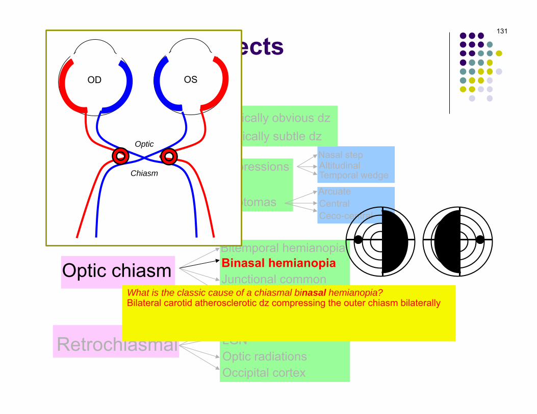
What is the classic cause of a chiasmal binasal hemianopia?Bilateral carotid disease
What is the actual etiology for the vast majority of real-world binasal defects?Glaucoma
130
Visual Field Defects
Retina
Optic nerve
Optic chiasm
Retrochiasmal
Bitemporal hemianopiaBinasal hemianopiaJunctional commonJunctional rare
Optic tractLGNOptic radiationsOccipital cortex
ArcuateCentralCeco-central
Nasal stepAltitudinalTemporal wedge
Depressions
Scotomas
Clinically obvious dzClinically subtle dz
What is the classic cause of a chiasmal binasal hemianopia?Bilateral carotid atherosclerotic dz compressing the outer chiasm bilaterally
What is the actual etiology for the vast majority of real-world binasal defects?Glaucoma
131
Optic
Chiasm
OD OS

Visual Field Defects
Retina
Optic nerve
Optic chiasm
Retrochiasmal
Bitemporal hemianopiaBinasal hemianopiaJunctional commonJunctional rare
Optic tractLGNOptic radiationsOccipital cortex
ArcuateCentralCeco-central
Nasal stepAltitudinalTemporal wedge
Depressions
Scotomas
Clinically obvious dzClinically subtle dz
What is the classic cause of a chiasmal binasal hemianopia?Bilateral carotid atherosclerotic dz compressing the outer chiasm bilaterally
What is the actual etiology for the vast majority of real-world binasal defects?Glaucoma
132
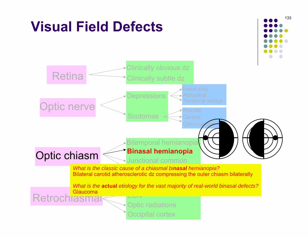
Visual Field Defects
Retina
Optic nerve
Optic chiasm
Retrochiasmal
Bitemporal hemianopiaBinasal hemianopiaJunctional commonJunctional rare
Optic tractLGNOptic radiationsOccipital cortex
ArcuateCentralCeco-central
Nasal stepAltitudinalTemporal wedge
Depressions
Scotomas
Clinically obvious dzClinically subtle dz
What is the classic cause of a chiasmal binasal hemianopia?Bilateral carotid atherosclerotic dz compressing the outer chiasm bilaterally
What is the actual etiology for the vast majority of real-world binasal defects?Glaucoma
133

Visual Field Defects
Retina
Optic nerve
Optic chiasm
Retrochiasmal
Bitemporal hemianopiaBinasal hemianopiaJunctional commonJunctional rare
Optic tractLGNOptic radiationsOccipital cortex
ArcuateCentralCeco-central
Nasal stepAltitudinalTemporal wedge
Depressions
Scotomas
Clinically obvious dzClinically subtle dz
What does the term junctional refer to anatomically?The junction between the optic nerve and the chiasm
What does a junctional common VF defect look like?An optic nerve VF defect in one eye and a hemianopic-like defect in the other
134

Visual Field Defects
Retina
Optic nerve
Optic chiasm
Retrochiasmal
Bitemporal hemianopiaBinasal hemianopiaJunctional commonJunctional rare
Optic tractLGNOptic radiationsOccipital cortex
ArcuateCentralCeco-central
Nasal stepAltitudinalTemporal wedge
Depressions
Scotomas
Clinically obvious dzClinically subtle dz
What does the term junctional refer to anatomically?The junction between the optic nerve and the chiasm
What does a junctional common VF defect look like?An optic nerve VF defect in one eye and a hemianopic-like defect in the other
135
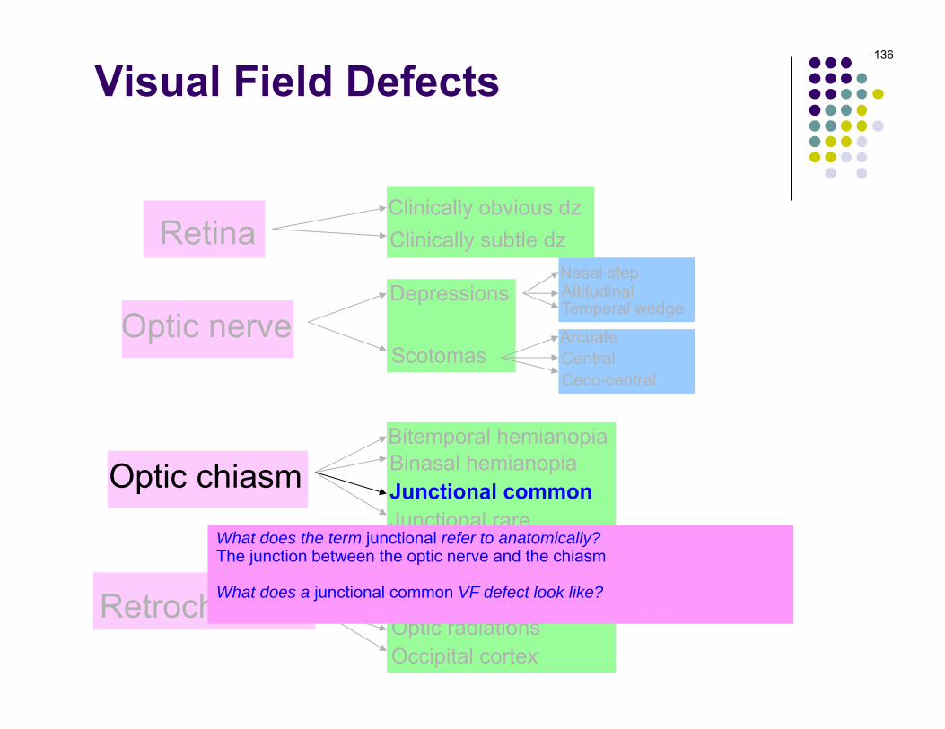
Visual Field Defects
Retina
Optic nerve
Optic chiasm
Retrochiasmal
Bitemporal hemianopiaBinasal hemianopiaJunctional commonJunctional rare
Optic tractLGNOptic radiationsOccipital cortex
ArcuateCentralCeco-central
Nasal stepAltitudinalTemporal wedge
Depressions
Scotomas
Clinically obvious dzClinically subtle dz
What does the term junctional refer to anatomically?The junction between the optic nerve and the chiasm
What does a junctional common VF defect look like?An optic nerve VF defect in one eye and a hemianopic-like defect in the other
136

Visual Field Defects
Retina
Optic nerve
Optic chiasm
Retrochiasmal
Bitemporal hemianopiaBinasal hemianopiaJunctional commonJunctional rare
Optic tractLGNOptic radiationsOccipital cortex
ArcuateCentralCeco-central
Nasal stepAltitudinalTemporal wedge
Depressions
Scotomas
Clinically obvious dzClinically subtle dz
What does the term junctional refer to anatomically?The junction between the optic nerve and the chiasm
What does a junctional common VF defect look like?An optic nerve VF defect in one eye and a hemianopic-like defect in the other
i.e., it respects the vertical meridian
137

Visual Field Defects
Retina
Optic nerve
Optic chiasm
Retrochiasmal
Bitemporal hemianopiaBinasal hemianopiaJunctional commonJunctional rare
Optic tractLGNOptic radiationsOccipital cortex
ArcuateCentralCeco-central
Nasal stepAltitudinalTemporal wedge
Depressions
Scotomas
Clinically obvious dzClinically subtle dz
What does a junctional rare VF defect look like?A hemianopic-like defect in one eye, but no lesion in the other
138

Visual Field Defects
Retina
Optic nerve
Optic chiasm
Retrochiasmal
Bitemporal hemianopiaBinasal hemianopiaJunctional commonJunctional rare
Optic tractLGNOptic radiationsOccipital cortex
ArcuateCentralCeco-central
Nasal stepAltitudinalTemporal wedge
Depressions
Scotomas
Clinically obvious dzClinically subtle dz
What does a junctional rare VF defect look like?A hemianopic-like defect in one eye, but no lesion in the other
139
Q
Which of the following is not associated with bitemporal visual-field loss? Sectoral RP Glaucoma Fuchs coloboma Chiasmal lesion Toxic/hereditary/nutritional optic neuropathy
140

Glaucoma. True bitemporal VF loss respects the vertical midline, and is associated almost exclusively with lesions compressing the chiasm, specifically the mid-chiasm. Other causes of bitemporal loss do not respect the midline (except by happenstance). Sectoral RP is symmetric bilaterally, and thus can affect the temporal VF bilaterally. Fuchs coloboma (aka tilted disc syndrome) is associated with bitemporal loss that resolves with proper astigmatic correction. Toxic/hereditary/nutritional optic neuropathy is associated with bilateral cecocentral VF loss, which can mimic bitemporal loss. Glaucoma almost always affects the nasal VF long before the temporal field is involved--if anything, glaucoma is far more likely to cause binasal VF cuts.
141
A Which of the following is not associated with bitemporal visual-field loss?
Sectoral RP Glaucoma Fuchs coloboma Chiasmal lesion Toxic/hereditary/nutritional optic neuropathy
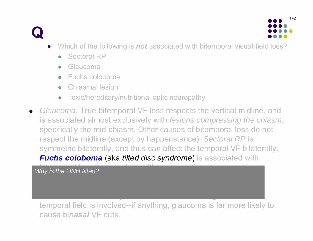
Glaucoma. True bitemporal VF loss respects the vertical midline, and is associated almost exclusively with lesions compressing the chiasm, specifically the mid-chiasm. Other causes of bitemporal loss do not respect the midline (except by happenstance). Sectoral RP is symmetric bilaterally, and thus can affect the temporal VF bilaterally. Fuchs coloboma (aka tilted disc syndrome) is associated with bitemporal loss that resolves with proper astigmatic correction. Toxic/hereditary/nutritional optic neuropathy is associated with bilateral cecocentral VF loss, which can mimic bitemporal loss. Glaucoma almost always affects the nasal VF long before the temporal field is involved--if anything, glaucoma is far more likely to cause binasal VF cuts.
142
Q Which of the following is not associated with bitemporal visual-field loss?
Sectoral RP Glaucoma Fuchs coloboma Chiasmal lesion Toxic/hereditary/nutritional optic neuropathy
Why is the ONH tilted?Abnormal closure of the embryonic optic fissure at the optic-nerve/globe junction results in an oblique (read: tilted) orientation of the ONH. The abnormal closure also creates a modest staphyloma in the inferonasal region of the globe.

Glaucoma. True bitemporal VF loss respects the vertical midline, and is associated almost exclusively with lesions compressing the chiasm, specifically the mid-chiasm. Other causes of bitemporal loss do not respect the midline (except by happenstance). Sectoral RP is symmetric bilaterally, and thus can affect the temporal VF bilaterally. Fuchs coloboma (aka tilted disc syndrome) is associated with bitemporal loss that resolves with proper astigmatic correction. Toxic/hereditary/nutritional optic neuropathy is associated with bilateral cecocentral VF loss, which can mimic bitemporal loss. Glaucoma almost always affects the nasal VF long before the temporal field is involved--if anything, glaucoma is far more likely to cause binasal VF cuts.
143
A Which of the following is not associated with bitemporal visual-field loss?
Sectoral RP Glaucoma Fuchs coloboma Chiasmal lesion Toxic/hereditary/nutritional optic neuropathy
Why is the ONH tilted?Abnormal closure of the embryonic optic fissure at the optic-nerve/globe junction results in an oblique (read: tilted) orientation of the ONH. The abnormal closure also creates a modest staphyloma in the inferonasal region of the globe.

Glaucoma. True bitemporal VF loss respects the vertical midline, and is associated almost exclusively with lesions compressing the chiasm, specifically the mid-chiasm. Other causes of bitemporal loss do not respect the midline (except by happenstance). Sectoral RP is symmetric bilaterally, and thus can affect the temporal VF bilaterally. Fuchs coloboma (aka tilted disc syndrome) is associated with bitemporal loss that resolves with proper astigmatic correction. Toxic/hereditary/nutritional optic neuropathy is associated with bilateral cecocentral VF loss, which can mimic bitemporal loss. Glaucoma almost always affects the nasal VF long before the temporal field is involved--if anything, glaucoma is far more likely to cause binasal VF cuts.
144
Q Which of the following is not associated with bitemporal visual-field loss?
Sectoral RP Glaucoma Fuchs coloboma Chiasmal lesion Toxic/hereditary/nutritional optic neuropathy
Why is the ONH tilted?Abnormal closure of the embryonic optic fissure at the optic-nerve/globe junction results in an oblique (read: tilted) orientation of the ONH. The abnormal closure also creates a modest staphyloma in the inferonasal region of the globe.
Why on earth do these pts get a VF defect?It’s actually pretty simple. Recall that these eyes have inferonasal staphylomas. This results in an extra-long ‘axial length’ for the photoreceptors overlying the staphyloma. Because of this extra axial length, the correction used during the performance of a visual-field test—a correction determined on the basis of the (non-staphylomatous) fovea—is not myopic enough for the staphylomatous inferonasal region. As a result, the uncorrected refractive error in this region produces a refractive scotoma in the superotemporal visual field.
This implies that the VF deficits could be resolved if the proper refractive correction was employed. Does it?Indeed it does, and this cinches the diagnosis
This implies also that the bitemporal VF loss shouldn’t respect the vertical midline. Does it?No (except by pure dumb luck)

Glaucoma. True bitemporal VF loss respects the vertical midline, and is associated almost exclusively with lesions compressing the chiasm, specifically the mid-chiasm. Other causes of bitemporal loss do not respect the midline (except by happenstance). Sectoral RP is symmetric bilaterally, and thus can affect the temporal VF bilaterally. Fuchs coloboma (aka tilted disc syndrome) is associated with bitemporal loss that resolves with proper astigmatic correction. Toxic/hereditary/nutritional optic neuropathy is associated with bilateral cecocentral VF loss, which can mimic bitemporal loss. Glaucoma almost always affects the nasal VF long before the temporal field is involved--if anything, glaucoma is far more likely to cause binasal VF cuts.
145
A Which of the following is not associated with bitemporal visual-field loss?
Sectoral RP Glaucoma Fuchs coloboma Chiasmal lesion Toxic/hereditary/nutritional optic neuropathy
Why is the ONH tilted?Abnormal closure of the embryonic optic fissure at the optic-nerve/globe junction results in an oblique (read: tilted) orientation of the ONH. The abnormal closure also creates a modest staphyloma in the inferonasal region of the globe.
Why on earth do these pts get a VF defect?It’s actually pretty simple. Recall that these eyes have inferonasal staphylomas. This results in an extra-long ‘axial length’ for the photoreceptors overlying the staphyloma. Because of this extra axial length, the correction used during the performance of a visual-field test—a correction determined on the basis of the (non-staphylomatous) fovea—is not myopic enough for the staphylomatous inferonasal region. As a result, the uncorrected refractive error in this region produces a refractive scotoma in the superotemporal visual field.
This implies that the VF deficits could be resolved if the proper refractive correction was employed. Does it?Indeed it does, and this cinches the diagnosis
This implies also that the bitemporal VF loss shouldn’t respect the vertical midline. Does it?No (except by pure dumb luck)

Glaucoma. True bitemporal VF loss respects the vertical midline, and is associated almost exclusively with lesions compressing the chiasm, specifically the mid-chiasm. Other causes of bitemporal loss do not respect the midline (except by happenstance). Sectoral RP is symmetric bilaterally, and thus can affect the temporal VF bilaterally. Fuchs coloboma (aka tilted disc syndrome) is associated with bitemporal loss that resolves with proper astigmatic correction. Toxic/hereditary/nutritional optic neuropathy is associated with bilateral cecocentral VF loss, which can mimic bitemporal loss. Glaucoma almost always affects the nasal VF long before the temporal field is involved--if anything, glaucoma is far more likely to cause binasal VF cuts.
146
Q Which of the following is not associated with bitemporal visual-field loss?
Sectoral RP Glaucoma Fuchs coloboma Chiasmal lesion Toxic/hereditary/nutritional optic neuropathy
Why is the ONH tilted?Abnormal closure of the embryonic optic fissure at the optic-nerve/globe junction results in an oblique (read: tilted) orientation of the ONH. The abnormal closure also creates a modest staphyloma in the inferonasal region of the globe.
Why on earth do these pts get a VF defect?It’s actually pretty simple. Recall that these eyes have inferonasal staphylomas. This results in an extra-long ‘axial length’ for the photoreceptors overlying the staphyloma. Because of this extra axial length, the correction used during the performance of a visual-field test—a correction determined on the basis of the (non-staphylomatous) fovea—is not myopic enough for the staphylomatous inferonasal region. As a result, the uncorrected refractive error in this region produces a refractive scotoma in the superotemporal visual field.
This implies that the VF deficits could be resolved if the proper refractive correction was employed. Does it?Indeed it does, and this cinches the diagnosis
This implies also that the bitemporal VF loss shouldn’t respect the vertical midline. Does it?No (except by pure dumb luck)

Glaucoma. True bitemporal VF loss respects the vertical midline, and is associated almost exclusively with lesions compressing the chiasm, specifically the mid-chiasm. Other causes of bitemporal loss do not respect the midline (except by happenstance). Sectoral RP is symmetric bilaterally, and thus can affect the temporal VF bilaterally. Fuchs coloboma (aka tilted disc syndrome) is associated with bitemporal loss that resolves with proper astigmatic correction. Toxic/hereditary/nutritional optic neuropathy is associated with bilateral cecocentral VF loss, which can mimic bitemporal loss. Glaucoma almost always affects the nasal VF long before the temporal field is involved--if anything, glaucoma is far more likely to cause binasal VF cuts.
147
A Which of the following is not associated with bitemporal visual-field loss?
Sectoral RP Glaucoma Fuchs coloboma Chiasmal lesion Toxic/hereditary/nutritional optic neuropathy
Why is the ONH tilted?Abnormal closure of the embryonic optic fissure at the optic-nerve/globe junction results in an oblique (read: tilted) orientation of the ONH. The abnormal closure also creates a modest staphyloma in the inferonasal region of the globe.
Why on earth do these pts get a VF defect?It’s actually pretty simple. Recall that these eyes have inferonasal staphylomas. This results in an extra-long ‘axial length’ for the photoreceptors overlying the staphyloma. Because of this extra axial length, the correction used during the performance of a visual-field test—a correction determined on the basis of the (non-staphylomatous) fovea—is not myopic enough for the staphylomatous inferonasal region. As a result, the uncorrected refractive error in this region produces a refractive scotoma in the superotemporal visual field.
This implies that the VF deficits could be resolved if the proper refractive correction was employed. Does it?Indeed it does, and this cinches the diagnosis
This implies also that the bitemporal VF loss shouldn’t respect the vertical midline. Does it?No (except by pure dumb luck)

Glaucoma. True bitemporal VF loss respects the vertical midline, and is associated almost exclusively with lesions compressing the chiasm, specifically the mid-chiasm. Other causes of bitemporal loss do not respect the midline (except by happenstance). Sectoral RP is symmetric bilaterally, and thus can affect the temporal VF bilaterally. Fuchs coloboma (aka tilted disc syndrome) is associated with bitemporal loss that resolves with proper astigmatic correction. Toxic/hereditary/nutritional optic neuropathy is associated with bilateral cecocentral VF loss, which can mimic bitemporal loss. Glaucoma almost always affects the nasal VF long before the temporal field is involved--if anything, glaucoma is far more likely to cause binasal VF cuts.
148
Q Which of the following is not associated with bitemporal visual-field loss?
Sectoral RP Glaucoma Fuchs coloboma Chiasmal lesion Toxic/hereditary/nutritional optic neuropathy
Why is the ONH tilted?Abnormal closure of the embryonic optic fissure at the optic-nerve/globe junction results in an oblique (read: tilted) orientation of the ONH. The abnormal closure also creates a modest staphyloma in the inferonasal region of the globe.
Why on earth do these pts get a VF defect?It’s actually pretty simple. Recall that these eyes have inferonasal staphylomas. This results in an extra-long ‘axial length’ for the photoreceptors overlying the staphyloma. Because of this extra axial length, the correction used during the performance of a visual-field test—a correction determined on the basis of the (non-staphylomatous) fovea—is not myopic enough for the staphylomatous inferonasal region. As a result, the uncorrected refractive error in this region produces a refractive scotoma in the superotemporal visual field.
This implies that the VF deficits could be resolved if the proper refractive correction was employed. Does it?Indeed it does, and this cinches the diagnosis
This implies also that the bitemporal VF loss shouldn’t respect the vertical midline. Does it?No (except by pure dumb luck)

Glaucoma. True bitemporal VF loss respects the vertical midline, and is associated almost exclusively with lesions compressing the chiasm, specifically the mid-chiasm. Other causes of bitemporal loss do not respect the midline (except by happenstance). Sectoral RP is symmetric bilaterally, and thus can affect the temporal VF bilaterally. Fuchs coloboma (aka tilted disc syndrome) is associated with bitemporal loss that resolves with proper astigmatic correction. Toxic/hereditary/nutritional optic neuropathy is associated with bilateral cecocentral VF loss, which can mimic bitemporal loss. Glaucoma almost always affects the nasal VF long before the temporal field is involved--if anything, glaucoma is far more likely to cause binasal VF cuts.
149
A Which of the following is not associated with bitemporal visual-field loss?
Sectoral RP Glaucoma Fuchs coloboma Chiasmal lesion Toxic/hereditary/nutritional optic neuropathy
Why is the ONH tilted?Abnormal closure of the embryonic optic fissure at the optic-nerve/globe junction results in an oblique (read: tilted) orientation of the ONH. The abnormal closure also creates a modest staphyloma in the inferonasal region of the globe.
Why on earth do these pts get a VF defect?It’s actually pretty simple. Recall that these eyes have inferonasal staphylomas. This results in an extra-long ‘axial length’ for the photoreceptors overlying the staphyloma. Because of this extra axial length, the correction used during the performance of a visual-field test—a correction determined on the basis of the (non-staphylomatous) fovea—is not myopic enough for the staphylomatous inferonasal region. As a result, the uncorrected refractive error in this region produces a refractive scotoma in the superotemporal visual field.
This implies that the VF deficits could be resolved if the proper refractive correction was employed. Does it?Indeed it does, and this cinches the diagnosis
This implies also that the bitemporal VF loss shouldn’t respect the vertical midline. Does it?No (except by pure dumb luck)