Vision in 1 Lecture Prof. Jack Pettigrew Vision Touch and Hearing Research Centre, University of...
37
Vision in 1 Lecture Prof. Jack Pettigrew Vision Touch and Hearing Research Centre, University of Queensland 4072 Australia.
-
Upload
evelin-spain -
Category
Documents
-
view
213 -
download
0
Transcript of Vision in 1 Lecture Prof. Jack Pettigrew Vision Touch and Hearing Research Centre, University of...

Vision in 1 Lecture
Prof. Jack PettigrewVision Touch and Hearing Research Centre, University of Queensland 4072 Australia.

Vision:
1. Parallel Visual Pathways:• Diversity of retinal ganglion cells and their destinations• E.g. Melanopsin and the circadian clock system: SCN , jet
lag etc• 2. “Ventral” (conscious) vs. “dorsal” (unconscious) visual
streamsBlindsight: Veridical vs. Non-veridical

(Blind spot)

Melanopsin-containinglight sensitive retinal ganglion cell
MM MM
M
PPDS PLS

Parallel Visual Paths: >9 Separate Destinations of Retinal Ganglion Cells
1. SCN of hypothalamus…melanopsin system.CIRCADIAN CLOCK
2. dLGN (4 P & 2M layers)..geniculostriate. CONSCIOUS VISION ………ventral (&dorsal) stream OBJECT
ID
3. Pulvinar-LP complex……MT…dorsal stream…..VISUOMOTOR
4. Pretectal complex…………….NEAR REFLEX TRIAD
5. Midbrain superior colliculus………VISUAL ORIENTATION
6. Accessory optic system…………….VISUAL STABILISATION
Dorsal TN, lateral TN, medial TN (pitch, roll & yaw)
7. Habenula, raphe etc ……………..
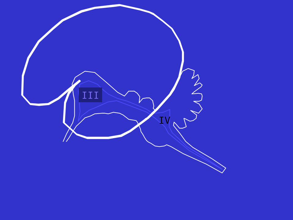
III
IV

III
IV
Forebrain
MidbrainHindbrain

AnterogradeTransportof ocular
label
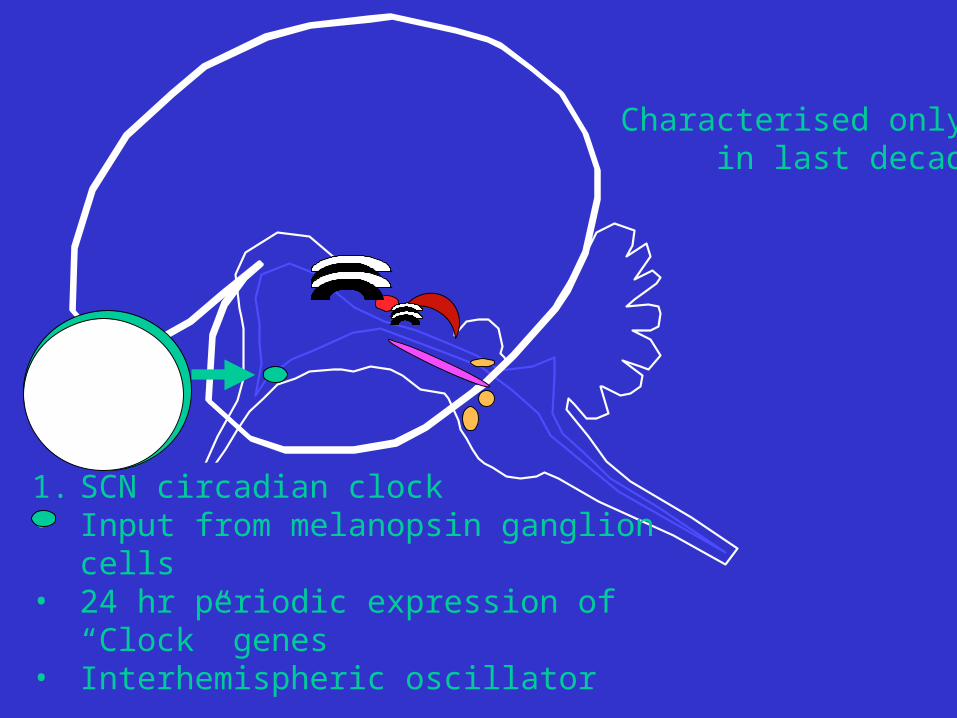
1. SCN circadian clock• Input from melanopsin ganglion cells• 24 hr periodic expression of “Clock” genes• Interhemispheric oscillator
Characterised only in last decade

Labelling in Suprachiasmatic Nucleus (SCN): lac-Z-melanopsin construct


1. SCN circadian clock
2. dLGN geniculo-striate path• Huge in primates• Binocular vision emphasised: 3 layers for each eye• Conscious vision• Developmentally plastic
V1Striate cortex

1. SCN circadian clock
2. DLGN geniculostriate
3.Pulvinar-LP complexOld tectal system: primary system in most vertebratesDominated by striate input in primates
MT

1. SCN
2. dLGN
3.Pulvinar-LP complex 4. Pretectum
Near triad, focus, miosis, vergence

1. SCN circadian clock
2. DLGN geniculostriate
3.Pulvinar-LP complexOld tectal system
5. Midbrain-Sup.Colliculus:• orientation• “visual grasp reflex”• “hard wired:• lateral visual field
4. Pretectum
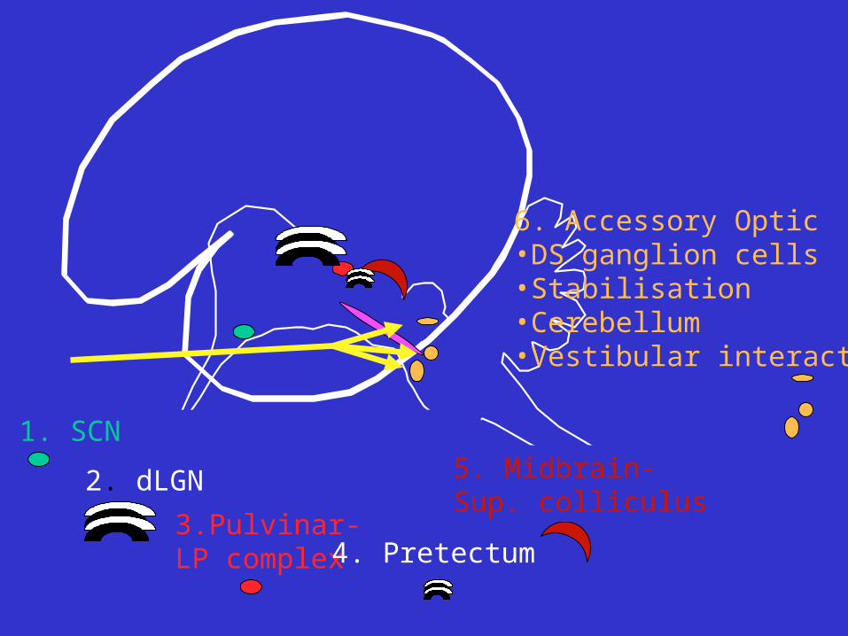
1. SCN
2. dLGN
3.Pulvinar-LP complex
6. Accessory Optic•DS ganglion cells•Stabilisation•Cerebellum•Vestibular interaction
4. Pretectum
5. Midbrain-Sup. colliculus
1. SCN
2. dLGN
3.Pulvinar-LP complex
6. Accessory Optic
4. Pretectum
5. Midbrain-Sup. colliculus
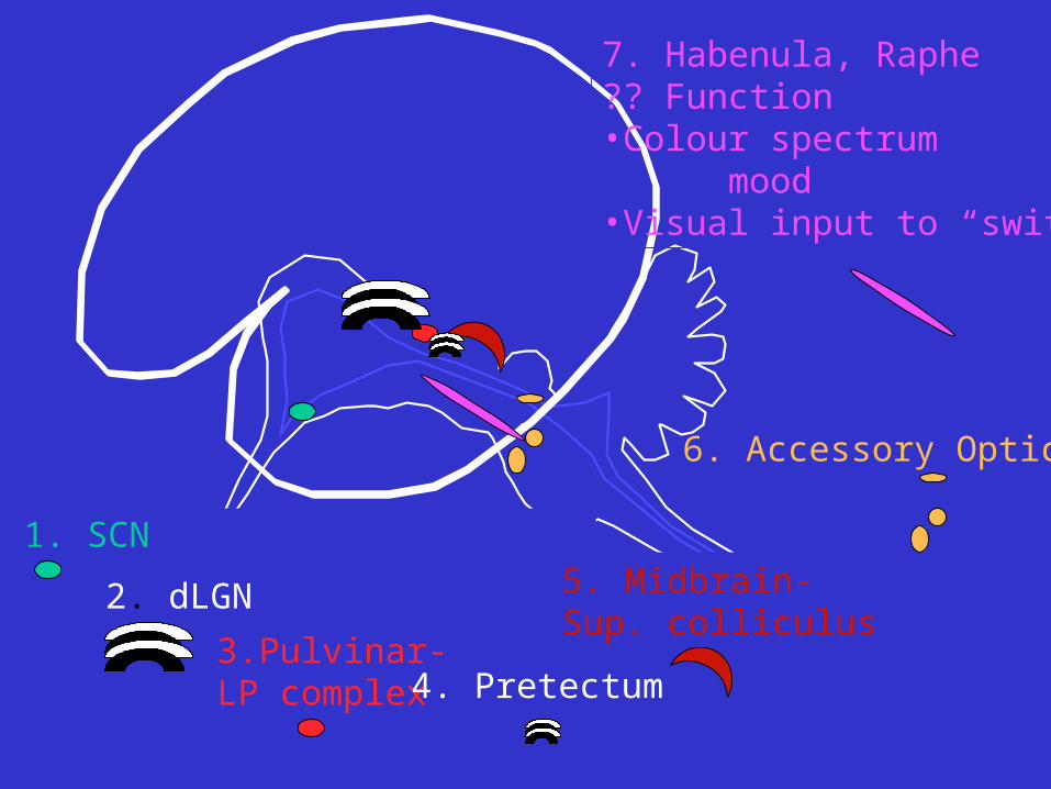
7. Habenula, Raphe?? Function•Colour spectrum mood•Visual input to “switch”

V1P
1. SCN
2. dLGN
3.Pulvinar-LP complex
6. Accessory Optic
4. Pretectum
5. Midbrain-Sup. colliculus
7. Habenula, Raphe

V1
MT
M
M
1. SCN
2. dLGN
3.Pulvinar-LP complex
6. Accessory Optic
4. Pretectum
5. Midbrain-Sup. colliculus
7. Habenula, RapheMM
M
V1
MT
DM
P
M
M
2. dLGN
3.Pulvinar-LP complex
5. Midbrain-Sup. colliculus
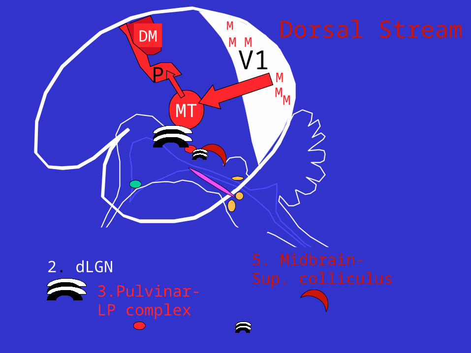
Dorsal Stream
M
M
M M
V1
MT
DM
P
M
M
2. dLGN
3.Pulvinar-LP complex
5. Midbrain-Sup. colliculus
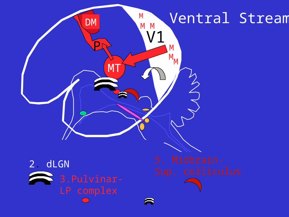
Ventral Stream
M
M
M M

V1
MT
DM
P
M
M
2. dLGN
3.Pulvinar-LP complex
5. Midbrain-Sup. colliculus
Ventral Stream
M
M
M M

V1
MT
DM
P
M
M
2. dLGN
3.Pulvinar-LP complex
5. Midbrain-Sup. colliculus
Ventral Stream
M
M
M M

V1
MT
DM
P
M
M
Ventral Stream
IT
IT visual Cortex in temporal pole:•Complex object recognition, including faces (FF area)•Highly plastic (lesions here affect visual memory)•Major output projection to limbic system, hippocampus
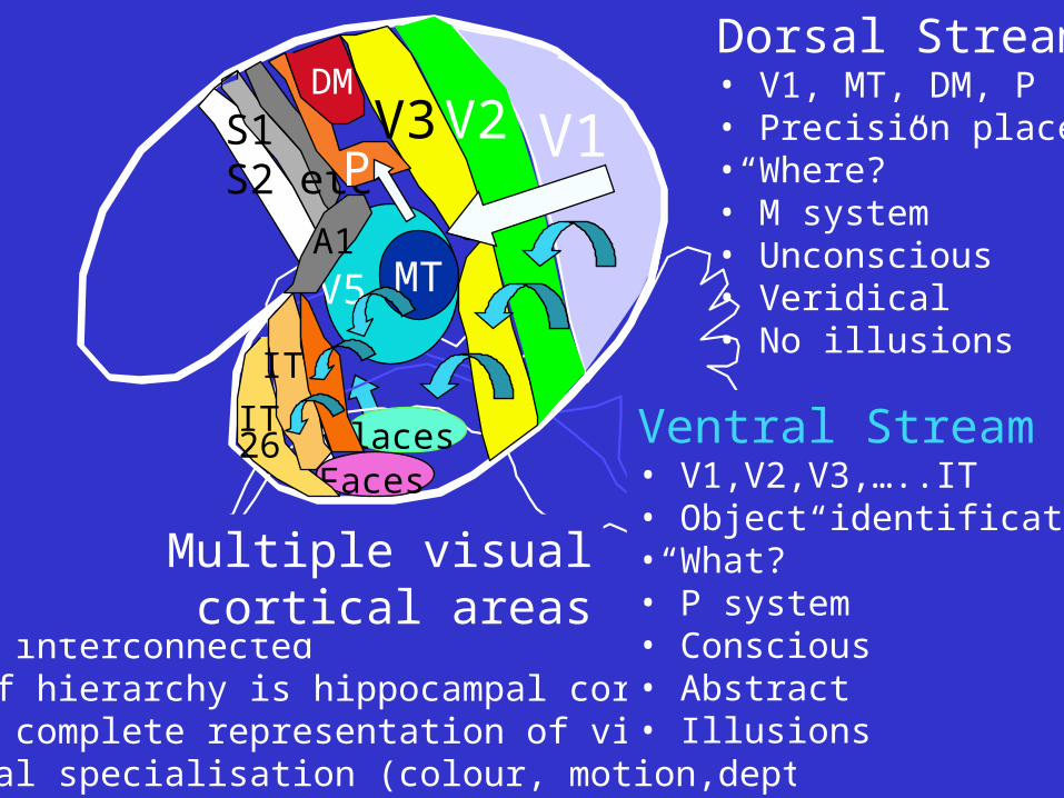
Hierarchical; V1 V2 etc ITMultiply interconnected“Apex” of hierarchy is hippocampal cortexEach has complete representation of visual fieldFunctional specialisation (colour, motion,depth etc)
Dorsal Stream• V1, MT, DM, P• Precision place•“Where?”• M system• Unconscious• Veridical• No illusions
V1V2
MTV5
V3
FacesPlacesIT
IT
26
DM
S1S2 etcP
A1
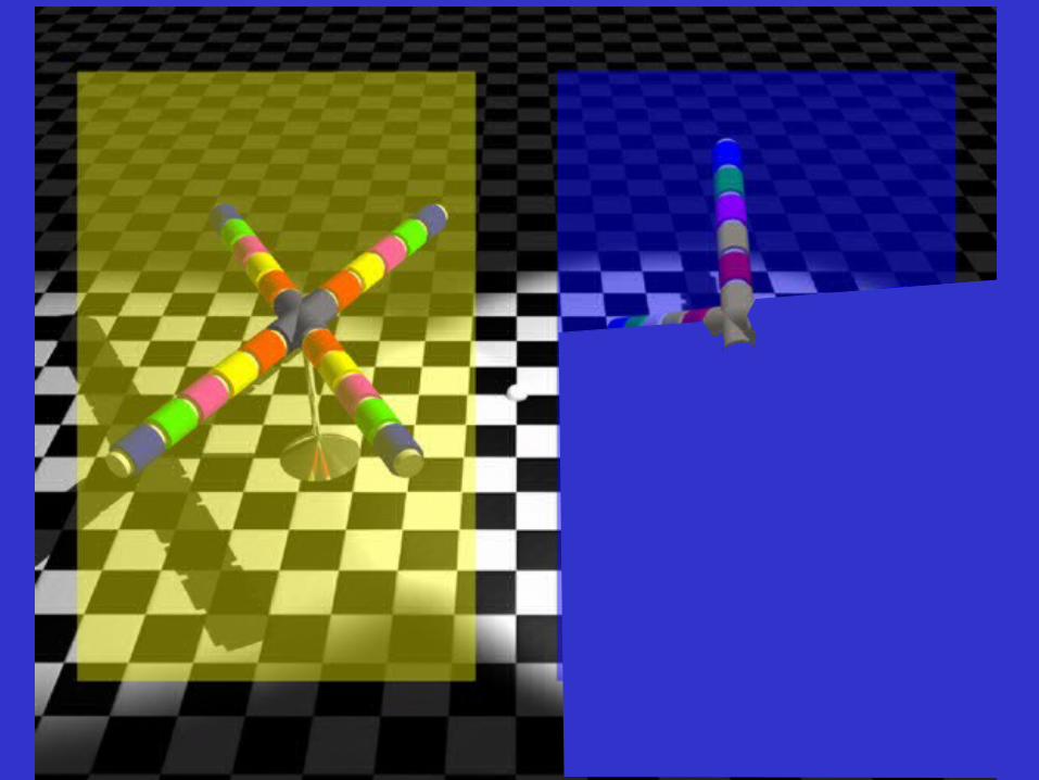
Ventral Stream• V1,V2,V3,…..IT• Object identification •“What?”• P system• Conscious• Abstract• Illusions
Multiple visual cortical areas


QuickTime™ and aGIF decompressor
are needed to see this picture.


QuickTime™ and aGIF decompressor
are needed to see this picture.


QuickTime™ and aGIF decompressor
are needed to see this picture.


QuickTime™ and aGIF decompressor
are needed to see this picture.