VIRUSES AND VIROIDS AS AGENTS OF PLANT DISEASE John
22
VIRUSES ANDVIROIDS AS AGENTS OF PLANT DISEASE John Randles and Helen Ogle Contents 7.1 Introduction ............ .............. 104 7.2 Characteristics oJunuses ............ ........ ios 7.3 Struchtre and archttecture ... 108 Rod-shapeduintses ........... l}g Isometric uiruses ................ 11O BacilLif,orrnuiruses ...... , ...... 111 7.4 Symptoms oJuirusiryfectton ... .................. iil Factors qffecting sgmptomexpresston ............. li4 7.5 TfansmissionoJ utruses ............. .......... 114 7.6 ldentificationoJuintses ............ ............ ilo 7.7 CLassification and nomencl.ahtre ............ .............. iiT 7.8 Viroids as agents oJdisease ................ i2l 7.9 ControloJuirus anduiroiddiseases ..... 123 D ir ect contr oLmeas ur es Indtrect controLmeasures . 125 7.1O nfiher reading .. i2O 7.1 Introduction Viruses are very small obligate parasites that are so simply constructed that they do not consist of cells, but of particles. The particles of a simple virus consist of a protein coat surrounding a genome of either ribonucleic acid (RNA) or deoxyribo- nucleic acid (DNA). They have been recognised as agents of disease in plants and other living organisms since the late 1880s. However, their main characteristics have been understood for less than 50 years. Virus diseases were first recognised when they caused a number of spectacular epidemics in humans (e.g. small pox), animals (e.g. foot and mouth disease) and plants (e.9. citrus tristeza). Some plant diseases formerly thought to be caused by viruses, but for which Koch's postulates had not been satisfied, were found in the I96Os to be caused by either phytoplasmas (e.g. aster yellows, tomato big bud, see Chapter 6) or viroids (e.g. potato spindle tuber, citrus exocortis and coconut cadang cadang diseases). Phytoplasmas are wall-less bacteria, whereas viroids are small, infectious, naked circular RNA molecules. Viroid- and virus-like sy.rnptoms have been recognised in plants for a very long time. Viroid-like symptoms were described in citrus fruit about 600 BC while a yellow leaf disease was recorded in Eupatorium sp. about 752 and tulip flowers with virus-like colour breaking (variegation or stripingi) appeared in paintings by Dutch artists about t6OO. In 1886, Adolf Mayer, a German agricultural chemist working at Wageningen in the Netherlands, found that a mosaic disease of tobacco could be transmitted to healthy plants by rubbing them with sap extracted from plants showing disease symptoms. In 1892, the Russian botanist, Dmitrii Ivanowski. showed thai
Transcript of VIRUSES AND VIROIDS AS AGENTS OF PLANT DISEASE John
VIRUSES AND VIROIDS AS AGENTS OF PLANT DISEASE
John Randles and Helen OgleContents
7.1 In t roduct ion. . . . . . . . . . . . . . . . . . . . . . . . . . 1047.2 Characterist ics oJunuses.... . . . . . . . . . . . . . . . . ios7.3 Struchtre and archttecture ... 108
Rod-shapeduintses ... . . . . . . . . l}gIsometric uiruses ... . . . . . . . . . . . . . 11OBacilLif ,orrnuiruses ... . . . , . . . . . . 111
7.4 Symptoms oJui rus i ry fect ton. . . . . . . . . . . . . . . . . . . . . i i lFactors qffecting sgmptomexpresston ............. li4
7.5 Tfansmission oJ utruses.... . . . . . . . . . . . . . . . . . . . 1147.6 ldent i f icat ionoJuintses . . . . . . . . . . . . . . . . . . . . . . . . i lo7.7 CLassif ication and nomencl.ahtre .. . . . . . . . . . . . . . . . . . . . . . . . . i iT7.8 Viroids as agents oJ disease ... . . . . . . . . . . . . . i2l7.9 ControloJuirus anduiroiddiseases ..... 123
D ir e ct c ontr oL me as ur e sIndtrect controLmeasures . 125
7.1O nfiher reading .. i2O
7.1 Introduction
Viruses are very small obligate parasites that are so simply constructed that theydo not consist of cells, but of particles. The particles of a simple virus consist of aprotein coat surrounding a genome of either ribonucleic acid (RNA) or deoxyribo-nucleic acid (DNA). They have been recognised as agents of disease in plants andother living organisms since the late 1880s. However, their main characteristicshave been understood for less than 50 years. Virus diseases were first recognisedwhen they caused a number of spectacular epidemics in humans (e.g. small pox),animals (e.g. foot and mouth disease) and plants (e.9. citrus tristeza). Some plantdiseases formerly thought to be caused by viruses, but for which Koch'spostulates had not been satisfied, were found in the I96Os to be caused by eitherphytoplasmas (e.g. aster yellows, tomato big bud, see Chapter 6) or viroids (e.g.potato spindle tuber, citrus exocortis and coconut cadang cadang diseases).Phytoplasmas are wall-less bacteria, whereas viroids are small, infectious, nakedcircular RNA molecules.
Viroid- and virus-like sy.rnptoms have been recognised in plants for a very longtime. Viroid-like symptoms were described in citrus fruit about 600 BC while ayellow leaf disease was recorded in Eupatorium sp. about 752 and tulip flowerswith virus-like colour breaking (variegation or stripingi) appeared in paintings byDutch artists about t6OO.
In 1886, Adolf Mayer, a German agricultural chemist working at Wageningenin the Netherlands, found that a mosaic disease of tobacco could be transmittedto healthy plants by rubbing them with sap extracted from plants showingdisease symptoms. In 1892, the Russian botanist, Dmitrii Ivanowski. showed thai
7. Viruses and utroids as agents oJ plant disease r05
sap from diseased plants retained its infectivity after passing through a filter thateliminated bacteria. Six years later, the Dutch botanist, Martinus Beijerinck,confirmed Ivanowski's results and used the term virus (Latin for venom orpoisonous fluid) to describe the causal agent of the disease and to distinguish itfrom bacteria. The transmission of viruses by mechanical sap inoculation, bygrafting and by insects provided additional evidence that viruses are infective.
A new era in virus research commenced in the middle 1930s when Wendell M.Stanley, an American biochemist, and Rupert J. Best, an Australian biochemist,independently purified tobacco mosaic virus (TMV) and showed that the highmolecular weight crystalline 'protein' that they recovered was infectious. Thefollowing year, Frederick C. Bawden and N.W. Pirie in the United Kingdomshowed that TMV particles contained protein and nucleic acid. Preparations ofTL{V were shown to contain rod-shaped particles in electron micrographsproduced in 1939. In 1956, RNA was shown to be the infectious component ofTNIV.
7.2 Characteristics of viruses
Most plant viruses have very simple particles with a 'core' of nucleic acid enclosedin a protein coat or shell called a capsid. The capsid is made up of many proteinsubunits of one or more types called capsomeres and protects the nucleic acidfrom enz5rmatic degradation. in most cases, infectivity is retained for only shortperiods (seconds or minutes) once the coat protein has been removed. In somecases, the virus particle has a lipid rich outer coat or envelope. The viral nucleicacid surrounded by protein subunits is called a nucleocapsid while the termvirion is used to describe the complete infectious virus particle. A virion may beeither a nucleocapsid alone or a nucleocapsid with additional components suchas a lipid envelope, an en4rrne or other structural proteins.
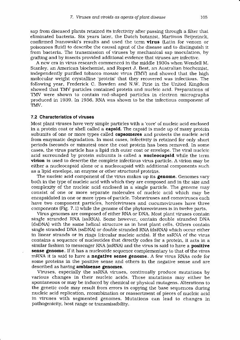
The nucleic acid component of the virus makes up its genome. Genomes varyboth in the type of nucleic acid with which they are composed and in the size andcomplexity of the nucleic acid enclosed in a single particle. The genome mayconsist of one or more separate molecules of nucleic acid which may beencapsidated in one or more types of par[icle. Tobraviruses and comoviruses eachhave two component particles, hordeiviruses and cucumoviruses have threecomponents (Fig. 7.1) while the genome of the phytoreoviruses is in twelve parts.
Virus genomes are composed of either RNA or DNA. Most plant viruses containsingle stranded RNA (ssRNA). Some however, contain double stranded DNA(dsDNA) with the same helical structure as in host plant cells. Others containsingle stranded DNA (ssDNA) or double stranded RNA (dsRNA) which occur eitherin linear strands or in rings (circular nucleic acids). If the ssRNA of the viruscontains a sequence of nucleotides that directly codes for a protein, it acts in asimilar fashion to messenger RNA (mRNA) and the virus is said to have a positivesense genome. If it has a nucleotide sequence complementary to that of the virusmRNA it is said to have a negative sense genome. A few virus RNAs code forsome proteins in the positive sense and others in the negative sense and aredescribed as having ambisense genomes.
Viruses, especially the ssRNA viruses, continually produce mutations byvarious changes in their nucleic acids. These mutations may either bespontaneous or may be induced by chemical or physical mutagens. Alterations tothe genetic code may result from errors in copying the base sequences duringnucleic acid replication, recombination or reassortment of pieces of nucleic acidin viruses with segmented genomes. Mutations can lead to changes inpathogenicity, host range or transmissibility.
dsDNl (RT)
ffi\Y2Caulimovirus
C:-Badnavirus
106 Jofut Randles and Helen OgLe
z.G
z-.Y
dsFN.A
ReoviridaeFhytoreovirus
FijivirusOryzavirus
G Aq4 E#Partitiviridae
AlphacryptovirusEetacryptovirus
l--,t+J
1 0 0 n m
ssRNA (+)
ffi\:zSequiviridaeTombusviridae
6e#DianthovirusLutecvirusMachlomovirusMarafivirusNecrovirusSobemovirusTymovirus
fAffi\;. --Enamovirusldaeovirus
Bramoviridae
ffifr,4gl \J/ \9
Af f i rA .g 4 y v rv - z
llarvirus
A A f f i Ae (r# ffiti ffilo r y H qAlfamovirus
<C>
fA fn\t' -'
ComoviriCae
Tobamovirus
Tobravirus
Hordeivirus
Furovirus
Cap i I lovirus. Trichov i rus
7.L The v.iruses wtrich infect plants. The properties used to distinguis! theviruses are the type of nucleic acid in the virus genome (single ordouble stranded DNA or RNA), the shape, size and number of theirparticles and the presence or absence of an envelope around the virusparlicles. The names of the genera or groups are often derived from thenarne of the first-named member in the group or some characteristic oftlle particle. (From Murphy et al., 1995.)
ssDNAGeminiviridae
@@"Subgroup l , l lGeminiv i rus"
@off i"Subgroup l l lGeminiv i rus"
Fotexvirus
l+tlr-.rl: - l ti t=---------11.t # F! - F! - F
l -1 .t - bl r - FlF--�ll + b, {#1.l - F
I r------11a - l.i l------------'l I
\#tRhabdoviriCae
CytorhabdovirusNuc!eorhabdovi rus
Bunyavir!dae
Clostercvirus
Figure
7. Viruses and utroids as agents of plant disease t07
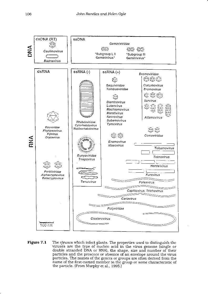
Most viruses encode at least four, and often more, proteins. These are the viruscapsid protein(s), some proteins (usually two) involved in virus replication and aprotein(s) involved in movement of the virus from one cell to the next (Fig. 7.2).Other vira-l proteins may include a helper protein that facilitates transmission bya vector and proteins of unknown function. Because viruses can be purified andsometimes crystallised, they have provided excellent models for studyinginteractions between proteins as well as contributing generally to studies of thestructure of biological molecules. Recent work has also shown the interplaybetween mutations in the genome of vimses and changes on the surface of theparticle. These in turn affect the ability of the virus to induce and bind to aunique diagnostic antibody, to be picked up by a vector and to infect aninoculated plant cell.
genomic RNA
-l_Ji----J-
II t ranslatronY
I+
lranslation
M r 1 2 6 . 0 0 0 p o l y p e p t i d e
Mr 1 83 ,000 po l ypep t i de
subgenomic mFNAf o r t h e m o v e m e n t p r o t e i n
m o v e m e n t p r o t e r n-+
subgenomrc mRNAl ^ , r h ^ ^ ^ ^ - , rr o r r n e c a p s r o p r o t e r n
I
{ t ranslat ion
c a p s r d p r o t e i n->
Figure 7.2 A diagram showing the genome of tobacco mosaic virus. The genome is a9ingl9 linear strand of RNA. The genes are shown as open reading frames(nucleic acid sequences which can be potentially
-translated into a
polypeptide sequence) which are translated into the enzymes associatedwith replication of ttre virus (molecular mass of 126,000 and 183,0O0), themovement protein which assists in the passage of the virus through theplasmodesmata into cells adjacent to tl-e infected cells and the capsid orcoat protein. (From Murphy et al, 199b.)
Viruses induce disease by multiplyrng in their host. They use amino acids andnucleotides synthesised in the cells of their hosts to build viral nucleic acids andproteins. The energy required for this process is supplied by the host and thevirus uses its mRNA to direct the host ceil 's protein-synthesising system tomanufacture viral proteins. A few viruses are 'defective'
in the senJe ihat tfreyrequire another virus (a helper virus) to supply the en4rmes required for theirreplication. Such dependent viruses are known as satellite viruses. Some
108 John Randles and Helen OoLe
viruses also have satellite RNAS which depend on the helper virus for repiicationand which are also encapsidated into particles using the coat protein of thehelper virus. The genomes of both tlpes of satellite are unrelated to those of thehelper virus. However, they frequently modiff the severity of the symptomsinduced by the helper virus.
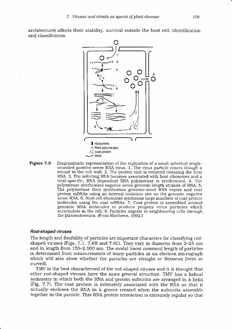
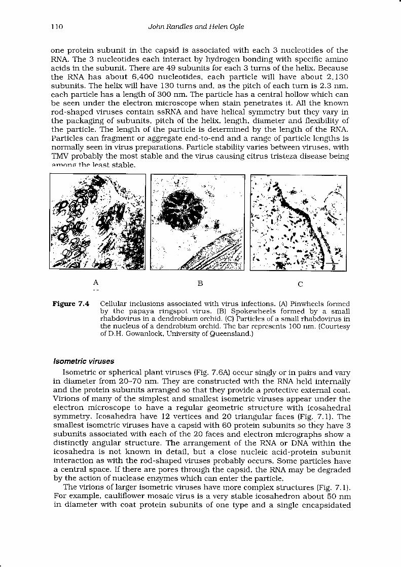
At the cellular level, when a virus infects a plant, its first point of contact iswith the outer cell membrane or plasmalemma. The protein coat may be removedfrom the genome here or inside the ceil (Fig. 7.3). If the genome is RNA, it acts asa mRNA, using the ribosomes from the cl.toplasm, cellular transfer RNAs andamino acids to translate the first of the virus-encoded genes into the replicationassociated protein(s). It appears that the host's nucleus or membranes areassociated with the synthesis of new virus RNA by the virus replicase enzyme.The newly slmthesised RNA includes the gene for the virus coat protein which isalso translated in the host cell. DNA genomes of viruses are transcribed to mRNAbefore virus replication commences. Assembly of the genome and the coat proteinto produce new virus particles then completes the replication cycle. Replication ofmany kinds of viruses takes place in distinctive virus-induced regions of the hostcell's cytoplasm called viroplasms. These sites often contain virus particles orbecome enriched with virus components which are described as inclusion bodies(Fig. 7.4). Some types of inclusion body are specific for the type of virus. Forexample, pinwheel shaped or laminate inclusion bodies as well as virus particlesare often visible in cells infected with the elongated potyviruses. Viruses whichare enclosed in a lipid envelope acquire it by passing through specific sites on cellmembranes. As well as producing new virus particles, replication induces anumber of cytopathic changes in the cells of the host plant including thepresence of intracellular inclusions or virus particles, alterations to cellularorganelles and phloem necrosis.
Viruses cannot penetrate plant surfaces on their own. They either avoid theneed to invade the plant's surface by being transmitted in seed or duringvegetative propagation or they enter the plant by some method that involvespenetration through a wound in the surface layers of the plant and introductio-nof the virus into the cytoplasm of cells in new host plants. Wounds could be madeduring mechanical inoculation or by vectors (see Chapter 15).
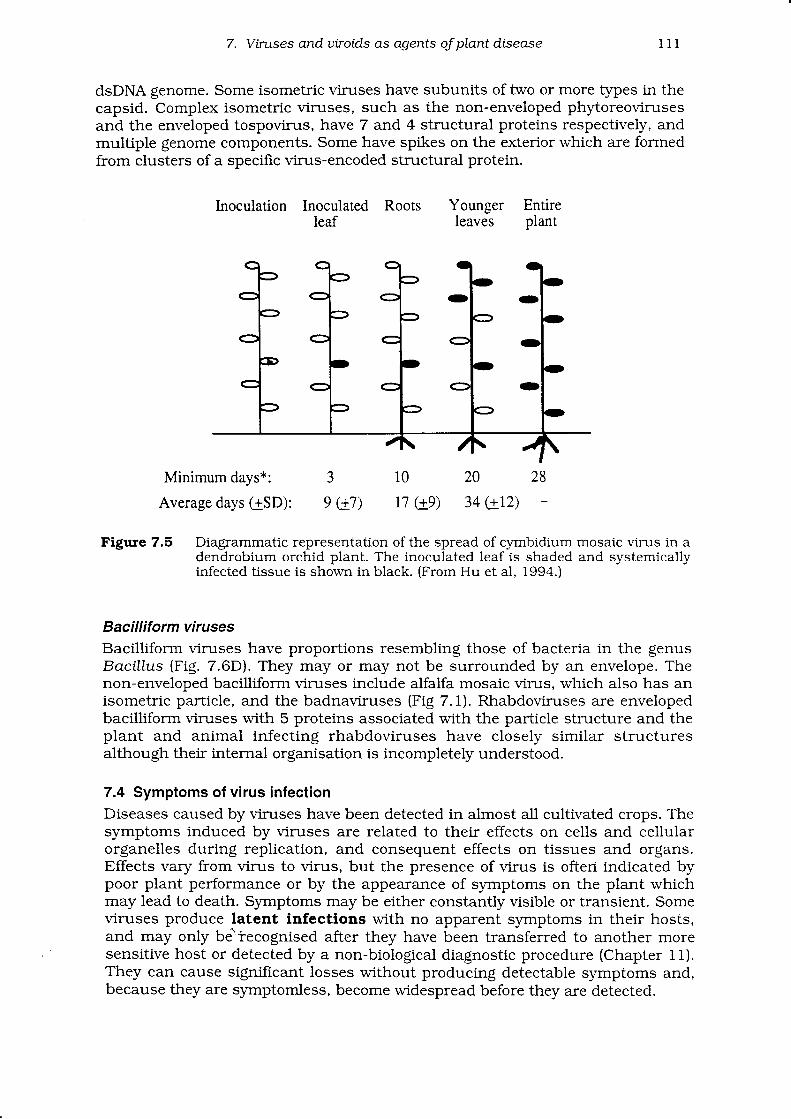
Some viruses are confined to one cell or a few cells at the point of inoculation.Others spread through the plant systemically. Virus particles have no means ofmoving themselves and are dependent on their hosts for movement within andbetween cells and for long distance transport within the plant. Either virusparticles or the genome are chaperoned through plasmodesmata by processesinvolving the virus movement protein. When the virus reaches the vasculartissue, it is distributed rapidly through the plant via the phloem and becomessystemic. Viruses generally move first to the roots and top leaves before infectingthe remaining leaves from the top of the plants downwards (Fig 7.5).
Viruses vary widely in the number of hosts they can infect. Cucumber mosaicvirus has a wide host range infecting 476 species of plants in 67 families whilelettuce necrotic yellows has a relatively narrow host range infecting specles inonly three families.
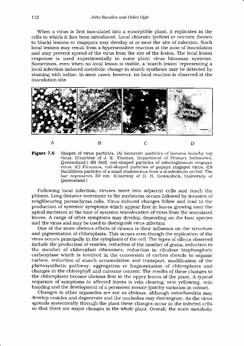
7.3 Structure and architectureVirus particles are too small to be seen with a light microscope but their shapeand size can be readily determined using a transmission electron microscope.They vary greatly in size and shape and may be rod-shaped, isometric orbacil l i form (Fig. 7.6).The way in which part icles are constructed (their
architecture) affects their stability,and classilication.
7. Vintses anduiroid.s as agents of plantdisease 109
survival outside the host ceil. identification
o
\ / -I ribosomeso RNA polymerase
j;ogo'.ot"'n
Figure 7.3 Diagrammatic representation of the replication of a small spherical single-stranded positive-sense RNA virus. 1, The virus particle enters though awound in the cell wall. 2, The protein coat is removed releasing the viralRNA. 3, The infecting RNA becomes associated with host ribosomes and aviral-specific, RNA dependent RNA polymerase is synthesised. 4, Thepoll'rnerase slmthesises negative sense genomic-length strands of RNA. 5,The polymerase then synthesises genomic-sized RNA copies and coatprotein mRNAs using an internal initiation site on the genomic negativesense RNA. 6, Host cell ribosomes synthesise large numbers of coat proteinmolecules using the coat mRNAs. 7, Coat protein is assembled aroundgenomic RNA molecules to produce progeny virus particles whichaccumulate in the cell. 8, Particles migrate to neighbouring cells throughthe plasmodesmata. (From Matthews, 1992.)
Rod-shaped viruses
The length and flexibility of particles are important characters for classifying rod-shaped viruses (Figs. 7.I, 7.68 and 7.6C). They vary in diameter from 3-25 nmand in length from 15O-2,OOO nm. The modal (most common) length of particlesis determined from measurements of many particles in an electron micrographwhich will also show whether the parlicles are straight or flexuous (bent orcurved).
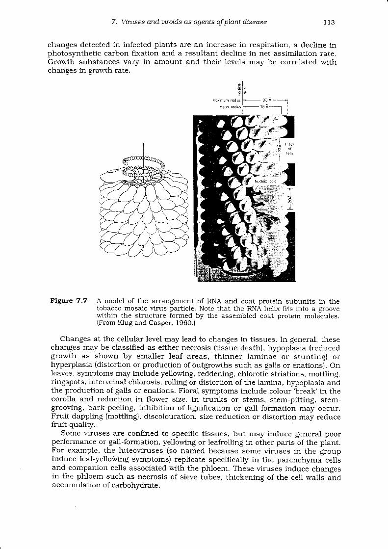
Tl/tV is the best characterised of the rod-shaped viruses and it is thought thatother rod-shaped viruses have the same general structure. TIrI\/ has a helicalsymmetry in which both the RNA and protein subunits are arranged in a helk(Fig. 7.7). The coat protein is intimately associated with the RNA so that itactuaily encloses the RNA in a groove created when the subunits assembletogether in the particle. This RNA protein interaction is extremely regular so that
r 1 0 John Randles and Helen OaIe
one protein subunit in the capsid is associated with each 3 nucleotides of theRNA. The 3 nucleotides each interact by hydrogen bonding',^rith specific aminoacids in the subunit. There are 49 subunits for each 3 turns of the helix. Becausethe RNA has about 6,400 nucleotides, each particle will have about 2,130subunits. The helix will have 130 turns and, as the pitch of each turn is 2.3 nm,each particie has a length of 3OO nm. The particle has a central hollow which canbe seen under the electron microscope when stain penetrates it. All the knownrod-shaped viruses contain ssRNA and have helical symmetry but they vary inthe packaging of subunits, pitch of the helix, length, diameter and flexibility ofthe particle. The length of the particle is determined by the length of the RNA.Particles can fragment or aggregate end-to-end and a range of parLicle lengths isnormally seen in virus preparations. Parlicle stability varies between viruses, withTiWV probably the most stable and the virus causing citrus tristeza disease beingqrnnnd the least stable.
A C
Figure 7.4 Cellular inclusions associated with virus infections. (A) Pinwheels formedby the papaya ringspot virus. (B) Spokewheels formed by a smallrhabdovirus in a dendrobium orchid. (C) Particles of a small rhabdovirus inthe nucleus of a dendrobium orchid. The bar represents IOO nm. (Courtesyof D.H. Gowanlock, University of Queensland.)
Isometric viruses
Isometric or spherical plant viruses (Fig. 7.6A) occur singly or in pairs and varyin diameter from 2O-7O nm. They are constructed with the RNA held internallyand the protein subunits arranged so that they provide a protective external coat.Virions of many of the simplest and smallest isometric viruses appear under theelectron microscope to have a regular geometric structure with icosahedralsymmetry. Icosahedra have 12 vertices and 2O triangular faces (Fig. 7.1). Thesmallest isometric viruses have a capsid with 60 protein subunits so they have 3subunits associated with each of the 20 faces and electron micrographs show adistinctly angular structure. The arrangement of the RNA or DNA within theicosahedra is not known in detail, but a close nucleic acid-protein subunitinteraction as with the rod-shaped viruses probably occurs. Some particles havea central space. If there are pores through the capsid, the RNA may be degradedby the action of nuclease en4lrnes which can enter the particle.
The virions of larger isometric viruses have more complex structures (Fig. 7. t).For example, cauliflower mosaic virus is a very stable icosahedron about 5O nmin diameter with coat protein subunits of one type and a single encapsidated
B
7. Vintses and utroid.s as agents oJ plant disease
dsDNA genome. Some isometric vimses have subunits of two or more types in thecapsid. Complex isometric viruses, such as the non-enveloped phyloreovirusesand the enveloped tospovirus, have 7 and 4 structural proteins respectively, andmultiple genome components. Some have spikes on the exterior which are formedfrom clusters of a specific virus-encoded structural protein.
1 1 1
Minimum days*:
Average days (-rSD):
lnoculation Inoculated Rootsleaf
Younger Entireleaves plant
20
34 en)3
e c7)10
17 Ge)
28
Figure 7.5 DiagrammaUc representation of the spread of cyrnbidium mosaic virus in adendrobium orchid plant. The inoculated leaf is shaded and systemicallyinfected tissue is shown in black. (From Hu et al, 1994.)
Bacilliform viruses
Bacilliform viruses have proporlions resembling those of bacteria in the genusBacillus (Fig. 7.6D). They may or may not be surrounded by an envelope. Thenon-enveloped bacilliform viruses include alfalfa mosaic virus, which also has anisometric particle, and the badnaviruses (Fig 7.1). Rhabdoviruses are envelopedbacilliform viruses with 5 proteins associated with the parlicle structure and theplant and animal infecting rhabdoviruses have closely similar structuresalthough their internal organisation is incompletely understood.
7.4 Symptoms of virus infectionDiseases caused by viruses have been detected in almost all cultivated crops. Thesymptoms induced by viruses are related to their effects on cells and cellularorganelles during replication, and consequent effects on tissues and organs.Effects vary from virus to virus, but the presence of virus is ofteri indicated bypoor plant performance or by the appearance of symptoms on the plant whichmay lead to death. Symptoms may be either constantly visible or transient. Someviruses produce latent infections with no apparent symptoms in their hosts,and may only be" iecognised after they have been transferred to another moresensitive host or detected by a non-biological diagnostic procedure (Chapter 1I).They can cause significant losses without producing detectable symptoms and,because they are symptomless, become widespread before they are detected.
t12 John Randle s and Helen OgLe
When a virus is first inoculated into a susceptible plant, it replicates in thecells to which it has been introduced. Local chlorotic (yellow) or necrotic (brownto black) lesions or ringspots may develop at or near the site of infection. Suchlocal lesions may result from a hypersensitive reaction at the zone of inoculationand may prevent spread of the virus from the site of the lesion. The local lesionresponse is used experimentally in some plant virus bioassay systems.Sometimes, even when no local lesion is visible, a 'starch lesion' representing alocal infection-induced metabolic change in starch synthesis may be detected bystaining with iodine. In most cases, however, no local reaction is observed at theinoculation site.
c
Ftgure 7.6 Shapes of virus particles. (A) Isometric particles of banana bunchy topvirus. (Courtesy of J. E. Thomas, Department of Primary Industries,Queensland.) (B) Stiff, rod-shaped particles of odontoglossum ringspotvirus. (C) Flexuous, rod-shaped particles of papaya ringspot virus. (D)Bacilliform particles of a small rhabdovirus from a dendrobium orchid. Thebar represents 5O nm. (Courtesy of D. H. Gowanlock, University ofQueensland.)
Following local infection, viruses move into adjacent cells and reach thephloem. lnng distance movement to the meristems occurs followed by invasion ofneighbouring parenchyma cells. Virus induced changes follow and lead to theproduction of systemic symptoms which appear first in leaves growing near theapical meristem at the time of systemic translocation of virus from the inoculatedleaves. A range of other symptoms may develop, depending on the host speciesand the virus and may be used to distinguish virus infection
One of the more obvious effects of viruses is their influence on the structureand pigmentation of chloroplasts. This occurs even though the replication of thevirus occurs principally in the cytoplasm of the cell. The types of effects observedinclude the production of vesicles, reduction of the number of grana, reduction inthe number of chloroplast ribosomes, reduction in ribulose bisphosphatecarboxylase which is involved in the conversion of carbon dioxide to organiccarbon, reduction of starch accumulation and transport, modification of thephotosynthetic pathway, aggregation or fragmentation of chloroplasts andchanges in the chlorophyll and carotene content. The results of these changes tothe chloroplasts become obvious first in the upper leaves of the plant. A typicalsequence of symptoms in affected leaves is vein clearing, vein yellowing, veinbanding and the development of a persistent mosaic (patchy variation in colour).
Changes in other organelles are not as obvious, although mitochondria maydevelop vesicles and degenerate and the nucleolus may disintegrate. As the virusspreads systemically through the plant these changes occur in the infected cellsso that there are major changes in the whole plant. Overall, the main metabolic
DBA
7. Viruses and utroid.s as agents oJ plant disease
changes detected in infected plants are an increase in respiration, a decline inphotosynthetic carbon fixation and a resultant decline in net assimilation rate.Growth substances vary in amount and their levels may be correlated withchanges in growth rate.
1 1 3
g.9
&
Mqximum /odius
l\4eon rodius
Figure 7.7 A model of the arrangement of RNA and coat protein subunits in thetobacco mosaic virus particle. Note that the RNA helix fits into a groovewithin the structure formed by the assembled coat protein molecules.(From Klug and Casper, 1960.)
Changes at the cellular level may lead to changes in tissues. In general, thesechanges may be classified as either necrosis (tissue death), hypoplasia (reducedgrowth as shown by smaller leaf areas, thinner laminae or stunting) orhyperplasia (distortion or production of outgrowths such as galls or enations). Onleaves, symptoms may include yellowing, reddening, chlorotic striations, mottling,ringspots, interveinal chlorosis, rolli.ng or distortion of the lamina, hypoplasia andthe production of galls or enations. Floral symptoms include colour 'break' in thecorolla and reduction in flower size. In trunks or stems, stem-pitting, stem-grooving, bark-peeling, inhibition of lignification or gall formation may occur.Fn.it dappling (mottling), discolouration, size reduction or distortion may reducefruit quality.
Some viruses are confined to specific tissues, but may induce general poorperformance or gall-formation, yellowing or leafrolling in other parts of the plant.For example, the luteoviruses (so named because some viruses in the groupinduce leaf-yellowing symptoms) replicate specifically in the parenchyma cellsand companion cells associated with the phloem. These viruses induce changesin the phloem such as necrosis of sieve tubes, thickening of the cell wails andaccumulation of carbohvdrate.
1 1 4 John Randles and Helen OgLe
The productivity of virus-infected plants may be reduced by virus-inducedhormonal changes, reduced availability of photoslmthates, direct effects on thechloroplasts, reduced translocation of starch, reduced stomatal opening, lesseffective leaf posture, reduced leaf initiation, reduced fixation of nitrogen inlegumes, reduced nutrient uptake, epinasty (downward bending of the petiole)and leaf rolling, increased senescence and premature abscission of leaves, ortumour, gall or enation production. In addition to lost productivity, there areadverse effects on the quality of produce, the competitive ability of the plant in acommunity such as a pasture or crop and the longevity of the plant.
Some viruses are lethal. One example of a lethal virus disease is 'quick decline'of citrus trees propagated on sour orange rootstocks, in which cytopathic effectsof the citrus tristeza virus on the rootstock cause rapid death of the scion.Another is coconut foliar decay virus infection of certain coconut varieties inVanuatu which is associated with damage to the phloem, frond necrosis anddeath of palms.
Mixed virus infections are common in vegetatively propagated plants, andhistorically the 'rrnning out' of potato varieties and the decline in productivity ofstrawberrSr crops, are good examples of the effect of cumulative infections with anumber of viruses on plant performance. Mixed infections may result in only anadditive effect of the combined viruses, but there are many examples ofslmergism, where mixed infections produce a more severe disease than expectedfrom the additive effects. Co-infection of tobacco with potato virus Y caused up toa lo-fold increase in the amount of potato virus X and this was correlated withthe development of very severe symptoms. However, interference with virusmultiplication typically occurs between strains of the same virus and this 'cross
protection' response forms the basis of one means of reducing the severity oflosses due to virus infection.
Factors affecti ng symptom expressionThe environment affects the susceptibility of plants to infection and theexpression of s1'rnptoms in plants. For example, shading plants before inoculationcan significantly increase their susceptibility to mechanical inoculation. The timeof day when inoculation is carried out can also affect the number of successfulinoculations obtained. Sometimes high light intensity and long days may favourvirus replication, but high light intensity may also lead to the induction of miidersymptoms than at low light intensity. Drought reduces replication and symptomseverity, but this effect has been little studied. Nutritional conditions favourablefor plant growth generally also favour virus replication. Temperature has a majoreffect on virus replication. Up to about 32'C the speed of virus replicationgenerally increases with increasing temperature and the time between inoculationand producilon of first symptoms decreases correspondingly. Temperatures aboveabout 33'C inhibit replication. As plants age they become less susceptible tovirus infection. The developmental stage is also important as susceptibility ofexperimental hosts has been found to be low or nil after flowering hascommenced.
7.5 Transmission of virusesThe virus life-cycle includes a phase within the protoplast of the host, duringwhich replication, maturation of virions and translocation takes place, and aphase outside the host when the intact virion moves to a new host. Thetransmission of viruses to new hosts is an essential part of the cycle of disease,and usually involves a vector (see Chapter l5), or carrier, of the virion to a new
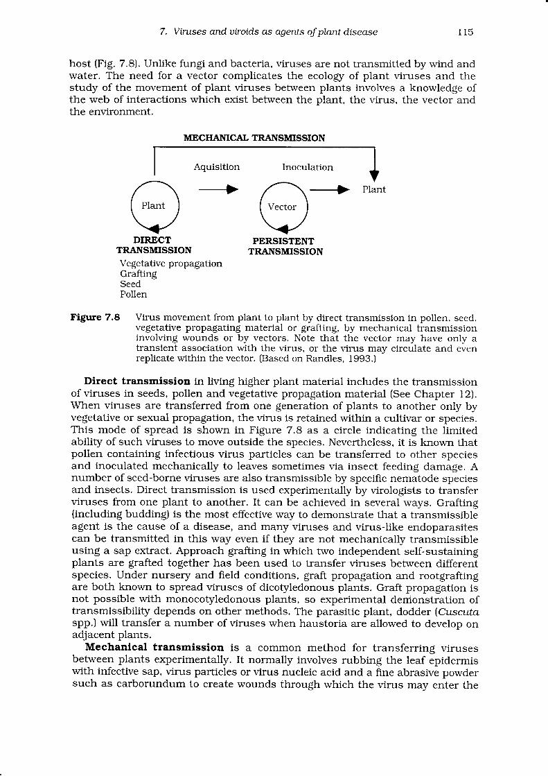
7. Vintses anduiroids as agents oJplantdisease 1 1 5
host (Fig. 7.8). Unlike fungi and bacteria, viruses are not transmitted by wind andwater. The need for a vector complicates the ecology of plant viruses and thestudy of the movement of plant viruses between plants involves a knowiedge ofthe web of interactions which exist between the plant. the virus. the vector andthe environment.
Aquisition Inoculation
,^./ \I Plant )\ /\ . /vDIR-ECT
TRANSMISSIONVegetative propagationGraftingSeedPollen
MECI{AT{ICAL TRANSMISSION
-+ piant
I Vector ]
VPERSISTENT
TRANSMISSION
Figure 7.8 Virus movement from plant to plant by direct transmission in pollen, seed,vegetative propagating material or grafting, by mechanical transmissioninvolving wounds or by vectors. Note that the vector may have only atransient association with the virus, or the virus may circulate and evenreplicate within the vector. (Based on Randles, 1993.)
Direct transmission in living higher plant material includes the transmissionof viruses in seeds, pollen and vegetative propagation material (See Chapter l2).When viruses are transferred from one generation of plants to another only byvegetative or sexual propagation, the virrs is retained \Mithin a cultivar or species.This mode of spread is shown in Figure 7.8 as a circle indicating the limitedability of such viruses to move outside the species. Nevertheless, it is known thatpollen containing infecilous virus particles can be transferred to other speciesand inoculated mechanically to leaves sometimes via insect feeding damage. Anumber of seed-borne viruses are also transmissible by specific nematode speciesand insects. Direct transmission is used experimentally by virologists to transferviruses from one plant to another. It can be achieved in several ways. Grafting(including budding) is the most effective way to demonstrate that a transmissibleagent is the cause of a disease, and many viruses and virus-like endoparasitescan be transmitted in this way even if they are not mechanically transmissibleusing a sap extract. Approach grafting in which two independent self-sustainingplants are grafted together has been used to transfer viruses between differentspecies. Under nursery and field conditions, graft propagation and rootgraftingare both known to spread viruses of dicotyledonous plants. Graft propagation isnot possible with monocot5rledonous plants, so experimental derrionstration oftransmissibility depends on other methods. The parasitic plant, dodder (Cuscutaspp.) will transfer a number of viruses when haustoria are allowed to develop onadjacent plants.
Mechanical transmission is a common method for transferring virusesbetween plants experimentally. It normally involves rubbing the leaf epidermiswith infective sap, virus particles or virus nucleic acid and a fine abrasive powdersuch as carborundum to create wounds through which the virus may enter the
l 1 6 J ohn Randle s and Helen OoIe
leaf. Not all viruses are mechanically transmissible, particularly those that arelimited to the phloem or other specific tissues. Despite the ease of mechanicaltransmission under experimental conditions, transfer of virus from one host toanother without the intervention of a vector is not common in nature. It is onlyknown to be important for very stable vimses such as tobacco mosaic virus andpotato virrs X. These viruses can contaminate structures, tools and soil debris,and wounding of a host plant allowing contact of the tissue with a source of viruscan lead to infection.
Vectors (see Chapter 15) can transmit viruses across plant species barriers,and their activity and type of association with the virus determines the range,rate, timing and pattern of spread of the viruses they transmit. Figure 7.8indicates that the vector may have a transient association with the virus so that itacquires the virus during feeding on infected plants and is able to transmit itimmediately. This mode of transmission is described as non-persistent, and istypical of transmission of many viruses by aphid vectors. The virus is associatedwith ducts within the vector's stylet and most viruses transmitted in this wayhave a number of different vector species. Vectors remain infective for up to about4 hours. A variant of this type of transmission is the semi-persistenttransmission found in some leafhopper-virus and aphid-virus systems in whichinfectivity is retained for up to about 1OO hours. Alternatively, the vector mayhave a much longer feeding time followed by a period during which it is unable toinfect plants on which it feeds. This is described as persistent transmissionbecause the vectors typically retain the ability to transmit virus for the rest oftheir lives.
7.6 ldentification of viruses
Disease symptoms caused by viruses are frequently similar to the symptomsassociated with nutrient deficiencies or excesses, or to chemical toxicitiesassociated with agricultural chemicals such as herbicides. Plant pathologistshave two ways to implicate a virus as the probable cause of a disorder. The firstway is to map the distribution of the affected plants. Viruses which are vector-borne frequently show patterns either with distinct foci, or random distributions,depending on how far the vector has travelled or how much plant to plant spreadhas occurred within the crop (see Chapter 12 for more detail on interpretingpatterns of disease distribution). Rarely are all plants affected in a site. Incontrast, abiotic nutrient or chemically induced diseases generally show a patternassociated with soil type or the application of chemicals where all plants showsymptoms. It is only when the incidence of a virus disease approaches 1OO0/o thatit is difficult to determine whether a virus is the cause of a disease. For example,severe cucumber mosaic virus epidemics in narrow leafed lupin crops causealmost all plants to be stunted and infertile as might be expected after a herbicideapplication. The second method is to demonstrate transmissibility of .thes1'rnptom either by graft or mechanical sap inoculation to plants of the same andrelated species under experimental conditions. This is the first step in applyingKoch's postulates for establishing the cause of disease.
Virologists rely on good insect-free greenhouse and associated facilities fordiagnosis by infectivity assays, host range studies, vector studies and thepropagation of plants so that virus concentrations reach sufficiently high levelsfor either purification or component analysis. Herbaceous indicator plants whichexpress different symptoms when inoculated with different viruses assist virusidentification (Table 7.I). In the laboratory, routine diagnostic methods based onserology and electron microscopy are now being supplemented by component
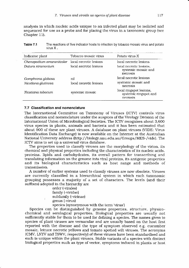
7. Virtses anduiroid.s as agents oJplantdisease
analysis in which nucleic acids unique to an infected plant may be isolated andsequenced for use as a probe and for placing the virus in a taxonomic group (seeChapter 11).
Table 7.1 The reactions of five indicator hosts to infection bv tobacco mosaic virus and ootatovirus X .
t17
Indicator plant Tobacco mosaic virus Potato virus X
Che nop o dium amar anttc olorDatura stramonium
Gomplvena gLobosaNicottana gtutinosa
Nicottana tabacum
local necrotic lesionslocal necrotic lesions
nillocal necrotic lesions
systemic mosaic
local necrotic lesionslocal necrotic lesions.
systemic mosaic andnecrosis
local necrotic lesionssystemic mosaic and
necrosislocal ringspot lesions,
systemic ringspot andnecrosis
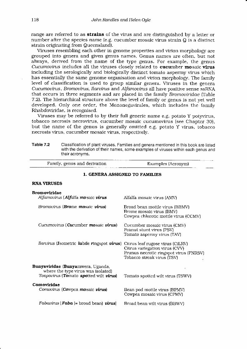
7.7 Classif ication and nomenclature
The International Committee on Taxonomy of Viruses (ICTV) controls virusclassification and nomenclature under the auspices of the Virologg Division of theInternational Union of Microbiological Societies. The ICTV recognises about 3,600virus species in plants, animals and bacteria and it has been estimated thatabout 9OO of these are plant viruses. A database on plant viruses MIDE: VirusIdentification Data Exchange) is now available on the Internet at the AustralianNational University address (http:/ /biologr.anu.edu.aulGroups/MES-/vide). TheICTV aims to set up a universal virus database.
The properties used to classiff viruses are the morphology of the virion, itschemical and physical properties including the characteristics of its nucleic acids,proteins, lipids and carbohydrates, its overall pattern for transcribing andtranslating information on the genome into viral proteins, its antigenic propertiesand its biological characterist ics such as host range and methods oftransmission.
A number of earlier systems used to classi$r viruses are now obsolete. Virusesare currently classified in a hierarchical system in which each taxonomicgrouping possesses a majority of a set of characters. The levels (and theirsuffixes) adopted in the hierarchy are
order (-virales)family (-viridae)subfamily (-virinae)genus (-virus)species (synonymous with the term'virus')
Species can be distinguished by genome properties, structure, physico-chemical and serological properties. Biological properties are usually notsufficiently stable for them to be used for defining a species. The names given tospecies of plant viruses are verrracular and are usually based on the host firstreported with the disease and the type of symptom observed e.g. cucumbermosaic, lettuce necrotic yellows and tomato spotted wilt viruses. The acronyms(CMV, LNYV and TSWV, respectively) of these viruses have been standardised andeach is unique within the plant viruses. Stable variants of a species with distinctbiological properties such as type of vector, symptoms induced in plants or host
1 1 8 John Randles and Helen OgIe
range are referred to as strains of the virus and are distinguished by a letter ornumber after the species name (e.g. cucumber mosaic virus strain Q is a distinctstrain originating from Sueensland).
Vimses resembling each other in genome properlies and virion morpholory aregrouped into genera and given genus names. Genus names are often, but notalways, derived from the name of the type genus. For example, the genusCtrcumouittts includes ail the viruses closely related to cucumber mosaic virusincluding the serologically and biologically distinct tomato asperrny virus whichhas essentially the same genome organisation and virion morphology. The familylevel of classification is used to group similar genera. Viruses i.n the generaCtrcumouirus, Bromouints,Ilaruirus and ALfamouirus all have positive sense ssRNAthat occurs in three segments and are placed in the family Bromouirtdae (Table7.2). The hierarchical structure above the level of family or genus is not yet welldeveloped. Only one order, the Mononegavirales, which includes the familyRhabdoviridae, is recognised.
Viruses may be referred to by their full generic name e.g. potato Y potyrrirus,tobacco necrosis necrovirus, cucumber mosaic cucumovirus (see Chapter 30),but the name of the genus is generally omitted e.g. potato Y virus, tobacconecrosis virus, cucumber mosaic virus, respectively.
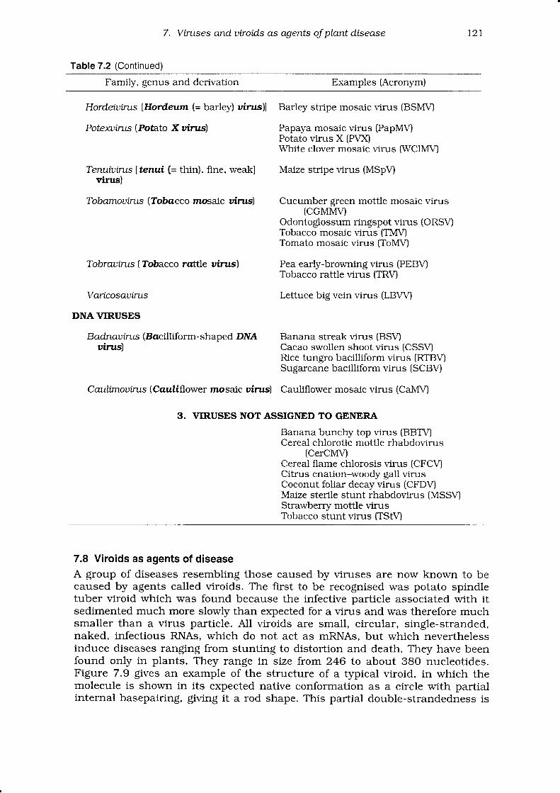
Table7.2 Classification of plant viruses. Families and genera mentioned in this book are listedwith the derivation of their names, some examples of viruses within each genus andtheir acronvms.
Family, genus and derivation Examples (Acronym)
1. GENERAASSIGNED TO FAIVIILIES
RNA VIRUSES
BromoviridaeAlJamouins (Alfalfa mosaic uirus
Bromouints (Brome mosaic uinrs)
Cucumouins (Crcumber mosaic uinrc)
Ilaruints (fsometric labile ringspot uirus)
Buryaviridae (Brrnyamwera, Uganda,where the type virus was isolated)
Tospourus (Tomato spotted wilt uinrs)
ComoviridaeComouints (Cowpea mosaic uirus)
Fabautns |Faba (= broad bean) uinrsl
Alfalfa mosaic virus (AMU
Broad bean mottle virus (BBIVI\4Brome mosaic virus (BMV)Cowpea chlorotic mottle virus (CCMV)
Cucumber mosaic virus (CMV)Peanut stunt virus (PSV)Tomato asperrny virus (TAVJ
Citrus leaf rugose virus (CiLRVJCitrus variegation virus (CVVPrunus necrotic ringspot virus (PNRSVJTobacco streak virus (TSU
Tomato spotted wilt virus (TSWV)
Bean pod mottle virus (BPMVJCowpea mosaic virus (CPMV)
Broad bean wilt virus (BBWU
7. Vintses and uiroids as agents oJ plant disease 1 1 9
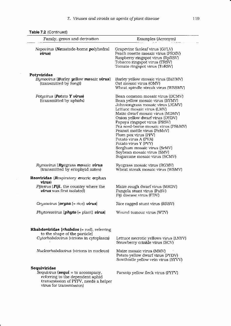
Table 7.2 (Continued)
Family, genus and derivation Examples (Acronym)
Nepournrs (Nernatode-bome polyhedraluirus)
PotyvlridaeBgmouirus (Barley yellow mosaic uirus)
(transmitted by fungi)
Potguirus (Potato Y uinrs)(transmitted by aphids)
Rymourms (Rgegrass mosaic uirus{transmitted by eriophyid mites)
Reoviridae (Respiratory enteric orphanvirus)
.Fy-.iurn-rs (I.iji, the country where theuirus was first isolated)
Oryzauirtts loryza (= rice) uirusl
Phgtoreouirus lphgto (= plant) uinrsl
Rlrabdoviridae lrhabd.os (= 1e6;, referringto the shape of the particlel
Cgtorhabdourus (virions in cytoplasm)
Nucleorhabdouirus {virions in nucleus)
SequiviridaeSequiurrus (segui = to accompany,
referring to the dependent aphidtransmission of PYFV, needs a helpervirus for transmission)
Grapevine fanleaf virus (GFLVPeach rosette mosaic virus (PRMQRaspberry ringspot virus (RpRSV)Tobacco ringspot virus (TRSVJTomato ringspot virus (ToRSV
Barley yellow mosaic virus (BaYMUOat mosaic virus (OMV)Wheat spindle streak virus IWSSMVI
Bean common mosaic virus (BCMV)Bean yellow mosaic virus (BYMVJohnsongrass mosaic virus (JGMUkttuce mosaic virus (LMV)Maize dwarf mosaic virus (MDMV)Onion yellow dwarf virus (OYDV)Papaya ringspot virus (PRSV)Pea seed-borne mosaic virus (Psbl\,[\/lPeanut motfle virus (PeMoV)Plum pox virus (PPVJPotato virus A (PVA)Potato virus Y (PVY)Sorghum mosaic virus (SrMVlSoybean mosaic virus (SMV)Sugarcane mosaic virus (SCMV)
Ryegrass mosaic virus (RGMV)Wheat streak mosaic virus (WSMV)
Maize rough dwarf virus (MRDV)Pangola stunt virus (PaSVFiji disease virus (FDV
Rice ragged stunt virus (RRSV)
Wound tumour virus [WTV)
Lettuce necrotic yellows virus (LNYV)Strawberr5r crinkle virus (SCVJ
Maize mosaic virus (MMVJPotato yellow dwarf virus (PYDV)Sowthisfle yellowvein virus (SYW)
Parsnip yellow fleck virus (PVnYl
r20
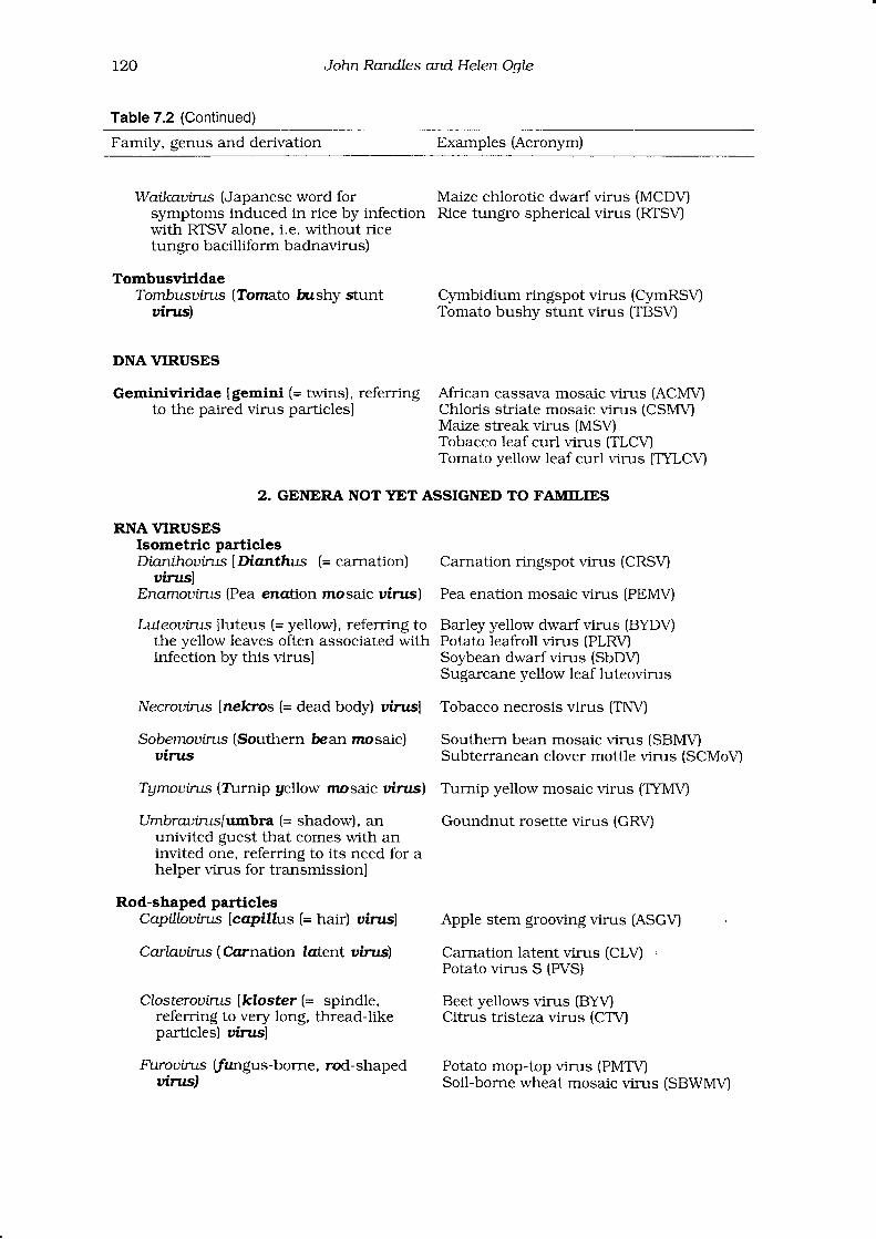
Table 7.2 (Continued)
John Randles and HeLen OoLe
Family, genus and derivation Examples (Acronym)
Watkauirus (Japanese word for Maize chlorotic dwarf virus (MCD\'Isymptoms induced in rice by infection Rice tungro spherical virus (RTSVJwith RTSV alone, i.e. without ricetungro bacilliform badnavirus)
TombusviridaeTombusuints (Tomato bushy stunt Cymbidium ringspot virus (CymRSVJ
uirus) Tomato bushy stunt virus (TBSVI
DNA VIRUSES
Geminiviridae [gemini (= twins), referring African cassava mosaic virus (ACMVIto the paired virus particlesl Chloris striate mosaic virus (CSMVJ
Maize streak virus (MSVTobacco leaf curl virus (TLCV)Tomato yellow leaf curl virus (TYLCV)
2. GENERA NOT YET ASSIGNED TO FAMILIES
RNA VIRUSESIsometric particlesDianthouirus [Dianthus (= carnation) Carnation ringspot virus (CRSV)
uiruslEnamouints (Pea enation mosaic uirus) Pea enation mosaic virus (PEMV)
Luteouirts fluteus (= yellow), referring to Barley yellow dwarf virus (BVO$the yellow leaves often associated with Potato leafroll virus (PLRV)infection by this virusl Soybean dwarf virus (SbDV)
Sugarcane yellow leaf luteovirus
Necrourrus lnekros (= dead body) uirus] Tobacco necrosis virus (TNVJ
Sobemouirus (Southern bean mosaic) Southern bean mosaic virus (SBlrI\4uinrs Subterranean clover mottle virus (SCMoVl
Tgmouirus (T\rrnip gellow mosaic uirus) Turnip yellow mosaic virus (TYMV)
Umbrauints[umbra (= shadow), an Goundnut rosette virus (GRV)univited guest that comes with aninvited one, referring to its need for ahelper virus for transmissionl
Rod-shaped particlesCapi\ouirus [capillus (= hair) uinrs] Apple stem grooving virus (ASGVI
Carlnuin:s (Ccrrnation tcrtent uinrs) Carnation latentvirus (CLVJPotato virus S (PVS)
Closterouutts lkloster (= spindle, Beet yellows virus (BYV)referring to very long, thread-like Citrus tristeza virus (CTV)parlicles) uinrsl
F-i.rourlrs [fungus-borne, rod-shaped Potato mop-top virus (PMTVJuittts) Soil-borne wheat mosaic virus (SBWMVI
7. Virtses and uiroid.s as agents oJ plant disease r21
Table 7.2 (Continued)
Family, genus and derivation Examples (Acronym)
Hordeiuints lHordeum (= barley) uirus)l Barley stripe mosaic virus (BSMVI
Potexuints (Potato X uirus) Papaya mosaic virus (PapMUPotato virus X (PVXWhite clover mosaic virus IWCIMV)
Tenuiuirus ltenui (= thin), fine, weakl Marze stripe virus (MSpV)virus)
Tobamouirus (?lobacco mosaic uinrs) Cucumber green mottle mosaic virus(CGMI\,M
Odontoglossum ringspot virus (ORS\'ITobacco mosaic virus (TMVJTomato mosaic virus (ToMVJ
Tobrauints (Tobacco rattle uirus) Pea early-browningvirus (PEBVTobacco rattle virus (TRV)
Varicosauints Lettuce big vein virus (LBVU
DNA VTRUSES
Badnadrus (Bacilliform-shaped DJYA Banana streak virus (BSUuirus) Cacao swollen shoot virus (CSS\4
Rice tungro bacilliform virus (RTBUSugarcane bacilliform virus (SCBV
Caulimouirus (Cauliflower mosaic uinrs) Cauliflower mosaic virus (CaMV)
3. VIRUSES NOT ASSIGNED TO GENERA
Banana bunchy top virus (BBTI'ICereal chlorotic mottle rhabdovirus
(CerCMV)Cereal flame chlorosis virus (CFCVJCitrus enation-woody gall virusCoconut foliar decay virus (CFDV)Maize sterile stunt rhabdovirus (MSSV)Strawberry mottle virusTobacco stunt virus (TSIV)
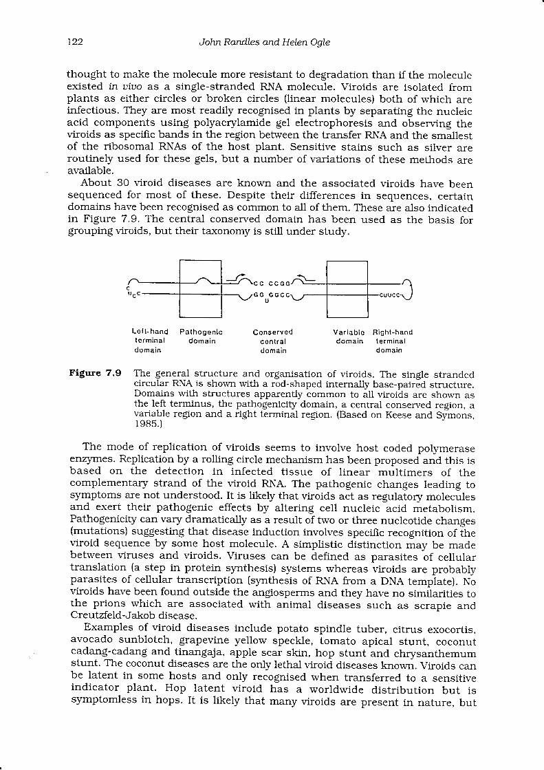
7.8 Viroids as agents of diseaseA group of diseases resembling those caused by viruses are now known to becaused by agents called viroids. The first to be recognised was potato spindletuber viroid which was found because the infective particle associated with itsedimented much more slowly than expected for a virus and was therefore muchsmaller than a virus particle. All viroids are small, ci.rcular, single-stranded,naked, infectious RNAs, which do not act as mRNAs, but which neverthelessinduce diseases ranging from stunting to distortion and death. They have beenfound only in plants. They range in size from 246 to about 380 nucleotides.Figure 7.9 gives an example of the structure of a typical viroid, in which themolecule is shown in its expected native conformation as a circle with partialinternal basepairing, giving it a rod shape. This partial double-strandedness is
r22 J ohn Randle s and HeLen OaIe
thought to make the molecule more resistant to degradation than if the moleculeexisted in uiuo as a single-stranded RNA molecule. Viroids are isolated fromplants as either circles or broken circles (linear molecules) both of which areinfectious. They are most readily recognised in plants by separating the nucleicacid components using polyacrylamide gel electrophoresis and observing theviroids as specific bands in the region between the transfer RNA and the smallestof the ribosomal RNAs of the host plant. Sensitive stains such as siiver areroutinely used for these gels, but a number of variations of these methods areavailable.
About 30 viroid diseases are known and the associated viroids have beensequenced for most of these. Despite their differences in sequences, certaindomains have been recognised as common to all of them. These are a-lso indicatedin Figure 7.9. The central conserved domain has been used as the basis forgrouping viroids, but their taxonomy is stiil under study.
c c c c o G
G G G G C
L o l t - h a n d P a t h o g o n l cte rmina l domaindomain
Conservedc on l ra ldomain
Vsr iab le R igh t -handdomain le rmina l
d o m a i n
Figure 7.9 The general structure and organisation of viroids. The single strandedcircular RNA is shown with a rod-shaped internally base-paired structure.Domains with structures apparently common to all viroids are shown asthe left terminus, the pathogenicity domain, a central conserved region, avariable region and a right terminal region. (Based on Keese and Symons,r985.)
The mode of replication of viroids seems to involve host coded poiymeraseenzyrnes. Replication by a rolling circle mechanism has been proposed and this isbased on the detection in infected t issue of l inear mult imers of thecomplementary strand of the viroid RNA. The pathogenic changes leading tosymptoms are not understood. It is likely that viroids act as regulatory moleculesand exert their pathogenic effects by altering cell nucleic acid metabolism.Pathogenicity can vary dramatically as a result of two or three nucleotide changes(mutations) suggesting that disease induction involves specific recognition of theviroid sequence by some host molecule. A simplistic distinction may be madebetween viruses and viroids. Viruses can be defined as parasites of cellulartranslation (a step in protein synthesis) systems whereas viroids are probablyparasites of cellular transcription (synthesis of RNA from a DNA template). Noviroids have been found outside the angiosperrns and they have no similarities tothe prions which are associated with animal diseases such as scrapie andCreutzfeld-Jakob disease.
Examples of viroid diseases include potato spindle tuber, citrus exocortis,avocado sunblotch, grapevine yellow speckle, tomato apical stunt, coconutcadang-cadang and tinangaja, apple scar skin, hop stunt and chrysanthemumstunt. The coconut diseases are the only lethal viroid diseases known. Viroids canbe latent in some hosts and only recognised when transferred to a sensitiveindicator plant. Hop latent viroid has a worldwide distribution but issymptomless in hops. It is likely that many viroids are present in nature, but
7. Viruses and utroi.d.s as agents oJ ptant disease t23
their detection will probably rely on the use of molecular diagnosis rather thanbiological methods. These methods have already detected viroid complexes incitrus and grapevine, for example, and sequence analysis has shown thatrecombination between viroids occurring in the same host is the probableexplanation for the origin of some new viroids. Citrus exocortis viroid has beenproposed as a possible dwarfing agent for citrus, as it has no effect on the fruit ofsweet orange scion varieties, but it reduces growth of some rootstock varieties.
Cytopathic changes associated with viroid infection include alterations to thecell wall, plasmalemma, chloroplasts and cellular proteins. The nucleus andnucleolus accumulate viroid in some systems, whereas the chloroplastaccumulates it in others. Viroids are translocated in plants but the method isthought to differ from that of viruses because no viroid coded movement proteinis likely to occur in the plant. Most viroids are spread via vegetative propagationbut they are also readily transmitted by mechanical inoculation and citrusexocortis is reported to spread during hedging operations in citrus groves. Someare pollen and seed-borne. Insect transmission is generally not thought to beimportant. Coconut cadang-cadang viroid can be transmitted by a high pressureinjection procedure to young coconut seedlings, but this method does not workfor mature palms. The natural mode of spread of this important disease is notknown. No transmission has been demonstrated by the use of contaminatedtools.
Diagnostic procedures for viroids are based on detecting viroid-like moleculesin plants. The properties of size and circularity can be identified most readily bygel electrophoretic procedures. Viroids change from structured partially base-paired rods to open unstructured circles as a result of denaturation of theinternal hydrogen bonds. Procedures such as 'return' and '2-dimensional' gelelectrophoresis use this property to detect and isolate viroids. The uniquenucleotide sequences of viroids can be exploited for diagnosis by hybridisationand polymerase chain reaction (PCR) based assays (Chapter I l).
7.9 Control of virus and viroid diseasesAnnual losses due to viruses and viroids are thought to be of the order of US$60billion per year worldwide. They are regarded as being second only to the fungi inimportance as plant pathogens. The types of losses vary. For example, inperennial crops, each diseased plant can take years to replace. Where infection ischronic, as in vegetatively propagated crops, mild non-apparent infections mayreach a high incidence and cause constant losses of about lO-2Oo/o a year. Inannual crops, losses vary from year to year, depending on how severely diseasedevelops. The most appropriate means of controlling viruses and viroids dependon the type of loss, but the principles are the same for both groups of pathogens.
Accurate diagnosis is the key to the control of viruses and viroids becausecontrol strategies depend on knowing the biology and epidemiology of thepathogen. Symptoms are unreliable for diagnosis and specific diagnostic tests areneeded (Chapter 11). Control measures can be broadly defined as direct orindirect. Direct control involves removal of infected material, 'cure' of infection orhost resistance to the disease while in indirect control measures, an attempt ismade to interfere with the disease cycle. Many of these control measures arediscussed in more detail in Chapters 22-28.
Di rect control measuresDisease will not develop if the crop is immune to virus infection or is free fromvirus when planted and there is no source of infection in the field or near enoughto allow virus to spread to the crop.
r24 John Randles and Helen Ogte
Resistant cultivars provide one of the cheapest and most effective solutions tothe problem of virus diseases. Resistant cultivars suppress or retard themultiplication of a virus or the development of pathogenic symptoms so plantsshow little or no sign of disease and virus replication within the plant is reducedor negligible. Some plants prevent the virus from infecting. For example, somelines of tobacco are not infected by cucumber mosaic, tobacco mosaic andtobacco necrosis viruses. The underlying mechanism is not fully understood butis thought to be related to the thickness of the leaf cuticle and the nature of theepidermal hairs. Some host cultivars are readily infected by virus, but the viruscannot replicate as readily as in other cultivars. In other cultivars, the systemicspread of the virus may be limited. Maize breeders have used this property tobreed cultivars resistant to maize dwarf mosaic virus. The net effect is that theplant is resistant to disease. Attempts have been made to develop cultivarsresistant to the major virus diseases.
More recently, virus-derived genes such as the coat or movement protein geneshave been transformed into plants by genetic engineering procedures to inhibitvirus replication in infected plants, giving rise to what is termed transgenicresistance.
Resistance should not be confused with tolerance. Some cultivars can beinfected by viruses, but show negligible or mild symptoms. Virus tolerance isheritable and has been used to breed citrus cultivars that show few symptomseven though infected with tristeza virus. Similarly, barley cultivars have beendeveloped that are tolerant of infection with barley yellow dwarf virus.
Crops can be kept free from virus by preventing the entry of materialcontaining virus. This is achieved by imposing quarantine restrictions which mayprevent the entry of infected material into a country, state, region or even anindividual farm. Planting material known to be infected by virus can be freed ofinfection in a number of ways. Seeds free from infection can be obtained if a seedcrop can be grown in an area where the disease does not occur. Lettuce mosaicvirus has been controlled through the use of virus-free seed. If the virus is carriedon the outside of the seed as tomato mosaic virus is. the seed can be treated withhydrochloric acid, trisodium phosphate or sodium hypochlorite to inactivate thevirus. Heat therapy can be used to remove viruses from infected tissues. Heattherapy often reduces the amount of virus in infected tissues, but rarely totallyremoves the virus infection. New growth produced while plants are grown at 35-39'C for several weeks is sometimes virus-free and can be removed for separatepropagation. This technique works because replication of most, but not all,viruses is inhibited above 33'C. Potato viruses A and Y and potato spindle tuberviroid can be eliminated from infected potatoes by storage at 5-15'C followed bymeristem-tip culture, probably due to reduced replication.
Plants can also be freed from virus infection by interfering with virusreplication biochemically in a process often referred to as chemotherapy. Someantibiotics inhibit either translation or transcription in virus-infected cells.Analogues which replace nucleotides with spurious nucleotides can inhibit virusreplication. One of these, virazole, which blocks purine ring synthesis has beenused in association with meristem culture to obtain virus-free explants fromvirus-infected plants. Generally, the phytotoxicity of inhibitory chemicalsprecludes their use except in specialised applications.
Once plant material is free from viruses, it can be protected from reinfection bymaintenance in tissue culture or by culturing in insect-free glass or gauzehouses. Living hosts of the virus such as weeds or ornamental or crop plantsshould be removed and plant remains that harbour the virus also should be
7. Vtruses and utroid.s as agents oJ plant disease r25
destroyed to ensure that there is no source of inoculum to reinfect virus-freeplants.
I ndi rect eontrol measu res
Indirect control measures are directed at breaking the disease cycle so thatinfection does not continue. It may be possible to avoid a disease by modiffingcropping procedures. In tropical and subtropical climates, sequential planting ofcrops to ensure continuous production also means that there is always a sourceof inoculum for a growing crop. Overlapping onion and shallot crops often causeoutbreaks of onion yellow dwarf virus in New Zealand. The disease cycle can bebroken by introducing a crop-free period. This may be difficult to do where cropssuch as taro (Colocasia escuLenta) are grown all year round. Alternatively, diseasecan be avoided by growing the crop in areas where the disease does not occur.Similarly, it may be possible to grow a crop in an area free from vectors that maycarry the disease from plant to plant. These practices are often used in theproduction of disease-free planting material.
Airborne virus vectors may be avoided by changing the sowing or transplantingtime of the crop. Late planting will avoid early vector flights and vice versa.However, the effects of changing planting date on yield must be taken intoconsideration. For example, virus infection may be reduced by early planting, butyields may become uneconomic. Changing plant density may also alter virusinfection, with less infection occurring at higher densities. However, competitionbetween plants may lower yields.
Vectors must also be controlled. This may involve the use of chemicals toreduce the vector population below the level at which damage occurs or the use ofa variety of non-chemical barriers to limit spread by airborne vectors. In general,insecticides are most effective against viruses that are carried in a persistentmanner in their vector. Vectors that transmit in a non-persistent manner will dieeventually after feeding on a plant sprayed with a systemic insecticide, but maytransmit the virus in the mean time. In fact, the spray may agitate the insect sothat it moves around and probes more plants than normal leading to a highertransmission rate than expected. The costs and environmental effects of sprayingmust also be considered. Vectors of non-persistent viruses can be prevented fromtransmitting by spraying plants with paraffin or mineral oils. The oil does notdirectly inactivate the virus, but may interfere with transmission as the vector'sstylet probes the leaf. However, the oils may be phytotodc and it may be difficultto keep a continuous coverage on plants.
Soil may be treated by steaming, fumigation or solarisation to kill vectors andkeep the production system vims-free.
Barriers may be effective in preventing the spread of aphid-borne viruses. Thebarriers may be other crops or they may be physical barriers. For example, tall-growing rnaize crops protect lower-growing crops such as cucurbits. Sticlry yellowpolythene strips distributed around crops or on the windward side of cropsreduce the incidence of potato virus Y and cucumber mosaic virus in capsicumsbecause the aphids are attracted to the strips and get caught on the stickysurf,ace. Coarse white nets suspended over crops will reduce infection by reducingthe chances of flying vectors detecting host plants.
Reflective mulches laid on the ground around crop plants are effective incontrolling aphid-borne viruses. The mulches are thought to act as a repellent byreflecting LIV light which discourages the aphids from landing. Mulches tend tobe expensive and their effectiveness decreases as plants grow and cover themulch with their leaves. Straw mulches successfullv control the whiteflv