Victoria A. Blaho and Timothy Hla* Center for Vascular ... · Center for Vascular Biology,...
36
1 An update on the biology of sphingosine 1-phosphate receptors Victoria A. Blaho and Timothy Hla* Center for Vascular Biology, Department of Pathology and Laboratory Medicine, Weill Cornell Medical College, 1300 York Ave., New York, NY 10065 * Corresponding author (email: [email protected]) by guest, on June 22, 2018 www.jlr.org Downloaded from
Transcript of Victoria A. Blaho and Timothy Hla* Center for Vascular ... · Center for Vascular Biology,...

1
An update on the biology of sphingosine 1-phosphate receptors
Victoria A. Blaho and Timothy Hla*
Center for Vascular Biology, Department of Pathology and Laboratory Medicine, Weill
Cornell Medical College, 1300 York Ave., New York, NY 10065
* Corresponding author (email: [email protected])
by guest, on June 22, 2018w
ww
.jlr.orgD
ownloaded from
2
Abstract
Sphingosine 1-phosphate (S1P) is a membrane-derived lysophospholipid that acts
primarily as an extracellular signaling molecule. Signals initiated by S1P are transduced
by five G protein-coupled receptors (GPCR), named S1P1-5 (1). Cellular and temporal
expression of the S1P receptors (S1PR) determine their specific roles in various organ
systems, but they are particularly critical for regulation of the cardiovascular, immune,
and nervous systems, with the most well-known contributions of S1PR signaling being
modulation of vascular barrier function, vascular tone, and regulation of lymphocyte
trafficking. However, out knowledge of S1PR biology is rapidly increasing as they
become attractive therapeutic targets in several diseases, such as chronic inflammatory
pathologies, autoimmunity, and cancer. Understanding how the S1P receptors regulate
interactions between biological systems will allow for greater efficacy in this novel
therapeutic strategy as well as characterization of complex physiological networks.
Because of the rapidly expanding body of research, this review will focus on the most
recent advances in S1P receptors.
Introduction
Sphingosine 1-phosphate (2S-amino-1-(dihydrogen phosphate)-4E-octadecene-1,3R-diol)
is a simple, membrane-derived lysophospholipid with regulatory roles in almost all facets
of mammalian biology (2). Concentrations of S1P in blood and lymph plasmas are high,
in the high nanomolar to low micromolar ranges, whereas S1P concentrations in tissues
are kept low, creating an S1P gradient (3). S1P signals primarily through five highly-
specific G protein-coupled receptors (GPCR), with nanomolar dissociation constants (4).
Expression patterns of the five S1PRs vary in tissues and also during development and
ageing. S1P1, S1P2, and S1P3 are essentially ubiquitously expressed, whereas expression
of S1P4 and S1P5 are highly restricted to distinct cell types (1).
Production of S1P can be initiated by external or internal signals, which leads to
activation of the biosynthetic pathway beginning with metabolism of membrane
by guest, on June 22, 2018w
ww
.jlr.orgD
ownloaded from
3
sphingomyelin (SM) to ceramide (Cer) by sphingomyelinases (SMase) (5, 6). Ceramide,
an important signaling molecule itself, can be metabolized by ceramidase (CDase) to
sphingosine (Sph) (7). Sph is then phosphorylated by one of two sphingosine kinases,
Sphk1 or Sphk2, resulting in S1P genesis (8-10) (Figure 1).
Although there are proposed intracellular roles for S1P, it is often transported out of the
cell where it can act in autocrine or paracrine manner on S1PR (11, 12). Transport out of
the cell may occur via several transporters; however, the only bona fide transporter to
date is Spns2, which is also capable of FTY720 export (13-22). Once outside of the cell,
S1P can bind to two known carriers, albumin or Apolipoprotein M (ApoM) (6, 23, 24)
(Figure 1). Approximately 35% of plasma S1P is bound to albumin and 65% to ApoM,
which is found on a small percentage (~5%) of high-density lipoprotein (HDL) particles
(24). This ApoM+HDL-bound S1P has been proposed as a primary contributor to the
vasoprotective properties of HDL (25-27). How albumin or ApoM deliver S1P to specific
S1PR has yet to be characterized.
Agonists and antagonists
There are several well-characterized agonists and antagonists of S1PR; however, most
compounds have been directed toward modulating the activity of S1P1. FTY720
(fingolimod/Gilenya; Novartis) is the prototypical S1PR agonist and was approved by the
U.S. Food and Drug Administration as a first line oral therapy for relapsing-remitting
multiple sclerosis (MS) (18, 28). Although FTY720 acts as an agonist at picomolar to
nanomolar concentrations on S1P1 and S1P3-5, it also acts as a functional antagonist for
S1P1 by inducing receptor endocytosis and degradation of this receptor (29-31). This
promiscuity may be responsible for adverse affects, such as acute bradycardia (decreased
heart rate) and hypertension, seen in fingolimod-treated patients (32, 33). Initial results
from rodent studies indicated that FTY720-P activation of S1P3 was responsible for both
bradycardia and hypertension; however, treatment of humans with more selective
agonists indicated that S1P1 agonism was responsible for reduced heart rate, whereas
S1P3 signaling contributed to the development of hypertension (34-37). The divergent
utilization of S1P1 and S1P3 in rodents versus primates for the regulation of these
by guest, on June 22, 2018w
ww
.jlr.orgD
ownloaded from

4
coordinated physiological functions highlights the difficulties encountered upon
extrapolation from rodent model-based characterization of S1PR function to human
disease therapies.
SEW2871 is an S1P1-specific agonist that activates ERK1/2, AKT, and Rac signals at
nanomolar concentrations and induces receptor internalization and recycling; however, it
has a relatively short half-life in vivo (38). AUY954 is another commonly used S1P1-
selective agonist with an EC50 of approximately 1nM, inducing phosphorylation of ERK
and AKT (39). At high concentrations, AUY954 also has some activity on S1P5 (39).
Conversely, W146 antagonizes AKT and ERK phosphorylation and is the only widely
utilized S1P1-specific antagonist (40). Administration of W146 enhances vascular
leakage and induced pulmonary edema (40, 41). VPC23019 is a useful in vitro tool as a
dual S1P1/3 antagonist; however, poor stability and in vivo efficacy limit its use (42-44).
The only known compound with activity at S1P2 is JTE-013, an antagonist with an IC50
of approximately 20 nM, which blocks S1P2 signaling through ROCK and PTEN (45,
46). The S1P2 specificity of JTE-013 has been called into question by several studies that
indicate it may have activity at S1P4 as well as non-S1PR-mediated effects (44, 47-49).
Vascular and lymphatic systems
Many effects of S1P on the vasculature are due to expression of S1P1 by the endothelium.
S1P1, originally named EDG1 (endothelial differentiation gene) was discovered during a
search for immediate early genes regulating endothelial cell differentiation (50).
Although S1pr1-/- embryos developed a vascular network, they died in utero at E12.5-
E14.5 due to defective coverage of large vessels by pericytes and vascular smooth muscle
cell (VSMC) (51, 52). Specifically, the aorta exhibited severe morphological
abnormalities, endothelial hypersprouting, and altered VSMC recruitment and
localization (Figure 2) (53, 54). The generation of inducible, cell-specific S1P1 knockout
mice has clarified the roles of EC or VSMC S1P1 in the regulation of post-natal vascular
development, maturation, and function. In the developing retinal vasculature, S1P1
expression is restricted to the EC and increases with vessel maturity, as the lowest levels
by guest, on June 22, 2018w
ww
.jlr.orgD
ownloaded from

5
of expression are found at the vascular leading front (55). Post-natal deletion of EC S1pr1
did not affect mural cell recruitment or vessel coverage in the retina; however, angiogenic
hypersprouting occurred, characterized by dilated vessels and increases in the number of
branch points and tip cells. Induced over-expression of EC S1P1 suppressed vascular
sprouting (55). Changes in the vascular architecture of EC S1pr1-/- were accompanied by
increased vascular permeability, resulting from altered VE cadherin localization at
endothelial cell-cell junctions (54, 55). These data confirmed numerous earlier, in vitro
studies describing the necessity of EC S1P1 for the maintenance of vascular barrier
function through adherens junction formation induced by activation of Rac after Gαi
coupling to S1P1 (Figure 2)(56, 57).
Maintenance and formation of adherens junctions was dependent on S1P1 signaling
initiated not only by ligand, but also by fluid shear stress (Figure 2). Examination of
murine aortae found that areas of turbulent flow (the lesser curvature) had poor
endothelial cell alignment and S1P1 relocalized from the EC surface to endocytic
vesicles, whereas the in the descending aorta, an area of laminar flow, S1P1 and VE
cadherin co-localized to the cell surface (55). Additionally, maintenance of vascular
homeostasis by the endothelial glycocalyx, which is also susceptible to changes in flow
dynamics, was dependent upon S1P1-induced inhibition of matrix metalloproteinase (58).
Mice with endothelium-specific deletion of S1P1 developed severe pathology in a model
of renal ischemia/reperfusion injury, both in the kidneys and the liver, characterized by
elevated plasma creatinine, ALT, and tissue necrosis (59). Conversely, of the five S1PR,
S1P2 mRNA in the kidney was most increased upon renal ischemia/reperfusion, and mice
deficient in S1P2 developed significantly less pathology compared to wild-type controls
(60). When S1pr2-/- mice were treated with the S1P1 antagonist, W146, before
ischemia/reperfusion, they were no longer protected from renal injury, suggesting that
S1P1 and S1P2 in the renal vasculature endothelium play protective and injurious roles,
respectively, in kidney injury and disease (60).
by guest, on June 22, 2018w
ww
.jlr.orgD
ownloaded from

6
The pro-inflammatory tendency of S1P2 is supported by in vitro studies suggesting a
paracrine feedback loop involving EC TNFα induction of S1P2 expression leading to
activation of NF-kB and increases in intracellular adhesion molecule (ICAM)-1 and
vascular cell adhesion molecule (VCAM)-1 (61). In vivo studies utilizing S1pr2-/- mice
and a model of acute inflammation, endotoxemia, further support the conclusion that
S1P2 is an important regulator of vascular activation and therefore, permeability (62).
Induction of endotoxemia in mice lacking S1pr2 in the stroma and not in the bone
marrow compartment resulted in decreased vascular permeability and VCAM-1 and
ICAM-1 expression, and more rapid resolution (62). Similarly, in vitro, S1P2 actively
suppressed angiogenic sprouting through leukemia associated RhoGEF (LARG)
activation of RhoC (63). These recent studies reaffirm the conclusion that an antagonistic
relationship exists between S1P1 and S1P2 in the vascular endothelium during tissue
injury and disease.
Lymphatic endothelium also expresses S1PR, although more interest has focused on the
role it may play in S1P metabolism (21) (64). Examination of murine iliac collecting
lymph vessels demonstrated that while S1P does not induce nitric oxide or prostaglandin
release, signaling via S1P2 regulates tonic contractility of lymph vessels, as shown using
S1P2 inhibition by JTE013 (65).
Immune System
S1P receptors regulate many aspects of immune cell biology. The best known is the
regulation by S1P1 of lymphocyte migration out of the secondary lymphoid organs into
the blood and lymph (Figure 3) (66). Regulation of migration occurs by S1P1
counteracting the retention signals provided by the chemokine receptor CCR7 (67).
However, this is not the only role for S1P1 in lymphocytes, and roles for the other four
S1PR in the immune system have recently been revealed.
by guest, on June 22, 2018w
ww
.jlr.orgD
ownloaded from

7
The contribution of S1PRs to regulation of the immune response has been studied
extensively in the context of experimental autoimmune encephalomyelitis (EAE), the
most commonly used animal model of multiple sclerosis (MS) (68). Although EAE/MS
are considered to be primarily diseases of the immune system, the role of S1PR on neural
cells is also gaining an appreciation and will be discussed later. FTY720 is a sphingosine
analogue that is phosphorylated, acts on S1P1, 3-5, and was the first FDA approved oral
therapy for MS (69). The presumed mechanism of action has been the trapping of
autoreactive T and B cells in the lymphoid organs, away from the central nervous system
(70) (71). However, T cell S1P1 may also regulate the activation and differentiation status
of these immune cells. Deletion of T cell S1P1 significantly suppresses the ability of these
cells to be polarized to Th17 in vitro (72). Conversely, when EAE was induced in mice
expressing an internalization-defective S1P1 (S5A), this significantly increased
polarization of T cells to the Th17 phenotype resulting in increased disease pathology and
immune cell infiltration into the CNS (72).
S1P1 is also expressed on CD4 T cells isolated from human rheumatoid arthritis (RA)
patients (73). S1P enhances TNFα-induced expression of receptor activator of nuclear
factor kB (RANK) ligand by these cells, an effect replicated in a synovial cell-like cell
line, MH7 (73). In collagen-induced models of RA, an S1P1-specific antagonist prevented
or ameliorated disease by up-regulating lymphocyte CD69 expression, which down-
regulates S1P1 surface expression, blocking thymic egress (73, 74) (75).
S1P1 also affect other populations of T cells, such as T regulatory cells (Treg), which, as
the name implies, play an important role in controlling immune responses, and T memory
cells (76, 77). S1P1 suppresses Treg development via the AKT/mTOR pathway and affects
their migration from the thymus and out of the periphery by counteracting CCR7
retention signals, similar to the mechanism regulating the egress of effector T cells from
lymph nodes (77) (78) (67). S1P1 signals may also modulate nuclear localization of the
transcription factor FOXP3, which is necessary for Treg generation (78). In human
patients, FTY720 significantly increased the number of T regulatory cells while
decreasing central memory T cells (79). In a specific subset of T memory cells, non-
by guest, on June 22, 2018w
ww
.jlr.orgD
ownloaded from
8
lymphoid resident memory cells (TRM), cytokines that induce the TRM phenotype also
down-regulate the transcription factor KLF2 and it’s target gene, S1pr1 (80).
Subsequently, TRM are unable to sense S1P in circulation and are maintained in the
periphery.
Although S1P1 has been the focus of much research, not much is known of the roles of
the other S1PR. In CD8 effector T cells, S1P4 may influence their trafficking to LN,
although it appears not to be a primary regulator (81). S1pr4-/- mice have decreased Th17
T cell polarization; however, reduced Th17 differentiation is likely T cell extrinsic and
primarily due to functions of S1P4 in dendritic cells (81).
S1PR expression choreographs many aspects of B cell subset localization within
lymphoid organs, thereby affecting their functionality; however, there are some direct
effects of S1P signaling on B cell survival (Figure 3) (82, 83). While S1P1 has some
regulatory functions in B cells, it appears that S1P2 has a greater impact on these cells.
Aged S1pr2-/- mice develop diffuse large B cell lymphoma (DLBCL), characterized by
increased germinal center (GC) B cells and spontaneous GC formation, which correlates
with an approximate 26% mutation incidence for S1PR2 in human DLBCL (84). Under
homeostatic conditions, S1P2 signals via G12/13 to activate Rho/ROCK, antagonizing
activation of AKT and pro-survival signals (82). B cell S1P2 also regulates follicular
positioning of B cells by directing their clustering to GC in response to follicular
dendritic cell (DC)-derived S1P (82, 85). The ability of follicular B cells to exit the
follicle was, however, dependent upon S1P1 expression (86). Additionally, trafficking of
marginal zone (MZ) B cells between the MZ and the follicle was regulated by S1P1,
which maintains these cells in the MZ in order for them to capture blood-borne antigens
(86-88).
Studies of non-obese diabetic (NOD) mice have shown that up-regulation of S1P3 by MZ
B cells and their T2 MZ precursors may also play a role in enhancing MZ retention in
these mice (89, 90). S1P3 has already been shown to regulate B cell migration in vitro but
not in vivo in wild type mice (83, 87). However, it may be important for positioning of
by guest, on June 22, 2018w
ww
.jlr.orgD
ownloaded from
9
immature B cells and their progenitors within the bone marrow, whereas S1P1
participates in directing their migration from the bone marrow parenchyma into sinusoids
and subsequently into circulation (83).
Natural killer cells (NK) are considered innate lymphoid cells that develop from
lymphoid progenitors in the BM but do not undergo genomic changes that occur in the B
or T cell receptor genes (91, 92). They are important for anti-tumor immunity and are
prolific producers of interferon γ (IFNγ)(92). Mouse NK cells have low levels of
transcript for S1pr1, S1pr2, and S1pr4 and high S1pr5 mRNA levels (Figure 3) (93, 94).
S1P5 normally antagonizes NK CXCR4 bone marrow retention signals, and S1pr5-/- mice
have decreased numbers of NK cells in the periphery and increased numbers in lymph
nodes and BM due to defective migration (93, 95). This phenotype is also observed in the
mouse model of Niemann Pick disease, type C (NPC), a lysosomal storage disorder
presenting as an accumulation of cholesterol and sphingolipids in the lysosome and
decreased concentrations of circulating S1P in human patients (96, 97). Studies utilizing
FTY720 indicated that S1P1 also contributes to NK migration from LN to lymph, but the
contribution is relatively minor compared to that of S1P5, which is not subject to CD69
regulation (75, 94). Decidual NK cells (dNK) are a specialized NK subset that regulates
trophoblast invasion during early pregnancy by secreting pro-angiogenic and growth
factors, including vascular endothelial growth factor (VEGF) (98). S1P1 and S1P5 are
increased in human dNK compared to circulating NK, and S1P5 expression decreases
after the first trimester (99). FTY720 treatment decreased dNK S1P5 expression, VEGF
production, and trophoblast invasion in vitro (99).
Macrophages are important sentinel cells that develop from monocytes to fight infection
and repair damaged tissue (100). S1PR expressed by monocytes and macrophages
regulate their migration and activation, and the receptors responsible are cell subtype-
and situation-specific (Figure 4) . In general, S1P1 and S1P3 appear to induce migration
toward S1P, whereas S1P2 expression repulses macrophages from S1P (101) (102).
S1pr2-/- mice on a pro-atherogenic genetic background (Apoe-/-) developed significantly
less atherosclerosis, accompanied by decreased macrophage and monocyte retention in
by guest, on June 22, 2018w
ww
.jlr.orgD
ownloaded from

10
atherosclerotic plaques, indicating effects on migration, tissue retention, and activation
(103). In comparison, S1pr3-/- mice on the same Apoe-/- background do not have altered
development of atherosclerosis, but do have decreased monocytes and macrophages with
atherosclerotic lesions (101). In wild-type mice, treatment with FTY720 results in
decreased circulating monocytes; however, use of the S1P1/4/5 agonist, BAF312 yielded
similar results, both at homeostasis and during EAE, indicating that S1P3 is not the sole
regulator of monocyte circulation (104). This could be a cell subtype specific effect, or
dependent on environment, as local administration of FTY720 appeared to enhance
recruitment of anti-inflammatory, pro-angiogenic monocytes (105). This supports an
earlier report that macrophage S1P3 induces a pro-regenerative phenotype in a model of
renal ischemia/reperfusion (106).
A report utilizing the zymosan peritonitis model proposed that the resulting apoptotic
neutrophils induced S1P1 expression on recruited macrophages and that S1P1 is necessary
for emigration from the inflamed peritoneum, but has no role in efferocytosis or
activation (107). S1P2 on alveolar macrophages (AM) may regulate their phagocytic
capacity, as S1pr2-/- AM displayed decreased phagocytosis of the fungus Cryptococcus
neoformans due to decreased expression of Fc receptors necessary for phagocytosis of
antibody-opsonized fungus (108).
Neutrophils are the first immune cell line of defense and can shape the immune response
(109). Neutrophils express mRNA for all S1PR; however, the level of expression and the
ability of S1P to affect changes in their responses depends upon their activation status
(Figure 4) (110). More recently, it was reported that S1P lyase (Sgpl) -/- mice are unable
to degrade S1P and have neutrophilia (111). Although S1P4 deficiency in Sgpl knockouts
resulted in circulating neutrophil numbers that were close to WT, S1P4 was not
specifically deleted in neutrophils, raising the possibility that multiple cell types were
responsible for the effect. Specific deletion of neutrophil S1P1 did not normalize
neutrophil numbers in Sgpl-/- mice. However, in rat models of hyperalgesia dependent
upon neutrophil infiltration, S1P1 was necessary for neutrophil recruitment (112).
by guest, on June 22, 2018w
ww
.jlr.orgD
ownloaded from

11
Specific S1P1 antagonism blocked neutrophil infiltration, whereas agonism increased
sensitivity.
Eosinophils (Eos) and mast cells (MC) are both involved in anti-parasite immune
responses and allergic immunity (113). Eos from mice over-expressing IL-5, an
eosinophil growth factor, express high levels of S1P3 and demonstrate increased
chemotactic responses to S1P in vitro (Figure 4) (114). In a model of allergic rhinitis,
FTY720 treatment significantly decreased the numbers of infiltrating MC and Eos,
resulting in resolution (115). In vitro, FTY720 induced MC apoptosis in a dose-
dependent manner (115). Similar to lymphocytes, S1P1 regulates MC migration toward
antigen, whereas S1P2 regulates their activation status upon FcεRI ligation, inducing
degranulation and CCL2 secretion (116).
Dendritic cells (DC) are professional antigen presenting cells and as such, are required
for proper induction and direction of the acquired immune response (117). Both human
and mouse DC express mRNA for S1P1-5 and exhibit varied responses to S1P stimulation
in vitro and in vivo (Figure 4) (118, 119) (120). Langerhans cell, skin resident DC,
require S1P1 for migration to LN, whereas kidney resident DC require S1P3 for
maturation in ischemia/reperfusion (121, 122). This is also the case in models of sepsis,
where DC S1P3 is required for IL-1β production (123). In EAE, although S1P1 agonism
decreased disease pathology, it did not affect entry into the CNS of a subset of DC
(plasmacytoid DC (pDC)). However, pDC in the CNS were necessary for the efficacy of
S1P1 agonist treatment (124).
S1P4 was cloned from mature human DC, yet not much is known about the role this
receptor plays in these cells (125). In models of autoimmune disease, Th2-type immune
responses, such as allergic airway inflammation and cutaneous hypersensitivity, S1pr4-/-
mice had increased pathology and up to 50% increase in DC in draining LN after topical
antigen application (81). This implies that S1P4 may antagonize S1P1 in DC, regulating
their ability to migrate from the periphery after antigen uptake.
by guest, on June 22, 2018w
ww
.jlr.orgD
ownloaded from
12
Nervous System
Neural progenitors express mRNA for S1P1-5 and respond to S1P stimulation with
induction of Ca2+ mobilization (Figure 5) (126). S1P regulates embryonic nervous system
development, as the neuroepithelial layers of the developing telencephalon in S1pr1-/-
embryos have significantly increased apoptosis and decreased mitosis (127). S1P2 may
also play a role in regulating neural progenitors, as post-ischemic administration of the
S1P2 antagonist JTE-013 or short hairpin RNA against S1P2 significantly increased
progenitor migration to the ischemic region (128). This indicates that S1P2 may repel
neural progenitors from areas of high S1P concentration in the same manner as it
regulates macrophage migration (102). Indirectly, S1P signaling on astrocytes affects
neural progenitor by increasing lamin production, thereby encouraging maturation and
neurite outgrowth by progenitors (129). Interestingly, neural stem cells were protected
from radiation-induced apoptosis by nanomolar FTY720 treatment in vitro, although it is
unknown which receptor is involved in this protection (130).
Although analyses of entire mouse dorsal root ganglion found that S1P3 was the most
highly expressed S1PR, single cell mRNA analysis of individual neurons found that S1P1
was most highly expressed, regardless of neuronal subtype, indicating that high
expression of S1P3 occurs in ganglion cell types other than neurons (Figure 5) (131, 132).
One group found that pain responses induced by intradermal S1P injection or models of
post-operative pain were significantly decreased in S1pr3-/- mice, whereas minimal
differences were seen in S1pr1-/- mice (131); however, another group found that mice
lacking S1P1 specifically in nociceptor neurons were protected from S1P-induced pain
(133). Finally, in the murine model of the neurodevelopmental disease Rett syndrome,
FTY720 or S1P1-specific agonist SEW2871 in vivo treatment increased neuron
production of brain-derived neurotrophic factor (BDNF) and decreased neurological
symptoms (134).
Oligodendrocytes are the myelinating cells of the CNS and the primary cell type affected
in MS and in the mouse EAE model (135). Process retraction, Rho/ROCK-mediated
inhibition of immature oligodentrocyte precursor migration, and Gi/AKT-mediated
by guest, on June 22, 2018w
ww
.jlr.orgD
ownloaded from
13
survival in mature oligodendrocytes occurs via S1P5 (Figure 5) (136, 137). Ex vivo
studies using cerebellar slice cultures indicated that S1PR agonism, particularly S1P1,
could prevent or reverse demyelination, explaining the ability of FTY720 to induce
remyelination and process extension in the same system (138, 139). Data from a different
in vitro system, myelinated neurospheres, indicated that FTY720 decreased microglial
activation and oligodenrocyte apoptosis, and induced remyelination primarily by S1P5
agonism (140). An in vivo study provides conflicting evidence to these in vitro studies,
reporting no effects on myelin repair with FTY720 treatment; however, the models of
demyelination utilized in both the in vitro and in vivo studies were induced chemically
and are meant to exclude possible effects of immune or vascular cells (141). As such,
they cannot model complex neuroinflammatory disease and care must therefore be taken
when attempting to extrapolate results to in vivo disease, such as EAE or MS.
The resident immune cells of CNS, microglia, express all S1PR (142). In vitro studies
indicated that FTY720 down-regulated production of pro-inflammatory molecules by
microglia while increasing neurotrophic factor production, resulting in an overall
neuroprotective phenotype (142). FTY720 also inhibited secretory vesicle mobility and
exocytic release by astroglia, thus inhibiting the release of proinflammatory mediators by
this cell type, as well (143). Astrocytic gliosis also occurs in EAE and MS (Figure 5)
(71). In vitro treatment of a human astrocyte cell line with FTY720 suppressed S1P-
induced production of pro-inflammatory cytokines (144). In vivo, specific deletion of
astrocyte S1P1 resulted in decreased EAE pathology and a loss of FTY720 efficacy,
indicating that the primary target of FTY720 during EAE was S1P1 specifically on
astrocytes (145). Additionally, in a model of spinal cord injury, FTY720 affected the later
stages of vascular permeability and astrogliosis, partially through agonism of S1P1 (146).
Another target of FTY720, S1P3, was also found on reactive astrocytes in human MS
lesions and up-regulated by LPS stimulation of astrocytes in vitro, although it is unknown
if expression of S1P3 is protective or pathogenic in the context of MS/EAE (147). Mice
deficient in the one S1PR not targeted by FTY720, S1P2, are prone to seizures resulting
in 40% mortality and have enhanced hippocampal gliosis accompanied by behavioral
defects (148). Importantly, MS patients treated with fingolimod show reduced brain
by guest, on June 22, 2018w
ww
.jlr.orgD
ownloaded from

14
volume loss and lesional activity, suggesting the importance of S1PR pathways in
neuroprotection (149-151).
The blood brain barrier (BBB) forms through unique interactions between brain
endothelial cells, astrocyte foot-processes, and pericytes, and regulates interactions
between the immune and nervous systems (152). Alterations in the BBB are implicated or
present in numerous neurological diseases, including MS, stroke, and dementias (153).
S1P5 was highly expressed by human brain capillary endothelial cells, and antagonism of
S1P5 in an in vitro model of BBB decreased vascular permeability and monocytic
transmigration (154). Studies of FTY720 treatment in the context of transient cerebral
ischemia and reperfusion have demonstrated neuroprotection in mouse and rat models;
however, these effects may be due to effects on interactions between the
neurovasculature and immune cells (155, 156). FTY720 treatment reduced brain edema
as well as expression of the vascular adhesion molecule ICAM-1, resulting in decreased
neutrophil infiltration (155). Additionally, when transient cerebral ischemia was induced
in lymphocyte-deficient Rag1-/- mice, the protective effect of FTY720 was lost, further
implying that FTY720-mediated protection is due to effects on the neurovasculature and
it’s interactions with immune cells (156). Conversely, a study utilizing a model of
permanent cerebral ischemia demonstrated no effect on pathology with FTY720
treatment, whereas another group demonstrated efficacy after delaying FTY720 treatment
for 3 days after photothrombosis induction, with increased functional capacity and
decreased astrogliosis (157, 158). Thus, protection by FTY720 may be dependent on
method of ischemia induction and temporal regulation of cell activation and recruitment.
Involvement of S1PRs in cancer and oncogenesis
S1P receptors have also been implicated in cancer pathogenesis, playing roles in tumor
maintenance similar to their roles in maintenance of homeostasis, such as modulation of
survival and proliferation (159-161). Wild-type hamster lung fibroblasts were protected
from nutrient deprivation-induced apoptosis by expression of S1P1, which induced the
by guest, on June 22, 2018w
ww
.jlr.orgD
ownloaded from
15
anti-apoptotic protein Mcl1 via the P13K and PKC pathways (162). Lung
adenocarcinoma cell lines respond to S1P with increased proliferation and invasion
through S1P3-mediated expression of epidermal growth factor receptor (EGFR) (163).
ER+ breast cancer cells also responded to S1P via S1P3 to coordinately regulate EGFR
localization and signaling (164). High expression of S1P1 or S1P3 by ER+ breast cancer
cells correlated with poor prognosis and high S1P1 expression induced decreased
expression of pro-apoptotic markers (165, 166). In estrogen receptor negative (ER-)
breast cancer cells, S1P4 expression activated the ERK1/2 pathway and correlated with
poor prognosis (167). In vitro, several breast cancer cell lines respond to S1P or S1P1
agonist SEW2871 with increased proliferation (168).
Another malignancy that S1P signaling may play a prominent role in is colonic
inflammation and the resultant cancer (169). In a model of ulcerative colitis, considered a
possible precursor for colon cancer, increased colonic bleeding and mortality resulted
from S1P1 deletion (170). In a model of colitis-associated cancer, S1P1 signaling was
necessary for persistent activation of NF-kB and STAT3 transcription factors needed for
maintaining the chronic inflammatory state and could be blocked by FTY720 treatment
(171). In human colon cancer cells, expression of the chemotherapeutic resistance and
cancer stem cell marker CD44 was regulated by S1P2-induced ERK phosphorylation
(172). Interestingly, FTY720 treatment impaired the mucosal immune response to the
extracellular bacterium, Citrobacter rodentium, including decreased DC numbers, as well
as macrophages and T cells in the colon, while increasing bacterial burden (173). These
data suggest that FTY720 or other S1PR modulators could be beneficial or detrimental,
depending upon how they influence the immune response.
In prostate adenocarcinoma, Sphk1-derived S1P activated AKT pro-survival pathways
through activation of S1P2 (174). AKT and BAD pro-survival pathways were also
reduced by FTY720 administration to neuroblastoma cells in vitro and an in vivo
xenograft model, resulting in decreased cancer cell viability (175).
by guest, on June 22, 2018w
ww
.jlr.orgD
ownloaded from

16
S1PR expression in several hematological malignancies has also been described,
including S1P1 expression by classical Hodgkin’s lymphoma (CHL) cells, B cell chronic
lymphocytic leukemia (B-CLL), activated B cell-like diffuse large B cell lymphoma
(ABC-DLBCL) (176-178). Chronic myeloid leukemia (CML) cells expressed S1P2,
which resulted in increased stability of the BCR-Abl1 fusion protein and subsequently,
increased proliferation (179). Expression of S1PR by blood cancer cells may directly
regulate their survival or by controlling the localization of cells within permissive
environments such as the lymph nodes.
Concluding Remarks
S1PR are gaining appreciation as powerful modulators of homeostasis and pathogenesis.
In all biological systems, S1PR play some role in regulating cell survival, migration,
phenotype, activation status, and proliferation. In the current review, we have attempted
to summarize the most recent advances in the field of S1PR biology and to provide novel
insights into the biological responses regulated. As more cell-specific animal models of
gene deletion or over-expression are created, and agonists and antagonists with greater
S1PR subtype specificity are developed, further studies with such tools will clarify the
contributions of specific S1PR in each physiological or pathological context. This is
especially true of the less explored members of the S1PR family, S1P4 and S1P5.
Additionally, we anticipate that the development of more compounds for clinical use will
expand our understanding of the complex signaling networks regulated by S1PR and their
role in human homeostasis and disease.
Acknowledgements: This work is supported by NIH grants HL67330, HL70694 and HL89934 to TH
References 1. Chun, J., T. Hla, K. R. Lynch, S. Spiegel, and W. H. Moolenaar. 2010. International Union of Basic and Clinical Pharmacology. LXXVIII. Lysophospholipid receptor nomenclature. Pharmacological reviews 62: 579-587.
by guest, on June 22, 2018w
ww
.jlr.orgD
ownloaded from
17
2. Blaho, V. A., and T. Hla. 2011. Regulation of Mammalian Physiology, Development, and Disease by the Sphingosine 1-Phosphate and Lysophosphatidic Acid Receptors. Chem. Rev. 3. Schwab, S. R., J. P. Pereira, M. Matloubian, Y. Xu, Y. Huang, and J. G. Cyster. 2005. Lymphocyte sequestration through S1P lyase inhibition and disruption of S1P gradients. Science 309: 1735-1739. 4. Pham, T.-C. T., J. I. Fells, D. A. Osborne, E. J. North, M. M. Naor, and A. L. Parrill. 2008. Molecular recognition in the sphingosine 1-phosphate receptor family. Journal of molecular graphics & modelling 26: 1189-1201. 5. Schmelz, E. M., K. J. Crall, R. Larocque, D. L. Dillehay, and A. H. Merrill. 1994. Uptake and metabolism of sphingolipids in isolated intestinal loops of mice. J Nutr 124: 702-712. 6. Yatomi, Y. 2008. Plasma sphingosine 1-phosphate metabolism and analysis. Biochim Biophys Acta 1780: 606-611. 7. Hannun, Y., and L. Obeid. 2008. Principles of bioactive lipid signalling: lessons from sphingolipids. Nat Rev Mol Cell Biol 9: 139-150. 8. Igarashi, Y., and Y. Yatomi. 1998. Sphingosine 1-phosphate is a blood constituent released from activated platelets, possibly playing a variety of physiological and pathophysiological roles. Acta biochimica Polonica 45: 299-309. 9. Kohama, T., A. Olivera, L. Edsall, M. M. Nagiec, R. Dickson, and S. Spiegel. 1998. Molecular cloning and functional characterization of murine sphingosine kinase. The Journal of biological chemistry 273: 23722-23728. 10. Liu, H., M. Sugiura, V. E. Nava, L. C. Edsall, K. Kono, S. Poulton, S. Milstien, T. Kohama, and S. Spiegel. 2000. Molecular cloning and functional characterization of a novel mammalian sphingosine kinase type 2 isoform. The Journal of biological chemistry 275: 19513-19520. 11. Olivera, A., N. E. Buckley, and S. Spiegel. 1992. Sphingomyelinase and cell-permeable ceramide analogs stimulate cellular proliferation in quiescent Swiss 3T3 fibroblasts. J Biol Chem 267: 26121-26127. 12. Van Brocklyn, J. R., M. J. Lee, R. Menzeleev, A. Olivera, L. Edsall, O. Cuvillier, D. M. Thomas, P. J. Coopman, S. Thangada, C. H. Liu, T. Hla, and S. Spiegel. 1998. Dual actions of sphingosine-1-phosphate: extracellular through the Gi-coupled receptor Edg-1 and intracellular to regulate proliferation and survival. The Journal of cell biology 142: 229-240. 13. Sato, K., E. Malchinkhuu, Y. Horiuchi, C. Mogi, H. Tomura, M. Tosaka, Y. Yoshimoto, A. Kuwabara, and F. Okajima. 2007. Critical role of ABCA1 transporter in sphingosine 1-phosphate release from astrocytes. Journal of neurochemistry 0: 071106212736004-??? 14. Mitra, P., C. A. Oskeritzian, S. G. Payne, M. A. Beaven, S. Milstien, and S. Spiegel. 2006. Role of ABCC1 in export of sphingosine-1-phosphate from mast cells. Proceedings of the National Academy of Sciences of the United States of America 103: 16394-16399. 15. Takabe, K., R. H. Kim, J. C. Allegood, P. Mitra, S. Ramachandran, M. Nagahashi, K. B. Harikumar, N. C. Hait, S. Milstien, and S. Spiegel. 2010. Estradiol induces export of sphingosine 1-phosphate from breast cancer cells via ABCC1 and ABCG2. The Journal of biological chemistry 285: 10477-10486.
by guest, on June 22, 2018w
ww
.jlr.orgD
ownloaded from
18
16. Kawahara, A., T. Nishi, Y. Hisano, H. Fukui, A. Yamaguchi, and N. Mochizuki. 2009. The sphingolipid transporter spns2 functions in migration of zebrafish myocardial precursors. Science 323: 524-527. 17. Hisano, Y., N. Kobayashi, A. Kawahara, A. Yamaguchi, and T. Nishi. 2010. The sphingosine 1-phosphate transporter, SPNS2, functions as a transporter of the phosphorylated form of the immunomodulating agent FTY720. The Journal of biological chemistry. 18. Chun, J., and H.-P. Hartung. 2010. Mechanism of action of oral fingolimod (FTY720) in multiple sclerosis. Clinical neuropharmacology 33: 91-101. 19. Fukuhara, S., S. Simmons, S. Kawamura, A. Inoue, Y. Orba, T. Tokudome, Y. Sunden, Y. Arai, K. Moriwaki, J. Ishida, A. Uemura, H. Kiyonari, T. Abe, A. Fukamizu, M. Hirashima, H. Sawa, J. Aoki, M. Ishii, and N. Mochizuki. 2012. The sphingosine-1-phosphate transporter Spns2 expressed on endothelial cells regulates lymphocyte trafficking in mice. J Clin Invest 122: 1416-1426. 20. Nijnik, A., S. Clare, C. Hale, J. Chen, C. Raisen, L. Mottram, M. Lucas, J. Estabel, E. Ryder, H. Adissu, S. M. G. Project, N. C. Adams, R. Ramirez-Solis, J. K. White, K. P. Steel, G. Dougan, and R. E. W. Hancock. 2012. The role of sphingosine-1-phosphate transporter spns2 in immune system function. The Journal of Immunology 189: 102-111. 21. Mendoza, A., B. Bréart, W. D. Ramos-Perez, L. A. Pitt, M. Gobert, M. Sunkara, J. J. Lafaille, A. J. Morris, and S. R. Schwab. 2012. The Transporter Spns2 Is Required for Secretion of Lymph but Not Plasma Sphingosine-1-Phosphate. CellReports 2: 1104-1110. 22. Nagahashi, M., E. Y. Kim, A. Yamada, S. Ramachandran, J. C. Allegood, N. C. Hait, M. Maceyka, S. Milstien, K. Takabe, and S. Spiegel. 2013. Spns2, a transporter of phosphorylated sphingoid bases, regulates their blood and lymph levels, and the lymphatic network. FASEB J 27: 1001-1011. 23. Murata, N., K. Sato, J. Kon, H. Tomura, M. Yanagita, A. Kuwabara, M. Ui, and F. Okajima. 2000. Interaction of sphingosine 1-phosphate with plasma components, including lipoproteins, regulates the lipid receptor-mediated actions. The Biochemical journal 352 Pt 3: 809-815. 24. Christoffersen, C., H. Obinata, S. B. Kumaraswamy, S. Galvani, J. Ahnström, M. Sevvana, C. Egerer-Sieber, Y. A. Muller, T. Hla, L. B. Nielsen, and B. Dahlbäck. 2011. Endothelium-protective sphingosine-1-phosphate provided by HDL-associated apolipoprotein M. Proc.Natl.Acad.Sci.U.S A. 25. Kimura, T., K. Sato, E. Malchinkhuu, H. Tomura, K. Tamama, A. Kuwabara, M. Murakami, and F. Okajima. 2003. High-Density Lipoprotein Stimulates Endothelial Cell Migration and Survival Through Sphingosine 1-Phosphate and Its Receptors. Arteriosclerosis, Thrombosis, and Vascular Biology 23: 1283-1288. 26. Argraves, K. M., and W. S. Argraves. 2007. HDL serves as a S1P signaling platform mediating a multitude of cardiovascular effects. Journal of lipid research 48: 2325-2333. 27. Tran-Dinh, A., D. Diallo, S. Delbosc, L. M. Varela-Perez, Q. Dang, B. Lapergue, E. Burillo, J. Michel, A. Levoye, J. Martin-Ventura, and O. Meilhac. 2013. HDL and endothelial protection. Br J Pharmacol 169: 493-511.
by guest, on June 22, 2018w
ww
.jlr.orgD
ownloaded from

19
28. Brinkmann, V., A. Billich, T. Baumruker, P. Heining, R. Schmouder, G. Francis, S. Aradhye, and P. Burtin. 2010. Fingolimod (FTY720): discovery and development of an oral drug to treat multiple sclerosis. Nature reviews Drug discovery 9: 883-897. 29. Mandala, S., R. Hajdu, J. Bergstrom, E. Quackenbush, J. Xie, J. Milligan, R. Thornton, G.-J. Shei, D. Card, C. Keohane, M. Rosenbach, J. Hale, C. L. Lynch, K. Rupprecht, W. Parsons, and H. Rosen. 2002. Alteration of lymphocyte trafficking by sphingosine-1-phosphate receptor agonists. Science 296: 346-349. 30. Brinkmann, V., M. D. Davis, C. E. Heise, R. Albert, S. Cottens, R. Hof, C. Bruns, E. Prieschl, T. Baumruker, P. Hiestand, C. A. Foster, M. Zollinger, and K. R. Lynch. 2002. The immune modulator FTY720 targets sphingosine 1-phosphate receptors. The Journal of biological chemistry 277: 21453-21457. 31. Oo, M. L., S. Thangada, M.-T. Wu, C. H. Liu, T. L. Macdonald, K. R. Lynch, C.-Y. Lin, and T. Hla. 2007. Immunosuppressive and anti-angiogenic sphingosine 1-phosphate receptor-1 agonists induce ubiquitinylation and proteasomal degradation of the receptor. The Journal of biological chemistry 282: 9082-9089. 32. Budde, K., R. L. Schmouder, R. Brunkhorst, B. Nashan, P. W. Lücker, T. Mayer, S. Choudhury, A. Skerjanec, G. Kraus, and H. H. Neumayer. 2002. First human trial of FTY720, a novel immunomodulator, in stable renal transplant patients. Journal of the American Society of Nephrology : JASN 13: 1073-1083. 33. Cohen, J. A., F. Barkhof, G. Comi, H.-P. Hartung, B. O. Khatri, X. Montalban, J. Pelletier, R. Capra, P. Gallo, G. Izquierdo, K. Tiel-Wilck, A. de Vera, J. Jin, T. Stites, S. Wu, S. Aradhye, and L. Kappos. 2010. Oral Fingolimod or Intramuscular Interferon for Relapsing Multiple Sclerosis. New England Journal of Medicine 362: 402-415. 34. Forrest, M., S.-Y. Sun, R. Hajdu, J. Bergstrom, D. Card, G. Doherty, J. Hale, C. Keohane, C. Meyers, J. Milligan, S. Mills, N. Nomura, H. Rosen, M. Rosenbach, G.-J. Shei, I. I. Singer, M. Tian, S. West, V. White, J. Xie, R. L. Proia, and S. Mandala. 2004. Immune cell regulation and cardiovascular effects of sphingosine 1-phosphate receptor agonists in rodents are mediated via distinct receptor subtypes. J Pharmacol Exp Ther 309: 758-768. 35. Sanna, M. G., J. Liao, E. Jo, C. Alfonso, M.-Y. Ahn, M. S. Peterson, B. Webb, S. Lefebvre, J. Chun, N. Gray, and H. Rosen. 2004. Sphingosine 1-phosphate (S1P) receptor subtypes S1P1 and S1P3, respectively, regulate lymphocyte recirculation and heart rate. The Journal of biological chemistry 279: 13839-13848. 36. Fryer, R. M., A. Muthukumarana, P. C. Harrison, S. Nodop Mazurek, R. R. Chen, K. E. Harrington, R. M. Dinallo, J. C. Horan, L. Patnaude, L. K. Modis, and G. A. Reinhart. 2012. The clinically-tested S1P receptor agonists, FTY720 and BAF312, demonstrate subtype-specific bradycardia (S1P(1)) and hypertension (S1P(3)) in rat. PLoS One 7: e52985. 37. Moberly, J. B., D. M. Ford, H. Zahir, S. Chen, T. Mochizuki, K. E. Truitt, and T. L. Vollmer. 2012. Pharmacological effects of CS-0777, a selective sphingosine 1-phosphate receptor-1 modulator: results from a 12-week, open-label pilot study in multiple sclerosis patients. J Neuroimmunol 246: 100-107. 38. Jo, E., M. G. Sanna, P. J. Gonzalez-Cabrera, S. Thangada, G. Tigyi, D. A. Osborne, T. Hla, A. L. Parrill, and H. Rosen. 2005. S1P1-selective in vivo-active agonists from high-throughput screening: off-the-shelf chemical probes of receptor interactions, signaling, and fate. Chemistry & biology 12: 703-715.
by guest, on June 22, 2018w
ww
.jlr.orgD
ownloaded from
20
39. Pan, S., Y. Mi, C. Pally, C. Beerli, A. Chen, D. Guerini, K. Hinterding, B. Nuesslein-Hildesheim, T. Tuntland, and S. Lefebvre. 2006. A Monoselective Sphingosine-1-Phosphate Receptor-1 Agonist Prevents Allograft Rejection in a Stringent Rat Heart Transplantation Model. Chemistry & biology 13: 1227-1234. 40. Sanna, M. G., S.-K. Wang, P. J. Gonzalez-Cabrera, A. Don, D. Marsolais, M. P. Matheu, S. H. Wei, I. Parker, E. Jo, W.-C. Cheng, M. D. Cahalan, C.-H. Wong, and H. Rosen. 2006. Enhancement of capillary leakage and restoration of lymphocyte egress by a chiral S1P1 antagonist in vivo. Nat Chem Biol 2: 434-441. 41. Oo, M. L., S.-H. Chang, S. Thangada, M. T. Wu, K. Rezaul, V. Blaho, S.-I. Hwang, D. K. Han, and T. Hla. 2011. Engagement of S1P1-degradative mechanisms leads to vascular leak in mice. The Journal of clinical investigation 121: 2290-2300. 42. Davis, M. D., J. J. Clemens, T. L. Macdonald, and K. R. Lynch. 2005. Sphingosine 1-phosphate analogs as receptor antagonists. J Biol Chem 280: 9833-9841. 43. Awad, A. S., H. Ye, L. Huang, L. Li, F. W. Foss, T. L. Macdonald, K. R. Lynch, and M. D. Okusa. 2006. Selective sphingosine 1-phosphate 1 receptor activation reduces ischemia-reperfusion injury in mouse kidney. American journal of physiology Renal physiology 290: F1516-1524. 44. Salomone, S., and C. Waeber. 2011. Selectivity and specificity of sphingosine-1-phosphate receptor ligands: caveats and critical thinking in characterizing receptor-mediated effects. Frontiers in pharmacology 2: 9. 45. Osada, M., Y. Yatomi, T. Ohmori, H. Ikeda, and Y. OZAKI. 2002. Enhancement of sphingosine 1-phosphate-induced migration of vascular endothelial cells and smooth muscle cells by an EDG-5 antagonist. Biochem.Biophys.Res Commun. 299: 483-487. 46. Sanchez, T., A. Skoura, M. T. Wu, B. Casserly, E. O. Harrington, and T. Hla. 2007. Induction of vascular permeability by the sphingosine-1-phosphate receptor-2 (S1P2R) and its downstream effectors ROCK and PTEN. Arteriosclerosis, Thrombosis, and Vascular Biology 27: 1312-1318. 47. Pyne, N. J., and S. Pyne. 2011. Selectivity and specificity of sphingosine 1-phosphate receptor ligands: "off-targets" or complex pharmacology? Front Pharmacol 2: 26. 48. Salomone, S., E. M. Potts, S. Tyndall, P. C. Ip, J. Chun, V. Brinkmann, and C. Waeber. 2008. Analysis of sphingosine 1-phosphate receptors involved in constriction of isolated cerebral arteries with receptor null mice and pharmacological tools. British Journal of Pharmacology 153: 140-147. 49. Long, J. S., Y. Fujiwara, J. Edwards, C. L. Tannahill, G. Tigyi, S. Pyne, and N. J. Pyne. 2010. Sphingosine 1-Phosphate Receptor 4 Uses HER2 (ERBB2) to Regulate Extracellular Signal Regulated Kinase-1/2 in MDA-MB-453 Breast Cancer Cells. Journal of Biological Chemistry 285: 35957-35966. 50. Hla, T., and T. Maciag. 1990. An abundant transcript induced in differentiating human endothelial cells encodes a polypeptide with structural similarities to G-protein-coupled receptors. The Journal of biological chemistry 265: 9308-9313. 51. Liu, Y., R. Wada, T. Yamashita, Y. Mi, C. X. Deng, J. P. Hobson, H. M. Rosenfeldt, V. E. Nava, S. S. Chae, M. J. Lee, C. H. Liu, T. Hla, S. Spiegel, and R. L. Proia. 2000. Edg-1, the G protein-coupled receptor for sphingosine-1-phosphate, is essential for vascular maturation. The Journal of clinical investigation 106: 951-961.
by guest, on June 22, 2018w
ww
.jlr.orgD
ownloaded from

21
52. Kono, M., Y. Mi, Y. Liu, T. Sasaki, M. L. Allende, Y.-P. Wu, T. Yamashita, and R. L. Proia. 2004. The sphingosine-1-phosphate receptors S1P1, S1P2, and S1P3 function coordinately during embryonic angiogenesis. The Journal of biological chemistry 279: 29367-29373. 53. Allende, M. L., T. Yamashita, and R. L. Proia. 2003. G-protein-coupled receptor S1P1 acts within endothelial cells to regulate vascular maturation. Blood 102: 3665-3667. 54. Gaengel, K., C. Niaudet, K. Hagikura, B. Laviña, B. L. Siemsen, L. Muhl, J. J. Hofmann, L. Ebarasi, S. Nyström, S. Rymo, L. L. Chen, M.-F. Pang, Y. Jin, E. Raschperger, P. Roswall, D. Schulte, R. Benedito, J. Larsson, M. Hellström, J. Fuxe, P. Uhlén, R. Adams, L. Jakobsson, A. Majumdar, D. Vestweber, A. Uv, and C. Betsholtz. 2012. The sphingosine-1-phosphate receptor S1PR1 restricts sprouting angiogenesis by regulating the interplay between VE-cadherin and VEGFR2. Developmental Cell 23: 587-599. 55. Jung, B., H. Obinata, S. Galvani, K. Mendelson, B.-s. Ding, A. Skoura, B. Kinzel, V. Brinkmann, S. Rafii, T. Evans, and T. Hla. 2012. Flow-Regulated Endothelial S1P Receptor-1 Signaling Sustains Vascular Development. Developmental Cell 23: 600-610. 56. Lee, M. J., S. Thangada, K. P. Claffey, N. Ancellin, C. H. Liu, M. Kluk, M. Volpi, R. I. Sha'afi, and T. Hla. 1999. Vascular endothelial cell adherens junction assembly and morphogenesis induced by sphingosine-1-phosphate. Cell 99: 301-312. 57. Garcia, J. G., F. Liu, A. D. Verin, A. Birukova, M. A. Dechert, W. T. Gerthoffer, J. R. Bamberg, and D. English. 2001. Sphingosine 1-phosphate promotes endothelial cell barrier integrity by Edg-dependent cytoskeletal rearrangement. J Clin Invest 108: 689-701. 58. Zeng, Y., R. H. Adamson, F.-R. E. Curry, and J. M. Tarbell. 2013. Sphingosine-1-phosphate protects endothelial glycocalyx by inhibiting syndecan-1 shedding. Am. J. Physiol. Heart Circ. Physiol. 59. Ham, A., M. Kim, J. Y. Kim, K. M. Brown, M. Fruttiger, V. D. D'Agati, and H. Thomas Lee. 2013. Selective deletion of the endothelial sphingosine-1-phosphate 1 receptor exacerbates kidney ischemia-reperfusion injury. Kidney Int. 60. Park, S. W., M. Kim, K. M. Brown, V. D. D'Agati, and H. T. Lee. 2012. Inhibition of sphingosine 1-phosphate receptor 2 protects against renal ischemia-reperfusion injury. J Am Soc Nephrol 23: 266-280. 61. Zhang, W., J. An, H. Jawadi, D. L. Siow, J.-F. Lee, J. Zhao, A. Gartung, K. R. Maddipati, K. V. Honn, B. W. Wattenberg, and M.-J. Lee. 2013. Sphingosine-1-phosphate receptor-2 mediated NFκB activation contributes to tumor necrosis factor-α induced VCAM-1 and ICAM-1 expression in endothelial cells. Prostaglandins Other Lipid Mediat 106C: 62-71. 62. Zhang, G., L. Yang, G. S. Kim, K. Ryan, S. Lu, R. K. O'Donnell, K. Spokes, N. Shapiro, W. C. Aird, M. J. Kluk, K. Yano, and T. Sanchez. 2013. Critical role of sphingosine-1-phosphate receptor 2 (S1PR2) in acute vascular inflammation. Blood 122: 443-455. 63. Del Galdo, S., C. Vettel, D. M. z. Heringdorf, and T. Wieland. 2013. The activation of RhoC in vascular endothelial cells is required for the S1P receptor type 2-induced inhibition of angiogenesis. Cell Signal 25: 2478-2484. 64. Pham, T. H. M., P. Baluk, Y. Xu, I. Grigorova, A. J. Bankovich, R. Pappu, S. R. Coughlin, D. M. McDonald, S. R. Schwab, and J. G. Cyster. 2010. Lymphatic endothelial
by guest, on June 22, 2018w
ww
.jlr.orgD
ownloaded from

22
cell sphingosine kinase activity is required for lymphocyte egress and lymphatic patterning. Journal of Experimental Medicine 207: 17-27. 65. Kimizuka, K., Y. Kawai, D. Maejima, K. Ajima, M. Kaidoh, and T. Ohhashi. 2013. Sphingosine 1-phosphate (S1P) induces S1P2 receptor-dependent tonic contraction in murine iliac lymph vessels. Microcirculation 20: 1-16. 66. Matloubian, M., C. G. Lo, G. Cinamon, M. J. Lesneski, Y. Xu, V. Brinkmann, M. L. Allende, R. L. Proia, and J. G. Cyster. 2004. Lymphocyte egress from thymus and peripheral lymphoid organs is dependent on S1P receptor 1. Nature 427: 355-360. 67. Pham, T. H. M., T. Okada, M. Matloubian, C. G. Lo, and J. G. Cyster. 2008. S1P1 receptor signaling overrides retention mediated by G alpha i-coupled receptors to promote T cell egress. Immunity 28: 122-133. 68. McCarthy, D. P., M. H. Richards, and S. D. Miller. 2012. Mouse models of multiple sclerosis: experimental autoimmune encephalomyelitis and Theiler's virus-induced demyelinating disease. Methods Mol Biol 900: 381-401. 69. Chun, J., and V. Brinkmann. 2011. A mechanistically novel, first oral therapy for multiple sclerosis: the development of fingolimod (FTY720, Gilenya). Discov Med 12: 213-228. 70. Cohen, J. A., and J. Chun. 2011. Mechanisms of fingolimod's efficacy and adverse effects in multiple sclerosis. Annals of Neurology 69: 759-777. 71. Brinkmann, V. 2009. FTY720 (fingolimod) in Multiple Sclerosis: therapeutic effects in the immune and the central nervous system. Br J Pharmacol 158: 1173-1182. 72. Garris, C. S., L. Wu, S. Acharya, A. Arac, V. A. Blaho, Y. Huang, B. S. Moon, R. C. Axtell, P. P. Ho, G. K. Steinberg, D. B. Lewis, R. A. Sobel, D. K. Han, L. Steinman, M. P. Snyder, T. Hla, and M. H. Han. 2013. Defective sphingosine 1-phosphate receptor 1 (S1P1) phosphorylation exacerbates TH17-mediated autoimmune neuroinflammation. Nat Immunol 14: 1166-1172. 73. Takeshita, H., M. Kitano, T. Iwasaki, S. Kitano, S. Tsunemi, C. Sato, M. Sekiguchi, N. Azuma, K. Miyazawa, T. Hla, and H. Sano. 2012. Sphingosine 1-phosphate (S1P)/S1P receptor 1 signaling regulates receptor activator of NF-κB ligand (RANKL) expression in rheumatoid arthritis. Biochem.Biophys.Res Commun. 419: 154-159. 74. Bankovich, A. J., L. R. Shiow, and J. G. Cyster. 2010. CD69 suppresses sphingosine 1-phosophate receptor-1 (S1P1) function through interaction with membrane helix 4. Journal of Biological Chemistry 285: 22328-22337. 75. Shiow, L. R., D. B. Rosen, N. Brdičková, Y. Xu, J. An, L. L. Lanier, J. G. Cyster, and M. Matloubian. 2006. CD69 acts downstream of interferon-alpha/beta to inhibit S1P1 and lymphocyte egress from lymphoid organs. Nature 440: 540-544. 76. Campbell, D. J., and M. A. Koch. 2011. Phenotypical and functional specialization of FOXP3+ regulatory T cells. Nat Rev Immunol 11: 119-130. 77. Liu, G., K. Yang, S. Burns, S. Shrestha, and H. Chi. 2010. The S1P(1)-mTOR axis directs the reciprocal differentiation of T(H)1 and T(reg) cells. Nat Immunol. 78. Ishimaru, N., A. Yamada, T. Nitta, R. Arakaki, M. Lipp, Y. Takahama, and Y. Hayashi. 2012. CCR7 with S1P1 signaling through AP-1 for migration of Foxp3+ regulatory T-cells controls autoimmune exocrinopathy. Am J Pathol 180: 199-208.
by guest, on June 22, 2018w
ww
.jlr.orgD
ownloaded from
23
79. Serpero, L. D., G. Filaci, A. Parodi, F. Battaglia, F. Kalli, D. Brogi, G. L. Mancardi, A. Uccelli, and D. Fenoglio. 2013. Fingolimod Modulates Peripheral Effector and Regulatory T Cells in MS Patients. J Neuroimmune Pharmacol. 80. Skon, C. N., J.-Y. Lee, K. G. Anderson, D. Masopust, K. A. Hogquist, and S. C. Jameson. 2013. Transcriptional downregulation of S1pr1 is required for the establishment of resident memory CD8(+) T cells. Nat Immunol 14: 1285-1293. 81. Schulze, T., S. Golfier, C. Tabeling, K. Räbel, M. H. Gräler, M. Witzenrath, and M. Lipp. 2011. Sphingosine-1-phospate receptor 4 (S1P₄) deficiency profoundly affects dendritic cell function and TH17-cell differentiation in a murine model. FASEB J 25: 4024-4036. 82. Green, J. A., K. Suzuki, B. Cho, L. D. Willison, D. Palmer, C. D. C. Allen, T. H. Schmidt, Y. Xu, R. L. Proia, S. R. Coughlin, and J. G. Cyster. 2011. The sphingosine 1-phosphate receptor S1P2 maintains the homeostasis of germinal center B cells and promotes niche confinement. Nature Immunology 12: 672-680. 83. Pereira, J. P., Y. Xu, and J. G. Cyster. 2010. A role for S1P and S1P1 in immature-B cell egress from mouse bone marrow. PloS one 5: e9277. 84. Cattoretti, G., J. Mandelbaum, N. Lee, A. H. Chaves, A. M. Mahler, A. Chadburn, R. Dalla-Favera, L. Pasqualucci, and A. J. MacLennan. 2009. Targeted disruption of the S1P2 sphingosine 1-phosphate receptor gene leads to diffuse large B-cell lymphoma formation. Cancer Research 69: 8686-8692. 85. Wang, X., B. Cho, K. Suzuki, Y. Xu, J. A. Green, J. An, and J. G. Cyster. 2011. Follicular dendritic cells help establish follicle identity and promote B cell retention in germinal centers. Journal of Experimental Medicine 208: 2497-2510. 86. Arnon, T. I., R. M. Horton, I. L. Grigorova, and J. G. Cyster. 2012. Visualization of splenic marginal zone B-cell shuttling and follicular B-cell egress. Nature: 1-7. 87. Cinamon, G., M. Matloubian, M. J. Lesneski, Y. Xu, C. Low, T. Lu, R. L. Proia, and J. G. Cyster. 2004. Sphingosine 1-phosphate receptor 1 promotes B cell localization in the splenic marginal zone. Nat Immunol 5: 713-720. 88. Cinamon, G., M. A. Zachariah, O. M. Lam, F. W. Foss, and J. G. Cyster. 2008. Follicular shuttling of marginal zone B cells facilitates antigen transport. Nat Immunol 9: 54-62. 89. Mariño, E., M. Batten, J. Groom, S. Walters, D. Liuwantara, F. Mackay, and S. T. Grey. 2008. Marginal-zone B-cells of nonobese diabetic mice expand with diabetes onset, invade the pancreatic lymph nodes, and present autoantigen to diabetogenic T-cells. Diabetes 57: 395-404. 90. Stolp, J., E. Mariño, M. Batten, F. Sierro, S. L. Cox, S. T. Grey, and P. A. Silveira. 2013. Intrinsic molecular factors cause aberrant expansion of the splenic marginal zone B cell population in nonobese diabetic mice. The Journal of Immunology 191: 97-109. 91. Kondo, M., I. L. Weissman, and K. Akashi. 1997. Identification of clonogenic common lymphoid progenitors in mouse bone marrow. Cell 91: 661-672. 92. Yu, J., A. G. Freud, and M. A. Caligiuri. 2013. Location and cellular stages of natural killer cell development. Trends Immunol. 93. Walzer, T., L. Chiossone, J. Chaix, A. Calver, C. Carozzo, L. Garrigue-Antar, Y. Jacques, M. Baratin, E. Tomasello, and E. Vivier. 2007. Natural killer cell trafficking in vivo requires a dedicated sphingosine 1-phosphate receptor. Nat Immunol 8: 1337-1344.
by guest, on June 22, 2018w
ww
.jlr.orgD
ownloaded from
24
94. Jenne, C. N., A. Enders, R. Rivera, S. R. Watson, A. J. Bankovich, J. P. Pereira, Y. Xu, C. M. Roots, J. N. Beilke, A. Banerjee, S. L. Reiner, S. A. Miller, A. S. Weinmann, C. C. Goodnow, L. L. Lanier, J. G. Cyster, and J. Chun. 2009. T-bet-dependent S1P5 expression in NK cells promotes egress from lymph nodes and bone marrow. The Journal of Experimental Medicine 206: 2469-2481. 95. Mayol, K., V. Biajoux, J. Marvel, K. Balabanian, and T. Walzer. 2011. Sequential desensitization of CXCR4 and S1P5 controls natural killer cell trafficking. Blood 118: 4863-4871. 96. Speak, A. O., D. Te Vruchte, L. C. Davis, A. J. Morgan, D. A. Smith, N. M. Yanjanin, L. Simmons, R. Hartung, H. Runz, E. Mengel, M. Beck, J. Imrie, E. Jacklin, J. E. Wraith, C. Hendriksz, R. Lachmann, C. Cognet, R. Sidhu, H. Fujiwara, D. S. Ory, A. Galione, F. D. Porter, E. Vivier, and F. M. Platt. 2013. Altered distribution and function of natural killer cells in murine and human Niemann-Pick disease type C1. Blood. 97. Fan, M., R. Sidhu, H. Fujiwara, B. Tortelli, and J. Zhang. 2013. Identification of Niemann-Pick C1 disease biomarkers through sphingolipid profiling. Journal of lipid …. 98. Hanna, J., D. Goldman-Wohl, Y. Hamani, I. Avraham, C. Greenfield, S. Natanson-Yaron, D. Prus, L. Cohen-Daniel, T. I. Arnon, I. Manaster, R. Gazit, V. Yutkin, D. Benharroch, A. Porgador, E. Keshet, S. Yagel, and O. Mandelboim. 2006. Decidual NK cells regulate key developmental processes at the human fetal-maternal interface. Nature Medicine 12: 1065-1074. 99. Zhang, J., C. E. Dunk, and S. J. Lye. 2013. Sphingosine signalling regulates decidual NK cell angiogenic phenotype and trophoblast migration. Hum. Reprod. 28: 3026-3037. 100. Murray, P. J., and T. A. Wynn. 2011. Protective and pathogenic functions of macrophage subsets. Nat Rev Immunol 11: 723-737. 101. Keul, P., S. Lucke, K. von Wnuck Lipinski, C. Bode, M. Gräler, G. Heusch, and B. Levkau. 2010. Sphingosine-1-Phosphate Receptor 3 Promotes Recruitment of Monocyte/Macrophages in Inflammation and Atherosclerosis. Circulation Research. 102. Michaud, J., D.-S. Im, and T. Hla. 2010. Inhibitory role of sphingosine 1-phosphate receptor 2 in macrophage recruitment during inflammation. J Immunol 184: 1475-1483. 103. Skoura, A., J. Michaud, D.-S. Im, S. Thangada, Y. Xiong, J. Smith, and T. Hla. 2010. Sphingosine-1-Phosphate Receptor-2 Function in Myeloid Cells Regulates Vascular Inflammation and Atherosclerosis. Arteriosclerosis, Thrombosis, and Vascular Biology. 104. Lewis, N. D., S. A. Haxhinasto, S. M. Anderson, D. E. Stefanopoulos, S. E. Fogal, P. Adusumalli, S. N. Desai, L. A. Patnaude, S. M. Lukas, K. R. Ryan, A. J. Slavin, M. L. Brown, and L. K. Modis. 2013. Circulating monocytes are reduced by sphingosine-1-phosphate receptor modulators independently of S1P3. The Journal of Immunology 190: 3533-3540. 105. Awojoodu, A. O., M. E. Ogle, L. S. Sefcik, D. T. Bowers, K. Martin, K. L. Brayman, K. R. Lynch, S. M. Peirce-Cottler, and E. Botchwey. 2013. Sphingosine 1-phosphate receptor 3 regulates recruitment of anti-inflammatory monocytes to microvessels during implant arteriogenesis. Proc.Natl.Acad.Sci.U.S A 110: 13785-13790. 106. Sola, A., A. Weigert, M. Jung, E. Vinuesa, K. Brecht, N. Weis, B. Brune, N. Borregaard, and G. Hotter. 2011. Sphingosine-1-phosphate signalling induces the
by guest, on June 22, 2018w
ww
.jlr.orgD
ownloaded from

25
production of Lcn-2 by macrophages to promote kidney regeneration. J. Pathol. 225: 597-608. 107. Weichand, B., N. Weis, A. Weigert, N. Grossmann, B. Levkau, and B. Brune. 2013. Apoptotic cells enhance sphingosine-1-phosphate receptor 1 dependent macrophage migration. Eur J Immunol. 108. McQuiston, T., C. Luberto, and M. Del Poeta. 2011. Role of sphingosine-1-phosphate (S1P) and S1P receptor 2 in the phagocytosis of Cryptococcus neoformans by alveolar macrophages. Microbiology (Reading, England) 157: 1416-1427. 109. Nathan, C. 2006. Neutrophils and immunity: challenges and opportunities. Nature Reviews Immunology 6: 173-182. 110. Rahaman, M., R. W. Costello, K. E. Belmonte, S. S. Gendy, and M.-T. Walsh. 2006. Neutrophil sphingosine 1-phosphate and lysophosphatidic acid receptors in pneumonia. American journal of respiratory cell and molecular biology 34: 233-241. 111. Allende, M. L., M. Bektas, B. G. Lee, E. Bonifacino, J. Kang, G. Tuymetova, W. Chen, J. D. Saba, and R. L. Proia. 2011. Sphingosine-1-phosphate lyase deficiency produces a pro-inflammatory response while impairing neutrophil trafficking. Journal of Biological Chemistry 286: 7348-7358. 112. Finley, A., Z. Chen, E. Esposito, S. Cuzzocrea, R. Sabbadini, and D. Salvemini. 2013. Sphingosine 1-phosphate mediates hyperalgesia via a neutrophil-dependent mechanism. PLoS ONE 8: e55255. 113. Abraham, S. N., and A. L. S. John. 2010. Mast cell-orchestrated immunity to pathogens. Nature Publishing Group 10: 440-452. 114. Sugita, K., K. Kabashima, J.-I. Sakabe, R. Yoshiki, H. Tanizaki, and Y. Tokura. 2010. FTY720 Regulates Bone Marrow Egress of Eosinophils and Modulates Late-Phase Skin Reaction in Mice. The American journal of pathology. 115. KleinJan, A., M. van Nimwegen, K. Leman, H. C. Hoogsteden, and B. N. Lambrecht. 2013. Topical treatment targeting sphingosine-1-phosphate and sphingosine lyase abrogates experimental allergic rhinitis in a murine model. Allergy 68: 204-212. 116. Oskeritzian, C. A., M. M. Price, N. C. Hait, D. Kapitonov, Y. T. Falanga, J. K. Morales, J. J. Ryan, S. Milstien, and S. Spiegel. 2010. Essential roles of sphingosine-1-phosphate receptor 2 in human mast cell activation, anaphylaxis, and pulmonary edema. The Journal of Experimental Medicine 207: 465-474. 117. Geissmann, F., M. G. Manz, S. Jung, M. H. Sieweke, M. Merad, and K. Ley. 2010. Development of monocytes, macrophages, and dendritic cells. Science 327: 656-661. 118. Idzko, M., E. Panther, S. Corinti, A. Morelli, D. Ferrari, Y. Herouy, S. Dichmann, M. Mockenhaupt, P. Gebicke-Haerter, F. Di Virgilio, G. Girolomoni, and J. Norgauer. 2002. Sphingosine 1-phosphate induces chemotaxis of immature and modulates cytokine-release in mature human dendritic cells for emergence of Th2 immune responses. The FASEB journal : official publication of the Federation of American Societies for Experimental Biology 16: 625-627. 119. Maeda, Y., H. Matsuyuki, K. Shimano, H. Kataoka, K. Sugahara, and K. Chiba. 2007. Migration of CD4 T cells and dendritic cells toward sphingosine 1-phosphate (S1P) is mediated by different receptor subtypes: S1P regulates the functions of murine mature dendritic cells via S1P receptor type 3. J Immunol 178: 3437-3446.
by guest, on June 22, 2018w
ww
.jlr.orgD
ownloaded from
26
120. Czeloth, N., G. Bernhardt, F. Hofmann, H. Genth, and R. Förster. 2005. Sphingosine-1-phosphate mediates migration of mature dendritic cells. J Immunol 175: 2960-2967. 121. Gollmann, G., H. Neuwirt, C. H. Tripp, H. Mueller, G. Konwalinka, C. Heufler, N. Romani, and M. Tiefenthaler. 2008. Sphingosine-1-phosphate receptor type-1 agonism impairs blood dendritic cell chemotaxis and skin dendritic cell migration to lymph nodes under inflammatory conditions. International Immunology 20: 911-923. 122. Bajwa, A., L. Huang, H. Ye, K. Dondeti, S. Song, D. L. Rosin, K. R. Lynch, P. I. Lobo, L. Li, and M. D. Okusa. 2012. Dendritic cell sphingosine 1-phosphate receptor-3 regulates Th1-Th2 polarity in kidney ischemia-reperfusion injury. The Journal of Immunology 189: 2584-2596. 123. Niessen, F., F. Schaffner, C. Furlan-Freguia, R. Pawlinski, G. Bhattacharjee, J. Chun, C. K. Derian, P. Andrade-Gordon, H. Rosen, and W. Ruf. 2008. Dendritic cell PAR1-S1P3 signalling couples coagulation and inflammation. Nature 452: 654-658. 124. Galicia-Rosas, G., N. Pikor, J. A. Schwartz, O. Rojas, A. Jian, L. Summers-Deluca, M. Ostrowski, B. Nuesslein-Hildesheim, and J. L. Gommerman. 2012. A sphingosine-1-phosphate receptor 1-directed agonist reduces central nervous system inflammation in a plasmacytoid dendritic cell-dependent manner. The Journal of Immunology 189: 3700-3706. 125. Gräler, M., and G. Bernhardt. 1998. EDG6, a Novel G-Protein-Coupled Receptor Related to Receptors for Bioactive Lysophospholipids, Is Specifically Expressed in Lymphoid Tissue* 1. Genomics. 126. Harada, J., M. Foley, M. A. Moskowitz, and C. Waeber. 2004. Sphingosine-1-phosphate induces proliferation and morphological changes of neural progenitor cells. Journal of neurochemistry 88: 1026-1039. 127. Mizugishi, K., C. Li, A. Olivera, J. Bielawski, A. Bielawska, C.-X. Deng, and R. L. Proia. 2007. Maternal disturbance in activated sphingolipid metabolism causes pregnancy loss in mice. The Journal of clinical investigation 117: 2993-3006. 128. Kimura, A., T. Ohmori, Y. Kashiwakura, R. Ohkawa, S. Madoiwa, J. Mimuro, K. Shimazaki, Y. Hoshino, Y. Yatomi, and Y. Sakata. 2008. Antagonism of sphingosine 1-phosphate receptor-2 enhances migration of neural progenitor cells toward an area of brain. Stroke 39: 3411-3417. 129. Spohr, T. C. L. d. S. e., R. S. Dezonne, J. Nones, C. dos Santos Souza, M. Einicker-Lamas, F. C. A. Gomes, and S. K. Rehen. 2012. Sphingosine 1-phosphate-primed astrocytes enhance differentiation of neuronal progenitor cells. J. Neurosci. Res. 90: 1892-1902. 130. Stessin, A. M., D. B. Gursel, A. Schwartz, B. Parashar, F. G. Kulidzhanov, A. M. Sabbas, J. Boockvar, D. Nori, and A. G. Wernicke. 2012. FTY720, sphingosine 1-phosphate receptor modulator, selectively radioprotects hippocampal neural stem cells. Neurosci. Lett. 516: 253-258. 131. Kays, J. S., C. Li, and G. D. Nicol. 2012. Expression of sphingosine 1-phosphate receptors in the rat dorsal root ganglia and defined single isolated sensory neurons. Physiol. Genomics 44: 889-901. 132. Camprubí-Robles, M., N. Mair, M. Andratsch, C. Benetti, D. Beroukas, R. Rukwied, M. Langeslag, R. L. Proia, M. Schmelz, A. V. Ferrer Montiel, R. V. Haberberger, and M. Kress. 2013. Sphingosine-1-phosphate-induced nociceptor
by guest, on June 22, 2018w
ww
.jlr.orgD
ownloaded from
27
excitation and ongoing pain behavior in mice and humans is largely mediated by S1P3 receptor. J Neurosci 33: 2582-2592. 133. Mair, N., C. Benetti, M. Andratsch, M. G. Leitner, C. E. Constantin, M. Camprubí-Robles, S. Quarta, W. Biasio, R. Kuner, and I. L. Gibbins. 2011. Genetic evidence for involvement of neuronally expressed S1P1 receptor in nociceptor sensitization and inflammatory pain. PLoS ONE 6: e17268. 134. Deogracias, R., M. Yazdani, M. P. Dekkers, J. Guy, M. C. S. Ionescu, K. E. Vogt, and Y.-A. Barde. 2012. Fingolimod, a sphingosine-1 phosphate receptor modulator, increases BDNF levels and improves symptoms of a mouse model of Rett syndrome. Proceedings of the National Academy of Sciences of the United States of America 109: 14230-14235. 135. Herndon, R. M. 2003. The pathology of multiple sclerosis and its variants. Multiple sclerosis: Immunology, pathology, and pathophysiology: 185-197. 136. Jaillard, C., S. Harrison, B. Stankoff, M. S. Aigrot, A. R. Calver, G. Duddy, F. S. Walsh, M. N. Pangalos, N. Arimura, K. Kaibuchi, B. Zalc, and C. Lubetzki. 2005. Edg8/S1P5: an oligodendroglial receptor with dual function on process retraction and cell survival. The Journal of neuroscience : the official journal of the Society for Neuroscience 25: 1459-1469. 137. Novgorodov, A. S., M. El-Alwani, J. Bielawski, L. M. Obeid, and T. I. Gudz. 2007. Activation of sphingosine-1-phosphate receptor S1P5 inhibits oligodendrocyte progenitor migration. The FASEB journal : official publication of the Federation of American Societies for Experimental Biology 21: 1503-1514. 138. Sheridan, G. K., and K. K. Dev. 2012. S1P1 receptor subtype inhibits demyelination and regulates chemokine release in cerebellar slice cultures. Glia 60: 382-392. 139. Miron, V. E., S. K. Ludwin, P. J. Darlington, A. A. Jarjour, B. Soliven, T. E. Kennedy, and J. P. Antel. 2010. Fingolimod (FTY720) enhances remyelination following demyelination of organotypic cerebellar slices. The American journal of pathology 176: 2682-2694. 140. Jackson, S. J., G. Giovannoni, and D. Baker. 2011. Fingolimod modulates microglial activation to augment markers of remyelination. J Neuroinflammation 8: 76. 141. Hu, Y., X. Lee, B. Ji, K. Guckian, D. Apicco, R. B. Pepinsky, R. H. Miller, and S. Mi. 2011. Sphingosine 1-phosphate receptor modulator fingolimod (FTY720) does not promote remyelination in vivo. Mol. Cell. Neurosci. 48: 72-81. 142. Noda, H., H. Takeuchi, T. Mizuno, and A. Suzumura. 2013. Fingolimod phosphate promotes the neuroprotective effects of microglia. J Neuroimmunol 256: 13-18. 143. Trkov, S., M. Stenovec, M. Kreft, M. Potokar, V. Parpura, B. Davletov, and R. Zorec. 2012. Fingolimod--a sphingosine-like molecule inhibits vesicle mobility and secretion in astrocytes. Glia 60: 1406-1416. 144. Seki, N., H. Kataoka, K. K. Sugahara, A. Fukunari, and K. Chiba. 2013. Role of Sphingosine 1-phosphate (S1P) receptor 1 in experimental autoimmune encephalomyelitis. Pharmacology and Pharmacy: 638-646. 145. Choi, J. W., S. E. Gardell, D. R. Herr, R. Rivera, C.-W. Lee, K. Noguchi, S. T. Teo, Y. C. Yung, M. Lu, G. Kennedy, and J. Chun. 2010. FTY720 (fingolimod) efficacy in an animal model of multiple sclerosis requires astrocyte sphingosine 1-phosphate
by guest, on June 22, 2018w
ww
.jlr.orgD
ownloaded from

28
receptor 1 (S1P1) modulation. Proceedings of the National Academy of Sciences of the United States of America. 146. Norimatsu, Y., T. Ohmori, A. Kimura, S. Madoiwa, J. Mimuro, A. Seichi, Y. Yatomi, Y. Hoshino, and Y. Sakata. 2012. FTY720 improves functional recovery after spinal cord injury by primarily nonimmunomodulatory mechanisms. Am J Pathol 180: 1625-1635. 147. Fischer, I., C. Alliod, N. Martinier, J. Newcombe, C. Brana, and S. Pouly. 2011. Sphingosine Kinase 1 and Sphingosine 1-Phosphate Receptor 3 Are Functionally Upregulated on Astrocytes under Pro-Inflammatory Conditions. PLoS ONE 6: e23905. 148. Akahoshi, N., Y. Ishizaki, H. Yasuda, Y. L. Murashima, T. Shinba, K. Goto, T. Himi, J. Chun, and I. Ishii. 2011. Frequent spontaneous seizures followed by spatial working memory/anxiety deficits in mice lacking sphingosine 1-phosphate receptor 2. Epilepsy Behav 22: 659-665. 149. Barkhof, F., J. A. Cohen, E. Radue, L. Kappos, P. Calabresi, D. Haring, N. Sfikas, P. Von Rosenstiel, and G. Francis. 2013. Brain volume changes, on-study correlations and the link to disability in three fingolimod phase 3 studies. In. ECTRIMS 29th Congress 1-1. 150. Kappos, L., J. A. Cohen, F. Barkhof, L. Cappiello, Y. Zhang, and P. Von Rosenstiel. 2013. Relapse rates and disability remain consistently low with long-term fingolimod therapy: five year interim results of the LONGTERMS exptension study. In ECTRIMS 29th Congress. 1-1. 151. Radue, E.-W., P. O'Connor, C. H. Polman, R. Hohlfeld, P. Calabresi, K. Selmaj, N. Mueller-Lenke, C. Agoropoulou, F. Holdbrook, A. de Vera, L. Zhang-Auberson, G. Francis, P. Burtin, L. Kappos, and F. R. E. E. o. D. O. T. i. M. S. F. S. Group. 2012. Impact of fingolimod therapy on magnetic resonance imaging outcomes in patients with multiple sclerosis. Arch. Neurol. 69: 1259-1269. 152. Abbott, N. J., A. A. K. Patabendige, D. E. M. Dolman, S. R. Yusof, and D. J. Begley. 2010. Structure and function of the blood-brain barrier. Neurobiology of Disease 37: 13-25. 153. Neuwelt, E. A., B. Bauer, C. Fahlke, G. Fricker, C. Iadecola, D. Janigro, L. Leybaert, Z. Molnár, M. E. O'Donnell, J. T. Povlishock, N. R. Saunders, F. Sharp, D. Stanimirovic, R. J. Watts, and L. R. Drewes. 2011. Engaging neuroscience to advance translational research in brain barrier biology. Nat. Rev. Neurosci. 12: 169-182. 154. Van Doorn, R., M. A. Lopes Pinheiro, G. Kooij, K. Lakeman, B. Van Het Hof, S. M. A. van der Pol, D. Geerts, J. Van Horssen, P. Van Der Valk, E. van der Kam, E. Ronken, A. Reijerkerk, and H. E. De Vries. 2012. Sphingosine 1-phosphate receptor 5 mediates the immune quiescence of the human brain endothelial barrier. J Neuroinflammation 9: 133. 155. Wei, Y., M. Yemisci, H.-H. Kim, L. M. Yung, H. K. Shin, S.-K. Hwang, S. Guo, T. Qin, N. Alsharif, V. Brinkmann, J. K. Liao, E. H. Lo, and C. Waeber. 2011. Fingolimod provides long-term protection in rodent models of cerebral ischemia. Ann Neurol. 69: 119-129. 156. Kraft, P., E. Göb, M. K. Schuhmann, K. Göbel, C. Deppermann, I. Thielmann, A. M. Herrmann, K. Lorenz, M. Brede, G. Stoll, S. G. Meuth, B. Nieswandt, W. Pfeilschifter, and C. Kleinschnitz. 2013. FTY720 ameliorates acute ischemic stroke in
by guest, on June 22, 2018w
ww
.jlr.orgD
ownloaded from
29
mice by reducing thrombo-inflammation but not by direct neuroprotection. Stroke 44: 3202-3210. 157. Liesz, A., L. Sun, W. Zhou, S. Schwarting, É. Mracskó, M. Zorn, H. Bauer, C. Sommer, and R. Veltkamp. 2011. FTY720 reduces post-ischemic brain lymphocyte influx but does not improve outcome in permanent murine cerebral ischemia. PLoS ONE 6: e21312. 158. Brunkhorst, R., N. Kanaan, A. Koch, and N. Ferreiros. 2013. FTY720 treatment in the convalescence period improves functional recovery and reduces reactive astrogliosis in photothrombotic stroke. PLoS ONE. 159. Pyne, S., and N. J. Pyne. 2013. New perspectives on the role of sphingosine 1-phosphate in cancer. Handb Exp Pharmacol: 55-71. 160. Kunkel, G. T., M. Maceyka, S. Milstien, and S. Spiegel. 2013. Targeting the sphingosine-1-phosphate axis in cancer, inflammation and beyond. Nat Rev Drug Discov. 12: 688-702. 161. Ogretmen, B., and Y. A. Hannun. 2004. Biologically active sphingolipids in cancer pathogenesis and treatment. Nat Rev Cancer 4: 604-616. 162. Hsu, A., W. Zhang, J.-F. Lee, J. An, P. Ekambaram, J. Liu, K. V. Honn, C. M. Klinge, and M.-J. Lee. 2012. Sphingosine-1-phosphate receptor-3 signaling up-regulates epidermal growth factor receptor and enhances epidermal growth factor receptor-mediated carcinogenic activities in cultured lung adenocarcinoma cells. Int. J. Oncol. 40: 1619-1626. 163. Rutherford, C., S. Childs, J. Ohotski, L. McGlynn, M. Riddick, S. MacFarlane, D. Tasker, S. Pyne, N. J. Pyne, J. Edwards, and T. M. Palmer. 2013. Regulation of cell survival by sphingosine-1-phosphate receptor S1P1 via reciprocal ERK-dependent suppression of Bim and PI-3-kinase/protein kinase C-mediated upregulation of Mcl-1. Cell Death Dis 4: e927. 164. Sukocheva, O., C. Wadham, and P. Xia. 2013. Estrogen defines the dynamics and destination of transactivated EGF receptor in breast cancer cells: role of S1P₃ receptor and Cdc42. Exp Cell Res 319: 455-465. 165. Watson, C., J. S. Long, C. Orange, C. L. Tannahill, E. Mallon, L. M. McGlynn, S. Pyne, N. J. Pyne, and J. Edwards. 2010. High expression of sphingosine 1-phosphate receptors, S1P1 and S1P3, sphingosine kinase 1, and extracellular signal-regulated kinase-1/2 is associated with development of tamoxifen resistance in estrogen receptor-positive breast cancer patients. Am J Pathol 177: 2205-2215. 166. Ohotski, J., J. Edwards, B. Elsberger, C. Watson, C. Orange, E. Mallon, S. Pyne, and N. J. Pyne. 2013. Identification of novel functional and spatial associations between sphingosine kinase 1, sphingosine 1-phosphate receptors and other signaling proteins that affect prognostic outcome in estrogen receptor-positive breast cancer. Int. J. Cancer 132: 605-616. 167. Ohotski, J., J. S. Long, C. Orange, B. Elsberger, E. Mallon, J. Doughty, S. Pyne, N. J. Pyne, and J. Edwards. 2012. Expression of sphingosine 1-phosphate receptor 4 and sphingosine kinase 1 is associated with outcome in oestrogen receptor-negative breast cancer. Br J Cancer 106: 1453-1459. 168. Emery, S. M., M. R. Alotaibi, Q. Tao, D. E. Selley, A. H. Lichtman, and D. A. Gewirtz. 2013. Combined antiproliferative effects of the aminoalkylindole WIN55,212-2
by guest, on June 22, 2018w
ww
.jlr.orgD
ownloaded from
30
and radiation in breast cancer cells. Journal of Pharmacology And Experimental Therapeutics. 169. Pyne, N. J., and S. Pyne. 2013. Sphingosine 1-phosphate is a missing link between chronic inflammation and colon cancer. Cancer Cell 23: 5-7. 170. Montrose, D. C., E. J. Scherl, B. P. Bosworth, X. K. Zhou, B. Jung, A. J. Dannenberg, and T. Hla. 2013. S1P₁ localizes to the colonic vasculature in ulcerative colitis and maintains blood vessel integrity. J Lipid Res 54: 843-851. 171. Liang, J., M. Nagahashi, E. Y. Kim, K. B. Harikumar, A. Yamada, W.-C. Huang, N. C. Hait, J. C. Allegood, M. M. Price, D. Avni, K. Takabe, T. Kordula, S. Milstien, and S. Spiegel. 2013. Sphingosine-1-phosphate links persistent STAT3 activation, chronic intestinal inflammation, and development of colitis-associated cancer. Cancer Cell 23: 107-120. 172. Kawahara, S., Y. Otsuji, M. Nakamura, M. Murakami, T. Murate, T. Matsunaga, H. Kanoh, M. Seishima, Y. Banno, and A. Hara. 2013. Sphingosine kinase 1 plays a role in the upregulation of CD44 expression through extracellular signal-regulated kinase signaling in human colon cancer cells. Anticancer Drugs 24: 473-483. 173. Murphy, C. T., L. J. Hall, G. Hurley, A. Quinlan, J. MacSharry, F. Shanahan, K. Nally, and S. Melgar. 2012. The sphingosine-1-phosphate analogue FTY720 impairs mucosal immunity and clearance of the enteric pathogen Citrobacter rodentium. Infect.Immun. 80: 2712-2723. 174. Beckham, T. H., J. C. Cheng, P. Lu, Y. Shao, D. Troyer, R. Lance, S. T. Marrison, J. S. Norris, and X. Liu. 2013. Acid ceramidase induces sphingosine kinase 1/S1P receptor 2-mediated activation of oncogenic Akt signaling. Oncogenesis 2: e49. 175. Li, M.-H., T. Hla, and F. Ferrer. 2013. FTY720 inhibits tumor growth and enhances the tumor-suppressive effect of topotecan in neuroblastoma by interfering with the sphingolipid signaling pathway. Pediatr Blood Cancer 60: 1418-1423. 176. Kluk, M. J., K. P. Ryan, B. Wang, G. Zhang, S. J. Rodig, and T. Sanchez. 2013. Sphingosine-1-phosphate receptor 1 in classical Hodgkin lymphoma: assessment of expression and role in cell migration. Lab Invest 93: 462-471. 177. Capitani, N., L. Patrussi, L. Trentin, O. M. Lucherini, E. Cannizzaro, E. Migliaccio, F. Frezzato, C. Gattazzo, F. Forconi, P. Pelicci, G. Semenzato, and C. T. Baldari. 2012. S1P1 expression is controlled by the pro-oxidant activity of p66Shc and is impaired in B-CLL patients with unfavorable prognosis. Blood 120: 4391-4399. 178. Liu, Y., J. Deng, L. Wang, H. Lee, B. Armstrong, A. Scuto, C. Kowolik, L. M. Weiss, S. Forman, and H. Yu. 2012. S1PR1 is an effective target to block STAT3 signaling in activated B cell-like diffuse large B-cell lymphoma. Blood 120: 1458-1465. 179. Salas, A., S. Ponnusamy, C. E. Senkal, M. Meyers-Needham, S. P. Selvam, S. A. Saddoughi, E. Apohan, R. D. Sentelle, C. Smith, C. R. Gault, L. M. Obeid, H. M. El-Shewy, J. Oaks, R. Santhanam, G. Marcucci, Y. Baran, S. Mahajan, D. Fernandes, R. Stuart, D. Perrotti, and B. Ogretmen. 2011. Sphingosine kinase-1 and sphingosine 1-phosphate receptor 2 mediate Bcr-Abl1 stability and drug resistance by modulation of protein phosphatase 2A. Blood 117: 5941-5952. Figure Legends Figure 1. Synthesis and export of sphingosine 1-phosphate (S1P). S1P synthesis primarily begins with metabolism of membrane sphingomyelin. Once synthesized, S1P
by guest, on June 22, 2018w
ww
.jlr.orgD
ownloaded from

31
can be irreversibly degraded to phosphoethanolamine and hexadecenal by S1P lyase, or actively transported out of the cell. Once outside of the cell, S1P is found bound to ApoM or albumin. Abbreviations: Spns2: spinster 2; ABC: ATP-binding cassette; ApoM: apolipoprotein M. Figure 2. Expression of S1PR and responses by endothelial cells Endothelial cells express S1P1, S1P2, and S1P3 protein. Endothelial cells may express different S1PR depending on activation status. The subcellular localization of heterotrimeric G proteins are not meant to be on the plasma membrane. Figure 3. Expression of S1PR and responses by cells of the acquired immune system. T cells express S1P1 and S1P4, B cells express S1P1, S1P2, S1P3, and S1P4, and natural killer (NK) cells express S1P1 and S1P5. Cells do not necessarily express all of the illustrated S1PR at one time, but may have differential expression during different stages of maturation or activation. The subcellular localization of heterotrimeric G proteins are not meant to be on the plasma membrane. Figure 4. Expression of S1PR and responses by cells of the innate immune system. Monocytes and/or macrophages express S1P1-4, neutrophils express S1P1, S1P3, and S1P4, eosinophils and mast cells express all S1PR, and dendritic cells express S1P1, S1P3, and S1P4. Cells do not necessarily express all of the illustrated S1PR at one time, but may have differential expression during different stages of maturation or activation. The subcellular localization of heterotrimeric G proteins are not meant to be on the plasma membrane. Figure 5. Expression of S1PR and responses by neural cells. Neural progenitors express S1P1 and S1P2, neurons express S1P1 and S1P3, oligodendrocytes express S1P1 and S1P5, and astrocytes express S1P1 and S1P2. S1P1 couples exclusively to Gαi. S1P2 and S1P3 can couple to GαI, Gα12/13, or Gαq, and S1P5 can couple to GαI or Gα12/13. Cells do not necessarily express all of the illustrated S1PR at one time, but may have differential expression during different stages of maturation or activation. The subcellular localization of heterotrimeric G proteins are not meant to be on the plasma membrane.
by guest, on June 22, 2018w
ww
.jlr.orgD
ownloaded from
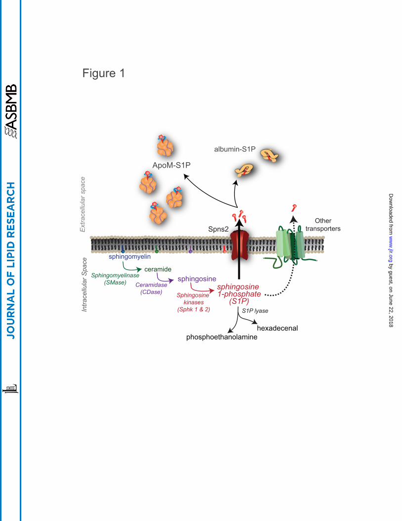
ceramidesphingosine
sphingosine 1-phosphate
(S1P)
sphingomyelin
ApoM-S1P
albumin-S1P
Ext
race
llula
r spa
ceIn
trace
llula
r Spa
ce
Sphingomyelinase(SMase) Ceramidase
(CDase) Sphingosine kinases
(Sphk 1 & 2)
Spns2Other
transporters
Figure 1
phosphoethanolaminehexadecenal
S1P lyase
by guest, on June 22, 2018w
ww
.jlr.orgD
ownloaded from

motilitymigration
vascularbarrier
vasodilation
proliferationsurvival
S1P1
shear sensing
S1P2S1P3
endotoxin-induced
vascular leak
heart rate(mouse)
adhesionmolecules
Figure 2
heart rate(human)
by guest, on June 22, 2018w
ww
.jlr.orgD
ownloaded from

Figure 3
lymphnode
egress
S1P1S1P4
Th17polarization
Th1
Threg
mRNA S1pr2S1pr3S1pr5
T cell
lymphnode &
bone marrow egress
S1P1
S1P4
cellsurvival
MZretention
B cell
S1P3S1P2
GC positioning
MZtrafficking
bonemarrow
positioning
chemotaxis
lymphnode
egress
S1P5
mRNA S1pr1S1pr2S1pr4
NK cell
bone marrowegress
S1P1
decidual angiogenesis
by guest, on June 22, 2018w
ww
.jlr.orgD
ownloaded from
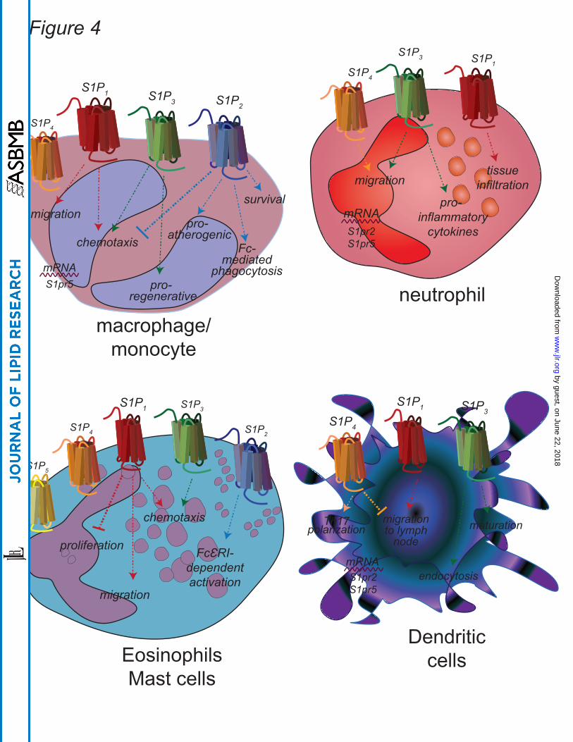
Figure 4
S1P1
S1P4
pro-atherogenic
mRNA S1pr5
macrophage/monocyte
S1P3 S1P2
chemotaxis Fc-mediated
phagocytosispro-
regenerative
survivalmigration
tissueinfiltration
S1P1
neutrophil
S1P4
pro-inflammatory
cytokines
migration
S1P3
mRNA S1pr2S1pr5
S1P1
S1P5
S1P4
S1P3
S1P2
chemotaxis
migration
FcƐRI-dependentactivation
proliferation
EosinophilsMast cells
mRNA S1pr2S1pr5
migration to lymph
node
S1P4
S1P1 S1P3
maturation
endocytosis
Th17polarization
Dendriticcells
by guest, on June 22, 2018w
ww
.jlr.orgD
ownloaded from
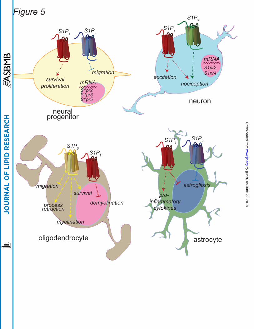
mRNA S1pr2S1pr3S1pr5
S1P1
survivalproliferation
S1P2
migration
neural progenitor
S1P3
S1P1
mRNA S1pr2S1pr4
nociceptionexcitation
neuron
S1P5
migration
processretraction
survival
S1P1
demyelination
myelination
oligodendrocyte
S1P1
pro-inflammatory
cytokines
astrogliosis
S1P2
astrocyte
Figure 5 by guest, on June 22, 2018
ww
w.jlr.org
Dow
nloaded from