Ventricular septal defect - Pediatrics Clerkship · ventricular septal defect.17–20 Environmental...
10
Seminar www.thelancet.com Vol 377 March 26, 2011 1103 Lancet 2011; 377: 1103–12 Published Online February 23, 2011 DOI:10.1016/S0140- 6736(10)61339-6 Department of Cardiology, Royal Children’s Hospital, and Murdoch Children’s Research Institute, Melbourne, Vic, Australia (D J Penny MD); and Baylor College of Medicine, and Texas Children’s Hospital, Houston, TX, USA (G W Vick III MD) Correspondence to: Dr D J Penny, Texas Children’s Hospital, 6621 Fannin, Houston, TX 77303, USA [email protected] Ventricular septal defect Daniel J Penny, G Wesley Vick III Ventricular septal defects account for up to 40% of all congenital cardiac malformations. The diagnosis encompasses a broad range of anomalies, including isolated defects and those associated with other congenital cardiac malformations. Presentation, symptoms, natural history, and management of ventricular septal defects depend on size and anatomical associations of the anomaly, patient’s age, and local diagnostic and interventional expertise. In this Seminar, we describe the anatomical range of ventricular septal defects and discuss present management of these malformations. Genetic determinants, diagnostic techniques, physiological considerations, and management challenges are examined in detail. Unfortunately, in many circumstances, evidence on which to guide optimum management is scarce. We present some longer term considerations of ventricular septal defects in adolescents and adults, with particular emphasis on patients with raised pulmonary vascular resistance and Eisenmenger’s syndrome. Introduction Ventricular septal defect is one of the commonest congenital malformations of the heart, accounting for up to 40% of all cardiac anomalies. 1 Frequency of this defect varies with age at examination, since many small malformations present at birth close shortly afterwards; it is also dependent on sensitivity of the examination technique. Prevalence in newborn babies of up to 5% has been reported from screening with highly sensitive colour doppler echocardiography. 2 Most are tiny muscular defects that disappear during the first year of life. Since many patients can be asymptomatic, and many anomalies close with time, the precise prevalence of ventricular septal defect within populations varies between studies, depending on mode of diagnosis and age of the population. In reports in which echocardiography was used in the diagnostic algorithm, a prevalence of up to 3·94 per 1000 patients has been recorded, which is greater than in previous work that relied on either clinical examination or post-mortem investigations. 3,4 Ventricular septal defect is not only a common isolated cardiac malformation but also an intrinsic component of several complex malformations, including tetralogy of Fallot or univentricular atrioventricular connection. It might also be associated with lesions, including transposition of the great arteries, congenitally corrected transposition, and aortic coarctation or interruption. However, in this Seminar we will concentrate on patients for whom ventricular septal defect is the predominant malformation. Genetics and cause Our understanding of the origins of ventricular septal defect is limited by our knowledge of mechanisms that lead to normal cardiac septation. At present, information suggests that the septum has both mesenchymal and muscular components. 5 The mesenchymal element originates mainly from fusion of the conotruncal and atrioventricular endocardial cushions. Mechanisms that initiate development of the muscular septum are less well defined, and at least two processes have been proposed. Some researchers postulate that the muscular septum forms from coalescence of the part of the ventricular wall that is interposed between the enlarging free walls of the developing right and left ventricles, therefore, as the ventricular cavities become deeper the septum grows passively inwards. 6 An alternative hypothesis suggests that the muscular septum originates from a cluster of cells, the so-called primitive interventricular septum, which expands actively towards the cushions of the atrioventricular canal. 7 Several factors probably lead to development of ventricular septal defects. Failure of complete formation of the primitive interventricular septum could contribute to trabecular defects, although many muscular defects in the trabecular septum probably result from excessive undermining beneath and between trabeculae, during formation of the trabecular part of the septum. Failure of fusion of the atrioventricular cushions—with each other or with the primary septum—could result in an inlet defect, whereas malalignment or poor development of outlet cushions might add to outlet defects. Finally, failure of complete closure of the area that forms the membranous septum, in association with incomplete development of components of the muscular septum, could contribute to a perimembranous defect. Most forms of congenital heart disease, including ventricular septal defect, have multifactorial origins. 8,9 An underlying inherited genetic predisposition could act synergistically with epigenetic factors, direct and indirect environmental causes, and purely stochastic effects to produce cardiac anomalies. Monogenic defects are, in some cases, clearly causative. 8 Such defects have attracted much interest Search strategy and selection criteria We searched PubMed with the term “ventricular septal defect”. We mainly selected publications from the past decade but did not exclude commonly cited references and highly regarded older publications. In view of recent advances in management of patients with Eisenmenger’s syndrome, we undertook a separate search of PubMed with the term “Eisenmenger syndrome”. Further, we reviewed reference lists of articles identified from these searches and selected those we judged especially relevant.
Transcript of Ventricular septal defect - Pediatrics Clerkship · ventricular septal defect.17–20 Environmental...

Seminar
www.thelancet.com Vol 377 March 26, 2011 1103
Lancet 2011; 377: 1103–12
Published OnlineFebruary 23, 2011DOI:10.1016/S0140-6736(10)61339-6
Department of Cardiology, Royal Children’s Hospital, and Murdoch Children’s Research Institute, Melbourne, Vic, Australia (D J Penny MD); and Baylor College of Medicine, and Texas Children’s Hospital, Houston, TX, USA (G W Vick III MD)
Correspondence to:Dr D J Penny, Texas Children’s Hospital, 6621 Fannin, Houston, TX 77303, [email protected]
Ventricular septal defectDaniel J Penny, G Wesley Vick III
Ventricular septal defects account for up to 40% of all congenital cardiac malformations. The diagnosis encompasses a broad range of anomalies, including isolated defects and those associated with other congenital cardiac malformations. Presentation, symptoms, natural history, and management of ventricular septal defects depend on size and anatomical associations of the anomaly, patient’s age, and local diagnostic and interventional expertise. In this Seminar, we describe the anatomical range of ventricular septal defects and discuss present management of these malformations. Genetic determinants, diagnostic techniques, physiological considerations, and management challenges are examined in detail. Unfortunately, in many circumstances, evidence on which to guide optimum management is scarce. We present some longer term considerations of ventricular septal defects in adolescents and adults, with particular emphasis on patients with raised pulmonary vascular resistance and Eisenmenger’s syndrome.
IntroductionVentricular septal defect is one of the commonest congenital malformations of the heart, accounting for up to 40% of all cardiac anomalies.1 Frequency of this defect varies with age at examination, since many small malformations present at birth close shortly afterwards; it is also dependent on sensitivity of the examination technique. Prevalence in newborn babies of up to 5% has been reported from screening with highly sensitive colour doppler echocardiography.2 Most are tiny muscular defects that disappear during the fi rst year of life.
Since many patients can be asymptomatic, and many anomalies close with time, the precise prevalence of ventricular septal defect within populations varies between studies, depending on mode of diagnosis and age of the population. In reports in which echo cardiography was used in the diagnostic algorithm, a prevalence of up to 3·94 per 1000 patients has been recorded, which is greater than in previous work that relied on either clinical examination or post-mortem investigations.3,4
Ventricular septal defect is not only a common isolated cardiac malformation but also an intrinsic component of several complex malformations, including tetralogy of Fallot or univentricular atrioventricular connection. It might also be associated with lesions, including transposition of the great arteries, congenitally corrected transposition, and aortic coarctation or interruption. However, in this Seminar we will concentrate on patients for whom ventricular septal defect is the predominant malformation.
Genetics and causeOur understanding of the origins of ventricular septal defect is limited by our knowledge of mechanisms that lead to normal cardiac septation. At present, information suggests that the septum has both mesenchymal and muscular components.5 The mesenchymal element originates mainly from fusion of the conotruncal and atrioventricular endocardial cushions. Mechanisms that initiate development of the muscular septum are less well defi ned, and at least two processes have been proposed. Some researchers postulate that the muscular septum forms from coalescence of the part of the ventricular wall
that is interposed between the enlarging free walls of the developing right and left ventricles, therefore, as the ventricular cavities become deeper the septum grows passively inwards.6 An alternative hypo thesis suggests that the muscular septum originates from a cluster of cells, the so-called primitive inter ventricular septum, which expands actively towards the cushions of the atrioventricular canal.7
Several factors probably lead to development of ventricular septal defects. Failure of complete formation of the primitive interventricular septum could contribute to trabecular defects, although many muscular defects in the trabecular septum probably result from excessive undermining beneath and between trabeculae, during formation of the trabecular part of the septum. Failure of fusion of the atrioventricular cushions—with each other or with the primary septum—could result in an inlet defect, whereas malalignment or poor development of outlet cushions might add to outlet defects. Finally, failure of complete closure of the area that forms the membranous septum, in association with incomplete development of components of the muscular septum, could contribute to a perimembranous defect.
Most forms of congenital heart disease, including ventricular septal defect, have multifactorial origins.8,9 An underlying inherited genetic predisposition could act synergistically with epigenetic factors, direct and indirect environmental causes, and purely stochastic eff ects to produce cardiac anomalies.
Monogenic defects are, in some cases, clearly causative.8 Such defects have attracted much interest
Search strategy and selection criteria
We searched PubMed with the term “ventricular septal defect”. We mainly selected publications from the past decade but did not exclude commonly cited references and highly regarded older publications. In view of recent advances in management of patients with Eisenmenger’s syndrome, we undertook a separate search of PubMed with the term “Eisenmenger syndrome”. Further, we reviewed reference lists of articles identifi ed from these searches and selected those we judged especially relevant.

Seminar
1104 www.thelancet.com Vol 377 March 26, 2011
because their molecular characterisation has facilitated identifi cation of important constituents of signalling pathways that govern cardiac development.10–12 Mutations in the transcription factors TBX5 and GATA4 have received particular attention. These factors are coexpressed in the heart and their interaction is vital for normal cardiac septation.13 TBX5 is expressed not only in the heart but also in the upper limb buds and eyes. The mutation reported most often in this transcription factor is associated with the autosomal dominant Holt-Oram syndrome,14,15 characterised by abnormalities of forelimbs and several cardiac malformations, including ventricular septal defect. A TBX5 poly-morphism is also associated with ventricular septal defect (without limb abnormalities) in the Chinese Han population.16 Researchers have identifi ed GATA4 sequence variants in familial cases of septal defects (particularly atrial) and in some patients with sporadic ventricular septal defect.17–20
Environmental factors such as teratogens, maternal infections, and untreated maternal metabolic illnesses (eg, phenylketonuria and pregestational diabetes) have been associated with ventricular septal defect.21 Purely stochastic events could also have an important role. Cardiac development is very elaborate, requiring precise operations for successful completion,22,23 which are likely to malfunction occasionally.
AnatomyVentricular septal defect, in many respects, can be deemed one of the simpler forms of congenital malformation of the heart. However, no universal consensus exists for its classifi cation.24–27 To be brief, we will present one system to describe the anatomy of ventricular septal defects because we believe the controversy surrounding these descriptors is beyond the scope of this Seminar.
Broadly speaking, defects can be classifi ed according to their location, either within the muscular septum (muscular defects) or at its margins. Ventricular septal defects at the margins of the muscular septum can be related to hinge-points of the leafl ets of the atrioventricular valves (perimembranous), those of the arterial valves (juxta-arterial or subarterial), or both (fi gure 1).28
Muscular defects are located within the muscular septum. They are surrounded exclusively by muscular rims and, when viewed from the cavity of the right ventricle, can open into the right-ventricular inlet, outlet, or apex.
Perimembranous defects open into the right ven-tricle where the subpulmonary outfl ow tract turns superiorly relative to the atrioventricular junction. Such malformations are characterised by presence of fi brous continuity between leafl ets of the tricuspid and aortic valves. They can extend to open into either the inlet or outlet of the right ventricle (resulting in deviation of the
Figure 1: Location of various types of ventricular septal defect(Left) Location of defects viewed from the right ventricle. (Upper right) Typical doubly committed and juxta-arterial defect. (Lower right) Doubly committed, juxta-arterial, and perimembranous defect. Modifi ed from reference 28 with permission of Elsevier.
Perimembranous
Muscular
Doubly committed andjuxta-arterial
Aortic valve
Pulmonaryvalve
Muscular rim
Pulmonary-aortic-tricuspidcontinuity
Seminar
www.thelancet.com Vol 377 March 26, 2011 1105
outlet septum) or they might be large enough to open to all parts of the ventricle, the so-called confl uent defect.
Doubly committed and juxta-arterial defects (also referred to as subarterial or supracristal defects) are found in an area that, in the normal heart, constitutes a freestanding tube of muscular tissue—the muscular infundibulum, which supports the pulmonary valve. An anomaly in this region will result in characteristic continuity between aortic and pulmonary valves. Most usually, these malformations have a posteroinferior rim of muscle, although they can extend into the peri-membranous zone; thus, fi brous continuity also arises with the tricuspid valve, the so-called doubly committed and juxta-arterial and perimembranous defect (fi gure 1).
PathophysiologySeveral key components determine the pathophysiological response to a ventricular septal defect. Primary factors are the amount and direction of interventricular shunting and the degree of volume loading to the cardiac chambers. Secondary eff ects include prolapse of the aortic valve and obstruction to the pulmonary or systemic outfl ow tract.
The amount of interventricular fl ow is determined by the size of the defect and relative resistances of pulmonary and systemic vascular beds. Small malformations themselves, so-called restrictive defects, provide intrinsic resistance to fl ow. The size of fl ow through larger non-restrictive defects is determined by relative resistances of pulmonary and systemic vascular beds. No agreed precise criteria exist for defi nition of a non-restrictive defect, although various cutoff s have been proposed, according to cross-sectional area of the defect versus area of the aortic orifi ce, diameter relative to body surface area, or velocity of fl ow across the malformation.
When a defect is non-restrictive, major determinants of the resultant interventricular fl ow and symptoms are relative resistances of the pulmonary and systemic vascular beds. Importantly, this relation can be very variable and dependent, in particular, on age of the patient. Left-to-right shunting might initially be minimal in babies, with fairly large defects due to high pulmonary vascular resistance characteristic of the early neonatal period. As pulmonary vascular resistance falls, left-to-right interventricular shunting rises and the patient becomes increasingly symptomatic due to excessive pulmonary blood fl ow.29
In some patients with ventricular septal defects, pulmonary vascular disease can develop in later childhood or in early adult life. In a few individuals, the typical postnatal decline in pulmonary vascular resistance could be delayed or arrested in the presence of a ventricular septal defect; therefore, they might never develop symptoms attributable to excessive left-to-right shunting and only present at a later stage with signs of pulmonary vascular disease. If a large lesion is left uncorrected then, over time, the amount of interventricular left-to-right shunting could decrease
and, eventually, its direction might reverse, leading to cyanosis and Eisenmenger’s syndrome.30,31
Eisenmenger’s syndrome—resulting from chronic elevations of pressure and fl ow—is associated with functional and structural alterations within the pulmonary vasculature.32,33 Key functional modifi cations are increased pulmonary vasoreactivity and resistance and structural microvascular changes, which include medial hypertrophy, migration of smooth muscle distally into typically unmuscularised microvessels, and ultimately, formation of so-called plexiform lesions.34 Abnormalities within the endothelium contribute to all stages of this progression. Activation of endothelial-dependent vasoconstrictor pathways and aberrations of endogenous endothelium-dependent vasodilator processes play a part in functional and structural remodelling, both in animal models of shunt-related pulmonary vascular disease35,36 and in the clinical condition.37
In patients with large ventricular septal defects without pulmonary vascular disease, a rise in volume loading of the left atrium and ventricle (due to increased pulmonary blood fl ow and, in turn, augmented pulmonary venous return) results in left heart dilation throughout the cardiac cycle. In response to the amplifi cation in wall stress, eccentric left-ventricular hypertrophy develops. Presence of relevant longstanding pulmonary hyper-tension could ultimately lead to right-ventricular hypertrophy and dilation. These features will predominate as a patient enters the terminal stages of severe Eisenmenger’s syndrome, which is characterised typically by pending or actual right heart failure.
Secondary structural cardiac anomalies could contribute substantially to the clinical course of patients with ventricular septal defects. Continued surveillance of all aff ected individuals is essential to monitor development of these defects, because they can aff ect clinical management. Malformations located near the aortic valve (doubly committed, perimembranous, or muscular) can be complicated by aortic-valve prolapse and regurgitation, which result from generation of Venturi forces, in which the high-velocity jet sucks the leafl et of the aortic valve into the restrictive defect.38 Several additional mechanisms could contribute to this eff ect, including absence of structural support for leafl ets and abnormal commissural suspension.
Mid-cavity obstruction of the right ventricle due to hypertrophy of muscle bands creates the entity known as double-chambered right ventricle. This process results in formation of a proximal high-pressure chamber and a distal low-pressure chamber within the cavity of the right ventricle.39 In some patients, modest anterior deviation of the outlet septum can happen. The reported prevalence of double-chambered right ventricle in individuals with ventricular septal defect varies widely within published work. An association is well recognised between double-chambered right ventricle and discrete subaortic stenosis.40

Seminar
1106 www.thelancet.com Vol 377 March 26, 2011
Defects aff ecting the muscular outlet septum can be associated with its posterior deviation into the left-ventricular outfl ow tract, resulting in muscular subaortic stenosis. This malformation usually presents in early infancy and might be associated additionally with aortic coarctation or interruption.
DiagnosisClinical examination can show evidence of volume loading of the left ventricle from a large ventricular septal defect, with lateral displacement of the cardiac apex. A pansystolic murmur could be present, with intensity of the murmur indicating velocity of fl ow across the malformation, such that smaller defects are generally loudest and can be associated with a thrill. Large anomalies—leading to an increase in mitral infl ow—could generate a diastolic rumble at the apex. Patients with Eisenmenger’s syndrome typically have cyanosis and clubbing, with a prominent right-ventricular heave, an accentuated pulmonary component of the second heart sound, and usually no murmur.
The electrocardiogram can be normal in patients with small ventricular septal defects. Volume loading of the left ventricle might result in left-ventricular hypertrophy, whereas raised right-ventricular pressure due to either pulmonary hypertension or obstruction to the pulmonary outfl ow tract could lead to right-ventricular hypertrophy.
Cross-sectional echocardiography is the mainstay of modern diagnosis of ventricular septal defect.41–43 The echocardiographer will aim not only to undertake a comprehensive study of the heart—based around a sequential approach—but also to provide several key pieces of data related to the malformation, including: size and location (fi gure 2); anatomical relations to tricuspid, aortic, and pulmonary valves (fi gure 3); associated obstruction to outfl ow from right or left ventricles; associated prolapse of aortic valve; assessment of right-ventricular pressure; and assessment of the amount of loading of the right and left heart (left-ventricular dimension at end-systole and end-diastole should be measured and normalised for body surface area).
Integration of spectral and colour doppler with two-dimensional (2D) echocardiography greatly assists with identifi cation and characterisation of ventricular septal defects.44–46 Reliable estimates of right-ventricular and pulmonary artery pressures, and of pressure diff erences between left and right ventricles, can usually be obtained with 2D-directed continuous-wave doppler.47 The need for cardiac catheterisation to obtain pressure data is thereby eliminated in most cases.
The echocardiographer should also assess extracardiac vascular structures, since clinically important anomalies of the aorta—especially coarctation—and pulmonary arteries, pulmonary veins, and systemic veins can be seen. Transoesophageal echocardiography has assumed an important role in intraoperative assessment of ventricular septal defect because it greatly facilitates confi rmation of repair and early identifi cation and correction of any residual lesion.48,49 Three-dimensional echocardiography is becoming widely available and could provide important diagnostic assistance for assessment of unusually positioned ventricular septal defects and those associated with complex congenital heart malformations.50
Nowadays, cardiac catheterisation is undertaken rarely in patients with uncomplicated defects. This procedure is usually reserved either to measure pulmonary vascular resistance in individuals with suspected or actual pulmonary vascular disease or to close the malformation by a transcatheter approach.
MRI is used increasingly to assess patients with many forms of congenital heart disease, both before and after surgery. Although, in most individuals with ventricular heart defects, adequate diagnostic information can be obtained from clinical examination and echocardiography, MRI might be of use, particularly in patients with poor echocardiographic images.51 MRI could provide additional useful information for defi nition of anatomy in individuals with complex defects—eg, those with
Figure 2: Diagnosis of muscular ventricular septal defectEchocardiograms show apical four-chamber (left) and parasternal long-axis view (right) of a large apical muscular defect (arrows). LA=left atrium. LV=left ventricle. RV=right ventricle. Ao=aortic valve.
Figure 3: Diagnosis of perimembranous defectEchocardiogram shows parasternal short-axis view of a perimembranous defect, with fi brous continuity between tricuspid (TV) and aortic (Ao) valves (arrows). RV=right ventricle. LA=left atrium. RA=right atrium.
LVLVLA
LA
Ao
RV
RV
TV
RALA
RV
Ao

Seminar
www.thelancet.com Vol 377 March 26, 2011 1107
double-chambered right ventricle.52 This imaging technique is especially useful for measurement of pulmonary-to-systemic fl ow ratio. With MRI, accurate quantifi cation of stroke volume of both the right and left ventricles can be made.53,54 Pulmonary-artery resistance can also be measured, provided simultaneous catheter or doppler determination of pulmonary-artery pressure is done.55 Associated extracardiac defects, such as coarctation of the aorta and pulmonary-artery branch stenoses, which are sometimes diffi cult to visualise by echocardiography in older patients, can be delineated clearly.56
Fetal diagnosis of ventricular septal defect is becoming increasingly frequent as imaging techniques improve. Interest has been shown in outcomes for fetuses with malformations seen by colour fl ow-mapping alone in the presence of apparently normal greyscale cross-sectional imaging. In a series of 146 such fetuses, 35 had extracardiac anomalies. Of 113 babies assessed a year after birth, the defect had closed in utero in 37 and during the fi rst year of postnatal life in 50.57
Clinical scenariosSymptomatic young infant with pulmonary hypertensionA baby with such symptoms would typically become breathless with failure to thrive within the fi rst few weeks of life. In this situation, we would usually recommend surgery within 3 months of birth. While awaiting surgery, medical treatment with low doses of diuretics with or without angiotensin-converting-enzyme inhibitors is typically used, although the evidence-base for these strategies is sparse. Monitoring of blood pressure and renal function should be done because renal failure and hypotension have been reported, particularly with angiotensin-converting-enzyme inhibitors.58 The early postoperative period can be complicated by pulmonary hypertension, which results from increased pulmonary vasoreactivity after cardiopulmonary bypass. Use of inhaled nitric oxide to treat postoperative pulmonary hypertension has become widespread,59 although in a Cochrane review, the paucity of data supporting its use early after cardiac surgery was highlighted.60
Asymptomatic patient without pulmonary hypertension but with volume overloaded left heartIn this scenario, many centres would recommend closure of ventricular septal defects with the aim of avoiding potential late left-ventricular dysfunction secondary to ongoing dilation. In an observational study of 96 patients (mean follow-up almost 8 years), without any intervention, the left-ventricular end-diastolic dimension Z score fell in 29 of 33 patients and declined to less than 2 in 26 of these.61 Although this series was small, the fi ndings suggest that the optimum approach for this group of patients, who would typically be most suitable for transcathether closure, might instead be conservative.
Asymptomatic patient with small ventricular septal defect and no left-ventricular dilationMuch information is available from natural history studies on the long-term outlook for individuals with ventricular septal defects. Gabriel and colleagues62 reported long-term outcomes in 229 patients with malformations judged “not to require surgical closure during childhood”. At a mean age of 30 years, mortality was zero, 95% were symptom-free, and left-ventricular size was normal in 89% and borderline in 10%. At the time of follow-up, four of 222 patients had experienced an episode of endocarditis. This study’s fi ndings confi rmed that with careful selection of patients, a conservative approach is warranted in this subgroup.
In a regional cohort of 290 patients with ventricular septal defect, spontaneous closure was noted in 123 of 180 cases with completely muscular borders compared with only 31 of 107 with perimembranous defects. Furthermore, muscular malformations closed spon-taneously in children aged up to 88 months, whereas no perimembranous defect closed in infants older than 62 months.63 Of 450 patients with perimembranous defects, at mean follow-up of 3 years, subaortic ridge developed in 26 (6%), aortic-valve prolapse in 53 (12%), and aortic regurgitation in 33 (7%).64
Asymptomatic patient with small defect and prolapse or regurgitation of aortic valveBest management for this population has historically been controversial, and up to now, no randomised trials have been published to defi ne the optimum strategy. In an audit of patients who underwent surgery for aortic regurgitation in the setting of ventricular septal defect, those with severe preoperative regurgitation had less favourable long-term outcomes and a higher requirement for reoperation because of suboptimum valve repair.65 As a result, individuals with peri-membranous ventricular septal defects and more than trivial aortic regurgitation should be referred for surgery.38 A reduced threshold for surgery might be justifi able in patients with juxta-arterial defects because of their high risk of aortic regurgitation and low rate of spontaneous closure.38
Patient with Eisenmenger’s syndrome Until recent times, treatment of individuals with Eisenmenger’s syndrome was only supportive.32,66 Dehydration and exposure to high altitudes should be avoided because these situations compound pre-existing hyperviscosity and arterial hypoxaemia. Historically, venesection to reduce the eff ects of polycythaemia was routine in many centres, although its benefi ts are questionable in asymptomatic patients.67 Indeed, venesection can worsen iron defi ciency and exercise intolerance and can amplify risk of stroke.67,68 Anticoagulation has been used to manage Eisenmenger’s syndrome in the past, although supportive evidence is
Seminar
1108 www.thelancet.com Vol 377 March 26, 2011
scarce and bleeding risk could be considerable.69 Female patients must be made aware that pregnancy is associated with substantial maternal and fetal risk (see Issues in adults with ventricular septal defects).
Recognition of the role of disrupted endothelial messengers in pathogenesis of Eisenmenger’s syndrome has broadened pharmacotherapeutic options for this disorder.33 Endothelial-based treatments, particularly those aimed either at blockade of the potent vasoconstrictor endothelin 1 or at prevention of catabolism of the nitric oxide-dependent vasodilator cGMP, are used increasingly in this population of patients.70–72 In the BREATHE-5 study, bosentan—a dual endothelin receptor-antagonist—lengthened 6-min walk distances in a randomised placebo-controlled trial over 16 weeks73 and in an open-label extension.74 These improvements continued for an additional 24 weeks. Findings of an open-label study showed increases in functional capacity that were maintained, particularly in adults.75 Improvements in quality of life and functional capacity were reported in patients with Eisenmenger’s
syndrome in response to oral sildenafi l, a phos-phodiesterase inhibitor that raises cGMP levels.76 Advanced endothelial-based treatments increase survival in individuals with Eisenmenger’s syndome.77
These advances, combined with the observation that many patients with Eisenmenger’s syndrome are responsive to vasodilators in the catheterisation laboratory,78 raise the possibility that aggressive endothelial-based treatments could restore operability in individuals who had previously been judged inoperable. Several published reports accord with this idea, although systematic studies are scarce.79
EndocarditisTraditionally, antibiotic prophylaxis was recommended routinely in patients with ventricular septal defects to prevent procedure-associated endocarditis. This guidance is based on recognition that such individuals are at increased risk of endocarditis, that this disorder could result from bacteraemia, that dental procedures might result in bacteraemia, and that treatment with antibiotics might reduce risk of bacteraemia and endocarditis. However, later evidence indicates that endocarditis is most likely to result from chronically poor dental hygiene and activities of daily living, which—coupled with the paucity of data in support of the eff ectiveness of antibiotic prophylaxis for prevention of endocarditis—has resulted in revised guidelines. These recommendations suggest that patients with uncomplicated ventricular septal defects do not need antibiotics, but they put strong emphasis on primary prevention of dental infections, with meticulous daily dental hygiene and regular dental review. However, antibiotic prophylaxis for dental and other procedures continues to be recommended for 6 months after complete surgical or transcatheter closure of a ventricular septal defect and indefi nitely when a residual defect is present in relation to patch material, because this situation could inhibit endothelialisation.80
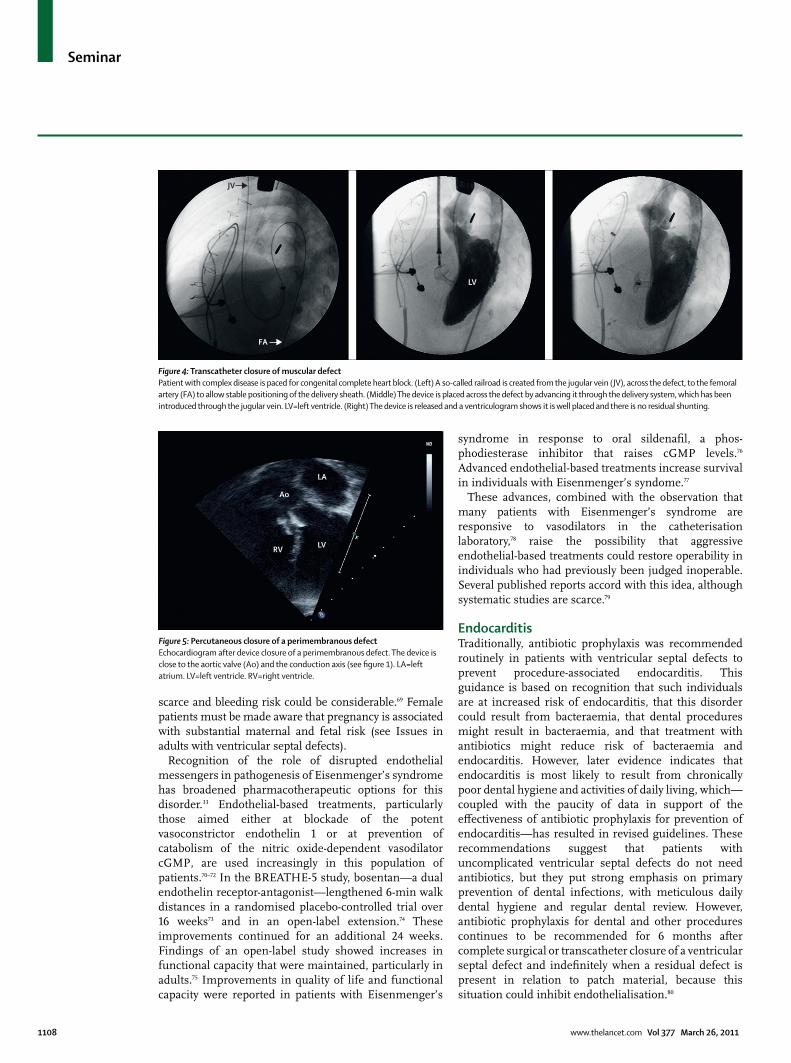
Figure 4: Transcatheter closure of muscular defectPatient with complex disease is paced for congenital complete heart block. (Left) A so-called railroad is created from the jugular vein (JV), across the defect, to the femoral artery (FA) to allow stable positioning of the delivery sheath. (Middle) The device is placed across the defect by advancing it through the delivery system, which has been introduced through the jugular vein. LV=left ventricle. (Right) The device is released and a ventriculogram shows it is well placed and there is no residual shunting.
Figure 5: Percutaneous closure of a perimembranous defectEchocardiogram after device closure of a perimembranous defect. The device is close to the aortic valve (Ao) and the conduction axis (see fi gure 1). LA=left atrium. LV=left ventricle. RV=right ventricle.
LV
JV
FA
LA
LVRV
Ao

Seminar
www.thelancet.com Vol 377 March 26, 2011 1109
Closure of ventricular septal defectsSurgeryPatch closure of a ventricular septal defect through sternotomy, with cardiopulmonary bypass, has been done for more than 50 years. With enhanced selection of patients, early surgery, and advances in perioperative care, operative mortality is low and substantial postoperative morbidity is rare.81 Usually, access to the defect is obtained through either the atrioventricular or semilunar valves, thus avoiding ventriculotomy. In some individuals, intraoperative temporary detachment of anterior and septal leafl ets of the tricuspid valve can increase exposure of the defect.82 Most patients after surgery report normal quality of life, compared with age-matched controls, although behavioural and school performance diffi culties might be present in early childhood.83
With expansion of cardiac surgery to the developing world,84 interest has grown in techniques to close defects in patients who did not have access to surgery in infancy, who have subsequently developed raised pulmonary vascular resistance. Surgical closure in these high-risk individuals
can result in substantial morbidity and mortality, because increases in pulmonary-arterial and right-ventricular pressure result in right-ventricular failure. A modifi ed surgical technique with creation of a so-called valved patch, which allows unidirectional right-to-left shunting across a deliberate residual defect, can be undertaken with low operative mortality,85 although the benefi ts of this procedure over conventional closure have been questioned.86
In the past, banding of the pulmonary artery was done frequently as an interim palliative procedure to reduce pulmonary blood fl ow, particularly in infants. This technique is now undertaken rarely, except in individuals with either many ventricular septal defects or apical malformations, in whom surgical access to the anomalies is anticipated to be especially diffi cult or impossible. Application of either absorbable or balloon-dilatable bands has been performed in patients with multiple muscular defects, which potentially could avoid the need for removal of the band if anomalies reduce in size over time.87
The main surgical challenges are with defects that are near the apex and are diffi cult to access through either the
Unrepaired defect Repaired defect
Survival Excellent survival for small defects; large defects can be associated with pulmonary vascular disease; might develop aortic regurgitation
Excellent survival; occasional residual shunt; might develop right-ventricular or left-ventricular outfl ow-tract obstruction; might develop aortic regurgitation
Haemodynamic issues Left-to-right shunt; left-ventricular dilation and impaired function; aortic regurgitation; pulmonary vascular disease
Residual shunt; ventricular function; aortic regurgitation; right-ventricular or left-ventricular outfl ow-tract obstruction
Arrhythmia and pacing Rare Rare atrioventricular block; rare ventricular block
Investigations
Chest radiography Cardiomegaly Cardiomegaly
ECG Rhythm and conduction; right-ventricular hypertrophy
Rhythm and conduction; right-ventricular hypertrophy
Echocardiography Number and size of defects; left-ventricular and right-ventricular function; aortic regurgitation
Residual shunt; ventricular function; aortic regurgitation
Transoesophageal echocardiography If transthoracic image is inadequate If transthoracic image is inadequate
Cardiac catheterisation Pulmonary vascular disease; associated lesions Rarely needed
Holter test Only if symptomatic Only if symptomatic
Exercise test If symptomatic; sports counselling If symptomatic; sports counselling
Indications for intervention Left-to-right shunt with heart overload; reversible pulmonary hypertension; aortic regurgitation; associated abnormalities (right-ventricular outfl ow tract, subaortic stenosis); previous endocarditis
Left-to-right shunt with heart overload; reversible pulmonary hypertension; aortic regurgitation; associated abnormalities (right-ventricular outfl ow tract, subaortic stenosis); previous endocarditis
Endocarditis prophylaxis Not needed If residual defect
Pregnancy No contraindications with uncomplicated defects; contraindicated with pulmonary vascular disease
No contraindications with closed defects; contraindicated with pulmonary vascular disease
Physical exercise No restriction for small defects No restriction for closed defects
Follow-up care
Aortic regurgitation Exclusively specialist centre Exclusively specialist centre
Complicated haemodynamics Exclusively specialist centre Exclusively specialist centre
Pulmonary vascular disease Shared care between specialist centre and general adult centre
Shared care between specialist centre and general adult centre
Small defect Managed in non-specialist centre with access to specialist centre if needed
Managed in non-specialist centre with access to specialist centre if needed
Modifi ed from reference 92 with permission of Oxford University Press.
Table: Issues related to ongoing surveillance of patients with ventricular septal defects

Seminar
1110 www.thelancet.com Vol 377 March 26, 2011
atrioventricular or semilunar valves. In some situations, malformations can be accessed through a ventriculotomy, although transcatheter approaches have been used.
Transcatheter closureOver the past decade or so, transcatheter techniques for closure of ventricular septal defects have been developed. These methods have been especially useful for muscular defects, which can be the most diffi cult to access surgically (fi gure 4).88 Much interest has been generated in development of transcatheter approaches to close perimembranous defects (fi gure 5). At present, this technique is not undertaken in most units because of the unacceptably high rate of post-procedure heart block associated with currently available devices. Of particular concern is that this risk does not subside or fall with time, with late-onset heart block being fairly prevalent.89,90 As softer devices are developed, this method could potentially be reintroduced in the future.
Hybrid techniquesIn infants with muscular ventricular septal defects, in whom both transcatheter and standard surgical approaches are diffi cult, a hybrid technique for closure has been implemented, which brings together surgery and interventional methods. With this method, a sternotomy is done in the standard way and the device is placed in the right ventricle through its anterior wall, under transoesophageal and fl uoroscopic guidance.91
Issues in adults with ventricular septal defectsExerciseAdults with small ventricular septal defects, normal pulmonary arterial pressure, normal ventricular function, and no associated lesions should have a normal tolerance for exercise and, therefore, no exercise restrictions should be imposed (table).92 Those with pulmonary arterial hypertension usually self-restrict their amount of exercise. In a large study of adults with congenital heart disease who underwent formal testing, patients with Eisenmenger’s syndrome achieved the lowest levels of peak oxygen consumption during exercise and, furthermore, the extent of the reduction in peak oxygen consumption was an important marker of prognosis.93
PregnancyWomen with small ventricular septal defects without pulmonary hypertension do not seem to be at increased cardiovascular risk during pregnancy (table). Those with moderate defects could have raised pulmonary blood fl ow during pregnancy, an indication of increased circulating volume, although this eff ect could—to a degree—be counterbalanced by reduction in systemic vascular resistance. By contrast, pregnancy in women with Eisenmenger’s syndrome is associated with a very high risk of maternal and fetal death and pre-mature delivery.94
In a study of 17 ongoing pregnancies in ten women with Eisenmenger’s syndrome,95 one maternal death was reported and another woman deteriorated greatly, requiring high-level intensive care. Four spontaneous abortions and one stillbirth happened. Of the 12 deliveries of live infants, ten were premature.95 Against this background, women with Eisenmenger’s syndrome should be counselled strongly against pregnancy and should be referred to a specialist for contraception advice.96 Although sterilisation might be deemed appropriate for some women, it should only be done at a specialist centre with careful periprocedural care of these high-risk patients. Early termination is often recommended for women with Eisenmenger’s syndrome who become pregnant. For those who choose to continue pregnancy, obstetric care should be undertaken at a specialist centre, with access to intensive care. Even after successful delivery, maternal risk continues beyond the time of birth; therefore, close monitoring must be maintained in the postpartum period. Data suggest that augmentation of endothelial-based treatments—eg, combination of intra venous epoprostenol and oral sildenafi l—might improve outcome for pregnant women.97
ContributorsBoth authors wrote the report and approved the fi nal version.
Confl icts of interestWe declare that we have no confl icts of interest.
References1 Hoff man JI. Incidence of congenital heart disease: I—postnatal
incidence. Pediatr Cardiol 1995; 16: 103–13.
2 Roguin N, Du ZD, Barak M, Nasser N, Hershkowitz S, Milgram E. High prevalence of muscular ventricular septal defect in neonates. J Am Coll Cardiol 1995; 26: 1545–48.
3 Hoff man JIE, Kaplan S. The incidence of congenital heart disease. J Am Coll Cardiol 2002; 39: 1890–900.
4 Hoff man JIE, Kaplan S, Liberthson RR. Prevalence of congenital heart disease. Am Heart J 2004; 147: 425–39.
5 Contreras-Ramos A, Sánchez-Gómez C, García-Romero HL, Cimarosti LO. Normal development of the muscular region of the interventricular septum: I—signifi cance of the ventricular trabeculations. Anat Histol Embryol 2008; 37: 344–51.
6 Goor AD, Edwards EJ, Lillehei W. The development of the interventricular septum of the human heart: correlative morphogenetic study. Chest 1970; 58: 453–67.
7 De La Cruz MV, Moreno-Rodriguez R. Embryological development of the apical trabeculated region of both ventricles: the contribution of the primitive interventricular septum in ventricular septation. In: De La Cruz MV, Markwald R, eds. Living morphogenesis of the heart. Basel: Birkhäuser, 1998: pp 120–30.
8 Pierpont ME, Basson CT, Benson DW, et al. Genetic basis for congenital heart defects: current knowledge—a scientifi c statement from the American Heart Association Congenital Cardiac Defects Committee, Council on Cardiovascular Disease in the Young, endorsed by the American Academy of Pediatrics. Circulation 2007; 115: 3015–38.
9 Nora JJ. Multifactorial inheritance hypothesis for the etiology of congenital heart diseases: the genetic-environmental interaction. Circulation 1968; 38: 604–17.
10 Bruneau BG. The developmental genetics of congenital heart disease. Nature 2008; 451: 943–48.
11 Srivastava D, Olson EN. A genetic blueprint for cardiac development. Nature 2000; 407: 221–26.
12 Benson DW. Genetic origins of pediatric heart disease. Pediatr Cardiol 2010; 31: 422–29.

Seminar
www.thelancet.com Vol 377 March 26, 2011 1111
13 Maitra M, Schluterman MK, Nichols HA, et al. Interaction of Gata4 and Gata6 with Tbx5 is critical for normal cardiac development. Dev Biol 2009; 326: 368–77.
14 Basson CT, Bachinsky DR, Lin RC, et al. Mutations in human TBX5 [corrected] cause limb and cardiac malformation in Holt-Oram syndrome. Nat Genet 1997; 15: 30–35.
15 Li QY, Newbury-Ecob RA, Terrett JA, et al. Holt-Oram syndrome is caused by mutations in TBX5, a member of the Brachyury (T) gene family. Nat Genet 1997; 15: 21–29.
16 Liu C-X, Shen A-D, Li X-F, et al. Association of TBX5 gene polymorphism with ventricular septal defect in the Chinese Han population. Chin Med J 2009; 122: 30–34.
17 Zhang W-M, Li X-F, Ma Z-Y, et al. GATA4 and NKX2.5 gene analysis in Chinese Uygur patients with congenital heart disease. Chin Med J 2009; 122: 416–19.
18 Zhang W, Li X, Shen A, Jiao W, Guan X, Li Z. GATA4 mutations in 486 Chinese patients with congenital heart disease. Eur J Med Genet 2008; 51: 527–35.
19 Rajagopal SK, Ma Q, Obler D, et al. Spectrum of heart disease associated with murine and human GATA4 mutation. J Mol Cell Cardiol 2007; 43: 677–85.
20 Garg V, Kathiriya IS, Barnes R, et al. GATA4 mutations cause human congenital heart defects and reveal an interaction with TBX5. Nature 2003; 424: 443–47.
21 Jenkins KJ, Correa A, Feinstein JA, et al. Noninherited risk factors and congenital cardiovascular defects: current knowledge: a scientifi c statement from the American Heart Association Council on Cardiovascular Disease in the Young: endorsed by the American Academy of Pediatrics. Circulation 2007; 115: 2995–3014.
22 Srivastava D. Genetic regulation of cardiogenesis and congenital heart disease. Ann Rev Pathol 2006; 1: 199–213.
23 Bajolle F, Zaff ran S, Bonnet D. Genetics and embryological mechanisms of congenital heart diseases. Arch Cardiovasc Dis 2009; 102: 59–63.
24 Soto B, Becker AE, Moulaert AJ, Lie JT, Anderson RH. Classifi cation of ventricular septal defects. Br Heart J 1980; 43: 332–43.
25 Jacobs JP, Burke RP, Quintessenza JA, Mavroudis C. Congenital Heart Surgery Nomenclature and Database Project: ventricular septal defect. Ann Thorac Surg 2000; 69 (4 Suppl): S25–35.
26 Soto B, Ceballos R, Kirklin JW. Ventricular septal defects: a surgical viewpoint. J Am Coll Cardiol 1989; 14: 1291–97.
27 Van Praagh R, Geva T, Kreutzer J. Ventricular septal defects: how shall we describe, name and classify them? J Am Coll Cardiol 1989; 14: 1298–99.
28 Benson LN, Yoo D-J, Al Habshan F, Anderson RH. Ventricular septal defects. In: Anderson RH, Baker EJ, Redington A, Rigby ML, Penny D, Wernovsky G, eds. Paediatric cardiology, 3rd edn. Philadelphia: Churchill Livingstone, 2009: pp 591–624.
29 Rudolph AM. Circulatory adjustments after birth: eff ects on ventricular septal defect. Br Heart J 1971; 33 (suppl): 32–34.
30 Duff els MGL, Engelfriet PM, Berger RMF, et al. Pulmonary arterial hypertension in congenital heart disease: an epidemiologic perspective from a Dutch registry. Int J Cardiol 2007; 120: 198–24.
31 Van de Bruaene A, Delcroix M, Pasquet A, et al. The Belgian Eisenmenger syndrome registry: implications for treatment strategies. Acta Cardiol 2009; 64: 447–53.
32 Diller GP, Gatzoulis MA. Pulmonary vascular disease in adults with congenital heart disease. Circulation 2007; 115: 1039–50.
33 Beghetti M, Galiè N. Eisenmenger syndrome: a clinical perspective in a new therapeutic era of pulmonary arterial hypertension. J Am Coll Cardiol 2009; 53: 733–40.
34 Rabinovitch M. Pulmonary hypertension: pathophysiology as a basis for clinical decision making. J Heart Lung Transplant 1999; 11: 1041–53.
35 Oishi PE, Wiseman DA, Sharma S, et al. Progressive dysfunction of nitric oxide synthase in a lamb model of chronically increased pulmonary blood fl ow: a role for oxidative stress. Am J Physiol Lung Cell Mol Physiol 2008; 295: L756–66.
36 Fasules JW, Tryka F, Chipman CW, Van Devanter SH. Pulmonary hypertension and arterial changes in calves with a systemic-to-left pulmonary artery connection. J Appl Physiol 1994; 77: 867–75.
37 Diller GP, van Eijl S, Okonko DO, et al. Circulating endothelial progenitor cells in patients with Eisenmenger syndrome and idiopathic pulmonary arterial hypertension. Circulation 2008; 117: 3020–30.
38 Tweddell JS, Pelech AN, Frommelt PC. Ventricular septal defect and aortic valve regurgitation: pathophysiology and indications for surgery. Semin Thorac Cardiovasc Surg Pediatr Card Surg Annu 2006: 9: 147–52.
39 Telagh R, Alexi-Meskishivili V, Hetzer R, Lange PE, Berger F, Abdul-Khaliq H. Initial clinical manifestations and mid- and long-term results after surgical repair of double-chambered right ventricle in children and adults. Cardiol Young 2008; 18: 268–74.
40 Vogel M, Smallhorn JF, Freedom RM, Coles J, Williams WG, Trusler GA. An echocardiographic study of the association of ventricular septal defect and right ventricular muscle bundles with a fi xed subaortic abnormality. Am J Cardiol 1988; 61: 857–60.
41 Cheatham JP, Latson LA, Gutgesell HP. Ventricular septal defect in infancy: detection with two dimensional echocardiography. Am J Cardiol 1981; 47: 85–89.
42 Minette MS, Sahn DJ. Ventricular septal defects. Circulation 2006; 114: 2190–97.
43 Sutherland GR, Godman MJ, Smallhorn JF, Guiterras P, Anderson RH, Hunter S. Ventricular septal defects: two dimensional echocardiographic and morphological correlations. Br Heart J 1982; 47: 316–28.
44 Ludomirsky A, Huhta JC, Vick GW, Murphy DJ, Danford DA, Morrow WR. Color Doppler detection of multiple ventricular septal defects. Circulation 1986; 74: 1317–22.
45 Ortiz E, Robinson PJ, Deanfi eld JE, Franklin R, Macartney FJ, Wyse RK. Localisation of ventricular septal defects by simultaneous display of superimposed colour Doppler and cross sectional echocardiographic images. Br Heart J 1985; 54: 53–60.
46 Sommer RJ, Golinko RJ, Ritter SB. Intracardiac shunting in children with ventricular septal defect: evaluation with Doppler color fl ow mapping. J Am Coll Cardiol 1990; 16: 1437–44.
47 Houston AB, Lim MK, Doig WB, Reid JM, Coleman EN. Doppler assessment of the interventricular pressure drop in patients with ventricular septal defects. Br Heart J 1988; 60: 50–56.
48 Ayres NA, Miller-Hance W, Fyfe DA, et al. Indications and guidelines for performance of transesophageal echocardiography in the patient with pediatric acquired or congenital heart disease: report from the task force of the Pediatric Council of the American Society of Echocardiography. J Am Soc Echocardiogr 2005; 18: 91–98.
49 Bezold LI, Pignatelli R, Altman CA, et al. Intraoperative transesophageal echocardiography in congenital heart surgery: the Texas Children’s Hospital experience. Texas Heart Inst J 1996; 23: 108–15.
50 Chen FL, Hsiung MC, Nanda N, Hsieh KS, Chou MC. Real time three-dimensional echocardiography in assessing ventricular septal defects: an echocardiographic-surgical correlative study. Echocardiography 2006; 23: 562–68.
51 Kilner PJ, Geva T, Maemmerer H, Trindade PT, Schwitter J, Webb GD. Recommendations for cardiovascular magnetic resonance in adults with congenital heart disease from the respective working groups of the European Society of Cardiology. Eur Heart J 2010; 31: 794–805.
52 Sato Y, Matsumoto N, Matsuo S, et al. Double chambered right ventricle: depiction at three-dimensional whole heart magnetic resonance imaging. Int J Cardiol 2007; 119: e14–16.
53 Beerbaum P, Körperich H, Barth P, Esdorn H, Gieseke J, Meyer H. Noninvasive quantifi cation of left-to-right shunt in pediatric patients: phase-contrast cine magnetic resonance imaging compared with invasive oximetry. Circulation 2001; 103: 2476–82.
54 Körperich H, Gieseke J, Barth P, et al. Flow volume and shunt quantifi cation in pediatric congenital heart disease by real-time magnetic resonance velocity mapping: a validation study. Circulation 2004; 109: 1987–93.
55 Muthurangu V, Taylor A, Andriantsimiavona R, et al. Novel method of quantifying pulmonary vascular resistance by use of simultaneous invasive pressure monitoring and phase-contrast magnetic resonance fl ow. Circulation 2004; 110: 826–34.
56 Vick GW. Recent advances in pediatric cardiovascular MRI. Curr Opin Pediatr 2003; 15: 454–62.
57 Axt-Fliedner R, Schwarze A, Smrcek J, Germer U, Krapp M, Gembruch U. Isolated ventricular septal defects detected by color Doppler imaging: evolution during fetal and fi rst year of postnatal life. Ultrasound Obstet Gynecol 2006; 27: 266–73.

Seminar
1112 www.thelancet.com Vol 377 March 26, 2011
58 Gantenbein MH, Bauersfeld U, Baenziger O, et al. Side eff ects of angiotensin converting enzyme inhibitor (captopril) in newborns and young infants. J Perinat Med 2008; 46: 448–52.
59 Namachivayam P, Theilen U, Butt WW, Cooper SM, Penny DJ, Shekerdemian LS. Sildenafi l prevents rebound pulmonary hypertension after withdrawal of nitric oxide in children. Am J Respir Crit Care Med 2006; 174: 1042–47.
60 Bizzarro M, Gross I. Inhaled nitric oxide for the postoperative management of pulmonary hypertension in infants and children with congenital heart disease. Cochrane Database Syst Rev 2005: 4: CD005055.
61 Kleinman CS, Tabibian M, Starc TJ, Hsu DT, Gersony WM. Spontaneous regression of left ventricular dilation in children with restrictive ventricular septal defects. J Pediatr 2007; 150: 583–86.
62 Gabriel HM, Heger M, Innerhofer P, et al. Long-term outcome of patients with ventricular septal defect considered not to require surgical closure during childhood. J Am Coll Cardiol 2002; 39: 1066–71.
63 Turner SW, Hornung T, Hunter S. Closure of ventricular septal defects: a study of factors infl uencing spontaneous and surgical closure. Cardiol Young 2002; 12: 357–63.
64 Eroğlu AG, Oztunç F, Saltik L, Bakari S, Dedeoğlu S, Ahunbay G. Evolution of ventricular septal defect with special reference to spontaneous closure rate, subaortic ridge and aortic valve prolapse. Pediatr Cardiol 2003; 24: 31–35.
65 Kostolny M, Schreiber C, von Arnim V, Vogt M, Wottke M, Lange R. Timing of repair in ventricular septal defect with aortic insuffi ciency. Thorac Cardiovasc Surg 2006; 54: 512–15.
66 Kumar RK, Sandoval J. Advanced pulmonary vascular disease: the Eisenmenger syndrome. Cardiol Young 2009; 19: 622–26.
67 Spence MS, Balaratnam MS, Gatzoulis MA. Clinical update: cyanotic adult congenital heart disease. Lancet 2007; 370: 1530–32.
68 Amnesh N, Warnes CA. Cerebrovacular events in adult patients with cyanotic congenital heart disease. J Am Coll Cardiol 1996; 28: 768–72.
69 Galiè N, Manes A, Palazzini M, et al. Management of pulmonary arterial hypertension associated with congenital systemic-to-pumonary shunts and Eisenmenger’s syndrome. Drugs 2008; 68: 1049–66.
70 Fine N, Dias B, Shoemaker G, Mehta S. Endothelin receptor antagonist therapy in congenital heart disease with shunt-associated pulmonary arterial hypertension: a qualitative systematic review. Can J Cardiol 2009; 25: e63–68.
71 Mehta PK, Simpson L, Lee EK, Lyle TA, McConnell ME, Book WM. Endothelin receptor antagonists improve exercise tolerance and oxygen saturations in patients with Eisenmenger syndrome and congenital heart defects. Tex Heart Inst J 2008; 35: 256–61.
72 Wort SJ. Sildenafi l in Eisenmenger syndrome: safety fi rst. Int J Cardiol 2007; 120: 314–16.
73 Galiè N, Beghetti M, Gatzoulis MA, et al. Bosentan therapy in patients with Eisenmenger syndrome: a multicenter, double-blind, randomized, placebo-controlled study. Circulation 2006; 114: 48–54.
74 Gatzoulis MA, Beghetti M, Galiè N, et al. Longer-term bosentan therapy improves functional capacity in Eisenmenger syndrome: results of the BREATHE-5 open-label extension study. Int J Cardiol 2008; 127: 27–32.
75 van Loon RLE, Hoendermis ES, Duff els MGJ, et al. Long-term eff ect of bosentan in adults versus children with pulmonary arterial hypertension associated with systemic-to-pulmonary shunt: does the benefi cial eff ect persist? Am Heart J 2007; 154: 776–82.
76 Tay ELW, Papaphylactou M, Diller GP, et al. Quality of life and functional capacity can be improved in patients with Eisenmenger syndrome with oral sildenafi l therapy. Int J Cardiol 2010; DOI:10.1016/j.ijcard.2010.02.020.
77 Dimopoulos K, Inuzuka R, Goletto S, et al. Improved survival among patients with Eisenmenger syndrome receiving advanced therapy for pulmonary arterial hypertension. Circulation 2010; 121: 20–25.
78 Limsuwan A, Khosithseth A, Wanichkul S, Khowsathit P. Aerosolized iloprost for pulmonary vasoreactivity testing in children with long-standing pulmonary hypertension related to congenital heart disease. Catheter Cardiovasc Interv 2009; 73: 98–104.
79 Dimopoulos K, Peset A, Gatzoulis MA. Evaluating operability in adults with congenital heart disease and the role of pretreatment with targeted pulmonary arterial hypertension therapy. Int J Cardiol 2008; 129: 163–71.
80 Wilson W, Taubert KA, Gewitz M, et al. Prevention of infective endocarditis: guidelines from the American Heart Association: a guideline from the American Heart Association Rheumatic Fever, Endocarditis, and Kawasaki Disease Committee, Council on Cardiovascular Disease in the Young, and the Council on Clinical Cardiology, Council on Cardiovascular Surgery and Anesthesia, and the Quality of Care and Outcomes Research Interdisciplinary Working Group. Circulation 2007; 116: 1736–54.
81 Scully BB, Morales DLS, Zafar F, McKenzie ED, Fraser CD, Heinle JS. Current expectations for surgical repair of isolated ventricular septal defects. Ann Thorac Surg 2010; 89: 544–49.
82 Bol Raap G, Meijboom FJ, Kappetein AP, Galema TW, Yap S-C, Bogers AJJC. Long-term follow-up and quality of life after closure of ventricular septal defect in adults. Eur J Cardiothorac Surg 2007; 32: 215–19.
83 Hövels-Gürich HH, Konrad K, Skorzenski D, et al. Long-term neurodevelopmental outcome and exercise capacity after corrective surgery for tetralogy of Fallot or ventricular septal defect in infancy. Ann Thorac Surg 2006; 81: 958–66.
84 Jonas RA. Congenital heart surgery in developing countries. Semin Thorac Cardiovasc Surg Pediatr Card Surg Annu 2008; 11: 3–6.
85 Novick WM, Sandoval N, Lazorhysynets VV, et al. Flap valve double patch closure of ventricular septal defects in children with increased pulmonary vascular resistance. Ann Thorac Surg 2005; 79: 21–28.
86 Gan H-L, Zhang J-Q, Zhang Z-G, Luo Y, Zhou Q-W, Bo P. The unidirectional valve patch provides no benefi ts to early and long-term survival in patients with ventricular septal defect and severe pulmonary artery hypertension. J Thorac Cardiovasc Surg 2010; 139: 950–55.
87 Brown S, Boshoff D, Rega F, Eyskens B, Meyns B, Gewillig M. Dilatable pulmonary artery banding in infants with low birth weight or complex congenital heart disease allows avoidance or postponement of subsequent surgery. Eur J Cardiothorac Surg 2010; 37: 296–301.
88 Lim DS, Forbes TJ, Rothman A, Lock JE, Landzberg MJ. Transcatheter closure of high-risk muscular ventricular septal defects with the CardioSEAL occluder: initial report from the CardioSEAL VSD registry. Catheter Cardiovasc Interv 2007; 70: 740–44.
89 Dumitrescu A, Lane GK, Wilkinson JL, Goh TH, Penny DJ, Davis AM. Trancatheter closure of perimembranous ventricular septal defect. Heart 2007; 93: 867.
90 Collins NJ, Benson L, Horlick E. Late complete heart block in an adult patient undergoing percutaneous ventricular septal defect closure. J Invasive Cardiol 2008; 20: E200–03.
91 Crossland DS, Wilkinson JL, Cochrane AD, D’udekem Y, Brizard CP, Lane GK. Initial results of primary device closure of large muscular ventricular septal defects in early infancy using perventricular access. Cathet Cardiovasc Interv 2008; 72: 386–91.
92 Deanfi eld J, Thaulow E, Warnes C, et al, for the Task Force on the Management of Grown Up Congential Heart Disease of the European Society of Cardiology. Management of grown up congenital heart disease. Eur Heart J 2003; 24: 1035–84.
93 Diller G-P, Dimopoulos K, Okonko D et al. Exercise intolerance in adult congenital heart disease: comparative severity, correlates, and prognostic implication. Circulation 2005; 112: 828–35.
94 Bédard E, Dimopoulos K, Gatzoulis MA. Has there been any progress made on pregnancy outcomes among women with pulmonary arterial hypertension? Eur Heart J 2009; 30: 256–65.
95 Dranenkiene A, Opitz CF, Gumbiené L, Doma B, Ewert R, Bühlmeyer K. Pregnancy in patients with Eisenmenger’s syndrome: experiences from Vilnius 1967–2003. Dtsch Med Wochenschr 2004; 129 (suppl 1): S35–39 [in German].
96 Silversides CK, Sermer M, Siu SC. Choosing the best contraceptive method for the adult with congenital heart disease. Curr Cardiol Rep 2009; 11: 298–305.
97 Goland S, Tsai F, Habib M, Janmohamed M, Goodwin TM, Elkayam U. Favorable outcome of pregnancy with an elective use of epoprostenol and sildenafi l in women with severe pulmonary hypertension. Cardiology 2010; 115: 205–08.