Use of Innovative Physics Forceps for Extractions in Preparation for ...
12
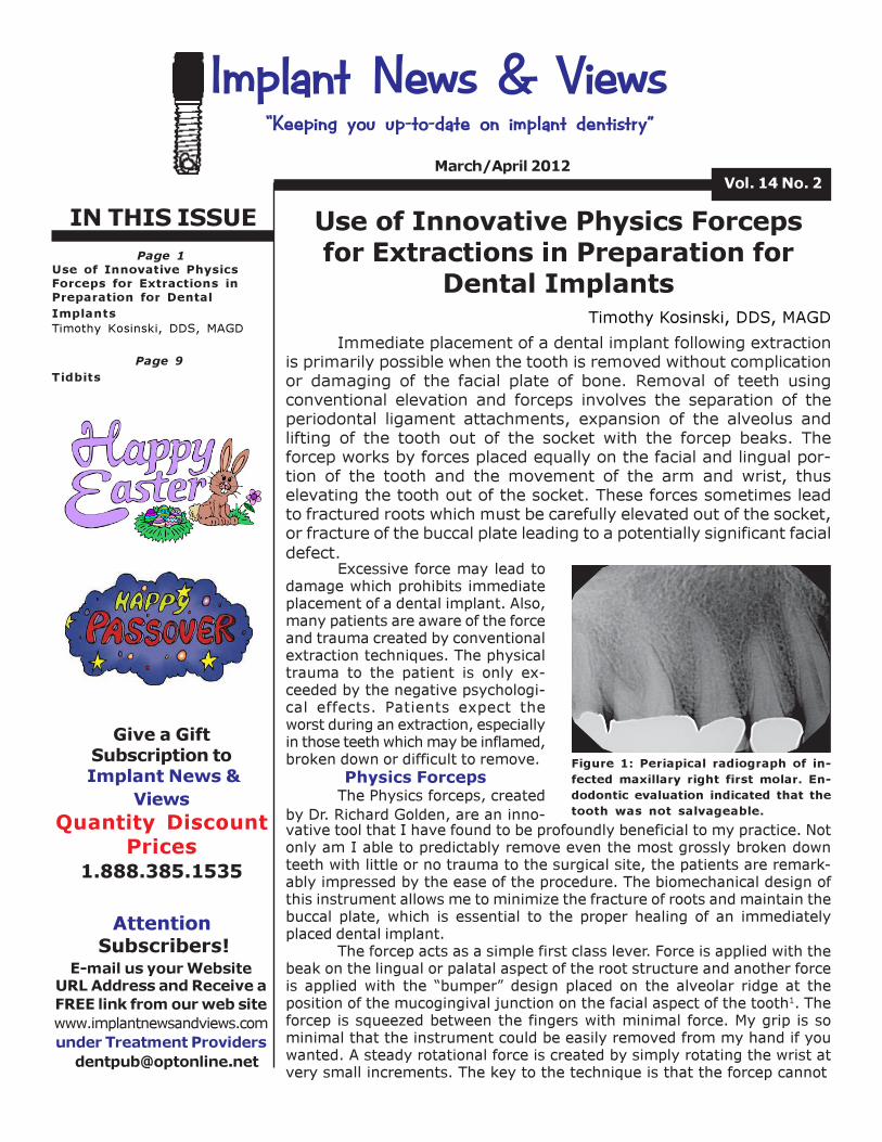
IN THIS ISSUE Vol. 14 No. 2 March/April 2012 Implant News & Views “Keeping you up-to-date on implant dentistry” Page 1 Use of Innovative Physics Forceps for Extractions in Preparation for Dental Implants Timothy Kosinski, DDS, MAGD Attention Subscribers! E-mail us your Website URL Address and Receive a FREE link from our web site www.implantnewsandviews.com under Treatment Providers [email protected] Page 9 Tidbits Timothy Kosinski, DDS, MAGD Use of Innovative Physics Forceps for Extractions in Preparation for Dental Implants Immediate placement of a dental implant following extraction is primarily possible when the tooth is removed without complication or damaging of the facial plate of bone. Removal of teeth using conventional elevation and forceps involves the separation of the periodontal ligament attachments, expansion of the alveolus and lifting of the tooth out of the socket with the forcep beaks. The forcep works by forces placed equally on the facial and lingual por- tion of the tooth and the movement of the arm and wrist, thus elevating the tooth out of the socket. These forces sometimes lead to fractured roots which must be carefully elevated out of the socket, or fracture of the buccal plate leading to a potentially significant facial defect. Excessive force may lead to damage which prohibits immediate placement of a dental implant. Also, many patients are aware of the force and trauma created by conventional extraction techniques. The physical trauma to the patient is only ex- ceeded by the negative psychologi- cal effects. Patients expect the worst during an extraction, especially in those teeth which may be inflamed, broken down or difficult to remove. Physics Forceps The Physics forceps, created by Dr. Richard Golden, are an inno- Figure 1: Periapical radiograph of in- fected maxillary right first molar. En- dodontic evaluation indicated that the tooth was not salvageable. vative tool that I have found to be profoundly beneficial to my practice. Not only am I able to predictably remove even the most grossly broken down teeth with little or no trauma to the surgical site, the patients are remark- ably impressed by the ease of the procedure. The biomechanical design of this instrument allows me to minimize the fracture of roots and maintain the buccal plate, which is essential to the proper healing of an immediately placed dental implant. The forcep acts as a simple first class lever. Force is applied with the beak on the lingual or palatal aspect of the root structure and another force is applied with the “bumper” design placed on the alveolar ridge at the position of the mucogingival junction on the facial aspect of the tooth 1 . The forcep is squeezed between the fingers with minimal force. My grip is so minimal that the instrument could be easily removed from my hand if you wanted. A steady rotational force is created by simply rotating the wrist at very small increments. The key to the technique is that the forcep cannot Give a Gift Subscription to Implant News & Views Quantity Discount Prices 1.888.385.1535
Transcript of Use of Innovative Physics Forceps for Extractions in Preparation for ...
IN THIS ISSUE
Vol. 14 No. 2March/April 2012
Implant News & Views “Keeping you up-to-date on implant dentistry”
Page 1Use of Innovative PhysicsForceps for Extractions inPreparation for Dental
Implants
Timothy Kosinski, DDS, MAGD
Attention Subscribers!
E-mail us your WebsiteURL Address and Receive a
FREE link from our web site
www.implantnewsandviews.com
under Treatment Providers
Page 9
Tidbits
Timothy Kosinski, DDS, MAGD
Use of Innovative Physics Forcepsfor Extractions in Preparation for
Dental Implants
Immediate placement of a dental implant following extraction
is primarily possible when the tooth is removed without complication
or damaging of the facial plate of bone. Removal of teeth using
conventional elevation and forceps involves the separation of the
periodontal ligament attachments, expansion of the alveolus and
lifting of the tooth out of the socket with the forcep beaks. The
forcep works by forces placed equally on the facial and lingual por-
tion of the tooth and the movement of the arm and wrist, thus
elevating the tooth out of the socket. These forces sometimes lead
to fractured roots which must be carefully elevated out of the socket,
or fracture of the buccal plate leading to a potentially significant facial
defect.Excessive force may lead to
damage which prohibits immediateplacement of a dental implant. Also,many patients are aware of the forceand trauma created by conventionalextraction techniques. The physicaltrauma to the patient is only ex-ceeded by the negative psychologi-cal effects. Patients expect theworst during an extraction, especiallyin those teeth which may be inflamed,broken down or difficult to remove.
Physics ForcepsThe Physics forceps, created
by Dr. Richard Golden, are an inno-
Figure 1: Periapical radiograph of in-
fected maxillary right first molar. En-
dodontic evaluation indicated that the
tooth was not salvageable.
vative tool that I have found to be profoundly beneficial to my practice. Notonly am I able to predictably remove even the most grossly broken downteeth with little or no trauma to the surgical site, the patients are remark-ably impressed by the ease of the procedure. The biomechanical design ofthis instrument allows me to minimize the fracture of roots and maintain thebuccal plate, which is essential to the proper healing of an immediatelyplaced dental implant.
The forcep acts as a simple first class lever. Force is applied with thebeak on the lingual or palatal aspect of the root structure and another forceis applied with the “bumper” design placed on the alveolar ridge at theposition of the mucogingival junction on the facial aspect of the tooth1. Theforcep is squeezed between the fingers with minimal force. My grip is sominimal that the instrument could be easily removed from my hand if youwanted. A steady rotational force is created by simply rotating the wrist atvery small increments. The key to the technique is that the forcep cannot
Give a Gift
Subscription to
Implant News &
Views
Quantity Discount
Prices1.888.385.1535

Page 2 Implant News & Views
Fig. 9
Fig. 3
Fig. 4 Fig. 5
be used like a traditionally designed forcep, meaning there is no arm force used to elevate the tooth out ofthe socket. My experience is that it may take a minute or two of minimal rotational force of my wrist in verysmall increments to remove the tooth. This is opposite of everything we have been taught about extractionsin the past. We need to slow down, prevent squeezing of the instrument and let the rotational forces “pop”the tooth out of the socket.
Case 1 [Figures 1-12]
The patient presented with a symptomatic maxillary right first molar tooth. Endodontic and radio-graphic evaluation indicated a fractured root. The decision was made with our patient that extraction of the
tooth would be completed and the socket site grafted for future implant placement.
Figure 2: Maxillary molar teeth
can often be removed in total
without sectioning. The palatal
portion of the root is flattened
subgingivally with a bur. This al-
lows a stable purchase point for
the beak of the Physics forcep.
Figure 3: The beak of the forcep
is engaged subgingivally on the
palatal side of the tooth to be
extracted. The “bumper” is posi-
tioned on the facial aspect of the
tooth on the alveolar ridge at the
approximate position of the
mucogingival junction.
Figure 4: The tooth “pops” out of
the socket. Another instrument
like a bird beak pliers is used to
remove the disengaged root from
its socket.
Figure 5: The maxillary right firstmolar as it looked upon removal.
Figure 6: It was clear that the
apex of the root was indeed frac-tured and was easily separated.The entire root and fractured
piece came out in total for an
atraumatic, simple extraction.
Figure 7: Blood is syringed from
the socket site with an insulin
syringe.
Figure 8: The patient’s blood is
mixed with Tri Calcium Phosphatecrystals which will be used to
graft the socket site in prepara-tion for future dental implantplacement. Because of the posi-
tion of the three maxillary sock-ets, it is often difficult to imme-
diately place a dental implant in
ideal position.
Figure 9: The graft/blood mixture
is placed into the socket.
Figure 10: Post operative radio-graph of the graft material in
place.
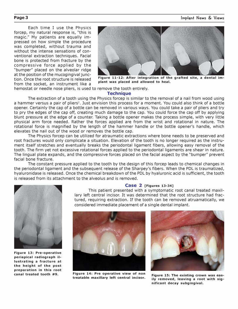
Fig. 6
Fig. 7
Page 3 Implant News & Views
Figure 11-12: After integration of the grafted site, a dental im-
plant was placed and allowed to heal.
Each time I use the Physicsforcep, my natural response is, “this ismagic.” My patients are equally im-pressed on how simple the procedurewas completed, without trauma andwithout the intense sensations of con-ventional extraction techniques. Facialbone is protected from fracture by thecompressive force applied by the“bumper” placed on the alveolar ridgeat the position of the mucogingival junc-tion. Once the root structure is releasedfrom the socket, an instrument like ahemostat or needle nose pliers, is used to remove the tooth entirely.
TechniqueThe extraction of a tooth using the Physics forcep is similar to the removal of a nail from wood using
a hammer versus a pair of pliers2. Just envision this process for a moment. You could also think of a bottleopener. Certainly the cap of a bottle can be removed in various ways. You could take a pair of pliers and tryto pry the edges of the cap off, creating much damage to the cap. You could force the cap off by applyingblunt pressure at the edge of a counter. Taking a bottle opener makes the process simple, with very littlephysical arm force needed. Rather the forces applied are from the wrist and rotational in nature. Therotational force is magnified by the length of the hammer handle or the bottle opener’s handle, whichelevates the nail out of the wood or removes the bottle cap.
The Physics forcep can be utilized for atraumatic extractions where bone needs to be preserved androot fractures would only complicate a situation. Elevation of the tooth is no longer required as the instru-ment itself stretches and eventually breaks the periodontal ligament fibers, allowing easy removal of thetooth. The firm yet not excessive rotational forces applied to the periodontal ligaments are shear in nature.The lingual plate expands, and the compressive forces placed on the facial aspect by the “bumper” preventfacial bone fracture.
The constant pressure applied to the tooth by the design of this forcep leads to chemical changes inthe periodontal ligament and the subsequent release of the Sharpey’s fibers. When the PDL is traumatized,hyaluronidase is released. Once the chemical breakdown of the PDL by hyaluronic acid is sufficient, the tooth
is released from its attachment to the alveolus and is removed.
Case 2 [Figures 13-34]
This patient presented with a symptomatic root canal treated maxil-lary left central incisor. It was determined that the root structure had frac-tured, requiring extraction. If the tooth can be removed atruamatically, we
considered immediate placement of a single dental implant.
Figure 13: Pre-operative
periapical radiograph il-
lustrating a fracture at
the height of the post
preparation in this root
canal treated tooth #9. Figure 14: Pre operative view of non
treatable maxillary left central incisor.Figure 15: The existing crown was eas-
ily removed, leaving a root with sig-
nificant decay subgingival.
Page 4 Implant News & Views
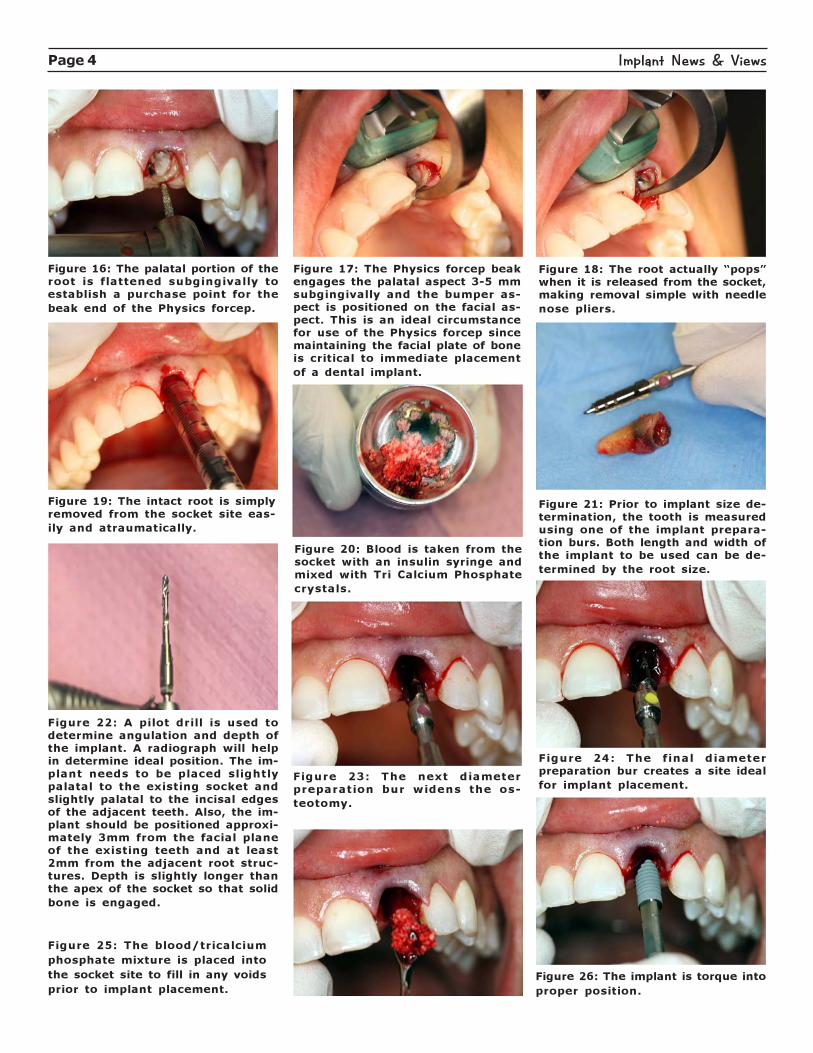
Figure 16: The palatal portion of theroot is flattened subgingivally toestablish a purchase point for the
beak end of the Physics forcep.
Figure 17: The Physics forcep beakengages the palatal aspect 3-5 mmsubgingivally and the bumper as-pect is positioned on the facial as-pect. This is an ideal circumstancefor use of the Physics forcep sincemaintaining the facial plate of boneis critical to immediate placement
of a dental implant.
Figure 18: The root actually “pops”when it is released from the socket,making removal simple with needle
nose pliers.
Figure 19: The intact root is simply
removed from the socket site eas-
ily and atraumatically.
Figure 20: Blood is taken from the
socket with an insulin syringe and
mixed with Tri Calcium Phosphate
crystals.
Figure 21: Prior to implant size de-
termination, the tooth is measured
using one of the implant prepara-
tion burs. Both length and width of
the implant to be used can be de-
termined by the root size.
Figure 22: A pilot drill is used to
determine angulation and depth ofthe implant. A radiograph will helpin determine ideal position. The im-
plant needs to be placed slightly
palatal to the existing socket andslightly palatal to the incisal edgesof the adjacent teeth. Also, the im-
plant should be positioned approxi-
mately 3mm from the facial planeof the existing teeth and at least2mm from the adjacent root struc-
tures. Depth is slightly longer thanthe apex of the socket so that solid
bone is engaged.
Figure 23: The next diameterpreparation bur widens the os-
teotomy.
Figure 24: The final diameter
preparation bur creates a site ideal
for implant placement.
Figure 25: The blood/tricalcium
phosphate mixture is placed into
the socket site to fill in any voids
prior to implant placement.
Figure 26: The implant is torque into
proper position.
Page 5 Implant News & Views
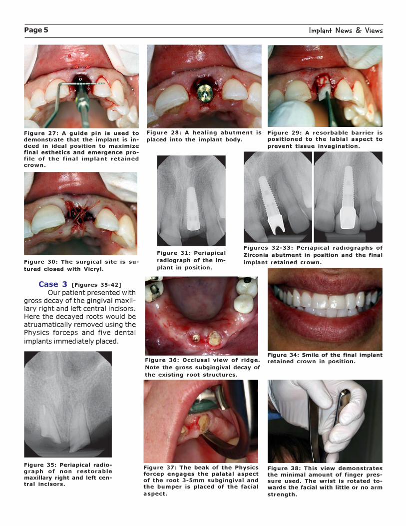
Figure 28: A healing abutment is
placed into the implant body.Figure 27: A guide pin is used todemonstrate that the implant is in-deed in ideal position to maximizefinal esthetics and emergence pro-file of the final implant retainedcrown.
Figure 29: A resorbable barrier ispositioned to the labial aspect to
prevent tissue invagination.
Figure 30: The surgical site is su-
tured closed with Vicryl.
Figure 31: Periapical
radiograph of the im-
plant in position.
Figures 32-33: Periapical radiographs of
Zirconia abutment in position and the final
implant retained crown.
Figure 34: Smile of the final implantretained crown in position.
Case 3 [Figures 35-42]
Our patient presented withgross decay of the gingival maxil-lary right and left central incisors.Here the decayed roots would beatruamatically removed using thePhysics forceps and five dental
implants immediately placed.
Figure 35: Periapical radio-graph of non restorablemaxillary right and left cen-
tral incisors.
Figure 36: Occlusal view of ridge.
Note the gross subgingival decay of
the existing root structures.
Figure 37: The beak of the Physics
forcep engages the palatal aspectof the root 3-5mm subgingival andthe bumper is placed of the facial
aspect.
Figure 38: This view demonstratesthe minimal amount of finger pres-
sure used. The wrist is rotated to-wards the facial with little or no arm
strength.
Page 6 Implant News & Views
Fig. 3
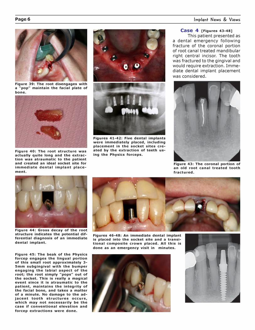
Figure 39: The root disengages witha “pop” maintain the facial plate of
bone.
Figure 40: The root structure was
actually quite long and the extrac-
tion was atraumatic to the patient
and created an ideal socket site for
immediate dental implant place-
ment.
Figures 41-42: Five dental implants
were immediately placed, including
placement in the socket sites cre-
ated by the extraction of teeth us-
ing the Physics forceps.
Case 4 [Figures 43-48]
This patient presented asa dental emergency followingfracture of the coronal portionof root canal treated mandibularright central incisor. The toothwas fractured to the gingival andwould require extraction. Imme-diate dental implant placement
was considered.
Figure 43: The coronal portion of
an old root canal treated tooth
fractured.
Figure 44: Gross decay of the root
structure indicates the potential dif-ferential diagnosis of an immediate
dental implant.
Figure 45: The beak of the Physics
forcep engages the lingual portionof this small root approximately 3-
5mm subgingival with the bumperengaging the labial aspect of the
root; the root simply “pops” out ofthe socket. This is really a magicalevent since it is atraumatic to the
patient, maintains the integrity ofthe facial bone, and takes a matter
of a minute. No damage to the ad-jacent tooth structures occurs,which may not necessarily be the
case if conventional elevation and
forcep extractions were done.
Figures 46-48: An immediate dental implant
is placed into the socket site and a transi-tional composite crown placed. All this is
done as an emergency visit in minutes.
Page 7 Implant News & Views
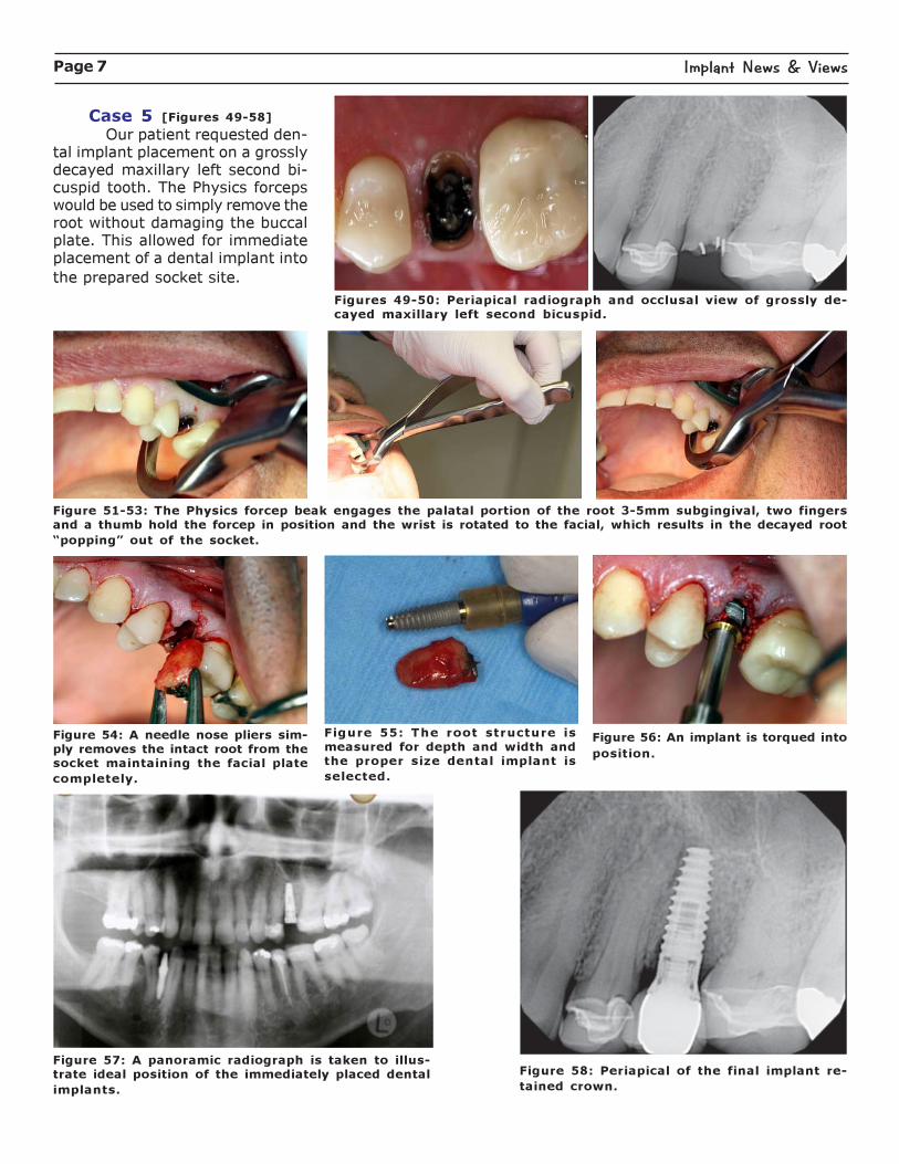
Fig. 4Case 5 [Figures 49-58]
Our patient requested den-tal implant placement on a grosslydecayed maxillary left second bi-cuspid tooth. The Physics forcepswould be used to simply remove theroot without damaging the buccalplate. This allowed for immediateplacement of a dental implant into
the prepared socket site.
Figures 49-50: Periapical radiograph and occlusal view of grossly de-cayed maxillary left second bicuspid.
Figure 51-53: The Physics forcep beak engages the palatal portion of the root 3-5mm subgingival, two fingers
and a thumb hold the forcep in position and the wrist is rotated to the facial, which results in the decayed root
“popping” out of the socket.
Figure 54: A needle nose pliers sim-
ply removes the intact root from thesocket maintaining the facial plate
completely.
Figure 55: The root structure is
measured for depth and width and
the proper size dental implant is
selected.
Figure 56: An implant is torqued into
position.
Figure 57: A panoramic radiograph is taken to illus-trate ideal position of the immediately placed dental
implants.
Figure 58: Periapical of the final implant re-
tained crown.
Page 8 Implant News & Views
Fig. 10
Fig. 9
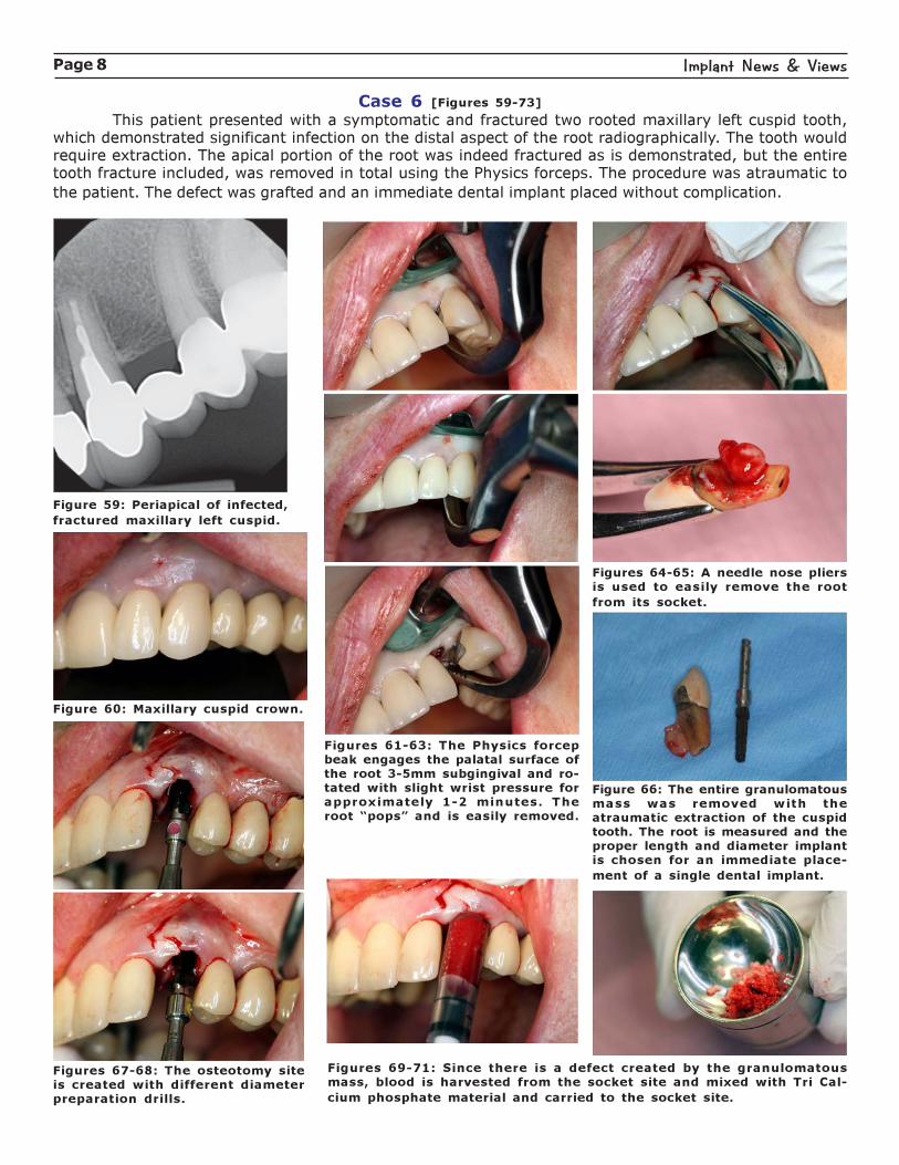
Case 6 [Figures 59-73]
This patient presented with a symptomatic and fractured two rooted maxillary left cuspid tooth,which demonstrated significant infection on the distal aspect of the root radiographically. The tooth wouldrequire extraction. The apical portion of the root was indeed fractured as is demonstrated, but the entiretooth fracture included, was removed in total using the Physics forceps. The procedure was atraumatic to
the patient. The defect was grafted and an immediate dental implant placed without complication.
Figure 59: Periapical of infected,
fractured maxillary left cuspid.
Figure 60: Maxillary cuspid crown.
Figures 61-63: The Physics forcepbeak engages the palatal surface of
the root 3-5mm subgingival and ro-
tated with slight wrist pressure forapproximately 1-2 minutes. Theroot “pops” and is easily removed.
Figures 64-65: A needle nose pliers
is used to easily remove the root
from its socket.
Figure 66: The entire granulomatousmass was removed with the
atraumatic extraction of the cuspidtooth. The root is measured and the
proper length and diameter implantis chosen for an immediate place-
ment of a single dental implant.
Figures 67-68: The osteotomy siteis created with different diameter
preparation drills.
Figures 69-71: Since there is a defect created by the granulomatousmass, blood is harvested from the socket site and mixed with Tri Cal-
cium phosphate material and carried to the socket site.

Page 9 Implant News & Views
Figures 72-73: The implant is car-
ried to the surgical site and posi-
tioned ideally in the bone.
Figure 71.
Timothy Kosinski, DDS, MAGDmaintains a private practice inBingham Farms, MI with an emphasison cosmetic and implant dentistry.He is an Adjunct Assistant Professorat the University of Detroit MercySchool of Dentistry, serves on theeditorial review Board of Reality, andis a Diplomat of the American Boardof Oral Implantology/Implant Den-tistry, the International Congress ofOral Implantologists and the Ameri-can Society of Osseointegration. Heis a Fellow of the American Academyof Implant Dentistry and received hisMastership in the AGD. Dr. Kosinskihas published over 70 articles on thesurgical and prosthetic phases ofimplant dentistry and was a con-tributor to the textbooks, Principlesand Practices of Implant Dentistry,and 2010’s Dental Implantation andTechnology. He can be reached [email protected] or 248-646-8651.
References1. Golden, R. “Less ThanFour Minute Extraction ofAny Tooth.”www.dentistrytoday.com/oral-surgery-00/5991. 12/3/2011. 1-6.2. Misch,C. and Perez, H.“Atraumatic Extractions.”Dentistry Today. Vol. 27.
No.8, Aug. 2008. 1-3.
Tidbits
Tidbits
Tidbits
Online Discussion GroupsWe try to monitor various online discussion groups to share their views on implants with our readers. Wefound a great group with a wealth of ideas on LinkedIn. To learn about the benefits & how to join, go tohttp://www.linkedin.com. The group is called Dental Implant Professionals and has over 4,000 mem-bers. You need to apply, but it’s free membership. Here’s a recent disscussion about how to get started andchoose an implant system.
Makarov AleksandrHow do you choose an implant system to work with? What are the criteria to chose implants and how muchdo you have to know about implant structure, surface and so on...?
Jochen KonnekerThere are many factors to consider. No system is perfect. Go to genieoss.com and look under surgeon-
implant design. There you will find information about the most important features for implants.
Jo Ann PulverAs a speaker for dental implant companies for the past 15 years, I have seen the most success regarding theselection of a dental implant system when the general dentist confers with his/her Oral Surgeon or Periodon-tist. It is important that this discussion remain between the dental professionals and not enter into the“selling” process with patients. The discussion of excessive technical detail often leads to derailment of thecase presentation and therefore causes patients to think the process is too involved or complicated for
them.

Page 10 Implant News & Views
Michael CorselloEducation! If you do not know what implant systems to even consider then you have a great learningopportunity. I would advise a maxi-course such as offered by Implant Seminars (Dr Garg) or MischInstitute. Simply conferring with an Oral Surgeon/ Periodontist is a good starting point but as you canimagine there is a lot to consider. First there are the two major considerations, surgical and prosthetic. Youreally need to understand both to even begin to consider what primary system you will be using. A doctorwho has been trained and has experience in both disciplines is really your best source of informal information.A formal training program will enlighten you on the implications in bone physiology, soft tissue, prosthetics,systemic conditions, implant coatings, thread design etc... This enables you to understand the factors whichwill influence implant selection. For example what implant are you going to use in a grafted site or poor bonequality? When do you need to submerge and when do you want a single phase design? Furthermore there willnot be one company which you will want to use for every case, at least not at this time.
Ian BrabyI would agree with you that, by and large, most systems available today are pretty equal. But that is not thecase with an individual’s experience and, on that basis, I would suggest that those embarking on theirimplant careers should use one of the more popular systems as the companies offer a wealth of support whenstarting out. Once established, you can begin to consider substituting your implant system of choice with aclone system - the same implant at a cheaper cost - as you should no longer require the level of support thatthe major companies can offer. I offer this as someone who was 15 years with a major company and nowworks with a clone company.
Andrew WoodAs a dental technician who has worked with various different implant systems for some twenty plus yearsnow, I find that good communication and discussion with your technician is also very important. Many of thelimitations with various implant systems are not always obvious from the technical info given in brochuresand advertising material, whereas finding out from your technician the benefits and disadvantages of par-ticular implant systems in specific cases with regard to the function as well as cosmetic result, may help youin your final diagnosis.
Paul FarrellI will preface my comments by disclosing that I am and Opinion Leader and a member of the internationalConnecting Science Circle for Thommen Medical (see my profile). As a surgeon I have, over the past 25years, made my way through multiple implant systems. As I have taken an interest in being the best that Ican be with my role within the Team approach to working with dental implants, I have changed systemsperiodically make the treatment process as simple, reliable, comfortable, and as cost efficient as possible forall concerned. This is difficult to achieve in the current “climate’ of implant dentistry as 95% of the discussionat major meetings is focused on less than 5% of the patients that are treated routinely with implants. Thishappens at a level that is of little value to many of the practitioners that have less experience and are tryingto learn how to plan and work more effectively with implants in daily practice. The system that I useaddresses as many of these issues as possible for restorative dentists with all levels of experience and abilitywhere implant case planning and use in private practice are concerned.
Danny Hiller
4 years Dental School 7 years implant sales - In my experience most of the implants today osseointegratewell enough. Most of the failures I’ve seen are due to something other than the implant itself. And, that’swhere the evaluation really needs to take place. Fundamentally, those who place implants are not equal intheir abilities. Also, a system may work better for one practitioner, but not for another - for whatever reason,whether it be training, psychological, or design. Also, when I mention failure, I don’t just mean a lost implantor excessive loss of bone. I’m also including esthetic, phonetic, and functional failure. So that being said, Ithink there are a few underlying principles that will increase the likelihood of success for your overall implantpractice, rather than just looking at an implant: 1) Get educated - There are some great courses out there.Though, if you’re just getting started it will cost around $100,000 to get fully trained (restorative & surgical).You’re going to need bone and tissue grafting courses, ridge augmentation, and sinus lift courses (in thebeginning - probably not going to do these kind of cases, but you’ll eventually move to these cases). Mostoverlooked, are the emergency courses - you’re going to need to know what to do, just in case. Unlike whatmost people think, implant surgery can go real south in a real hurry (I’ve seen it). 2) Fully understand that
Page 11 Implant News & Views
most of your cases will probably be a team effort between the surgeon, the restorative doc, and the lab -sometimes the rep, if they’re any good. I’ve seen implants placed in the embrasures, I’ve seen multipleimplants placed where one would have sufficed. The implant always integrates when it’s in the wrong place.It all becomes a restorative nightmare, leading to a potential treatment failure. So, it’s incumbent upon thesurgeon that he know restorative, and vice versa. And, if you are doing the surgery and the restorative, thenyou need to know the concepts of both. 3) Understand the system you choose. A surgeon typically justlearns the surgical side of the system, and the restorative doc, rarely knows the system and leaves himselfat the mercy of the surgeon and the rep. The Docs I like to work with the most, do both. Because they doboth, they have to understand the system they use and its limitations. I also think it behooves implantologiststo learn the new technologies such as what 3M and Atlantis have to offer, because it might increaseefficiencies = profit. 4) Make sure that your practice is set up to handle implant dentistry. I’ve walked intooffices that want to start implant dentistry but don’t have digital radiography. Also, your assistants, hygien-ists, and front office have to be on board or you may end up losing a lot of money. Implantology is a sub-specialty, I would treat my supply room as such and keep implant supplies separate. 5) Understand yourlimitations...toughest thing for a doctor to say is “I’m not very good at this, I should probably leave it tosomeone else”. I know when I was in school, I had a knack for endo. I was terrible at removable. It’s hard tofind a dentist who is truly outstanding at everything. I’m just saying, you can save yourself a lot of stress byknowing when to refer. So, the original question was - What implant system, and how do you determine...Isay, whatever works in your hands and makes sense for your practice (I have a preference, yes). Are therenuances...yes - and I do know what they are. Unlike most reps, I like to read research articles. Maybe ifanyone’s interested I’ll elaborate on what I see in the industry.
Nedim SulejmanagiæTotaly agree that most implant systems are same. All of them are made of at least grade 2 titanium, which isenough to withstand biting and other forces in mouth. I would suggest to choose internal connection whenplacing single implants to avoid rotational forces on the screw itself. Otherwise for us doctors, patients bonequality is most important
Dan PflugerAs the restorative lab we like a system that keeps bone loss near zero. All three of the following are a goodbet. I like Ankylos for the Morse taper concept but wish you could do custom abutments. Zirconia abutmentsare a little pricey. Good support. Nobel Biocare has a good system and support, we like the custom abutmentselection and product line. Straumann has very good support. They incorporated the Morse taper andinternal hex very well. Custom abutments and restorative options are many. We see many other systems, butthe support is very spotty.
Jon BergstresserThe ability to hold the bone height is, in my opinion the most important aspect of an implant. If the boneheight drops down around the implant, the tissue will recede to reflect the new, (usually lower) bone height.There is a lot of research on which implants have the best bone retention. The interface connection reallymatters by the way, minimizing micro movement at the implant /abutment interface is important. From atechnician standpoint I want to be able to rely on the soft tissues to be stable to have a healthy “pink” aswell as a natural tooth display.
Andreas DanielssonMany implants today do have great designs, surfaces, user-friendliness etc. Use the Osstell ISQ - the onlyobjective, scientific way to measure implant stability, to quality assure your treatment (www.osstell.com).
Lars HanssonI see that a lot of you use Ankylos, and we know that that system has been only stock abutments for manyyears and has limited the use sometimes. But now we can actually offer Cad Cam abutments in bothZirconium and Titanium for Ankylos. That I think will make Ankylos even a more interesting implant to choose.The prosthetics are a must and the preservation of bone.
Jeff YeiderMy experience with oral implants started in 1974, with Dentists hammering in blade implants cast from nonprecious alloys. I found Branemark had started the implant revolution. I have worked with every system from

Page 12 Implant News & Views
Implant News & Views“Keeping you up-to-date on
implant dentistry”
Published byDental Education Publications
EditorKeith Rossein, DDS
Letters-to-the-EditorMust be received typed, doublespaced [Limit 200 words], signed and
with a contact phone number.
Mailing AddressPlease send all requests, letters-to-
the-editor, contributions, “tidbits,”
subscriptions and/or suggestions to:
Dental Education Publications500 Birch Road
Malverne, NY 11565
1 (888) 385-1535Outside US -1 (516) 593-3806
Fax 1 (516) 599-3734e-mail [email protected]
www.implantnewsandviews.com
DisclaimerThe views and opinions expressed by
contributing authors and other pro-
fessionals are not necessarily the
views or beliefs of the publication.
While Implant News & Views is infor-
mational, it is not intended to replace
your own professional judgment or
advice from your own professionalconsultants.
DEP or its contributors have no fi-nancial interest in any service, prod-
ucts or programs, unless otherwisestated.
Subscription RatesOne year [6 issues] at $49.00
US FUNDS ONLY
Copyright ProtectedReproduction of any part or all of
this publication is prohibitedwithout written permission.
Copyright © 2012
Dental Education Publications
Astra to Zimmer , advocated, tested, and lectured for some in between,as I helped in the development of the first all ceramic pressing systems,as well as developing the first USA version of milled Zirconia while advo-cating for the world’s largest dental supplier. I know what I’m talkingabout when it comes to dentistry. So here is my advice on how tochoose a system, it’s not the system, It’s the technical ability of theimplantologist, and your restorative lab’s ability. Find a good colleague,and a good lab.
Janet Rice, DDSI have been placing implants for 20+ years and worked with most theimplant companies. I currently only place NobelBiocare. I could buy cheaperbut quality assurance is so important. And a company that you know willbe there in the future. I have seen companies come and go. Branemarkis a name that started it all and I have been placing them exclusively forover10 yrs. with great results. Chose a company with a commitment.Like Branemark’s NobelBiocare.
Marino VilbiI have been using compatible and original dental implants systems formany years. Different studies prove that most of compatibles are of thesame quality of the originals giving the same long term result. Big com-panies have to justify the expensive price with the so called “support”.How many experienced professionals (implantologists or technicians) doactually call a company support to solve out a complication? Personally Ichose the company by the following criteria: 1) Good quality and reliabil-ity of the implants 2) Compatibility with major brands 3) Guarantee ofthe devices - I also do not under estimate the following points: 1)Company dealer able to waste minimum of my time to refill the stock and(even better) 2) Possibility to make orders on the net at any time of theday at any day of the week. 3) Yearly statistic of my purchases. 4)Online invoice 5) Same price for every professional.
Dr. Armen Hartoonian DMD, CAGSAny implant should be placed where prosthesis will be,therefore, occlu-sion is one the most important factor for implant success. There aremany factors for any implant system to be successful. Prosthesis tohave narrow occlusal plane (baccalo-lingualy) the less lateral forcesduring chewing, therefore, better results. Also other most importantthing about the implant system is the top part design where prosthesisplaced (longer internal the better result), Nobel Biocare design is one of
the best, but there are other good systems.
Give a Gift Subscription to Implant News & Views
Quantity Discount Prices 1.888.385.1535 or
www.implantnewsandviews.com
$29 each for 10-24 subscriptions or $19 each for 25 or moresubscriptions.