Urinary System Urethra - Academic Computer...
58
1. Kidneys 2. Ureters 3. Urinary bladder 4. Urethra Urinary System
-
Upload
truongdiep -
Category
Documents
-
view
213 -
download
0
Transcript of Urinary System Urethra - Academic Computer...

1. Kidneys
2. Ureters
3. Urinary bladder
4. Urethra
Urinary System

Kidney Functions
• Primary functions:– Filter and remove waste
from plasma,
– Regulate blood volume and pressure,
– Regulate blood osmolarity.
• Secondary functions:– Produce renin,
– Produce EPO,
– Regulate pH,
– Formation of calcitriol
– Perform gluconeogenesis.

Secrete:- Aldosterone- Cortisol- Epinephrine- Norepinephrine- Other hormones
Adrenal Glands

Renal cortex
Renal sinuscontains:
Minor calyxMajor calyx
Renal pelvis
Renal artery
Renal vein
Ureter
Fibrous capsule
Renal lobe
Renal papilla
Corticomedullary junction
Renal pyramid
Renal column
Renal medulla
Basic Kidney Anatomy

Formed in the renal cortex
Travels thru the medullary pyramids
Drips out of the pyramids into the minor calyces
Flows thru major calyces
Flows thru the renal pelvis
Flows thru the ureter
Flows thru and is stored in the urinary bladder
Flows thru the urethra
Basic Pathway of Urine Flow

Renal vein
Nephron
Renal artery
Segmentalartery
Arcuate veinInterlobar vein
Interlobar artery Arcuate artery Interlobular artery
Interlobular vein
Basic Pathway of
Renal Blood Flow

Peritubularcapillaries
(associatedwith convoluted
tubules)
Medulla
Cortex
DCT
Nephron loop
Arcuatevessels
PCTRenalcorpuscleInterlobular
vein
Glomerulus
Efferent arteriole
Vasa recta(associated withnephron loop)
Afferent arteriole
Basic Pathway of
Renal Blood Flow

Tubular secretionTubular reabsorptionGlomerular filtration
The movement of substances fromthe blood within the glomerulus
into the capsular space
The movement of substancesfrom the tubular fluid back
into the blood
The movement of substancesfrom the blood into the
tubular fluid
Collectingtubule
Collecting duct
Vasa recta
Capsular space
Glomerularcapsule
Glomerulus
Afferentarteriole
Efferentarteriole
Nephron loop
DCT
Descendinglimb
Ascendinglimb
PCT
Peritubular capillaries

By regulating how much water we reabsorb, we regulate blood volume
By regulating what chemicals we reabsorb and what we secrete, we regulate blood [electrolyte] and blood pH

Renal Math

Nephron - sites of urine formation
• 1 million per kidney.
• 5 parts– Glomerulus– Glomerular Capsule– Proximal Convoluted Tubule– Loop of Henle– Distal Convoluted Tubule
• Empty into collecting ducts.

Glomerular Capsule• 2 layers • Encloses the glomerulus.• Receives filtered blood (filtrate)

• Parietal layer– Simple
squamous– Contains filtrate.
• Visceral layer– Filters– Made of podocytes.
Glomerular Capsule


Proximal Convoluted Tubule• Primary site of R&S
• Receives filtrate from the GC
• Very twisty.
• Simple cuboidal.
• Lots of microvilli.
• Lots of mitochondria.

Loop of Henle• Receives filtrate from
the PCT
• Creates a conc. gradient in the renal medulla which allows for water reabsorption from the collecting duct

Distal Convoluted Tubule
• Receives filtrate from the loop of Henle
• Site of hormonal adjustment of water & salt secretion/reabsorption.
• Simple cuboidal
• Fewer microvilli and mitochondria.

Collecting Duct• Receives urine from several
nephrons
• Extends through the renal medulla and empties into a minor calyx
• Simple cuboidal and columnar.
• Site of hormonal adjustment of water reabsorption.

Glomerulus• High BP
– Due to the size differential btwn the afferent and efferent arterioles
– This high BP is the driving force behind the filtration

•Surround renal tubules
•Receive blood from the
efferent arteriole
•Sites of R&S
•Low BP and high OP
Peritubular Capillaries

•Receive blood from
the efferent arteriole
•Work with the loops of Henle
to facilitate water reabsorption
in the collecting duct
Vasa Recta

Interlobularartery
Afferent arteriole Glomerular capillaries
Efferent arteriole
Glomerular capsuleRest of renal tubulecontaining filtrate
Peritubularcapillary
To interlobular vein
Urine
Glomerular filtrationTubular reabsorptionTubular secretion
Three majorrenal processes:
3 Steps of Urine Formation

Filtration MembraneVisceral layer of glomerular capsule
Podocyte cell bodyFiltration slitsPedicels
Capillary lumen
Glomerular capillary
Filtration membraneEndothelium offenestrated capillaryBasement membraneof capillary
Filtration slitsof visceral layer

Endothelium (blocks formed elements)Basement membrane (blocks largeproteins)Filtration slits of visceral layer (blocksmall proteins)
Filtrateincludes water,
glucose, amino acids,ions, urea, many
hormones, vitaminsB and C, ketones, and
very small amountsof protein
Capsular spaceCapillary
Filtered
Smallprotein
Leukocyte
Largeprotein
Platelet
Erythrocyte
Not filtered
Filtration Membrane

Capsular space
Glomerulus
HPg 60 out
OPg 32 in
HPc 18 in
Blood enteringglomerulus
Afferentarteriole
Efferentarteriole
Glomerularcapsule
Glomerular hydrostatic pressure (HPg)
Capsular hydrostatic pressure (HPc)
Blood colloid osmotic pressure (OPg)
60 mm Hg out
32 mm Hg in
18 mm Hg in
–
–
Net filtration pressure (NFP) 10 mm Hg out=
PCT
NFP 10 out
Filtration Pressure

Glomerular Filtration Rate (ml/min)• 125 mL/min
• Depends on glomerular BP

Myogenic Mechanism of Maintaining GFR
Decrease in systemic blood pressure
Efferentarteriole
Glomerulus
Widened arteriole lumen allows more blood intoglomerulus to offset adecrease in systemicblood pressure
Afferentarteriolevasodilates

Myogenic Mechanism of Maintaining GFR
Increase in systemic blood pressure
Efferentarteriole
Glomerulus
Afferentarteriolevasoconstricts
Narrowed arteriole lumenallows less blood intoglomerulus to offset anincrease in systemicblood pressure

Tubuloglomerular Mechanism

Effect of the Sympathetic Nervous System on GFR
Increased sympathetic activity
NE and E cause afferent arteriole to constrict
GBP and thus GFR fall
What is an advantage of this process?

BP Falls
JG cells release renin into the plasma
Renin converts the plasma protein Angiotensinogen into Angiotensin I
Angiotensin I is converted into Angiotensin II by ACE – the Angiotensin Converting Enzyme
Increased sympathetic
activity
Renin-Angiotensin System

Ag II causes Vasoconstriction
Adrenal cortex to release
Aldosterone
Pituitary gland to release ADH
Increased TPR
Increased sodium reabsorption in DCT
Increased water reabsorption
Increased BV
Thirstiness Increased BP

PCT Reabsorption• What kind of “stuff” needs to be
reabsorbed?
• Reabsorbed molecules travel from the PCT to the PTC.

Activetransport Passivetransport
Peri-tubular
capillary
Filtratein tubulelumen
Transcellular
Paracellular
Paracellular
CapillaryEndothelial cell
Luminalmembrane
Solutes
H2O
Tubule cell
Interstitialfluid
Transcellular
Basolateralmembranes
PCT Reabsorption

Filtratein tubulelumen
GlucoseAmino acidsSome ionsVitamins
Lipid-solublesubstances
Nucleus
Tubule cell
Paracellularroute
Interstitialfluid
Peri-tubular
capillary
Tight junction
Primary active transport
Passive transport (diffusion)
Secondary active transport Transport protein Ion channel or aquaporin
Cl–, Ca2+, K+
and otherions, urea
Cl–
3Na+
2K+
3Na+
2K+
K+
H2O
Na+
6
5
4
3
2
1
PCT Reabsorption
Obligatory reabsorption

Transport Maximum
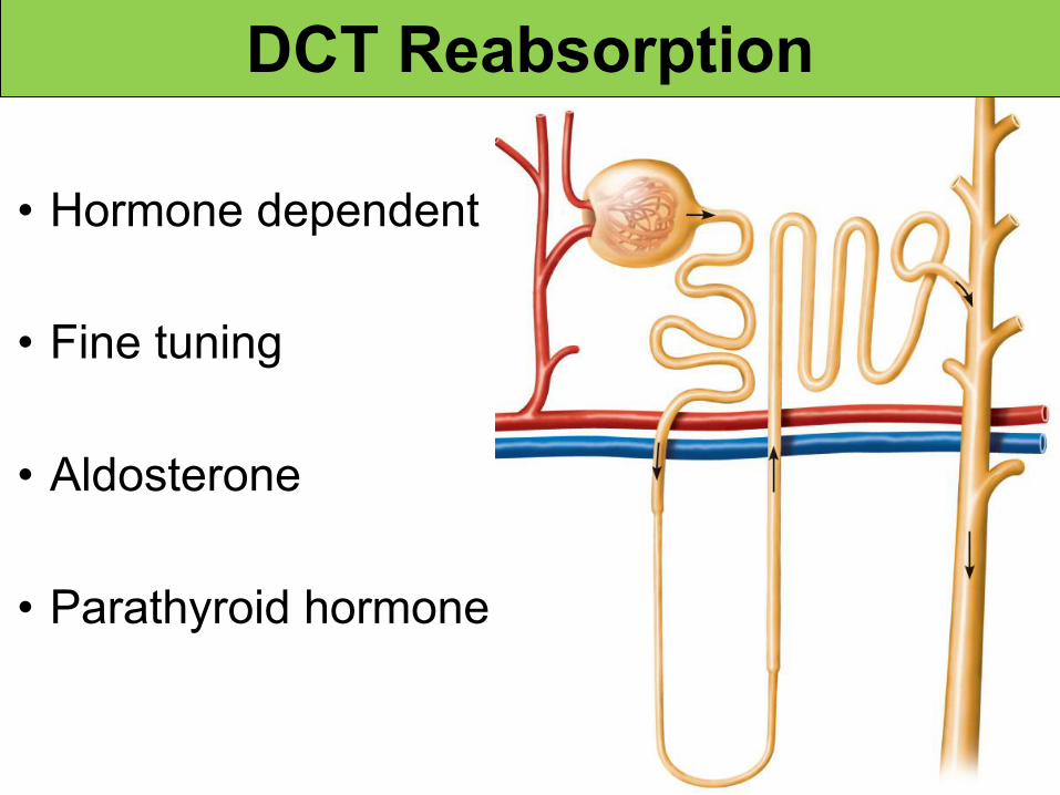
DCT Reabsorption
• Hormone dependent
• Fine tuning
• Aldosterone
• Parathyroid hormone
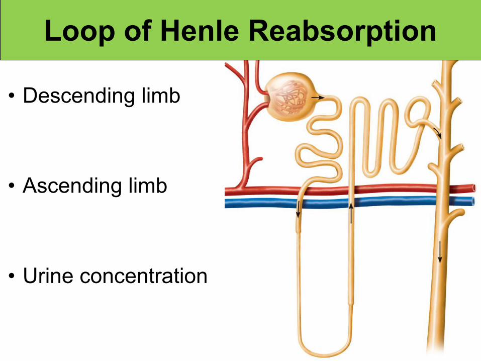
Loop of Henle Reabsorption
• Descending limb
• Ascending limb
• Urine concentration
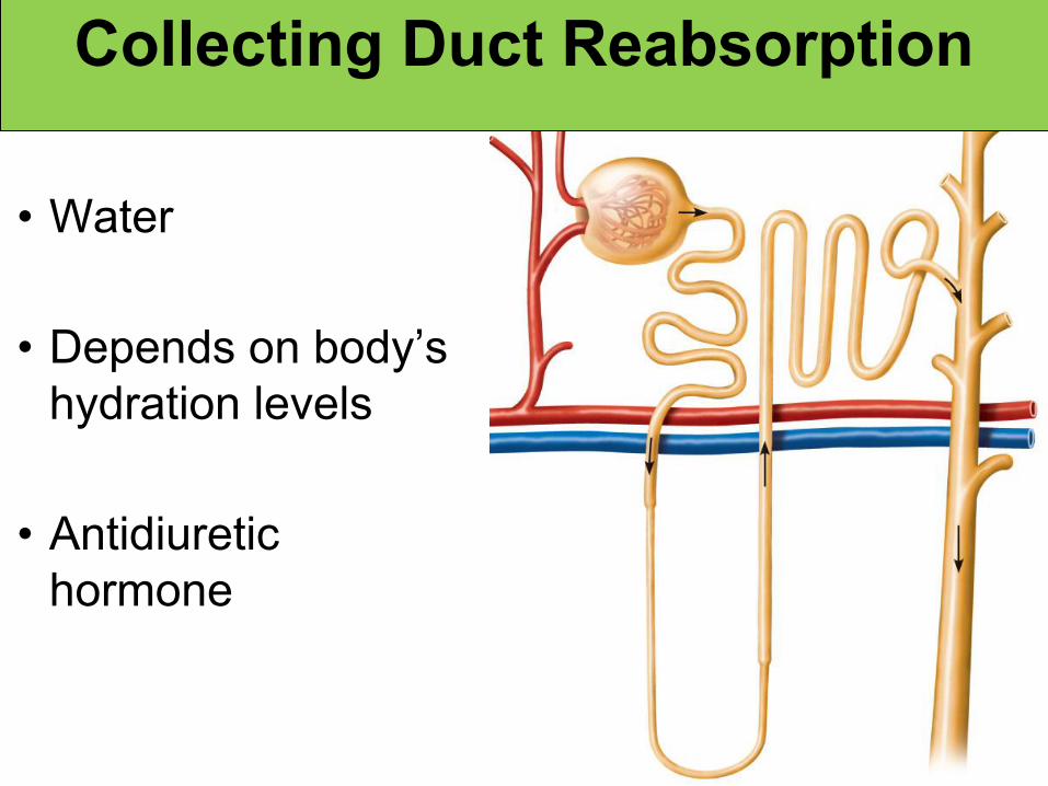
• Water
• Depends on body’s hydration levels
• Antidiuretic hormone
Collecting Duct Reabsorption

Tubular Secretion

Regulating Blood Concentration
• Plasma osmolarity is constantly measured by neurons in the hypothalamus.
• In response to changes in plasma osmolarity, hypothalamic neurons adjust their release of antidiuretic hormone from the posterior pituitary.

ADH increases the reabsorption of water from the collecting duct.
Regulating Blood Concentration

High Blood Osmolarity
Sensed by osmoreceptors in the hypothalamus
Posterior pituitary gland increases ADH release
ADH increases the permeability of the collecting duct to water
Increased water reabsorptionBlood
osmolarity falls
Facultative reabsorption

What will increased ADH do to:
• Water reabsorption
• Urine output
• Urine color
• Blood volume
• Blood pressure


Loop of Henle
Osmolalityof interstitialfluid(mOsm)
Innermedulla
Outermedulla
Cortex Active transport Passive transportWater impermeable
H2O
H2O
H2O
H2O
H2O
H2O
H2O
NaCI
NaCI
NaCI
NaCI
NaCI
Medullary Osmotic Gradient

Aldosterone• Produced by the adrenal cortex.• Produced when:
– Plasma Na+ is too low.– Plasma K+ is too high.– Stimulated by angiotensin II.
Aldosterone acts on the DCT to increase the secretion of K+ and the reabsorption of Na+ and water.

How would excess aldosterone affect
• Plasma sodium levels
• Plasma potassium levels
• Urine output
• Blood volume
• Blood pressure

Diuretics• Osmotic
– Anything that increases filtrate osmolarity increases urine output.
• Caffeine– Inhibits renal sodium reabsorption.
• Alcohol– Inhibits ADH release.

Urine
• Characteristics– Volume
– pH of 4.5 - 8
– Clear to yellow
• Components– 95% water
– 5% solutes• Uric acid• Urea• Creatinine• Ions

Ureters
Adventitia
Muscularis
Mucosa
Mucosalfolds
Ureter

Ureter
PeritoneumMucosalfoldsDetrusor muscle
Ureteral openingsTrigoneNeck of urinary bladder
Internal urethral sphincter
Urethra
External urethral sphincter(in urogenital diaphragm)
Urinary Bladder


Pelvic Diaphragm

Labiumminus
Labiummajus
Urethra
Externalurethral orifice
Urogenitaldiaphragm
External urethral sphincter
Internal urethral sphincter
Urinary bladderTrigone
Ureteral openings
Urethra

Penis
Externalurethral orifice
Spongyurethra
Urogenitaldiaphragm
External urethralsphincter
Prostate gland
Internal urethralsphincter
Prostaticurethra
Membranousurethra
Urinary bladderTrigone
Ureteral openings
Urethra
Urethra

Micturition