Urinary system DevelopmentTeratology. Intermediary mesoderm: Pronephros Pronephros 3rd week, C 3rd...
49
Urinary system Urinary system Development Development Teratology Teratology
-
Upload
jesse-thomas -
Category
Documents
-
view
215 -
download
3
Transcript of Urinary system DevelopmentTeratology. Intermediary mesoderm: Pronephros Pronephros 3rd week, C 3rd...

Urinary systemUrinary system
DevelopmentDevelopment
Teratology Teratology

Intermediary mesoderm:Intermediary mesoderm:
• PronephrosPronephros
3rd week, C3rd week, C
Ductus mesonephricus Ductus mesonephricus (Wolffi)(Wolffi)
• MesonephrosMesonephros 4th week, C6-L34th week, C6-L3
• MetanephrosMetanephros 5th week, L4-S5th week, L4-S

pronephros
mesonephros
metanephros+ureteric bud
- paraaxial - intermediary mesoderm- lateral


Kidney development

Nephron developmentNephron developmentglomerulus

Ascensus renis

Cloaca development
Sinus urogenitalis:-canalis vesicourethralis-pars pelvina-pars phalica

Cloaca Cloaca
• Septum urorectaleSeptum urorectale – – sinus urogenitalissinus urogenitalis + + rectumrectum (cloacal membrane – urogenital m. + anal m.)(cloacal membrane – urogenital m. + anal m.)
• Sinus urogenitalisSinus urogenitalis - - canalis vesicourethraliscanalis vesicourethralis urinary bladder, urinary bladder, ♀♀
uretrauretra - - pars pelvinapars pelvina ♂ ♂ pars pars prostatica et diaphragmatica uretrhrae prostatica et diaphragmatica uretrhrae - - pars phalicapars phalica ♂ ♂ pars phalica urethraepars phalica urethrae
♀ ♀ vestibulum vaginaevestibulum vaginae♀♀ - vestibulum vaginae

Wolffian duct (ductus Wolffian duct (ductus mesonephricus) and ureteric budmesonephricus) and ureteric bud
prostate
seminalvesicles
Cloacal epithelium – from ndoderm
Epithelium of trigonum vesicae – from mesoderm

Congenital malformations of Congenital malformations of urinary systemurinary system
HypoplasiaHypoplasia – small kidney – small kidney RefluxReflux – – HydronephrosisHydronephrosis – urine stasis – urine stasis

Congenital malfromations Congenital malfromations (CM)(CM)
• 1. CM of kidney 1. CM of kidney
• 2. CM of pelvis and ureter2. CM of pelvis and ureter
• 3. CM of urinary bladder3. CM of urinary bladder
• 4. CM of urethra4. CM of urethra

1. CM of kidney1. CM of kidney• anomalies of numberanomalies of number
• anomalies of shape anomalies of shape
• anomalies of postion (ectopia)anomalies of postion (ectopia)
• anomalies of parenchyma (nephrodysplasia)anomalies of parenchyma (nephrodysplasia)
• anomalies of vesselsanomalies of vessels
Kidney malformations arrise at the begining of Kidney malformations arrise at the begining of development (development (development of metanefros isn‘t development of metanefros isn‘t inducedinduced by by ureteric bud or both kidney are closely together – before week ureteric bud or both kidney are closely together – before week
66) or later () or later (during incomplete ascensus renisduring incomplete ascensus renis – – after week 8after week 8).).

Agenesis renisAgenesis renis
• bilateralbilateral (1 : 3000; prenatal dg. – (1 : 3000; prenatal dg. – oligohydramnionoligohydramnion, , hypotrophic fetushypotrophic fetus, , skeleton skeleton deformities and lung hypoplasia due to fetus oppresiondeformities and lung hypoplasia due to fetus oppresion) ) - - (death by uremia and drespiratory distress) (death by uremia and drespiratory distress)
• unilateralunilateral (1 : 1500) + agenesis of ipsilateral (1 : 1500) + agenesis of ipsilateral ureter and renal vessels; ureter and renal vessels;
• etiologyetiology: absence of metanephros, ureteric bud : absence of metanephros, ureteric bud did not develop or did not reach metanephros did not develop or did not reach metanephros (regression) – metanephros development was (regression) – metanephros development was not inducednot induced
• – – genetic dispositiongenetic disposition

1 – kidnye agenesis 1 – kidnye agenesis 2 – kidnye agenesis + cross 2 – kidnye agenesis + cross
ectopia of ureterectopia of ureter
1 2

Supernumerary kidney Supernumerary kidney (2-3 % (2-3 % newborns)newborns) RRen duplexen duplex
• unilateral or bilaterl unilateral or bilaterl
• + + pelvis duplexpelvis duplex and partially or and partially or completely completely ureter fissus ureter fissus or or ureter ureter duplexduplex
• etiologyetiology: 2 ureteric buds or : 2 ureteric buds or branching of one bud in proximal endbranching of one bud in proximal end

1 – ren duplex et ureter duplex, 2 – ureter 1 – ren duplex et ureter duplex, 2 – ureter fissusfissus
1 2

Shape malformations of kidney:Shape malformations of kidney:Horse-shaped kidney (ren Horse-shaped kidney (ren
arcuatus)arcuatus) 1:5001:500 • etiologyetiology: fusion of lower pole of both : fusion of lower pole of both
metanephros in front of large vessels metanephros in front of large vessels (aorta + v.cava inf.)(aorta + v.cava inf.)
• fused parenchyma = isthmus „brakes“ fused parenchyma = isthmus „brakes“ ascensus renis bellow detachment of a. ascensus renis bellow detachment of a. mesenterica inf. (mesenterica inf. (+position anomaly - +position anomaly - ektopiaektopia) and rotation () and rotation (++ malrotationmalrotation; ; hilus – ventrally), ureters run in front of hilus – ventrally), ureters run in front of isthmus –isthmus –+ renal vessels duplication+ renal vessels duplication

A – ren arcuatusB – ren fungiformisC – ren sigmoideus
A B
C
Anomaly of the shape + ektopia:
urine stasis – hydronefrosis+ vesicaureteric reflux secondary infections

Position anomalies:Position anomalies:Ectopia of kidneyEctopia of kidney uni-, bilat.uni-, bilat.
• - - ren pelvicusren pelvicus ( (ren sacralisren sacralis, , ren ren lumbalislumbalis): retention of kidney during ): retention of kidney during ascensus renis ascensus renis
• - - cross ectopiacross ectopia: both ureters grow : both ureters grow into metanephros on one side or into metanephros on one side or during ascensus renis one kidney during ascensus renis one kidney transfers on the oposit side and fuse transfers on the oposit side and fuse with the other kidneywith the other kidney

Ren pelvicus
+ ren + ureter duplex
Cross ectopia

Malrotation Malrotation (or(or hyperrotation)hyperrotation) of of
kidneykidney
• is connected is connected with ectopia or with ectopia or anomaly of kidney anomaly of kidney shape shape
• hilus – ventrally hilus – ventrally (embryonic (embryonic
position)position) or dorsaly or dorsaly
• Notice:(normal adult position of Notice:(normal adult position of hilus is medial)hilus is medial)

• diffuse cystic malformation (diffuse cystic malformation (always always bilatbilat.) – cystic degeneration of kidney.) – cystic degeneration of kidney
• 2 forms of polycystic disease: 2 forms of polycystic disease: - - autosomally dominantautosomally dominant type type
˝adult˝ ˝adult˝ macrocysticmacrocystic form form - - autosomally recesiveautosomally recesive type type
˝infantil˝ ˝infantil˝ microcysticmicrocystic form form
Defekts of parenchymaDefekts of parenchyma::Polycystic kidneysPolycystic kidneys nephrodysplasia polycysticanephrodysplasia polycystica

autosomally dominant type autosomally dominant type APCD – Adult Polycystic APCD – Adult Polycystic DiseaseDisease
• Disease manifests in adulthood (after 30th); Disease manifests in adulthood (after 30th); 1:400 - 1000, probability of transmission to 1:400 - 1000, probability of transmission to offspring is 50 %;offspring is 50 %;
• etiol.:etiol.: patol. genes on 4th and 16th chromosomes patol. genes on 4th and 16th chromosomes – – insufficient polycystin productioninsufficient polycystin production (membrane protein (membrane protein necessary for differentiation of cells in renal tubules)necessary for differentiation of cells in renal tubules). .
• Klinic manifestation:Klinic manifestation: bilat. enlarged kidney, macroscopic bilat. enlarged kidney, macroscopic cysts, abdominal and/or lumbal pain, hematuria, cysts, abdominal and/or lumbal pain, hematuria, hypertension, infections, renal insufficiency and failure.hypertension, infections, renal insufficiency and failure.
• Dg.:Dg.: (FA), abdomen palpation, sono event. CT (FA), abdomen palpation, sono event. CT• Th.:Th.: symptomatic, decelerate progression of symptomatic, decelerate progression of
disease, renal failure – disease, renal failure – renal renal funcions have to be compensated (hemodialysis, peritoneal funcions have to be compensated (hemodialysis, peritoneal dialysis, transplantation)dialysis, transplantation)

Polycystic kidney – macroscopic cysts are seen also on the kidney surface

autosomally recesive typeautosomally recesive type IPCDIPCD - ˝infantil˝ form - ˝infantil˝ form
• 1 : 40.000, probability of transmission to offspring - 1 : 40.000, probability of transmission to offspring - 25 % children of healthy parents „disease carriers˝; 25 % children of healthy parents „disease carriers˝;
• + anomalies also in liver, spleen, lungs,+ anomalies also in liver, spleen, lungs,• etiol.etiol.:: unclear - defect of ureter development unclear - defect of ureter development
(nephrons are not connected with collecting ducts) (nephrons are not connected with collecting ducts) • Klinic manifestation:Klinic manifestation: bilat. enlarged kidneey, hypertension, bilat. enlarged kidneey, hypertension,
decreased glomerular filtration, renal failure. To a lesser decreased glomerular filtration, renal failure. To a lesser extent of damage 50-80 % children can live about 15 years. extent of damage 50-80 % children can live about 15 years. Some children die shortly after birth by lung failure.Some children die shortly after birth by lung failure.
• Prenatal dg. in week 9 of i.u.dev.Prenatal dg. in week 9 of i.u.dev. – FA, DNA markers. – FA, DNA markers. • Th.:Th.: same as in same as in PCHLADPCHLAD

Polycystic kidney – cysts are not seen on the surface of kidney

Hypoplasia renisHypoplasia renis
• Insuficiently developed Insuficiently developed kidney – small, small kidney – small, small amount of histologically amount of histologically normal and functional normal and functional nephronsnephrons
• usually unilateralusually unilateral• compenzational compenzational
hypertrofia of the other hypertrofia of the other kidneykidney

A – unilateral renal agenesis B – pelvic ren + ureter bifidus
C – kidney malrotation + ren duplexD – crossed ectopia
E – „pancake“ kidney
F – supernumerary kidney

Wilms‘ tumorWilms‘ tumor (nephroblastom)(nephroblastom)
• The most frequent type of tumors in The most frequent type of tumors in chidren under 5 years, rare in chidren under 5 years, rare in adulthood adulthood
• 90% treatment succes, also in case of 90% treatment succes, also in case of greater distribution (metastasis)greater distribution (metastasis)
• familiar occurrence – tumor contais familiar occurrence – tumor contais cells of mesonephroscells of mesonephros
• etiologyetiology:: - hereditary basis - hereditary basis

Thesaurismosis Thesaurismosis („(„storage diseasestorage disease“)“)
A metabolic disorder in which a substance is stored in certain cells A metabolic disorder in which a substance is stored in certain cells of some organs, usually in large amounts, due to dof some organs, usually in large amounts, due to defect efect production of enzymes splitting this substance. It causes production of enzymes splitting this substance. It causes functional failure of storing organs functional failure of storing organs
Etiol.: Etiol.: defected gen in auto- or heterosomes, usually recessive defected gen in auto- or heterosomes, usually recessive inheritanceinheritance
• Anderson-Fabry diseaseAnderson-Fabry disease (storage of (storage of cerebrosidescerebrosides = neutral = neutral sphingolipidssphingolipids), ),
• von Gierke diseasevon Gierke disease (storage of (storage of glycogenglycogen), ), • Gaucher diseaseGaucher disease (storage of (storage of glukocerebrosidesglukocerebrosides),),• Fanconi syFanconi sy. (storage of . (storage of cystinecystine; ; cystinózacystinóza, , cystinuriecystinurie))• Primary hyperoxaluriaPrimary hyperoxaluria – cong. defect of glykooxalates – cong. defect of glykooxalates
production (storage of production (storage of oxalatesoxalates; ; urolithiasisurolithiasis). ). • Cong. defects of metabolism of Cong. defects of metabolism of purinespurines – familiary gouty – familiary gouty
juvenile nephropathy + artritis already in the 2nd dacede of life. juvenile nephropathy + artritis already in the 2nd dacede of life.

Anomalies of renal vascularizationAnomalies of renal vascularization
• Arise during ascensus renis – accesory Arise during ascensus renis – accesory arteries from a. iliaca and aorta (there arteries from a. iliaca and aorta (there are NOT collaterals between arteries! – are NOT collaterals between arteries! – obstruction causes infarction of renal obstruction causes infarction of renal parenchyma) parenchyma)
• supernumerary veins (with collaterals)supernumerary veins (with collaterals)
• accesory arteries – 25 %, veins - 12,5 accesory arteries – 25 %, veins - 12,5 % %

Renal renculiRenal renculi
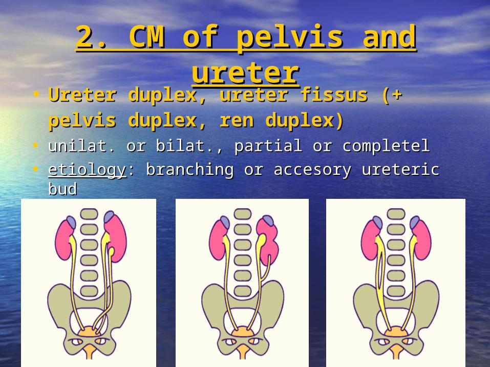
2. CM of pelvis and 2. CM of pelvis and ureterureter
• Ureter duplex, ureter fissus (+ pelvis Ureter duplex, ureter fissus (+ pelvis duplex, ren duplex)duplex, ren duplex)
• unilat. or bilat., partial or completelunilat. or bilat., partial or completel
• etiologyetiology: branching or accesory ureteric bud: branching or accesory ureteric bud

Ectopic defects of ureterEctopic defects of ureter
• ectopia of orificium ectopia of orificium ureterisureteris –ureter opens –ureter opens into urethra, uterus or into urethra, uterus or vagina (vagina (rarely into rarely into ductus deferensductus deferens))
• cross ectopia of ureter, cross ectopia of ureter, „retrocava″ ureter„retrocava″ ureter – –

Congenital stenosis, obstructions, Congenital stenosis, obstructions, atresisatresis
Physiol. ureter narrowings:Physiol. ureter narrowings:
1.1. pelvi-uretral junction, pelvi-uretral junction,
2.2. crossing with vasa iliaca, crossing with vasa iliaca,
3.3. pars intramuralis – pars intramuralis – ureter-vesical ureter-vesical junction.junction.
1
2
3

3. CM of urinary bladder3. CM of urinary bladder
• Extrophia Extrophia 1 : 40.000 1 : 40.000 (2-3 ♂ (2-3 ♂ :: 1♀) 1♀)
• Ventral abdominal wall Ventral abdominal wall and ventral wall of and ventral wall of urinary bladder are not urinary bladder are not formed; urinary bladder formed; urinary bladder is opened and inner is opened and inner surface of its dorsal wall surface of its dorsal wall is visible (+ epispadia is visible (+ epispadia and cleft of symphysis and cleft of symphysis (diastasis)(diastasis)

ExtrophiaExtrophia
• etiol.etiol.: defect of mezenchyme : defect of mezenchyme migration between ectoderm of migration between ectoderm of abdominal wall and cloaca in week 4abdominal wall and cloaca in week 4
• Reconstruction of the wall (24 - 48 h Reconstruction of the wall (24 - 48 h after birth), epispadia (about 2nd year).after birth), epispadia (about 2nd year).

Sy. prune – bellySy. prune – belly
• 1 : 40 0001 : 40 000• Trias of syndromesTrias of syndromes::
1.1. agenesis of muscles in abdominal agenesis of muscles in abdominal wall wall 2.2. obstruction of urinary bladderobstruction of urinary bladder in region of internal
urethral ostium (dilatation of bladder, megaureters and hydronephrosis – above obstruction)
3.3. bilateral kryptorchismkryptorchism• Etiol.: unclear
hormonal treatment (estrogens) of mother during the 1st trimestr was frequently find in anamnesis
• Prognosis: viability depends on number of functional nephrons at birth.

defect obliteration of ductus defect obliteration of ductus allantoideusallantoideus ––• urachal cysts and fistulae (a, b)urachal cysts and fistulae (a, b)
• urachus patens (c)urachus patens (c)
a b c

4. CM or urethra4. CM or urethra
• Clefts of urethra:Clefts of urethra:
HypospadiaHypospadia insufficient fusion insufficient fusion of plicae genitales of plicae genitales
EpispadiaEpispadia see see extrophia extrophia

Sources of pictures:Sources of pictures:
• http://www.embryology.ch/genericpages/moduleohttp://www.embryology.ch/genericpages/moduleorganoen.htmlrganoen.html
• embryology.med.embryology.med.unswunsw..eduedu.au/.../.au/.../BGDlabXYXXBGDlabXYXX_5._5.htmhtm..
• www.embryology.ch/.../genitinterne06.htmlwww.embryology.ch/.../genitinterne06.html..• www.emedicine.com/ped/topic704.htmwww.emedicine.com/ped/topic704.htm.. • embryology.med.embryology.med.unswunsw..eduedu.au/.au/DefectDefect/page4./page4.htmhtm. . • www.childrenskidneydisease.org/Stories.aspwww.childrenskidneydisease.org/Stories.asp. .
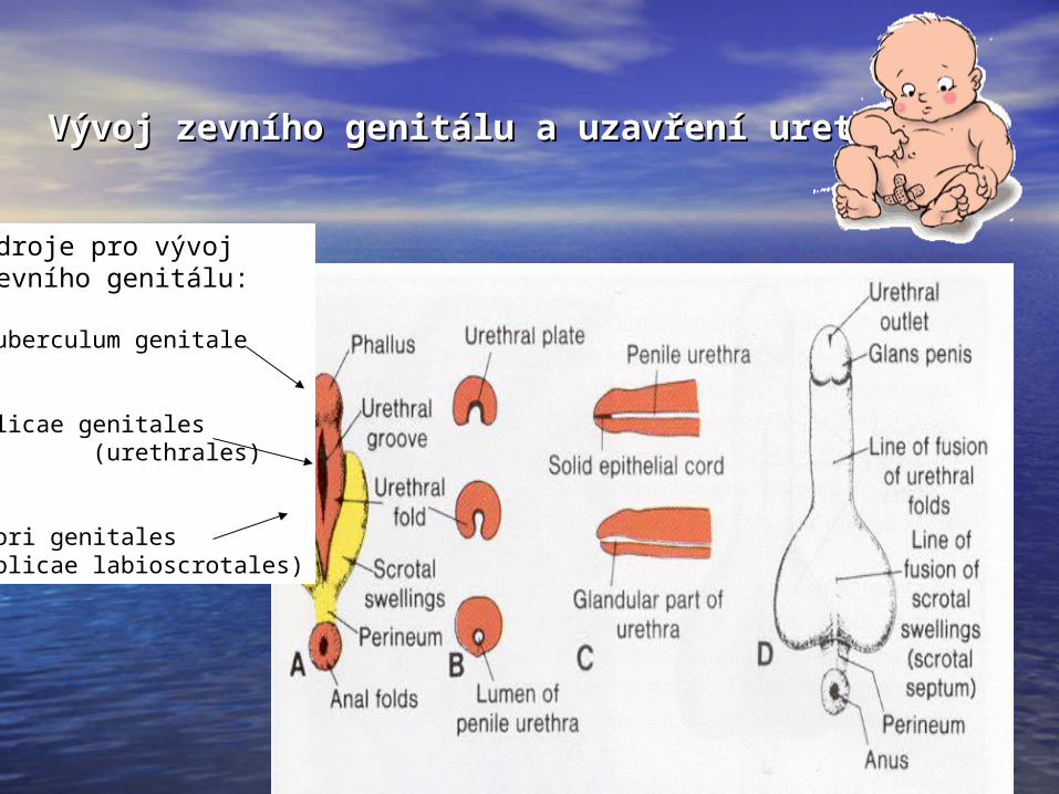
Vývoj zevního genitálu a uzavření uretry Vývoj zevního genitálu a uzavření uretry ♂♂
Zdroje pro vývojzevního genitálu:
Tuberculum genitale
Plicae genitales (urethrales)
Tori genitales(plicae labioscrotales)

Vývoj nefronuVývoj nefronu


Močový měchýř, ureterMočový měchýř, ureter