Cognitive Map of the Pedagogical Situation as a Component ...

Unpacking the Cognitive Map: The Parallel Map Theoryof Hippocampal Function
Lucia F. JacobsUniversity of California, Berkeley
Francoise SchenkUniversity of Lausanne
In the parallel map theory, the hippocampus encodes space with 2 mapping systems. The bearing mapis constructed primarily in the dentate gyrus from directional cues such as stimulus gradients. The sketchmap is constructed within the hippocampus proper from positional cues. The integrated map emergeswhen data from the bearing and sketch maps are combined. Because the component maps work inparallel, the impairment of one can reveal residual learning by the other. Such parallel function mayexplain paradoxes of spatial learning, such as learning after partial hippocampal lesions, taxonomic andsex differences in spatial learning, and the function of hippocampal neurogenesis. By integratingevidence from physiology to phylogeny, the parallel map theory offers a unified explanation forhippocampal function.
The cognitive map theory articulated by John O’Keefe and LynnNadel in 1978 not only was the first unified theory of hippocampalfunction but also has been the most influential (Best & White,1999). This theory postulated that the hippocampus creates amental representation of allocentric space. This representation, thecognitive map, is more flexible than other mental representationsof space and allows the navigator to create novel routes betweenfamiliar sites.
O’Keefe and Nadel’s (1978) cognitive map theory was sup-ported by a diversity of empirical results, such as the impairmentof spatial navigation by hippocampal lesions (Jarrard, 1983; Mor-ris, Hagan, & Rawlins, 1986). It was also supported by tworemarkable discoveries. The first was the elucidation of the hip-pocampal place unit by O’Keefe (e.g., O’Keefe & Dostrovsky,1971). O’Keefe found that activity of these hippocampal pyrami-dal cells was localized to specific locations in a test environmentand that they retained their specificity even in the absence of visualinput (O’Keefe & Conway, 1980). This provided concrete evi-dence for the role of the hippocampus in coding locations in space.
A second discovery was the demonstration of a hippocampalmechanism for rapid, long-lasting, synapse-specific associative
learning. This is the process of long-term potentiation (LTP; Bliss& Lomo, 1973). LTP mediation by the N-methyl-D-aspartate(NMDA) receptor provided a physiological theory of the Hebbiansynapse (Bliss & Collingridge, 1993). Evidence that spatial learn-ing and hippocampal LTP are impaired by NMDA-receptor antag-onists provided new support for the cognitive map theory (Morriset al., 1986).
Twenty years later, however, the cognitive map theory remainscontroversial. Although it laid the foundation for current theoriesof how the hippocampus encodes space (McNaughton et al., 1996;O’Keefe & Burgess, 1996; Redish, 1999), the present theoriesdiffer from one another, and no one model of spatial encoding hasreceived universal acceptance. There are also critics of the cogni-tive map theory itself (Eichenbaum, Dudchenko, Wood, Shapiro,& Tanila, 1999). The disagreement about hippocampal function isfueled in part by different approaches to the question (e.g., non-human vs. human studies) and in part by paradoxical results.Results from the rodent literature include the recovery of placelearning after hippocampal lesions (Whishaw, Cassel, & Jarrard,1995), the tuning of hippocampal cells to nonspatial information(Wood, Dudchenko, & Eichenbaum, 1997), the ameliorative ef-fects of maze pretraining on place learning in the presence ofblocked synaptic plasticity (Bannerman, Good, Butcher, Ramsay,& Morris, 1995; Saucier & Cain, 1995), and other dissociations ofsynaptic plasticity and spatial learning (Abeliovich et al., 1993;Huang et al., 1995; Nosten-Bertrand et al., 1996; Richter-Levin,Thomas, Hunt, & Bliss, 1998).
Research guided by questions of hippocampal function in hu-mans has focused on the hippocampus’s role in universal cognitiveprocesses, such as intermediate-term memory (Rawlins, 1985),declarative memory (Squire, 1992), contextual processing (Gluck& Myers, 2001), episodic memory (Burgess, Maguire, & O’Keefe,2002; O’Keefe & Nadel, 1978; Squire & Zola-Morgan, 1991;Vargha-Khadem et al., 1997), or relational processing (Cohen &Eichenbaum, 1993; Eichenbaum et al., 1999). For example,Eichenbaum et al. proposed that the hippocampus’s role in spatial
Lucia F. Jacobs, Department of Psychology, University of California,Berkeley; Francoise Schenk, Institute of Physiology, University of Lau-sanne, Lausanne, Switzerland.
This research was supported by a Prytanean Faculty Award and bygrants from the University of California, from the J.D. French Alzheimer’sFoundation to Lucia F. Jacobs, and from the Swiss National ScienceFoundation to Francoise Schenk. For comments, suggestions, encourage-ment, and argument, we thank Kim Beeman, Andrea Chiba, Jack Cowan,Brian Derrick, Michael Dickinson, Jack Gallant, Michael Bastiani, LeslieKay, Bruce Miller, Richard Morris, Lynn Nadel, Jacques Paillard, JonSeger, Matthew Shapiro, Miriam Smolover, David Steinsaltz, GeorgStriedter, and Matthew Wilson.
Correspondence concerning this article should be addressed to Lucia F.Jacobs, Department of Psychology, University of California, Tolman Hall,Berkeley, California 94720-1650. E-mail: [email protected]
Psychological Review Copyright 2003 by the American Psychological Association, Inc.2003, Vol. 110, No. 2, 285–315 0033-295X/03/$12.00 DOI: 10.1037/0033-295X.110.2.285
285

learning is an application case of its function for a more generallearning process, the learning of relations. Here, spatial represen-tations do not constitute a map of space but instead contribute tothe general principle of “linking events within episodes” (Eichen-baum et al., 1999, p. 216). As a consequence, this “memory space”(Eichenbaum et al., 1999, p. 218) codes spatial and nonspatialrelations among events, processing spatial relations for navigationand serial relations for solving more abstract, nonspatial stimulusrelations, such as those found in transitive inference (Dusek &Eichenbaum, 1997).
Spatial models of the hippocampus, in contrast, often take abottom-up, computational approach to the question of how therodent hippocampus maps space. These models have included newgeneration models from O’Keefe and Burgess (1996), the multiplechart theory of McNaughton et al. (1996; Samsonovich & Mc-Naughton, 1997), and the multiframe theory of Redish andTouretsky (1997). For example, the Redish and Touretsky modelon spatial navigation combines four different navigation systems(taxon, praxic, locale, and route) and five spatial representations(local view, head direction, path integrator, place code, and goalmemory). The anatomical realization of the model is provided bythe connections among the several brain structures assumed tomediate these representations (Redish, 1999). We cannot reviewall of the models in detail, and the interested reader is referred tothe above citations for more information.
At present, no one theory, whether spatial or nonspatial, quali-tative or quantitative, modeling human or nonhuman behavior, hasgained universal acceptance. Instead, the different approaches ap-pear to be developing in parallel (Best & White, 1999; Cohen &Eichenbaum, 1991; McNaughton, 1996; Nadel, O’Keefe, Shapiro,McNaughton, & Disterhoft, 1998). It is clear that the rodenthippocampus plays some critical role in spatial orientation and thatthis role is not fully understood. If neurobiology cannot be under-stood except in light of behavior (Shepherd, 1994) and, asDobzhansky (1951) argued, biology cannot be understood exceptin light of evolution, then to understand the hippocampus, we mustunderstand both its evolution and its role in spatial navigation. Webegin with a reexamination of this behavior.
The Nature of Spatial Navigation
We start by defining spatial navigation and discussing what iscommonly accepted as spatial information. We do this becausethere is much confusion over terms and it is important for us tospecify exactly what we mean by a word such as landmark. Wethen outline a new theory of navigation based on the idea that thecognitive map is composed of parallel component maps. We thenaddress the origins of the dual nature of navigation in vertebratesand the development of a more powerful representation of space,the cognitive map, in birds and mammals. Because we propose thatdifferent classes of maps are constructed by the mammalian hip-pocampus and that these maps rely on different cues, we are alsorequired to introduce new terminology to define experimentalconditions precisely in terms of such cues.
Navigation can be defined as purposive movement throughabsolute space. For absolute space, we ascribe to O’Keefe andNadel’s (1978) original definition:
Absolute space embodies the notion of a framework or containerwithin which material objects can be located but which is conceived
as existing independently of particular objects or objects in general.Objects are located relative to the places of the framework and onlyindirectly, via this framework, to other objects. Movement of a body(including the observer) changes its position within the framework butdoes not alter the framework or the relationship of other objects to theframework. In contrast, relative space designates a set of relationshipsamongst objects or sensory inputs which in themselves are inherentlynonspatial. Objects are located relative to other objects and relativespace does not exist independent of the existence of objects. (p. 7)
Further, O’Keefe and Nadel distinguished between the mentalrepresentations of psychological space and its complement, phys-ical space, defined as “any space attributed to the external worldindependent of the existence of minds” (pp. 6–7).
Thus, there is physical space, and there are mental maps of thatspace. A map can be defined in diverse ways; we ascribe toNeisser’s (1976) broad definition: “not pictures in the head, butplans for obtaining information from potential environments” (p.131). Hence, the defining characteristic of the class of mentalrepresentations known as a map is that it provides a navigator,moving in space, with an expectation of the sensory input given acertain movement in a certain direction at a certain speed or arounda certain obstacle. With a cognitive map (i.e., a mental map ofabsolute space; O’Keefe & Nadel, 1978, p. 2), the navigator canplace itself within the framework and navigate directly amongobjects.
This ability is also known as place learning. This differs fromorientation in relative space, such as egocentric or orientation to asimple cue. In an experimental setting, an animal is said to dem-onstrate place learning when it automatically creates a new route tothe goal, and this new route is constructed from new egocentricrelations to objects. Thus, a rat that can spontaneously chart adirect course in the water maze, regardless of its release position inthe pool, is said to demonstrate place learning (Morris, 1984).
Sources of Spatial Information
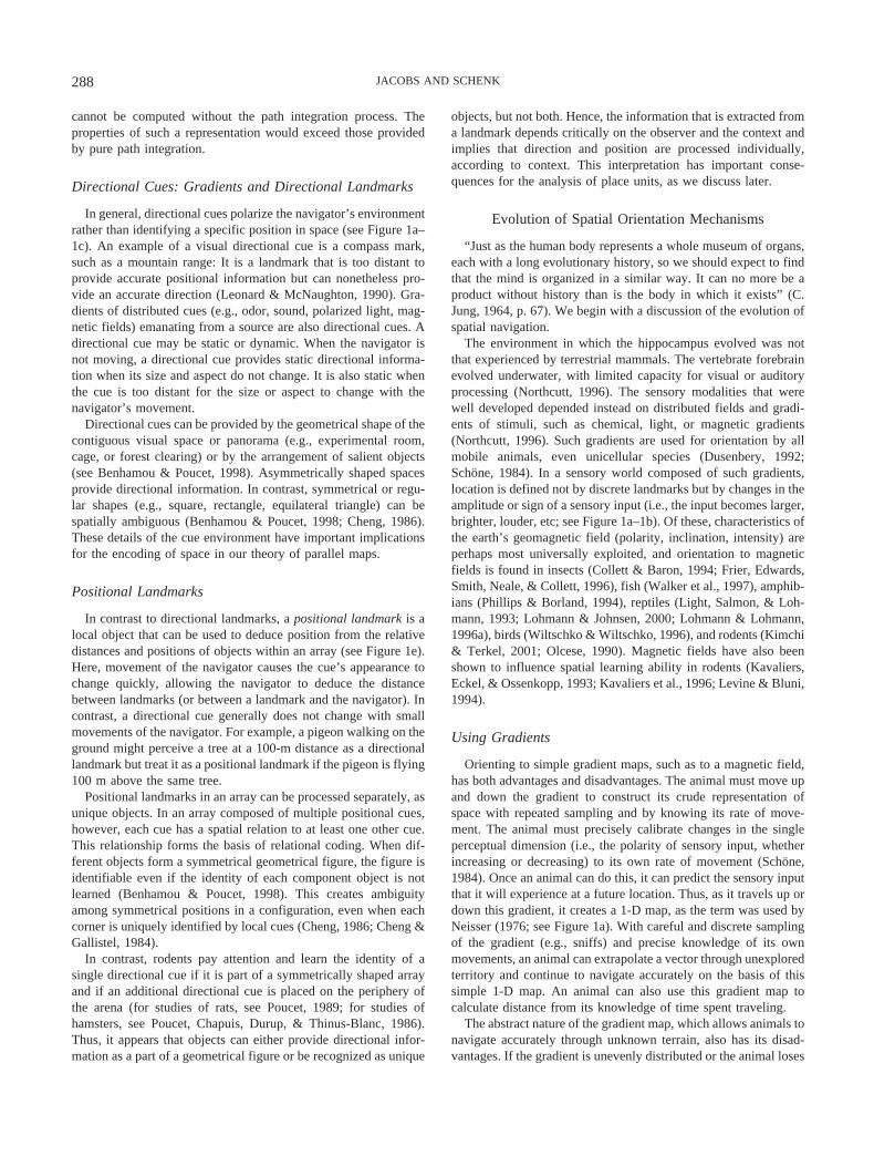
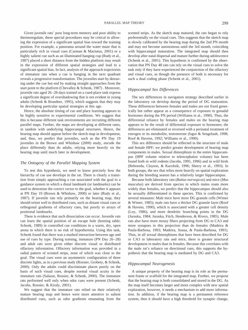
As an animal explores, it uses internal and external cues to relateits current position to its start point in the environment. Internalcues such as self-generated movement cues inform the navigatorhow far and in which direction it has moved from a given position.External cues such as landmarks can be used in two different ways,both for direction and for position (Collett, Cartwright, & Smith,1986; Leonard & McNaughton, 1990), as illustrated in Figure 1cand 1e.
Self-Generated Movement Cues
Locomotion generates a dynamic sensory flow in diverse mo-dalities (proprioceptive, tactile, auditory, olfactory, and visual).The navigator integrates some or all of this information to updatethe current position relative to the start point. Path integration isthe outcome of the process that regularly updates a directionalvector. The vector is generated by the animal’s movement duringan exploratory bout and is based on this dynamic sensory flow andthe efferent copy of the intended action. The path integrationvector encodes the distance and direction from the start point ofexploration, where the vector is apparently reset. Thus, path inte-gration allows the navigator to beeline to its most recent startposition at any time. It appears to be an ancient component of
286 JACOBS AND SCHENK

spatial orientation, as it is found throughout invertebrate andvertebrate taxa (Leonard & McNaughton, 1990; Maaswinkel &Whishaw, 1999; Wehner, Michel, & Antonsen, 1996).
We point out one caveat: Although we can define path integra-tion, it is much less clear when and how it is combined with otherspatial information. We suggest there is an important distinctionbetween pure path integration and the reliance of the hippocampuson path integration. Perhaps path integration itself (a single, one-dimensional gradient produced from vestibular and external sen-sory feedback) is simply a vector that is exported to the hippocam-
pus, which then assigns meaning to this vector. In this case, pathintegration would not be a property of the hippocampus but aprocess whose output is used by the hippocampus in constructingone-dimensional (1-D) and two-dimensional (2-D) maps. The vec-tor obtained from path integration could be a primitive workingmemory representation, one that is reset at the start of everyexploratory bout. It might then acquire more dimensions when it isassociated with external points, such as an identifiable start posi-tion. This association of the working memory vector with externallandmarks would lead to a richer representation of space, one that
Figure 1. Determining direction and position with spatial cues. a: Gradient—A field of graded intensity. b:Compass mark—A distal landmark that can provide only directional information; it is too distant to providepositional information. c: Array geometry—Direction deduced from the polarization of an array of positionallandmarks. d: Two-dimensional (2-D) gradient map—A 2-D map constructed from directional cues. e: 2-Dtopographic map—A 2-D map constructed from positional cues.
287PARALLEL MAP THEORY
cannot be computed without the path integration process. Theproperties of such a representation would exceed those providedby pure path integration.
Directional Cues: Gradients and Directional Landmarks
In general, directional cues polarize the navigator’s environmentrather than identifying a specific position in space (see Figure 1a–1c). An example of a visual directional cue is a compass mark,such as a mountain range: It is a landmark that is too distant toprovide accurate positional information but can nonetheless pro-vide an accurate direction (Leonard & McNaughton, 1990). Gra-dients of distributed cues (e.g., odor, sound, polarized light, mag-netic fields) emanating from a source are also directional cues. Adirectional cue may be static or dynamic. When the navigator isnot moving, a directional cue provides static directional informa-tion when its size and aspect do not change. It is also static whenthe cue is too distant for the size or aspect to change with thenavigator’s movement.
Directional cues can be provided by the geometrical shape of thecontiguous visual space or panorama (e.g., experimental room,cage, or forest clearing) or by the arrangement of salient objects(see Benhamou & Poucet, 1998). Asymmetrically shaped spacesprovide directional information. In contrast, symmetrical or regu-lar shapes (e.g., square, rectangle, equilateral triangle) can bespatially ambiguous (Benhamou & Poucet, 1998; Cheng, 1986).These details of the cue environment have important implicationsfor the encoding of space in our theory of parallel maps.
Positional Landmarks
In contrast to directional landmarks, a positional landmark is alocal object that can be used to deduce position from the relativedistances and positions of objects within an array (see Figure 1e).Here, movement of the navigator causes the cue’s appearance tochange quickly, allowing the navigator to deduce the distancebetween landmarks (or between a landmark and the navigator). Incontrast, a directional cue generally does not change with smallmovements of the navigator. For example, a pigeon walking on theground might perceive a tree at a 100-m distance as a directionallandmark but treat it as a positional landmark if the pigeon is flying100 m above the same tree.
Positional landmarks in an array can be processed separately, asunique objects. In an array composed of multiple positional cues,however, each cue has a spatial relation to at least one other cue.This relationship forms the basis of relational coding. When dif-ferent objects form a symmetrical geometrical figure, the figure isidentifiable even if the identity of each component object is notlearned (Benhamou & Poucet, 1998). This creates ambiguityamong symmetrical positions in a configuration, even when eachcorner is uniquely identified by local cues (Cheng, 1986; Cheng &Gallistel, 1984).
In contrast, rodents pay attention and learn the identity of asingle directional cue if it is part of a symmetrically shaped arrayand if an additional directional cue is placed on the periphery ofthe arena (for studies of rats, see Poucet, 1989; for studies ofhamsters, see Poucet, Chapuis, Durup, & Thinus-Blanc, 1986).Thus, it appears that objects can either provide directional infor-mation as a part of a geometrical figure or be recognized as unique
objects, but not both. Hence, the information that is extracted froma landmark depends critically on the observer and the context andimplies that direction and position are processed individually,according to context. This interpretation has important conse-quences for the analysis of place units, as we discuss later.
Evolution of Spatial Orientation Mechanisms
“Just as the human body represents a whole museum of organs,each with a long evolutionary history, so we should expect to findthat the mind is organized in a similar way. It can no more be aproduct without history than is the body in which it exists” (C.Jung, 1964, p. 67). We begin with a discussion of the evolution ofspatial navigation.
The environment in which the hippocampus evolved was notthat experienced by terrestrial mammals. The vertebrate forebrainevolved underwater, with limited capacity for visual or auditoryprocessing (Northcutt, 1996). The sensory modalities that werewell developed depended instead on distributed fields and gradi-ents of stimuli, such as chemical, light, or magnetic gradients(Northcutt, 1996). Such gradients are used for orientation by allmobile animals, even unicellular species (Dusenbery, 1992;Schone, 1984). In a sensory world composed of such gradients,location is defined not by discrete landmarks but by changes in theamplitude or sign of a sensory input (i.e., the input becomes larger,brighter, louder, etc; see Figure 1a–1b). Of these, characteristics ofthe earth’s geomagnetic field (polarity, inclination, intensity) areperhaps most universally exploited, and orientation to magneticfields is found in insects (Collett & Baron, 1994; Frier, Edwards,Smith, Neale, & Collett, 1996), fish (Walker et al., 1997), amphib-ians (Phillips & Borland, 1994), reptiles (Light, Salmon, & Loh-mann, 1993; Lohmann & Johnsen, 2000; Lohmann & Lohmann,1996a), birds (Wiltschko & Wiltschko, 1996), and rodents (Kimchi& Terkel, 2001; Olcese, 1990). Magnetic fields have also beenshown to influence spatial learning ability in rodents (Kavaliers,Eckel, & Ossenkopp, 1993; Kavaliers et al., 1996; Levine & Bluni,1994).
Using Gradients
Orienting to simple gradient maps, such as to a magnetic field,has both advantages and disadvantages. The animal must move upand down the gradient to construct its crude representation ofspace with repeated sampling and by knowing its rate of move-ment. The animal must precisely calibrate changes in the singleperceptual dimension (i.e., the polarity of sensory input, whetherincreasing or decreasing) to its own rate of movement (Schone,1984). Once an animal can do this, it can predict the sensory inputthat it will experience at a future location. Thus, as it travels up ordown this gradient, it creates a 1-D map, as the term was used byNeisser (1976; see Figure 1a). With careful and discrete samplingof the gradient (e.g., sniffs) and precise knowledge of its ownmovements, an animal can extrapolate a vector through unexploredterritory and continue to navigate accurately on the basis of thissimple 1-D map. An animal can also use this gradient map tocalculate distance from its knowledge of time spent traveling.
The abstract nature of the gradient map, which allows animals tonavigate accurately through unknown terrain, also has its disad-vantages. If the gradient is unevenly distributed or the animal loses
288 JACOBS AND SCHENK
track of its rate of sampling or its rate of movement, the map is nolonger reliable, and there is little opportunity for self-correction.The value and reliability of a simple 1-D map can be enhanced,however, when it is combined with other 1-D maps, the intersec-tion of which yields a Cartesian coordinate (i.e., 2-D) map (seeFigure 1d). Once this 2-D map has been created, it can be used topredict the length and orientation of a vector through untraveledspace.
Evidence for Gradient Maps
The hypothesis that vertebrates create 2-D maps from distrib-uted stimuli has generated much discussion but few rigorous tests(Wallraff, 1996). There is indirect support for this hypothesis,however, from studies on orientation to plumes of olfactory cuesby homing pigeons (Papi, 1992) and magnetic field orientation insea turtles (Lohmann & Lohmann, 1996b).
Green sea turtles (Chelonia mydas) migrate as adults to theirplace of hatching, Ascension Island in the southern AtlanticOcean, a site thousands of kilometers from the location to whichthey initially dispersed. They navigate through the southern At-lantic Ocean using traditional paths, even on their first return to theisland after many years (Luschi, Hays, Del Seppia, Marsh, & Papi,1998). Therefore, on the basis of little experience and in theabsence of obvious landmarks, the turtles solve an oceanic watermaze, returning to a tiny island in the middle of the southernAtlantic. A population genetic analysis of Ascension Island turtleshas shown that their genotype is unique to this island. Because noturtle of a different genotype has ever been found on the island,this suggests that the population has been genetically isolated,perhaps by the unique navigational algorithm they use to return tothe island (Lohmann, Hester, & Lohmann, 1999). Such geneticprogramming of long-distance orientation has also been demon-strated in migrating birds (Helbig, Berthold, & Wiltschko, 1989;Wiltschko & Wiltschko, 1996).
Lohmann et al. (1999) and others (e.g., Akesson, 1996) havesuggested that the mystery of this precise orientation lies in theturtles’ ability to decode the geomagnetic map in this locale. Theangle of inclination of the earth’s magnetic field and the geomag-netic field strength are close to orthogonal in the south Atlantic.Ascension Island’s location can therefore be specified with someprecision by the intersection of these magnetic gradients (Loh-mann et al., 1999). In the laboratory, loggerhead sea turtles candeduce direction both from magnetic inclination and from fieldstrength (Lohmann & Lohmann, 1996a). Hence, it is feasible thatwild turtles can read location from the bicoordinate grid formed bythis intersection (Lohmann et al., 1999). As in other cases ofextraordinary migration, such abilities have probably evolvedslowly, as animals adapt their movements to slowly shifting pat-terns of resource distribution (Alerstam, 1990).
Despite the widespread use of orientation to distributed stimuliin animals (Dusenbery, 1992), this class of stimuli has been absentfrom previous models of spatial navigation in mammals and frommodels of hippocampal function. To anticipate our later argument,we note that the hippocampus’s ability to create vectors fromdistributed stimuli is at the heart of its ability to encode thecognitive map. Our proposition that the cognitive map is based onsuch directional, 1-D maps forms the basis of our theory. Only byencoding gradients can the hippocampus create the mental repre-
sentation of a novel short cut between locales that have not beenpreviously connected in the animal’s spatial experience. We beginby discussing hippocampal function among vertebrates.
The Evolution of the Hippocampus
The hippocampal formation (HPF) is the mammalian homo-logue of the medial pallium, one of three regions (dorsal, medial,and lateral) of the vertebrate telencephalon or pallium (Northcutt,1995). The medial pallium is found in all jawed vertebrates and,hence, is a remarkably conserved structure (Bruce & Neary, 1995).The homology of structures derived from the medial pallium inbirds, mammals, and reptiles has been established by converginglines of evidence, including patterns of embryology, connectivity,histochemical boundaries, and homeotic gene expression (Fernan-dez, Pieau, Reperant, Boncinelli, & Wassef, 1998; Medina &Reiner, 2000). Although a medial pallium homologue can also bedefined in fish and amphibians (Butler & Hodos, 1996), there areno studies of its functional or behavioral significance in thesegroups. Thus, at present, we can discuss medial pallium functiononly in those taxa for which studies of spatial learning exist:reptiles, birds, and mammals. Our goal here is to present a briefsynthesis of the vertebrate literature, tempering our conclusionswith the knowledge that much research remains to be done.
Cognitive traits leave few fossils. The accepted method ofelucidating the function of an ancestor is to compare function andstructure across extant taxa. If contemporary groups share a com-mon trait, one can parsimoniously conclude that the similarityarises from common descent. Hence, the shared trait may beancestral and may have been present in the common ancestor(Harvey & Pagel, 1991). The common ancestor of birds andmammals, for example, existed over 200 million years ago (Ro-mer, 1977). It is possible that the similarities seen in extant speciesare thus homologies of structure and function.
Despite other differences in telencephalon structure, in bothbirds and mammals the relative size of the hippocampus is pre-dicted by the spatial behavior of the species under natural condi-tions. For example, in both songbirds and rodents, the fitness ofindividuals of certain classes (e.g., females vs. males, scatterhoarding species vs. larder hoarding species) may depend moreheavily on spatial memory or spatial exploration. These individu-als have larger hippocampi, relative to the remaining telencepha-lon, than do individuals not subject to these selection pressures(Jacobs, Gaulin, Sherry, & Hoffman, 1990; Jacobs & Spencer,1994; Sherry, Forbes, Khurgel, & Ivy, 1993; Sherry, Jacobs, &Gaulin, 1992; Sherry, Vaccarino, Buckenham, & Herz, 1989).Lesions of the hippocampus in birds and mammals also producesimilar deficits in locating a place in an array of distal cues(Bingman, 1990; Bingman, Bagnoli, Ioale, & Casini, 1989; Morriset al., 1986; Sherry & Vaccarino, 1989; Strasser & Bingman,1997). Thus, the function and physiology of the medial palliumhomologue appear to be similar in birds and mammals.
At first glance, the function of the medial pallium homologue,the medial cortex, in reptiles appears to parallel that seen in birdsand mammals. The relative volume of the medial cortex, forexample, is larger in a lizard species that forages actively for preycompared with a species that waits for prey to arrive (Day, Crews,& Wilczynski, 1999). As we discuss below, a more recent behav-
289PARALLEL MAP THEORY
ioral study by Day, Crews, and Wilczynski (2001) paints a slightlydifferent picture of the role of the medial cortex.
Homology of Structure
The evidence for homologies at the level of hippocampal sub-fields—that is, the dentate gyrus (DG) or the hippocampus proper(HP)—is somewhat speculative at this point. Comparative studiesof medial cortex structure in reptiles suggest that the small-celledarea of the ventral medial cortex is homologous to the DG, theventral-most region of the mammalian hippocampus (Hoogland,Martinez-Garcia, Geneser, & Vermeulen-Vanderzee, 1998). Like-wise, the reptilian dorsomedial cortex may be homologous to theentorhinal cortex (EC) and the subiculum (Hoogland &Vermeulen-VanderZee, 1990; Martinez-Garcia & Olucha, 1990).Subfield homologies are less clear in birds. Szekely (1999) hasconcluded, on the basis of a comparison of intra-and extrahip-pocampal projections between birds and mammals, that the HPhomologue in birds is ventral to the DG homologue. Because thisdorsal–ventral orientation is the opposite of that seen in mammalsand reptiles, we suspect the final definition of subfield homologiesin vertebrates awaits further research, preferably informed bypatterns of embryology (Striedter, 1997) and gene expression(Fernandez et al., 1998; Medina & Reiner, 2000).
Because the phylogenetic history of birds, reptiles, and mam-mals separated hundreds of million of years ago, one could spec-ulate that the spatial function of the medial pallium predated theseparation of these lineages and, hence, can be back dated at leastthis far. This does not mean that all hippocampal function ishomologous in birds and mammals. Because cognitive mappingability requires association structures (e.g., mammalian neocortex)and these areas are not homologous in birds and mammals, theability to construct a cognitive map must be the result of conver-gent evolution in these groups. What may be homologous amongbirds, mammals, and reptiles is, instead, the role of the medialpallium in allocentric orientation to distributed stimuli. The simi-larities among these taxa thus would have arisen from this ances-tral trait, even if the expression of this trait has diverged widelyamong vertebrates with the further evolution and specialization ofthe forebrain. It is possible that all the similarities are the result ofconvergent evolution, although this is not the most parsimoniousexplanation in light of the evidence for homology of structure. Thisquestion can only be resolved with further comparative studies ofmedial pallium structure and function.
Homology of Function: Spatial Learning in Reptiles
Despite patterns of medial pallium allometry (i.e., size relativeto telencephalon) that are similar to those seen in birds andmammals, new studies by Day et al. (1999) suggest that reptilesmay not use visual cues for spatial orientation in the same way asdo birds and mammals. Reptiles, of course, represent a diversegroup that includes the order of turtles and the suborders of snakesand lizards (Romer, 1977). There have been few studies of spatialorientation in reptiles. Three studies published recently representalmost the entire body of work on the use of cues during spatialnavigation in reptiles (Day et al., 2001; Holtzman, Harris, Aran-guren, & Bostock, 1999; Lopez et al., 2000). It is unfortunate thatthese new studies were each conducted by a different laboratory,
using a different task and studying a different reptile group (snake,turtle, and lizard). On the other hand, the studies do have animportant feature in common, as each task measured referencememory for a single location.
In the first study, by Holtzman et al. (1999), corn snakes (Elapheguttata guttata) searched for an escape hole among a ring ofnonescape holes in a reptilian version of the Barnes (1979) maze,a hippocampal task in rodents. The circular arena was surroundedby a high wall, and the only landmark was a single, conspicuouscue card; the pathway of the snake from a central release point wasused as evidence that it had learned the correct location for escape.The snakes did orient more quickly to the escape hole overrepeated trials. Although this was interpreted as true place learning(Holtzman, 1998; Holtzman et al., 1999), in the published searchpaths, all initial orientations of the snakes were first directedtoward the single cue card. Thus, the snakes’ performance couldalso be ascribed to an egocentric encoding of a cue location ratherthan to true allocentric place learning.
Day et al.’s (2001) study of two species of lacertid lizard(Acanthodactylus boskianus, Acanthodactylus scutellatus) alsofailed to find evidence for orientation to a landmark array. In thisstudy, the goal was a single heated rock in a ring of seven unheatedrocks. Although lizards decreased their latency to the goal, theirmovements were not affected by any landmark rotation or re-moval, as assayed by probe tests with eight cold rocks. Medialcortex lesions did not alter the lizards’ nonresponse to landmarks,as might be expected. Thus, lizards did not orient in the same wayas birds and rodents given similar experimental conditions (Su-zuki, Augerinos, & Black, 1980; Vander Wall, 1982).
In contrast to the snake and lizard studies, allocentric placelearning was recently demonstrated in turtles (Pseudemys scripta).In this study, the goal was a food bait in one arm of a water-filledplus maze (Lopez et al., 2000). Experimental groups learned toassociate either a cue or a place with the bait. Probe trials wereconducted in which part or all of the maze was curtained, blockingdistal cues. When distal landmarks (e.g., lab furniture, distinctivelycolored room walls) were blocked by a curtain, only turtles in theplace group showed a decline in performance.
The negative findings from the lizard study contrast sharplywith the positive findings from the turtle study, which the authorsrightly described as the first demonstration of unequivocal placelearning in reptiles. Rather than concluding that this ability isspecial to turtles, however, we suggest an alternative explanationthat also explains the results from the other groups. We proposethat in all studies, reptiles oriented primarily to directional, notpositional, cues, whether visual or nonvisual. In the snake study,the evidence is their initial orientation to the single cue card. In thelizard study, the authors concluded that the lizards were learning toorient to a goal but that the experiment did not control the cues thelizards were using (Day et al., 2001). We suggest that the lizardscould have been orienting to directional cues, such as magneticfields or gradients of auditory or light cues, as these types ofstimuli are used by reptiles.
Results from the turtles orienting on a plus maze are alsoconsistent with this hypothesis. There are two ways to solve a plusmaze: choose the correct arm, as defined by an array of positionallandmarks (see Figure 1e), or choose the correct direction, asdefined by directional landmarks (see Figure 1a–1c). Thus, accu-rate performance on a plus maze could be the result of orientation
290 JACOBS AND SCHENK

to a directional cue or to an array of positional landmarks. In thisstudy, the walls of the experimental room differed in color, andthese distinctively colored walls could have functioned as coarsedirectional landmarks. Turtles could thus have oriented using thisdirectional information and accurately chosen the correct arm, withno knowledge of place (a correct choice was scored as entry intothe arm). As we discuss later, this interpretation may also explainthe residual spatial learning seen after genetic lesions of thehippocampus in mice when orienting in a water-filled plus maze(Silva, Paylor, Wehner, & Tonegawa, 1992).
Role of the Septum
In all jawed vertebrates, the medial pallium receives significantinput from the septum (Butler & Hodos, 1996; Swanson & Risold,2000). Because of extensive homologies between the septum inmammals and in reptiles (Font, Lanuza, Martinez-Marcos,Hoogland, & Martinez-Garcia, 1998), theories of the behavioralfunction of the septohippocampal connections in mammals may berelevant to spatial navigation in reptiles. In particular, Numan’s(2000) theory of septal function dissociates the contributions of theseptohippocampal system from that of the corticohippocampalsystem in spatial navigation. He concluded from physiological andbehavioral evidence that “it is possible that the hippocampus andits connections with surrounding cortical areas encode the relationsbetween external stimuli, and that the hippocampus and its rela-tions with the septum encode and maintain the self-motion cues”(Numan, 2000, p. 316) We suggest that the septohippocampalsystem, which is highly developed in reptiles (Hoogland &Vermeulen-Vanderzee, 1990), plays a critical role in their spatialnavigation. Like a mammal with lesions in the corticohippocampalsystem, intact reptiles may rely heavily on egocentric orientation tocues, orienting primarily to directional cues such as gradients andvisual beacons but not encoding the relationship among externalstimuli.
In conclusion, we suggest that studies of spatial orientation inreptiles are consistent with the hypothesis that reptiles orient todirectional cues but not to landmark arrays. Striedter (1997) hasshown that the medial cortex in adult reptiles is structurally similarto the hippocampus of mammals or birds at an earlier embryolog-ical stage. This is interesting in light of the striking similarity inbehavior between juvenile rodents and adult reptiles. Both show a
dependence on directional cues (Schenk, Grobety, Lavenex, &Lipp, 1995), and this may reflect a rough equivalence of hip-pocampal development. More research is needed to test this hy-pothesis of the role of the medial cortex in directional cue-basedorientation in reptiles. If confirmed, studies of medial cortexfunction would support our hypothesis for the ancestral gradient-encoding function of the medial pallium.
The Parallel Map Theory
The term cognitive map was first introduced by E. C. Tolman(1948) to describe what he saw as mental representations in therodent, and it is a term he coined for his bold reply to the tenets ofstrict behaviorism. The cognitive map is currently understood asthe mental representation that conveys the ability to computeshortcuts through untraveled terrain (Gallistel, 1990; O’Keefe &Nadel, 1978). To date, cognitive maps have been found only ingroups that have evolved significant associational structures (i.e.,birds and mammals) but not in reptiles (as reviewed above) orinsects (Dyer, 1994). Within mammals, hippocampal organizationincreases in complexity in groups with greater development of theneocortex (Schwerdtfeger, 1984; West, 1990; West & Schwerdt-feger, 1985).
The concept of the cognitive map is usually assumed to be aunitary mental representation. We propose instead that the cogni-tive map is constructed from two parallel maps that, when inte-grated, allow the navigator to calculate cognitive map shortcuts.These parallel maps differ in how they represent space, what cuesare used to represent space, and what hippocampal structures areinvolved in creating the representation (see Table 1). We call thisnew formulation of the cognitive map the parallel map theory(PMT) of spatial navigation.
The Maps
The first parallel map is the bearing map. It is constructed fromthe integration of self-movement cues and directional cues. Thesecues are used to form a mental representation of a 1-D vector. Thebearing map is created from the intersection of vectors, whichforms a 2-D coordinate system (see Figure 2a). Because thebearing map can be based entirely on gradients, the navigator canuse a simple movement algorithm to accurately navigate over long
Table 1The Parallel Components of the Integrated Map
Parallel componentHippocampal structure
(must be intact)Environmental stimuli
(must be present)
Bearing map Subcortical channel Directional cuesDentate gyrus Compass mark, orCA3 Gradient of distributed cues, orMedial septum Asymmetric room or arena, orFimbria fornix Polarized landmark array
Sketch map Cortical channel Positional cuesCA1 Array of perceptually unique local landmarksNMDA receptor in CA1
Integrated map Both channels intact Both directional and positional cues availableSubiculum
Note. CA � subfields of the hippocampus proper; NMDA � N-methyl-D-aspartate.
291PARALLEL MAP THEORY
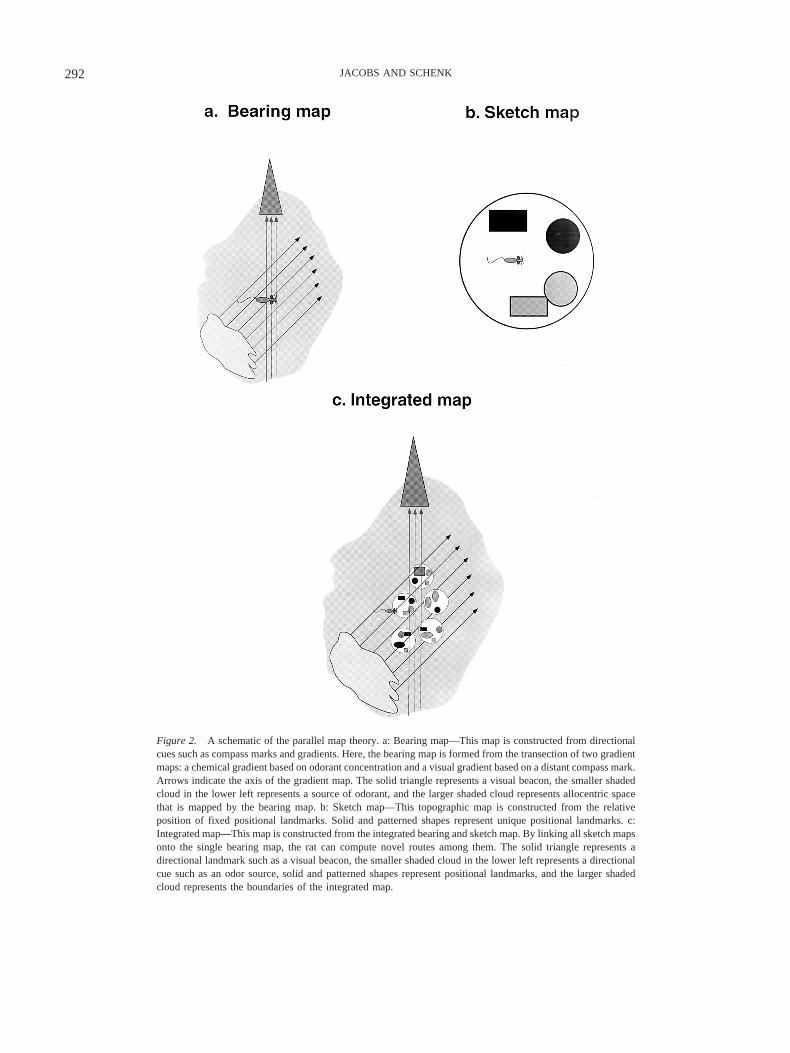
Figure 2. A schematic of the parallel map theory. a: Bearing map—This map is constructed from directionalcues such as compass marks and gradients. Here, the bearing map is formed from the transection of two gradientmaps: a chemical gradient based on odorant concentration and a visual gradient based on a distant compass mark.Arrows indicate the axis of the gradient map. The solid triangle represents a visual beacon, the smaller shadedcloud in the lower left represents a source of odorant, and the larger shaded cloud represents allocentric spacethat is mapped by the bearing map. b: Sketch map—This topographic map is constructed from the relativeposition of fixed positional landmarks. Solid and patterned shapes represent unique positional landmarks. c:Integrated map—This map is constructed from the integrated bearing and sketch map. By linking all sketch mapsonto the single bearing map, the rat can compute novel routes among them. The solid triangle represents adirectional landmark such as a visual beacon, the smaller shaded cloud in the lower left represents a directionalcue such as an odor source, solid and patterned shapes represent positional landmarks, and the larger shadedcloud represents the boundaries of the integrated map.
292 JACOBS AND SCHENK

distances. The navigator must only calibrate self-motion cues withchanges in the intensity of a distributed cue to extrapolate its futureposition in the coordinate system. Thus, the bearing map allowsthe navigator to maintain an accurate representation of its position,even in unknown territory. Starting with a primitive gradientalgorithm, the bearing map creates a coarse-grained mental repre-sentation of space that nonetheless provides a powerful tool forspatial navigation, particularly long-distance navigation.
The second parallel map is the sketch map. It is constructed froman arrangement of positional cues. These unique local landmarksare encoded relative to each other as a topographic map (seeFigure 2b). The sketch map differs in fundamental ways from thebearing map. First, all landmarks in the map must be individuallylearned, and thus, there can no extrapolation or generalizationacross novel terrain, as in the bearing map. The relations (distance,direction) among landmarks must also be learned. The sketch mapis thus a fine-grained mental representation that is best suited forlocal navigation. The positional codes within sketch maps areallocentric, as each cue refers to another component of the sketchmap.
PMT can be described at several levels of analysis. What wehave just described is the first level, the conceptual account of twomaps. In this account, one map is based on directional cues, someof which may be extrapolated, and the other is based on a set ofmemorized positional cues. The concept of the bearing map has noreal predecessors in theoretical models of spatial navigation inmammals, including previous formulations of the cognitive map.In contrast, the second, topographic sketch map has much incommon with previous cognitive map theories, as we describebelow. PMT thus describes two new concepts, the bearing map andthe idea that the cognitive or integrated map is composed ofparallel maps (see Figure 2c). We use the term integrated mapinstead of cognitive map because the original term was neverprecisely defined by Tolman in 1948, which has led to differentusages in different disciplines (see review by Bennett, 1996).
Anatomy of the Parallel Maps
A second level of analysis is the physical scaffold underlyingthe three mental representations (bearing map, sketch map, andintegrated map). Despite recent progress (Amaral & Witter, 1995;Deadwyler & Hampson, 1999; Witter, Wouterlood, Naber, & vanHaeften, 2000), the full complexity of hippocampal anatomy hasyet to be mapped onto a complete theory of its function. WhatSwanson, Kohler, and Bjorklund said in 1987 remains true:
It appears safe to say that nowhere is the gap between structure andfunction greater than in this region [hippocampus]. The major reasonfor this is that while the hippocampal formation contains the simplestcortical fields from an anatomical point of view, it receives, processes,and transmits the most complex array of information of any corticalregion. (p. 126)
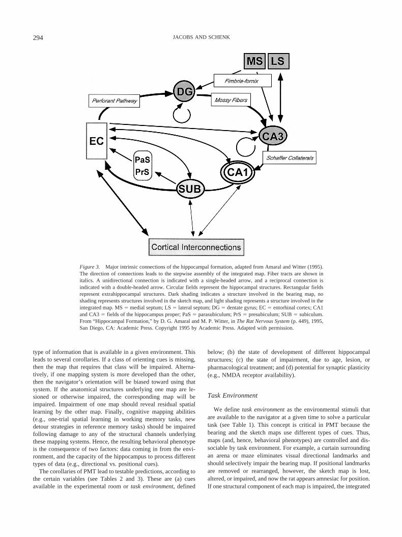
Keeping this in mind, we present a working model of thehippocampal structures underlying the encoding and use of theparallel map system (see Figure 3). We propose that the maps aremediated by different neural structures: The bearing map is medi-ated by subcortical hippocampal channels projecting to the DG andthe CA3 subfield of the HP, the sketch map is mediated by theCA1 subfield and its cortical connections, and the integrated map
is mediated by the synchronization of activity between these twochannels. The anatomical basis of the parallel maps is shownschematically in Figure 3.
The assignment of map to structure is an important feature ofPMT, as is its emphasis on separate functions of the DG and theHP, which create parallel and independent maps. Although thehippocampal subfield functions have received some attention, hav-ing been modeled (e.g., Deadwyler & Hampson, 1999; Granger,Wiebe, Taketani, & Lynch, 1996) and empirically disassociated(Gilbert, Kesner, & Lee, 2001; McNaughton, Barnes, Meltzer, &Sutherland, 1989; Mizumori, McNaughton, Barnes, & Fox, 1989),these studies have proposed complementary functions rather thanparallel ones. We are taking this line of reasoning in a newdirection, first by proposing that DG and CA1 mediate parallelmaps, and second by adding the concept of the gradient map. PMT,based on parallel maps and representations of gradients, thus leadsto a set of unique predictions (see Tables 2 and 3).
In summary, the integrated map relies on the presence of twoparallel map processes. It emerges from the simultaneous activa-tion of the parallel bearing and sketch maps and, hence, from thesynchronization of physiological activity between the DG and theHP. When both maps are active and accessible, the navigator cancreate the integrated map by double labeling positional cues withinand between sketch maps, as outlined in Table 4. The emergentproperties of the integrated map supply an ability that neither mapalone can accomplish: navigation among familiar locations thatinvolves movement across unfamiliar terrain. With this, the navi-gator can extrapolate its movement beyond the knowledge ofmemorized landmarks. The strength of this system lies in itsredundancy, a design feature found in spatial navigation through-out the animal kingdom (Keeton, 1974). Because the maps work inparallel, if one is impaired then the navigator may continue toorient accurately, using the residual ability afforded by the remain-ing map. It cannot, of course, use the integrated map, but it mayretain a considerable ability to navigate, as is apparent from theliterature on partial lesions of the hippocampus. Because the inte-grated map is an emergent property of the coactivation of thebearing and sketch maps, all three maps can be isolated with avariety of techniques (e.g., cue manipulation, lesion, unit activity),allowing each element of the theory to be tested (see Tables 2and 3).
The final level of analysis for PMT lies beyond experimentaltests of its predictions, however, and that challenge is to mapconcept to structure with computational models. PMT narrows thesearch for such a model by mapping specific functions to differentsubfields, and now feasible and realistic computational models ofthe parallel maps need to be developed from knowledge of theanatomy and topography of the HPF (Amaral & Witter, 1995;Witter et al., 2000). We are currently working with collaborators todevelop a model of a hippocampal parallel mapping process.
We now discuss each map in turn, describing its characteristicsand its physiological properties and reviewing the evidence for theeffect of its experimental dissociation on spatial navigation in thelaboratory rodent.
Predictions From a Parallel Map System
Our theory proposes that spatial abilities depend not only on theintegrity of two independent brain mapping systems but also on the
293PARALLEL MAP THEORY
type of information that is available in a given environment. Thisleads to several corollaries. If a class of orienting cues is missing,then the map that requires that class will be impaired. Alterna-tively, if one mapping system is more developed than the other,then the navigator’s orientation will be biased toward using thatsystem. If the anatomical structures underlying one map are le-sioned or otherwise impaired, the corresponding map will beimpaired. Impairment of one map should reveal residual spatiallearning by the other map. Finally, cognitive mapping abilities(e.g., one-trial spatial learning in working memory tasks, newdetour strategies in reference memory tasks) should be impairedfollowing damage to any of the structural channels underlyingthese mapping systems. Hence, the resulting behavioral phenotypeis the consequence of two factors: data coming in from the envi-ronment, and the capacity of the hippocampus to process differenttypes of data (e.g., directional vs. positional cues).
The corollaries of PMT lead to testable predictions, according tothe certain variables (see Tables 2 and 3). These are (a) cuesavailable in the experimental room or task environment, defined
below; (b) the state of development of different hippocampalstructures; (c) the state of impairment, due to age, lesion, orpharmacological treatment; and (d) potential for synaptic plasticity(e.g., NMDA receptor availability).
Task Environment
We define task environment as the environmental stimuli thatare available to the navigator at a given time to solve a particulartask (see Table 1). This concept is critical in PMT because thebearing and the sketch maps use different types of cues. Thus,maps (and, hence, behavioral phenotypes) are controlled and dis-sociable by task environment. For example, a curtain surroundingan arena or maze eliminates visual directional landmarks andshould selectively impair the bearing map. If positional landmarksare removed or rearranged, however, the sketch map is lost,altered, or impaired, and now the rat appears amnesiac for position.If one structural component of each map is impaired, the integrated
Figure 3. Major intrinsic connections of the hippocampal formation, adapted from Amaral and Witter (1995).The direction of connections leads to the stepwise assembly of the integrated map. Fiber tracts are shown initalics. A unidirectional connection is indicated with a single-headed arrow, and a reciprocal connection isindicated with a double-headed arrow. Circular fields represent the hippocampal structures. Rectangular fieldsrepresent extrahippocampal structures. Dark shading indicates a structure involved in the bearing map, noshading represents structures involved in the sketch map, and light shading represents a structure involved in theintegrated map. MS � medial septum; LS � lateral septum; DG � dentate gyrus; EC � entorhinal cortex; CA1and CA3 � fields of the hippocampus proper; PaS � parasubiculum; PrS � presubiculum; SUB � subiculum.From “Hippocampal Formation,” by D. G. Amaral and M. P. Witter, in The Rat Nervous System (p. 449), 1995,San Diego, CA: Academic Press. Copyright 1995 by Academic Press. Adapted with permission.
294 JACOBS AND SCHENK
map becomes impaired, which eliminates cognitive mapping abil-ity (see Tables 2 and 3).
Map Phenotype
By knowing the task environment (i.e., the input to the mappingsystem) and the locus of impairment (i.e., the residual mappingsystem), we can predict the behavioral phenotype of the navigator,or map phenotype. We define this as an animal’s spatial strategy ina given experimental condition. A particular strategy may be intactor absent; if it is the remaining intact strategy, it will underlie anyresidual spatial learning observed (see Tables 2 and 3).
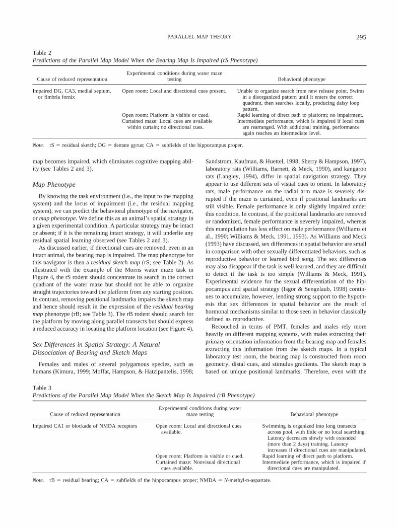
As discussed earlier, if directional cues are removed, even in anintact animal, the bearing map is impaired. The map phenotype forthis navigator is then a residual sketch map (rS; see Table 2). Asillustrated with the example of the Morris water maze task inFigure 4, the rS rodent should concentrate its search in the correctquadrant of the water maze but should not be able to organizestraight trajectories toward the platform from any starting position.In contrast, removing positional landmarks impairs the sketch mapand hence should result in the expression of the residual bearingmap phenotype (rB; see Table 3). The rB rodent should search forthe platform by moving along parallel transects but should expressa reduced accuracy in locating the platform location (see Figure 4).
Sex Differences in Spatial Strategy: A NaturalDissociation of Bearing and Sketch Maps
Females and males of several polygamous species, such ashumans (Kimura, 1999; Moffat, Hampson, & Hatzipantelis, 1998;
Sandstrom, Kaufman, & Huettel, 1998; Sherry & Hampson, 1997),laboratory rats (Williams, Barnett, & Meck, 1990), and kangaroorats (Langley, 1994), differ in spatial navigation strategy. Theyappear to use different sets of visual cues to orient. In laboratoryrats, male performance on the radial arm maze is severely dis-rupted if the maze is curtained, even if positional landmarks arestill visible. Female performance is only slightly impaired underthis condition. In contrast, if the positional landmarks are removedor randomized, female performance is severely impaired, whereasthis manipulation has less effect on male performance (Williams etal., 1990; Williams & Meck, 1991, 1993). As Williams and Meck(1993) have discussed, sex differences in spatial behavior are smallin comparison with other sexually differentiated behaviors, such asreproductive behavior or learned bird song. The sex differencesmay also disappear if the task is well learned, and they are difficultto detect if the task is too simple (Williams & Meck, 1991).Experimental evidence for the sexual differentiation of the hip-pocampus and spatial strategy (Isgor & Sengelaub, 1998) contin-ues to accumulate, however, lending strong support to the hypoth-esis that sex differences in spatial behavior are the result ofhormonal mechanisms similar to those seen in behavior classicallydefined as reproductive.
Recouched in terms of PMT, females and males rely moreheavily on different mapping systems, with males extracting theirprimary orientation information from the bearing map and femalesextracting this information from the sketch maps. In a typicallaboratory test room, the bearing map is constructed from roomgeometry, distal cues, and stimulus gradients. The sketch map isbased on unique positional landmarks. Therefore, even with the
Table 2Predictions of the Parallel Map Model When the Bearing Map Is Impaired (rS Phenotype)
Cause of reduced representationExperimental conditions during water maze
testing Behavioral phenotype
Impaired DG, CA3, medial septum,or fimbria fornix
Open room: Local and directional cues present. Unable to organize search from new release point. Swimsin a disorganized pattern until it enters the correctquadrant, then searches locally, producing daisy looppattern.
Open room: Platform is visible or cued. Rapid learning of direct path to platform; no impairment.Curtained maze: Local cues are available
within curtain; no directional cues.Intermediate performance, which is impaired if local cues
are rearranged. With additional training, performanceagain reaches an intermediate level.
Note. rS � residual sketch; DG � dentate gyrus; CA � subfields of the hippocampus proper.
Table 3Predictions of the Parallel Map Model When the Sketch Map Is Impaired (rB Phenotype)
Cause of reduced representationExperimental conditions during water
maze testing Behavioral phenotype
Impaired CA1 or blockade of NMDA receptors Open room: Local and directional cuesavailable.
Swimming is organized into long transectsacross pool, with little or no local searching.Latency decreases slowly with extended(more than 2 days) training. Latencyincreases if directional cues are manipulated.
Open room: Platform is visible or cued. Rapid learning of direct path to platform.Curtained maze: Nonvisual directional
cues available.Intermediate performance, which is impaired if
directional cues are manipulated.
Note. rB � residual bearing; CA � subfields of the hippocampus proper; NMDA � N-methyl-D-aspartate.
295PARALLEL MAP THEORY

same task environment, females and males may differ in how theymap the space: Females learn and remember the relations amongunique positional landmarks, whereas males perceive positionallandmarks as points in the shape of an array. Perhaps for thisreason, reducing the ambient light after acquisition produces animmediate and severe impairment in female but not male orienta-tion on the radial arm maze (Williams & Meck, 1993). Thus, evenin the same environment, there are two ways to encode the space,with females and males showing a natural dissociation of these twostrategies and of the parallel mapping systems.
This sex difference has certain implications for studies of spatialnavigation in polygamous rats and mice. If a study uses all malesubjects, because males rely more heavily on distal cues and roomgeometry, this could lead to a bias in defining spatial navigation inthat paradigm as highly dependent on such directional cues. Thismight be particularly evident in studies in which cues are inconflict. In such studies, a male’s use of cue hierarchy should showgreater reliance on directional cues. For example, a recent study ofcue use in male rats found a significant preference for directionalover positional landmarks in orientation. The rats were also morelikely to extract directional than positional information from theconfiguration of positional landmarks. They oriented first to thegeometry of the landmarks and only then to the identity of thepositional landmarks (Benhamou & Poucet, 1998). In this case,positional objects appear to serve as directional landmarks. Inanother study using all males, rats used the geometrical relation-ship of proximal landmarks to deduce direction (Greene & Cook,1997). We do not question the validity of these results but onlypoint out that it is possible that the same study repeated withfemales might find a different set of rules, such as orientationprimarily to landmark identity and only secondarily to arraygeometry.
Dissociating Parallel Maps
We now turn the remaining discussion to experimental evidencein support of PMT. We first discuss the functional and structuralcharacteristics of each map, its development and plasticity. Wethen discuss how task environments and specific lesions of hip-pocampal structures should result in predictable map phenotypes.Finally, we will discuss patterns of unit activity in the hippocam-pus and their relation to PMT.
The Bearing Map
Oliver Wendell Holmes (1889) once wrote, “I find the greatthing in this world is not so much where we stand, as in whatdirection we are moving” (p. 127). It is no less true for spatial thanfor conceptual navigation. The bearing map supplies the directionand the global location. It is the locus of primary mapping, and theanatomical channel underlying it is the permanent scaffold of thisprimary map. Because it is the scaffold, we postulate that itnecessarily increases in size and complexity as an animal matures,explores new territory, and adds knowledge about new gradientsand directions in its environment.
The bearing map is constructed from simple 1-D gradients.Structures in the bearing map channel create a 2-D coordinatesystem from this 1-D input (see Figure 2a and Table 4). Theseexternal gradient maps are calibrated to internal gradient mapssupplied by path integration. The 2-D coordinate system of thebearing map becomes the scaffold not only for all directionalinformation but also for encoding the relative position of sketchmaps. Once a sketch map has been defined in relation to thebearing map, the distance and direction of vectors that connectdisparate sketch maps can be computed. This is the integrated map
Table 4Steps in the Construction of the Integrated Map
Step and locus Process Representation
1. MS Self-movement cues calibrated with theta rhythm as pacemaker.Derives rate of movement and location relative to prior point.
1-D algorithm, stored in working memory.
2. DG 1-D map calculated from medial septum input and sensory inputfrom entorhinal cortex.
1-D map, stored in working memory.
Autoassociative network within DG recalls 2-D bearing map andadds new 1-D map, creating a newly expanded 2-D bearingmap through pattern completion. Transmits updated bearingmap to CA3.
2-D bearing map, stored in reference memory.
3. CA3 Localizes current position on the 2-D map from DG by matchingpattern with current EC input. Transmits position tosubcortical structures through bilateral projection to lateralseptal nuclei and to cortical structures through Schaffercollaterals to CA1.
Local aspect of the bearing map, stored in working memory.
4. CA1 Creates a minimap of the local panorama, computing within-sketch vectors on the basis of head direction input.
Sketch map, stored in working memory, and integrated map,also stored in working memory.
Receives bearing map position from CA3; localizes currentsketch map on bearing map. Transmits this part of the newlycomputed integrated map to subiculum.
Integrated map, an emergent map from the integration ofbearing and sketch maps, stored in working memory.
5. Subiculum Updates reference memory of integrated map with newinformation from CA1. Computes final position of thisfragment on the integrated map. Transmits integrated map toassociative cortices.
Current integrated map, stored in reference memory.
Note. This is a proposed account of how the parallel map theory could be assembled by components of the hippocampal formation. The schema mustbe considered speculative, as we cannot yet even incorporate the CA2 field. MS � medial septum; 1-D � one-dimensional; DG � dentate gyrus; CA �subfields of the hippocampus proper; 2-D � two-dimensional; EC � entorhinal cortex.
296 JACOBS AND SCHENK

representation, and, using this map, the navigator can set out onnovel routes among known locales (see Figure 2c).
Structural Components of the Bearing Map
Navigators create gradient maps when they can calibrate theirmovements relative to regular changes in stimulus intensity. Thisrequires three components: an input from internal movement (e.g.,vestibular input), a sensory input, and a neural structure that cancreate 1-D maps and 2-D maps from such gradient information.
We propose that these roles are mediated in stepwise fashion bythe septal nuclei, DG and CA3. Rather like an assembly line, eachcomponent adds a new piece to the bearing map (see Figure 3 andTable 4). The medial septum (MS) coordinates the input frominternal movement by supplying the pacemaker needed to assessself-movement along a gradient. The pacemaker of this processmay be the hippocampal theta rhythm, a rhythmical, slow activitypattern (Hasselmo, 2000). Theta occurs during active locomotion,and its frequency and amplitude are related to the many parametersassociated with movement, such as speed (O’Keefe, 1993), antic-ipation of movement (Morris & Hagen, 1983), and type of body
movements (Whishaw & Vanderwolf, 1973). Theta ceases whenthe animal is immobile or engaged in other behaviors that do notinvolve movement in a trajectory (Vanderwolf, 1969). Given itsclose relationship to movement, it is not surprising that the MS andits modulation of theta are important in spatial navigation (Whi-shaw, 2000).
Given the pacemaker’s input, the next step is the integration ofexternal sensory input in the DG. The DG is the locus of aconvergence of subcortical and cortical (i.e., EC) inputs. It also hasa complex internal structure, in which projections from the poly-morphic layer synapse within the molecular layer, which containsprojections back to the polymorphic layer (Amaral & Witter,1995). This complex internal structure could underlie the functionof the DG to create the 2-D bearing map. After the sensory inputfrom EC is integrated with self-movement by calibration to thetheta rhythm to create the 1-D map, this autoassociative structurein the DG completes the pattern of partially intersecting 1-D maps,creating a 2-D representation. This is then used to create or updatethe bearing map. This new information is then projected out of theDG to the CA3 through the mossy fiber projection.
Figure 4. Predictions of the parallel map theory for the Morris water maze. The four patterns of spatialperformance result from the presence or absence of the parallel maps. Residual learning, resulting from the lossof a single map (bearing or sketch), allows the rat to solve the maze, using transects when the bearing map isintact and local loops when the sketch map is intact. The residual sketch map phenotype is expressed in twostages: initial impairment of orientation and recovery to local loops with additional training. With both mapsimpaired, the rat shows a permanent loss of spatial orientation, represented by global loops; performanceimproves only with the development of nonspatial algorithms. With both maps intact, the rat encodes anintegrated map, which allows it to choose a direct path to the platform, regardless of start point. The solid circlerepresents a hidden platform; the thick line represents a predicted swim trajectory.
297PARALLEL MAP THEORY
CA3 has long been a focus of theoretical models because of itsunusual autoassociative architecture (Kali & Dayan, 2000; Marr,1971; McNaughton & Morris, 1987; Rolls, 1996). We speculatethat one function of this architecture could be to convert gradientmap information to data that can be incorporated into a topo-graphic map (see Figure 1d–1e). This process may be necessary tointegrate the bearing and sketch maps. Because CA3 is the conduitbetween DG and CA1, this could be a role it plays. CA3 wouldthus translate the current location on the bearing map into a codethat could be read by CA1. For example, the navigator’s locationcould be recoded as a new object in a topographic map (i.e., on thecurrent sketch map). This allows CA1 to calculate the new topol-ogy of the relationship among the navigator and the surroundingobjects.
To create an integrated map, however, CA1 also needs direc-tional information from the bearing map, and, hence, CA3 musttransmit the location on the bearing map as well. We suggest thatCA3 could do this by segregating its output to CA1 into gradientand topographic information projections. The area in CA3 that isthe most likely locus for the projection of gradient map data toCA1 is the locus of heaviest projection from DG to CA (i.e., theproximal third of CA3; also the more septal area). This proximalthird has also been called the projection zone (Ishizuka, Weber, &Amaral, 1990) because the axons project directly to distal CA1with few signs of synaptic contacts enroute. We speculate that thelocus for the projection of topographic data to CA1 could takeplace in the middle and distal areas of CA3 (midseptotemporal),which has been called the association zone on the basis of thedensity of within-field projections and its input from the EC(Ishizuka et al., 1990).
Finally, because all CA3 cells project both to CA1 and to thelateral septum (Swanson, Sawchenko, & Cowan, 1980), projec-tions from proximal CA3 and middle–distal CA3 might work inparallel, allowing the updated position to be supplied as an afferentcopy to the bearing map channel (i.e., septal nuclei) and the sketchmap channel (CA1). In such a scenario, the CA3 would projectboth location (for further encoding) and expected direction (forcurrent action). If so, then the lateral septum should show place-specific activity involved in executing the next movement on theplanned trajectory. There is some evidence for this. The lateralseptum is considered to be the medial territory of the striatum(Swanson & Risold, 2000). Characteristics of a structure involvedin place learning include LTP-like responses with fornix stimula-tion in the mouse (Garcia, Vouimba, & Jaffard, 1993) and placeresponses that have been recorded in freely moving subjects, bothin rats (Bezzi, Leutgeb, Treves, & Mizumori, 2000) and primates(Ono & Nishijo, 1999). Because the subiculum also projects to thelateral septum (LS; Amaral & Witter, 1995), we speculate thatmovement may be driven by instructions either from the bearingmap (through CA3) or from the integrated map (through thesubiculum). Hence, the parallel map structure could well be mir-rored in parallel output commands to the motor system, a hypoth-esis that deserves more discussion than is possible here.
In summary, we propose that the bearing map is created in theDG and projected through the mossy fiber projection to CA3,which then transmits current location to CA1, in terms of both thegradient and the topographic location, and to the LS, which me-diates movement along the trajectories set up in the bearing map.
Development of the Bearing Map
As Tinbergen (1963) argued, to understand a behavior, one musttake into account not only its physiological mechanism but also itsevolutionary history, its current ecological significance, and itsdevelopment. Having discussed the history and structure of thebearing map, we now address its development and plasticity, onthe basis of studies of the laboratory rat.
Structure and activity. The hippocampus is a remarkably late-maturing brain structure, with final maturation occurring atabout 21 days after birth in the rat (Bayer, 1982). In some sense,HPF development does not stop but simply proceeds throughdifferent phases, in which structure, physiology, and behaviorcontinue to change.
Not only do hippocampal subfields develop late relative to othertelencephalon structures, but they also develop in a mosaic pattern.The adult cytoarchitectonic pattern in DG develops by postnatal(PN) Day 20, though mossy fibers continue to develop in adult-hood (Amaral & Dent, 1981) and neurogenesis continues in thegranule layer throughout life in rodents and primates (Bayer,Yackel, & Puri, 1982; Gould, Beylin, Tanapat, Reeves, & Shors,1999). CA1 develops later, and significant dendritic arborization inthe stratum lacunosum occurs in the 2nd PN month (Pokorny &Yamamoto, 1981). In female rats, CA1 dendritic arborizationfluctuates naturally with levels of estrogen, as during estrus(Gould, Wooley, Frankfurt, & McEwen, 1990). Because of theclose connections between HPF and the neocortex, such plasticitymay result in as yet undetected structural changes that may con-tinue throughout life (Alvarado & Bachevalier, 2000). We discussthe role of neurogenesis in a later section.
Metabolic activity also develops in a mosaic pattern. Onceagain, DG precedes CA1, with a marked increase in DG activityoccurring around weaning and CA1 increasing a month later(Glick, Weaver, & Meibach, 1980; Meibach, Ross, Cox, & Glick,1980). DG activity is correlated with gamma-aminobutryic acid(GABA)-ergic inhibition, which does not mature until the end ofthe 1st PN month in the rat (Swann, Brady, & Martin, 1989),although activity similar to LTP is found after 2 PN weeks (Bat-tistin & Cherubini, 1994).
Behavior. Like the hippocampal subfields, spatial learning inrats develops in different stages during the 4th PN week, even insimple tasks such as spontaneous alternation (Blozovski & Hess,1989; Douglas, Peterson, & Douglas, 1973; Egger, 1973; Waters,Klintsova, & Foster, 1997). By the time rats are weaned and havebegun exploring their environment (PN Day 20–26), they are ableto orient to a single goal, whether it is a water maze platform or anescape hole in an open field (Chevalley & Schenk, 1987; Rudy,Stadler-Morris, & Albert, 1987). This change in navigational strat-egy suggests that adult spatial behavior appears at the end of thepostweaning period, around 24 days PN (Rudy et al., 1987).
However, an earlier appearance of a spatial bias toward thetraining position has been observed after particular types of train-ing. When training occurs in one day with long intertrial intervalsand the pups’ internal temperature is carefully controlled, a sig-nificant bias toward the training quadrant appears as early as PNDay 19 (Brown & Whishaw, 2000). When training takes place ina small pool with highly salient visual cues in the immediatevicinity of the pool, a significant bias is observed at PN Day 20–22(Carman & Mactutus, 2001).
298 JACOBS AND SCHENK
Given juvenile rats’ poor long-term memory and poor ability tothermoregulate, these special procedures may be critical in allow-ing the expression of a significant spatial bias toward the trainingposition. For example, a panorama around the water maze that isparticularly rich in visual cues (Carman & Mactutus, 2001) or ahighly salient cue such as an illuminated hanging cup (Rudy et al.,1987) placed a short distance from the hidden platform may resultin the expression of different spatial strategies and lead to asignificant spatial bias. In fact, analysis of the approach trajectoriesof immature rats when a cue is hanging in the next quadrantreveals a progressive transformation. The juveniles start by detour-ing under the cue but end by making straight approaches from thestart point to the platform (Chevalley & Schenk, 1987) . Moreover,juvenile rats aged 26–28 days trained on a cued-place task expressa significant degree of overshadowing that is not evident in youngadults (Schenk & Brandner, 1995), which suggests that they maybe developing particular spatial strategies at this age.
Hence, the absolute timing of cue and place learning appears tobe highly sensitive to experimental conditions. We suggest thatthis is because different task environments are recruiting differentparallel maps. Further, the maps are developing at different rates,in tandem with underlying hippocampal structures. Hence, thebearing map should appear before the sketch map in development,and, thus, we predict that juveniles, such as the PN Day 19juveniles in the Brown and Whishaw (2000) study, encode theplace differently than do adults, relying more heavily on thebearing map until much later in development.
The Ontogeny of the Parallel Mapping System
To test this hypothesis, we need to know precisely how thehierarchy of cue use develops in the rat. There is clearly a transi-tion from simply approaching a cue associated with the goal to aguidance system in which a distal landmark (or landmarks) can beused to determine the correct vector to the goal, whether it appearsat PN Day 19 (Brown & Whishaw, 2000) or later (Rudy et al.,1987). If juvenile rats rely primarily on the bearing map, theyshould orient well to distributed cues, such as distant visual cues ororthogonal gradients of olfactory cues, but poorly to arrays ofpositional landmarks.
There is evidence that such dissociation can occur. Juvenile ratscan learn the spatial position of an escape hole (homing table;Schenk, 1989) in controlled cue conditions in a warm, dry, openarena in which there is no risk of hypothermia. Using this task,Schenk found that there was a marked interaction between age anduse of cues by type. During training, immature (PN Day 26–28)and adult rats were given either discrete visual or distributedolfactory information. Olfactory information was provided in aradial pattern of scented strips, none of which was close to thegoal. The visual cues were an asymmetric configuration of threediscrete lights, as in a previous study (Rossier, Grobety, & Schenk,2000). Only the adults could discriminate places purely on thebasis of such visual cues, despite normal visual acuity in theimmature rats (Salazar, Rossier, & Schenk, 2000). The immaturerats performed well only when odor cues were present (Schenk,Jacobs, Rossier, & Kiraly, 2001).
We suggest that the immature rats relied on their relativelymature bearing map and hence were more attentive to salientdistributed cues, such as odor gradients emanating from the
scented strips. As the sketch map matured, the rats began to relypreferentially on the visual cues. This suggests that the sketch mapis slowly calibrated by the bearing map during the 2nd PN monthand may not become autonomous until the 3rd month, coincidingwith hippocampal maturation. The integrated map should thendevelop after natal dispersal and mature further during adolescence(Schenk et al., 2001). This hypothesis is confirmed by the obser-vation that PN Day 48 rats can rely on the visual cues to solve thetask only if they have experienced the conjunction of the olfactoryand visual cues, as though the presence of both is necessary forsuch a dual coding phase (Schenk et al., 2001).
Hippocampal Sex Differences
The sex differences in navigation strategy described earlier inthe laboratory rat develop during the period of DG maturation.These differences between females and males are not fixed genet-ically but rather appear as a consequence of exposure to gonadalhormones during the PN period (Williams et al., 1990). Thus, thedifferential reliance by females and males on the bearing mapappears to be the result of differential exposure to hormones; thedifferences are eliminated or reversed with a perinatal treatment ofestrogen or its metabolite, testosterone (Isgor & Sengelaub, 1998;Roof & Havens, 1992; Williams et al., 1990).
This sex difference should be reflected in the structure of maleand female HPF; we predict greater development of bearing mapcomponents in males. Sexual dimorphism in the entire hippocam-pus (HPF volume relative to telencephalon volume) has beenfound both in wild rodents (Jacobs, 1995, 1996) and in wild birds(Reboreda, Clayton, & Kacelnik, 1996; Sherry et al., 1993). Inboth groups, the sex that relies more heavily on spatial explorationduring the breeding season has a relatively larger hippocampus.
Because both laboratory rats (Rattus norvegicus) and mice (Musmusculus) are derived from species in which males roam morewidely than females, we predict that the hippocampus should alsobe sexually differentiated in these species. This is confirmed byseveral measures: Male mice have more DG granule cells (Wimer& Wimer, 1985); male rats have a thicker DG granule layer (Roof& Havens, 1990), which is associated with a greater cell density(Loy, 1986), and more dendritic branching points in the DG(Juraska, 1984; Juraska, Fitch, Henderson, & Rivers, 1985). Malerats also have more mossy fibers projecting from DG to CA3 andmore synapses in this projection than do females (Madeira &Paula-Barbosa, 1993; Madeira, Sousa, & Paula-Barbosa, 1991).Thus, in all sexual dimorphisms that have been described for DGor CA3 in laboratory rats and mice, there is greater structuraldevelopment in males than in females. Because this correlates withthe male rat’s reliance on directional cues, this supports the hy-pothesis that the bearing map is mediated by DG and CA3.
Hippocampal Neurogenesis
A unique property of the bearing map is its role as the perma-nent frame or scaffold for the integrated map. Further, we proposethat the bearing map is both consolidated and stored in the DG. Asthe map itself becomes larger and more complex with new spatialexploration, however, it needs a mechanism to add more informa-tion. In addition, if the bearing map is a permanent referencesystem, then it should have a high threshold for synaptic change,
299PARALLEL MAP THEORY
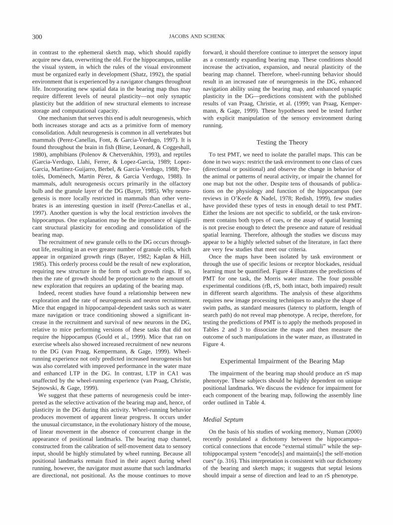
in contrast to the ephemeral sketch map, which should rapidlyacquire new data, overwriting the old. For the hippocampus, unlikethe visual system, in which the rules of the visual environmentmust be organized early in development (Shatz, 1992), the spatialenvironment that is experienced by a navigator changes throughoutlife. Incorporating new spatial data in the bearing map thus mayrequire different levels of neural plasticity—not only synapticplasticity but the addition of new structural elements to increasestorage and computational capacity.
One mechanism that serves this end is adult neurogenesis, whichboth increases storage and acts as a primitive form of memoryconsolidation. Adult neurogenesis is common in all vertebrates butmammals (Perez-Canellas, Font, & Garcia-Verdugo, 1997). It isfound throughout the brain in fish (Birse, Leonard, & Coggeshall,1980), amphibians (Polenov & Chetverukhin, 1993), and reptiles(Garcia-Verdugo, Llahi, Ferrer, & Lopez-Garcia, 1989; Lopez-Garcia, Martinez-Guijarro, Berbel, & Garcia-Verdugo, 1988; Por-toles, Domenech, Martin Perez, & Garcia Verdugo, 1988). Inmammals, adult neurogenesis occurs primarily in the olfactorybulb and the granule layer of the DG (Bayer, 1985). Why neuro-genesis is more locally restricted in mammals than other verte-brates is an interesting question in itself (Perez-Canellas et al.,1997). Another question is why the local restriction involves thehippocampus. One explanation may be the importance of signifi-cant structural plasticity for encoding and consolidation of thebearing map.
The recruitment of new granule cells to the DG occurs through-out life, resulting in an ever greater number of granule cells, whichappear in organized growth rings (Bayer, 1982; Kaplan & Hill,1985). This orderly process could be the result of new exploration,requiring new structure in the form of such growth rings. If so,then the rate of growth should be proportionate to the amount ofnew exploration that requires an updating of the bearing map.
Indeed, recent studies have found a relationship between newexploration and the rate of neurogenesis and neuron recruitment.Mice that engaged in hippocampal-dependent tasks such as watermaze navigation or trace conditioning showed a significant in-crease in the recruitment and survival of new neurons in the DG,relative to mice performing versions of these tasks that did notrequire the hippocampus (Gould et al., 1999). Mice that ran onexercise wheels also showed increased recruitment of new neuronsto the DG (van Praag, Kempermann, & Gage, 1999). Wheel-running experience not only predicted increased neurogenesis butwas also correlated with improved performance in the water mazeand enhanced LTP in the DG. In contrast, LTP in CA1 wasunaffected by the wheel-running experience (van Praag, Christie,Sejnowski, & Gage, 1999).
We suggest that these patterns of neurogenesis could be inter-preted as the selective activation of the bearing map and, hence, ofplasticity in the DG during this activity. Wheel-running behaviorproduces movement of apparent linear progress. It occurs underthe unusual circumstance, in the evolutionary history of the mouse,of linear movement in the absence of concurrent change in theappearance of positional landmarks. The bearing map channel,constructed from the calibration of self-movement data to sensoryinput, should be highly stimulated by wheel running. Because allpositional landmarks remain fixed in their aspect during wheelrunning, however, the navigator must assume that such landmarksare directional, not positional. As the mouse continues to move
forward, it should therefore continue to interpret the sensory inputas a constantly expanding bearing map. These conditions shouldincrease the activation, expansion, and neural plasticity of thebearing map channel. Therefore, wheel-running behavior shouldresult in an increased rate of neurogenesis in the DG, enhancednavigation ability using the bearing map, and enhanced synapticplasticity in the DG—predictions consistent with the publishedresults of van Praag, Christie, et al. (1999; van Praag, Kemper-mann, & Gage, 1999). These hypotheses need be tested furtherwith explicit manipulation of the sensory environment duringrunning.
Testing the Theory
To test PMT, we need to isolate the parallel maps. This can bedone in two ways: restrict the task environment to one class of cues(directional or positional) and observe the change in behavior ofthe animal or patterns of neural activity, or impair the channel forone map but not the other. Despite tens of thousands of publica-tions on the physiology and function of the hippocampus (seereviews in O’Keefe & Nadel, 1978; Redish, 1999), few studieshave provided these types of tests in enough detail to test PMT.Either the lesions are not specific to subfield, or the task environ-ment contains both types of cues, or the assay of spatial learningis not precise enough to detect the presence and nature of residualspatial learning. Therefore, although the studies we discuss mayappear to be a highly selected subset of the literature, in fact thereare very few studies that meet our criteria.
Once the maps have been isolated by task environment orthrough the use of specific lesions or receptor blockades, residuallearning must be quantified. Figure 4 illustrates the predictions ofPMT for one task, the Morris water maze. The four possibleexperimental conditions (rB, rS, both intact, both impaired) resultin different search algorithms. The analysis of these algorithmsrequires new image processing techniques to analyze the shape ofswim paths, as standard measures (latency to platform, length ofsearch path) do not reveal map phenotype. A recipe, therefore, fortesting the predictions of PMT is to apply the methods proposed inTables 2 and 3 to dissociate the maps and then measure theoutcome of such manipulations in the water maze, as illustrated inFigure 4.
Experimental Impairment of the Bearing Map
The impairment of the bearing map should produce an rS mapphenotype. These subjects should be highly dependent on uniquepositional landmarks. We discuss the evidence for impairment foreach component of the bearing map, following the assembly lineorder outlined in Table 4.
Medial Septum
On the basis of his studies of working memory, Numan (2000)recently postulated a dichotomy between the hippocampus–cortical connections that encode “external stimuli” while the sep-tohippocampal system “encode[s] and maintain[s] the self-motioncues“ (p. 316). This interpretation is consistent with our dichotomyof the bearing and sketch maps; it suggests that septal lesionsshould impair a sense of direction and lead to an rS phenotype.
300 JACOBS AND SCHENK
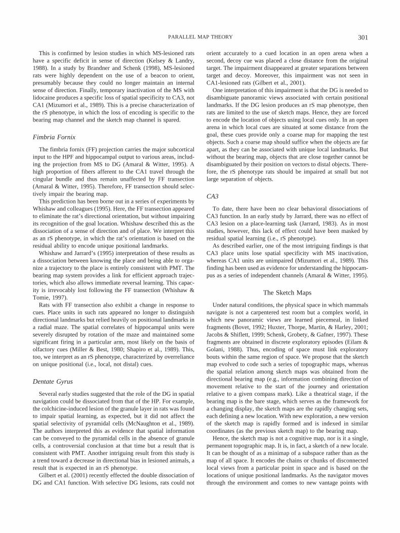
This is confirmed by lesion studies in which MS-lesioned ratshave a specific deficit in sense of direction (Kelsey & Landry,1988). In a study by Brandner and Schenk (1998), MS-lesionedrats were highly dependent on the use of a beacon to orient,presumably because they could no longer maintain an internalsense of direction. Finally, temporary inactivation of the MS withlidocaine produces a specific loss of spatial specificity to CA3, notCA1 (Mizumori et al., 1989). This is a precise characterization ofthe rS phenotype, in which the loss of encoding is specific to thebearing map channel and the sketch map channel is spared.
Fimbria Fornix
The fimbria fornix (FF) projection carries the major subcorticalinput to the HPF and hippocampal output to various areas, includ-ing the projection from MS to DG (Amaral & Witter, 1995). Ahigh proportion of fibers afferent to the CA1 travel through thecingular bundle and thus remain unaffected by FF transection(Amaral & Witter, 1995). Therefore, FF transection should selec-tively impair the bearing map.
This prediction has been borne out in a series of experiments byWhishaw and colleagues (1995). Here, the FF transection appearedto eliminate the rat’s directional orientation, but without impairingits recognition of the goal location. Whishaw described this as thedissociation of a sense of direction and of place. We interpret thisas an rS phenotype, in which the rat’s orientation is based on theresidual ability to encode unique positional landmarks.
Whishaw and Jarrard’s (1995) interpretation of these results asa dissociation between knowing the place and being able to orga-nize a trajectory to the place is entirely consistent with PMT. Thebearing map system provides a link for efficient approach trajec-tories, which also allows immediate reversal learning. This capac-ity is irrevocably lost following the FF transection (Whishaw &Tomie, 1997).
Rats with FF transection also exhibit a change in response tocues. Place units in such rats appeared no longer to distinguishdirectional landmarks but relied heavily on positional landmarks ina radial maze. The spatial correlates of hippocampal units wereseverely disrupted by rotation of the maze and maintained somesignificant firing in a particular arm, most likely on the basis ofolfactory cues (Miller & Best, 1980; Shapiro et al., 1989). This,too, we interpret as an rS phenotype, characterized by overrelianceon unique positional (i.e., local, not distal) cues.
Dentate Gyrus
Several early studies suggested that the role of the DG in spatialnavigation could be dissociated from that of the HP. For example,the colchicine-induced lesion of the granule layer in rats was foundto impair spatial learning, as expected, but it did not affect thespatial selectivity of pyramidal cells (McNaughton et al., 1989).The authors interpreted this as evidence that spatial informationcan be conveyed to the pyramidal cells in the absence of granulecells, a controversial conclusion at that time but a result that isconsistent with PMT. Another intriguing result from this study isa trend toward a decrease in directional bias in lesioned animals, aresult that is expected in an rS phenotype.
Gilbert et al. (2001) recently effected the double dissociation ofDG and CA1 function. With selective DG lesions, rats could not
orient accurately to a cued location in an open arena when asecond, decoy cue was placed a close distance from the originaltarget. The impairment disappeared at greater separations betweentarget and decoy. Moreover, this impairment was not seen inCA1-lesioned rats (Gilbert et al., 2001).
One interpretation of this impairment is that the DG is needed todisambiguate panoramic views associated with certain positionallandmarks. If the DG lesion produces an rS map phenotype, thenrats are limited to the use of sketch maps. Hence, they are forcedto encode the location of objects using local cues only. In an openarena in which local cues are situated at some distance from thegoal, these cues provide only a coarse map for mapping the testobjects. Such a coarse map should suffice when the objects are farapart, as they can be associated with unique local landmarks. Butwithout the bearing map, objects that are close together cannot bedisambiguated by their position on vectors to distal objects. There-fore, the rS phenotype rats should be impaired at small but notlarge separation of objects.
CA3
To date, there have been no clear behavioral dissociations ofCA3 function. In an early study by Jarrard, there was no effect ofCA3 lesion on a place-learning task (Jarrard, 1983). As in moststudies, however, this lack of effect could have been masked byresidual spatial learning (i.e., rS phenotype).
As described earlier, one of the most intriguing findings is thatCA3 place units lose spatial specificity with MS inactivation,whereas CA1 units are unimpaired (Mizumori et al., 1989). Thisfinding has been used as evidence for understanding the hippocam-pus as a series of independent channels (Amaral & Witter, 1995).
The Sketch Maps
Under natural conditions, the physical space in which mammalsnavigate is not a carpentered test room but a complex world, inwhich new panoramic views are learned piecemeal, in linkedfragments (Bovet, 1992; Huxter, Thorpe, Martin, & Harley, 2001;Jacobs & Shiflett, 1999; Schenk, Grobety, & Gafner, 1997). Thesefragments are obtained in discrete exploratory episodes (Eilam &Golani, 1988). Thus, encoding of space must link exploratorybouts within the same region of space. We propose that the sketchmap evolved to code such a series of topographic maps, whereasthe spatial relation among sketch maps was obtained from thedirectional bearing map (e.g., information combining direction ofmovement relative to the start of the journey and orientationrelative to a given compass mark). Like a theatrical stage, if thebearing map is the bare stage, which serves as the framework fora changing display, the sketch maps are the rapidly changing sets,each defining a new location. With new exploration, a new versionof the sketch map is rapidly formed and is indexed in similarcoordinates (as the previous sketch map) to the bearing map.
Hence, the sketch map is not a cognitive map, nor is it a single,permanent topographic map. It is, in fact, a sketch of a new locale.It can be thought of as a minimap of a subspace rather than as themap of all space. It encodes the chains or chunks of disconnectedlocal views from a particular point in space and is based on thelocations of unique positional landmarks. As the navigator movesthrough the environment and comes to new vantage points with
301PARALLEL MAP THEORY
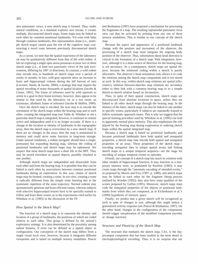
new panoramic views, a new sketch map is formed. Thus, undernatural conditions, as a mammal explores new terrain, it createsmultiple, disconnected sketch maps. Some maps may be linked toeach other by common positional landmarks. Yet even with linksthrough common landmarks, this representation alone (i.e., multi-ple sketch maps) cannot pass the test of the cognitive map: con-structing a novel route between previously disconnected sketchmaps.
As a caveat, we note that the spatial experience of the laboratoryrat may be qualitatively different from that of the wild rodent. Alab rat exploring a single open arena possesses at most two or threesketch maps (i.e., at least two panoramic views of the task envi-ronment, differing by 180° in direction). In contrast, a wild rodentmay encode tens or hundreds of sketch maps over a period ofweeks or months. In fact, wild gray squirrels show an increase inbrain and hippocampal volume during the fall harvest of nuts(Lavenex, Steele, & Jacobs, 2000), a strategy that may require thespatial encoding of many thousands of spatial locations (Jacobs &Liman, 1991). The frame of reference used by wild squirrels toorient to a goal in their home territory, however, does appear to besimilar to that used by the laboratory rat in a test room: anextramaze, allothetic frame of reference (Jacobs & Shiflett, 1999).
Once the sketch map is encoded, the next step is to encode theorientation of the sketch maps within the bearing map and to eachother, creating the integrated map (see Table 4). Whether or not aparticular sketch map is integrated, however, it continues to remainactive and independent until it is no longer accurate. If there is achange in the location or identity of landmarks in the topographicarray, then the sketch map is overwritten by a new sketch map. Ifthere are no changes in the array, then the map is maintained inmemory and could serve some function in long-term storage.Under natural conditions, a rodent navigator should have a single,permanent but expanding bearing map, whereas the coding ofpositional landmarks and sketch maps may be ephemeral. Wesuspect that most sketch maps have a short life in the HPF. Theymay be stored elsewhere as spatial objects, possibly chunked toone another.
Although sketch maps are independent and dissociable fromeach other and from the bearing map, it is possible that they can belinked to each other by associations between common positionallandmarks during an exploration. In this way, chains of sketchmaps may be formed, creating a route. In our view, creating a routeis radically different from the simple route learning due to thesystematic repetition of the same trajectory. Normal rodents mayspontaneously generate and learn efficient routes, whereas subjectswith selective hippocampal lesions have to be specially trained tofollow and learn these routes, as in the experiments cited earlier byWhishaw et al. (1995) in the discussion of the FF.
How Spatial Is the Sketch Map?
The function of a sketch map is to represent the identity andlocation of a group of landmarks, the positions of which are codedrelative to each other. This group is defined by the animal’sexploratory strategy. It is also determined by the proximity amongsalient features. It even can be defined as a spatial object orconfiguration. Our conception of the sketch map differs from asingle visual local view, however, because it integrates differentviewpoints and is based on multiple sensory modalities. Poucet
and Benhamou (1997) have proposed a mechanism for processingthe fragments of a map. The resulting supramodal panoramic localview can thus be activated by priming from any one of thesesensory modalities. This is similar to our concept of the sketchmap.
Because the aspect and appearance of a positional landmarkchange with the position and movement of the observer, theprocessing of a sketch map must integrate the ongoing bodyposition of the observer. Thus, information about head direction iscritical to the formation of a sketch map. Path integration, how-ever, although it is a main source of direction for the bearing map,is not necessary. As a consequence, sketch maps are spatial ob-jects, because the relational coding within a sketch is purelyallocentric. The observer’s head orientation only allows it to codethe relations among the sketch map components and is not storedas such. In this way, within-sketch map relations are spatial (allo-centric), whereas between-sketches map relations are secondaryeither to their link with a common bearing map or to a simplesketch-to-sketch relation based on locomotion.
Thus, in spite of their spatial construction, sketch maps aredisconnected from absolute locations in space, unless they arelinked to all other sketch maps through the bearing map. In theabsence of the latter, sketch maps can also be linked to one anotherin specific routes, particularly if subjects are repeatedly trained tofollow systematic approach routes. This is why we suggest that thespecial training procedure used by Whishaw et al. (1995) can leadto apparently normal place memory. This also emphasizes the roleplayed by the bearing map during exploration in ordering sketchmaps within the spatial integrated map.
Because a sketch map is based on positional landmarks andbecause positional landmarks have both spatial and nonspatialproperties, a sketch map may encode both spatial and nonspatialproperties of an array. These properties of the sketch map—encoding nonspatial data in unique spatial arrays and linkingsketch maps in a unique temporal sequence—set the stage forencoding of unique temporal events or episodes.
Overall, our concept of a sketch map has much in common withother models of hippocampal function. It may function as a tem-porary memory store, as postulated by Rawlins (1985); it maycreate maps through the “automatic encoding of attended events,”as proposed by Morris and Frey (1997, p. 1489), and sketch mapsmay be linked to each other by the fragment fitting processoutlined by Worden (1992); they also have some qualities of thescenes proposed by Gaffan (1991). Moreover, sketch maps mustcode the nonspatial properties of the objects or positional land-marks from which they are composed, as in Eichenbaum et al.’s(1999) hypothesis of memory space.
Finally, we predict that a given sketch will be recognized assuch in spite of changes in size, although this might induce ageneralized activity response (see Poucet & Benhamou, 1997). Onthe other hand, changes in the configuration of the componentsshould trigger reexploration of the modified component (noveltyor change reaction).
Structure and Plasticity of the Sketch Map
The structure that mediates the sketch map, CA1, is the hip-pocampal component that has been studied in the most detail inelectrophysiological recording. Thus, it is no surprise that our
302 JACOBS AND SCHENK
description is similar to other models in which CA1 encodes atopographic allocentric representation. Our model differs in that aCA1-mediated representation is not necessarily the cognitive map;it may be a temporary sketch map that is later incorporated into theintegrated map.
Because the sketch maps are temporary representations, theyshould be served by a more rapid form of plasticity than that in thebearing map. For example, rapid updating of the sketch mapshould occur when an animal attends to a change in positionallandmarks. This can occur when a new area is explored or whenpositional landmarks have been shifted or replaced in a familiararea.
Another implication of the sketch map is for the role of thehippocampus in reducing interference (Shapiro & Olton, 1994).The precise remapping of sketch maps to unique positional land-marks should be correlated with a reduction in interference. Wesuggest that this is because it is difficult to differentiate (and,hence, not overwrite) sketch maps without knowing their relativeposition on the bearing map.
There is currently a general agreement that the major form ofsynaptic plasticity for CA1 function is NMDA-mediated LTP(Morris, 1990). This form of plasticity is appropriate for the rapidencoding and reconfiguration required for constructing and updat-ing a sketch map. In fact, a rat’s spatial exploration of new terrainhas been shown to reverse NMDA-mediated LTP in the rat (Xu,Anwyl, & Rowan, 1998). CA1 is also sensitive to changes inpositional landmarks. With c-fos response as an assay, CA1 wasselectively activated after a rat viewed familiar objects in a novelscene. After it viewed the novel scenes, c-fos response increased inpostrhinal areas and CA1 but decreased in DG and the subiculum(Wan, Aggleton, & Brown, 1999). This differential activation ofDG and CA1 supplies additional evidence for the independent butcomplementary nature of these two structures, which is appropriatefor the major components of the parallel maps.
There are predictable sex differences in CA1 plasticity. Malesappear to rely preferentially on the bearing map, whereas femalesrely preferentially on the sketch map, and females should be moresensitive to changes in the topology of positional cues. Females doexpress an immediate reaction to the change of positional land-marks in spite of the fact that the directional information contri-bution to the bearing map has not changed (Williams & Meck,1993). Hence, we expect that plasticity in the sketch map might bea hallmark of the female hippocampus. Circulating levels of es-trogen have a significant effect on CA1 structure and function inadult female rats. Naturally occurring levels of estrogen causesignificant dendritic branching in the estrus female (Gould et al.,1990). This is accompanied by increased levels of LTP in thepresence of natural peaks in estrogen concentration (Warren, Hum-phreys, Juraska, & Greenough, 1995).
Experimental Impairment of the Sketch Map
Many studies have examined the role of CA1 function and theNMDA receptor in spatial learning. Few studies have attempted toisolate the specific function of CA1, however. PMT predicts thatCA1 lesion or the blockade of NMDA-mediated LTP should notabolish spatial learning but instead should reveal the residuallearning capabilities of the bearing map (i.e., an rB phenotype).
CA1
The behavioral dissociation of DG function by Gilbert et al.(2001) was one half of a double dissociation between DG andCA1. The task that created an impairment in CA1-lesioned ratswas quite different: It was the temporal ordering of visits to armsof the radial arm maze, a task that Chiba, Kesner, and Reynolds(1994) had previously shown to be impaired in hippocampal-lesioned rats. Yet even though CA1-lesioned and hippocampal-lesioned rats were impaired on this task, the DG-lesioned rats werenot (Gilbert et al., 2001). This result is consistent with an rBphenotype—a selective loss of the ability to link different pan-oramic views or sketch maps. The loss of temporal order is alsopredicted from the lesion of the sketch map channel, because oneoperation possible with sketch maps is to link and sequentiallyactivate them.
The Role of the NMDA Receptor
The dissociation of NMDA-mediated LTP and spatial learninghas posed a serious challenge to the theory of the hippocampus asa cognitive map (Bannerman et al., 1995; Saucier & Cain, 1995).As we interpret these results, because this receptor is found in highconcentration in CA1, the loss or blockade of these receptorsshould produce the rB phenotype. In contrast, techniques that areselective for the receptors of transmitters found in greater abun-dance in the DG should produce the rS phenotype. Such paradox-ical dissociations of LTP and spatial learning thus may be theoutcome of residual learning by the unaffected channel.
If the NMDA blockade selectively impairs CA1, it shouldproduce the rB phenotype (see Table 2). Therefore, rats experi-encing the NMDA blockade should be able to navigate usingdirectional cues. The water maze is a task that requires bothbearing and sketch maps and particularly requires a high-resolution sketch map for high spatial accuracy, as accurate ap-proach trajectories must be computed to hit the platform or tocorrect misses. Therefore, NMDA blockade and, hence, impair-ment of the sketch map should produce a greater deficit in thewater maze than in the radial arm maze, which relies more heavilyon directional cues. If the rat knows how to solve the water mazeusing its bearing map, an accurate performance later under NMDAblockade should be possible because of the residual bearing map.
We suggest that this accounts for the effect of pretraining onNMDA blockade (Bannerman et al., 1995; Saucier & Cain, 1995).Our interpretation is as follows: Intact rats with prior training hadhad experience solving the task using the full complement ofspatial representation (bearing, sketch, and integrated maps). Inaddition, the rats were male and, hence, were likely to have reliedon the bearing map. When the NMDA-blockade selectively im-paired the sketch map, these pretrained rats were able to recall theirstrategy of using a bearing map. This change in swim behavior(illustrated in Figure 4) should be detectable with quantitativeanalyses of swim path shape but also can be seen in the raw datapublished by Bannerman et al. (1995). Finally, although the ratsshowed the rB phenotype, this representation had only a lowresolution and was inadequate for unimpaired performance in thewater maze, and, hence, the rats’ performance rose only to anintermediate level.
The pretraining effect was abolished, however, when the pre-training occurred in a curtained maze. Here, NMDA blockade
303PARALLEL MAP THEORY

during the second maze abolished accurate orientation (Banner-man et al., 1995). Our interpretation is that the curtained mazeeliminated directional cues during pretraining and, hence, pre-vented the rats from learning to solve the water maze using thebearing map. Therefore, these rats were prevented from learningduring NMDA blockade.
Our prediction that the NMDA blockade after pretraining shouldproduce a lesser impairment in the radial arm maze is supported bytwo studies (Caramanos & Shapiro, 1994; Shapiro & O’Connor,1992). We would interpret these results as the rat’s successfulreliance on a residual bearing map to solve the highly directionalradial arm maze.
Genetic engineering techniques can also be used to selectivelylesion neurotransmitter receptors in hippocampal subfields. Re-sults from two such studies are consistent with PMT predictions ofresidual learning and plasticity in the remaining channel. Knockoutof CA1-localized NMDA-mediated LTP impaired the spatial se-lectivity of place cells (McHugh, Blum, Tsien, Tonegawa, &Wilson, 1996). This could be interpreted as need for the NMDA-mediated tuning in the high-resolution sketch map. Even thoughthese mice should have shown the rB phenotype, the place spec-ificity of the sketch map would be impaired.
Residual learning was also observed when the knockout of aneuronal glycoprotein eliminated LTP in the DG but did noteliminate CA1 LTP (Nosten-Bertrand et al., 1996). We suggestthat in this study, the rS phenotype produced by the knockoutprocedure allowed the animals to recover to normal performancelevels. In contrast, in the study by McHugh et al. (1996), the rBphenotype would not allow recovery of place specificity in dorsalCA1 pyramidal cells.
Harking back to spatial learning by turtles, we note that micewith calcium-calmodulin-kinase II (CCKII) mutations showed dif-ficulty orienting in an open water maze but could nonethelesssolve a water-filled plus maze (Silva et al., 1992). In our view, themice had an rB phenotype and could not locate a place, only adirection (i.e., our interpretation of the performance by turtles in awater-filled plus maze). We suggest that orientation by CCKIImice and turtles was mediated by their homologous brain struc-tures: the small-celled area of the medial cortex in the turtles, andthe DG in the mice. It would be interesting to test this propositionby combining selective lesions with precise behavioral assays ofthe bearing map in mice and turtles. These results point, however,to the great potential of these genetic techniques (e.g., a doubledissociation of receptor types and, thus, potentially of the parallelmaps).
The Integrated Map
The integrated map emerges from the coding of the sketch mapsin relation to the bearing map (see Table 4). Once this occurs,novel routes and shortcuts can be calculated from the resultantintegrated map. The navigator can also chunk elemental sketchmaps to form larger sketch maps. Thus, the integrated map pro-vides two important cognitive capacities. It permits the navigatorto induce the length and direction of the paths needed to travelfrom one sketch map to another, on the basis of their relativelocation on the bearing map. It emerges from the interaction of thetwo maps and allows long-term storage of chunks of sketch maps(or, potentially, episodes) for later retrieval from long-term mem-
ory. By this means, it is possible to derive the direction of move-ment that is needed to move from one sketch map to another, evenif this movement is discontiguous. In fact, it is the function of theintegrated map to compute this vector. It allows the navigator tocompute the shortest route between two sketch maps that have nopositional landmarks in common.
Coactivation
The fact that discrete sketch maps (or chains of sketch maps)have been stored with an index of their situation on a bearing mapallows them to be positioned relative to each other in a singleintegrated map. This spatial index for the sketch maps is due to thecoactivation of the two maps. This facilitates the direct access (orrecall) during later movements in that locale. And because sketchmaps are assigned to unique locations on the single bearing map,it is possible to distinguish them unambiguously from each other.
Structural Components of the Integrated Map
Forming an integrated map requires two components: a repre-sentation of the bearings with the environment, and a representa-tion of the relationships among discrete objects. The integratedmap also requires a reference memory representation of the currentor most recent integrated map of the environment. To have fullintegrated map capacity, all components must therefore be func-tional: the components of the bearing map (MS, DG, CA3), thesketch map (CA1), and the subiculum, which mediates the refer-ence memory of the integrated map. (There are no doubt upstreamstructures projecting to these components, particularly subcorticalareas, that are important for map integration.) The two componentmaps (bearing and sketch) must also have intact afferent projects,such as the FF projection from MS to DG, the mossy fiberprojection from DG to CA3, and the Schaffer collaterals from CA3to CA1 (see Figure 3).
Map Assembly
The neural architecture of the HPF is uniquely characterized byone-way connections from CA3 to CA1 to the subiculum (Amaral& Witter, 1995). We interpret this architecture as the assembly linenecessary to construct the integrated map (see Table 4). Wepropose that 1-D maps that are initially encoded in DG are thencombined there to form the 2-D bearing map. This information isthen projected to CA3. CA3 projects the location as a position onthe bearing map (and as a discrete object) to CA1. CA1 integratesthe location on the bearing map with the current sketch map andthereby calibrates the sketch map with the bearing map.
Once the integrated map has been formed, CA1 extracts newroutes from the integrated map. CA1 is a complex and topograph-ically organized structure, with direct projections from sensoryareas such as the EC, olfactory bulb, and neocortical areas as wellas thalamic nuclei (Amaral and Witter, 1995). This complex ar-chitecture may reflect CA1�s role as the constructor of sketch mapsand the locus of the integration of these maps with the bearingmap. In this case, CA1 topology reflects two functions: its role asthe encoding of new sketch maps as well as the locus of the currentstatus of the integrated map. From the integrated map (probablydorsal CA1), the role of this area is to extract the temporal order
304 JACOBS AND SCHENK
of positional landmarks on novel routes, calculated from the inte-grated map.
Consolidation of the Integrated Map
Both bearing and sketch maps must be stored in long-termmemory to be retrieved later, albeit in different locations. Wesuggest that the storage of the bearing map is within the DG and,hence, that the DG provides the index to all allocentric encoding(see Teyler & DiScenna, 1986, for an earlier proposal of indexingby the hippocampus). In contrast, the sketch maps are most likelystored in the temporoparietal neocortex. The question of whetherthe integrated map is stored as such is less clear. This depends onthe definition of what is a single sketch map and whether there isoverlap among sketch maps encoded for a given environment. Ifsketch maps are coded when directional information is available,the relation among sketch components may be associated with atleast one major gradient and with the vector from path integration.Such dual coding of successive sketch maps would become morefrequent as the environment is intensively patrolled, leading to thelong-term storage of linked sketch maps. This may be why over-trained memory for familiar places can be retrieved even afterhippocampal damage (Teng & Squire, 1999). Although we do notsuggest that a global integrated map is stored in long-term mem-ory, we predict that consolidation may stabilize the relationshipsamong sketch maps if they have been linked by locomotion.However, preoperative training in the water maze, although itfacilitates acquisition, does not seem to facilitate task reacquisitionand has little effect on probe trials (Morris, Schenk, Tweedie, &Jarrard, 1990). This suggests that very little of the integrated maphas been transferred out of the hippocampus during consolidation.
Reference Memory Representation of the Integrated Map
After the integrated map is created by the hippocampus, it is stillnecessary to have a reference memory representation to make themap available for use and updating. Unlike DG and HP, thesubiculum has reciprocal connections with hippocampal, thalamic,and cortical areas. We suggest, therefore, that it might store thereference memory representation of the integrated map that hasbeen constructed by the hippocampus. If so, lesions of the subic-ulum would differ in subtle ways from hippocampal lesions: Theywould impair reference memory for the integrated map but notworking memory. Place units in the subiculum should also differfrom those in the hippocampus, as we discuss later.
Experimental Impairment of the Integrated Map
Damage to one channel should simultaneously impair the rele-vant map and the integrated map.
Subiculum
A selective lesion of the subiculum should impair the referencememory of the relationship among sketch maps (i.e., the integratedmap). Hence, result of this should be an rB phenotype. The animalshould show partial recovery, because it could learn to navigatewith the remaining bearing map. Such qualitative differences be-tween subiculum-and hippocampus-lesioned groups have been ob-served. Rats with subicular lesions (rB) appeared to swim around
the entire pool. This swim pattern was quite different from theprogressively adjusted loops commonly seen in rats with an rSphenotype (e.g., rats with septal lesions). Moreover, the rats witha subiculum lesion seemed to have little memory between sessions(i.e., impaired reference memory), although they were capable ofexpressing memory for a new platform position after four trials ina match-to-place test (i.e., intact working memory; Morris et al.,1990).
On the basis of the foregoing discussion, a pure subicular lesion(rB phenotype) should impair learning of the new location butleave residual capacities based on the bearing map intact. Thisshould result in a residual ability to learn new positions based onthe bearing map, in which new locations can be encoded. Consis-tent with this interpretation, subiculum-lesioned rats showed im-provement in platform location within trial blocks (Morris et al.,1990). This suggests that they could find the platform by swim-ming transects, orienting with the bearing map (see Figure 4).
Hippocampus Lesions Sparing the Subiculum
The challenge to any spatial theory of hippocampal function isto explain residual spatial learning after a complete hippocampallesion, defined as the absence of cell bodies and/or all neural tissueof the DG and HP. Yet learning by hippocampal-lesioned rats canoccur under certain conditions, and, with additional training, ahippocampal-lesioned rat can show significant recovery. The ex-planation for this may lie in the nature of the training. Recoverydoes not occur quickly but only with special training, such asalternating trials with a visible and an invisible platform at thesame position (Whishaw et al., 1995; Whishaw & Gorny, 1999;Whishaw & Jarrard, 1995). The logistical difficulty with thislesion is that hippocampal-lesioned rats cannot generate controlledtrajectories (i.e., trajectories associated with predictable changes insensory input, based on the 2-D bearing map). They thereforecannot form sketch maps of positional cues, which are scarce in aswimming pool when no salient cue is provided in the vicinity ofthe pool. The solution of attracting the rat to the visible platformto facilitate learning of these cues is an elegant one. Training therats in this way allows them to link egocentric views that couldthen become an effective substitute for true allocentric placelearning.
For example, under such prolonged training, a secondary systemmay be slowly brought online to compensate for the loss ofhippocampal function. The parietal cortex appears to act as aspatial map, associating objects and places (Burgess, Jeffrey, &O’Keefe, 1999; Long & Kesner, 1998). Therefore, hippocampal-lesioned rats could, with special training, slowly develop a parietalmap to navigate to the platform. The subiculum could also play arole in this type of learning, as it could encode a new referencemap for the environment, constructed from this limited parietalinput. Neither of these strategies produce intact learning ability,but, with enough training, there should be some recovery offunction. If either of these components (parietal, subiculum) weremissing, however, there would be no recovery.
This prediction is confirmed in a study of the locus of iboteniclesions on spatial learning: Rats whose lesion included both thehippocampus and some part of the subiculum showed no recovery(Morris et al., 1990). This would be expected if the rat had lost notonly its sense of direction (lesioned bearing map channel) and its
305PARALLEL MAP THEORY
ability to calculate a new position (lesioned sketch map channel)but also the ability to retain any new learned spatial relations(lesioned subiculum).
Lesion placement. In addition to complete hippocampal le-sions, removing tissue from either dorsal or ventral hippocampalregions produces different patterns of impairment. Dorsal lesionsproduce a greater impairment than do ventral lesions (E. Moser,Moser, & Andersen, 1993; M.-B. Moser & Moser, 1998; M.-B.Moser, Moser, Forrest, Andersen, & Morris, 1995). In addition, thedegree of impairment is proportionate to the amount of tissue lost;small dorsal lesions impaired the probe test performance, whereaslarge (more than 20% of the hippocampus) lesions impaired la-tency to the platform (E. Moser et al., 1993). We suggest that thesmall dorsal lesions selectively impaired the CA1 and the sketchmap and, hence, the ability to encode the local position of theplatform. In other words, the dorsal lesion produced an rB pheno-type, and the ventral lesion produced an rS phenotype. The dorsallesion (rB phenotype) allows escape latencies to remain low orintermediate, as in the case of the pretraining effect (Bannerman etal., 1995). Ventral lesions (rS) should have less effect, because ofthe greater use of the sketch map in the water maze. With largerlesions, however, both maps are lost, and escape latencies increase.This prediction could be tested by correlating the loss of tissue foreach subfield (DG, CA3, or CA1) in a dorsal or ventral lesion ofdifferent magnitudes with quantitative analyses of swim pathsproduced by the rat.
Hippocampal residual learning and path integration. In ourview, path integration is not a hippocampus-dependent represen-tation but instead is a source of data for the bearing map, providingit with a default direction value. PMT therefore predicts that ratswith hippocampal lesions are not impaired in path integration (a1-D representation) but instead are impaired in integrating pathintegration into a spatial representation (a 2-D representation).Thus, the key question is whether lesioned rats are impaired in pathintegration or in orienting with a vector derived from path inte-gration. This prediction is supported by evidence that hippocampalrats are only impaired in tasks requiring a spatial representation butnot in pure path integration studies (Alyan & McNaughton, 1999).In another study, hippocampal rats could only home through pathintegration using combined self-movement, surface, and visualcues but were unable to do so with only self-movement cues(Maaswinkel, Jarrard, & Whishaw, 1999). Such learning, depend-ing on the training, could be residual learning by the subiculum,even in the absence of CA1. Hippocampal lesions also abolish therB phenotype, which is indispensable for orienting in a dark regionof an open field with no olfactory cues (Schenk et al., 1995).
Entorhinal Cortex
The EC occupies a special role in hippocampal function. Itprojects to every subfield and has reciprocal connections with CA1(Witter et al., 2000). It is thus an active partner for both sketch andbearing maps, and lesions of the EC alone should reveal significantresidual capacities, similar to lesions of the DG and HP. Indeed,like rats with hippocampal lesions (Morris et al., 1990), EC-lesioned rats show stereotyped swim loops that become progres-sively more likely to cross the platform location (Schenk & Morris,1985). The actual expressed phenotype critically depends on thetask and the tissue remaining in the subiculum, if in fact there is a
residual capacity by the subiculum and parietal to develop sketch-like maps. In fact, EC-lesioned rats do show similar residualcapacities to hippocampal-lesioned rats, in contrast to rats with acombined EC and subiculum lesion, which show no recovery(Schenk & Morris, 1985). This also raises the question of thesignificance of the direct EC–CA1 connection (Witter, Griffioen,Jorritsma-Byham, & Krijnen, 1988). This connection may play animportant role for sketch map reorganization, as is required in theacquisition of learning a new place in a familiar environment.
Hippocampal Unit Activity
Neural activity in specific HPF areas, as measured by unit orfield recordings (and functional imaging, once finer spatial reso-lution has been achieved), should be activated differentially de-pending on the map that is being constructed or accessed for aparticular task.
Predictions
The parallel maps may be active individually at any time,though, under most conditions, they should be active simulta-neously (see Table 4). For example, new exploration along gradi-ents should selectively activate the bearing map channel. In con-trast, exploration into arrays of new positional landmarks or theintroduction of a new positional landmark should selectively acti-vate the sketch map channel. Rearrangement of these landmarks orother mismatch detection (O’Keefe, 1976) should also activate thesketch map channel units. Activation of one component may thenrequire an update of the integrated map, which should activate thewhole assembly line (see Table 4).
An obvious test, then, is to examine the activity of place cells indifferent HPF subfields in reaction to specific task environment orcue manipulation conditions. However, spatial selectivity shouldbe found in sites that encode place, which could be found in a 2-Drepresentation in the bearing map based on directional cues, or a2-D representation in the sketch map constructed from positionallandmarks, or in the integrated map. Spatial selectivity in itselfmay therefore not distinguish easily between mapping systems.The critical test is instead the response of units to changes in taskenvironment. For example, altering directional cues should acti-vate units in the bearing map channel, and manipulating positionallandmarks should primarily activate the sketch map channel, buthaving to navigate between two discontiguous sketch maps shouldactivate the integrated map.
There are significant obstacles to testing these predictions withthe current unit activity literature. First, as we have just discussed,it is not yet clear precisely how subfields differentiate by spatialspecificity. Second, the location of the unit must be preciselydefined. In addition to differences among subfields, there is also ahighly organized topography of projections among subfields. Out-put from CA3, for example, differentiates into three zones, each ofwhich remains segregated in CA1 (Ishizuka et al., 1990). A similartopography is seen in perforant path projections (Witter et al.,2000) and in the CA1 projection to the subiculum (Amaral &Witter, 1995). Such regular topography is likely to be involved infunctional differentiation of independent channels within the hip-pocampus (Amaral & Witter, 1995). We suspect that this topog-raphy is related to the segregation of bearing, sketch, and inte-
306 JACOBS AND SCHENK
grated maps in the hippocampus. Hence, unit activity may verywell differ among such segregated channels.
There is also evidence that hippocampal regions vary in unitactivity. For example, place fields in ventral HPF are sparser andless finely tuned than are those recorded in dorsal HPF (M. W.Jung, Wiener, & McNaughton, 1994), corresponding to the differ-entiation of dorsal–ventral function discussed earlier. A new focusof physiological studies has been the recording of hippocampalensembles, with the goal of discerning topographical patterns ofactivity. This has met with mixed results, perhaps because ofmethodological differences among studies (Hampson, Simeral, &Deadwyler, 1999; Redish et al., 2001). At this point, however,there is no clear consensus on the relationship between unit loca-tion and the pattern of activity.
Task environment is also critical in the evaluation of unitactivity in relation to PMT. Single-track environments (e.g., plusmaze, radial arm maze), favored in some studies (McNaughton etal., 1996; Mizumori, Ragozzino, Cooper, & Leutgeb, 1999;O’Keefe & Speakman, 1987; Redish et al., 2001), provide a testenvironment that is clearly organized into directional vectors. Ratsin such an environment, particularly male rats, might naturally relymore heavily on the bearing map. These rats might thereforerecruit a different pattern of cell ensembles than might rats navi-gating in less directional environments, such as the round cylin-drical arena with a single cue card, the task developed by Muller,Kubie, and Ranck (1987; Ranck, 1973). The card in this taskenvironment provides a simple directional landmark and perhapspositional landmarks from the edges of the card. Because otherdirectional cues are generally missing in this environment, how-ever, unit recordings might biased toward activity in the sketchmap in the cylindrical, cue-card arena and the bearing map channelin the radial arm maze.
Given these caveats, some unit activity studies may shed somelight on the mechanism underlying the parallel mapping systems.We address two categories of unit activity, place units and headdirection units.
Head Direction Units
As we discussed earlier, orienting in a sketch map requires adifferent type of directional information than does orienting in thebearing map. Mapping positional landmarks requires three types ofinformation. Visual input on the size, shape, and aspect of theobject contributes to its identification. Vestibular information me-diates the integration of the changes in aspect with changes inegocentric movements. Finally, knowledge of the direction inwhich the observer is looking (i.e., head direction) facilitates thecomputation of relative position. Processing the relation betweendifferent positional landmarks thus involves motion parallax andthe information coded by the head direction units. In comparison,path integration is related to whole body translation, and, thus,rotation should be more critical for gradient discrimination. Theneural basis of head direction units in structures that project to thehippocampus is becoming well elucidated (Taube & Muller, 1998;Taube, Muller, & Ranck, 1990). We have a different interpretationof the function of these units. We propose that the head directionsystem, involving the anterior dorsal thalamus, postsubiculum, andCA1, furnishes directional data not to the bearing map but to thesketch map. In fact, head direction and place information converge
in CA1 (Leutgeb, Ragozzino, & Mizumori, 2000), but becausehead direction units are related to major directional cues, thebearing map may play a critical role in updating the head directionunits. This may be one way the efficacy of the bearing map affectssketch map processing.
Place Units
We summarize relevant findings from the literature by theirrelationship to different maps.
Evidence for the bearing map. We have already discussed thedissociation of CA3 and CA1 unit activity with MS inactivation,for which MS inactivation resulted in a loss of spatial selectivity inCA3 but not in CA1 (Mizumori et al., 1989). This is concordantwith the critical role of the MS input for the bearing map and hencefor CA3 spatial coding but not for the sketch map, which retains itsspatial specificity.
A second observation that is consistent with activity in thebearing map is that place cells appear preconfigured: A rat intro-duced into a novel arena immediately shows large (i.e., low reso-lution) place fields at the beginning of exploration (Wilson &McNaughton, 1993). These coarse fields could be evidence for theexisting bearing map. Carrying the subject among laboratoryrooms would not disrupt such a map, although the map might bedisrupted by vestibular disorientation. This would also explainwhy place cells update their location using directional information(O’Keefe & Burgess, 1996).
Evidence for the sketch map. The sketch map allows thenavigator to refine the coarse resolution of the bearing map. If CA1is the primary area encoding new sketch maps, then it shouldrapidly encode and tune positional landmarks to create the workingmemory copy of the integrated map. Several studies have demon-strated the role of the NMDA receptor, which is found in highdensity in CA1, in fine tuning place fields (Kentros et al., 1998).The knockout of the gene for a key subunit of the NMDA receptorin CA1 affects the spatial selectivity of CA1 place cells; micelacking this receptor showed reduced LTP in the CA1 and hadcoarser resolution of place fields (McHugh et al., 1996).
Evidence for the integrated map. A noted characteristic ofplace unit studies is the diversity of place cell spatial specificity.For example, a study of CA1 and CA3 place units categorizedthree types according to their response to landmarks: those re-sponding to distal or local landmarks, and those responding to bothtypes (Gothard, Skaggs, Moore, & McNaughton, 1996). We spec-ulate that these three types correspond to units engaged in threemaps: bearing map units encoding distal (i.e., directional) land-marks, sketch map units encoding local (i.e., positional) land-marks, and integrated map units encoding both. If so, then thereshould be some segregation of function by subfield. CA3 neuronsshould specify place in relation to the directional cues, such as thegeometry of the array, whereas CA1 units could be involved inintegrated map or sketch map representations. Therefore, CA1units could respond to both classes of cues, though we speculatethat the area most distal to CA3, which receives input from theCA3 projection zone, should be more responsive to directionalcues than should units in the midseptotemporal regions of CA1,which receive inputs from the CA3 associational zone.
Another finding, related to cue control, is the differential controlof place units by the landmarks according to their position in the
307PARALLEL MAP THEORY
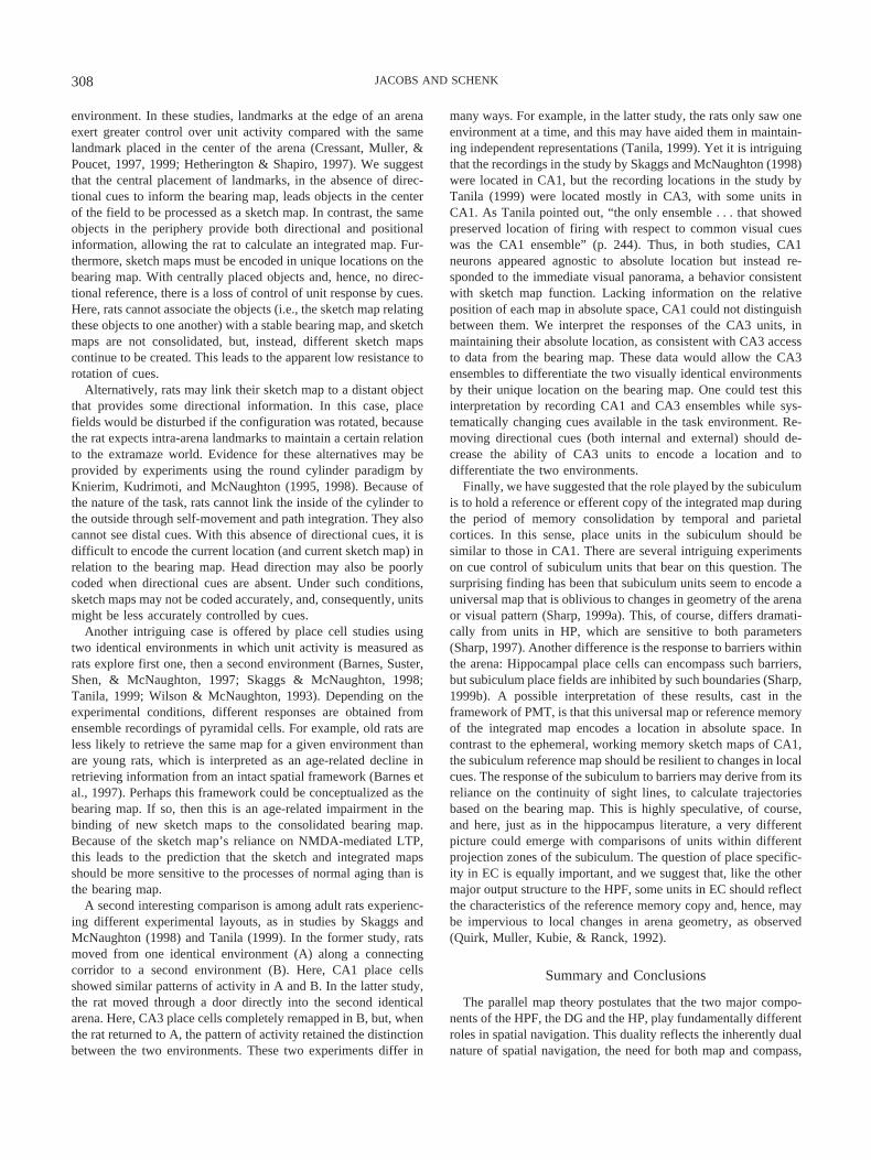
environment. In these studies, landmarks at the edge of an arenaexert greater control over unit activity compared with the samelandmark placed in the center of the arena (Cressant, Muller, &Poucet, 1997, 1999; Hetherington & Shapiro, 1997). We suggestthat the central placement of landmarks, in the absence of direc-tional cues to inform the bearing map, leads objects in the centerof the field to be processed as a sketch map. In contrast, the sameobjects in the periphery provide both directional and positionalinformation, allowing the rat to calculate an integrated map. Fur-thermore, sketch maps must be encoded in unique locations on thebearing map. With centrally placed objects and, hence, no direc-tional reference, there is a loss of control of unit response by cues.Here, rats cannot associate the objects (i.e., the sketch map relatingthese objects to one another) with a stable bearing map, and sketchmaps are not consolidated, but, instead, different sketch mapscontinue to be created. This leads to the apparent low resistance torotation of cues.
Alternatively, rats may link their sketch map to a distant objectthat provides some directional information. In this case, placefields would be disturbed if the configuration was rotated, becausethe rat expects intra-arena landmarks to maintain a certain relationto the extramaze world. Evidence for these alternatives may beprovided by experiments using the round cylinder paradigm byKnierim, Kudrimoti, and McNaughton (1995, 1998). Because ofthe nature of the task, rats cannot link the inside of the cylinder tothe outside through self-movement and path integration. They alsocannot see distal cues. With this absence of directional cues, it isdifficult to encode the current location (and current sketch map) inrelation to the bearing map. Head direction may also be poorlycoded when directional cues are absent. Under such conditions,sketch maps may not be coded accurately, and, consequently, unitsmight be less accurately controlled by cues.
Another intriguing case is offered by place cell studies usingtwo identical environments in which unit activity is measured asrats explore first one, then a second environment (Barnes, Suster,Shen, & McNaughton, 1997; Skaggs & McNaughton, 1998;Tanila, 1999; Wilson & McNaughton, 1993). Depending on theexperimental conditions, different responses are obtained fromensemble recordings of pyramidal cells. For example, old rats areless likely to retrieve the same map for a given environment thanare young rats, which is interpreted as an age-related decline inretrieving information from an intact spatial framework (Barnes etal., 1997). Perhaps this framework could be conceptualized as thebearing map. If so, then this is an age-related impairment in thebinding of new sketch maps to the consolidated bearing map.Because of the sketch map’s reliance on NMDA-mediated LTP,this leads to the prediction that the sketch and integrated mapsshould be more sensitive to the processes of normal aging than isthe bearing map.
A second interesting comparison is among adult rats experienc-ing different experimental layouts, as in studies by Skaggs andMcNaughton (1998) and Tanila (1999). In the former study, ratsmoved from one identical environment (A) along a connectingcorridor to a second environment (B). Here, CA1 place cellsshowed similar patterns of activity in A and B. In the latter study,the rat moved through a door directly into the second identicalarena. Here, CA3 place cells completely remapped in B, but, whenthe rat returned to A, the pattern of activity retained the distinctionbetween the two environments. These two experiments differ in
many ways. For example, in the latter study, the rats only saw oneenvironment at a time, and this may have aided them in maintain-ing independent representations (Tanila, 1999). Yet it is intriguingthat the recordings in the study by Skaggs and McNaughton (1998)were located in CA1, but the recording locations in the study byTanila (1999) were located mostly in CA3, with some units inCA1. As Tanila pointed out, “the only ensemble . . . that showedpreserved location of firing with respect to common visual cueswas the CA1 ensemble” (p. 244). Thus, in both studies, CA1neurons appeared agnostic to absolute location but instead re-sponded to the immediate visual panorama, a behavior consistentwith sketch map function. Lacking information on the relativeposition of each map in absolute space, CA1 could not distinguishbetween them. We interpret the responses of the CA3 units, inmaintaining their absolute location, as consistent with CA3 accessto data from the bearing map. These data would allow the CA3ensembles to differentiate the two visually identical environmentsby their unique location on the bearing map. One could test thisinterpretation by recording CA1 and CA3 ensembles while sys-tematically changing cues available in the task environment. Re-moving directional cues (both internal and external) should de-crease the ability of CA3 units to encode a location and todifferentiate the two environments.
Finally, we have suggested that the role played by the subiculumis to hold a reference or efferent copy of the integrated map duringthe period of memory consolidation by temporal and parietalcortices. In this sense, place units in the subiculum should besimilar to those in CA1. There are several intriguing experimentson cue control of subiculum units that bear on this question. Thesurprising finding has been that subiculum units seem to encode auniversal map that is oblivious to changes in geometry of the arenaor visual pattern (Sharp, 1999a). This, of course, differs dramati-cally from units in HP, which are sensitive to both parameters(Sharp, 1997). Another difference is the response to barriers withinthe arena: Hippocampal place cells can encompass such barriers,but subiculum place fields are inhibited by such boundaries (Sharp,1999b). A possible interpretation of these results, cast in theframework of PMT, is that this universal map or reference memoryof the integrated map encodes a location in absolute space. Incontrast to the ephemeral, working memory sketch maps of CA1,the subiculum reference map should be resilient to changes in localcues. The response of the subiculum to barriers may derive from itsreliance on the continuity of sight lines, to calculate trajectoriesbased on the bearing map. This is highly speculative, of course,and here, just as in the hippocampus literature, a very differentpicture could emerge with comparisons of units within differentprojection zones of the subiculum. The question of place specific-ity in EC is equally important, and we suggest that, like the othermajor output structure to the HPF, some units in EC should reflectthe characteristics of the reference memory copy and, hence, maybe impervious to local changes in arena geometry, as observed(Quirk, Muller, Kubie, & Ranck, 1992).
Summary and Conclusions
The parallel map theory postulates that the two major compo-nents of the HPF, the DG and the HP, play fundamentally differentroles in spatial navigation. This duality reflects the inherently dualnature of spatial navigation, the need for both map and compass,
308 JACOBS AND SCHENK
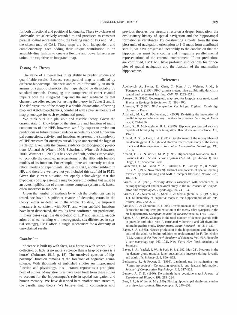
for both directional and positional landmarks. These two classes oflandmarks are selectively attended to and processed to constructparallel spatial representations, the bearing map of DG and CA3,the sketch map of CA1. These maps are both independent andcomplementary, each adding their unique contribution in anassembly-line fashion to create a flexible and powerful represen-tation, the cognitive or integrated map.
Testing the Theory
The value of a theory lies in its ability to predict unique andquantifiable results. Because each parallel map is mediated bydifferent hippocampal channels and relies differentially on mech-anisms of synaptic plasticity, the maps should be dissociable bystandard methods. Damaging one component of either channelimpairs both the integrated map and the map mediated by thatchannel; we offer recipes for testing the theory in Tables 2 and 3.The definitive test of the theory is a double dissociation of bearingmap and sketch map channels, combined with a precise measure ofmap phenotype for each experimental group.
We think ours is a plausible and testable theory. Given thecurrent state of knowledge of the structure and function of manycomponents of the HPF, however, we fully expect to revise ourpredictions as future research reduces uncertainty about hippocam-pal connections, activity, and function. At present, the complexityof HPF structure far outstrips our ability to understand the logic ofits design. Even with the current evidence for topographic projec-tions (Amaral & Witter, 1995; Scharfman, Witter, & Schwarcz,2000; Witter et al., 2000), it has been difficult, perhaps impossible,to reconcile the complex neuroanatomy of the HPF with feasiblemodels of its function. For example, there are currently no theo-retical models or experimental studies of CA2, another subfield inHP, and therefore we have not yet included this subfield in PMT.Given this current situation, we openly acknowledge that thishypothesis of map assembly may be correct in outline but must bean oversimplification of a much more complex system and, hence,often incorrect in the details.
Given the number of methods by which the predictions can betested, we have a significant chance of detecting errors in thetheory, either in detail or in the whole. To date, the empiricalliterature is consistent with PMT, and when subfield functionshave been dissociated, the results have confirmed our predictions.In many cases (e.g., the dissociation of LTP and learning, associ-ation of wheel running with neurogenesis, sex differences in spa-tial strategy), PMT offers a single mechanism for a diversity ofunexplained results.
Conclusion
“Science is built up with facts, as a house is with stones. But acollection of facts is no more a science than a heap of stones is ahouse” (Poincare, 1913, p. 18). The unsolved question of hip-pocampal function remains at the forefront of cognitive neuro-science. With thousands of published studies on hippocampalfunction and physiology, this literature represents a prodigiousheap of stones. Many structures have been built from these stonesto account for the hippocampus’s role in spatial navigation andhuman memory. We have described here another such structure,the parallel map theory. We believe that, in comparison with
previous theories, our structure rests on a deeper foundation, theevolutionary history of spatial navigation and the hippocampalformation in vertebrates. By constructing a model from the sim-plest units of navigation, orientation to 1-D maps from distributedstimuli, we have progressed inexorably to the conclusion that thehippocampus must be encoding and integrating parallel mentalrepresentations of the external environment. If our predictionsare confirmed, PMT will have profound implications for princi-ples of spatial navigation and the function of the mammalianhippocampus.
References
Abeliovich, A., Paylor, R., Chen, C., Kim, J. J., Wehner, J. M., &Tonegawa, S. (1993). PKC-gamma mutant mice exhibit mild deficits inspatial and contextual learning. Cell, 75, 1263–1271.
Akesson, S. (1996). Geomagnetic map used for long-distance navigation?Trends in Ecology & Evolution, 11, 398–400.
Alerstam, T. (1990). Bird migration. Cambridge, England: CambridgeUniversity Press.
Alvarado, M. C., & Bachevalier, J. (2000). Revisiting the maturation ofmedial temporal lobe memory functions in primates. Learning & Mem-ory, 7, 244–256.
Alyan, S., & McNaughton, B. L. (1999). Hippocampectomized rats arecapable of homing by path integration. Behavioral Neuroscience, 113,19–31.
Amaral, D. G., & Dent, J. A. (1981). Development of the mossy fibers ofthe dentate gyrus: I. A light and electron microscopic study of the mossyfibers and their expansions. Journal of Comparative Neurology, 195,51–86.
Amaral, D. G., & Witter, M. P. (1995). Hippocampal formation. In G.Paxinos (Ed.), The rat nervous system (2nd ed., pp. 443–493). SanDiego, CA: Academic Press.
Bannerman, D. M., Good, M. A., Butcher, S. P., Ramsay, M., & Morris,R. G. M. (1995, November 9). Distinct components of spatial learningrevealed by prior training and NMDA receptor blockade. Nature, 378,182–186.
Barnes, C. A. (1979). Memory deficits associated with senescence: Aneurophysiological and behavioral study in the rat. Journal of Compar-ative and Physiological Psychology, 93, 74–104.
Barnes, C. A., Suster, M. S., Shen, J., & McNaughton, B. L. (1997, July17). Multistability of cognitive maps in the hippocampus of old rats.Nature, 388, 272–275.
Battistin, T., & Cherubini, E. (1994). Developmental shift from long-termdepression to long-term potentiation at the mossy fibre synapses in therat hippocampus. European Journal of Neuroscience, 6, 1750–1755.
Bayer, S. A. (1982). Changes in the total number of dentate granule cellsin juvenile and adult rats: A correlated volumetric and 3H-thymidineautoradiographic study. Experimental Brain Research, 46, 315–323.
Bayer, S. A. (1985). Neuron production in the hippocampus and olfactorybulb of the adult rat brain: Addition or replacement? In F. Nottebohm(Ed.), Annals of the New York Academy of Sciences: Vol. 457. Hope fora new neurology (pp. 163–172). New York: New York Academy ofSciences.
Bayer, S. A., Yackel, J. W., & Puri, P. S. (1982, May 21). Neurons in therat dentate gyrus granular layer substantially increase during juvenileand adult life. Science, 216, 890–892.
Benhamou, S., & Poucet, B. (1998). Landmark use by navigating rats(Rattus norvegicus): Contrasting geometric and featural information.Journal of Comparative Psychology, 112, 317–322.
Bennett, A. T. D. (1996). Do animals have cognitive maps? Journal ofExperimental Biology, 199, 219–224.
Best, P. J., & White, A. M. (1999). Placing hippocampal single-unit studiesin a historical context. Hippocampus, 9, 346–351.
309PARALLEL MAP THEORY
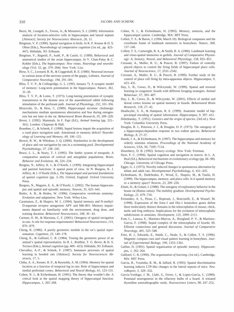
Bezzi, M., Leutgeb, S., Treves, A., & Mizumori, S. J. (2000). Informationanalysis of location-selective cells in hippocampus and lateral septum[Abstract]. Society for Neuroscience Abstracts, 26, 11.
Bingman, V. P. (1990). Spatial navigation in birds. In R. P. Kesner & D. S.Olton (Eds.), Neurobiology of comparative cognition (1st ed., pp. 423–447). Hillsdale, NJ: Erlbaum.
Bingman, V., Bagnoli, P., Ioale, P., & Casini, G. (1989). Behavioral andanatomical studies of the avian hippocampus. In V. Chan-Palay & C.Kohler (Eds.), The hippocampus: New vistas. Neurology and neurobi-ology (Vol. 52, pp. 379–394). New York: Liss.
Birse, S. C., Leonard, R. B., & Coggeshall, R. E. (1980). Neuronal increasein various areas of the nervous system of the guppy, Lebistes. Journal ofComparative Neurology, 194, 291–301.
Bliss, T. V. P., & Collingridge, G. L. (1993, January 7). A synaptic modelof memory: Long-term potentiation in the hippocampus. Nature, 361,31–39.
Bliss, T. V. P., & Lomo, T. (1973). Long-lasting potentiation of synaptictransmission in the dentate area of the anaesthetized rabbit followingstimulation of the perforant path. Journal of Physiology, 232, 331–356.
Blozovski, D., & Hess, C. (1989). Hippocampal nicotinic cholinergicmechanisms mediate spontaneous alternation and fear during ontogen-esis but not later in the rat. Behavioural Brain Research, 35, 209–220.
Bovet, J. (1992). Mammals. In F. Papi (Ed.), Animal homing (pp. 321–361). London: Chapman & Hall.
Brandner, C., & Schenk, F. (1998). Septal lesions impair the acquisition ofa cued place navigation task: Attentional or memory deficit? Neurobi-ology of Learning and Memory, 69, 106–125.
Brown, R. W., & Whishaw, I. Q. (2000). Similarities in the developmentof place and cue navigation by rats in a swimming pool. DevelopmentalPsychobiology, 37, 238–245.
Bruce, L. L., & Neary, T. J. (1995). The limbic system of tetrapods: Acomparative analysis of cortical and amygdalar populations. Brain,Behavior and Evolution, 46, 224–234.
Burgess, N., Jeffery, K. J., & O’Keefe, J. (1999). Integrating hippocampaland parietal functions: A spatial point of view. In N. Burgess, K. J.Jeffery, & J. O’Keefe (Eds.), The hippocampal and parietal foundationsof spatial cognition (pp. 3–29). Oxford, England: Oxford UniversityPress.
Burgess, N., Maguire, E. A., & O’Keefe, J. (2002). The human hippocam-pus and spatial and episodic memory, Neuron, 35, 625–641.
Butler, A. B., & Hodos, W. (1996). Comparative vertebrate anatomy:Evolution and adaptation. New York: Wiley-Liss.
Caramanos, Z., & Shapiro, M. L. (1994). Spatial memory and N-methyl-D-aspartate receptor antagonists APV and MK-801: Memory impair-ments depend on familiarity with the environment, drug dose, andtraining duration. Behavioral Neuroscience, 108, 30–43.
Carman, H. M., & Mactutus, C. F. (2001). Ontogeny of spatial navigationin rats: A role for response requirements? Behavioral Neuroscience, 115,870–879.
Cheng, K. (1986). A purely geometric module in the rat’s spatial repre-sentation. Cognition, 23, 149–178.
Cheng, K., & Gallistel, C. R. (1984). Testing the geometric power of ananimal’s spatial representation. In H. L. Roitblat, T. G. Bever, & H. S.Terrace (Eds.), Animal cognition (pp. 409–423). Hillsdale, NJ: Erlbaum.
Chevalley, A.-F., & Schenk, F. (1987). Immature processes of spatiallearning in hooded rats [Abstract]. Society for Neurosciences Ab-stracts, 17, 5.
Chiba, A. A., Kesner, R. P., & Reynolds, A. M. (1994). Memory for spatiallocation as a function of temporal lag in rats: Role of hippocampus andmedial prefrontal cortex. Behavioral and Neural Biology, 61, 123–131.
Cohen, N. J., & Eichenbaum, H. (1991). The theory that wouldn’t die: Acritical look at the spatial mapping theory of hippocampal function.Hippocampus, 1, 265–268.
Cohen, N. J., & Eichenbaum, H. (1993). Memory, amnesia, and thehippocampal system. Cambridge, MA: MIT Press.
Collett, T. S., & Baron, J. (1994, March 10). Biological compasses and thecoordinate frame of landmark memories in honeybees. Nature, 368,137–140.
Collett, T. S., Cartwright, B. A., & Smith, B. A. (1986). Landmark learningand visuo-spatial memories in gerbils. Journal of Comparative Physiol-ogy: A, Sensory, Neural, and Behavioral Physiology, 158, 835–851.
Cressant, A., Muller, R. U., & Poucet, B. (1997). Failure of centrallyplaced objects to control the firing fields of hippocampal place cells.Journal of Neuroscience, 17, 2531–2542.
Cressant, A., Muller, R. U., & Poucet, B. (1999). Further study of thecontrol of place cell firing by intra-apparatus objects. Hippocampus, 9,423–431.
Day, L. B., Crews, D., & Wilczynski, W. (1999). Spatial and reversallearning in congeneric lizards with different foraging strategies. AnimalBehaviour, 57, 393–407.
Day, L. B., Crews, D., & Wilczynski, W. (2001). Effects of medial anddorsal cortex lesions on spatial memory in lizards. Behavioural BrainResearch, 118, 27–42.
Deadwyler, S. A., & Hampson, R. E. (1999). Anatomic model of hip-pocampal encoding of spatial information. Hippocampus, 9, 397–412.
Dobzhansky, T. (1951). Genetics and the origin of species. (3rd ed.). NewYork: Columbia University Press.
Douglas, R. J., Peterson, J. J., & Douglas, D. P. (1973). The ontogeny ofa hippocampus-dependent response in two rodent species. BehavioralBiology, 8, 27–37.
Dusek, J. A., & Eichenbaum, H. (1997). The hippocampus and memory fororderly stimulus relations. Proceedings of the National Academy ofSciences, USA, 94, 7109–7114.
Dusenbery, D. B. (1992). Sensory ecology. New York: Freeman.Dyer, F. C. (1994). Spatial cognition and navigation in insects. In L. A.
Real (Ed.), Behavioral mechanisms in evolutionary ecology (pp. 66–98).Chicago: University of Chicago Press.
Egger, G. J. (1973). Novelty induced changes in spontaneous alternation byinfant and adult rats. Developmental Psychobiology, 6, 431–435.
Eichenbaum, H., Dudchenko, P., Wood, E., Shapiro, M., & Tanila, H.(1999). The hippocampus, memory, and place cells: Is it spatial memoryor a memory space? Neuron, 23, 209–226.
Eilam, D., & Golani, I. (1988). The ontogeny of exploratory behavior in thehouse rat (Rattus rattus): The mobility gradient. Developmental Psycho-biology, 21, 679–710.
Fernandez, A. S., Pieau, C., Reperant, J., Boncinelli, E., & Wassef, M.(1998). Expression of the Emx-1 and Dlx-1 homeobox genes definethree molecularly distinct domains in the telencephalon of mouse, chick,turtle and frog embryos: Implications for the evolution of telencephalicsubdivisions in amniotes. Development, 125, 2099–2111.
Font, C., Lanuza, E., Martinez-Marcos, A., Hoogland, P. V., & Martinez-Garcia, F. (1998). Septal complex of the telencephalon of lizards: III.Efferent connections and general discussion. Journal of ComparativeNeurology, 401, 525–548.
Frier, H. J., Edwards, E., Smith, C., Neale, S., & Collett, T. S. (1996).Magnetic compass cues and visual pattern learning in honeybees. Jour-nal of Experimental Biology, 199, 1353–1361.
Gaffan, D. (1991). Spatial organization of episodic memory. Hippocam-pus, 1, 262–264.
Gallistel, C. R. (1990). The organization of learning. (1st ed.). Cambridge,MA: MIT Press.
Garcia, R., Vouimba, R. M., & Jaffard, R. (1993). Spatial discriminationlearning induces LTP-like changes in the lateral septum of mice. Neu-roReport, 5, 329–332.
Garcia-Verdugo, J. M., Llahi, S., Ferrer, I., & Lopez-Garcia, C. (1989).Postnatal neurogenesis in the olfactory bulbs of a lizard. A tritiatedthymidine autoradiographic study. Neuroscience Letters, 98, 247–252.
310 JACOBS AND SCHENK

Gilbert, P., Kesner, R., & Lee, I. (2001). Dissociating hippocampal sub-regions: A double dissociation between dentate gyrus and CA1. Hip-pocampus, 11, 626–636.
Glick, S. D., Weaver, L. M., & Meibach, R. C. (1980, March 7). Lateral-ization of reward in rats: Differences in reinforcing thresholds. Science,207, 1093–1095.
Gluck, M. A., & Myers, C. E. (2001). Gateway to memory: An introductionto neural network modeling of the hippocampus and learning. Cam-bridge, MA: MIT Press.
Gothard, K., Skaggs, W. E., Moore, K. M., & McNaughton, B. L. (1996).Binding of hippocampal CA1 neural activity to multiple referenceframes in a landmark-based navigation task. Journal of Neuro-science, 16, 823–835.
Gould, E., Beylin, A., Tanapat, P., Reeves, A., & Shors, T. J. (1999).Learning enhances adult neurogenesis in the hippocampal formation.Nature Neuroscience, 2, 260–265.
Gould, E., Wooley, C. S., Frankfurt, M., & McEwen, B. S. (1990). Gonadalsteroids regulate dendritic spine density in hippocampal pyramidal cellsin adulthood. Journal of Neuroscience, 10, 1286–1291.
Granger, R., Wiebe, S. P., Taketani, M., & Lynch, G. (1996). Distinctmemory circuits composing the hippocampal region. Hippocampus, 6,567–578.
Greene, C. H., & Cook, R. G. (1997). Landmark geometry and identitycontrols spatial navigation in rats. Animal Learning & Behavior, 25,312–323.
Hampson, R. E., Simeral, J. D., & Deadwyler, S. A. (1999, December 9).Distribution of spatial and nonspatial information in dorsal hippocam-pus. Nature, 402, 610–614.
Harvey, P. H., & Pagel, M. D. (1991). The comparative method in evolu-tionary biology. Oxford, England: Oxford University Press.
Hasselmo, M. E. (2000). Septal modulation of theta dynamics: What is thefunction of the theta rhythm? In R. Numan (Ed.), The behavioralneuroscience of the septal region (pp. 92–114). New York: Springer.
Helbig, A. J., Berthold, P., & Wiltschko, W. (1989). Migratory orientationof blackcaps (Sylvia atricapilla): Population-specific shifts of directionduring the autumn. Ethology, 82, 307–315.
Hetherington, P. A., & Shapiro, M. L. (1997). Hippocampal place fields arealtered by the removal of single visual cues in a distance-dependentmanner. Behavioral Neuroscience, 111, 20–34.
Holmes, O. W. (1889). The autocrat of the breakfast-table. Boston: Hough-ton Mifflin. (Original work published 1885)
Holtzman, D. A. (1998). From slither to hither: Orientation and spatiallearning in snakes. Integrative Biology, 1, 81–89.
Holtzman, D. A., Harris, T. W., Aranguren, G., & Bostock, E. (1999).Spatial learning of an escape task by young corn snakes, Elaphe guttataguttata. Animal Behaviour, 57, 51–60.
Hoogland, P. V., Martinez-Garcia, F., Geneser, F. A., & Vermeulen-Vanderzee, E. (1998). Convergence of thalamic and cholinergic projec-tions in the “dentate area” of lizards. Brain, Behavior and Evolution, 51,113–122.
Hoogland, P. V., & Vermeulen-Vanderzee, E. (1990). Intrinsic and extrin-sic connections of the cerebral cortex of lizards. In W. K. Schwerdtfeger& W. J. A. J. Smeets (Eds.), The forebrain of reptiles (pp. 20–29). Basel,Switzerland: Karger.
Huang, Y.-Y., Kandel, E. R., Varshavsky, L., Brandon, E. P., Qi, M.,Idzerda, R. L., et al. (1995). A genetic test of the effects of mutations inPKA on mossy fiber LTP and its relation to spatial and contextuallearning. Cell, 83, 1211–1222.
Huxter, J. R., Thorpe, C. M., Martin, G. M., & Harley, C. W. (2001).Spatial problem solving and hippocampal place cell firing in rats:Control by an internal sense of direction carried across environments.Behavioural Brain Research, 123, 37–48.
Isgor, C., & Sengelaub, D. R. (1998). Prenatal gonadal steroids affect adult
spatial behavior, CA1 and CA3 pyramidal cell morphology in rats.Hormones and Behavior, 34, 183–198.
Ishizuka, N., Weber, J., & Amaral, D. G. (1990). Organization of intra-hippocampal projections originating from CA3 pyramidal cells in the rat.Journal of Comparative Neurology, 295, 580–623.
Jacobs, L. F. (1995). The ecology of spatial cognition: Adaptive patterns ofhippocampal size and space use in wild rodents. In E. Alleva, A. Fasolo,H.-P. Lipp, & L. Nadel (Eds.), Studies of the brain in naturalisticsettings (pp. 301–322). Dordrecht, the Netherlands: Kluwer Academic.
Jacobs, L. F. (1996). Sexual selection and the brain. Trends in Ecology andEvolution, 11, 82–86.
Jacobs, L. F., Gaulin, S. J. C., Sherry, D. F., & Hoffman, G. E. (1990).Evolution of spatial cognition: Sex-specific patterns of spatial behaviorpredict hippocampal size. Proceedings of the National Academy ofSciences, USA, 87, 6349–6352.
Jacobs, L. F., & Liman, E. R. (1991). Grey squirrels remember thelocations of buried nuts. Animal Behaviour, 41, 103–110.
Jacobs, L. F., & Shiflett, M. W. (1999). Spatial orientation on a verticalmaze in free-ranging fox squirrels (Sciurus niger). Journal of Compar-ative Psychology, 113, 116–127.
Jacobs, L. F., & Spencer, W. D. (1994). Natural space-use patterns andhippocampal size in kangaroo rats. Brain, Behavior and Evolution, 44,125–132.
Jarrard, L. E. (1983). Selective hippocampal lesions and behavior: Effectsof kainic acid lesions on performance of place and cue tasks. BehavioralNeuroscience, 97, 873–889.
Jung, C. (1964). Approaching the unconscious. In C. G. Jung, M.-L. vonFranz, & J. Freeman (Eds.), Man and his symbols (pp. 18–103). GardenCity, NY: Doubleday.
Jung, M. W., Wiener, S. I., & McNaughton, B. L. (1994). Comparison ofspatial firing characteristics of units in dorsal and ventral hippocampusof the rat. Journal of Neuroscience, 14, 7347–7356.
Juraska, J. M. (1984). Sex differences in developmental plasticity in thevisual cortex and hippocampal dentate gyrus. In G. J. de Vries (Ed.),Progress in brain research (Vol. 61, pp. 205–214). Amsterdam:Elsevier.
Juraska, J. M., Fitch, J. M., Henderson, C., & Rivers, N. (1985). Sexdifference in the dendritic branching of dentate granule cells followingdifferential experience. Brain Research, 333, 73–80.
Kali, S., & Dayan, P. (2000). The involvement of recurrent connections inarea CA3 in establishing the properties of place fields: A model. Journalof Neuroscience, 20, 7463–7477.
Kaplan, H., & Hill, K. (1985). Hunting ability and reproductive successamong male Ache foragers. Current Anthropology, 26, 223–246.
Kavaliers, M., Eckel, L. A., & Ossenkopp, K. P. (1993). Brief exposureto 60 Hz magnetic fields improves sexually dimorphic spatial learningperformance in the meadow vole, Microtus pennsylvanicus. Journal ofComparative Physiology: A, Sensory, Neural, and Behavioral Physiol-ogy, 173, 241–248.
Kavaliers, M., Ossenkopp, K. P., Prato, F. S., Innes, D. G. L., Galea,L. A. M., Kinsella, D. M., & Perrot-Sinal, T. S. (1996). Spatial learningin deer mice: Sex differences and the effects of endogenous opioidsand 60 Hz magnetic fields. Journal of Comparative Physiology: A,Sensory, Neural, and Behavioral Physiology, 179, 715–724.
Keeton, W. T. (1974). The mystery of pigeon homing. Scientific American,231(6), 96–107.
Kelsey, J. E., & Landry, B. A. (1988). Medial septal lesions disrupt spatialmapping ability in rats. Behavioral Neuroscience, 102, 289–293.
Kentros, C., Hargreaves, E. L., Hawkins, R. D., Kandel, E. R., Shapiro,M. L., & Muller, R. V. (1998, June 26). Abolition of long-term stabilityof new hippocampal place cell maps by NMDA receptor blockade.Science, 280, 2121–2126.
Kimchi, T., & Terkel, J. (2001). Magnetic compass orientation in the blind
311PARALLEL MAP THEORY
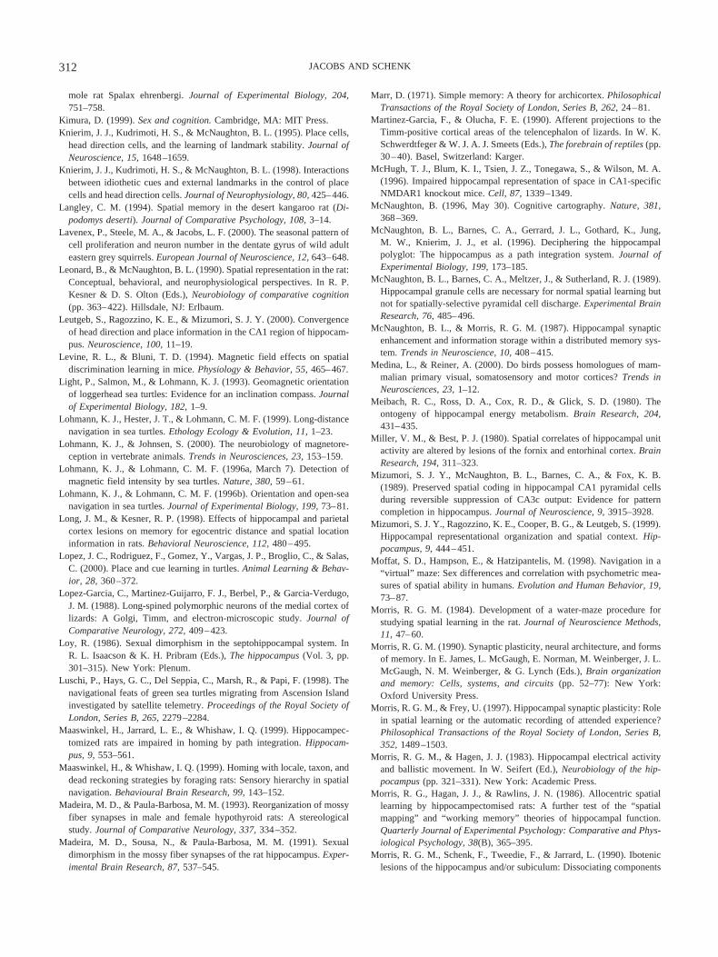
mole rat Spalax ehrenbergi. Journal of Experimental Biology, 204,751–758.
Kimura, D. (1999). Sex and cognition. Cambridge, MA: MIT Press.Knierim, J. J., Kudrimoti, H. S., & McNaughton, B. L. (1995). Place cells,
head direction cells, and the learning of landmark stability. Journal ofNeuroscience, 15, 1648–1659.
Knierim, J. J., Kudrimoti, H. S., & McNaughton, B. L. (1998). Interactionsbetween idiothetic cues and external landmarks in the control of placecells and head direction cells. Journal of Neurophysiology, 80, 425–446.
Langley, C. M. (1994). Spatial memory in the desert kangaroo rat (Di-podomys deserti). Journal of Comparative Psychology, 108, 3–14.
Lavenex, P., Steele, M. A., & Jacobs, L. F. (2000). The seasonal pattern ofcell proliferation and neuron number in the dentate gyrus of wild adulteastern grey squirrels. European Journal of Neuroscience, 12, 643–648.
Leonard, B., & McNaughton, B. L. (1990). Spatial representation in the rat:Conceptual, behavioral, and neurophysiological perspectives. In R. P.Kesner & D. S. Olton (Eds.), Neurobiology of comparative cognition(pp. 363–422). Hillsdale, NJ: Erlbaum.
Leutgeb, S., Ragozzino, K. E., & Mizumori, S. J. Y. (2000). Convergenceof head direction and place information in the CA1 region of hippocam-pus. Neuroscience, 100, 11–19.
Levine, R. L., & Bluni, T. D. (1994). Magnetic field effects on spatialdiscrimination learning in mice. Physiology & Behavior, 55, 465–467.
Light, P., Salmon, M., & Lohmann, K. J. (1993). Geomagnetic orientationof loggerhead sea turtles: Evidence for an inclination compass. Journalof Experimental Biology, 182, 1–9.
Lohmann, K. J., Hester, J. T., & Lohmann, C. M. F. (1999). Long-distancenavigation in sea turtles. Ethology Ecology & Evolution, 11, 1–23.
Lohmann, K. J., & Johnsen, S. (2000). The neurobiology of magnetore-ception in vertebrate animals. Trends in Neurosciences, 23, 153–159.
Lohmann, K. J., & Lohmann, C. M. F. (1996a, March 7). Detection ofmagnetic field intensity by sea turtles. Nature, 380, 59–61.
Lohmann, K. J., & Lohmann, C. M. F. (1996b). Orientation and open-seanavigation in sea turtles. Journal of Experimental Biology, 199, 73–81.
Long, J. M., & Kesner, R. P. (1998). Effects of hippocampal and parietalcortex lesions on memory for egocentric distance and spatial locationinformation in rats. Behavioral Neuroscience, 112, 480–495.
Lopez, J. C., Rodriguez, F., Gomez, Y., Vargas, J. P., Broglio, C., & Salas,C. (2000). Place and cue learning in turtles. Animal Learning & Behav-ior, 28, 360–372.
Lopez-Garcia, C., Martinez-Guijarro, F. J., Berbel, P., & Garcia-Verdugo,J. M. (1988). Long-spined polymorphic neurons of the medial cortex oflizards: A Golgi, Timm, and electron-microscopic study. Journal ofComparative Neurology, 272, 409–423.
Loy, R. (1986). Sexual dimorphism in the septohippocampal system. InR. L. Isaacson & K. H. Pribram (Eds.), The hippocampus (Vol. 3, pp.301–315). New York: Plenum.
Luschi, P., Hays, G. C., Del Seppia, C., Marsh, R., & Papi, F. (1998). Thenavigational feats of green sea turtles migrating from Ascension Islandinvestigated by satellite telemetry. Proceedings of the Royal Society ofLondon, Series B, 265, 2279–2284.
Maaswinkel, H., Jarrard, L. E., & Whishaw, I. Q. (1999). Hippocampec-tomized rats are impaired in homing by path integration. Hippocam-pus, 9, 553–561.
Maaswinkel, H., & Whishaw, I. Q. (1999). Homing with locale, taxon, anddead reckoning strategies by foraging rats: Sensory hierarchy in spatialnavigation. Behavioural Brain Research, 99, 143–152.
Madeira, M. D., & Paula-Barbosa, M. M. (1993). Reorganization of mossyfiber synapses in male and female hypothyroid rats: A stereologicalstudy. Journal of Comparative Neurology, 337, 334–352.
Madeira, M. D., Sousa, N., & Paula-Barbosa, M. M. (1991). Sexualdimorphism in the mossy fiber synapses of the rat hippocampus. Exper-imental Brain Research, 87, 537–545.
Marr, D. (1971). Simple memory: A theory for archicortex. PhilosophicalTransactions of the Royal Society of London, Series B, 262, 24–81.
Martinez-Garcia, F., & Olucha, F. E. (1990). Afferent projections to theTimm-positive cortical areas of the telencephalon of lizards. In W. K.Schwerdtfeger & W. J. A. J. Smeets (Eds.), The forebrain of reptiles (pp.30–40). Basel, Switzerland: Karger.
McHugh, T. J., Blum, K. I., Tsien, J. Z., Tonegawa, S., & Wilson, M. A.(1996). Impaired hippocampal representation of space in CA1-specificNMDAR1 knockout mice. Cell, 87, 1339–1349.
McNaughton, B. (1996, May 30). Cognitive cartography. Nature, 381,368–369.
McNaughton, B. L., Barnes, C. A., Gerrard, J. L., Gothard, K., Jung,M. W., Knierim, J. J., et al. (1996). Deciphering the hippocampalpolyglot: The hippocampus as a path integration system. Journal ofExperimental Biology, 199, 173–185.
McNaughton, B. L., Barnes, C. A., Meltzer, J., & Sutherland, R. J. (1989).Hippocampal granule cells are necessary for normal spatial learning butnot for spatially-selective pyramidal cell discharge. Experimental BrainResearch, 76, 485–496.
McNaughton, B. L., & Morris, R. G. M. (1987). Hippocampal synapticenhancement and information storage within a distributed memory sys-tem. Trends in Neuroscience, 10, 408–415.
Medina, L., & Reiner, A. (2000). Do birds possess homologues of mam-malian primary visual, somatosensory and motor cortices? Trends inNeurosciences, 23, 1–12.
Meibach, R. C., Ross, D. A., Cox, R. D., & Glick, S. D. (1980). Theontogeny of hippocampal energy metabolism. Brain Research, 204,431–435.
Miller, V. M., & Best, P. J. (1980). Spatial correlates of hippocampal unitactivity are altered by lesions of the fornix and entorhinal cortex. BrainResearch, 194, 311–323.
Mizumori, S. J. Y., McNaughton, B. L., Barnes, C. A., & Fox, K. B.(1989). Preserved spatial coding in hippocampal CA1 pyramidal cellsduring reversible suppression of CA3c output: Evidence for patterncompletion in hippocampus. Journal of Neuroscience, 9, 3915–3928.
Mizumori, S. J. Y., Ragozzino, K. E., Cooper, B. G., & Leutgeb, S. (1999).Hippocampal representational organization and spatial context. Hip-pocampus, 9, 444–451.
Moffat, S. D., Hampson, E., & Hatzipantelis, M. (1998). Navigation in a“virtual” maze: Sex differences and correlation with psychometric mea-sures of spatial ability in humans. Evolution and Human Behavior, 19,73–87.
Morris, R. G. M. (1984). Development of a water-maze procedure forstudying spatial learning in the rat. Journal of Neuroscience Methods,11, 47–60.
Morris, R. G. M. (1990). Synaptic plasticity, neural architecture, and formsof memory. In E. James, L. McGaugh, E. Norman, M. Weinberger, J. L.McGaugh, N. M. Weinberger, & G. Lynch (Eds.), Brain organizationand memory: Cells, systems, and circuits (pp. 52–77): New York:Oxford University Press.
Morris, R. G. M., & Frey, U. (1997). Hippocampal synaptic plasticity: Rolein spatial learning or the automatic recording of attended experience?Philosophical Transactions of the Royal Society of London, Series B,352, 1489–1503.
Morris, R. G. M., & Hagen, J. J. (1983). Hippocampal electrical activityand ballistic movement. In W. Seifert (Ed.), Neurobiology of the hip-pocampus (pp. 321–331). New York: Academic Press.
Morris, R. G., Hagan, J. J., & Rawlins, J. N. (1986). Allocentric spatiallearning by hippocampectomised rats: A further test of the “spatialmapping” and “working memory” theories of hippocampal function.Quarterly Journal of Experimental Psychology: Comparative and Phys-iological Psychology, 38(B), 365–395.
Morris, R. G. M., Schenk, F., Tweedie, F., & Jarrard, L. (1990). Iboteniclesions of the hippocampus and/or subiculum: Dissociating components
312 JACOBS AND SCHENK
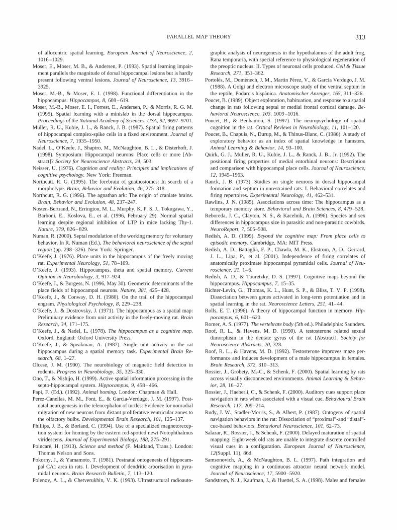
of allocentric spatial learning. European Journal of Neuroscience, 2,1016–1029.
Moser, E., Moser, M. B., & Andersen, P. (1993). Spatial learning impair-ment parallels the magnitude of dorsal hippocampal lesions but is hardlypresent following ventral lesions. Journal of Neuroscience, 13, 3916–3925.
Moser, M.-B., & Moser, E. I. (1998). Functional differentiation in thehippocampus. Hippocampus, 8, 608–619.
Moser, M.-B., Moser, E. I., Forrest, E., Andersen, P., & Morris, R. G. M.(1995). Spatial learning with a minislab in the dorsal hippocampus.Proceedings of the National Academy of Sciences, USA, 92, 9697–9701.
Muller, R. U., Kubie, J. L., & Ranck, J. B. (1987). Spatial firing patternsof hippocampal complex-spike cells in a fixed environment. Journal ofNeuroscience, 7, 1935–1950.
Nadel, L., O’Keefe, J., Shapiro, M., McNaughton, B. L., & Disterhoft, J.(1998). Symposium: Hippocampal neurons: Place cells or more [Ab-stract]? Society for Neuroscience Abstracts, 24, 503.
Neisser, U. (1976). Cognition and reality: Principles and implications ofcognitive psychology. New York: Freeman.
Northcutt, R. G. (1995). The forebrain of gnathostomes: In search of amorphotype. Brain, Behavior and Evolution, 46, 275–318.
Northcutt, R. G. (1996). The agnathan ark: The origin of craniate brains.Brain, Behavior and Evolution, 48, 237–247.
Nosten-Bertrand, N., Errington, M. L., Murphy, K. P. S. J., Tokugawa, Y.,Barboni, E., Koslova, E., et al. (1996, February 29). Normal spatiallearning despite regional inhibition of LTP in mice lacking Thy-1.Nature, 379, 826–829.
Numan, R. (2000). Septal modulation of the working memory for voluntarybehavior. In R. Numan (Ed.), The behavioral neuroscience of the septalregion (pp. 298–326). New York: Springer.
O’Keefe, J. (1976). Place units in the hippocampus of the freely movingrat. Experimental Neurology, 51, 78–109.
O’Keefe, J. (1993). Hippocampus, theta and spatial memory. CurrentOpinion in Neurobiology, 3, 917–924.
O’Keefe, J., & Burgess, N. (1996, May 30). Geometric determinants of theplace fields of hippocampal neurons. Nature, 381, 425–428.
O’Keefe, J., & Conway, D. H. (1980). On the trail of the hippocampalengram. Physiological Psychology, 8, 229–238.
O’Keefe, J., & Dostrovsky, J. (1971). The hippocampus as a spatial map:Preliminary evidence from unit activity in the freely-moving rat. BrainResearch, 34, 171–175.
O’Keefe, J., & Nadel, L. (1978). The hippocampus as a cognitive map.Oxford, England: Oxford University Press.
O’Keefe, J., & Speakman, A. (1987). Single unit activity in the rathippocampus during a spatial memory task. Experimental Brain Re-search, 68, 1–27.
Olcese, J. M. (1990). The neurobiology of magnetic field detection inrodents. Progress in Neurobiology, 35, 325–330.
Ono, T., & Nishijo, H. (1999). Active spatial information processing in thesepto-hippocampal system. Hippocampus, 9, 458–466.
Papi, F. (Ed.). (1992). Animal homing. London: Chapman & Hall.Perez-Canellas, M. M., Font, E., & Garcia-Verdugo, J. M. (1997). Post-
natal neurogenesis in the telencephalon of turtles: Evidence for nonradialmigration of new neurons from distant proliferative ventricular zones tothe olfactory bulbs. Developmental Brain Research, 101, 125–137.
Phillips, J. B., & Borland, C. (1994). Use of a specialized magnetorecep-tion system for homing by the eastern red-spotted newt Notophthalmusviridescens. Journal of Experimental Biology, 188, 275–291.
Poincare, H. (1913). Science and method (F. Maitland, Trans.). London:Thomas Nelson and Sons.
Pokorny, J., & Yamamoto, T. (1981). Postnatal ontogenesis of hippocam-pal CA1 area in rats. I. Development of dendritic arborisation in pyra-midal neurons. Brain Research Bulletin, 7, 113–120.
Polenov, A. L., & Chetverukhin, V. K. (1993). Ultrastructural radioauto-
graphic analysis of neurogenesis in the hypothalamus of the adult frog,Rana temporaria, with special reference to physiological regeneration ofthe preoptic nucleus: II. Types of neuronal cells produced. Cell & TissueResearch, 271, 351–362.
Portoles, M., Domenech, J. M., Martin Perez, V., & Garcia Verdugo, J. M.(1988). A Golgi and electron microscope study of the ventral septum inthe reptile, Podarcis hispanica. Anatomischer Anzeiger, 165, 311–326.
Poucet, B. (1989). Object exploration, habituation, and response to a spatialchange in rats following septal or medial frontal cortical damage. Be-havioral Neuroscience, 103, 1009–1016.
Poucet, B., & Benhamou, S. (1997). The neuropsychology of spatialcognition in the rat. Critical Reviews in Neurobiology, 11, 101–120.
Poucet, B., Chapuis, N., Durup, M., & Thinus-Blanc, C. (1986). A study ofexploratory behavior as an index of spatial knowledge in hamsters.Animal Learning & Behavior, 14, 93–100.
Quirk, G. J., Muller, R. U., Kubie, J. L., & Ranck, J. B., Jr. (1992). Thepositional firing properties of medial entorhinal neurons: Descriptionand comparison with hippocampal place cells. Journal of Neuroscience,12, 1945–1963.
Ranck, J. B. (1973). Studies on single neurons in dorsal hippocampalformation and septum in unrestrained rats: I. Behavioral correlates andfiring repertoires. Experimental Neurology, 41, 462–531.
Rawlins, J. N. (1985). Associations across time: The hippocampus as atemporary memory store. Behavioral and Brain Sciences, 8, 479–528.
Reboreda, J. C., Clayton, N. S., & Kacelnik, A. (1996). Species and sexdifferences in hippocampus size in parasitic and non-parasitic cowbirds.NeuroReport, 7, 505–508.
Redish, A. D. (1999). Beyond the cognitive map: From place cells toepisodic memory. Cambridge, MA: MIT Press.
Redish, A. D., Battaglia, F. P., Chawla, M. K., Ekstrom, A. D., Gerrard,J. L., Lipa, P., et al. (2001). Independence of firing correlates ofanatomically proximate hippocampal pyramidal cells. Journal of Neu-roscience, 21, 1–6.
Redish, A. D., & Touretzky, D. S. (1997). Cognitive maps beyond thehippocampus. Hippocampus, 7, 15–35.
Richter-Levin, G., Thomas, K. L., Hunt, S. P., & Bliss, T. V. P. (1998).Dissociation between genes activated in long-term potentiation and inspatial learning in the rat. Neuroscience Letters, 251, 41–44.
Rolls, E. T. (1996). A theory of hippocampal function in memory. Hip-pocampus, 6, 601–620.
Romer, A. S. (1977). The vertebrate body (5th ed.). Philadelphia: Saunders.Roof, R. L., & Havens, M. D. (1990). A testosterone related sexual
dimorphism in the dentate gyrus of the rat [Abstract]. Society forNeuroscience Abstracts, 20, 328.
Roof, R. L., & Havens, M. D. (1992). Testosterone improves maze per-formance and induces development of a male hippocampus in females.Brain Research, 572, 310–313.
Rossier, J., Grobety, M.-C., & Schenk, F. (2000). Spatial learning by ratsacross visually disconnected environments. Animal Learning & Behav-ior, 28, 16–27.
Rossier, J., Haeberli, C., & Schenk, F. (2000). Auditory cues support placenavigation in rats when associated with a visual cue. Behavioural BrainResearch, 117, 209–214.
Rudy, J. W., Stadler-Morris, S., & Albert, P. (1987). Ontogeny of spatialnavigation behaviors in the rat: Dissociation of “proximal”-and “distal”-cue-based behaviors. Behavioral Neuroscience, 101, 62–73.
Salazar, R., Rossier, J., & Schenk, F. (2000). Delayed maturation of spatialmapping: Eight-week old rats are unable to integrate discrete controlledvisual cues in a configuration. European Journal of Neuroscience,12(Suppl. 11), 86d.
Samsonovich, A., & McNaughton, B. L. (1997). Path integration andcognitive mapping in a continuous attractor neural network model.Journal of Neuroscience, 17, 5900–5920.
Sandstrom, N. J., Kaufman, J., & Huettel, S. A. (1998). Males and females
313PARALLEL MAP THEORY
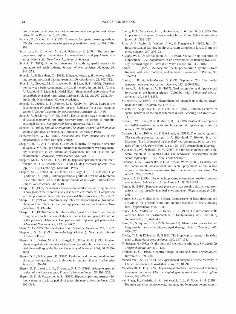
use different distal cues in a virtual environment navigation task. Cog-nitive Brain Research, 6, 351–360.
Saucier, D., & Cain, D. P. (1995, November 9). Spatial learning withoutNMDA receptor-dependent long-term potentiation. Nature, 378, 186–189.
Scharfman, H. E., Witter, M. P., & Schwarcz, R. (2000). The parahip-pocampal region: Implications for neurological and psychiatric dis-eases. New York: New York Academy of Sciences.
Schenk, F. (1989). A homing procedure for studying spatial memory inimmature and adult rodents. Journal of Neuroscience Methods, 26,249–258.
Schenk, F., & Brandner, C. (1995). Enhanced visuspatial memory follow-ing pre and postnatal choline treatment. Psychobiology, 23, 302–312.
Schenk, F., Grobety, M. C., Lavenex, P., & Lipp, H.-P. (1995). Dissocia-tion between basic components of spatial memory in rats. In E. Alleva,A. Fasolo, H.-P. Lipp, & L. Nadel (Eds.), Behavioural brain research innaturalistic and semi-naturalistic settings (Vol. 82, pp. 207–223). Dor-drecht, the Netherlands: Kluwer Academic.
Schenk, F., Jacobs, L. F., Rossier, J., & Kiraly, M. (2001). Steps in thedevelopment of spatial cognition in rats: Evidence for a dual mappingsystem [Abstract]. Society for Neuroscience Abstracts, 27, 1436.
Schenk, F., & Morris, R. G. M. (1985). Dissociation between componentsof spatial memory in rats after recovery from the effects of retrohip-pocampal lesions. Experimental Brain Research, 58, 11–28.
Schone, H. (1984). Spatial orientation: The spatial control of behavior inanimals and man. Princeton, NJ: Princeton University Press.
Schwerdtfeger, W. K. (1984). Structure and fiber connections of thehippocampus. Berlin: Springer-Verlag.
Shapiro, M. L., & O’Connor, C. (1992). N-methyl-D-aspartate receptorantagonist MK-801 and spatial memory representation: Working mem-ory is impaired in an unfamiliar environment but not in a familiarenvironment. Behavioral Neuroscience, 106, 604–612.
Shapiro, M. L., & Olton, D. S. (1994). Hippocampal function and inter-ference. In D. L. Schacter & E. Tulving (Eds.), Memory systems 1994(pp. 87–117). Cambridge, MA: MIT Press.
Shapiro, M. L., Simon, D. K., Olton, D. S., Gage, F. H. D., Nilsson, O., &Bjorklund, A. (1989). Intrahippocampal grafts of fetal basal forebraintissue alter place fields in the hippocampus of rats with fimbria-fornixlesions. Neuroscience, 32, 1–18.
Sharp, P. E. (1997). Subicular cells generate similar spatial firing patternsin two geometrically and visually distinctive environments: Comparisonwith hippocampal place cells. Behavioural Brain Research, 85, 71–92.
Sharp, P. E. (1999a). Complimentary roles for hippocampal versus subic-ular/entorhinal place cells in coding place, context, and events. Hip-pocampus, 9, 432–443.
Sharp, P. E. (1999b). Subicular place cells expand or contract their spatialfiring pattern to fit the size of the environment in an open field but notin the presence of barriers: Comparison with hippocampal place cells.Behavioral Neuroscience, 113, 643–662.
Shatz, C. J. (1992). The developing brain. Scientific American, 267, 61–67.Shepherd, G. M. (1994). Neurobiology (3rd ed.). New York: Oxford
University Press.Sherry, D. F., Forbes, M. R. L., Khurgel, M., & Ivy, G. O. (1993). Greater
hippocampal size in females of the brood parasitic brown-headed cow-bird. Proceedings of the National Academy of Sciences, USA, 90, 7839–7843.
Sherry, D. F., & Hampson, E. (1997). Evolution and the hormonal controlof sexually-dimorphic spatial abilities in humans. Trends in CognitiveScience, 1, 50–56.
Sherry, D. F., Jacobs, L. F., & Gaulin, S. J. C. (1992). Adaptive special-ization of the hippocampus. Trends in Neurosciences, 15, 298–303.
Sherry, D. F., & Vaccarino, A. L. (1989). Hippocampus and memory forfood caches in black-capped chickadees. Behavioral Neuroscience, 103,308–318.
Sherry, D. F., Vaccarino, A. L., Buckenham, K., & Herz, R. S. (1989). Thehippocampal complex of food-storing birds. Brain, Behavior and Evo-lution, 34, 308–317.
Silva, A. J., Paylor, R., Wehner, J. M., & Tonegawa, S. (1992, July 10).Impaired spatial learning in alpha-calcium-calmodulin kinase II mutantmice. Science, 257, 206–211.
Skaggs, W. E., & McNaughton, B. L. (1998). Spatial firing properties ofhippocampal CA1 populations in an environment containing two visu-ally identical regions. Journal of Neuroscience, 18, 8455–8466.
Squire, L. R. (1992). Memory and the hippocampus: A synthesis fromfindings with rats, monkeys, and humans. Psychological Review, 99,195–231.
Squire, L. R., & Zola-Morgan, S. (1991, September 20). The medialtemporal lobe memory system. Science, 253, 1380–1386.
Strasser, R., & Bingman, V. P. (1997). Goal recognition and hippocampalformation in the homing pigeon (Columba livia). Behavioral Neuro-science, 111, 1245–1256.
Striedter, G. F. (1997). The telencephalon of tetrapods in evolution. Brain,Behavior and Evolution, 49, 179–213.
Suzuki, S., Augerinos, G., & Black, A. H. (1980). Stimulus control ofspatial behavior on the eight-arm maze in rats. Learning and Motivation,11, 1–18.
Swann, J. W., Brady, R. J., & Martin, D. L. (1989). Postnatal developmentof GABA-mediated synaptic inhibition in rat hippocampus. Neuro-science, 28, 551–561.
Swanson, L. W., Kohler, C., & Bjorklund, A. (1987). The limbic region. I:The septohippocampal system. In A. Bjorklund, T. Hokfelt, & L. W.Swanson (Eds.), Handbook of chemical neuroanatomy, integrated sys-tems of the CNS, Part I (Vol. 5, pp. 125–136). Amsterdam: Elsevier.
Swanson, L. W., & Risold, P.-Y. (2000). On the basic architecture of theseptal region. In R. Numan (Ed.), The behavioral neuroscience of theseptal region (pp. 1–14). New York: Springer.
Swanson, L. W., Sawchenko, P. E., & Cowan, W. M. (1980). Evidence thatthe commissural, associational and septal projections of the regioninferior of the hippocampus arise from the same neurons. Brain Re-search, 197, 207–212.
Szekely, A. D. (1999). The avian hippocampal formation: Subdivisions andconnectivity. Behavioural Brain Research, 98, 219–225.
Tanila, H. (1999). Hippocampal place cells can develop distinct represen-tations of two visually identical environments. Hippocampus, 9, 235–246.
Taube, J. S., & Muller, R. U. (1998). Comparisons of head direction cellactivity in the postsubiculum and anterior thalamus of freely movingrats. Hippocampus, 8, 87–108.
Taube, J. S., Muller, R. U., & Ranck, J. B. (1990). Head-direction cellsrecorded from the postsubiculum in freely-moving rats. Journal ofNeuroscience, 10, 420–435.
Teng, E., & Squire, L. R. (1999, August 12). Memory for places learnedlong ago is intact after hippocampal damage. Nature (London), 400,675–677.
Teyler, T. J., & DiScenna, P. (1986). The hippocampal memory indexingtheory. Behavioral Neuroscience, 100, 147–154.
Tinbergen, N. (1963). On the aims and methods of ethology. Zeitschrift furTierpsychologie, 20, 410–433.
Tolman, E. C. (1948). Cognitive maps in rats and men. PsychologicalReview, 55, 189–208.
Vander Wall, S. B. (1982). An experimental analysis of cache recovery inClark’s nutcracker. Animal Behaviour, 30, 84–94.
Vanderwolf, C. H. (1969). Hippocampal electrical activity and voluntarymovement in the rat. Electroencephalography and Clinical Neurophys-iology, 26, 407–418.
van Praag, H., Christie, B. R., Sejnowski, T. J., & Gage, F. H. (1999).Running enhances neurogenesis, learning, and long-term potentiation in
314 JACOBS AND SCHENK

mice. Proceedings of the National Academy of Sciences, USA, 96,13427–13431.
van Praag, H., Kempermann, G., & Gage, F. H. (1999). Running increasescell proliferation and neurogenesis in the adult mouse dentate gyrus.Nature Neuroscience, 2, 266–270.
Vargha-Khadem, F., Gadian, D. G., Watkins, K. E., Connelly, A., VanPaesschen, W., & Mishkin, M. (1997, July 18). Differential effects ofearly hippocampal pathology on episodic and semantic memory. Sci-ence, 277, 376–380.
Walker, M. M., Diebel, C. E., Haugh, C. V., Pankhurst, P. M., Montgom-ery, J. C., & Green, C. R. (1997, November 27). Structure and functionof the vertebrate magnetic sense. Nature, 390, 371–376.
Wallraff, H. G. (1996). Seven theses on pigeon homing deduced fromempirical findings. Journal of Experimental Biology, 199, 105–111.
Wan, H., Aggleton, J. P., & Brown, M. W. (1999). Different contributionsof the hippocampus and perirhinal cortex to recognition memory. Jour-nal of Neuroscience, 19, 1142–1148.
Warren, S. G., Humphreys, A. G., Juraska, J. M., & Greenough, W. T.(1995). LTP varies across the estrous cycle: Enhanced synaptic plasticityin proestrus rats. Brain Research, 703, 26–30.
Waters, N. S., Klintsova, A. Y., & Foster, T. C. (1997). Insensitivity of thehippocampus to environmental stimulation during postnatal develop-ment. Journal of Neuroscience, 17, 7967–7973.
Wehner, R., Michel, B., & Antonsen, P. (1996). Visual navigation ininsects: coupling of egocentric and geocentric information. Journal ofExperimental Biology, 199, 129–140.
West, M. J. (1990). Stereological studies of the hippocampus: A compar-ison of the hippocampal subdivisions of diverse species including hedge-hogs, laboratory rodents, wild mice and men. In J. Storm-Mathisen, J.Zimmer, & O. P. Ottersen (Eds.), Progress in brain research (Vol. 83,pp. 13–36). Amsterdam: Elsevier.
West, M. J., & Schwerdtfeger, W. K. (1985). An allometric study ofhippocampal components: A comparative study of the brains of theEuropean hedgehog, the tree shrew, and the marmoset monkey. BrainBehavior and Evolution, 27, 93–105.
Whishaw, I. Q. (2000). The septohippocampal system and path integration.In R. Nunan (Ed.), The behavioral neuroscience of the septal region (pp.270–297). New York: Springer.
Whishaw, I. Q., Cassel, J.-C., & Jarrard, L. E. (1995). Rats with fimbria-fornix lesions display a place response in a swimming pool: A dissoci-ation between getting there and knowing where. Journal of Neuro-science, 15, 5779–5788.
Whishaw, I. Q., & Gorny, B. (1999). Path integration absent in scent-tracking fimbria-fornix rats: Evidence for hippocampal involvement in“sense of direction” and “sense of distance” using self-movement cues.Journal of Neuroscience, 19, 4662–4673.
Whishaw, I. Q., & Jarrard, L. E. (1995). Similarities vs. differences in place
learning and circadian activity in rats after fimbria-fornix section oribotenate removal of hippocampal cells. Hippocampus, 5, 595–604.
Whishaw, I. Q., & Tomie, J. A. (1997). Perseveration on place reversals inspatial swimming pool tasks: Further evidence for place learning inhippocampal rats. Hippocampus, 7, 361–370.
Whishaw, I. Q., & Vanderwolf, C. H. (1973). Hippocampal EEG andbehavior: Changes in amplitude and frequency of RSA (theta rhythm)associated with spontaneous and learned movement patterns in rats andcats. Behavioral Biology, 8, 461–484.
Williams, C. L., Barnett, A. M., & Meck, W. H. (1990). Organizationaleffects of early gonadal secretions on sexual differentiation in spatialmemory. Behavioral Neuroscience, 104, 84–97.
Williams, C. L., & Meck, W. H. (1991). The organizational effects ofgonadal steroids on sexually dimorphic spatial ability. Psychoneuroen-docrinology, 16, 155–176.
Williams, C. L., & Meck, W. H. (1993). Organizational effects of gonadalhormones induce qualitative differences in visuospatial navigation. InM. Haug, R. E. Whalen, C. Aron, & K. L. Olsen (Eds.), The developmentof sex differences and similarities in behavior (pp. 179–185). Dordrecht,the Netherlands: Kluwer Academic.
Wilson, M. A., & McNaughton, B. L. (1993, August 20). Dynamics of thehippocampal ensemble code for space. Science, 261, 1055–1058.
Wiltschko, W., & Wiltschko, R. (1996). Magnetic orientation in birds.Journal of Experimental Biology, 199, 29–38.
Wimer, R. E., & Wimer, C. (1985). Three sex dimorphisms in the granulecell layer of the hippocampus in house mice. Brain Research, 328,105–109.
Witter, M. P., Griffioen, A. W., Jorritsma-Byham, B., & Krijnen, J. L. M.(1988). Entorhinal projections to the hippocampal CA1 region in the rat:An underestimated pathway. Neuroscience Letters, 85, 193–198.
Witter, M. P., Wouterlood, F. G., Naber, P. A., & van Haeften, T. (2000).Anatomical organization of the parahippocampal-hippocampal network.In H. E. Scharfman, M. Witter, & R. Schwarcz (Eds.), The parahip-pocampal region: Implications for neurological and psychiatric diseases(Vol. 911, pp. 1–24). New York: New York Academy of Sciences.
Wood, E. R., Dudchenko, P. A., & Eichenbaum, H. (1997). Nonspatialcorrelates of hippocampal cellular activity in rats performing an olfac-tory continuous nonmatching to sample task on an open field [Abstract].Society for Neuroscience Abstracts, 23, 502.
Worden, R. (1992). Navigation by fragment fitting: A theory of hippocam-pal function. Hippocampus, 2, 165–188.
Xu, L., Anwyl, R., & Rowan, M. J. (1998, August 27). Spatial explorationinduces a persistent reversal of long-term potentiation in rat hippocam-pus. Nature, 394, 891–894.
Received February 20, 2001Revision received March 14, 2002
Accepted April 7, 2002 �
315PARALLEL MAP THEORY