University of Groningen Single-tooth implants in the ... · customized restoration of a...
13
University of Groningen Single-tooth implants in the aesthetic zone den Hartog, Laurens IMPORTANT NOTE: You are advised to consult the publisher's version (publisher's PDF) if you wish to cite from it. Please check the document version below. Document Version Publisher's PDF, also known as Version of record Publication date: 2011 Link to publication in University of Groningen/UMCG research database Citation for published version (APA): Hartog, L. D. (2011). Single-tooth implants in the aesthetic zone: a clinical trial of different implant neck designs and immediate loading Groningen: s.n. Copyright Other than for strictly personal use, it is not permitted to download or to forward/distribute the text or part of it without the consent of the author(s) and/or copyright holder(s), unless the work is under an open content license (like Creative Commons). Take-down policy If you believe that this document breaches copyright please contact us providing details, and we will remove access to the work immediately and investigate your claim. Downloaded from the University of Groningen/UMCG research database (Pure): http://www.rug.nl/research/portal. For technical reasons the number of authors shown on this cover page is limited to 10 maximum. Download date: 14-07-2018
Transcript of University of Groningen Single-tooth implants in the ... · customized restoration of a...

University of Groningen
Single-tooth implants in the aesthetic zoneden Hartog, Laurens
IMPORTANT NOTE: You are advised to consult the publisher's version (publisher's PDF) if you wish to cite fromit. Please check the document version below.
Document VersionPublisher's PDF, also known as Version of record
Publication date:2011
Link to publication in University of Groningen/UMCG research database
Citation for published version (APA):Hartog, L. D. (2011). Single-tooth implants in the aesthetic zone: a clinical trial of different implant neckdesigns and immediate loading Groningen: s.n.
CopyrightOther than for strictly personal use, it is not permitted to download or to forward/distribute the text or part of it without the consent of theauthor(s) and/or copyright holder(s), unless the work is under an open content license (like Creative Commons).
Take-down policyIf you believe that this document breaches copyright please contact us providing details, and we will remove access to the work immediatelyand investigate your claim.
Downloaded from the University of Groningen/UMCG research database (Pure): http://www.rug.nl/research/portal. For technical reasons thenumber of authors shown on this cover page is limited to 10 maximum.
Download date: 14-07-2018

This is an edited version of the manuscript: Den Hartog, L., Raghoebar, G.M., Stellingsma, K.,
Meijer, H.J. Immediate loading and customized restoration of a single implant in the maxillary
esthetic zone: a clinical report. Journal of Prosthetic Dentistry 2009; 102: 211-215
6.
Immediate loading and customized restoration
of a single-tooth implant in the maxillary aesthetic
zone: a clinical report

Cha
pter
6
102
AbstrAct
The replacement of a single missing anterior tooth with a dental implant is
a demanding therapy. This report describes a treatment in which an ante-
rior maxillary implant was immediately restored with a provisional restora-
tion. During the provisional phase, an optimal emergence profile was cre-
ated by adjusting the provisional restoration. An impression was made with
an individually fabricated impression post for an accurate reproduction of
the established emergence profile and finally a screw-retained all-ceramic
crown was placed. By implementing this protocol, an optimal definitive re-
sult could be achieved together with immediate patient satisfaction. How-
ever, cooperation between several disciplines and careful patient selection
were required.

Clin
ical
rep
ort
6
103
IntroductIon
The application of dental implant to restore a missing tooth in the anterior den-
tition is challenging. In the aesthetic zone, both the appearance of the implant
crown and the soft tissue contribute to a successful treatment outcome and
should consequently be in harmony with the surrounding dentition (Chang et al.
1999, Belser et al. 2004). To accomplish the desired result, meticulous pre-op-
erative treatment planning is crucial together with cooperation between several
disciplines during the restorative phase.
In recent literature, several treatment strategies using dental implants have
been reported to replace a missing tooth (Den Hartog et al. 2008). Among these,
interest has been attributed to immediate or early loading protocols in which a
provisional restoration is placed soon after implant placement. Clinical studies of
immediate or early loading have reported favorable treatment outcomes in terms
of implant survival, marginal bone resorption, soft tissue level and the incidence
of complications for treatment in which implants were inserted in healed sites
(Ericsson et al. 2000, Andersen et al. 2002, Cooper et al. 2007, Hall et al. 2007) as
well as implants placed in fresh extraction sockets (Crespi et al. 2008, de Rouck et
al. 2008). However, a recent systematic review (Den Hartog et al. 2008) showed
that well-designed controlled studies that compare these approaches with con-
ventional protocols are scarce and whether or not superior aesthetic outcomes
could be achieved remained inconclusive. Immediate or early implant loading
provides several advantages for the patient including a shorter overall treatment
time, avoidance of a second-stage operation and eliminating the need for a re-
movable prosthesis during the healing phase. However, these protocols require
careful pre-operative planning and patient selection. Furthermore, good primary
implant stability is a prerequisite (Esposito et al. 2007), in addition to the develop-
ment of a protected occlusion to create a non-occluding provisional crown.
Another important development has been the introduction of alumina- and
zirconia-based ceramic abutments made with computer-aided design/compu-
ter-aided manufacturing (CAD/CAM) technology. Ceramic abutments are high-
strength (Sundh & Sjögren 2008, Yüzügüllü & Avci 2008,) biocompatible (We-
lander et al. 2008) and have allowed new options to improve the natural appear-
ance of the implant crown. However, little is known about the long-term clinical
performance of these abutments (Linkevicius & Apse 2008).
The purpose of this clinical report was to demonstrate an immediate implant
loading protocol for restoration of a missing central incisor. After the provisional
restoration phase, an individually fabricated impression post was used and sub-
sequently a definitive screw-retained one piece all-ceramic crown was placed.

Cha
pter
6
104
Figure 1. Pre-operative view.
Figure 2. Exposure of the alveolar bone.
Figure 3. Screw-retained provisional crown placed same day after implant surgery.

Clin
ical
rep
ort
6
105
Clinical reportA 47-year-old woman consulted the Department of Oral and Maxillofacial Surgery
(University Medical Center Groningen, University of Groningen, Groningen, the
Netherlands) with a missing right central incisor (Figure 1) lost due to a trau-
matic injury. At the time of the consultation she was wearing a removable partial
denture and desired a durable and fixed restoration, without involvement of the
adjacent teeth. The patient was healthy, did not smoke and intraoral examination
revealed a healthy well-maintained dentition. Clinically, adequate bone volume
was present at the future implant site. In all dimensions, sufficient space was
available for an implant crown with an anatomical design. Radiographically, no
pathology of the bone and adjacent teeth was noted. Because of the favorable
starting point, it was decided to use an immediate loading protocol.
Preoperatively, diagnostic casts were made with a diagnostic arrangement
representing the future implant crown in an ideal position. Next, a transparent
acrylic resin template (Vertex Castapress; Vertex Dental, Zeist, the Netherlands)
was fabricated and a guide channel was prepared in the template to aid in proper
implant placement. Care was taken with the surgical guide so that the guidance
channel would direct the implant sufficiently toward the palate to accommodate
a screw-retained restoration.
One day before surgery, the patient started taking antibiotics (amoxicillin 500
mg, 3 times daily for seven days) and used a 0.2% chlorhexidine mouthwash
(Corsodyl; GlaxoSmithKline, Utrecht, the Netherlands) for oral disinfection. Fol-
lowing local anaesthesia (Ultracaine D-S Forte; Aventis Pharma, Hoevelaken, the
Netherlands) a slightly palatal crest-incision was made with extensions through
the buccal and palatal sulcus of the adjacent teeth. A minimal mucoperiosteal
flap was elevated to expose only the ridge crest (Figure 2). Then, an implant (No-
belReplace Tapered RP 16 mm, Nobel Biocare, Gothenburg, Sweden) was placed
according to the procedure prescribed by the manufacturer guided by the surgical
template. The shoulder of the implant was placed at a depth of 3 mm apical to the
buccal and cervical aspect of the prospective clinical crown to provide soft tissue
to develop an adequate emergence profile. Good primary implant stability was
obtained (> 45 Ncm, determined with Osseocare; Nobel Biocare). Next, an open-
tray impression was made at the implant level using a custom resin impression
tray (Lightplast base plates; Dreve Dentamid, Unna, Germany) and a polyether
impression material (Impregum Penta; 3M ESPE, St. Paul, Minnesota, USA.).
Finally, a healing abutment (NobelReplace; Nobel Biocare) was placed and the
wound was closed with sutures (Ethilon 5-0; Johnson & Johnson Gateway, Pis-
cataway, New Jersey, USA).
In the dental laboratory, a screw-retained provisional restoration was fabricated
consisting of an engaging temporary abutment (Temporary Abutment Engaging

Cha
pter
6
106
Figure 5. Assembly of provisional crown with implant analog and impres-sion of cervical portion of provisional crown.
Figure 4. View of peri-implant soft tissue after the provisional restoration phase. Note established emergence profile.
Figure 6. Customized impression post.

Clin
ical
rep
ort
6
107
NobelReplace; Nobel Biocare) against which composite resin (Solidex; Shofu Inc,
Kyoto, Japan) was modelled. Eight hours post-implant placement, the abutment
was removed and the provisional crown was placed and subsequently torqued to
32 Ncm (Figure 3). Special care was taken to prevent any centric and eccentric
occlusal contacts with the antagonist teeth. Furthermore, the provisional restora-
tion was contoured so that the peri-implant soft tissue was optimally supported.
In particular, the interproximal papillae were given enough space to regenerate.
The patient was instructed to follow a soft diet, to avoid exerting force on the pro-
visional restoration and to continue chlorhexidine rinses (Corsodyl; GlaxoSmith-
Kline, Utrecht, The Netherlands) for seven days. For pain control, ibuprofen 600
mg (Brufen Bruis 600; Abott B.V., Hoofddorp, The Netherlands, three times daily
for time needed) was prescribed and at two weeks post surgery the sutures were
removed.
The patient returned to the prosthodontist once a month for three months for
examination of the implant. During these sessions, implant mobility, oral hygiene
and occlusion were evaluated. Also, an important objective was the creation of an
ideal emergence profile by removing the provisional crown. Extraorally and where
needed, composite resin was removed or added to aspects of the crown to create
more space or more support for the soft tissue. In this process, special attention
was given to the shape of the proximal contour to provide an optimal condition
for the papillae to reach maturity.
Three months later (six months post-implant placement) an implant level im-
pression was made using an impression post (Impression Coping Implant Level
Open Tray NobelReplace; Nobel Biocare) that was customized in a way that the
obtained emergence profile could be transferred to the definitive restoration (Fig-
ure 4). To realize this, the provisional crown was assembled with an implant ana-
log (Implant Replica NobelReplace; Nobel Biocare) embedded in type IV dental
stone (GC Fuji Rock EP; GC Europe N.V., Leuven, Belgium). An addition silicone
impression (Futar D; Kettenbach GmbH & Co KG, Eschenburg, Germany) of the
cervical portion of the crown was made (Figure 5). Next, the latter was substituted
for an impression post and bis acrylic composite resin (Protemp; 3M ESPE, St.
Paul, Minn) was added to the post. After polishing the individualized post (Figure
6) it was inserted into the implant and an open-tray impression was made with a
polyether impression material (Impregum Penta; 3M ESPE) and a custom resin
impression tray (Lightplast base plates; Dreve Dentamid).
In the dental laboratory, a soft tissue cast was prepared. First, a waxing of
the definitive crown was made on a temporary abutment (Temporary Abutment
Engaging NobelReplace; Nobel Biocare). The screw access hole was located suf-
ficiently to the palate to create a screw-retained crown and to prepare an appropri-
ate abutment. Therefore, the waxing was cut back to the desired form and scanned
Cha
pter
6
108
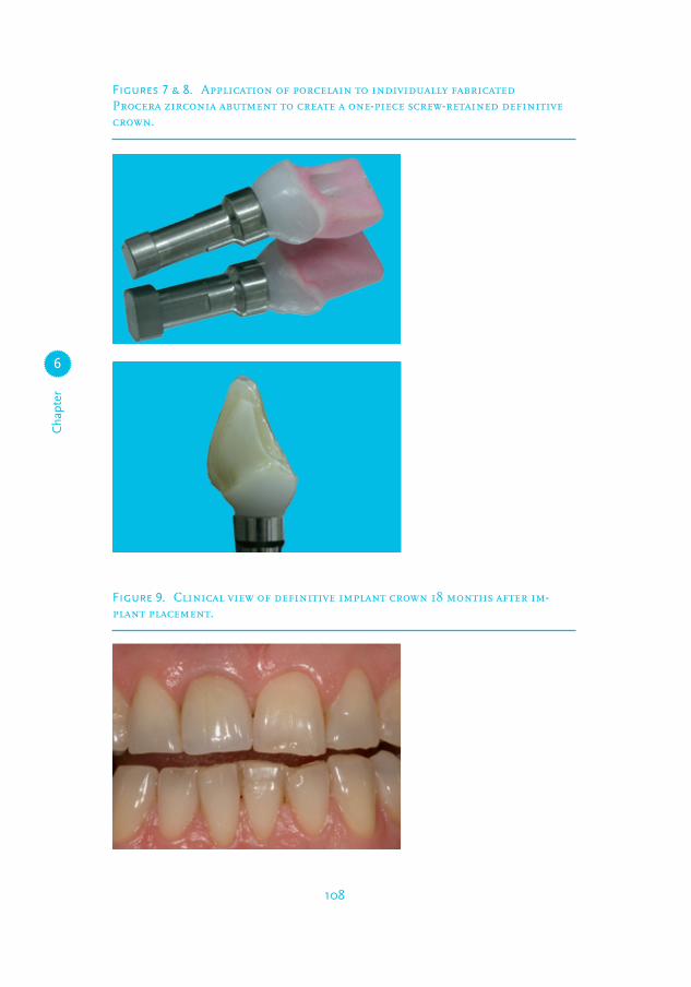
Figures 7 & 8. Application of porcelain to individually fabricated Procera zirconia abutment to create a one-piece screw-retained definitive crown.
Figure 9. Clinical view of definitive implant crown 18 months after im-plant placement.

Clin
ical
rep
ort
6
109
for fabrication of an individual zirconia abutment (Procera; Nobel Biocare AB).
Porcelain was added directly to the abutment to create a screw-retained one piece
definitive restoration (Figures 7 & 8). The restoration was placed and the abut-
ment screw was torqued with 32 Ncm. Finally, the screw hole was filled with a
cotton pellet and composite resin (Clearfil AP-X; Kuraray Medical Inc, Okayama,
Japan). The restoration has been in service for 18 months without complication
(Figure 9).
dIscussIon
This report describes an immediate loading protocol finalized with the placement
of a screw-retained all-ceramic restoration. A major prerequisite for immediate
loading is a high degree of primary stability in terms of high insertion torque
(Esposito et al. 2007). In this treatment, an initial insertion torque of at least 45
Ncm was reached. Although clinical studies on immediate single-tooth implant
loading reported varying minimal insertion torques for immediate loading, the
authors of this report adopted a threshold of 45 Ncm.
In this patient, a substantial maturation of the papillae occurred during the
provisional phase. Care was taken to ensure that the provisional crown did not
disturb this process, but served as a natural guide. Regeneration of papillae with
time has been reported in several studies (Jemt & Lekholm 2005, Schropp et
al. 2005), but the mechanism behind this phenomenon could not validly be ex-
plained. Some authors believed that this increase might be attributed to remod-
elling potential of the soft tissue to establish a proper biological height after the
surgical manipulation (Chang et al. 1999). It is widely accepted however, that the
interproximal bone level next to the adjacent teeth is important for the future level
of the interproximal papillae of the implant (Belser et al. 2004).
Finally, a screw-retained definitive restoration was fabricated. The advantages
of this type of restoration compared to a cement-retained restoration include
retrievability and no risks for cement remnants thereby excluding possible irrita-
tion of the peri-implant tissues. However, the presence of a screw access opening
decreases fracture resistance of the porcelain (Torrado et al. 2004). Furthermore,
screw-retained restorations necessitate precise implant positioning for a proper
palatal position of the screw access hole that does not interfere with the aesthet-
ics.

Cha
pter
6
110
References
Andersen, E., Haanaes, H.R. & Knutsen, B.M. (2002) Im-
mediate loading of single-tooth ITI implants in the anterior
maxilla: a prospective 5-year pilot study. Clinical Oral
Implants Research 13, 281-287.
Belser, U.C., Schmid, B., Higginbottom, F. & Buser, D.
(2004) Outcome analysis of implant restorations located
in the anterior maxilla: a review of the recent literature.
International Journal of Oral and Maxillofacial Implants 19
Suppl, 30-42.
Chang, M., Wennstrom, J.L., Odman, P. & Andersson, B.
(1999) Implant supported single-tooth replacements com-
pared to contralateral natural teeth. Crown and soft tissue
dimensions. Clinical Oral Implants Research 10, 185-194.
Cooper, L.F., Ellner, S., Moriarty, J., Felton, D.A., Paquette
D. & Molina, A. (2007) Three-year evaluation of single-tooth
implants restored 3 weeks after 1-stage surgery. International
Journal of Oral and Maxillofacial Implants 22, 791-800.
Crespi, R., Capparé, P., Gherlone, E. & Romanos, G.E.
(2008) Immediate versus delayed loading of dental implants
placed in fresh extraction sockets in the maxillary esthetic
zone: a clinical comparative study. International Journal of
Oral and Maxillofacial Implants 23, 753-758.
De Rouck, T., Collys, K. & Cosyn, J. (2008) Immediate
single-tooth implants in the anterior maxilla: a 1-year case
cohort study on hard and soft tissue response. Journal of
Clinical Periodontology 35, 649-657.
Den Hartog, L., Slater, J.J., Vissink, A., Meijer, H.J. &
Raghoebar, G.M. (2008) Treatment outcome of immediate,
early and conventional single-tooth implants in the aesthetic
zone: a systematic review to survival, bone level, soft tissue,
aesthetics and patient satisfaction. Journal of Clinical Peri-
odontology 35, 1073-1086.
Esposito, M., Grusovin, M.G., Willings, M., Coulthard, P.
& Worthington, H.V. (2007) Interventions for replacing
missing teeth: different times for loading dental implants.
Cochrane Database of Systematic Reviews 18, CD003878.
Ericsson, I., Nilson, H., Lindh, T., Nilner, K. & Randow, K
(2000). Immediate functional loading of Branemark single
tooth implants. An 18 months’ clinical pilot follow-up study.
Clinical Oral Implants Research 11, 26-33.
Hall, J.A., Payne, A.G., Purton, D.G., Torr, B., Duncan, W.J.
& De Silva, R.K. (2007) Immediately restored, single-tapered
implants in the anterior maxilla: prosthodontic and aesthetic
outcomes after 1 year. Clinical Implant Dentistry and Related
Research 9, 34-45.
Jemt, T. & Lekholm, U. (2005) Single implants and buccal
bone grafts in the anterior maxilla: measurements of buccal
crestal contours in a 6-year prospective clinical study. Clini-
cal Implant Dentistry and Related Research 7, 127-135.
Kan, J.Y., Rungcharassaeng, K. & Lozada, J. (2003) Immedi-
ate placement and provisionalization of maxillary anterior
single implants: 1-year prospective study. International
Journal of Oral and Maxillofacial Implants 18, 31-39.
Linkevicius, T. & Apse P. (2008) Influence of abutment
material on stability of peri-implant tissues: a systematic re-
view. International Journal of Oral and Maxillofacial Implants
23, 449-456
Meijndert, L., Raghoebar, G.M., Meijer H.J. & Vissink, A.
(2008) Clinical and radiographic characteristics of single-
tooth replacements preceded by local ridge augmentation: a
prospective randomized clinical trial. Clinical Oral Implants
Research 19, 1295-1303.
Palmer, R.M., Palmer, P.J. & Smith, B.J. (2000) A 5-year
prospective study of Astra single tooth implants. Clinical
Oral Implants Research 11, 179-182.
Palattella, P., Torsello, F. & Cordaro, L. (2008) Two-year
prospective clinical comparison of immediate replacement
vs. immediate restoration of single tooth in the esthetic
zone. Clinical Oral Implants Research 19, 1148-1153.
Schropp, L., Isidor, F., Kostopoulos, L. & Wenzel, A. (2005)
Interproximal papilla levels following early versus delayed
placement of single-tooth implants: a controlled clinical
trial. International Journal of Oral and Maxillofacial Implants
20, 753-761.
Sundh, A. & Sjögren G. (2008) A study of the bending
resistance of implant-supported reinforced alumina and
machined zirconia abutments and copies. Dental Materials
24, 611-617.
Torrado, E., Ercoli, C., Al Mardini, M., Graser, G.N., Tal-
lents, R.H. & Cordaro L. (2004) A comparison of the
porcelain fracture resistance of screw-retained and cement-
retained implant-supported metal-ceramic crowns. Journal of
Prosthetic Dentistry 91, 532-537.
Welander, M., Abrahamsson, I. & Berglundh T. (2008) The
mucosal barrier at implant abutments of different materials.
Clinical Oral Implants Research 19, 635-641.
Yüzügüllü, B. & Avci, M. (2008) The implant-abutment in-
terface of alumina and zirconia abutments. Clinical Implant
Dentistry and Related Research 10, 113-121.

Clin
ical
rep
ort
6
111