ESTHETIC CRITERIA ANALYSIS OF SINGLE … CRITERIA ANALYSIS OF SINGLE-TOOTH IMPLANT-SUPPORTED...
119
ESTHETIC CRITERIA ANALYSIS OF SINGLE-TOOTH IMPLANT-SUPPORTED RESTORATIONS IN THE ANTERIOR MAXILLA by ANTHONY J REGANATO MIA GEISINGER, COMMITTEE CHAIR MICHAEL REDDY NICOLAAS GEURS PHILIP VASSILOPOULOS DANIEL GIVAN A THESIS Submitted to the graduate faculty of The University of Alabama at Birmingham, in partial fulfillment of the requirements for the degree of Master of Science BIRMINGHAM, ALABAMA 2010
Transcript of ESTHETIC CRITERIA ANALYSIS OF SINGLE … CRITERIA ANALYSIS OF SINGLE-TOOTH IMPLANT-SUPPORTED...

ESTHETIC CRITERIA ANALYSIS OF SINGLE-TOOTH IMPLANT-SUPPORTED
RESTORATIONS IN THE ANTERIOR MAXILLA
by
ANTHONY J REGANATO
MIA GEISINGER, COMMITTEE CHAIR
MICHAEL REDDY
NICOLAAS GEURS
PHILIP VASSILOPOULOS
DANIEL GIVAN
A THESIS
Submitted to the graduate faculty of The University of Alabama at Birmingham,
in partial fulfillment of the requirements for the degree of
Master of Science
BIRMINGHAM, ALABAMA
2010

Copyright by
ANTHONY J REGANATO
2010

iii
ESTHETIC CRITERIA ANALYSIS OF SINGLE-TOOTH IMPLANT-SUPPORTED
RESTORATIONS IN THE ANTERIOR MAXILLA
ANTHONY J REGANATO
MASTERS OF CLINICAL DENTISTRY
ABSTRACT
Background: Although criteria concerning the functional assessment of implants are
prevalently employed for the determination of implant success, the evolution of esthetic
indices for objective evaluation of soft-tissue esthetics and the final prosthesis can be
useful for determining overall success. As patient demands for esthetic implant
restorations increase, treatment planning with a clear view of esthetic outcomes is
imperative to achieve overall success for our implant patients.
Objective: To compare implant restorations using Albrekttson’s implant success criteria
and a global esthetic assessment consensus by an expert panel. Individual implant and/or
implant restoration characteristics that are preferentially correlated with esthetic success
were examined and esthetic criteria recommendations were developed based on data
points obtained in this study
Materials and Methods: Patients who received single-tooth implant fixture placement
#6-11 at the University of Alabama at Birmingham School of Dentistry and who had final
restorations in function for > 1year were identified using a search of the computerized
database. Periodontal charting as well as radiographic, photographic, and study cast
analyses were implemented in this study for data collection.
Results: After statistical analysis was performed, approximately 75% (32 out of the 43
implants) were deemed to be an esthetic failure by 3 or more examiners on the expert
panel. Of the 6.98% (3 of the 43 implants) of the implants that failed to meet
iv
Albrektsson’s criteria for implant success, all were judged to be esthetically unsuccessful
by 3 or more examiners on the expert panel.
Conclusions: 1) The association between function and esthetic was not statistically
significant in this investigation; 2) There appears to be no association between single-
tooth implant-supported restorations in the anterior maxilla that meet Albrekttson’s
implant success criteria and those restorations that meet esthetic success standards as
determined by a global esthetic assessment; 3) Implant restorations that fulfill functional
success parameters do not necessarily translate into the achievement of esthetic success;
4) Further studies are needed in order to amend existing success criteria commonly used
for implant success standards.
Keywords: dental implants, esthetic success, functional success, esthetic criteria

v
DEDICATION
I would like to dedicate this thesis to my family and closest friends who stood by me and
supported me throughout this endeavor.
vi
ACKNOWLEDGEMENTS
I would like to acknowledge my mentor, Dr. Geisinger, for her significant help and
guidance with this project.
I would also like to thank my master’s committee AKA expert panel of Dr. Reddy, Dr.
Geurs, Dr. Vass, and Dr. Givan for their patience and participation in the global
assessment portion of this investigation.
I would like to acknowledge Sandra McNeal and Mark Litaker for working with us and
performing the statistical analysis on this project.
I would like to thank Kelly, Liz and Sandra Haigh for their administrative efforts.
Furthermore, I would like to thank everyone who believed in this project and those of
you who have derived further investigations based upon this master study

vii
TABLE OF CONTENTS
Page
ABSTRACT ........................................................................................................................... iii
DEDICATION ......................................................................................................................... v
ACKNOWLEDGMENTS ...................................................................................................... vi
LIST OF TABLES ................................................................................................................... x
CHAPTER
1 INTRODUCTION ................................................................................................................ 1
Implant Success .......................................................................................................... 1
General Treatment Planning Concepts in the Anterior Maxilla .................................. 2
Prerequisites for Implant Therapy................................................................................ 4
General Diagnostic Smile Analysis for the Anterior Maxilla ..................................... 5
General Surgical Factors ............................................................................................. 8
Surgical Esthetic Factors in the Anterior Maxilla ..................................................... 11
Other Treatment Considerations ............................................................................... 18
Esthetic Factors ......................................................................................................... 19
Existing Esthetic Criteria and Associated Studies ................................................... 19
2 PURPOSE OF THE STUDY ............................................................................................. 29
Study Goals ............................................................................................................... 29
Null Hypotheses ........................................................................................................ 30

viii
Specific Aims …...................................................................................................... 30
Objectives ............................................................................................................... 31
3 MATERIALS & METHODS .......................................................................................... 33
Absolute Criteria ..................................................................................................... 38
4 RESULTS ........................................................................................................................ 46
5 DISCUSSION .................................................................................................................. 57
Study Demographics ............................................................................................... 57
Functional Success and Failure .............................................................................. 58
Papilla Fill ............................................................................................................... 59
Soft Tissue Height Discrepancy .............................................................................. 61
Hard/Soft Tissue Deficiency ................................................................................... 64
Adequacy of Soft Tissue Thickness ........................................................................ 65
Soft Tissue Color/Texture Deviation ...................................................................... 66
Trans-Gingival Discoloration ................................................................................. 67
Recession ................................................................................................................ 69
Bleeding Upon Probing .......................................................................................... 70
Periodontal Pocket Depth ....................................................................................... 71
Incisal Edge Discrepancy ....................................................................................... 73
Crown Contour/Color Irregularity ......................................................................... 74
Patient Satisfaction ................................................................................................. 74
Global Assessment Discussion ............................................................................... 76
Esthetic Success and Failure .................................................................................. 77
Failure due to Soft Tissue Component, Prosthetic Component, or Both ............... 78

ix
Inter-Examiner Agreement ....................................................................................... 80
Esthetic Success and Failure by 3 or More Experts .................................................. 81
Soft Tissue & Prosthetic Esthetic Success Characteristics Compared with Overall
Esthetic Success ........................................................................................................ 81
6 CONCLUSION .................................................................................................................. 86
LIST OF REFERENCES ...................................................................................................... 89
APPENDIX
A IRB FORM ........................................................................................................... 97
B ABSOLUTE CRITERIA EXAMPLES ................................................................ 99
x
LIST OF TABLES
Table Page
1 Study Demographics – Patient Based ........................................................................... 47
2 Study Demographics – Implant Based .......................................................................... 48
3 Individual Variable Assessment ................................................................................... 53
4 Soft Tissue & Prosthetic Esthetic Success Characteristics Compared with Overall
Esthetic Success ............................................................................................................ 55

1
CHAPTER 1
INTRODUCTION
With the advancements that have occurred in implant design to improve
osseointegration and functional success, esthetics has become a critical goal for both
patients and practitioners in implant dentistry. When dental implants were first
introduced, the importance of implant esthetics was discounted in favor of concepts such
as function, structure, and biology.1 The most widely used implant success criteria,
proposed by Albrekttson and colleagues in 1986, focuses on clinical and radiographic
measures of functional implant success, such as the presence of mobility, periapical
radiolucency, crestal bone loss, patient reported discomfort, and/or the presence of
pathology/disease.2 However, as patient demands for esthetic implant restorations
increase, treatment planning with a clear view of esthetic outcomes is imperative to
achieve overall success for our implant patients.
Implant Success
The application of dental implants for single-tooth replacements has evolved into
a viable prosthodontic alternative to conventional fixed bridgework, resin bonded
restorations or removable partial dentures.3 Most studies use current success criteria for

2
the evaluation of dental implants.2,4,5
Most commonly, Albrektsson’s success criteria are
used to define implant success. This definition includes the following factors: 1) absence
of persistent signs/symptoms such as pain, infection, neuropathies, parathesias, and
violation of vital structures; 2) implant immobility; 3) no continuous peri-implant
radiolucency; 4) negligible progressive bone loss (less than 0.2mm annually) after
physiologic remodeling during the first year of function; and 5) patient/dentist
satisfaction with the implant-supported restoration.2,4,5
In general, multi-year studies of
implants in partially edentulous patients have commonly reported greater than 90%
success rates for both maxillary and mandibular implants.6-12
With regard to single-tooth
restorations on dental implants, literature studies have shown excellent survival rates,
varying from 96.1% to 98.9% after 7.5 years in function (Creugers et al. 2000).13
Numerous studies14-18
have also reported similar implant survival and success rates for
implants inserted in the esthetic zone compared to those placed in other segments of the
jaws.19
With the predictability of osseointegration and long-term success of implant
fixtures using different types of restorations and in various locations, the focus of implant
dentistry has shifted from survival of the implant/restorations and improvement in
function, to more esthetic concerns.20
General Treatment Planning Concepts in the Anterior Maxilla
Esthetics is often a prime goal of the clinician and patient when replacing missing
teeth in the anterior maxilla. The anterior maxillary teeth in the ―esthetic zone‖ usually
extend from first premolar to first premolar, but in some individuals can extend as far

3
distally as the first molar dependent upon individual patient factors.21
Articles focusing
on gingival esthetics have, in general, limited their investigations to the six maxillary
anterior teeth spanning from canine to canine.22
A challenge in assessing esthetics for
such investigations is the reality that the judgment of esthetics is subjective and that
esthetic norms vary between cultures and groups. This makes an absolute evaluation of
esthetics difficult to perform in a scientific way.23
An understanding of the patient’s
needs and desires as well as a correct balance between demands is important to establish
at the beginning of treatment planning and discussion toward the potential end result.
To be a viable treatment alternative for tooth replacement in the esthetic zone,
implant-supported restorations should be judged equally or more esthetic than traditional
crown and bridge restorations.20
Previous research indicates that the condition of the
peri-implant soft tissues is a critical determinant of esthetic success, particularly in the
anterior esthetic zone.24-26
Therefore, success of single-implant therapy in the anterior
maxilla is not only determined by high survival rates, but also influenced by the esthetic
perceptions of the patient and by the quality and stability of the peri-implant tissues.27
An
ideal prosthesis should fully recapitulate or enhance the esthetic features of the tooth or
teeth it replaces.28
Preferably, the appearance of the peri-implant soft tissue should be in
harmony with the mucosa around the adjacent teeth and the implant crown should be in
balance with the neighboring dentition (Meijer et al. 2005).27
The ability to achieve an indistinguishable restoration is a primary goal in the
replacement of any tooth, particularly in the maxillary anterior region.29
Achieving this

4
objective requires a restoration that mimics its natural counterpart in shape, contour,
texture, and color as well as gingival tissues that allow for a natural color, contour, and
emergence profile.30
One problem often encountered is that dental implants themselves
do not mimic the radicular cross-sections of teeth and are not oriented in the alveolar
bone in the same way.30
Therefore, to create an illusion of normal form, a significant
change in gingival contours must be developed by a restoration that reproduces the
appearance of a natural tooth on a foundation that rarely mimics the shape or location of
the natural roots.30
Ultimately, the management of the associated gingival tissues may
dictate the final esthetic outcome and a compromise in esthetics may be likely when this
consideration becomes subservient to the other treatment planning factors.30
Prerequisites for Implant Therapy
Patient screening is one of the fundamental steps in the treatment planning
sequence and correlates to the success of implant therapy. It is essential that a candidate
for implants be evaluated for potential contraindications to their placement.4 At present,
there are no reports of absolute medical contraindications for placement of implants, but
relative contraindications do exist.31
Adverse effects on implant survival have been
attributed to uncontrolled diabetes, alcoholism, heavy smoking, post-irradiated jaws, and
poor oral hygiene.32-35
However, individuals with a strong susceptibility to periodontitis
may be treated successfully with implants.36

5
Restorative requirements, interarch space and jaw relationships, location of
edentulous areas, and the quantity and quality of available bone should be evaluated
before implants are selected as a treatment option. Radiographs, including panoramic,
lateral, and occlusal views and periapical films, may be necessary to determine the height
of available bone and for selection of the dimensions of the implants.4 They also may be
needed to determine the proximity of potentially complicating structures including the
maxillary sinuses, foramina, mandibular canal, and adjacent teeth or roots.4 The use of 3-
dimensional computerized tomography (CT) scans might be advocated when more
accurate information regarding the topography of osseous structures is required.37,38
General Diagnostic Smile Analysis for the Anterior Maxilla
When beginning with esthetics, the treatment planning process must begin with
an appraisal of the position of the maxillary central incisors relative to the upper lip.1 In
the average smile, the lip is positioned to show 75 to 100 percent of the maxillary central
incisor and the interproximal gingiva.39
A high smile line reveals the total cervical-incisal
length of the maxillary anterior teeth and a contiguous band of gingiva.39
A low smile
line displays less than 75 percent of the anterior teeth.40
Previous studies have shown
that with advancing age, the amount of incisal display decreases proportionally.41,42
For
example, in a 30-year-old, 3 millimeters of incisal display at rest is appropriate.
However, in a 60-year-old, the incisal display could be 1 mm or less. The change in
incisal display with time probably relates to the resiliency and tone of the upper lip,
which tends to decrease with advancing age.1

6
Midline deviation and mal-alignment of the maxillary central incisors should be
evaluated during the diagnostic evaluation of the patient. If the midline is deviated to the
right or left, studies have shown that midline deviations of up to 3 or 4 mm are not
noticed by laypeople as long as the long axes of the teeth are parallel with the long axis
of the face.43,44
Taking this into consideration, the more important relationship to
evaluate may be the medio-lateral inclination of the maxillary central incisors.
Researchers have found that if the incisors are inclined by 2mm to the right or left,
laypeople regard this discrepancy as unesthetic.44,45
Correction of deviations and/or
malalignments may be necessary prior to implant placement to ensure an optimal esthetic
outcome.
Once the correct incisal edge position and midline relationship of the maxillary
incisors have been established, the next step is to evaluate the labio-lingual inclination of
the maxillary anterior teeth. Generally, the labial surface of the maxillary central incisors
should be perpendicular to the occlusal plane. This relationship permits maximum direct
light reflection from the labial surface of the maxillary central incisors, which enhances
their esthetic appearance.46
If teeth are retroclined or proclined, correction may require
either orthodontics or extensive restorative dentistry and, possibly, endodontics to
establish a more ideal labio-lingual inclination.47
After the position of the maxillary
central incisal edges have been determined, the incisal edges of the maxillary lateral
incisors and canines, as well as of the buccal cusps of the maxillary premolars and
molars, can be established.1

7
The position of the maxillary lateral incisors is equally significant to appraise in
comparison to the maxillary central incisors. The maxillary lateral incisor crown is more
slender than the central incisor and may lean more medially.48
In addition, the labial
surface is more convex than the central incisor, which may be a restorative consideration
when fabricating the appropriate contours of the final crown. Frequently, the root of the
maxillary lateral incisor is bent distally or disto-lingually near the apex, which may
impact adjacent implant placement.48
The key to determining the correct gingival levels is to determine the desired
tooth size relative to the projected incisal edge position. Therefore, the ideal gingival
levels are determined by establishing the correct width-to-length ratio of the maxillary
anterior teeth49-51
, by determining the desired amount of gingival display44
, and by
establishing symmetry between right and left sides of the maxillary dental arch.47
If the
existing gingival levels will produce a tooth that is too short relative to the projected
incisal edge position, then the gingival margins must be moved apically. The key factors
that determine the most appropriate method of correction include the sulcus depth, the
location of the cementoenamel junction relative to the bone level, the amount of existing
tooth structure, the root-to-crown ratio and the shape of the root.52
The next step in the process of establishing the correct esthetic position of the
maxillary anterior teeth is to assess the papilla levels relative to the overall crown length
of the maxillary central incisors. Research has shown that the average ratio is about 50

8
percent contact and 50 percent papilla.53
If the contact is significantly shorter than the
papilla, it usually indicates moderate-to-significant incisor abrasion, which tends to
shorten the crowns and, therefore, shortens the contact between the central incisors.54
If
the contact is significantly longer than the papilla, it could suggest that the gingival
contour or scallop over the central incisors is flat, which could be caused by altered
passive or altered active eruption of the teeth.55
General Surgical Factors
Three important guidelines have traditionally governed both submerged and non-
submerged endosseous dental implant systems.4 These are: 1) surgical procedures that
minimize thermal trauma to bone; 2) a primary healing period of variable duration to
permit osseointegration of the implant fixture; and 3) prevention of micromotion greater
than 100μm during the healing period.56
However, the necessity of an initial unloaded
period of three to six months to achieve osseointegration is an area of evolving research
with several reports suggesting that implants can be placed into function at the time of
surgery, if they are splinted.56-60
The importance of controlling heat generated by surgical
implant site preparation has been demonstrated in both animal and human studies.61,62
Thermal trauma to bone can be avoided by the use of low speed, high torque handpieces
and a graded series of both externally and internally cooled drills.63
Surgical procedures
may be performed under aseptic conditions, and a retrospective study addressing
implants placed under aseptic “clean” conditions as compared with “sterile” or operating

9
room conditions showed no significant differences in success rates using either
technique.64
Bone quality and bone volume influence successful outcomes of implant
therapy.4 The maxilla has thin porous bone on the labial aspect, very thin porous-to-
dense compact bone in the nasal region and thick cortical bone on the palatal aspect.39
The trabecular bone often is less dense than the mandible.39
Classically, bone quality of
edentulous areas of the jaw has been classified as type I through type IV.39
Type I bone
has homogeneous cortical bone with no cancellous bone, whereas type IV bone has an
extremely thin compact layer and cancellous bone of reduced density. Type II carries
mostly cortical bone and some cancellous bone, while Type III presents cancellous bone
surrounded with a 3- or 4-millimeter–thick layer of compact bone. Researchers have
reported predominance of Type III bone in the anterior and premolar regions of the
maxilla.39
The volume of available bone for implant placement is another important factor
to assess in the surgical evaluation of the patient. The alveolar bone reacts to dental
extraction by remodeling its structures, removing bone at its outer surfaces and
depositing bone in the empty sockets. The various factors affecting alveolar bone
resorption can be classified as mechanical, biological and anatomical. Five general
groups of diverse jaw shapes encountered after extraction are as follows: 1) most of the
alveolar ridge is present; 2) moderate residual ridge resorption has occurred; 3) advanced
residual ridge resorption has occurred and only basal bone remains; 4) some resorption

10
of the basal bone has occurred; 5) extreme resorption of the basal bone has taken place.39
Depending on the resorption pattern of the bone encountered and the time since
extraction, additional procedures may be needed for proper implant placement.
The quality and quantity of bone play a significant role in the success of implant
stabilization at the time of surgical insertion and during the early healing stages of
osseointegration. Lower success rates are associated with cancellous than with cortical
bone.65,66
The volume density of bone matrix in cortical bone is about 80 to 90% and in
cancellous bone about 20% to 25%.67
Therefore, cortical bone contributes to greater
implant-bone contact and implant fixation.4
If bone quality and quantity are inadequate for the placement of implants, bone
augmentation procedures may be indicated.4 These could include the use of either
bioabsorbable or non-resorbable barrier membranes and bone grafts, bone substitutes,
and/or growth factors to enhance bone regeneration.68,69
A review of the literature
indicated that implants in grafted bone do not demonstrate decreased success rates
compared with those placed in native bone.70
However, it was unclear as to which graft
materials are most efficacious.70
Accordingly, long-term, well-controlled, prospective
longitudinal comparative studies are needed in this rapidly advancing area of
reconstructive bone surgery.71,72
11
Surgical Esthetic Factors in the Anterior Maxilla
The main esthetic objectives of implant therapy from a surgical point of view are
the achievement of a harmonious gingival margin without abrupt changes in tissue
height, maintaining intact papillae, and obtaining or preserving a convex contour of the
alveolar crest. In addition, implant therapy in the anterior maxilla is challenging for the
clinician because of the esthetic demands of patients and difficult pre-existing anatomy.
In this area of the mouth, the clinician is often confronted with tissue deficiencies caused
by various anatomic and pathologic conditions. Tissue deficiencies in the esthetic zone
require proper pre-treatment planning for both function and esthetics that may not be
required elsewhere in the dentition during implant treatment.
An optimal esthetic implant restoration depends upon anatomic and surgical
parameters. These factors may include the following: (1) submucosal positioning of the
implant shoulder, (2) adequate 3-dimensional implant positioning, (3) long-term stability
of esthetic and peri-implant soft tissue contours, and (4) symmetry of clinical crown
volumes between the implant site and contralateral teeth.73
Due to these requirements,
implant placement in an optimal position must begin with a restorative plan and an
anatomic assessment of the edentulous site present in the anterior maxilla.73
Surgical considerations regarding optimal implant positioning play an important
role in the final prosthetic outcome. When determining the buccolingual positioning of
an implant, two factors play a critical role in clinical decisions: bone thickness with
adequate blood supply and the appropriate implant angulation for the proper emergence

12
profile. An implant should be surrounded with bone at least 1 mm thick on both the
buccal and lingual aspects. When a mean facial bone thickness of 1.8 mm or larger
remains after site preparation, the potential for bone loss decreases significantly and bone
apposition is more likely to occur.74
In addition, the implant body should be aligned with
adjacent teeth as well as with the dentition in the opposing arch for proper occlusion.75
If
an implant must be placed palatally, for each millimeter of palatal inclination, the
implant should be placed an additional millimeter apically to correct angulation.30
If the
buccolingual dimension of the maxillary arch is compromised, guided bone regeneration
(GBR) should be considered to allow implant placement at the appropriate buccal or
lingual position.39
The ideal placement of the implant in the apico-incisal or sagittal direction should
be 2 to 3 mm above an imaginary line that connects the anticipated gingival margin of
the adjacent maxillary teeth.76
This allows for the transition from the narrow circular
cross-section of the implant to that of the natural tooth size at the gingival margin. The
width of the cross-section of the tooth to be replaced determines the depth of placement
in an apico-coronal orientation. Replacement of a mesiodistally narrow tooth would
require placement of the fixture less apical than that of a wider tooth. The depth of
placement is important, as if the implant is positioned too far apically, it may result in
increased pocket depths around the transmucosal insert or the abutment, hindering proper
hygienic maintenance. On the other hand, the placement of an implant too incisally may
result in a short overcontoured crown that may be esthetically unacceptable.76

13
When determining the mesial-distal positioning of an implant, a minimum of 1.25
mm of clearance is required between the implant fixture and adjacent teeth for proper
osseointegration and decreased risk of damage to adjacent natural teeth.39
When
calculating the mesial-distal distance to select the appropriate implant diameter, one also
has to consider the space required for the fabrication of contact points between crowns.
Thus, a minimum of 1.5 to 2 mm of clearance from the adjacent tooth is recommended to
obtain optimum esthetics with appropriate space for prosthetic devices related to various
implant designs and also for peri-implant tissue health.39
Implant width and length
should be selected before the surgical procedure. The selection is based on the diameter
of the missing tooth, the proximity of the adjacent roots, and available bone volume at
the edentulous site.76
In central incisors and canines, implants with a regular-neck configuration
(shoulder diameter of 4.8 mm) are most often used. The minimal mesiodistal gap size for
such a standard-neck implant is 7mm, whereas 8 to 9 mm is ideal to allow a sufficient
distance to adjacent roots. The narrow-neck implant with a shoulder diameter of 3.5 mm
is most often used in lateral incisor areas with a minimal gap size of 5.5mm.73
The underlying bone structure plays a key role in the establishment of esthetic
soft tissues in the anterior maxilla. Two anatomic structures are important when
considering soft tissue esthetics: the bone height of the alveolar crest in the interproximal
areas and the height and thickness of the facial bone wall. The interproximal crest height
plays a role in the presence or absence of peri-implant papillae. Previous investigations at
14
natural teeth73
demonstrated that a distance of 6 mm or more from the alveolar crest to
the contact point reduces the probability of intact papillae. This observation has been
confirmed with implant-supported restorations as well.73
It has also been shown that the
height of peri-implant papillae in single-tooth gaps is independent of the proximal bone
level next to the implant but is dependent on the interproximal bone height of the
adjacent teeth.73
Having a facial bone wall of sufficient height and thickness is important for long-
term stability of harmonious gingival margins around implants and adjacent teeth.73
In
daily practice, implant patients frequently present with a bone wall that is missing or of
insufficient height and/or thickness because of the various causes of tooth loss. Attempts
to place implants in sites with facial bone defects in the absence of bone reconstruction
will frequently result in soft tissue recession, potentially exposing implant collars and
leading to loss of the harmonious gingival margin.73
The emergence profile of a dental implant depends upon both implant body
angulation and the existing status of the periodontal tissues. The clinical parameters that
have been reported earlier should be considered for an optimal emergence of the implant
restoration. In regard to implant angulation, implant bodies should be placed at angles
less than 25 degrees bucco-lingually since esthetic needs cannot be fulfilled easily with
implants placed with wider angles.77,78
It is important to remember that soft-tissue
augmentation is not possible without hard-tissue support.79
Therefore, a ridge deficiency
at the implant site should be within 3 mm of its optimal contour to allow the clinician to
15
modify the soft tissues suitably. To have ideal localization, implant placement in bone
requires placement of the implant platform 3 to 5 mm from the cementoenamel junction
of the adjacent tooth.80,81
Furthermore, both buccal and lingual bone walls should be at
least 1 to 2 mm in thickness.39
To allow for a proper emergence profile and to achieve optimal emergence,
maintaining 1mm of bone thickness labial to the implant is ideal.76
Placing the implant
further in a palatal direction will result in a ditched restoration with a modified ridge lap
design for the final restoration. That design hinders the hygiene and affects the esthetic
outcome. On the other hand, placing the implant too far labially may jeopardize the
esthetics, forcing fabrication of an overcontoured crown, particularly if the positioning is
so severe that it is impossible to correct with an angulated abutment. In some cases in
which there is no choice but to place the implant in a palatal direction because of
anatomical or clinical limitations, the implant should be placed 1 mm apically for every
mm that it is placed palatally, as previously mentioned.76
Up to the mid-1990s, alveolar bone loss at the crest was considered to be a
physiological response to healing during the first year after dental implant placement.5
This was thought to occur as a result of mechanical stress caused by the implant body at
alveolar crest level and was defined as “saucerization.”82
Currently, it is accepted that
this phenomenon occurs not only owing to mechanical stress created by the implant body
at the crest but also owing to lack of a space for biological width83
and the existence of
the microgap84
at the alveolar crest level. Cochran and colleagues84
reported that a space

16
of approximately 3 mm in height is required for peri-implant sulcus formation around
dental implants without alveolar bone loss. Also, soft-tissue changes can occur, which
may impact final soft tissue esthetics; an additional 0.75mm and 0.9 mm of tissue
recession can occur at six months and one year, respectively, after abutment
connection.85-87
Favorable peri-implant biotype may minimize the impact of physiologic hard/soft
remodeling. The thin scalloped periodontium found in less than 15% of cases is
characterized by a delicate soft tissue curtain, a scalloped underlying osseous form and
often has dehiscence and fenestrations and a reduced quantity and quality of keratinized
mucosa.88
Generally, interproximal tissue does not completely fill the space between
adjacent teeth. The tooth form in this type exhibits a contact point towards the incisal
third essentially triangular anatomic crowns and contact areas of teeth that are small
facio-lingually and apico-coronally. Due to extreme taper of the roots the bone
interproximally tends to be thicker.88
This form of gingiva reacts to insults by receding
facially and interproximally. As recession occurs and the inter-radicular bone resorbs,
the subsequent soft tissue loss compromises the overall esthetic result.88
Characteristics of the soft tissue biotype will play a prominent role in final
planning for the shoulder position of the implant. A thin biotype with highly scalloped
tissue will require the implant body and shoulder to be placed more palatal to mask any
titanium show through. When implants are placed toward the palate a slightly deeper
placement is required to allow for proper emergence profile.88

17
The thick flat periodontal biotype is characterized by a denser more fibrotic soft
tissue curtain, a flat thicker underlying osseous form and an increased quantity and
quality of attached keratinized gingiva. This tissue often reacts to insults by pocket
formation. Flat gingiva is associated with a tooth form that is more bulbous. Contact
areas are located more toward the middle third of the tooth; primarily square anatomic
crowns and contact areas that are wide facio-lingually and apico-coronally.88
The tooth morphology appears to be correlated with the soft tissue quality. The
triangular tooth shape is associated with the scalloped and thin periodontium. The
contact area is located in the coronal third of the crown underlining a long and thin
papilla. The square anatomic crown shape combines with a thick and flat periodontium.
The contact area is located at the middle third supporting a short and wide papilla.88
The physiological dentogingival junction of natural teeth includes the length of
the epithelial attachment, the length of the connective-tissue attachment and the depth of
the sulcus. This also is known as “the biological width.”89
The mean value of the
biological width around a natural tooth is 2.73 mm.89
The implant-epithelium junction is
similar to that in the natural dentition, except that it is shorter and thinner than the tooth-
epithelium junction.90
Because of the absence of a cementum layer around an implant,
most connective-tissue fibers in the supracrestal region are oriented in a direction parallel
to the implant surface.91,92
Furthermore, investigators have observed the presence of an
avascular zone, 50 to 100 micrometers wide, of dense circular connective-tissue fibers
18
that are in direct contact with the implant body at the supracrestal area.93
The biological
width around implants can have significant influence on the character of soft tissues and
depends on a variety of characteristics that include implant design, presence of adjacent
teeth and quality of soft tissue.39
Other Treatment Considerations
Various implant treatment strategies have been proposed for the accomplishment
of optimal esthetics. These include approaches to rehabilitate the underlying bone
structures by augmentation procedures with autologous bone and/ or bone substitutes
(Weber et al. 1997, Jensen et al. 2006, Pelo et al. 2007), techniques to manipulate and
enhance the architecture of the peri-implant soft tissue (Zetu & Wang 2005, Esposito et
al. 2007) and methods for alveolar ridge preservation following tooth extraction (Lekovic
et al. 1997, Irinakis & Tabesh 2007). Furthermore, implants and abutments with specific
configurations have been introduced to sustain the hard and soft tissues (Wohrle 2003,
Morton et al. 2004, Lazzara & Porter 2006, Maeda et al. 2007, Noelken et al. 2007)
together with provisionalization techniques to restore the soft tissue contour (Jemt 1999,
Al-Harbi & Edgin 2007), and the introduction of ceramic customized abutments and
ceramic implant crowns (Canullo 2007, Schneider 2008).27

19
Esthetic Factors
Meeting the goal of providing a single-tooth implant crown that equals or exceeds
the esthetic value of the tooth it replaces requires identifying and addressing easily
recognized anatomic constraints.28
Recent literature focuses on some of the following
objective criteria for evaluating dental esthetics: gingival health, balance of gingival
levels, gingival zenith, interdental closure, interdental contact location, tooth axis, basic
features of tooth form, relative tooth dimensions, tooth characterization, surface texture,
color, incisal edge configuration, lower lip line, smile symmetry, and midline/occlusal
plane orientation.28
The application of time proven and well-documented objective
criteria for dental esthetics to the anterior single-tooth implant scenario can guide
planning and assure execution of implant placement, abutment design, and crown
formation to achieve the highest and most reproducible esthetic goals of the clinician and
patient.28
Existing Esthetic Criteria and Associated Studies
Although criteria concerning the functional assessment of implants (stability,
radiographic bone loss, prosthetic complications, and peri-implant hygiene2) are
prevalently employed for the determination of implant success (survival), the evolution
of esthetic indices for objective evaluation of soft-tissue esthetics and the final prosthesis
can be useful for determining overall success.20
Measured by the abundance of implant
dentistry publications chiefly concerned with osseointegration processes and clinical
success rates, until late, few studies were concerned with the esthetic parameters of

20
implant restorations.20
In general, there is a lack of objective methods of measurement,
utilizing a rating score and carried out by a professional observer, in order to assess
esthetic quality.20
This may be due to the subjective nature of esthetics or that
historically implant patients may have had significant esthetic compromise prior to tooth
extraction. The esthetic result has rarely been included among success criteria for implant
therapy, although there is an increasing tendency to do so in the most recent studies, and
especially in those that deal with implant-supported rehabilitation in the maxillary
anterior areas.94
In order to evaluate and record esthetics, a fundamental distinction may be drawn
between subjective and objective methods.20
One subjective method is for the patient to
answer questionnaires in which he or she can express his or her satisfaction and any
deficiencies that may exist.20
However, this subjective assessment is not suitable for
evaluating any potential sources of error or scope for improvement in restoration.20
Objective methods by a professional examiner based on defined criteria are rare in the
field of esthetic implant dentistry.20
An objective rating score, with a division in
different items, not only gives insight in the esthetic result of a specific treatment, but
also facilitates analysis in order to improve surgical and/or prosthetic treatment.13
Currently, the few indices available, which aim to objectify the esthetic outcome of
single-tooth implant crowns, have shown reproducibility, based on calculations of intra-
and interobserver agreement.27
However, the validity of these indexes have not been
investigated and although they may show good face validity, the construct validity in
particular needs further research.27

21
To date, the most frequently used index is the Papilla Index of Jemt,95
which is
often used in combination with other indices or integrated with further measurements.94
This index is perhaps the first attempt to apply a scientific feature to the esthetic
judgment.94
The Papilla index defines five distinct levels to assess the size and volume
of interproximal papillae adjacent to single-tooth implants, ranging from the complete
absence of papillary tissue (index score 0) to hyperplastic papillae (index score 4) as
follows:95
The Papilla Index of Jemt95
:
Index score 0 – no papilla is present
– There is no curvature of the soft tissue contour adjacent to the single implant
restoration
Index score 1 – less than half of the height of the papilla is present
– Slight curvature of the soft tissue contour
Index score 2 – at least half of the height of the papilla is present, but not all
– Soft tissue contour fairly harmonious
Index score 3 – papilla fills up the entire proximal space
– Papilla is in good harmony with adjacent soft tissue contour
Index score 4 – papilla is hyperplastic and covers too much of the restoration/adjacent
tooth
– The soft tissue contour is more or less irregular
This index was used for the esthetic examination of 25 single-tooth implants, reporting a
significant spontaneous regeneration of papillae after a mean follow-up period of 18

22
months compared to the peri-implant soft tissue conditions present at the time of
insertion of the restorations.19
The investigator concluded that the proposed index was
suitable for the scientific assessment of soft tissue contours adjacent to single-tooth
implant restorations.19
By considering only a single variable, the Papilla index runs the risk of producing
misleading results and only a partial judgment.94
It is not a sensitive tool because it does
not consider other possible esthetic defects, such as the level of the marginal buccal
tissues, the surface color and appearance, the convexity of the alveolar process, and the
matching of the implant-supported element with the adjoining teeth and its symmetry
compared with the homologous contralateral tooth.94
The index thus lacks specificity,
possibly leading to an unsatisfactory esthetic judgment.94
Another system that considers just the interproximal papilla is that proposed by
Nordland and Tarnow.96
The function of this index is to estimate the appearance of the
natural teeth; it enables a description of the degree of filling of the interproximal space
by the interdental papilla based on three anatomical landmarks: the interdental contact
point, the facial apical extent of the cementoenamel junction (CEJ), and the
interproximal coronal extent of the CEJ.94
Nordland and Tarnow’s system is not
appropriate for implant-supported prosthetic elements, as all three of the anatomical
points that are considered can undergo changes and may no longer be precise reference
points.94

23
It has been proposed that the Jemt’s index is more suitable for the esthetic
evaluation of implant rehabilitations with regard to the presence and the height of the
interproximal papilla.94
Nevertheless, both of these indices consider only the
interproximal papilla while disregarding the surrounding peri-implant soft tissues
characteristics. An accurate and reliable method of esthetic analysis should be sensitive
to, and specific for, other esthetic defects based on the evaluation of more variables, each
of which could be assessed objectively and quantified.94
Subjective methods were also employed in past studies, either utilizing patient
questionnaires or a visual analog scale to measure satisfaction and esthetic success.
Chang and colleagues23
reexamined, at least 6 months after the prosthetic restoration, 20
patients treated with an implant-supported single crown in the anterior maxilla.94
The
authors compared the prosthetic element with the restored contralateral tooth and
considered numerous parameters concerning the form of the crown, its relationships with
the adjacent elements, and the dimensions of the soft tissues.94
The subjective evaluation
of the patients was found to be more positive (mean value of 96% on the VAS) than that
of the professionals and the index had a repeatability of 90% at the second observation,
which was carried out 1 month after the first.94
Slightly different is the Pink Esthetic Score, which takes into account many soft
tissue components and has been used by Fürhauser and colleagues97
to analyze the
esthetics of 30 implant-supported single crowns in the anterior maxilla.94
This pink
esthetic score (PES)14 evaluates the esthetic outcome of soft tissue around implant-

24
supported single crowns in the anterior zone by awarding seven points for the mesial and
distal papilla, soft-tissue level, soft-tissue contour, soft-tissue color, soft-tissue texture,
and alveolar process deficiency.20
With the exception of papilla formation, the
evaluation is performed by visually comparing reference teeth.20
This index not only
considers the height of the interproximal papilla, comparable to the Papilla index of
Jemt, but also considers other various soft tissue and hard tissue characteristics not
previously examined in past studies.94
Although the index produced highly reproducible
results, it was not possible to completely eliminate the influence of the individual, and, in
particular, that of the observer’s technical and cultural background.94
Even more complex and articulated is the ―Implant Crown Aesthetic Index‖
introduced by Meijer and colleagues,13
which takes into account some of the features of
the crown, such as diameter, position of the incisal edge, labial convexity, color,
translucency, and surface characteristics, as well as some soft tissue features, such as
labial margin position, interdental embrasure filling, labial mucosa contour, color, and
surface.94
The reliability of this index has been tested by examining the degree of
intraobserver and interobserver agreement among four professionals (two maxillofacial
surgeons and two prosthodontists) who were asked to evaluate the esthetic result of 24
implant-supported single crowns in the anterior maxilla, with the aid of photographs.94
The prosthodontists were found to be more reliable and objective observers in
comparison with the surgeons because they showed greater interobserver (between the
first and second evaluations) and intraobserver agreement.94

25
Meijer and colleagues13
showed the Implant Crown Aesthetic Index to have a
considerable degree of reliability and suggested that it could be used as a guide to
determine a professional’s ability to make coherent judgments.94
However, it should be
noted that with such a structured evaluation, a large discrepancy from the ideal situation
for only one particular variable can bring about a negative judgment of ―nonsatisfactory
esthetics,‖ even when there is a positive judgment as regards the other variables, while
small discrepancies, which may add penalty points to the final total, allow the attainment
of a satisfactory to moderate esthetic judgment.94
This may explain why Meijndert et al.
(2007), using the Implant Crown Aesthetic Index developed my Meijer, reported that in
34% of the cases, the esthetics were not acceptable, which is a rather high percentage.27
Juodzbalys and Wang98
developed a complex esthetic index (CEI) for rating the
esthetics of anterior maxillary implant-supported restorations with respect to the
surrounding soft and hard tissues. Fifty patients (31 males and 19 females; age: 18 to 50
years; mean age – SD: 32.4 – 9.1 years) previously treated with dental implants were
evaluated regarding the esthetic results of their restorations using the proposed CEI. Two
calibrated oral surgeons did the evaluation and recording. The evaluation was carried out
twice by each of the examiners 2 weeks apart.98
The proposed complex esthetic index is composed of three components: the soft
tissue index (S), predictive index (P), and implant-supported restoration index (R).
Within each category, specific parameters were evaluated and graded as adequate (rating
20%), compromised (rating 10%), or deficient (rating 0%). Soft tissue characteristics of

26
the S that were previously published8 include soft tissue contour variations, soft tissue
vertical deficiency, soft tissue color and texture variations, and mesial and distal papillae
appearance. The P primary assessed the following components: mesial and distal
interproximal bone height, gingival tissue biotype, apico-coronal position of the
implants, and horizontal contour deficiency. The R evaluated the color and translucency
of the implant-supported restoration, labial convexity in the abutment/implant junction,
implant/crown incisal edge position, crown width/length ratio, and surface roughness and
ridges of the implant-supported restoration in relationship to adjacent and contralateral
teeth. All mentioned R parameters, with the exception of crown width/length ratio, must
be in harmony with the adjacent and contralateral teeth.98
Accordingly, when the S, P, and R general ratings were adequate, the CEI rating
was 100%. When one of the indices registered between 60% and 90%, this was a
compromised and clinically acceptable result. When the CEI was <50%, this was a
deficient and clinically unacceptable esthetic outcome. An adequate CEI of S100, P100,
and R100 was observed by both examiners in 10% and 12% of cases in evaluations I and
II, respectively. Further analysis revealed the importance of each CEI component. It was
found that in situations where the CEI was S100, P100, and R <100%, the number of
cases was the same as it was using the adequate CEI. When the P was <100%, the
number of cases with adequate S and R was reduced more than twice. When the S was
<100%, the number of cases with adequate P and R was significantly reduced.98

27
It has become evident from examination of the different methods of evaluating
esthetic results presented in the literature that, at present, there is no commonly approved
reliable index.94
Most authors adopt the Papilla Index of Jemt,95
often integrating it with
other measurements, or else they propose new methods that are, in their opinion, more
complete, objective, and/or reproducible.94
Fürhauser and colleagues97
and Meijer and
colleagues13
attempt to objectify their esthetic judgment, creating a system that achieves
reproducibility and lacks, as far as possible, the direct influence of the particular
characteristics of each observer.94
Nevertheless, the reproducibility of the esthetic judgment is conditioned by some
variables proper of the direct observation, of the observation on photographs, and of both
the observation method, that must be considered as possible bias.94
It appears that the
evaluation of photographs, even if it removes some risks specific to direct observation, is
inadequate for those variables that require three dimensional observation, such as the
degree of labial convexity, or those that may be influenced by brightness, contrast, and
neatness of the image, such as the superficial appearance and the color of the soft tissues
in comparison with the surrounding mucosa.98
The significance of the esthetic appearance before implant treatment has been
identified as a critical factor and might strongly relate to the final esthetic outcome.27
To
illustrate, when the starting point is favorable, favorable esthetics are more likely after an
implant based single-tooth replacement, both from the patients’ and professionals’
perspectives, while an unfavorable starting point might lead to satisfactory results from

28
the patients’ perspective while the professionals objective judgment might be
unfavorable.27
This incongruity can easily lead to bias in esthetic implant research.27
In modern implant dentistry, treatment considerations must begin with well-
defined esthetic objectives. Identifying realistic esthetic goals and accounting for the
impact of the planned treatment on function, structure, and biology, a clinician will be
able to deliver the highest level of dental care to each patient1. As our patients become
increasingly aware of dental esthetics, establishing a higher standard of care that evolves
with the increasing demands of our patients will become a requirement for success.
29
CHAPTER 2
PURPOSE OF THE STUDY
Study Goals
- To compare implant restorations using the following methods: Albrekttson’s implant
success criteria; a global assessment for esthetic success as determined by an expert
panel
-To determine the rate at which implant restorations which are currently in function and
meet Albrekttson’s implant success criteria also achieve esthetic success
-To identify individual implant and/or implant restoration characteristics that are
preferentially correlated with esthetic success
-To develop esthetic criteria recommendations using data points obtained in this study

30
Null Hypotheses
1) Among single-tooth implant restorations that achieve success based upon
Albrekttson’s criteria that have been in functional load greater than one year, the
rate of achieving esthetic success as judged by global esthetic assessment
consensus by a committee of Board Certified Prosthodontists and Periodontists
does not differ from the rate of esthetic success among those implant restorations
which do not meet Albrekttson’s implant success criteria
2) There are no differences between single-tooth implant-supported restorations in
the anterior maxilla that meet Albrekttson’s implant success criteria and those
restorations that meet esthetic success standards as determined by a global
esthetic assessment consensus reached by a committee of Board Certified
Prosthodontists and Periodontists at measured data points
Specific Aims
1) Determine if implant esthetic success as determined by a global esthetic
assessment consensus reached by a committee of Board Certified Prosthodontists
and Periodontists correlates with success or failure as judged by Albrekttson‟s
implant success criteria.
2) Determine if the following individual implant parameters correlate with implant
global esthetic assessment
a. Papilla Fill

31
b. Soft Tissue Height Location
c. Presence of Hard/Soft Tissues Deficiencies
d. Gingival Characteristics ie. Color, Texture, & Thickness
e. Presence of Recession
f. Peri-implant Pocket Depths
g. Incisal Edge Location
h. Crown Contour Characteristics
3) Develop esthetic criteria recommendations utilizing those data points that were
correlated with global esthetic success.
Objectives
1) Collect data on single-tooth implant-supported restorations in the anterior
maxilla through the use of periodontal charting, study models, radiographic and
photographic analysis
2) Assess single-tooth implant-supported restorations in the anterior maxilla by
determining whether restorations meet esthetic success standards imparted by an
expert committee review
3) Evaluate single-tooth implant-supported restorations in the anterior maxilla on
the basis of absolute criteria using data points obtained in this study
4) Determine if individual patient identifiers and markers are correlated with
overall global esthetic success standards

32
5) Analyze the data obtained within this study using a chi-square test to examine
differences with categorical variables ( global assessment = expected outcome,
esthetic criteria scoring system assessment, observed outcome)
6) Determine whether a correlation exists between Albrekttson’s criteria for dental
implant success and esthetic success as measured by expert global assessment

33
CHAPTER 3
MATERIALS & METHODS
A cross sectional investigation of esthetic characteristics of single-tooth implant-
supported restorations in place intraorally for at least one year in the anterior maxilla, #6-
11, was performed in this study. Implant restorations and fixtures utilized in this
investigation were selected retrospectively from those placed in the Graduate
Periodontology clinic at the University of Alabama at Birmingham without predilection
to any specific implant system. A patient list of potential study participants was obtained
from the UAB patient database, which met the following inclusion criteria:
The existence of one or more non-adjacent, single-tooth, implant-supported
crown
Implant prosthesis in function intraorally for at least one year
Implants located in the anterior upper jaw, (teeth #6-11)
Patient age ≥ 19
Patients are able to give informed consent for themselves
Patients who can understand written English without the aid of ad hoc
interpretation

34
Patients were eliminated from this investigation as potential study participants regarding
the following exclusion criteria:
Women who report a current pregnancy
Patients with a previous diagnosis of an “ailing” or “failing” dental implant
Patients who have received a secondary surgical procedure at the fixture of
interest post-implant placement (e.g. guided bone regeneration, soft-tissue
grafting)
Patients who have received replacement of the original restoration at the site of
interest.
Patients with zirconium abutments as part of their implant restorations
Patients with non-restored missing teeth in the anterior maxilla (teeth #6-11 or
canine to canine)
Patients with notable gingival inflammation (GI = 2/3) in the anterior maxilla as
determined by the Löe and Silness Gingival Index
Patients with non-uniform tooth wear or cervical abrasion at any teeth in the
anterior maxilla #6-11
Patients with evidence of gingival abnormality in the anterior maxillary sextant
(#6-11), examples include, but are not limited to: gingival overgrowth,
hyperplasia, and exophytic growth/abnormalities
A total of 37 patients, 21 males and 16 females, participated in this study. Verbal
and written consent were obtained from each patient prior to the start of the evaluation

35
process. One sole examiner evaluated single-tooth implants and their corresponding
implant-supported restorations via the methods below:
1) Radiographic analysis of radiographs meeting Pritchard diagnostic criteria99
to
determine success via Albrekttson’s criteria. Pritchard radiographic diagnostic
criteria99
is listed as follows:
Criteria of Accuracy of Radiographs:
The image of the tips of molar cusps will be recorded with little or none of the
occlusal surface showing.
Open interproximal spaces, proximal contacts do not overlap unless the teeth are
actually out of line anatomically.
Distinct enamel cops and pulp chambers.
Information That Can be Obtained Only From Radiographs:
Root length and morphology.
Clinical-crown-to-clinical-root ratio.
Approximate gross amount of bone destruction.
Most coronal position of bone in the septal regions.
Condition of the alveolar bone and periodontal space on the mesial, distal, and
apical aspects of the root.
Position of the maxillary sinus in relation to the periodontal deformity.

36
Information That Cannot be Obtained from Radiographs:
Existence or absence of periodontal pockets.
Morphology of bone deformities.
Soft-to-hard tissue relationship.
Tooth mobility.
Position or condition of structures on the buccal, labial, and lingual aspects of
the tooth.
2) Periodontal charting documenting periodontal pocket depth (#) via the use of a
#15 UNC periodontal probe, BOP (+/-), edema (+/-), erythema (+/-), mobility
(+/-), and soft tissue thickness (Y = 1mm or more, as determined by absence of
trans-gingival appearance of the periodontal probe, N = less than 1mm, as
determined by trans-gingival appearance of the periodontal probe through the
gingival tissues)
3) Photographic analysis to determine soft and hard tissue characteristics; 3
pictures were taken at a follow-up time point with the appropriate image ratios as
follows:
1:1 clinical image of the single-tooth implant-supported restoration
1:1 clinical image of the single-tooth implant-supported restoration with a #15
UNC periodontal probe positioned vertically from the soft tissue margin along
the long axis of the restoration
1:2 clinical image of the retracted frontal profile view of the patient in maximum
intercuspation, with maxillary central incisors centered in the image and their
incisal edges horizontal to the interpupillary line
37
4) Study cast analysis to determine the presence of hard/soft tissue deficiencies
apical to the implant-supported restoration, and to calibrate measurements taken
from the clinical pictures taken in this study. Study casts were taken with full
arch stock impression trays and syringable alginate substitute impression
material. Impressions were poured up with microstone dental stone and study
models were trimmed accordingly so that model bases were parallel to the incisal
edges of the maxillary central incisors
Variables that were assessed in this study include Albrekttson’s implant success
criteria, papilla fill/height, soft tissue marginal height discrepancy, hard/soft tissue
deficiency of the buccal plate/gingival complex, soft tissue thickness assessment,
gingival color/texture deviation, soft tissue profile/contour deformity, gray trans-gingival
discoloration from the abutment/implant, recession, bleeding upon probing, periodontal
pocket depth, incisal edge discrepancy, crown contour irregularity, and patient
satisfaction. Comparison and analysis of quantifiable variables as well as overall
esthetics were used to evaluate the objectives of the study and determine the validity of
the null hypothesis.

38
Absolute Criteria
The following data points were collected in each patient case according to the
methods outlined below:
1) Albrekttson’s implant success criteria:
a. Y = meets all of the following criteria:
i. absence of persistent signs/symptoms such as pain, infection,
neuropathies, parathesias, and violation of vital structures, as
determined by clinical, radiographic, and patient assessment
ii. implant immobility, as determined by clinical assessment; the
back of two mirror handles were placed on each side (facial and
palatal) of the implant restoration and controlled lateral forces not
exceeding 10 Ncm were applied; mobility was determined by
visual inspection
iii. no continuous peri-implant radiolucency, as determined by
periapical radiographic assessment; the integrity of the
surrounding alveolus was scrutinized for fibro-encapsulation,
infection, etc.
iv. negligible progressive bone loss (less than 0.2mm annually) after
physiologic remodeling (1mm) during the first year of function, as
determined by periapical radiographic assessment; one millimeter
of physiologic remodeling was permissible to the first thread of
the implant; bone loss beyond that point was measured on the

39
periapical radiograph using a periodontal probe with millimeter
increments; the following calculation was used to determine the
amount of crestal bone loss:
1. crestal bone loss (measured as mm/year) = bone loss from
the first thread of the implant (measured in mm), divided
by the number of years since restoration delivery minus 1
v. patient satisfaction with the implant-supported restoration, as
determined by patient assessment; patients were asked if they were
satisfied with the quality of esthetics achieved with their implant
restorations only and not the overall esthetics in the anterior
maxillary region
b. N = fails any one criterion
2) Papilla fill:
a. Y = Complete (75-100% papilla fill present, which is determined in the
following manner: using a 1:1 image of the implant restoration in
question, a line was drawn connecting the mesial and distal contact points
of the implant restoration in question; a parallel line was then drawn
tangent to the gingival zenith; papilla fill was then measured in
millimeters from the tangent line to the apex of the papilla, divided by the
distance from the tangent line to the line connecting the contact points,
with conversion into a percentage)
b. N = Incomplete (<75% papilla fill present)

40
3) Soft tissue height discrepancy:
a. Y = present, as determined in the following manner: using a 1:2 image, a
horizontal line was drawn from the gingival zenith of the implant
restoration in question, across to the contralateral reference tooth; the
distance from the horizontal line to the gingival zenith of the contralateral
reference tooth was then measured in millimeters and scaled using the
length of the implant restoration in the 1:2 image, as measured in
millimeters, and the exact length of the implant restoration, as measured
in millimeters on the study cast model
b. N = not present
4) Hard/soft tissue discrepancy
a. Y = present/visible, as determined by visual inspection and palpation of
the facial gingival region apical to the implant restoration in question;
further verification may have been performed using the study cast models,
as needed
b. N = not present/visible
5) Adequacy of soft tissue thickness:
a. Y = probe not visible through the gingival tissues (>1mm thickness), as
determined by clinical assessment of the implant restoration in question; a
periodontal probe was inserted into the mid-facial aspect of the peri-

41
implant sulcus; visibility of the probe was associated with less than 1mm
of gingival thickness, whereas absence of probe visibility was associated
with 1mm or more of gingival thickness
b. N = probe visible through tissue (<1mm thickness)
6) Gingival color/texture deviation:
a. Y = deviation evident, as determined by clinical assessment of the
surrounding peri-implant gingival tissues; gingival color and texture
around the implant restoration were compared to the adjacent tissue
appearance
b. N = no deviation noted
7) Bleeding upon probing:
a. Y = Bleeding upon probing (BOP) present, as determined by clinical
assessment of the implant restoration in question; immediate as well as
delayed bleeding elicited from insertion of a periodontal probe into the
peri-implant sulcus were included in this assessment
b. N = BOP absent
8) Periodontal pocket depth:
a. Y = PPD > 4mm, as determined by clinical assessment of the implant
restoration in question; PPD was measured from the free gingival margin
to the base of the peri-implant sulcus; the deepest reading was recorded at

42
each site, corresponding to the mesial-facial, mid-facial, distal-facial,
mesial-palatal, -mid-palatal, and distal-palatal aspects
b. N = PPD <4mm
9) Recession:
a. Y = present, as determined by clinical assessment of the implant
restoration in question; recession was measured as the distance from the
implant restoration margin to the free gingival margin; recession with an
implant restoration likely corresponded to the appearance of the
underlying titanium abutment/implant body seen beyond the gingival
margin
b. N = not present
10) Incisal Edge Discrepancy:
a. Y = present, as determined in the following manner: using a 1:2 image, a
horizontal line was drawn from the incisal edge of the implant restoration
in question, across to the contralateral reference tooth; the distance from
the horizontal line to the incisal edge of the contralateral reference tooth
was then measured in millimeters and scaled using the length of the
implant restoration in the 1:2 image, as measured in millimeters, and the
exact length of the implant restoration, as measured in millimeters on the
study cast model
b. N = not present
43
11) Crown contour irregularity:
a. Y = irregular contour deviation present, as determined by clinical
assessment of the implant restoration in question; crown contour
irregularity may have included but was not limited to an excessively long
or short implant restoration, problematic height of contour, and/or poor
emergence profile.
b. N = irregular contour deviation absent
12) Patient Satisfaction:
a. Y = satisfied, as determined by patient assessment: patients were asked if
they were satisfied with the quality of esthetics achieved with their
implant restorations only and not the overall esthetics in the anterior
maxillary region
b. N = not satisfied
Additional information that was collected for future analysis is as follows: crestal
bone loss amount after 1 year of physiologic remodeling (1mm), crestal bone loss/year,
max periodontal pocket depth, amount of recession, months since restoration placed,
implant system used, type of crown: PFM vs. Porcelain, placed in regenerated bone vs.
native bone, cement vs. screw-retained crown, months from implant placement to
restoration temporization, months since implant placed, mesial papilla height and % fill,
distal papilla height and % fill, soft tissue height discrepancy (vs. contralateral tooth),
44
incisal edge discrepancy (vs. contralateral tooth), crown contour irregularity, crown color
irregularity (vs. contralateral tooth), hard/soft tissue deficiency, soft tissue thickness via
periodontal probe assessment using a UNC #15 (visible/not visible with probe inserted
into sulcus), self-reported smoking status: Y/Former/N (recorded via pack/year history),
self-reported diabetic status: Y/N, age (verified via DOB on driver’s license/medical
record), sex, implant site #, radiographic distance from implant to adjacent teeth,
radiographic distance from contact point to crestal bone height adjacent to implant,
radiographic distance from contact point to crestal bone height adjacent to dentition,
level of oral hygiene as determined by Silness plaque index at implant of interest, size of
the implant (length, diameter), and presence of fremitus (maxillary anterior region)
All data points were compiled and organized on an excel spreadsheet for tracking
purposes. Quantification of the absolute criteria data points was performed using binary
coding (0 or 1 indicating a Y or N response, according to the data point assessed) in
order to summarize the findings of the study. Descriptive analysis in this investigation
was acquired from the aforementioned method.
Global assessment of esthetics was carried out by an expert panel consisting of
four periodontists and one prosthodontist. A 1:2 clinical image, consisting of a retracted
frontal profile view of each patient, was presented to the expert panel for evaluation.
Esthetic success was determined via Y/N response based on the expert opinion and
individual bias of each member of the panel. In cases where an esthetic success was not
achieved, failure was attributed to soft tissue contours, prosthetic contours, or both. All

45
information was recorded on worksheets used during the evaluation session and
compiled on a master spreadsheet for statistical analysis.
Esthetic success and failure were calculated based on whether three or more
experts agreed on the rating. Frequencies, percentages and chi-squares were calculated
to determine the relationship between soft tissue, prosthetic, and esthetic success and
failure. All analysis was performed using SAS v9.2 (Cary, NC).

46
CHAPTER 4
RESULTS
37 Patients, 21 males and 16 females, participated in this study, yielding a total of
43 implants and corresponding single-tooth implant supported restorations. Of those 37
patients, 3 patients were African American, 1 patient was Hispanic, and the remainder
was Caucasian (33 patients, see Table #1). Of the 43 implants included in this
investigation, 6 patient cases were evaluated having multiple (2) non-adjacent single-
tooth implant-supported restorations; 4 patient cases assessed implants in the #7
(maxillary right lateral incisor) and #10 (maxillary left lateral incisor) posit ions; 2 patient
cases assessed implants in the #6 (maxillary right canine) and #11 (maxillary right
canine) positions; therefore, a total of 31 implants were evaluated as solitary implants in
the anterior maxillary region (see Table #2).

47
Table #1: Study Demographics – Patient
Based
Study Variables N = # of Implants % Study Population Based on Implants N = # of Patients
% Study Population Based on Patients
43 Total Implants
37 Total Patients
Age
< 40 years of age 2 5% 1 3%
40-50 3 7% 2 5%
51-60 4 9% 4 11%
61-70 17 40% 14 38%
71-80 14 33% 14 38%
81-90 3 7% 2 5%
Race
Caucasian 38 88% 33 89%
African American 4 9% 3 8%
Hispanic 1 2% 1 3%
Gender
Male 23 53% 21 57%
Female 20 47% 16 43%
Smoking
Smoker 0 0% 0 0%
Former smoker 2 5% 2 5%
Non-smoker 41 95% 35 95%
Diabetes
Diabetic 4 9% 4 11%
Non-Diabetic 39 91% 33 89%

48
Table #2: Study Demographics –
Implant Based
Study Variables N = # of Implants
% Study Population Based
on Implants N = # of Patients
% Study Population
Based on Patients
43 Total
Implants
37 Total Patients
Tooth Number
Tooth #6 7 16% N/A N/A
Tooth #7 10 23% N/A N/A
Tooth #8 4 9% N/A N/A
Tooth #9 5 12% N/A N/A
Tooth #10 10 23% N/A N/A
Tooth #11 7 16% N/A N/A
Implant System
Nobel Biocare 27 63% 26 70%
Biohorizons 8 19% 5 14%
Zimmer 5 12% 4 11%
Other 3 7% 2 5%
Implant
diameter
< 4.0 mm 26 60% 21 57%
4.0mm - 4.9mm 13 30% 12 32%
5.0 - 5.9mm 4 9% 4 11%
Implant length
< 10mm 0 0% 0 0%
10-10.9mm 3 7% 2 5%
11-11.9mm 0 0% 0 0%
12-12.9mm 1 2% 1 3%
13-13.9mm 20 47% 19 51%
14-14.9mm 0 0% 0 0%
15-15.9mm 11 26% 8 22%
16-16.9mm 8 19% 7 19%
Months from Restoration
12-24 months 22 51% 19 51%
25-36 months 8 19% 6 16%
37-48 months 5 12% 4 11%
49-60 months 2 5% 2 5%
> 60 months 6 14% 6 16%

49
Table #2: Study Demographics
(continued) Months since implant placement
<12 months 0 0% 0 0%
12-24 months 9 21% 9 24%
25-36 months 11 26% 10 27%
37-48 months 6 14% 5 14%
49-60 months 9 21% 6 16%
> 60 months 8 19% 7 19%
Of the 43 implants evaluated in this study, 3 implants (6.98% of implants) failed
to fulfill Albrektsson’s criteria for success. Of the 3 failures, all were associated with
crestal bone loss greater than 0.2mm per year after accounting for 1mm of crestal bone
loss due to physiologic remodeling after the first year in function. Therefore, functional
success was achieved in 93.02% of the implants assessed in this investigation, according
to the parameters of Albrektsson’s criteria.
Of the 43 implants, adequate papilla fill (75-100% papilla fill present with both
mesial and distal papillae) was found with 27 of those implants, which correlates to a
63% success rate on papilla fill; therefore 37% of the implants evaluated in this
investigation failed to meet this esthetic parameter, thus exhibiting inadequate papilla fill
(less than 75% papilla fill with either mesial or distal papillae) and contributing to overall
esthetic deficiency.

50
Soft tissue height symmetry (with the contralateral reference tooth) was found
with 18 out of the 43 implants, which correlates to a 42% success rate on soft tissue
height assessment; therefore 58% of the implants evaluated in this investigation failed to
meet this esthetic parameter, thus exhibiting soft tissue height discrepancy (1mm or more
of asymmetry with the contralateral reference tooth) and contributing to overall esthetic
deficiency.
Absence of hard/soft tissue deficiency (with the adjacent gingival areas) was
found with 37 out of the 43 implants, which correlates to a 86% success rate on hard/soft
tissue assessment, apical to the implant restoration in question; therefore 14% of the
implants evaluated in this investigation failed to meet this esthetic parameter, thus
exhibiting hard/soft tissue deficiency (obvious deficiency upon visual inspection and
palpation of the gingival region apical to the implant restoration) and contributing to
overall esthetic deficiency.
Adequacy of soft tissue thickness (1mm or more of thickness) was found with 33
out of the 43 implants, which correlates to a 77% success rate on soft tissue assessment
of the peri-implant sulcus in question; therefore 23% of the implants evaluated in this
investigation failed to meet this esthetic parameter, thus exhibiting inadequate soft tissue
thickness (less than 1mm of thickness as determined by periodontal probe visibility) and
contributing to overall esthetic deficiency.
51
Gingival color/texture symmetry (with the adjacent gingival areas) was found
with 20 out of the 43 implants, which correlates to a 47% success rate on gingival
color/texture assessment, apical to the implant restoration in question; therefore 53% of
the implants evaluated in this investigation failed to meet this esthetic parameter, thus
exhibiting color/texture deviation from the adjacent gingival areas and contributing to
overall esthetic deficiency.
Absence of trans-gingival gray discoloration (with the adjacent gingival areas)
was found with 28 out of the 43 implants, which correlates to a 65% success rate on
trans-gingival hue assessment, apical to the implant restoration in question; therefore
35% of the implants evaluated in this investigation failed to meet this esthetic parameter,
thus exhibiting gray trans-gingival discoloration when compared to the adjacent gingival
areas and contributing to overall esthetic deficiency.
Absence of gingival recession was found with 36 out of the 43 implants, which
correlates to an 84% success rate on recession assessment; therefore 16% of the implants
evaluated in this investigation failed to meet this esthetic parameter, thus exhibiting
gingival recession (1mm or more of recession or appearance of the underlying titanium
abutment/implant body) and contributing to overall esthetic deficiency.
Absence of bleeding upon probing was found with 42 out of the 43 implants,
which correlates to a 98% success rate on BOP assessment; therefore 2% of the implants
evaluated in this investigation failed to meet this parameter, thus exhibiting immediate or

52
delayed bleeding upon probing and potentially compromising the soft tissue integrity and
esthetic appearance of the peri-implant gingival tissues.
Peri-implant pocket depths of 4mm or less were found with 35 out of the 43
implants, which correlates to an 81% success rate on PPD assessment; therefore 19% of
the implants evaluated in this investigation failed to meet this parameter, thus exhibiting
pocket depths greater than 4mm and potentially compromising the soft tissue integrity
and esthetic appearance of the peri-implant gingival tissues.
Incisal edge symmetry (with the contralateral reference tooth) was found with 33
out of the 43 implants, which correlates to a 77% success rate on incisal edge
assessment; therefore 23% of the implants evaluated in this investigation failed to meet
this esthetic parameter, thus exhibiting incisal edge discrepancy (1mm or more of
asymmetry with the contralateral reference tooth) and contributing to overall esthetic
deficiency.
Acceptable crown contours (when compared to the adjacent and contralateral
reference teeth) were found with 32 out of the 43 implants, which correlates to a 74%
success rate on crown contour assessment; therefore 26% of the implants evaluated in
this investigation failed to meet this esthetic parameter, thus exhibiting crown contour
irregularity (when compared with the adjacent and contralateral reference teeth) and
contributing to overall esthetic deficiency.
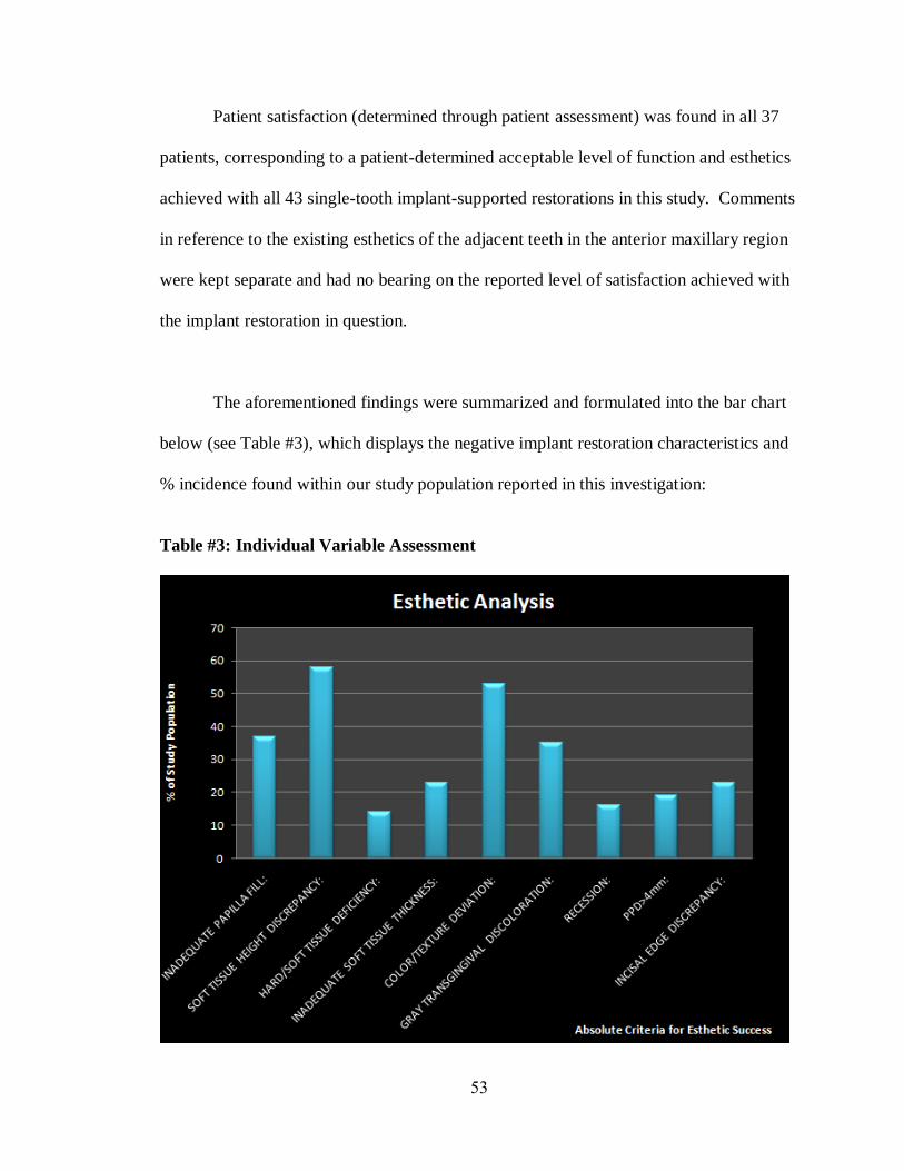
53
Patient satisfaction (determined through patient assessment) was found in all 37
patients, corresponding to a patient-determined acceptable level of function and esthetics
achieved with all 43 single-tooth implant-supported restorations in this study. Comments
in reference to the existing esthetics of the adjacent teeth in the anterior maxillary region
were kept separate and had no bearing on the reported level of satisfaction achieved with
the implant restoration in question.
The aforementioned findings were summarized and formulated into the bar chart
below (see Table #3), which displays the negative implant restoration characteristics and
% incidence found within our study population reported in this investigation:
Table #3: Individual Variable Assessment
54
Global esthetic assessment was performed by an expert panel consisting of four
periodontists and one prosthodontist. Esthetic success was determined by each member
of the panel and recorded as follows: 15Y/43 (35%), 14Y/43 (33%), 15Y/43 (35%),
12Y/43 (28%), and 4Y/43 (9%). The lowest esthetic tabulation (4Y/43) was concluded
by the sole prosthodontist of the panel. On average, 28% of the implants (12 of 43)
included in this investigation achieved esthetic success, which corresponds to a 72%
overall esthetic failure rate. On average, 11 of the 43 implants (26%) did not achieve
esthetic success due to soft tissue contours, 8 of the 43 implants (19%) did not achieve
esthetic success due to prosthetic contour, and 12 of the 43 implants (28%) did not
achieve esthetic success due to a combination of both components. 23 of the 43 implants
(53%) in this study were deemed failures due to partial involvement of the soft tissue
contours, whereas 20 of the 43 implants (47%) were deemed failures due to partial
involvement of the prosthetic contours.
Inter-examiner agreement was calculated from the overall esthetic scores of the
expert panel. 6 implant cases had 100% inter-examiner agreement, 11 implant cases had
80% inter-examiner agreement, 12 implant cases had 60% inter-examiner agreement, and
14 implant cases had less than 60% inter-examiner agreement.
Approximately 75% (32 out of the 43 implants) were deemed to be an esthetic
failure by 3 or more examiners on the expert panel. Approximately 6.98% (3 of the 43
implants) of the implants examined in this study failed to meet Albrektsson’s criteria for

55
implant success. All of these implants (3 of the 43 implants) were judged to be
esthetically unsuccessful by 3 or more examiners on the expert panel. The association
between function and esthetic was not statistically significant in this investigation.
Therefore, there appears to be no association between single-tooth implant-supported
restorations in the anterior maxilla that meet Albrekttson’s implant success criteria and
those restorations that meet esthetic success standards as determined by a global esthetic
assessment. Implant restorations that fulfill functional success parameters do not
necessarily translate into the achievement of esthetic success.
Table #4: Soft Tissue & Prosthetic Esthetic Success Characteristics Compared with Overall Esthetic
Success
Esthetic Success Esthetic Failure P-value
Soft Tissue Success % 25.58% (N = 11) 23.26% (N = 10) p<0.001
Soft Tissue Failure % 0 51.16 (N = 22) p<0.001
Prosthetic Success % 25.58% (N = 11) 34.88% (N = 15) p<0.001
Prosthetic Failure % 0 39.53% (N = 17) p<0.001
(Based on N = 43 total implants)
According to the statistical analysis (see Table #4), soft tissue success was
correlated to esthetic success in 25.58% (N = 11) of all implants. Similarly, prosthetic
success was correlated to esthetic success in 25.58% (N = 11) of all implants. Soft tissue
success was correlated to esthetic failure in 23.26% (N = 10) of all implants whereas

56
prosthetic success was correlated to esthetic failure in 34.88% (N = 15) of all implants.
Soft tissue failure was correlated to esthetic failure in 51.16% (N = 22) of all implants.
Prosthetic failure was correlated to esthetic failure in 39.52% (N = 17) of all implants.

57
CHAPTER 5
DISCUSSION
Study Demographics
Our investigation evaluated 43 single-unit implant-supported restorations in the
anterior maxillary region in 37 patients treated in the Graduate Periodontology and
Prosthodontic clinics at the University of Alabama at Birmingham. Based upon the
study population assessed in this study, 57% of the patients were male (vs. 43% female)
and the majority of patients examined were within the 61-70 year old age group (38% of
the patient population) as well as the 71-80 year old age group (38% of the patient
population), accounting for 76% of all patients. 89% of the patient population was
Caucasian, 8% were African American, and only 3% were Hispanic. 95% of the patients
reported a negative tobacco history and 89% were non-diabetic. These last two known
risk factors (smoking & diabetes) indicate that our patient population was relatively
healthy with minimal systemic involvement that might alter the overall functional and
esthetic success of the implants in this study.
Based on 43 total implants analyzed in this study, 46% of all implants were
located in the lateral incisor position (23% at #7 site and 23% at #10 site), followed by

58
the canine position (32% overall, 16% at #6 site and 16% at #11 site), and the central
incisor position (21% overall, 9% in #8 site and 12% at #9 site). 63% of all implants
placed were Nobel Biocare fixtures, followed by Biohorizons (19%), and Zimmer (12%).
The majority of the implants placed in this study were less than 4mm in diameter (60%
of all implants) and were within a length range of 13-13.9mm (47% of all implants). The
majority of the implants in this study were assessed 12-24 months after restoration
delivery (51% of all implants) and 25-36 months after implant placement (26% of all
implants). Since our most frequently evaluated implant occurred in the lateral incisor
position (46% of all implants), it follows suit that the most common diameter (<4mm)
and length (13-13.9mm) found in this study would conform to the size parameters of a
lateral incisor, which is commonly replaced by a long and narrow platform implant
fixture.
Functional Success and Failure
93.02% of all implants included in this study were functionally successful
according to Albrektsson‟s success criteria. 6.98% of all implants were functional
failures according to the same criteria. A failed implant has been described as one that is
clinically mobile.4 In contrast, an implant that shows progressive loss of supporting bone,
but that is clinically immobile, is a failing implant.4 Early implant failures denote a lack
of initial integration while late failures and failing implants occur after initial integration,
physiological remodeling, and loading.4 Problems limited to the soft tissues surrounding
implants and not involving the supporting bone have been defined as “ailing implants”

59
and, more recently, as biological complications.4 Endosseous dental implants rarely fail
beyond the first year after restoration.4
Literature studies show excellent survival rates of single-tooth restorations on
dental implants, varying from 96.1% to 98.9% after 7.5 years in function (Creugers et al.
2000).13
Multi-year studies of implants in partially edentulous patients have generally
reported greater than 90% success rates for both maxillary and mandibular implants.6-12
The implant survival meta-analysis on implants in the esthetic zone up to 1 year after
implant restoration, revealed an overall survival rate of 95.5% [95% CI: (93.0–97.1)]
irrespective of the type of intervention. It should be stated that, with respect to the loss of
implants that are more than 1 year in function, a very low event rate was calculated as
0.007 [95% CI: (0.003–0.019)].27
In our investigation, functional success was
determined to be 93.02%, which is comparable to the overall survival rate of implants in
the anterior esthetic zone (95.5%) found within the meta-analysis mentioned above.27
Papilla Fill
37% of all implants exhibited inadequate papilla fill (less than 75% papilla fill
with either mesial or distal papilla) according to the parameters of our evaluation.
According to Furhauser‟s study of the pink esthetic score (PES), complete papilla fill
was achieved in 62% (mesial papilla) and 56% (distal papilla) of all cases (N = 30) in
their first assessment, and in 58.8% (mesial papilla) and 51.6% (distal papilla) of all
cases (N = 30) in their second assessment. The average of these two assessments yielded

60
complete papilla fill in 60.4% (mesial papilla) and 53.8% (distal papilla) of all cases,
with an overall papilla fill of 57.1%. Therefore, the PES investigation demonstrated
inadequate papilla fill in 42.9% of all cases, which is comparable to the findings in our
study (37% inadequate papilla fill).97
The one variable reported to be most important for peri-implant esthetics in the
literature, i.e. the papilla, was most often restored to match that of natural teeth. Our
study showed inadequate papilla fill in 37% of all cases, whereas the PES study showed
inadequate papilla fill in 42.9% of all cases. These findings suggest that more than 50%
of all cases exhibited acceptable papilla fill. The majority of studies reported in the
Gehrke et al. article show that the soft tissue between an implant supported single-tooth
reconstruction and the adjacent teeth has a substantial influence on the esthetic outcome
from the patient‟s and clinician‟s point of view.20
This may well suggest that awareness
of the importance of the peri-implant soft-tissue variable improves therapeutic
approaches and outcomes.97
Single-implant cases obviously take benefit of tissue support of the adjacent
dentition (Grunder 2000, Kan et al. 2003b, Belser et al. 2004).27
When considering the
heights of interimplant papillae for instance, studies indicated that these papillae might
show inadequacy for complete enclosure of the interimplant area with soft-tissue, thereby
failing to duplicate the interproximal soft-tissue appearance of the adjacent teeth (Tarnow
et al. 1992, 2003, Lee et al. 2005).27
This deficiency may affect the esthetic outcome
unfavorably. The soft tissue height proximal to single-implants is on average much

61
higher and is related to the interproximal bone level at the side of the adjacent teeth
(Grunder 2000, Kan et al. 2003b).27
Hence, single-implant therapy may lead to more
favorable treatment outcomes. The findings of our study (37% inadequate papilla fill)
suggest that in more than 50% of all implants evaluated, adequate papilla fill/height was
achievable, mainly due to intact adjacent bone support.
Several studies (Schropp et al. 2005a, Romeo et al. 2008, Jemt 1997)27
have
revealed that an increase of tissue volume in the embrasures could be observed during
follow-up. For instance, Jemt & Lekholm (2003) found a mean papilla index of 1.1 at
crown placement (score 1 and 2 denote, respectively, less than half of the height and at
least half of the height of the proximal area filled by soft tissue) while at 2 year follow-
up a mean score of 2.4 was found (score 3: complete closure of proximal space with soft
tissue).27
The majority of the papillae analyzed were associated with papilla index scores
of 2 or 3 after follow-up, but no significant differences were observed between the
different test and control groups.27
Considering that the majority of implants in our study
were evaluated 12-24 months after restoration delivery (51% of all implants), further
improvement in papilla fill may be a possible finding with future follow-up.
Soft Tissue Height Discrepancy
58% of all implants exhibited a soft tissue height discrepancy (1mm or more of
asymmetry with the contralateral reference tooth) according to the parameters of our
evaluation. According to Furhauser‟s PES study, symmetrical soft tissue margins were

62
achieved in 42.3% of all cases (N = 30) in their first assessment, and in 44.5% of all
cases (N = 30) in their second assessment. The average of these two assessments yielded
symmetrical soft tissue margins in 43.4% of all cases. Therefore, the PES investigation
demonstrated soft tissue marginal discrepancies in 56.6% of all cases, which is very
similar to the findings in our study (58% soft tissue height discrepancy).97
According to the PES study, the level of the soft-tissue margin (56.6% of cases
with discrepancy) and the color of the peri-implant soft-tissue (58.8% of cases with color
deviation, 53.9% of cases with soft-tissue color/texture deviation) fared the worst in their
assessment.97
In our assessment, the level of the soft-tissue margin (58% of cases with
discrepancy) and the color/texture of the peri-implant soft-tissue (53% of cases with
color/texture deviation) fared the worst as well. Schropp et al. (2005a) reported that the
level of the marginal peri-implant mucosa was acceptable in significantly more cases
where implants were installed in early healed extraction sites compared with
conventionally healed sites; of the latter almost two thirds of the crowns were assessed to
be too short.27
This may suggest that these parameters should be given more attention
when considering how to achieve optimal esthetics with single-tooth implant-supported
restorations.
The gingival zenith (soft tissue margin) represents the most apical part of the
clinical crown. It also represents both the faciolingual and the mesiodistal location of the
crown in relationship to the edentulous ridge. As such, it has a remarkable influence on
the morphology of the planned restoration. The gingival zenith affects other objective
63
criteria, including the balance of gingival levels (too inferior or superior), the tooth axis
(too distal or mesial), the tooth dimension (too inferior or superior), and the tooth form
(triangular becomes ovoid if too inferior). Without the control of the gingival zenith, the
clinician‟s ability to define dental implant esthetics is vastly diminished.28
At least four factors affect the gingival zenith. One is, of course, the relative
location of the tissues to the planned gingival zenith. Second is the depth of the dental
implant placement. Third is the response of the buccal bone and mucosa to the implant
procedure and components. The fourth is the prosthodontic management of the gingival
zenith architecture. Ideally, the planned gingival zenith is symmetric with the
contralateral tooth and harmonious with the gingival levels of adjacent teeth.28
The technique of tissue modeling with subgingival crown contours intentionally
forces the gingival margin out to the buccal to align it with and mimic the tissue along
the adjacent natural teeth. This tipping to the buccal also elevates the gingival margin in
an apical direction. Therefore it is imperative that the surgeon maintain gingival
thickness and contour when placing and exposing the implant. Following exposure of the
implant, the bucco-gingival margin around the healing abutment may be apical to, even
with, or coronal to the gingival margins on the adjacent natural teeth. If the beginning
point of the gingival margin is apical to its natural counterpart, there will be no option
but to fabricate a restoration that is longer than desired for bilateral symmetry.30
64
Hard/Soft Tissue Deficiency
14% of all implants exhibited a hard/soft tissue deficiency (obvious deficiency
upon visual inspection and palpation of the gingival region apical to the implant
restoration) according to the parameters of our evaluation. According to Furhauser‟s
PES study, absence of alveolar process deficiency was achieved in 54% of all cases (N =
30) in their first assessment, and in 49.1% of all cases (N = 30) in their second
assessment. The average of these two assessments yielded an absence of alveolar
process deficiency in 51.6% of all cases. Therefore, the PES investigation demonstrated
alveolar process deficiency in 48.4% of all cases. In our study, hard and soft tissue
deficiencies were assessed as one entity, yielding an overall deficiency of 14% of cases.
No definitive correlations can be drawn from these findings.97
In the past, hard and soft tissue deficiencies at the labial aspect resulted in
implant placement that was lingual to the ideal aforementioned position. Currently, labial
deficiencies can be corrected surgically with high success rates and should not be a
reason for lingual positioning of the platform.30
The only reason for placing the platform
slightly to the lingual aspect is if the clinician‟s method of choice is a screw-retained
prosthesis.30
However, one has to be careful positioning the implant lingually. Placing
the implant too far to the lingual aspect will result in a restoration that has an abrupt
buccal emergence profile, which does not facilitate proper oral hygiene.30
If the ideal gingival zenith is greater than 3 mm incisal and 2 mm buccal from the
existing bone crest, then bone augmentation procedures may be considered in addition to
65
gingival grafting in order to correct for any hard/soft tissue deficiencies.28
The gingival
zenith, therefore, becomes the therapeutic reference point when evaluating ridge
morphology defects. A positive esthetic result is suggested when the adjacent tooth
attachment levels are intact and there is adequate bone relative to the reference point.28
Without underlying bone to support the buccal contour in full dimension, the
esthetic volume of the edentulous space ultimately will be deficient. Based on the
location of the planned gingival zenith, therefore, decisions regarding the need for bone
augmentation, socket preservation, and/or soft tissue augmentation procedures can be
prudently accessed. In our study, only 14% of all implants demonstrated hard/soft tissue
deficiency, which may imply that the importance of site development is already a well
understood concept.
Adequacy of Soft Tissue Thickness
23% of all implants exhibited inadequate soft tissue thickness (less than 1mm of
thickness as determined by periodontal probe visibility) according to the parameters of
our evaluation. Soft tissue thickness can be correlated to the tissue biotype of the peri-
implant mucosa. A thin scalloped periodontium is found in less than 15% of cases and is
characterized by a delicate soft tissue curtain, a scalloped underlying osseous form,
possible dehiscences and/or fenestrations, and a reduced quantity and quality of
keratinized mucosa.88
This form of gingiva reacts to insults by receding facially and
interproximally. As recession occurs and the inter-radicular bone resorbs, the subsequent

66
soft tissue loss compromises the overall esthetic result.88
When soft tissue thickness is
less than 1mm, a thin scalloped periodontium is likely present with comparable
occurrence rates (15% occurrence of a thin scalloped periodontium vs. 23% occurrence
of inadequate soft tissue thickness). Since this type of tissue biotype responds to insults
by recession, a possible association between the rate of inadequate soft tissue thickness
(23%) and the rate of recession (16%) found in our study may be made.
Soft Tissue Color/Texture Deviation
53% of all implants exhibited soft tissue color/texture deviation from the adjacent
gingival areas, according to the parameters of our evaluation. According to Furhauser‟s
PES study, symmetrical soft tissue color was achieved in 45.3% of all cases (N = 30) in
their first assessment, and in 37.1% of all cases (N = 30) in their second assessment. The
average of these two assessments yielded symmetrical soft tissue color in 41.2% of all
cases. Symmetrical soft tissue texture was achieved in 53.1% of all cases (N = 30) in
their first assessment, and in 49.1% of all cases (N = 30) in their second assessment. The
average of these two assessments yielded symmetrical soft tissue texture in 51.1% of all
cases. Therefore, the PES investigation demonstrated soft tissue discoloration in 58.8%
of all cases as well as soft tissue texture deviation in 48.9% of all cases. By combining
these two parameters, overall soft tissue color/texture deviation was found in 53.9% of
all cases, which is very similar to the findings in our study (53% soft tissue color/texture
deviation).97
Soft tissue color/texture fared poorly in both our study and in the PES
study, which may suggest that this parameter should be given more attention when

67
considering how to achieve optimal esthetics with single-tooth implant-supported
restorations.
According to the Gehrke et al. study regarding the reproducibility of the Implant
Crown Aesthetic Index, outcomes for the anatomic shape, color, and texture of the peri-
implant mucosa obtained a poorer score than other parameters.20
In a study reported in
the Cooper et al. article, the color of the periimplant soft tissue matched that of the
reference tooth in no more than just over one-third of cases.28
Our study, in comparison,
found soft tissue color/texture deviations in 53% of all cases, which may further suggest
that the peri-implant mucosa requires thorough consideration in achieving overall
esthetic success.
Trans-Gingival Discoloration
35% of all implants exhibited gray trans-gingival discoloration (when compared
to the adjacent gingival areas) according to the parameters of our evaluation. According
to Furhauser‟s PES study, symmetrical soft tissue color was achieved in 45.3% of all
cases (N = 30) in their first assessment, and in 37.1% of all cases (N = 30) in their second
assessment. The average of these two assessments yielded symmetrical soft tissue color
in 41.2% of all cases. Therefore, the PES investigation demonstrated soft tissue
discoloration in 58.8% of all cases. In our study, gray trans-gingival discoloration (35%)
was assessed separately from soft tissue color/texture deviation (53%). No definitive
correlations can be drawn from these findings.97

68
There is a limit to the implant width that a buccal cortical plate at a given site can
accommodate. Even if there is room for a very wide implant, the superficial placement of
this wide platform may result in an optical reflection, a „„show through,‟‟ of the implant
through the thin bony plate. Once created, such an esthetic deficiency cannot be
corrected.30
Replacement of a tooth in the anterior maxillary region often relies on the
use of a regular or narrow platform implant, which might avoid the previously mentioned
problems with trans-gingival discoloration.
The soft tissues must be carefully considered when planning implants in the
anterior maxillary region in order to avoid any gray trans-gingival discoloration. Thin,
friable, poorly keratinized tissues are more susceptible to inflammatory and mechanical
insult. This can be potentially disastrous in an esthetically demanding area, because post-
restorative recession will compromise even the most well-fabricated restoration. These
tissues are also highly susceptible to a “graying effect" from the underlying implant body
or abutment as it passes from the bone crest toward the peri-implant sulcus. Although
this problem is often eliminated via soft tissue grafting, such an approach is impractical
if it will result in a greater buccal bulk around the implant than around the adjacent
teeth.100

69
Recession
16% of all implants exhibited gingival recession (1mm or more of recession or
appearance of the underlying titanium abutment/implant body) according to the
parameters of our evaluation. According to the Furhauser article, Sheller et al. (1998)
reported recession in 10% of the implant-supported single-tooth replacements that they
evaluated, which is comparable to the findings of our study (16% of all cases with
recession present).97
Achieving optimal gingival esthetics around anterior single implants is a
challenging procedure and maintaining it over time can be an equally demanding task.
Despite the high success rates achieved with osseointegrated implants,6-12
the peri-
implant mucosal response is not clearly understood. For anterior single implants, up to
16% of gingival recession has been previously reported.101
On the other hand,
spontaneous rebound of the receded gingiva has also been observed following a few
years of function.101
The peri-implant mucosa changes had been postulated as an attempt
to establish a stable biological dimension.101
Peri-implant mucosa, like its natural teeth
counterpart (the dentogingival complex), comprises similar histologic components
(gingival sulcus and connective tissue attachments) and dimension ( ~ 3 mm).101
While
the dimension of each component may change over time, these changes do not
significantly affect the overall dimension.101
The risk of recession may potentially increase if a number of factors are not
considered. These factors may included but should not be limited to the following:
70
initial presentation (Seibert classification), implant position capability (relative to
planned gingival zenith), bone formation and resorption at the implant, peri-implant
mucosa integration, character of the implant abutment interface, inflammation, local
factors (plaque, etc.), patient factors (e.g., biotype), abutment form, submucosal contour
of the provisional crown, bone modeling/remodeling, and potential adjacent tooth
eruption.28
Bleeding Upon Probing
2% of all implants exhibited immediate or delayed bleeding upon probing
according to the parameters of our evaluation. According to the Hartog et al. article,
studies that assessed the presence of plaque on the surfaces of the implant restoration
showed high variance in outcome from 0.5% to 61% of sites examined. Accordingly, the
same phenomenon could be observed with bleeding upon probing.27
Therefore, even
when plaque is present as an etiological factor and BOP has been identified around the
peri-implant mucosa, greater likelihood with BOP inconsistencies may occur.
The biologic width is made up, as one proceeds coronally, of connective tissue
attachment to root cementum, followed by a hemidesmosomal attachment of the
junctional epithelium to the root, and followed by the gingival sulcus.100
The biologic
width serves a number of protective functions. If impinged upon by restorative materials
and not corrected, an inflammatory process often ensues and bleeding upon probing may
become readily evident. Implant restorations need to adhere to the principles of biologic

71
width in order to minimize biological impingement upon the attachment to the implant.
Violation of biologic width may be correlated to the presence of BOP and should be
avoided in order to maintain the health of the peri-implant mucosa.
Periodontal Pocket Depth
19% of all implants exhibited pocket depths greater than 4mm according to the
parameters of our evaluation. According to the Hartog et al. article, a study by Schropp
et al. (2005b) observed a mean reduction in probing depth of 0.5mm during a 2-year
observation period to a mean probing depth of 4.2 mm for single-unit implant-supported
restorations in the anterior maxilla.27
The mean probing depths presented by other studies
within the article were clearly lower.27
These studies support the notion of 4mm being
clinically compatible with peri-implant health.
Lang and co-workers demonstrated that, as the degree of inflammation in the soft
tissues increased, the probe penetration into the periimplant sulcus exceeded the
connective tissue level by an average of 0.52mm.100
Ericsson and Lindhe also
demonstrated a lesser resistance to probing the peri-implant mucosa, as compared to
probing the periodontal sulcus.100
This is due to the lack of a true connective tissue
attachment to the implant surface. Such vulnerability further underscores the need to
provide the patient with a cleansable, maintainable peri-implant milieu.100
Nonetheless,
greater probing depth around an implant compared with a natural tooth may be due to a
lack of a true attachment or the presence of inflammation in the peri-implant mucosa.

72
Probing depths around implants may be of little diagnostic value, unless
accompanied by pathologic signs (e.g., radiographic radiolucencies, purulent exudate,
bleeding) and/or symptoms (e.g., discomfort, pain). The benefit of probing the implant
sulcus has been challenged in the literature because sound scientific criteria are lacking.
On one hand, probing not only measures pocket depth, but also reveals tissue
consistency, bleeding, and the presence of exudate.102
Increasing probing depths over
time may indicate bone loss, but not necessarily indicate disease for an endosteal
implant. Stable, rigid, fixated implants have been reported with pocket depths ranging
from 2 to 6 mm. Lekholm et al. found that the presence of deep pockets was not
accompanied by accelerated marginal bone loss.102
Conversely, studies have shown than
sulcus depths greater than 5 to 6 mm around implants have a greater incidence of
anaerobic bacteria and may require intervention in the presence of inflammation or
exudate (e.g., surgery, antibiotic regimens).102
Overall, healthy, partially edentulous
implant patients consistently exhibit greater probing depths around implants than around
teeth.102
The potential for damage to the fragile attachment or marring of the implant
surface may exist during probing.102
On the other hand, there is no clinical or
experimental evidence supporting this hypothesis. Routine probing depths are not
suggested in the absence of other signs or symptoms and may be related to the presence
of local disease or preexisting tissue thickness before the implant was inserted.102
Future

73
research in the area of probing is needed before including this as a primary criterion in a
consensus for success, survival, and/or failure.
Incisal Edge Discrepancy
23% of all implants exhibited an incisal edge discrepancy (1mm or more of
asymmetry with the contralateral reference tooth) according to the parameters of our
evaluation. According to the Gehrke et al. study regarding the reproducibility of the
Implant Crown Aesthetic Index, very good ratings below one point (0 = excellent, 1 =
satisfactory) were given relatively often for the outcome of the position of the incisal
edge.20
Our study, in comparison, found an incisal edge discrepancy in less than one-
fourth (23%) of all cases, which may suggest that the restorative dentist assigned to each
case fabricated the final implant-supported restoration with careful consideration for
incisal edge symmetry more frequently than expected.
Jemt & Lekholm (2005) reported that majority of implant crowns were on
average 0.7mm longer than the contralateral natural crowns after 5-year follow-up.27
The
same value was recorded by Gotfredsen (2004) after 5-year and he found that 17 of the
20 (85%) implant crowns were too long.27
These findings do not agree with the findings
of Gehrke et al. as well as the findings of our investigation (23% of all implant
restorations exhibited incisal edge discrepancy). The incisal edge position is contingent
upon the adjacent incisal edges as well as the incisal edge of the contralateral reference
tooth. Therefore, existing adjacent and contralateral esthetics of the natural dentition

74
may play an important role in this determination and if the esthetics (the incisal edges
more specifically) is not optimal in both regions, a higher rate of discrepancy or variation
may be noted.
Crown Contour/Color Irregularity
26% of all implants exhibited crown contour irregularity (when compared the
adjacent and contralateral reference teeth) according to the parameters of our evaluation.
According to the Gehrke et al. study regarding the reproducibility of the Implant Crown
Aesthetic Index, very good ratings below one point (0 = excellent, 1 = satisfactory) were
given relatively often for the outcomes of the mesiodistal dimension and labial convexity
of the final restoration.20
Our study, in comparison, found crown contour irregularities in
approximately one-fourth (26%) of all cases, which may suggest that the restorative
dentist assigned to each case fabricated the final implant-supported restoration with
careful consideration for appropriate crown contours more frequently than expected.
Patient Satisfaction
100% of all implants satisfied patient expectations according to patient-
determined acceptable levels of function and esthetics. In a study by Baracat et al. of
patient expectations before and after dental implant therapy, the patients‟ esthetic and
functional post-treatment ratings of dental implant therapies were significantly higher
than their expectations before these therapies.103
This may suggest that low pre-treatment
75
expectations in either study may affect the potential overall acceptance of esthetics post-
treatment. Pjetursson and colleagues reported that 92% of their patients stated that
implant dental therapy outcomes satisfied their expectations, which is comparable to the
findings of our study (100% patient satisfaction).103
Chang and colleagues23
interviewed dentists and patients to evaluate the esthetic
outcome of implant supported single-tooth restorations using standardized
questionnaires. The results confirm a high level of patient satisfaction with the outcome
of treatment. The picture was contradictory in the assessment of these patient cases by
prosthodontists, who assigned the treatment outcome a much lower rating than the
patients. The author interpreted this result to the effect that the clinicians are either more
critical or they apply different standards when assessing esthetic outcome from those
applied by the patients involved themselves. In actual fact, a statistical analysis indicated
that parameters such as crown shape, contact point position, color, and topography of the
surrounding soft tissue had a significant influence on the rating of general satisfaction
with appearance, although it was not possible to detect any similar connections within
patient questioning.20
It is important to note that many factors appear to influence patient‟s attitudes
toward dental esthetics, such as culture, education, economy, traditions, dental care
system, patient/dentist relationship, media images of dental appearance, among others.103
In evaluating patient satisfaction, it is very difficult to account for which factors weigh
more heavily. Patient preferences and attitudes are difficult to eliminate from the

76
esthetic assessment, which may invalidate patient satisfaction ratings in any study of
esthetics.
Global Assessment Discussion
Esthetic success was determined by each member of the expert panel and
reported as follows: 35% esthetic success (15 of 43), 33% esthetic success (14 of 43),
35% esthetic success (15 of 43), 28% esthetic success (12 of 43), and 9% esthetic
success (4 of 43, as determined by the sole prosthodontist on the panel). These findings
were compiled in a single assessment as opposed to multiple assessments. According to
the Furhauser PES study, two assessments were performed, with the highest score of 14
(most esthetic) less often assigned in the second assessment.97
The lowest scores
(inadequate esthetics) were slightly more often assigned in the second run, suggesting
that the observers tended to become more critical in the second assessment.97
Therefore,
in order to eliminate observational bias, only one assessment was performed by our
expert panel.
The sole prosthodontist on the expert panel awarded an overall esthetic outcome
in only 4 of 43 (9%) implant cases, whereas the four periodontists on the expert panel
deemed an esthetic outcome in 32.75% (on average) of all cases. This may suggest that
the sole prosthodontist may have been overly critical in the esthetic assessment.
According to the PES study, prosthodontists, surgeons, and dental students all scored the
outcome significantly more generously, which suggests that no association can be made

77
between the specialty groups evaluating each case.97
Discrepancies found between
various occupational groups are probably not only dependent on the examiner‟s degree
of specialization, but also on individual viewpoints.20
Esthetic Success and Failure
Overall esthetic success was achieved with 28% of all implants included in this
investigation, prior to statistical assessment. Overall esthetic failure was reported with
72% of all implants included in this study, prior to statistical assessment. According to
Meijndert et al. (2007), using the Implant Crown Aesthetic Index developed my Meijer,
it was reported that in 34% of the cases, the esthetics were not acceptable, which varies
significantly from the results of our study.13
Therefore, esthetic success variance found
between various studies are probably not only dependent on the examiner‟s degree of
specialization, but also on individual viewpoints,20
which makes standardization of
esthetics and comparisons between studies difficult.
Most studies on implant success still do not provide, or evaluate, criteria for
esthetic success, so it is difficult to determine the prevalence of implant failures for
esthetic reasons. The few studies in the literature that have reported on esthetic
complications reveal that anywhere between 4% and 16% of single implant crowns in the
anterior maxilla fail for esthetic reasons.104
The most common esthetic complication is
gingival recession exposing the implant/abutment junction, with one study reporting up
to 61% of cases with at least 1 mm of gingival recession on the facial aspect.104
In
78
addition, poor shade selection for the prosthesis and lack of interdental papillae also
account for implant esthetic failures.104
Failure due to Soft Tissue Component, Prosthetic Component, or Both
26% of all implants did not achieve esthetic success due to soft tissue contours
only and 53% of all implants were deemed esthetic failures due to partial involvement of
the soft tissue contours, prior to statistical assessment. 19% of all implants did not
achieve esthetic success due to prosthetics only and 47% of all implants were deemed
esthetic failures due to partial involvement of the prosthetics, prior to statistical
assessment. 28% of all implants did not achieve esthetic success due to a combination of
both components, prior to statistical analysis.
According to a cross-sectional, retrospective study by Belser and colleagues19
, all
45 anterior maxillary single-tooth implants fulfilled strict success criteria for dental
implants with regard to osseointegration, including the absence of peri-implant
radiolucency, implant mobility, suppuration, and pain. Further evaluation was performed
with the pink esthetic score (PES), comprised of the following five variables: mesial
papilla, distal papilla, curvature of the facial mucosa, level of the facial mucosa, and root
convexity/ soft tissue color and texture at the facial aspect of the implant site, in
combination with the white esthetic score (WES). The WES specifically focuses on the
visible part of the implant restoration itself (i.e., the part of the implant crown that
emerges from the peri-implant mucosa) and is based on the five following parameters:

79
general tooth form; outline and volume of the clinical crown; color, which includes the
assessment of the dimension’s hue and value; surface texture, and translucency and
characterization.19
The mean total pink esthetic score/white esthetic score was 14.7 – 1.18 (range: 11
to 18, maximum 20). The mean total PES of 7.8 – 0.88 (range: 6 to 9, maximum 10)
documents favorable overall peri-implant soft tissue conditions.19
In comparison, our
investigation found a deficient soft tissue component in 53% of all implants evaluated,
suggesting unfavorable peri-implant esthetics. The two PES variables facial mucosa
curvature (1.9 – 0.29) and facial mucosa level (1.8 – 0.42) had the highest mean values,
whereas the combination variable root convexity/soft tissue color and texture (1.2 – 0.53)
proved to be the most difficult to fully satisfy.19
Soft tissue color/texture fared poorly in
both our study and in this PES/WES study, which may suggest that this parameter should
be given more attention when considering how to achieve optimal esthetics with single-
tooth implant-supported restorations.
The mean total PES/WES of 14.7 indicated an overall successful esthetic
outcome; only one crown scored <12, which was defined as the threshold of clinical
acceptability.19
In comparison, our study found an overall unsuccessful esthetic outcome
(74% esthetic failure before statistical analysis, 75% esthetic failure after statistical
analysis), which may be due to the quality of the objective absolute criteria included in
our study as well as the method of which esthetics was assessed.

80
The PES (mean score of 7.8) was clearly higher than the corresponding WES
(mean score of 6.9).19
This may be due to the fact that the PES is mainly influenced by
the local anatomy and the applied surgical procedure to regenerate the peri-implant bone
defects routinely present in post-extraction implant sites. Hence, the routine and skills of
the implant surgeon play an important role in the esthetic outcome of peri-implant soft
tissues.19
The WES was clearly less favorable than the PES, which contradicts the findings
of our investigation (53% with a soft tissue deficiency vs. 47% with a prosthetic
deficiency). This observation is also documented by the fact that 20% of the crowns
scored below the threshold of 6, which is the level of clinical acceptability from the
examiner‟s point of view. It is possible that the WES component of the applied esthetic
index is too rigorous, because all patients accepted the insertion of their implant crowns
during therapy. The patient‟s perception of dental restorations from an esthetic point of
view frequently differs significantly from that of dental professionals, which is
confirmed by reports from the literature.19
Inter-Examiner Agreement
Inter-examiner agreement was reported in this study as follows: 6 implants with
100% inter-examiner agreement, 11 implants with 80% inter-examiner agreement, 12
implants with 60% inter-examiner agreement, and 14 implants with less than 60% inter-
examiner agreement. Disagreement found between the examiners on the expert panel of

81
this investigation are probably not only dependent on the examiner‟s degree of
specialization, but also on individual viewpoints,20
which may support the lack of
agreement identified above.
Esthetic Success and Failure by 3 or More Experts
75% of all implants were deemed to be an esthetic failure by 3 or more
examiners on the expert panel. 6.98% of all implants failed to meet Albrektsson‟s
criteria for implant success and were also deemed esthetic failures. Therefore, the
association between function and esthetic was not statistically significant in this
investigation. In conclusion, there appears to be no association between single-tooth
implant-supported restorations in the anterior maxilla that meet Albrekttson’s implant
success criteria and those restorations that meet esthetic success standards as determined
by a global esthetic assessment. Furthermore, implant restorations that fulfill functional
success parameters do not necessarily translate into the achievement of esthetic success.
Soft Tissue & Prosthetic Esthetic Success Characteristics Compared with Overall
Esthetic Success
According to the statistical analysis performed in this study, soft tissue success
was correlated to esthetic success in 25.58% (N = 11) of all implants. This means that
when peri-implant esthetics are adequate or ideal, an overall esthetic result may only
occur in about a quarter of the study population. This implies that success with soft

82
tissue characteristics is a contributing factor to overall esthetic success, but it does not
dictate or predominantly influence the overall outcome. Soft tissue success was
correlated to esthetic failure in 23.26% (N = 10) of all implants, signifying that even
when soft tissue contours may have been satisfactory or optimal, esthetics was still not
achieved in about a quarter of the study population, as stated above. Soft tissue failure
was correlated to esthetic failure in 51.16% (N = 22) of all implants. This means that
when soft tissue esthetics are not achieved, overall esthetic failure occurs about 50% of
the time. It can be concluded that peri-implant esthetics may have a low positive
predictive value (overall esthetic success only 25.58% of the time when successful soft
tissue esthetics are achieved) on the esthetic outcome.
According to the statistical analysis performed in this study, prosthetic success
was correlated to esthetic success in 25.58% (N = 11) of all implants. This means that
when crown esthetics are adequate or ideal, an overall esthetic result may only occur in
about a quarter of the study population. This implies that success with prosthetic
characteristics is a contributing factor to overall esthetic success, but it does not dictate or
predominantly influence the overall outcome. Prosthetic success was correlated to
esthetic failure in 34.88% (N = 15) of all implants, signifying that even when prosthetic
characteristics may have been adequate or optimal, esthetics was still not achieved in
about a third of the study population, as stated above. Prosthetic failure was correlated to
esthetic failure in 39.52% (N = 17) of all implants. This means that when crown esthetics
are not achieved, overall esthetic failure occurs about 40% of the time. It can be
concluded that implant restoration esthetics may have a low positive predictive value

83
(overall esthetic success only 25.58% of the time when successful prosthetic esthetics are
achieved) on the esthetic outcome. Furthermore, it may be concluded that soft tissue
failure may factor more heavily on the overall esthetic failure (51.16%) of single-tooth
implant-supported restorations, compared to prosthetic failure (39.52% esthetic failure)
Another similar study evaluated the contribution of surgical, soft tissue, and
prosthetic components to overall esthetic success. Juodzbalys and Wang98
developed a
complex esthetic index (CEI) for rating the esthetics of anterior maxillary implant-
supported restorations with respect to the surrounding soft and hard tissues. Fifty
patients (31 males and 19 females; age: 18 to 50 years; mean age – SD: 32.4 – 9.1 years)
previously treated with dental implants were evaluated regarding the esthetic results of
their restorations using the proposed CEI. Two calibrated oral surgeons did the
evaluation and recording. The evaluation was carried out twice by each of the examiners
2 weeks apart.98
The proposed complex esthetic index is composed of three components: the soft
tissue index (S), predictive index (P), and implant-supported restoration index (R).
Within each category, specific parameters were evaluated and graded as adequate (rating
20%), compromised (rating 10%), or deficient (rating 0%). Soft tissue characteristics of
the S that were previously published8 include soft tissue contour variations, soft tissue
vertical deficiency, soft tissue color and texture variations, and mesial and distal papillae
appearance. The P primary assessed the following components: mesial and distal
interproximal bone height, gingival tissue biotype, apico-coronal position of the

84
implants, and horizontal contour deficiency. The R evaluated the color and translucency
of the implant-supported restoration, labial convexity in the abutment/implant junction,
implant/crown incisal edge position, crown width/length ratio, and surface roughness and
ridges of the implant-supported restoration in relationship to adjacent and contralateral
teeth. All mentioned R parameters, with the exception of crown width/length ratio, must
be in harmony with the adjacent and contralateral teeth.98
Accordingly, when the S, P, and R general ratings were adequate, the CEI rating
was 100%. When one of the indices registered between 60% and 90%, this was a
compromised and clinically acceptable result. When the CEI was <50%, this was a
deficient and clinically unacceptable esthetic outcome. An adequate CEI of S100, P100,
and R100 was observed by both examiners in 10% and 12% of cases in evaluations I and
II, respectively.98
In comparison, only 25% of all single-tooth implant-supported
restorations in our study achieved overall esthetic success.
Further analysis revealed the importance of each CEI component. It was found
that in situations where the CEI was S100, P100, and R (prosthetic component) <100%,
the number of cases was the same as it was using the adequate CEI. When the P (surgical
component) was <100%, the number of cases with adequate S and R was reduced more
than twice. When the S (soft tissue component) was <100%, the number of cases with
adequate P and R was significantly reduced.98
This may imply that the surgical and soft
tissue components of this index outweigh the contribution of the prosthetic component
and may require more attention when trying to obtain an esthetic result. From our study,

85
similar conclusions were drawn as follows: soft tissue failure may factor more heavily on
the overall esthetic failure (51.16%) of single-tooth implant-supported restorations,
compared to prosthetic failure (39.52% esthetic failure). Hence, soft tissue signifies an
important esthetic factor that must be carefully considered in implant dentistry and
optimally planned for in the final restorative result.

86
CHAPTER 6
CONCLUSION
The conclusions of our investigation are as follows:
1) The association between function and esthetic was not statistically significant in
this investigation.
2) There appears to be no association between single-tooth implant-supported
restorations in the anterior maxilla that meet Albrekttson’s implant success
criteria and those restorations that meet esthetic success standards as determined
by a global esthetic assessment.
3) Implant restorations that fulfill functional success parameters do not necessarily
translate into the achievement of esthetic success.
4) Soft tissue height (level of the soft tissue margin) and soft tissue color texture
may require more attention and consideration when trying to obtain an esthetic
result.
5) Soft tissue failure may factor more heavily on the overall esthetic failure
(51.16%) of single-tooth implant-supported restorations, compared to prosthetic
failure (39.52% esthetic failure)

87
6) Soft tissue signifies an important esthetic factor that must be carefully
considered in implant dentistry and optimally planned for when contemplating
single-tooth implant-supported restorations in the anterior maxilla
The esthetic result is still not systematically included among the implant therapy
success criteria, although a tendency to do so is developing in more recently published
articles and especially in those that appraise the implant-supported prosthetic
rehabilitations of the maxillary and mandibular anterior sectors. It has not been possible
to identify an esthetic evaluation index that is commonly approved by clinicians and
researchers because the parameters considered are often different between study groups
or they are used in various combinations. A common index would be useful not only to
assess the success of the therapy as a whole, but also as a valid tool of longitudinal
observation of the progressive maturation of the peri-implant soft tissues. It would then
be possible to appraise the esthetics not only in terms of purely descriptive dimensional
measurements, but also in terms of a progressive matching of the implant-supported
prosthetic restoration with the surrounding tissues.
The lack of an encoded index that has been approved by the international
scientific community represents a spur to search for a method with which to evaluate the
esthetic result that is as far as possible complete, objective, reproducible, reliable, and
free from methodological errors (bias). Finally, there is a need to integrate the
professional‟s “objective” evaluation and the “subjective” evaluation of the patient, the
latter harvested by means of satisfaction questionnaires whose answers are quantified on

88
Visual Analogue Scales (VASs). This would not only establish the degree of agreement
or disagreement between the two points of view but would also make the clinician aware
of the parameters that would render the implant-supported rehabilitation more
esthetically pleasing in the eyes of the patient.
89
LIST OF REFERENCES
1. Spears FM, Kokich VG, Mathews DP. Interdisciplinary management of anterior
dental esthetics. J Am Dent Assoc 2006;137;160-169
2. Albrekttson T, Zarb G. The long-term efficacy of currently used dental implants:
a review and proposed criteria for success. Int J Oral Maxillofac Implants 1:11-
25, 1986
3. Palmer RM, Palmer PJ, Newton JT. Dealing with esthetic demands in the anterior
maxilla. Periodontology 2000. Volume 33, Issue 1, Date: October 2003,
Pages: 105-118
4. AAP Position Paper: Dental Implants in Periodontal Therapy. J
Periodontol2000;71:1934-1942.
5. Smith DE, Zarb GA. Criteria for success of osseointegrated endosseous implants.
J Prosthet Dent 1989; 62:567-572.
6. Fritz ME. Implant therapy. II. Ann Periodontol 1996;1:796-815.
7. Lekholm U, van Steenberghe D, Herrimann I, et al.Osseointegrated implants in
the treatment of partially edentulous jaws: A prospective 5-year multicenter study.
Int J Oral Maxillofac Implants 1994;9:627-635.
8. Buser D, Weber HP, Bragger U, Balsiger C. Tissue integration of one-stage ITI
implants: 3-year results of a longitudinal study with hollow-cylinder and
hollowscrew implants. Int J Oral Maxillofac Implants 1991;6:405-412.
9. Fugazzotto PA, Gulbransen HJ, Wheeler SL, Lindsay J. The use of IMZ
osseointegrated implants in partially and completely edentulous patients: success
and failure rates of 2,023 implant cylinders up to 60+ months in function. Int J
Oral Maxillofac Implants1993;8:617-621.
10. Patrick D, Zosky J, Lubar R, Buchs A. The longitudinal clinical efficacy of Core-
Vent dental implants: a five-year report. J Oral Implants 1989;15:95-103.
11. Saadoun AP, LeGall ML. Clinical results and guidelines on Steri-Oss endosseous
implants. Int J Periodontics Restorative Dent 1992;12:486-495.
12. Sbordone L, Barone A, Ciaglia RN, Ramaglia L, Iacono VJ. Longitudinal study
of dental implants in a periodontally compromised population. J Periodontol
1999;70:1322-1329.

90
13. Meijer HJ, Stellingsma K, Meijndert L, Raghoebar GM. A new index for rating
aesthetics of implant-supported single crowns and adjacent soft tissues--the
Implant Crown Aesthetic Index. Clin Oral Implants Res. 2005 Dec;16(6):645-9.
14. Eckert SE, Wollan PC. Retrospective review of endosseous implants placed in
partially edentulous jaws. J Prosthet Dent 1998;79:415-421.
15. Lindhe T, Gunne J, Tillberg A, Molin M. A meta-analysis of implants in partial
edentulism. Clin Oral Implants Res 1998;9:80-90.
16. Wyatt CC, Zarb GA. Treatment outcomes of patients with implant-supported
fixed partial prostheses. Int J Oral Maxillofac Implants 1998;13:204-211.
17. Noack N, Willer J, Hoffmann J. Long-term results after placement of dental
implants: Longitudinal study of 1,964 implants over 16 years. Int J Oral
Maxillofac Implants 1999;14:748-755.
18. Naert I, Koutsikakis G, Duyck J, Quirynen M, van Steenberghe D, Jacobs R.
Biologic outcome of implant-supported restorations in the treatment of partial
edentulism. Part I: A longitudinal clinical evaluation. Clin Oral Implants Res
2002;13:381-389.
19. Belser UC, Gru¨tter L, Vailati F, Bornstein MM, Weber HP, and Buser D.
Outcome Evaluation of Early Placed Maxillary Anterior Single-Tooth Implants
Using Objective Esthetic Criteria: A Cross-Sectional, Retrospective Study in 45
PatientsWith a 2- to 4-Year Follow-Up Using Pink and White Esthetic Scores. J
Periodontol 2009;80:140-151.
20. Gehrke P, Lobert M, Dhom G. Reproducibility of the pink esthetic score--rating
soft tissue esthetics around single-implant restorations with regard to dental
observer specialization. J Esthet Restor Dent. 2008;20(6):375-84; discussion 385.
21. Palmer RM, Palmer PJ, Newton JT. Dealing with esthetic demands in the anterior
maxilla. Periodontology 2000. Volume 33, Issue 1, Date: October 2003,
Pages: 105-118
22. Charruel S, Perez C, Foti B, Camps J, Monnet-Corti V. Gingival Contour
Assessment: Clinical Parameters Useful for Esthetic Diagnosis and Treatment. J
of Perio 2008, Vol. 79, No. 5, Pages 795-801
23. Chang M, Odman PA, Wennström JL, Andersson B. Esthetic outcome of implant
supported single-tooth replacements assessed by patient and prosthodontist. Int J
Prosthodont 1999: 12: 335–341
24. Choquet V, Hermans M, Adriaenssens P. Clinical and radiographic evaluation of
the papilla level adjacent to single-tooth dental implants. A retrospective study in
the maxillary anterior region.J Periodontol 2001;72:1364–71.
25. Touati B. Custom-guided healing for improved esthetics in implant-supported
restorations. Int J Dent Symp 1995;3:36–9.

91
26. Touati B, Guez G, Saadoun A. Esthetic soft tissue integration and optimized
emergence profile: provisionalization and customized impression coping. Pract
Periodontics. Aesthet Dent 1999;11:305–15.
27. den Hartog L, Huddleston Slater JJR, Vissink A, Meijer HJA, Raghoebar
GM.Treatment outcome of immediate, early and conventional single-tooth
implants in the aesthetic zone: a systematic review to survival, bone level, soft-
tissue, aesthetics and patient satisfaction. J Clin Periodontol 2008; 35: 1073–1086.
28. Cooper LF. Objective criteria: guiding and evaluating dental implant esthetics. J
Esthet Restor Dent. 2008;20(3):195-205.
29. Sadan A, Blatz MB, Bellerino M, Block M. Prosthetic Design Considerations for
Anterior Single-Implant Restorations. J Esthet Restor Dent 16:165–175, 2004
30. Potashnick SR. Soft Tissue Modeling for the Esthetic Single-Tooth - Implant
Restoration. J Esthet Dent. 1998;10(3):121-31.
31. Cochran D. Implant therapy I. Ann Periodontol 1996;1:707-791.
32. Bain CA, Moy PK. The association between the failure of dental implants and
cigarette smoking. Int J Oral Maxillofac Surg 1993;8:609-615.
33. Weyant RJ. Characteristics associated with the loss and peri-implant tissue health
of endosseous dental implants. Int J Oral Maxillofac Implants 1994;9:95-102.
34. Weyant R, Burt BA. An assessment of survival rates and within-patient clustering
of failures for endosseous oral implants. J Dent Res 1993;72:2-8.
35. De Bruyn H, Callaert B. The effect of smoking on early implant failure. Clin Oral
Impl Res 1994;5:260-264.
36. Nevins M, Langer B. The successful use of osseointegrated implants for the
treatment of the recalcitrant periodontal patient. J Periodontol 1995;66:150-157.
37. Iacono VJ, Livers HN. Special radiographic technique for implant dentistry. In:
Wilson TG, Kornman KS, Newman MG, eds. Advances in Periodontics.
Chicago:Quintessence; 1992;341-345.
38. McGivney GP, Haughton V, Strandt JA, Eicholz JE, Lubar DM. A comparison of
computer-assisted tomography and data-gathering modalities in prosthodontics.Int
J Oral Maxillofac Implants 1986;1:55-58.
39. Leblebicioglu B, Rawal S, Mariotti A. A review of the functional and esthetic
requirements for dental implants. J Am Dent Assoc 2007 138: 321-329
40. Tjan AH, Miller GD, The JGP. Some esthetic factors in a smile. J Prosth Dent
1984;51(1):24-8.
41. Vig RG Brundo GC. The kinetics of anterior tooth display. J Prosthet Dent
1978;39:502-4.
42. Ackerman MB, Brensinger C, Landis JR. An evaluation of dynamic lip-tooth
characteristics during speech and smile in adolescents. Angle Orthod
2004;74(1):43-50.

92
43. Beyer JW, Lindauer SJ. Evaluation of dental midline position.Semin Orthod
1998;4(3):146-52.
44. Kokich VO, Kiyak HA, Shapiro PA. Comparing the perception of dentists and lay
people to altered dental esthetics. J Esthet Dent 1999;11:311-24.
45. Thomas JL, Hayes C, Zawaideh S. The effect of axial midline angulation on
dental esthetics. Angle Orthod 2003;73:359-64.
46. Rufenacht C. Fundamentals of esthetics. Carol Stream, Ill.: Quintessence; 1990.
47. Spear FM. The esthetic correction of anterior dental malalignment:conventional
vs. instant (restorative) orthodontics. J Calif Dent Assoc 2004;32(2):133-41.
48. Seibert J, Lindhe J. Esthetics and periodontal therapy. In: Lindhe J, ed. Textbook
of clinical periodontology. 2nd ed. Copenhangen, Denmark: Munksgaard;
1989:477-514.
49. Gillen RJ, Schwartz RS, Hilton TJ, Evans DB. An analysis of selected normative
tooth proportions. Int J Prosthodont 1994;7:410-7.
50. Sterrett JD, Oliver T, Robinson F, Fortson W, Knaak B, Russell CM.
Width/length rations of normal clinical crowns of the maxillary anterior dentition
in man. J Clin Periodontol 1999;26(3):153-7.
51. Wolfart S, Thormann H, Freitag S, Kern M. Assessment of dental appearance
following changes in incisor proportions. Eur J Oral Sci 2005;113(3):159-65.
52. Kokich VG, Kokich VO. Interrelationship of orthodontics with periodontics and
restorative dentistry. In: Nanda R, ed. Biomechanics and esthetic strategies in
clinical orthodontics. St. Louis: Elsevier; 2005:348-73.
53. Kurth JR, Kokich VG. Open gingival embrasures after orthodontic treatment in
adults: prevalence and etiology. Am J Orthod Dentofacial Orthop
2001;120(2):116-23.
54. Kokich VG, Kokich VO. Orthodontic therapy for the periodontal restorative
patient. In: Rose L, Mealey B, Genco R, Cohen D, eds. Periodontics: Medicine,
surgery, and implants. St. Louis: Mosby-Elsevier; 2004:718-44.
55. Spear F. Maintenance of the interdental papilla following anterior tooth removal.
Pract Periodontics Aesthet Dent 1999;11(1):21-8.
56. Brunski JB, Puleo DA, Nanci A. Biomaterials and biomechanics of oral and
maxillofacial implants: current status and future developments. Int J Oral
Maxillofac Implants 2000;15:15-46.
57. Henry P, Rosenberg J. Single-stage surgery for rehabilitation of the edentulous
mandible. Preliminary results. Practical Periodontics Aesthetic Dent 1994;6:15-
22.
58. Randow K, Ericsson I, Nilner K, Petersson A, Glantz P-O. Immediate functional
loading of Bränemark dental implants. An 18-month clinical follow-up study.
Clin Oral Impl Res 1999;10:8-15.
93
59. Tarnow DP, Emitiaz S, Classi A. Immediate loading of threaded implants at stage
I surgery in edentulous arches: ten consecutive case reports with 1- to 5-year data.
Int J Oral Maxillofac Implants 1997;12:319-324.
60. Ledermann P. Stegprothetische versorgung des zahnlosen unterkiefers mit hilfe
von plasmabeschichteten titanschraubenimplantaten (in German). D Zahnärztl Z
1979;34:907-911.
61. Eriksson A, Albrektsson T. Temperature threshold levels for heat-induced bone
tissue injury: a vital microscopic study in the rabbit. J Prosthet Dent 1983;50:101-
107.
62. Eriksson A, Albrektsson T, Grane B, McQueen D. Thermal injury to bone. A
vital-microscopic description of heat effects. Int J Oral Surg 1982;11:115-121.
63. Erikson A, Adell R. Temperatures during drilling for the placement of implants
using the osseointegration technique. J Oral Maxillofac Surg 1986;44:4-7.
64. Scharf D, Tarnow D. Success rates of osseointegration for implants placed under
sterile versus clean conditions. J Periodontol 1993;74:954-956.
65. van Steenberghe D, Lekholm U, Bolender C, et al. The applicability of
osseointegrated oral implants in the rehabilitation of partial edentulism: a
prospective multicenter study on 558 fixtures. Int J Oral Maxillofac Implants
1990;5:272-281.
66. Jaffin RA, Berman CL. The excessive loss of Bränemark fixtures in type IV
bone: a 5-year analysis. J Periodontol 1991;62:2-4.
67. Schenk RK, Buser D. Osseointegration: a reality. Periodontol 2000 1998;17:22-
35.
68. Hermann JS, Buser D. Guided bone regeneration for dental implants. Current
Opinion Periodontol 1996;3:168-177.
69. Hämmerle CH, Karring T. Guided bone regeneration at oral implant sites.
Periodontol 2000 1998;17:151-175.
70. Tolman DE. Reconstructive procedures with endosseous implants in grafted bone:
a review of the literature. Int J Oral Maxillofac Implants 1995;10:275-294.
71. Lorenzoni M, Pertl C, Polasky RA, Wegscheider WA. Guided bone regeneration
with barrier membranes—a clinical and radiographic follow-up study after 24
months. Clin Oral Impl Res 1999;10:16-23.
72. Buser D, Dula K, Hirt HP, Schenk RK. Lateral ridge augmentation using
autografts and barrier membranes. a clinical study with 40 partially edentulous
patients. J Oral Maxillofac Surg 1996;54:420-432.
73. Buser D, Martin W, Belser UC. Optimizing esthetics for implant restorations in
the anterior maxilla: anatomic and surgical considerations. Int J Oral Maxillofac
Implants. 2004;19 Suppl:43-61.

94
74. Spray JR, Black CG, Morris HF, Ochi S. The influence of bone thickness on
facial marginal bone response: stage 1 placement through stage 2 uncovering.
Ann Periodontol 2000;5(1):119-28.
75. Kim Y, Oh TJ, Misch CE, Wang HL. Occlusal considerations in implant therapy:
clinical guidelines with biomechanical rationale. Clin Oral Implants Res
2005;16(1):26-35.
76. El Askary, Abd El Salam BDS. Multifaceted Aspects of Implant Esthetics: The
Anterior Maxilla. Implant Dentistry: Sept 2001 - Vol 10 - Issue 3 - pp 182-191
77. Sethi A, Sochor P. Predicting esthetics in implant dentistry using multiplanar
angulation: a technical note. Int J Oral Maxillofac Implants 1995;10(4):485-90.
78. Belser UC, Schmid B, Higginbottom F, Buser D. Outcome analysis of implant
restorations located in the anterior maxilla: a review of the recent literature. Int J
Oral Maxillofac Implants 2004;19(supplement):30-42.
79. Evian CI, Al-Maseeh J, Symeonides E. Soft tissue augmentation for implant
dentistry. Compend Contin Educ Dent 2003;24(3):195-206.
80. Hermann JS, Buser D, Schenk RK, Schoolfield JD, Cochran DL. Biologic width
around one- and two-piece titanium implants: a histometric evaluation of
unloaded nonsubmerged and submerged implants in the canine mandible Clin
Oral Implants Res 2001;12(6):559-71.
81. Hartman GA, Cochran DL. Initial implant position determines the magnitude of
crestal bone remodeling. J Periodontol 2004;75(4):572-7.
82. Mihalko WM, May TC, Kay JF, Krause WR. Finite element analysis of interface
geometry effects on the crestal bone surrounding a dental implant. Implant Dent
1992:1(3):212-7.
83. Cochran DL, Hermann JS, Schenk RK, Higginbottom FL, Buser D. Biologic
width around titanium implants: a histometric analysis of the implanto-gingival
junction around unloaded and loaded nonsubmerged implants in the canine
mandible. J Periodontol 1997;68(2):186-98.
84. Piattelli A, Vrespa G, Petrone G, Iezzi G, Annibali S, Scarano A. Role of the
microgap between implant and abutment: a retrospective histologic evaluation in
monkeys. J Periodontol 2003;74(3):346-52.
85. Joly JC, de Lima AF, da Silva RC. Clinical and radiographic evaluation of soft
and hard tissue changes around implants: a pilot study. J Periodontol
2003;74(8):1097-103.
86. Chen ST, Wilson TG Jr, Hammerle CH. Immediate or early placement of
implants following tooth extraction: review of biologic basis, clinical procedures,
and outcomes. Int J Oral Maxillofac Implants 2004;19(supplement):12-25.
87. Botticelli D, Berglundh T, Lindhe J. Hard-tissue alterations following immediate
implant placement in extraction sites. J Clin Periodontol 2004;31(10):820-8.

95
88. Jivraj S. and Chee, W. Treatment planning of implants in the aesthetic zone.
British Dental Journal. Vol.201 No. 2 Jul 22 2006
89. Gargiulo AW, Wentz FM, Orban B. Dimensions and relations of the
dentogingival junction in humans. J Periodontol 1961;32:261-8.
90. Berglundh T, Lindhe J. Dimension of the periimplant mucosa: biologic width
revisited. J Clin Periodontol 1996;23(10):971-3.
91. Listgarten MA, Lang NP, Schroeder A. Periodontal tissues and their counterparts
around endosseous implants (corrected and republished with original paging,
article originally printed in Clin Oral Implants Res 1991;2[1]:1-19). Clin Oral
Impl Res 1991;2(3):1-19.
92. Berglundh T, Lindhe J, Ericsson I, Marinello CP, Liljenberg B, Thomsen P. The
soft tissue barrier at implants and teeth. Clin Oral Implants Res 1991;2(2):81-90.
93. Schroeder A, van der Zypen E, Stich H, Sutter F. The reactions of bone,
connective tissue, and epithelium to endosteal implants with sprayed titanium
surfaces. J Maxillofac Surg 1981;9(1):15-25.
94. Annibali S, Bignozzi I, La Monaca G, Cristalli MP. Usefulness of the Aesthetic
Result as a Success Criterion for Implant Therapy: A Review. Clin Implant Dent
Relat Res. 2009 Aug 6.
95. Jemt T. Regeneration of gingival papillae after single implant treatment. Int J
Periodontics Restorative Dent 1997;17:326–333.
96. Nordland WP, Tarnow DP. A classification system for loss of papillary height. J
Periodontol 1998; 69:1124–1126.
97. Fürhauser R, Florescu D, Benesch T, Haas R, Mailath G, Watzek G. Evaluation
of soft tissue around single-tooth implant crowns: the pink esthetic score. Clin
Oral Implants Res. 2005 Dec;16(6):639-44.
98. Juodzbalys G, Wang HL. Esthetic index for anterior maxillary implant-supported
restorations. J Periodontol. 2010 Jan;81(1):34-42.
99. Prichard JF. Interpretation of radiographs on periodontics. Int J Periodontics
Restorative Dent 1983;3(1):8–39.
100. Fugazzotto PA, Ryan R. Biology of implant esthetics: tooth replacement in the
anterior maxilla. J Esthet Dent. 1997;9(5):248-54.
101. Kan JY, Rungcharassaeng K, Umezu K, Kois JC. Dimensions of peri-implant
mucosa: an evaluation of maxillary anterior single implants in humans. J
Periodontol. 2003 Apr;74(4):557-62.
102. Misch CE, Perel ML, Wang HL, Sammartino G, Galindo-Moreno P, Trisi P,
Steigmann M, Rebaudi A, Palti A, Pikos MA, Schwartz-Arad D, Choukroun J,
Gutierrez-Perez JL, Marenzi G, Valavanis DK. Implant success, survival, and
failure: the International Congress of Oral Implantologists (ICOI) Pisa Consensus
Conference. Implant Dent. 2008 Mar;17(1):5-15.
103. Baracat LF, Teixeira AM, Dos Santos MB, da Cunha VD, Marchini L. Patients'
Expectations Before and Evaluation After Dental Implant Therapy. Clin Implant
Dent Relat Res. 2009 Aug 3.

96
104. Bashutski JD, Wang HL.Common implant esthetic complications.Implant Dent.
2007 Dec;16(4):340-8.

97
APPENDIX A
IRB FORM

98

99
APPENDIX B
ABSOLUTE CRITERIA EXAMPLES
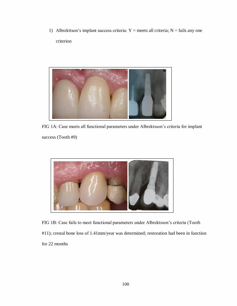
100
1) Albrekttson’s implant success criteria: Y = meets all criteria; N = fails any one
criterion
FIG 1A: Case meets all functional parameters under Albrektsson’s criteria for implant
success (Tooth #9)
FIG 1B: Case fails to meet functional parameters under Albrektsson’s criteria (Tooth
#11); crestal bone loss of 1.41mm/year was determined; restoration had been in function
for 22 months

101
2) Papilla fill: Y = Complete (75-100% papilla fill present); N = Incomplete (<75%
papilla fill present)
FIG 2A: Case demonstrates optimal papilla fill and height (75-100% papilla fill present),
as determined by clinical assessment
FIG 2B: Case demonstrates inadequate papilla fill and height (<75% papilla fill present),
as determined by clinical assessment

102
3) Soft tissue height discrepancy: Y = present; N = not present
FIG 3A: Case demonstrates soft tissue height discrepancy between the implant
restoration (tooth #9, maxillary left central incisor) and the contralateral reference tooth
#8 (maxillary right central incisor)
FIG 3B: Case demonstrates soft tissue height symmetry between the implant restoration
(tooth #10, maxillary left lateral incisor) and the contralateral reference tooth #7
(maxillary right lateral incisor)

103
4) Hard/soft tissue discrepancy: Y = present/visible; N = not present/visible
FIG 4A: Case demonstrates hard/soft tissue deficiency apical to the implant restoration
(tooth #11, maxillary left canine)
FIG 4B: Case demonstrates optimal hard/soft tissue profile apical to the implant
restoration (tooth #10, maxillary left lateral incisor)
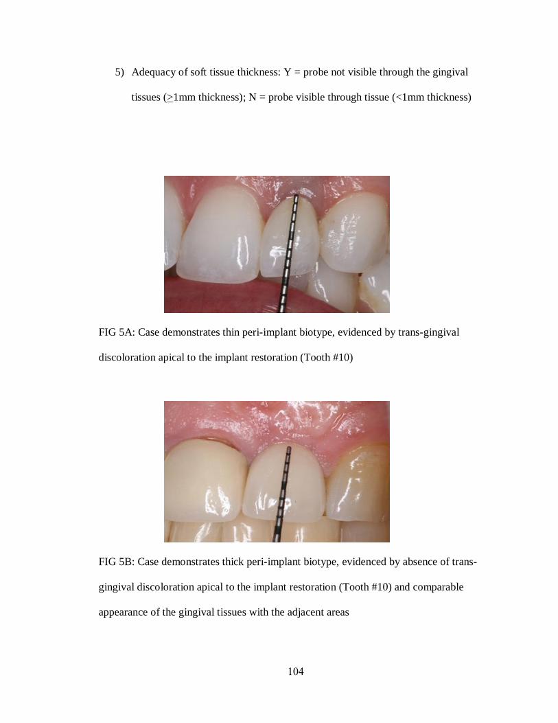
104
5) Adequacy of soft tissue thickness: Y = probe not visible through the gingival
tissues (>1mm thickness); N = probe visible through tissue (<1mm thickness)
FIG 5A: Case demonstrates thin peri-implant biotype, evidenced by trans-gingival
discoloration apical to the implant restoration (Tooth #10)
FIG 5B: Case demonstrates thick peri-implant biotype, evidenced by absence of trans-
gingival discoloration apical to the implant restoration (Tooth #10) and comparable
appearance of the gingival tissues with the adjacent areas

105
6) Gingival color/texture deviation: Y = deviation evident; N = no deviation noted
FIG 6A: Case demonstrates gingival color/texture deviation apical to the implant
restoration (tooth #6, maxillary right canine), when compared to the adjacent tissues;
FIG 6B: Case demonstrates gingival color/texture symmetry apical to the implant
restorations (tooth #7, maxillary right lateral incisor, and tooth #10, maxillary left lateral
incisor), when compared to the adjacent tissues;
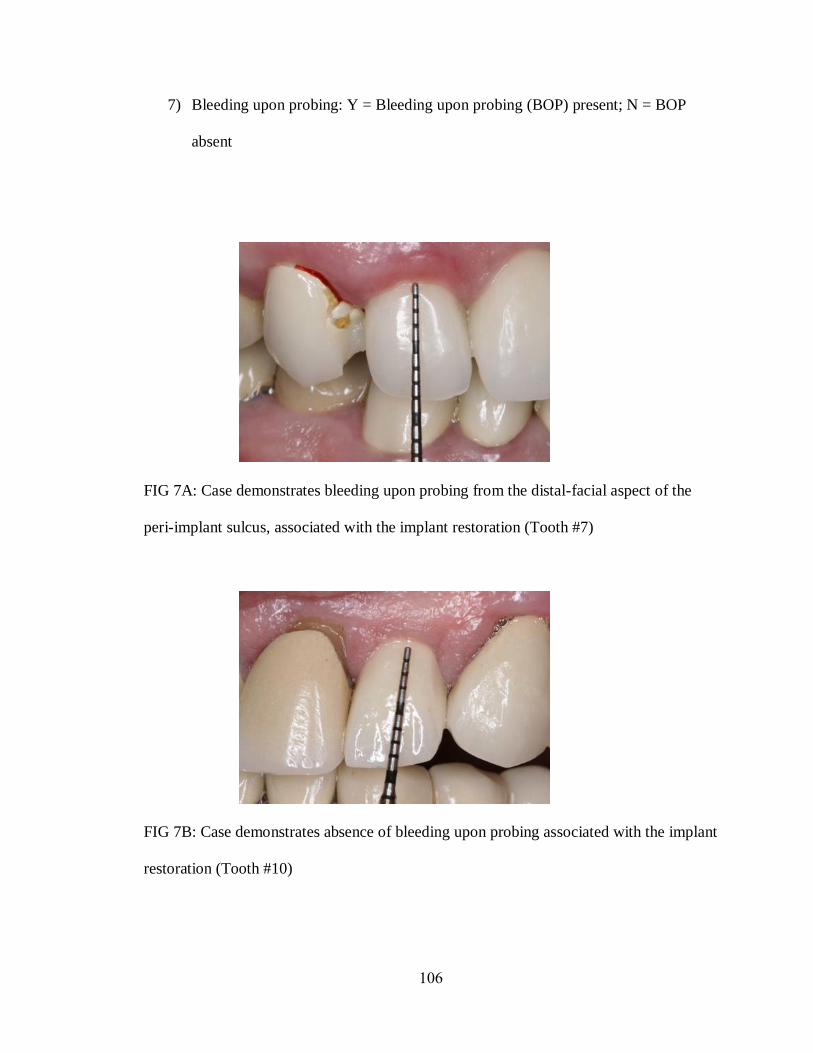
106
7) Bleeding upon probing: Y = Bleeding upon probing (BOP) present; N = BOP
absent
FIG 7A: Case demonstrates bleeding upon probing from the distal-facial aspect of the
peri-implant sulcus, associated with the implant restoration (Tooth #7)
FIG 7B: Case demonstrates absence of bleeding upon probing associated with the implant
restoration (Tooth #10)

107
8) Recession: Y = present; N = not present
FIG 8A: Case demonstrates recession apical to the margin of the implant restoration
(tooth #9, maxillary left central incisor), corresponding to the appearance of the
underlying titanium abutment/implant body seen beyond the gingival margin
FIG 8B: Case demonstrates absence of gingival recession apical to the implant restoration
(tooth #10, maxillary left lateral incisor)
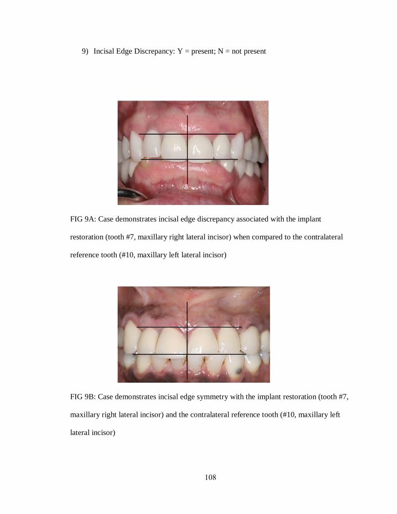
108
9) Incisal Edge Discrepancy: Y = present; N = not present
FIG 9A: Case demonstrates incisal edge discrepancy associated with the implant
restoration (tooth #7, maxillary right lateral incisor) when compared to the contralateral
reference tooth (#10, maxillary left lateral incisor)
FIG 9B: Case demonstrates incisal edge symmetry with the implant restoration (tooth #7,
maxillary right lateral incisor) and the contralateral reference tooth (#10, maxillary left
lateral incisor)
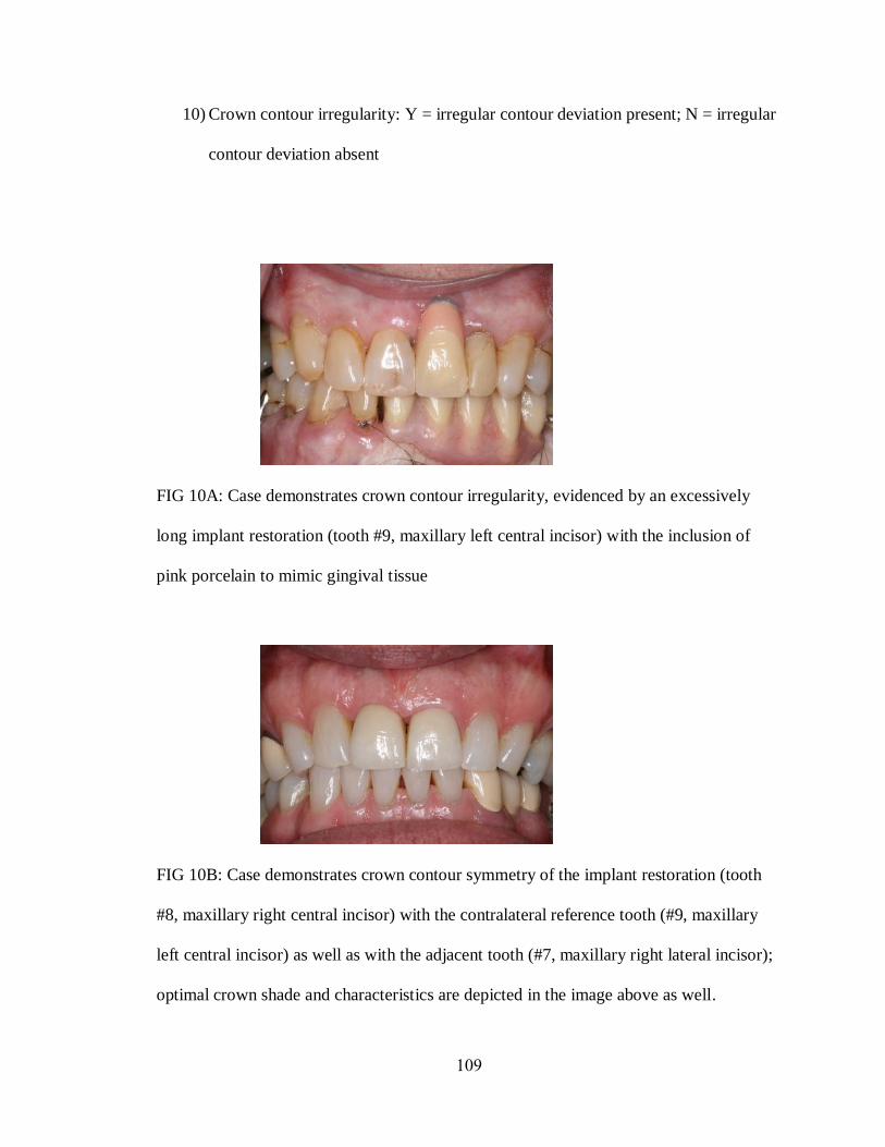
109
10) Crown contour irregularity: Y = irregular contour deviation present; N = irregular
contour deviation absent
FIG 10A: Case demonstrates crown contour irregularity, evidenced by an excessively
long implant restoration (tooth #9, maxillary left central incisor) with the inclusion of
pink porcelain to mimic gingival tissue
FIG 10B: Case demonstrates crown contour symmetry of the implant restoration (tooth
#8, maxillary right central incisor) with the contralateral reference tooth (#9, maxillary
left central incisor) as well as with the adjacent tooth (#7, maxillary right lateral incisor);
optimal crown shade and characteristics are depicted in the image above as well.