UNIVERSITI PUTRA MALAYSIA MOLECULAR …psasir.upm.edu.my/8384/1/FSMB_1998_1_A.pdf · Laboratory;...
25
UNIVERSITI PUTRA MALAYSIA MOLECULAR CHARACTERISATION OF ESCHERICHIA COLI SEROGROUP 0 157:H7 AHMAD ZAINURI MOHD DZOMIR FSMB 1998 1
-
Upload
phungkhuong -
Category
Documents
-
view
226 -
download
0
Transcript of UNIVERSITI PUTRA MALAYSIA MOLECULAR …psasir.upm.edu.my/8384/1/FSMB_1998_1_A.pdf · Laboratory;...

UNIVERSITI PUTRA MALAYSIA
MOLECULAR CHARACTERISATION OF ESCHERICHIA COLI SEROGROUP 0 157:H7
AHMAD ZAINURI MOHD DZOMIR
FSMB 1998 1
MOLECULAR CHARACTERISATION OF ESCHERICHIA COLI SEROGROUP 0 1 57:H7
By
AHMAD ZAINURI MOHD DZOMIR
Thesis Submitted in Fulfilment of the Requirements for the Degree of Master of Science in the Faculty
of Food Science and Biotechnology University Putra Malaysia
September 98

ACKNOWLEDGEMENTS
My deepest gratitude to my supervisor, Professor Dr. Gulam Rusul
Rahmat Ali for his continuous support, patience and advice given to me
throughout the course of this project. I also wish to thank my co
supervisors, Dr. Son Radu, for his valuable guidance and constant
encouragement, and Dr. Zaiton Hassan, for her suggestions and support.
Thanks also to all of the members and ex-members of the Microbiology
Laboratory; Ibu Endang, Liew Wang Shiang, Loke Chui Fung, Kak
Dayang, Kak Mai, Kak Sahilah, Sharmila, Sudha, Nimita, Rozila, Kamil,
Razali, Sam, Reddy, Senthil, Nasseer, Yuherman and Bushtari for their
kindness, friendship, helpful discussions and comments.
Lastly, I would like to thank my family, Pak Su, Mak Su and nenek for
their love and support.
ii

TABLE OF CONTENTS
Page
ACKNOWLEDGEMENTS. . . . . . . . . . . . . . . . . . . . . . . . . .. . . . . . . .. . . . . . . . . . . . . . . . . . . . . . ii
LIST OF TABLES. . . . . . . . . . . . . . . .. . . . . . . . . . . . . . . . . . . . . . . . . . . . . . . . . . . . . . . . . . . . . . . . . . . . vi
LIST OF PLATES. . . . . . . . . . . . . . . . . . . . . . . . . . . . . . . . . . . . . . . . . . .. . . . . . . . . . . . . . . .. . . . . . . . . V111
ABSTRACT . . . . . . . . . . . . . . . . . . . . . . . . . . . . . . . . . . . . . . . . . . . . . . . . . . . . . . . . . . . . . . . . . . . . . . . . . . . . ix
ABSTRAK. . . . . . . . . . .. . . . . . . .. . . . . . . . . . . . . . . . . . . . . . . .. . . . . . . . . . . . . . . .. . . . . .. . . . . . . . . . .. . xi
CHAPTER
I �ENE� INTR()J)l1CTI()N... ..... .. . ........ . . .. . . . . . . .. . . . .. .. . . .. 1
II LITE�T� �E�. . . .. . . . . ... .. . . . . . . ... . . . . .. . . . .. . . . . . . . . .. . ... !5 Microbiology of Escherichia coli 01!57:H7........................ !5
Biochemical Characteristics... . . . . . . . . . . . . . . . . . . . . . . . . . . . . . . . . . . . !5 Growth and Survival Properties. . . . . . . . . . . . . . . . . . . . . . . . .. . . . . . . . 7
Pathogenicity . . . . . . . . . . . . . .. . . . . . . .. . . . . . , . . . . . . . . . . . . , .. . . . . . , . . . . . . . . . . . . , . 8 Verocytotoxins . .. . . . . . . .. . . , . . . . . . . . . . . . . . . . . . . . . . . . . . . . . . . . .. . . , . . . .. . 9 Adherence and Attachment. . . . . . . . . . . . . . . . . . . . . . . . . . . . . . . . . . . . . . 1 0
Complication of E. coli 0 1!57:H7 Infections.. . . . . . . . . . . . . . . . . . . . . 1 2 Hemorrhagic Colitis.. . . . . . .. . . . . . . .. . . . . . . . . . . . . . . . . . . . .. . . . . . . . .. . . . 1 3 Hemolytic Uremic Syndrome (HUS).. . .. . . . .. . .. . . . . . .. . . . . .. . 14 Thrombotic Thrombocytopenic Purpura (TIP}.. . . . . . .. . . . 14
Epidemiology. . . .. . . . . . . . .. . . . . . .. . . . . . . .. . . . . . . .. . . . . . . . . . . . . . . . . . . . . . . . . . . . 1!5 Outbreak Investigations. . . . . . . . . . . . .. . . . . . . . . . . . . . . . . . . . . . . . . . . . . . . 1!5 Studies of Transmission. . . . . . . . . . . . . . . . . . . . . . . . . . . . . . . . . .. . . . . . . . . 1 9 Animal Reservoir. . . . . . . . . . . . . . . . . . . . . . . . . . . . . . . . . .. . . . . . . . . . . . . . . .. . . . 20
Detection and Characterisation of E. coli 0 1!57:H7 in Foods. . . . . . . . . . . . . . . . . . . . . . . . . .. . . . . . . . . . . . . . . . . . . . . . . .. . . . . . . . . . . . . . . . . . . . . . . . . 23
Conventional Methods.. . . . . . .. . . . . . . .. . . . . . . . . . . . . . . . . . . . .. . . . . . . .. 24 Alternative Methods... . . . . . . . . . . . . . . . . . . . .. . . . . . . . . . . . . . . . . . . . . . .. . . 27 Barriers to Implementation. . . .. . . . . . . . . . . . . . . . . . . . . . . . . . . . . . . . . . . 3 1
Typing of A New Clonally Derived Pathogen - Escherichia coli 0 1!57:H7. . . . . . . . . . .. . . . . . . . . . . . . . . . . . . . . . . . . . . . . . . . . . . . . . . . . . . . .. . . . . . . .. 32
Biochemical Promes. . . . . . . . . . . . . . . . . . . . . . . . . . . . . . . . . . . . . . . . .. . . . . . . . 33 Antibiotic Susceptibility Pattern and Plasmid Prome. . . 36 Pulsed-Field Gel Electrophoresis (PFGE} . . . .. . . . . . . . . . . . . '" 38 Arbitrarily Primed Polymerase Chain Reaction (AP-PCR).. . . . . . .. . . . . . . . . . . . . . . . . . . . . . . .. . . . . . . . . . . . . . . . . . . . . . . . . . . . .. . . . . . . . . . . 40
iii

Page
III ANTIBIOTIC SUSCEPTIBILITY PATTERN AND 43 PLASMID PROFILES . . . ... . . . . . . . . . . . . . . . . . . . . . . . . . .. . . . . . . . . . . . . . . . . . . . Introduction......... .. .. .. . . . . ............. .... ........ .... ...... ..... . ..... 43 Methods.............. ... . ............ . . . ... . . . . . ..... . . .......... . . ... .... . . .. 44
Bacterial Strains Used in this Study... ... . ...... .. ........ ... 44 Maintenance of Bacterial Strains....... .... . ....... . . .. . ....... 45 Antibiotic Discs......... ... . ............. .. . . .. . .... ........ . . . . . .. . . .. 45 Antibiotic Susceptibility Test. . . .... . . ...... . ....... . .... . . ..... .. 46 Plasmid DNA Isolation..... ................. .. . ..... . . . ... ..... .. ... 47 Agarose Gel Electrophoresis....... ... . . .. . . . . . . .. . . ........ . . . ... 48 Plasmid DNA Visualisation and Photography... ..... ... 49 Plasmid DNA Molecular Weight Determination......... .. 49
Results................... ........... ..... ...................................... 50 Susceptibilities of E. coli 0 1 57:H7 Isolates ........... . .... 50 Plasmid Promes of E. coli 0 157:H7 ........................... 54 Combination of Antibiotype and Plasmid Prome to Differentiate Isolates. . . .............................................. 57
Discussion ............. ................ , ... ...... ... ...... ............. ...... 60
IV. MACRORESTRICTION DIGESTION PATTERN BY PULSED-FIELD GEL ELECTROPHORESIS (PFGE). . . . . .. .. . .. . ... . . . .. . . . ... . . .... . . . .. . .. . . . . . . ... . . . .. .. . ... ..... ....... 65 Introduction......... ........... . . .... . . .. .. ............................ ..... 65 Methods....................... .................. .... ......................... . 66
Preparation of Intact Genomic DNA........................... 66 Digestion of the Agarose Embedded DNA............... .... 67 Electrophoresis................ ........................................ 67 DNA Bands Visualisation and Photography............. . .. 69 Genomic DNA Fragments Molecular Weight Determination ..... ...... ............... , ..... . ... ... ...... ............ . 69 Genetic Analysis... ... ... ...... ... ...... ... .. . ... . . . ... . . . . .. . .. ... ... 69
Results... ... ............... ....... . . . ..... ............ ........ ................. 70 Examining the Integrity of DNA Samples...... .. . ..... .. . . .. 70 Macrorestriction Digestion Pattern by PFGE............... 74 Genetic Distance between Different Macrorestriction Digestion Pattern..................................................... 80 Restriction Polymorphisms Resolved by PFGE Distinguished between Isolates............. .............. . . . ... 83
Discussion.. . ... . ............. ........... .. ... . ... ... . ........... ....... . . ... 87
iv

V DETECTION OF POLYMORPHISMS AMPLIFIED BY ARBITRARILY PRIMED POLYMERASE CHAIN
Page
REACTION (AP-PCR). . . . . . . . . . . . . . . . . . . . . . . . . . . . . . . . . . . . . . . . . . . . . . . . . . . . 90 Introduction . .. . . . ... .. . . . ..... . . . . . . . . . , . . . . . . . . . . . . . . . . . . . . . . . . . . . . . . . . . . . . 90 Methods....................................................................... 9 1
Bacterial Growth and DNA Extraction........................ 9 1 Spectrophotometric Quantitation of DNA................... 92 Standardisation of Primers Unit......... ....................... 92 AP-PCR Amplifications.............................................. 93 Electrophoresis.............................. ... . . . . . . . . . . . . . . . . . . . . . . . 94 Visualisation of DNA Bands and Photography............ 94
Results......................................................................... 95 Optimisation of Concentration of Arbitrary Primer, DNA template and Mg2+ Ion for Discriminatory Power...................................................................... 95 Identification of Useful Primers................................. 97 Strain Discrimination with Five 1 0-nt Primers with G+C Content of 50%. . . . . . . . . . . . . . . . . . . . . . . . . . . . . . . . . . . . . . . . . . . . . . . . . 99
Discussion .............. , . . . . . . .. . . . . . . . . . . . . . . . . . . . . . . . . . . . . . . . . . . . . . . . . . . . . 1 04
VI . GENERAL DISCUSSION AND CONCLUSION......... . . . . . . . . . 1 08
REFERENCES........................................................................ 1 12
VITAE.................................................................................... 1 23
v
LIST OF TABLES
Table
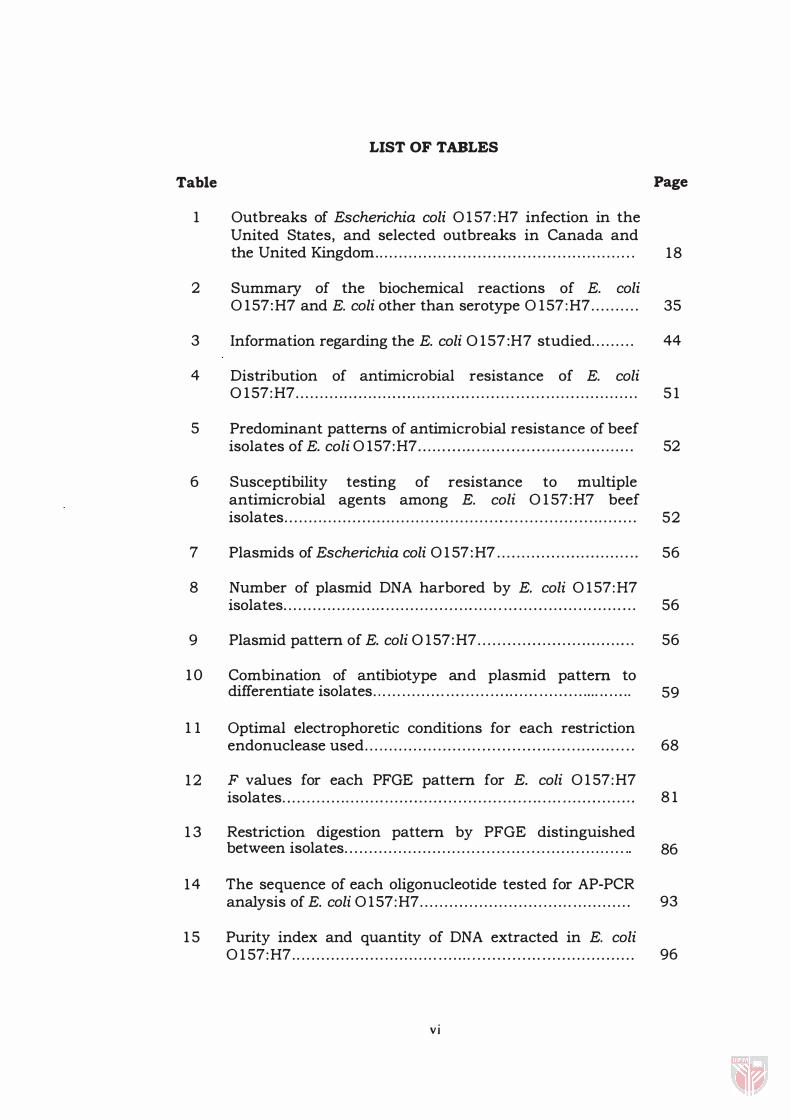
1 Outbreaks of Escherichia coli 0 1 57:H7 infection in the United States, and selected outbreaks in Canada and
Page
the United Kingdom. . . . . . . . . . . . . . . . . . . . . . . . . . . . . . . . . . . . . . . . . . . . . . . . . .. . . 18
2 Summary of the biochemical reactions of E. coli 0 1 57:H7 and E. coli other than serotype 0 1 57:H7. . . . . . . . . . 35
3 Information regarding the E. coli 0 1 57:H7 studied. . . . . . . . . 44
4 Distribution of antimicrobial resistance of E. coli 0 1 57:H7. . . . . . . . . . . . . . . . . . . . . . . . . . . . . . . . . . . . . . . . . . . . . . . . . . . . . . . . . . . . . . . . . . . . . . 5 1
5 Predominant patterns of antimicrobial resistance of beef isolates of E. coli 0 157:H7. . . . . . . . . . .. . . . . . . . . . . . . . . . . . . . . . . . . . . . . . . . . 52
6 Susceptibility testing of resistance to multiple antimicrobial agents among E. coli 0 1 57:H7 beef isolates. . . . . . . . . . . . . . . . . . . . . . . . . . . . . . . . . . . . . . . . . . . . .. . . . . . . . . . . . . . . . . . . . . . . . . . . 52
7 Plasmids of Escherichia coli 0 1 57:H7 . . . . . . . . . . . . . . . . . . . . . . . . . .. . . 56
8 Number of plasmid DNA harbored by E. coli 0 1 57:H7 isolates . . . . . . . . . . . . . . . . . . . . , . . . . . . . . . . . , . . . . . , . . . . . . . . . . . . . . . . . . . . . . . . . . . . . . . . . 56
9 Plasmid pattern of E. coli 0 1 57:H7. . . . . . . . . . . . . . . . . . . . . . . . . . . . . .. . 56
1 0 Combination of antibiotype and plasmid pattern to differentiate isolates. . . . . . . . . . . . . . . . . . . . . . . . . . . . . . . . . . . . . . . . . . . . . . . . . . . . . . 59
1 1 Optimal electrophoretic conditions for each restriction endonuclease used. . . . . . . . . . . . . . . . . . . . . . . . . . . . . . . . . . . . . . . . . . . . . . . . . . . . . . . 68
1 2 F values for each PFGE pattern for E. coli 0 1 57:H7 isolates . . . . . . . . . . . . . . . . . . . . , . . . . . . . . . . . . . . . . . . . . . . . . . . . . . . . . . . . . . . . . . . . . . . . . . ,. 8 1
1 3 Restriction digestion pattern by PFGE distinguished between isolates. . . . . . . . . . . . . . . . . . . . . . . . . . . . . . . . . . . . . . . . . . . . . . . . . . . . . . . . . .. 86
14 The sequence of each oligonucleotide tested for AP-PCR analysis of E. coli 0 1 57:H7. . . . . . . . . . . . . . . . . . . . . . . . . . . . . .. . . . . . . . . . . . . 93
1 5 Purity index and quantity of DNA extracted in E. coli 0 1 57:H7. . . . . . . . . . . . . . . . . . . . . . . . . . . . . . . . . . . . . . . . . . . . . . . . . . . . . . . . . . . . . . . . . . . . . . 96
vi

16 AP-PCR profile pattern and genetic types of E. coli
Page
0157:H7. .......... . ... ................. . ......... ........................... . 103
vii

LIST OF PLATES
Plate Page
1 Plasmid profiles of E. coli 0 1 57:H7 isolates . . . . . . . . . . . . . . . . . . . . 55
2 Screening of the 14 chromosomal DNAs of E. coli 0 1 57:H7 for integrity. . . . . . . . . . . . . . . . . . . . . . . . . . . . . . . . . . . . . . . . . . . . . . . . . . . 72
3 PFGE of the 10 E. coli 0 1 57:H7 genomic samples after performing a mock digestion. . . . . . . . . . . . . . . . . . . . . . . . . . . . . . . . . . . . . . . . 73
4 Comparison of Xbal digests of genomic DNA of E. coli 0 1 57:H7 beef isolates. . . . . . . . . . . . . . . . . . . . . . . . . . . . . . . . . . . . . . . . . . . . . . . . . . 77
5 Comparison of SpeI digests of genomic DNA of E. coli 0 1 57:H7 beef isolates. . . . . . . . . . . . . . . . . . . . . . . . . . . . . . . . . . . . . . . . . . . . . . . . . . 78
6 Comparison of HindIII digests of genomic DNA of E. coli 0 1 57:H7 beef isolates. . . . . . . . . . . . . . . . . . . . . . . . . . . . . . . . . . . . . . . . . . . . . . . . . . 79
7 Results of tests to identify useful primers for AP-PCR analysis. . . . . . . . . . . . . . . . . . . . . . . . . . . . . . . . . . . . . . . . . . . . . . . . . . . . . . . . . . . . . . . . . . . . . . 98
8 Representative arrays of fragments generated by arbitrary primer PCR with primer GEN 1 5002.
9 Random amplified polymorphic DNA patterns of 1 5 E.
1 00
coli 0 1 57:H7 beef isolates using primer GEN 1 5003. . . . . . . . 1 0 1
1 0 Representative arrays of fragments generated by arbitrary primer PCR with primer GEN 1 5009.
viii
102

Abstract of thesis presented to the Senate of Universiti Putra Malaysia in fulfilment of the requirement for the degree of Master of Science
MOLECULAR CHARACTERISATION OF ESCHERICHIA COLI
SEROGROUP 0 157:H7
BY
AHMAD ZAINURI MOHD DZOMIR
SEPTEMBER 1998
Chairman: Professor Dr. Gulam Rusul Rahmat Ali
Faculty: Food Science and Biotechnology
Fourteen E. coli 0 1 57:H7 beef isolates were characterised by using
four specific epidemiological markers: combination of antibiogram and
plasmid profiling, pulsed-field gel electrophoresis (PFGE) and arbitrary
primed polymerase chain reaction (AP-PCR). These markers were
assessed for their reliability, typability, rapidity and discriminatory
power in differentiating beef E. coli 0 1 57:H7 strains from different
locations, namely Bangsar, Kajang, Petaling Jaya and Serdang. The
majority of the isolates were resistant to penicillin G ( 1 00%), vancomycin
( 100%) , trimethoprim/ sulphametoxazole ( 100%) , bacitracin ( 100%) and
erythromycin (92 .8%) . Only 2 1.4% were resistant to carbenicillin. 1 4.3%
and 7. 1% were resistant to ampicillin and cephalotin, respectively.
ix

Plasmid analysis revealed three basic plasmid patterns among E. coli
0 1 57:H7 strains, profile 1 characterised by plasmid DNA of 60 and 2.5
MDa, profile 2 characterised by plasmid of 60 MDa, and profile 3
characterised by the absence of any plasmid in the strains. Grouping
according to combination of antibiogram and plasmid analysis indicated
eight different groups as two strains with similar antibiotype could be
distinguished into two different strains by their dissimilar plasmid
profile. However, the reliability of antibiogram and plasmid analysis in
typing E. coli 0 1 57:H7 can be questioned. Thus, other reliable methods
such as PFGE and AP-PCR were then applied. In the present study,
macrorestriction of genomic DNA of E. coli 0 1 57:H7 using Xbal, SpeI and
HindIII and analysed by PFGE successfully grouped ten out of fourteen
isolates into five groups and provided evidence of epidemiologically
related strains between strains of different and same locations. However,
AP-PCR using three short primers grouped the isolates into fourteen
distinct groups and differentiates isolates that were not differentiated by
PFGE. The overall analysis of the present study revealed AP-PCR as the
most suitable method to differentiate E. coli 0 1 57:H7 because it was
more discriminatory, less labor intensive and applicable to all isolates.
Using this method, it was clearly shown that all fourteen E. coli 0 1 57:H7
existed as independent isolates.
x

Abstrak tesis yang dikemukakan kepada Senat Universiti Putra Malaysia sebagai memenuhi keperluan untuk Ijazah Master Sains
PENCIRIAN MOLEKULAR ESCHERICHIA COLl SEROGROUP 0157:H7
OLEH
AHMAD ZAINURI MOHD DZOMIR
SEPTEMBER 1998
Pengerusi: Professor Dr. Gulam Rusul Rahmat Ali
Fakulti: Sains Makanan dan Bioteknologi
Empat belas isolat E. coli 0 1 57:H7 diciri menggunakan empat metod
epidemiologi iaitu gabungan ujian ketahanan terhadap antibiotik dan
analisis plasmid, PFGE dan AP-PCR. Marker diuji dari segi kemantapan,
kebolehan pencirian, kepantasan dan nilai pemisahan untuk
membezakan E. coli 0 1 57:H7 yang diisolat dari pelbagai tempat seperti
Bangsar, Kajang, Petaling Jaya dan Serdang. Kebanyakan isolat adalah
resistan kepada penicillin G ( 100%), vancomycin ( 100%), trimethoprim/
sulphametoxazole ( 1 00%) , bacitracin ( 100%) dan erythromycin (92 . 8%) .
Hanya 2 1 .4% resistan kepada carbenicillin. 1 4 .3% dan 7 . 1% resistant
kepada ampicillin dan cephalotin masing-masing. Analisis plasmid
menunjukkan tiga corak asas di antara E. coli 0 1 57:H7, profil 1 diciri
oleh kehadiran dua plasmid bersaiz saiz 60 MDa dan 2 .5 MDa, profil 2
xi

diciri oleh kehadiran satu plasmid bersaiz 60 MDa dan profil 3 diciri
oleh ketidakhadiran plasmid. Pengkelasan mengikut penggabungan
antibiograrn dan analisis plasmid menunjukkan lapan kumpulan
bardasarkan pada pendapat yang mengatakan dua isolat yang
mempunyai antibiotaip yang serupa boleh dibezakan dengan profil
plasmid yang berlainan. Walaubagaimanapun, kebolehan antibiograrn
dan analisis plasmid untuk menciri E. coli 0157:H7 boleh dipersoalkan.
Jadi, metod lain yang lebih mantap seperti PFGE dan AP-PCR telah
dicuba. Dalarn kajian ini, pemotongan DNA genomik E. coli 0157:H7
menggunakan enzim Xbal, SpeJ dan HindIII, dan dianalisis
menggunakan PFGE mengkelaskan sepuluh dari empatbelas isolat
kepada lima kumpulan dan memberi bukti perkaitan epidemiologi
antara isolat yang dipencil dari temp at yang sarna atau pun berlainan.
Dengan menggunakan AP-PCR dan tiga primer pendek, semua isolat
berjaya dibezakan ke dalarn empat belas kumpulan yang berlainan dan
ia juga dapat memisahkan isolat yang tidak boleh dibezakan dengan
PFGE. Secara keseluruhan, kajian ini mendapati bahawa AP-PCR
merupakan metod yang paling sesuai untuk membezakan E. coli
0157:H7 kerana ianya lebih cepat, mempunyai nilai pemisahan yg lebih
tinggi, kurang memerlukan tenaga kerja dan boleh diaplikasikan keatas
semua isolat. Dengan metod ini, arnat jelas ditunjukkan bahawa
empatbelas isolat yang dikaji tidak mempunyai perkaitan di antara
sarna satu lain.
xii

CHAPTER I
GENERAL INTRODUCTION
The bacteria constituting the species Escherichia coli were first
discovered by a German microbiologist, Theobald Escherich in 1 885 and
were commonly thought as normal flora of man and animals (Sojka,
1 965) . However, until late 1950's, certain strains were found to be
capable of inducing disease, and E. coli were therefore regarded as a
potential pathogen. These strains are classified into several groups based
on their distinct clinical manifestations and virulence determinants, for
example, the enterotoxigenic E. coli (ETEC) , enteropathogenic E. coli
(EPEC) and enteroinvasive E. coli (EIEC) (Olsvik et al., 1 99 1 ).
In 1 982, following two outbreaks of a distinctive bloody diarrheal
syndrome and a sporadic case of bloody diarrhea, a new bacterial
pathogen, E. coli 0 1 57:H7 was identified (Riley et al. , 1 983). E. coli
0 1 57:H7 comprises a fourth group of E. coli associated with human
diarrhea, i .e., enterohemorrhagic E. coli (EHEC). The first two outbreaks
that occurred in Michigan and Oregon, involved a national fast-food
chain distributing hamburgers. Since then, several additional outbreaks
have been reported in other parts of United States (Ryan et al. , 1 986,

2
Griffin et al. , 1988), Canada (Borczyk et al. , 1987) and with increased
surveillance, E. coli 0157:H7 outbreaks have also being reported in other
parts of the world including Mexico (Cravioto et aI. , 1990), China (Xu et
al. , 1990), Argentina (Lopez et al. , 1989), Belgium (Pierrard et al. , 1990)
and Malaysia (Son et al. , 1996).
As the incidence of by E. coli 0157:H7 is in the increase in many
parts of the world, specific surveillance of this pathogen is essential. For
identifying the sources and monitoring the spread of E. coli 0157:H7, a
number of epidemiologic markers have been use, including antibiotic
susceptibility pattern (antibiotype) , plasmid profile, pulsed field gel
electrophoresis (PFGE) , biotype, phage typing and restriction analyses of
chromosomal DNA by classical electrophoresis. The use of epidemiologic
markers enables the bacterial stains to be typed and establish degree of
relatedness between the strains.
As phenotypic systems have limitations in typing and stability, and
most typing systems on their own have a low discriminatory ability,
therefore, in the present study, we combined antibiotic susceptibility
pattern and plasmid profile for subtyping E. coli 0157:H7 beyond their
serotype. Among genotypic analysis techniques, PFGE of
macrorestriction fragments appears to be a highly sensitive method that
may detect subtle genetic variations among phylogenetically and
epidemiology related isolates of E. coli OI57:H7.
3
Recently, a polymerase chain reaction (PCR)-based method of
genotypic analyses that is sensitive and yet efficient for typing pathogenic
microbe has become very popular because of its convenience and
simplicity. Arbitrarily primed-PCR has been reported to be useful for
tracing route of infection and also for understanding the spread of
pathogen, especially 0157:H7 serotype. In the present study, we attempt
to type strains of E. coli 0157:H7 isolated from different samples and
locations according to three methods, namely the combination of plasmid
proflle and antibiotype, PFGE and arbitrarily primed (AP)-PCR. The study
here is motivated by the view that E. coli 0157:H7, though members of
one clone, can often be distinguished by methods that have been clarified
by previous workers.

4
Objectives of the Study
This study is carried out to determine the relatedness of a group of
14 E. coli 0157:H7 beef isolates using molecular techniques. The isolates
are from Bangsar, Kajang, Serdang and Petaling Jaya. Three methods are
used; combination of plasmid profiling and antibiotic susceptiblity
pattern, macrorestriction digestion pattern by PFGE and arbitrarily
primed polymerase chain reaction (AP-peR) to detect polymorphisms in
their genome. The results from each method are then discussed in order
to determine which is the most discriminative for the purpose of strain
differentiation.
CHAPTER II
LITERATURE REVIEW
Microbiology of Escherichia coli 0 157:H7
The strains of E. coli 0157:H7 that were isolated during the outbreak
in the Northwest Pacific, United States of America are typical of most E.
coli, with some exceptions. E. coli serotype 0157:H7 possess biochemical
markers, growth and survival characteristics that are significantly
different from those of other E. coli strains.
Biochemical Characteristics.
The inability to ferment sorbitol and the absence of f3-glucuronidase
activity are believed to be a specific phenotypic feature of E. coli
0157:H7. More than 90 % of E. coli isolates of human origin ferment
sorbitol within 24 hours; howeyer, E. coli 0157:H7 does not (Johnson et
al. , 1983; Ratnam et al., 1988). Furthermore, other workers reported that
E. coli 0157:H7 failed to ferment sorbitol as late as seven days (Wells et
al., 1983). Workers exploited this biochemical feature and developed a
modified MacConkey agar containing D-sorbitol (SMAC) instead of
5
6
lactose to detect E. coli 0 1 57:H7 (March and Ratnam, 1 986; Farmer and
Davis, 1 985) . The strain is similar to other E. coli on the lactose
containing agar such as Eosin Methylene Blue Agar (EMBA) making it
very difficult to differentiate between them. However, on SMAC agar E.
coli 0 1 57:H7 could be recognised as colorless colonies showing that they
failed to ferment sorbitol. Ratnam et al. ( 1988) described supplementary
biochemical markers, such as lysine and ornithine decarboxylation to
increase the specificity of the sorbitol-only screened E. coli 0 1 57:H7. The
inclusion of these tests reduced the number of organism to be serotyped
and improved the specificity of the biochemical screen to 33.6% (Haldene
et ai., 1 986) .
In addition, E. coli 0 1 57:H7 strains are different from other E.
coli strains, as they do not posses �-glucuronidase activity. More than
90% of E. coli produce the enzyme �-glucuronidase, which is the basis
for a rapid flu orogenic assay for E. coli (Feng and Hartman, 1 982) . The
indicator, 4-methyl-umbelliferone glucuronide (MUG) used in this assay
is hydrolysed by �-glucuronidase enzyme possess by normal E. coli and
converted into 4-methylumbelliferone that fluoresces under radiation
with ultra-violet (UV) light (366 nm). In contrast to most E. coli strains, E.
coli 0 1 57:H7 is not capable of producing �-glucuronidase. When
irradiated with long wave UV light, no fluorescence is formed, hence E.
coli 0 1 57:H7 could be differentiated from other E. coli.
7
Growth and Survival Properties
Doyle and Shoeni ( 1984) showed that E. coli 0 1 57:H7 can survive
well up to 9 months at -20°C and at -80°C in ground beef. They also
observed another important characteristic of E. coli 0 1 57:H7 i .e . , they
grew poorly at 44-44.5°C, which is the temperature generally used for the
isolation of E. coli 0 1 57:H7. Hence, traditional procedures for detecting
E. coli in foods would not likely detect E. coli 0 1 57:H7. Temperature
range of 16.4 to 42 .5°C and 48 hours incubation has been suggested by
Raghukeer and Matches ( 1990) for detecting E. coli 0 157:H7 in foods.
E. coli 0 1 57:H7 has no unusual heat resistance. Most of the
organisms will be killed if the food was pasteurised or heated to some
extent. D' Aoust et al. ( 1 988) observed that more than 1 04 E. coli
0 1 57:H7 per ml were killed following pasteurisation of milk (72°C,
16 .2s). In addition, Kotula et al. ( 1977) observed ground beef patties that
were undercooked 'well done' (4 minutes per side at 1 49°F griddle
temperature) showed a reduction in the coliform count from four logs to
less than 1 / gram. Coliform count in retail raw ground beef are viable,
but may be expected to be 1 000/gram or more (Restaino and Lyon,
1 987) . This means that high initial coliform counts that results in a rare
hamburger are likely to permit the survival of coliforms after cooking.

8
Outbreaks of E. col i 0 1 57:H7 caused by drinking apple cider
indicates that E. col i 0 1 57:H7 is resistant to acidic pH and distinguished
0 1 57: H7 serotype from other E. col i (Miller and Kaspar, 1 994) . E. col i
0 1 57:H7 can grow at pH levels ranging from 4.0 to 9 .0 (Glass et al . ,
1 992) which is not significantly different from the pH range of 4 .4 to 9 .0
reported for other E. col i. However, the ability of E. col i 0 1 57:H7 to
survive at a pH of less than 4.0 was reported by Miller and Kaspar
( 1994) . Their study revealed that E. col i 0 1 57:H7 can survive in
unpasteurised apple cider at pH 3.6 to 4 .0 for as long as 3 1 days at BOC.
Similarly, Zhao and Doyle ( 1994) observed that E. col i 0 1 57:H7, when
initially present at 6 .5 x 1 03 CFU/g, can survive in mayonnaise (pH 3.6
to 3 .9) at 20°C for 2 1 days and at 5°C for 55 days.
Pathogenicity
The pathogenesis of infections with E. col i 0 1 57:H7 and other
enterohemorrhagic E. col i is not completely understood. E. col i 0 1 57:H7
do not elaborate the heat-labile and heat-stable enterotoxins that are
produced by enterotoxigenic E. col i and not enteroinvasive, as judged by
both Sereny test and the absence of invasion into epithelial cells in tissue
culture. However, E. col i 0 1 57:H7 do produce high levels of cytotoxin,
variously termed as verotoxin or Shiga-like toxin. In addition to toxin,
adherence to mucosal surfaces in the gastrointestinal tract is an

9
important primary step that results in bacterial colonisation of the
intestine necessary for delivery of elaborated toxins to enterocytes.
Verocytotoxins
Verocytotoxin-producing E. coli (VTEC) were first described by
Konowalchuk (1977), who identified a cytotoxin active on cultured Vero
cells (African green monkey kidney cells) and produced by certain strains
of E. coli; it was termed Vero cytotoxin (VT) . VT is clearly
indistinguishable, biologically and immunogically from the heat stabile
(ST) and heat-labile (LT) enterotoxins of E. coli, and reported to be closely
related to Shiga toxin which is produced by strains of Shigella dysentriae
type 1. Hence, these toxins were designated as Shiga-like toxins. Both
Shiga and Shiga-like toxin share the same receptor, and have similar
structure and modes of action (Robinson et al. , 1980). The gene coding
for Shiga-like toxin I is very similar to that of Shiga toxin, while the gene
for Shiga-like toxin II shows 58 percent overall homology with that for
Shiga-like toxin.
Shiga-like toxins from E. coli 0157:H7 were first reported in 1983
when OBrien and co-workers found that isolates from two outbreaks in
the United States produced high levels of a cell-associated cytotoxin for
HeLa and Vero cells. On further analysis, it is clear that this organism
produces two kind of cytotoxins, one of which can be neutralised by anti-
10
Shiga toxin. The neutralisable cytotoxin was designated as Shiga-like
toxin I (SLT-I), and the other Shiga-like toxin II (SLT-II). If both toxins are
produced by the same strains, SLT-I predominates in cell lysates while
SLT-II is more active toxin in culture filtrate. The two Shiga-like toxins
are antigenically distinct and differ in their biological effects: Shiga-like
toxin II is less toxic to Vero cells, but more toxic for mice, and causes
hemorrhagic colitis in the adult rabbit, while Shiga-like toxin I does not
(Evans et al., 1977) .
Phage conversion is responsible for controlling the production of
several important bacterial toxins, including diphtheria toxin,
streptococcal erythrogenic toxin, botulinum toxin and staphylococcal
enterotoxin (Betley et al. , 1986) . Recently, the production of Shiga-like
toxin was shown to be determined by specific phages in selected strains
of both EPEC and EHEC (0 1 57) serotype isolated from humans (Smith et
ai. , 1983) which indirectly suggests that E. coli 0 1 57:H7 may have
acquired these toxins through phage mediated transfer (Strockbine et ai. ,
1986) .
Adherence and Attachment
Another property that makes E. coli 0 1 57:H7 virulent is its ability to
adhere to intestinal cells. The mechanism of bacterial attachment to the
intestinal mucosal cell remains controversial because there is no direct

II
evidence from human cases to asses the nature of intestinal colonisation
by E. col i 0 1 57:H7. However, experiments to observe the phenomenon
have been conducted in animal and cell cultures.
Studies in gnotobiotic piglets and cell cultures showed no evidence
of the extensive invasion and intracellular multiplication as seen with the
invasive bacteria such as Shigella (Tzipori et al . , 1 986). E. col i 0 1 57:H7
produce a distinctive microscopic lesion characterised by intimate
attachment of the bacteria to the apical intestinal mucosal cell and
localised destruction of the microvilli (Tzipori et al., 1 988; Tzipori et al . ,
1 989). The bacteria also exhibit localised adherence to cells in culture,
with dense concentration of actin microfilaments in a cup-like structure
in the cytoplasm beneath the attached bacterium (Knutton et ai. , 1 989) .
This attaching-efficacing lesion resembles that produced by
enteropathogenic E. coli strains in piglets and cell culture (Knutton et al.,
1 989; Tzipori et al . , 1989) .
The mechanism may indicate an initial contact followed by more
intimate attachment, such as has been described by enteropathogenic E.
col i (Knutton et ai. , 1989) . The initial attachment may be mediated by the
60 MDa plasmid while the intimate attachment may be chromosomally
mediated (Toth et al., 1 990) . Most E. col i 0 1 57:H7 carry a 60 MDa
plasmid . There are several different opinions on the role of the plasmid.
Karch et ai. ( 1 987) determined that the plasmid was required for
1 2
expression of a fimbrial adhesin and adherence to Henle 407 intestinal
cells. The strains that were cured of the plasmid failed to express
fimbriae and lost the ability to adhere to intestinal cells. Recently, Toth et
al. ( 1 990) determined that the 60 MDa plasmid appear to modify the
eucaryotic cell adherence of E. coli 0 1 57:H7 and that adherence was
conferred to an E. coli transformant.
However, Junkin and Doyle ( 1 989) revealed that adherence to Henle
407 cells by E. coli 0 1 57:H7 strain 932 was not dependent on the 60
MDa plasmid mentioned. They showed that the test strain 932 adheres
to the human small intestine cell line INT407 at an average level of 7 . 1
bacteria per INT407 cell without the strain harboring the plasmid.
Complications of E. coli 0157:H7lnfections
Since the occurrence of the outbreaks in 1 982 , continued
epidemiologic, clinical, and laboratory investigations clearly established
E. coli 0 1 57:H7 to be an important etiologic agent of hemorrhagic colitis.
Typical hemorrhagic colitis can be distinguished clinically from bloody
diarrhea or dysentery seen in shigellosis, Campylobacter spp. and
enteroinvasive E. coli enteritis, amoebiasis, or other enteric illnesses such
as narcrotising enterocolitis or pseudomembraneous colitis by the lack of
prominent fever (Riley, 1987) . The illness caused by E. coli 0 1 57:H7
resolves in most patients with no sequel. Early outbreak studies in which