UNIT XII – ANIMAL PHYSIOLOGY II Digestive, Reproductive, Nervous, Muscular Systems Big Campbell...
61
UNIT XII – ANIMAL PHYSIOLOGY II Digestive, Reproductive, Nervous, Muscular Systems Big Campbell – Ch. 41, 46, 47, 48, 49, 50 Baby Campbell – Ch 21, 27, 28, 30 Hillis – Ch 32, 33, 34, 36, 39
Transcript of UNIT XII – ANIMAL PHYSIOLOGY II Digestive, Reproductive, Nervous, Muscular Systems Big Campbell...

UNIT XII – ANIMAL PHYSIOLOGY IIDigestive, Reproductive, Nervous, Muscular Systems
Big Campbell – Ch. 41, 46, 47, 48, 49, 50Baby Campbell – Ch 21, 27, 28, 30
Hillis – Ch 32, 33, 34, 36, 39
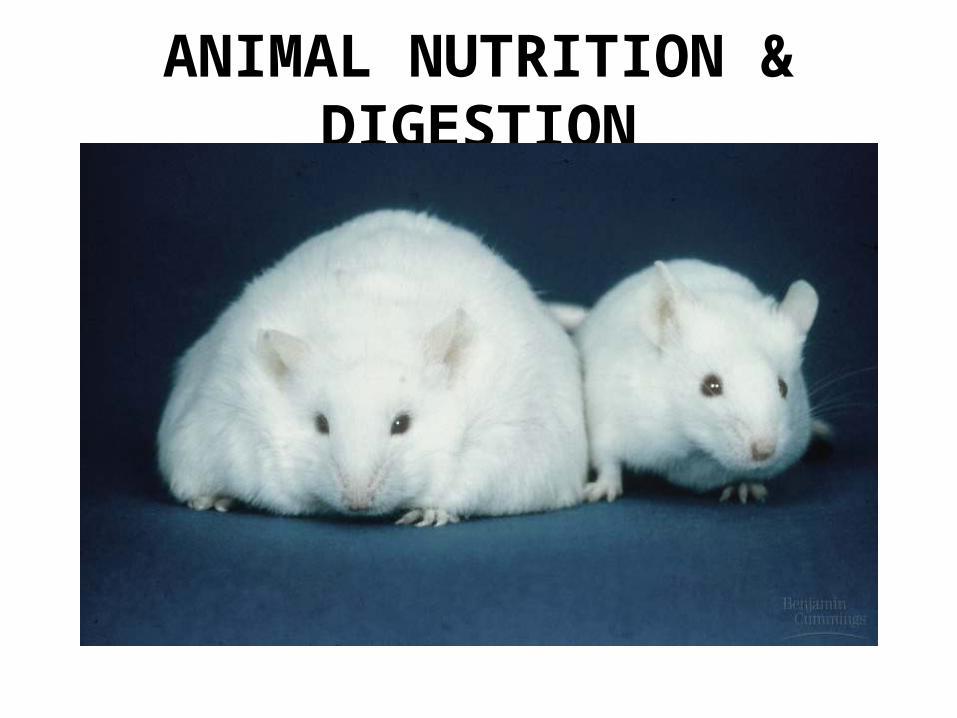
ANIMAL NUTRITION & DIGESTION
I. NUTRITION• Undernourishment
Caloric deficiency• Overnourishment
Excessive food intake Obesity
• Malnourishment Essential nutrient deficiency
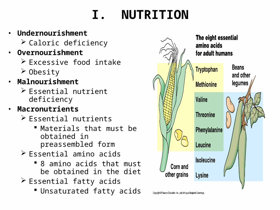
• Macronutrients Essential nutrients
Materials that must be obtained in preassembled form
Essential amino acids 8 amino acids that must be
obtained in the diet Essential fatty acids
Unsaturated fatty acids

I. NUTRITION, cont
• Micronutrients: Vitamins - Organic coenzymes
Water Soluble: B Vitamins – Required for general metabolism Vitamin C – Required for connective tissue production
Fat Soluble: Vitamin A – Vision Vitamin D – Ca2+
Vitamin E - ??? Vitamin K – blood clotting
Minerals - Inorganic cofactors Na Ca Fe K P I Cl
I. NUTRITION, contFeeding Types & Adaptations
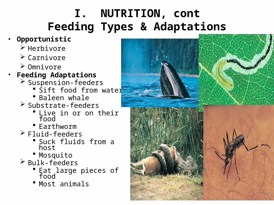
• Opportunistic Herbivore Carnivore Omnivore
• Feeding Adaptations Suspension-feeders
Sift food from water Baleen whale
Substrate-feeders Live in or on their food Earthworm
Fluid-feeders Suck fluids from a host Mosquito
Bulk-feeders Eat large pieces of food Most animals

II. DIGESTIONOverview Of Food Processing
• Ingestion• Digestion
Enzymatic hydrolysis Intracellular: breakdown within cells (sponges) Extracellular: breakdown outside cells (most animals)
Gastrovascular cavity vs. alimentary canal • Absorption• Elimination
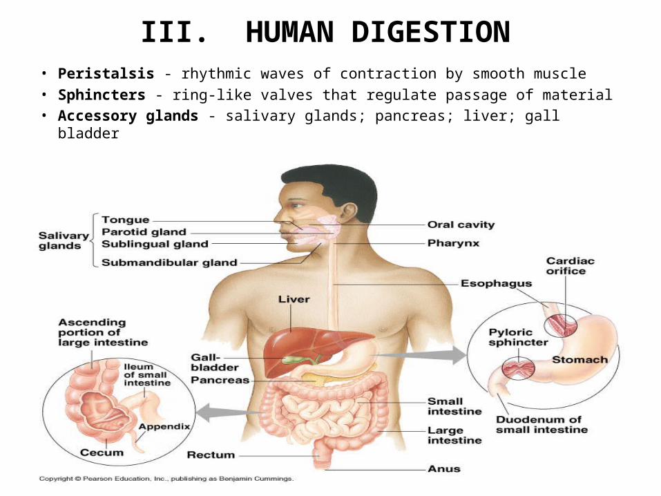
III. HUMAN DIGESTION• Peristalsis - rhythmic waves of contraction by smooth muscle• Sphincters - ring-like valves that regulate passage of material• Accessory glands - salivary glands; pancreas; liver; gall bladder
III. HUMAN DIGESTION, cont
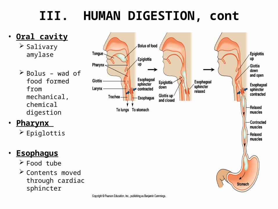
• Oral cavity Salivary amylase
Bolus – wad of food formed from mechanical, chemical digestion
• Pharynx Epiglottis
• Esophagus Food tube Contents moved
through cardiac sphincter

III. HUMAN DIGESTION, cont
• Stomach Gastric juices – Made up
of Mucus
Pepsin/pepsinogen
HCl
Partially-digested stomach contents known as chyme
Pass through pyloric sphincter to small intestine
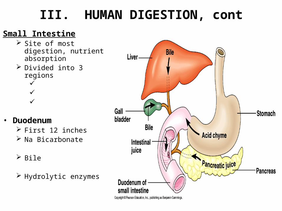
III. HUMAN DIGESTION, cont
Small Intestine Site of most digestion,
nutrient absorption Divided into 3 regions
• Duodenum First 12 inches Na Bicarbonate
Bile
Hydrolytic enzymes

III. HUMAN DIGESTION, cont• Jejunun & Ileum
Villi/microvilli Contain vessels from circulatory system , lymphatic system
Nutrient absorption carried out through diffusion, active transport Capillary networks
Amino acids, monosaccharides aborbed into circulatory system Transported to liver via hepatic portal vein
Lacteal Vessels from lymphatic system Transport chylomicrons – water-soluble droplets of fats mixed with cholesterol
• Hepatic portal vessel – carries contents from nutrient-rich capillaries to liver

III. HUMAN DIGESTION, cont

III. HUMAN DIGESTION, cont
• Large Intestine or Colon Cecum / Appendix Water reabsorbed from secreted
digestive juices Contents moved along by peristalsis
Diarrhea Constipation
Huge population of normal bacterial flora
Feces – undigested food (cellulose), dead bacteria
• Rectum Waste storage 2 sphincters Waste expelled through anus

III. HUMAN DIGESTION, cont

III. HUMAN DIGESTION, cont• Hormones Involved in Digestion
– Leptin Produced by adipose cells Increased amount of adipose tissue = increased levels of leptin = decreased appetite
– Gastrin Produced by stomach Food triggers release of gastrin → returns to stomach wall → stimulates secretion of
gastric juice– Enterogastrone
Produced by duodenum Inhibits peristalsis and acid secretion by stomach, slows digestion when chyme with high
fat concentration enters duodenum– Secretin
Produced by duodenum Stimulates pancreas to release Na bicarbonate to neutralize chyme
– Cholecystokinin (CCK) Produced by duodenum Stimulated by presence of amino acids/fatty acids in duodenum → Triggers release of
pancreatic digestive enzymes, bile from gallbladder
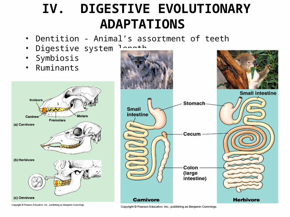
IV. DIGESTIVE EVOLUTIONARY ADAPTATIONS
• Dentition - Animal’s assortment of teeth• Digestive system length• Symbiosis• Ruminants

IV. EVOLUTIONARY ADAPTATIONS, cont
Ruminant Digestion

HUMAN REPRODUCTION

I. GAMETE PRODUCTION

II. REPRODUCTION – MALE HUMAN ANATOMY• Testes
Contained in scrotum Importance of temperature Seminiferous tubules – sperm formation Leydig Cells – produce testosterone & other
hormones Sertoli Cells
• Epididymis coiled tubules that sperm pass through from
testis• Vas deferens
Muscular tube that propels sperm during ejaculation
• Ejaculatory Duct Combines sperm from both testes; leads to
urethra• Glands
Seminal vesicles – Add fluid to protect nourish sperm, including fructose, mucus, enzymes; produces semen
Prostate gland - Secretes anticoagulant, nutrients into semen
Bulbourethral glands – Secretes acid neutralizer before ejaculation
• Penis/Urethra Ejaculation - Release of semen Blockage of urine flow controlled by
sphincters

II. REPRODUCTION – MALE, cont
• Human Sperm

III. REPRODUCTION – FEMALE HUMAN ANATOMY• Ovaries
Follicle – Egg capsule; nourishes and protects egg
Egg released during ovulation Corpus luteum – Secretes estrogen
and progesterone to maintain uterine lining; formed from follicle after egg is released
• Oviduct Also known as fallopian tube Egg moved along through action of
cilia• Uterus
Thick, muscular organ also known as womb
Endometrium – inner lining Cervix – opens into vagina

IV. REPRODUCTIVE CYCLES
• Estrous Cycle Seen in animals Uterine lining is reabsorbed by the uterus if pregnancy does not occur; no
bleeding Causes more pronounced behavioral changes Animals typically only copulate during ovulation; known as estrus
• Menstrual Cycle Seen in humans, other primates Oogenesis occurs during the ovarian cycle Ovarian cycle is synchronized with menstrual cycle through the action of
hormones Divided into phases
Follicular phase – growth of follicle Ovulation – release of egg Luteal phase – degeneration of corpus luteum

V. MENSTRUAL REPRODUCTIVE CYCLE
Follicular Phase• Small amounts of FSH and LH are secreted by the pituitary• The follicle is stimulated to grow, leading to secretion of estrogen (estradiol)• Initially, low levels of estrogen inhibit secretion of FSH, LH (negative
feedback)

V. MENSTRUAL REPRODUCTIVE CYCLE, cont
Ovulation• As estrogen concentration continues to increase increase in growing follicle,
at a critical concentration, estrogen concentration switches to positive feedback mechanism.
• FSH and LH production increase, especially LH• Causes release of follicle

V. MENSTRUAL REPRODUCTIVE CYCLE, cont
Luteal Phase• LH stimulates remaining follicular tissue to transform into corpus luteum• Due to effects of LH, corpus luteum secretes progesterone, estrogen• Increasing concentrations of progesterone, estrogen exert negative feedback on
pituitary, decreasing release of FSH, LH• As levels continue to decrease, corpus luteum disintegrates• Results in sharp decrease in estrogen, progesterone levels• At a certain point, levels drop beneath concentration required for negative feedback to
pituitary → pituitary then begins secreting FSH, LH → cycle begins again

VI. FERTILIZATION• Fertilization:
Sperm reaches egg Head of sperm contains a vesicle known as the acrosome; contains enzymes that help sperm penetrate
egg Acrosomal reaction – hydrolytic enzymes act on egg jelly coat Surface proteins on sperm bind with receptor molecules on egg Sperm cell membrane fuses with egg cell membrane Cell membrane of egg depolarizes, becomes impenetrable to sperm to prevent multiple fertilization
(polyspermy) Triggers increase in metabolic activity in fertilized egg (including completion of meiosis II)

EMBRYONIC DEVELOPMENT
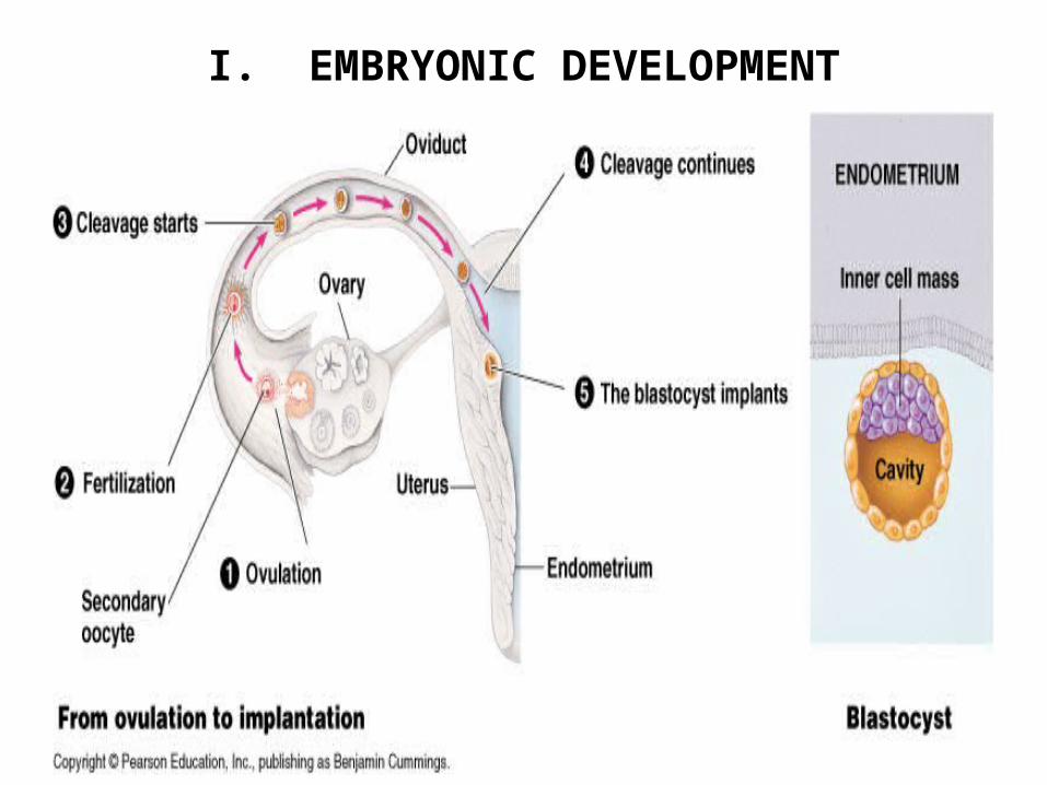
I. EMBRYONIC DEVELOPMENT

I. EMBRYONIC DEVELOPMENT
Cleavage• Cleavage produces a ball of cells
known as a blastula Cells known as blastomeres Cavity formed known as blastocoel
• Nutrients stored in the egg known as yolk
• Two sides of the blastula Vegetal pole – Side with high yolk
concentration; larger cells due to yolk; divide more slowly
Animal pole – Side with low yolk concentration; smaller cells; divide at a faster rate

I. EMBRYONIC DEVELOPMENT - Amniotes• Forms within a shell or
uterus• Extraembryonic
membranes Yolk sac – Contains
blood vessels that transport nutrients from yolk to embryo
Amnion – Fluid-filled sac; protection
Chorion – Formation of placenta
Allantois – Disposal sac for nitrogenous wastes; incorporated into umbilical cord in mammals
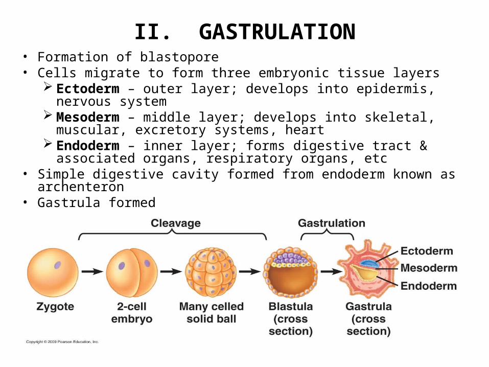
II. GASTRULATION• Formation of blastopore • Cells migrate to form three embryonic tissue layers
Ectoderm – outer layer; develops into epidermis, nervous system Mesoderm – middle layer; develops into skeletal, muscular, excretory
systems, heart Endoderm – inner layer; forms digestive tract & associated organs,
respiratory organs, etc• Simple digestive cavity formed from endoderm known as archenteron• Gastrula formed

III. ORGANOGENESIS
Organogenesis

IV. FETAL DEVELOPMENT• Gestation – pregnancy• First Trimester
Organogenesis By week 8, human fetus has all adult features Corpus luteum maintained by HCG; prevents
menstruation; also used to detect pregnancy• Second Trimester
Refinement of human features Corpus luteum degenerates; placenta begins
secreting progesterone• Third Trimester
Rapid growth Respiratory, circulatory systems prepare for
breathing• Parturition - birth
Estrogen levels increase; trigger formation of oxytocin receptors
Fetus, mother’s pituitary gland secrete oxytocin which triggers uterine contractions, preparation for lactation
Following birth, mother’s pituitary gland secretes prolactin; stimulates milk production, continued uterine contractions

NERVOUS SYSTEM
Phineus Gage1823 - 1860

I. NERVOUS SYSTEM
Human Nervous System

II. CELLS OF THE NERVOUS SYSTEM• Glia
o Support cellso Mostly nonconducting cells that provide support, insulation, protection
Astrocyctes Schwann cells - PNS Oligodendrocytes - CNS
• Neuron o Basic unit of function o Three types
Sensory Neurons Convey signals from sensory receptors to CNS
Interneurons Integrate, interpret data; relay signals to other neurons
Motor Neurons Convey signals from CNS to effector cells (glands or muscles)
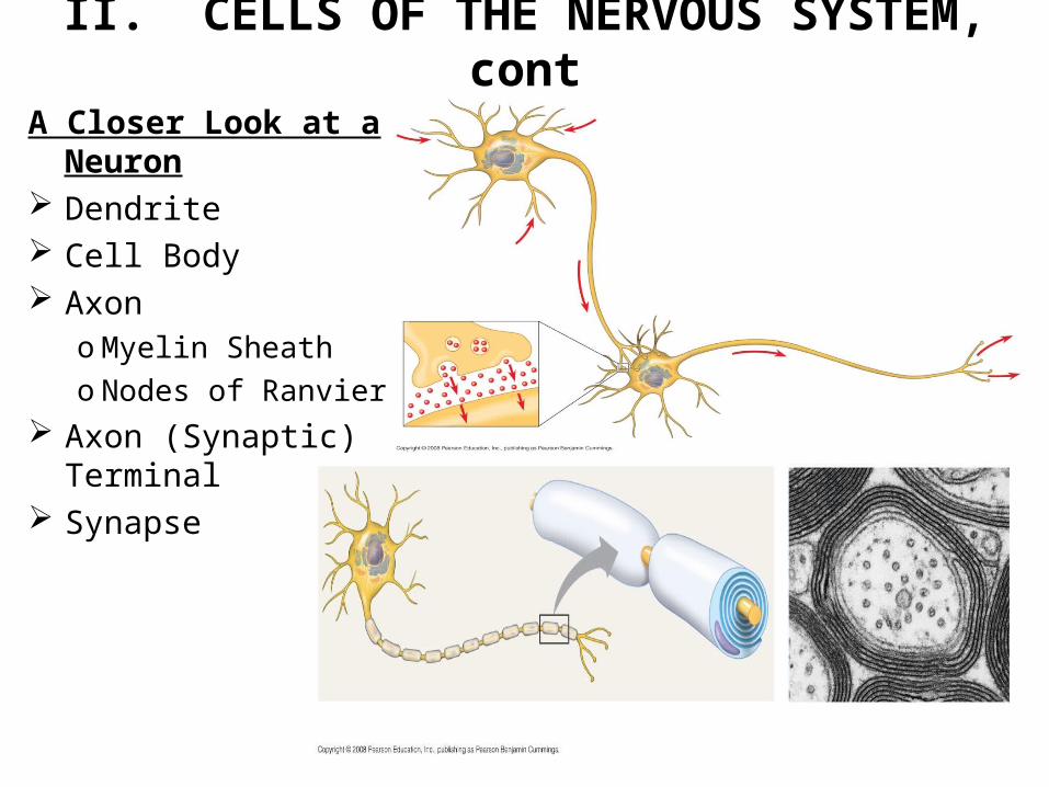
II. CELLS OF THE NERVOUS SYSTEM, cont
A Closer Look at a Neuron Dendrite Cell Body Axon
o Myelin Sheatho Nodes of Ranvier
Axon (Synaptic) Terminal Synapse

II. CELLS OF THE NERVOUS SYSTEM, cont
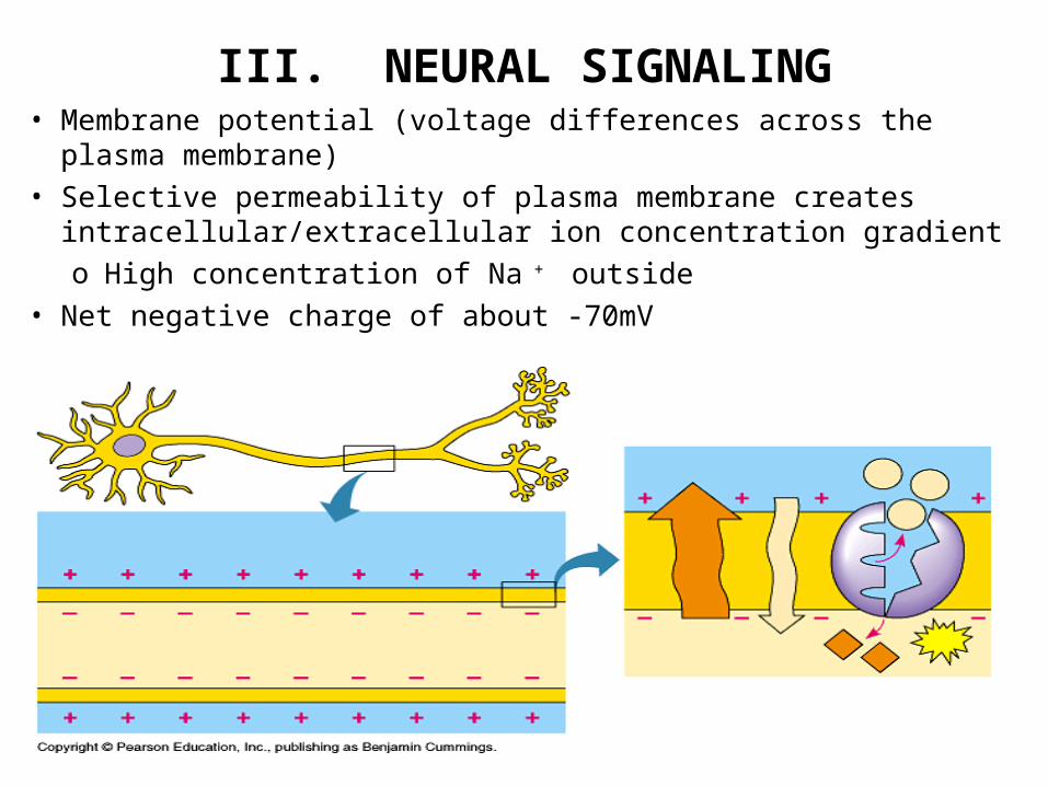
III. NEURAL SIGNALING• Membrane potential (voltage differences across the plasma membrane)• Selective permeability of plasma membrane creates
intracellular/extracellular ion concentration gradiento High concentration of Na + outside
• Net negative charge of about -70mV

III. NEURAL SIGNALING, cont• Neurons, muscle cells → excitable cells; cells that can change membrane potentials• Gated Ion Channels → open/close response to stimuli → photoreceptors; vibrations in air
(sound receptors); chemical (neurotransmitters) & voltage (membrane potential changes)• Hyperpolarization → opening of K+ channels; results in outflow of K+; increase in electrical
gradient• Depolarization → opening of Na+ channels; results in inflow of Na+

III. NEURAL SIGNALING, cont• Threshold – Stimulus strong enough
to increase voltage to ~ -50mV; triggers an action potential
• Caused by movement of ions through Na+, K+ voltage-gated channels
• Sequence of events: Resting State – Channels closed Depolarization – Na+ channels
open; inside of cell becomes + Repolariztion - Na+ channels
close; K+ channels open slowly → K+ ions leave → cell returns to negative
Hyperpolarization – Created by K+ gates; close very slowly → K+ ions continue flowing out of cell → brief period where cell is more negative than resting state. Known as refractory period – neuron is insensitive to depolarization until resting potential is restored

III. NEURAL SIGNALING, cont• Movement of the action potential is self-propagating• Regeneration of “new” action potentials only after refractory period• Forward direction only• Speed of action potential related to
Axon diameter Nodes of Ranvier; known as saltatory conduction

III. NEURAL SIGNALING, contTransmission of Impulse Across a
Synapse• Synaptic Cleft – small gap between
sending neuron and receiving cell• Synaptic vesicles contain
neurotransmitter molecules• Action potential causes synaptic
terminal to depolarize → Ca2+ channels open → Ca2+ flows in → causes vesicles to fuse with axon terminal membrane
• Neurotransmitters “spit out”; diffuse across synapse Excitatory Postsynaptic
Potentials (EPSPs) Inhibitory Postsynaptic
Potentials (IPSPs)• Examples of neurotransmitters
include acetylcholine, dopamine, epinephrine, norepinephrine, serotonin

III. NEURAL SIGNALING, cont

III. NEURAL SIGNALING, contA Review
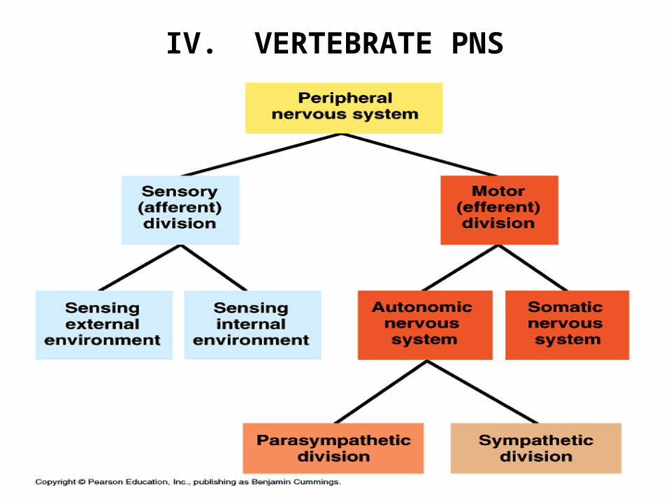
IV. VERTEBRATE PNS

IV. VERTEBRATE PNS, cont
• Nerves Bundles of
sensory & motor neurons
12 pairs of cranial nerves
31 pairs of spinal nerves

IV. VERTEBRATE PNS, cont• Reflex - “Automatic” response; sensory to motor neurons

V. VERTEBRATE CNS
• Brain
• Spinal Cord
• Protected by

V. VERTEBRATE CNS, cont
Human Brain• Forebrain
Cerebrum Cerebral Cortex Corpus Callosum
Thalamus Hypothalamus
• Midbrain – Receives & transmits sensory info to forebrain
• Hindbrain Cerebellum Pons Medulla oblongata

V. VERTEBRATE CNS, cont

ANIMAL MOVEMENT

I. INTRODUCTION TO MOVEMENT
• Gravity, friction must be overcome• Types of Movement
Swimming Flying Locomotion on land
• Importance of Skeleton Support Protection Essential to Movement Hydrostatic Skeleton
Found in Exoskeleton
Found in Endoskeleton
Found in

II. SKELETAL MUSCLE FUNCTION
• Typically at least 2 attachment sites Origin Insertion
• Muscles of appendicular skeleton make up antagonistic pairs Flexor Extensor
• Muscles Bundle of muscle fibers
Multinucleated cells Composed of myofibrils

II. SKELETAL MUSCLE FUNCTION, cont• Muscle myofibrils made up of two
types of myofilaments Thin Filaments
Two strands of actin Wrapped with protein complex
made up of tropomyosin and troponin complex
Thick filament Myosin
• Contracting unit of muscle tissue known as sarcomeres
• Sliding Filament Theory When stimulated, actin & myosin
filaments slide past each other; overlap increases
Shortens sarcomere length

II. SKELETAL MUSCLE FUNCTION, contSliding-Filament Model, I
• Myosin binds ATP; hydrolyzed to ADP + Pi
• Myosin head changes shape; termed high energy configuration• Myosin head binds to specific site on actin; forms a cross bridge
• ADP and Pi released; myosin relaxes to low energy configuration
• Causes actin to slide toward center of sarcomere• Binding of new ATP releases myosin head

II. SKELETAL MUSCLE FUNCTION, contMuscle Contraction Regulation, I
• Relaxation Tropomyosin
blocks myosin binding sites on actin
• ContractionCalcium binds to
troponin complexTropomyosin
changes shape Exposes myosin
binding sites

II. SKELETAL MUSCLE FUNCTION, contMuscle Contraction Regulation, II
• Stimulated by action potential in a ________________ neuron
• _____________________ triggers depolarization of muscle fiber by opening _____________ voltage-gated channels
• Action potential spreads to infoldings of cell membrane called T (transverse) tubules
• Sarcoplasmic reticulum = specialized ER that actively transports calcium ions
• When action potential reaches places where T tubules touch sarcoplasmic reticulum → Ca 2+ released
• Ca 2+ then binds to troponin, allowing myosin binding sites to be revealed

III. OVERVIEW OF SKELETAL MUSCLE FUNCTION

IV. SKELETAL MUSCLE ADAPTATIONS
• Energy Availability Adaptations
Creatine Phosphate
Myoglobin
• Fast-twitch Fibers• Slow-twitch Fibers