Underwood, C. and Ward, D. (2008) Sharks of the Order...
29
Birkbeck ePrints: an open access repository of the research output of Birkbeck College http://eprints.bbk.ac.uk Underwood, C. and Ward, D. (2008) Sharks of the Order Carcharhiniformes from the British Coniacian, Santonian and Campanian (Upper Cretaceous). Palaeontology 51 (3): 509-536. This is an exact copy of an article published in Palaeontology (ISSN 0031- 0239), published by Blackwell Publishing. © 2008 The Palaeontological Association. All articles available through Birkbeck ePrints are protected by intellectual property law, including copyright law. Any use made of the contents should comply with the relevant law. Citation for this version: Underwood, C. and Ward, D. (2008) Sharks of the Order Carcharhiniformes from the British Coniacian, Santonian and Campanian (Upper Cretaceous). London: Birkbeck ePrints. Available at: http://eprints.bbk.ac.uk/archive/00000726 Citation for the publisher’s version: Underwood, C. and Ward, D. (2008) Sharks of the Order Carcharhiniformes from the British Coniacian, Santonian and Campanian (Upper Cretaceous). Palaeontology 51 (3): 509-536. http://eprints.bbk.ac.uk Contact Birkbeck ePrints at [email protected]
Transcript of Underwood, C. and Ward, D. (2008) Sharks of the Order...

Birkbeck ePrints: an open access repository of the research output of Birkbeck College
http://eprints.bbk.ac.uk
Underwood, C. and Ward, D. (2008) Sharks of the Order Carcharhiniformes from the British Coniacian, Santonian and Campanian (Upper Cretaceous). Palaeontology 51 (3): 509-536. This is an exact copy of an article published in Palaeontology (ISSN 0031-0239), published by Blackwell Publishing. © 2008 The Palaeontological Association. All articles available through Birkbeck ePrints are protected by intellectual property law, including copyright law. Any use made of the contents should comply with the relevant law. Citation for this version: Underwood, C. and Ward, D. (2008) Sharks of the Order Carcharhiniformes from the British Coniacian, Santonian and Campanian (Upper Cretaceous). London: Birkbeck ePrints. Available at: http://eprints.bbk.ac.uk/archive/00000726 Citation for the publisher’s version: Underwood, C. and Ward, D. (2008) Sharks of the Order Carcharhiniformes from the British Coniacian, Santonian and Campanian (Upper Cretaceous). Palaeontology 51 (3): 509-536.
http://eprints.bbk.ac.uk Contact Birkbeck ePrints at [email protected]
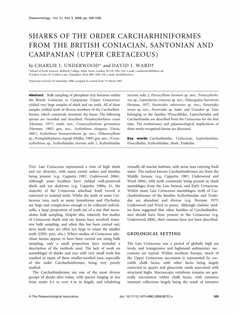
SHARKS OF THE ORDER CARCHARHINIFORMES
FROM THE BRITISH CONIACIAN, SANTONIAN AND
CAMPANIAN (UPPER CRETACEOUS)
by CHARLIE J. UNDERWOOD* and DAVID J. WARD�*School of Earth Sciences, Birkbeck College, Malet Street, London WC1E 7HX, UK; e-mail: [email protected]
�Crofton Court, 81 Crofton Lane, Orpington, Kent BR5 1HB, UK; e-mail: [email protected]
Typescript received 26 September 2006; accepted in revised form 19 March 2007
Abstract: Bulk sampling of phosphate-rich horizons within
the British Coniacian to Campanian (Upper Cretaceous)
yielded very large samples of shark and ray teeth. All of these
samples yielded teeth of diverse members of the Carcharhini-
formes, which commonly dominate the fauna. The following
species are recorded and described: Pseudoscyliorhinus reussi
(Herman, 1977) comb. nov., Crassescyliorhinus germanicus
(Herman, 1982) gen. nov., Scyliorhinus elongatus (Davis,
1887), Scyliorhinus brumarivulensis sp. nov., ?Palaeoscyllium
sp., Prohaploblepharus riegrafi (Muller, 1989) gen. nov., ?Creta-
scyliorhinus sp., Scyliorhinidae incertae sedis 1, Scyliorhinidae
incertae sedis 2, Pteroscyllium hermani sp. nov., Protoscyliorhi-
nus sp., Leptocharias cretaceus sp. nov., Palaeogaleus havreensis
Herman, 1977, Paratriakis subserratus sp. nov., Paratriakis
tenuis sp. nov., Paratriakis sp. indet. and ?Loxodon sp. Taxa
belonging to the families ?Proscylliidae, Leptochariidae and
Carcharhinidae are described from the Cretaceous for the first
time. The evolutionary and palaeoecological implications of
these newly recognised faunas are discussed.
Key words: Carcharhinidae, Cretaceous, Leptochariidae,
Proscylliidae, Scyliorhinidae, shark, Triakidae.
The Late Cretaceous represented a time of high shark
and ray diversity, with many extant orders and families
being present (e.g. Cappetta 1987; Underwood 2006).
Although some localities have yielded well-preserved
shark and ray skeletons (e.g. Cappetta 1980a, b), the
majority of the Cretaceous selachian fossil record is
restricted to isolated teeth. Whilst the teeth of some Cre-
taceous taxa, such as many lamniforms and Ptychodus,
are large and conspicuous enough to be collected individ-
ually, a large proportion of teeth are of a size that neces-
sitates bulk sampling. Despite this, relatively few studies
of Cretaceous shark and ray faunas have involved exten-
sive bulk sampling, and when this has been carried out,
sieve mesh sizes are often too large to retain the smaller
teeth (DJW, pers. obs.). Where studies of Cretaceous sela-
chian faunas appear to have been carried out using bulk
sampling, only a small proportion have included a
description of the methods used. The lack of work on
assemblages of sharks and rays with very small teeth has
resulted in many of these smaller-toothed taxa, especially
of the order Carcharhiniformes, being very poorly
studied.
The Carcharhiniformes are one of the most diverse
groups of sharks alive today, with species ranging in size
from under 0.3 to over 6 m in length, and inhabiting
virtually all marine habitats, with some taxa entering fresh
water. The earliest known Carcharhiniformes are from the
Middle Jurassic (e.g. Cappetta 1987; Underwood and
Ward 2004), with teeth commonly being present in shark
assemblages from the Late Jurassic and Early Cretaceous.
Within many Late Cretaceous assemblages, teeth of Car-
charhiniformes of the families Scyliorhinidae and Triaki-
dae are abundant and diverse (e.g. Herman 1977;
Underwood and Ward in press). Although cladistic stud-
ies have suggested that other families of Carcharhinifor-
mes should have been present in the Cretaceous (e.g.
Underwood 2006), their remains have not been described.
GEOLOGICAL SETTING
The Late Cretaceous was a period of globally high sea
levels, and transgressive and highstand sedimentary suc-
cessions are typical. Within northern Europe, much of
the Upper Cretaceous succession is represented by coc-
colith chalk facies, with other facies being largely
restricted to quartz and glauconite sands associated with
structural highs. Macroscopic vertebrate remains are gen-
erally uncommon within chalk facies, with extensive
museum collections largely being the result of intensive
[Palaeontology, Vol. 51, Part 3, 2008, pp. 509–536]
ª The Palaeontological Association doi: 10.1111/j.1475-4983.2008.00757.x 509

collecting during former periods of active manual quar-
rying. The English Chalk is also a particularly poor can-
didate for systematic bulk sampling because of the
extreme rarity of shark and ray teeth and the degree of
bioerosion encountered. This is in contrast to southern
Swedish chalks that can yield up to 10 tooth fragments
per kg (Mikael Siverson, pers. comm. 2000). Unlike
most of the English Chalk, phosphatic chalks, often
immediately overlying hardgrounds, commonly contain
abundant shark and ray remains. This is presumably due
to a combination of current winnowing and low rate of
sediment deposition and high overall levels of pore
water phosphate. Many of the teeth are well mineralised
and rates of bioerosion are lower than in non-phos-
phatic chalks. Localized areas of these phosphatic chalks
are common in the Santonian and Campanian of north-
west Europe and fill lenticular erosional troughs or ‘cu-
vettes’ with hardground complexes at their base (Jarvis
1980, 2006).
The majority of the English material described below
was collected from chalks with a high content of phos-
phatic material (Text-fig. 1) during a period of fieldwork
carried out by DJW in the late 1970s, which was inspired
by the work of Ian Jarvis, Peter Woodruff, and Andy
Gale. A Coniacian glauconitic sandstone from Northern
Ireland was sampled more recently by CJU. All of these
phosphatic horizons sampled yielded large numbers of
shark and ray teeth of many species. The vast majority of
these teeth are extremely small, and belong to small
nectobenthic taxa. For details of the localities sampled
and associated faunas, see the Appendix.
MATERIAL AND METHODS
All of the rock samples studied were moderately to
strongly lithified, and could only be broken down by use
of acids. Samples of the phosphatic chalks from southern
England were crushed into 2–5-cm fragments and dis-
solved in buffered dilute acetic or formic acid. The resi-
due from the 355 lm sieve fraction and above was picked
for vertebrate remains. The phosphate-rich greensands
from Minnis North were broken down in buffered formic
acid. The residue from the 500 lm sieve fraction and
above was picked for vertebrate remains, along with much
of the 355 lm fraction; this fine fraction yielded very few
teeth and these were highly diluted by grains of phos-
phate, quartz and glauconite.
A large proportion of teeth collected during this study
are imperfect, with roots of the majority showing some
degree of damage owing to microbial bioerosion (see
Underwood et al. 1999). There is no sign of physical
biostratinomic damage of teeth from any of the sites
other than Minnis North. Where teeth of a taxon are
distinctive enough to be recognisable from incomplete
specimens, counts of numbers of specimens can be
obtained, but where there are several taxa with similar
teeth within the same sample (as with some scyliorhi-
TEXT -F IG 1 . Map showing the
localities sampled.
510 P A L A E O N T O L O G Y , V O L U M E 5 1
nids) it is not possible to obtain an accurate count for
the number of imperfect specimens. Counts otherwise
include all teeth and partial teeth well preserved enough
to be identifiable. All figured specimens from England
are housed in the Natural History Museum, London
(BMNH P., shortened here to ‘P.’). The figured speci-
mens from Minnis North are in Ulster Museum
(BELUM K). All figured specimens were imaged by
SEM. The terminology used here for parts of the sela-
chian teeth largely follows that of Cappetta (1987).
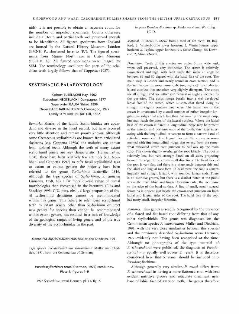
SYSTEMATIC PALAEONTOLOGY
Cohort EUSELACHII Hay, 1902
Subcohort NEOSELACHII Compagno, 1977
Superorder GALEA Shirai, 1996
Order CARCHARHINIFORMES Compagno, 1977
Family SCYLIORHINIDAE Gill, 1862
Remarks. Sharks of the family Scyliorhinidae are abun-
dant and diverse in the fossil record, but have received
very little attention and remain poorly known. Although
some Cretaceous scyliorhinids are known from articulated
skeletons (e.g. Cappetta 1980a) the majority are known
from isolated teeth. Although the teeth of many extant
scyliorhinid genera are very characteristic (Herman et al.
1990), there have been relatively few attempts (e.g. Nou-
bhani and Cappetta 1997) to refer fossil scyliorhinid taxa
to extant or extinct genera; the majority have been
referred to the genus Scyliorhinus Blainville, 1816.
Although the type species of Scyliorhinus, S. canicula
Linnaeus, 1758, has a far more diverse range of dental
morphologies than recognised in the literature (Ellis and
Shackley 1995; CJU, pers. obs.), a large proportion of fos-
sil scyliorhinid dentitions cannot be accommodated
within this genus. This failure to refer fossil scyliorhinid
teeth to extant genera other than Scyliorhinus or erect
new genera for species than cannot be accommodated
within extant genera, has resulted in a lack of knowledge
of the geological ranges of living genera and of the true
diversity of the Scyliorhinidae in the past.
Genus PSEUDOSCYLIORHINUS Muller and Diedrich, 1991
Type species. Pseudoscyliorhinus schwarzhansi Muller and Died-
rich, 1991, from the Cenomanian of Germany.
Pseudoscyliorhinus reussi (Herman, 1977) comb. nov.
Plate 1, figures 1–9
1977 Scyliorhinus reussi Herman, pl. 11, fig. 2.
in press Pseudoscyliorhinus sp. Underwood and Ward, fig.
1C–D.
Material. P. 66363–P. 66367 from a total of 124 teeth: 10, Box-
ford; 2, Winterbourne lower horizon; 2, Winterbourne upper
horizon; 2, Taplow upper horizon; 71, Stoke Clump; 35, Down-
end; 2, Minnis North.
Description. Teeth of this species are under 3 mm wide and,
when well preserved, very distinctive. The crown is relatively
symmetrical and high, with erect cusps that make an angle of
between 60 and 80 degrees with the basal face of the root. The
main cusp is slender and nearly round in cross section, and is
flanked by one, or more commonly two, pairs of much shorter
lateral cusplets that are often very slightly divergent. The cusps
are all straight and are either symmetrical or slightly inclined to
the posterior. The cusps merge basally into a well-developed
labial face of the crown, which is somewhat flared along its
straight to slightly concave basal edge. The labial face of the
crown is ornamented by a small number of rather irregular lon-
gitudinal ridges that reach less than half-way up the main cusp,
but may reach the apex of the lateral cusplets. Where the labial
base of the crown is flared, a longitudinal ridge may be present
at the anterior and posterior ends of the tooth, this ridge inter-
acting with the longitudinal ornament to form a narrow band of
reticulate ornament. The lingual face of the crown is orna-
mented with fine longitudinal ridges that extend from the some-
what excavated crown-root junction to half-way up the main
cusp. The crown slightly overhangs the root labially. The root is
relatively low, but very strongly flared on all sides, projecting
beyond the edge of the crown in all directions. The basal face of
the root is very flat, and there is a sharp angle between this and
the labial and lingual root faces. In basal view, the root is convex
lingually and straight labially, with rounded lateral ends. There
is no nutritive groove, but there is a distinct notch at the point
where the main labial and lingual foramina enter the root close
to the edge of the basal surface. A line of small, evenly spaced
foramina is present just below the crown-root junction on both
labial and lingual sides of the root. The basal face of the root
has many small, irregular foramina.
Remarks. This genus is readily recognised by the presence
of a flared and flat-based root differing from that of any
other scyliorhinids. The genus was diagnosed on the
Cenomanian species P. schwarzhansi Muller and Diedrich,
1991, with the very close similarities between this species
and the previously described Scyliorhinus reussi Herman,
1977 evidently not having been recognised at the time.
Although no photographs of the type material of
P. schwarzhansi were published, the diagnosis of Pseudo-
scyliorhinus equally well covers S. reussi. It is therefore
considered here that S. reussi should be included into
Pseudoscyliorhinus.
Although generally very similar, P. reussi differs from
P. schwarzhansi in having a more flattened root with less
evident nutritive groove and reticulate ornament near
base of labial face of anterior teeth. The genus therefore
U N D E R W O O D A N D W A R D : C A R C H A R H I N I F O R M E S S H A R K S F R O M T H E B R I T I S H U P P E R C R E T A C E O U S 511

has a recorded range of Cenomanian to Campanian. No
extant scyliorhinids have been recorded as having teeth
similar to Pseudoscyliorhinus; it is, therefore, not possible
to assess the affinities of this genus. It has, however, been
recorded that teeth of juvenile scyliorhinids have roots
that are larger and more flared than in the adults (Her-
man et al. 1990, p. 230), so it is possible that the tooth
morphology of Pseudoscyliorhinus was paedomorphic.
Genus CRASSESCYLIORHINUS gen. nov.
Derivation of name. From the thick and heavy form of the teeth
compared to all other scyliorhinids.
Type species. Scyliorhinus germanicus Herman, 1982.
Diagnosis. Teeth very robust and symmetrical with mod-
erate heterodonty. Erect central cusp conical and takes up
much of crown; lateral cusplets small. Ornament of
strong ridges in unworn teeth, often reticulate near base
of labial crown. Labial margin of crown indented in cen-
tre. Root bulky and strongly V-shaped in basal view with
somewhat flared distal lobes. Nutritive groove absent in
almost all teeth.
Remarks. The only described species of scyliorhinid with
a dentition similar to Crassescyliorhinus germanicus is Scy-
liorhinus musteliformis Herman, 1977, which differs in
having teeth with lower crowns. The close similarity
between these species suggests that S. musteliformis should
also be included in Crassescyliorhinus. The genus has a
recorded range of Santonian–Maastrichtian. The teeth of
Crassescyliorhinus are clearly distinct from those of all
other recorded post-Cretaceous scyliorhinid genera, being
most similar to those of the Cretaceous genus Cretoscylio-
rhinus Underwood and Mitchell, 1999, from which they
differ in being far more robust and in having less clearly
separated lateral cusplets, a greater degree of heterodonty
and a less ‘trilobed’ root basal face.
Crassescyliorhinus germanicus (Herman, 1982)
Plate 2, figures 10–18
1977 Scyliorhinus elongatus Herman, pl. 11, fig. 1H.
1982 Scyliorhinus germanicus Herman, pl. 2, fig. 10; pl. 4,
figs 4–5.
1989 Scyliorhinus germanicus Herman; Muller, pl. 11,
fig. 5, pl. 12, figs 1–4.
?2001 Scyliorhinus sp., Cappetta and Odin, pl. 1, fig. 8.
Material. P. 66368–P. 66372 from a total of several hundred
teeth: 54, Boxford; 68, Winterbourne lower horizon; 43, Winter-
bourne upper horizon; 9, Taplow lower horizon; 18, Taplow
upper horizon; several hundred, Downend.
Description. Teeth of this species are very robust and show a
moderate degree of monognathic heterodonty. Some presumed
anterior teeth are higher than wide, but the majority of teeth are
wider than high, reaching a width of 3 mm. Teeth from most
positions are symmetrical, with a low degree of asymmetry being
present in posterior teeth. Over half of the width of the crown is
taken up with a robust main cusp. This is conical and only
slightly flattened on the labial face, and is of similar width to
height on all except extreme anterior and posterior teeth. This is
flanked by one or two pairs of short but robust lateral cusplets
that are typically wider than high; a third cusplet may be present
on the anterior side of large, presumed anterolateral teeth. The
ends of the crown are rounded, with the basal edge of the crown
on the labial side having a strong and smoothly curved indenta-
tion on all except posterior teeth. The labial face of the crown is
typically highly ornamented with strong and sharp-edged ridges,
although these may be less well developed on some of the largest
and most robust lateral teeth. The labial ornament comprises
widely spaced longitudinal ridges, with up to six on the main
cusp, that bifurcate towards the crown base to form a polygonal
pattern, and reach the apex of the cusps where not removed by
wear. Ornament on the lingual face of the crown is similar if
not so strongly developed and rather more irregular. The crown
is a similar width or slightly narrower than the root, and
strongly overhangs it labially. The root is low, and distinctly V-
shaped when viewed basally. The basal face of the root is flat,
and is flared at its lateral ends. There is a bulbous lingual apex
of the root with a large, central foramen. Opposite this on the
EXPLANATION OF PLATE 1
Figs 1–9. Pseudoscyliorhinus reussi (Herman, 1977) comb. nov. 1, P. 66363, Lower Campanian, Downend, labial view. 2–4, P. 66366,
Boxford, in 2, labial, 3, oblique lateral, and 4, basal views. 5, P. 66364, Lower Campanian, Winterbourne, upper phosphate level,
?parasymphaseal tooth, labial view. 6, P. 66365, Lower Campanian, Winterbourne, upper phosphate level, ?small anterior tooth,
labial view. 7–9, P. 66367, Lower Campanian, Winterbourne, upper phosphate level, in 7, labial, 8, lingual, and 9, oblique lateral
views. 1–6, · 35; 7–9, · 20.
Figs 10–18. Crassescyliorhinus germanicus (Herman, 1982). 10–11, P. 66368, Upper Santonian, Winterbourne, lower phosphate level,
large anterolateral tooth in 10, labial, and 11, lingual views. 12–14, P. 66369, Middle Santonian, Boxford, anterior tooth in 12,
labial, 13, oblique lateral, and 14, lingual views. 15, P. 66370, Middle Santonian, Boxford, posterior tooth, labial view. 16–17, P.
66371, Boxford, anterior tooth in 16, labial, and 17, lingual views. 18, P. 66372, Middle Santonian, Boxford, posterolateral tooth,
labial view. 10–11, · 20; 12–18, · 35.
512 P A L A E O N T O L O G Y , V O L U M E 5 1

PLATE 1
UNDERWOOD and WARD, Crassescyliorhinus, Pseudoscyliorhinus
1 2
3
4
7
5
6
8
9 10
11
14
13
12
15 16 17
18
lingual side is a notch or groove containing the lingual foramen.
An open nutritive groove is rarely present. The lingual faces of
the root are concave and have a small number of well-developed
foramina. Numerous small foramina are present on the basal
face of the root.
Remarks. The teeth recorded here are very similar to
those previously figured (e.g. Herman 1977, 1982; Muller
1989), although the small number of specimens figured
before has not previously allowed recognition of the
degree of heterodonty within the species recognised here,
and the teeth figured here are largely of morphologies not
previously illustrated.
Genus SCYLIORHINUS Blainville, 1816
Type species. Squalus canicula Linnaeus, 1758.
Remarks. Scyliorhinus has commonly been used as a con-
venient genus within which to place virtually all fossil scy-
liorhinid (and sometimes other carcharhiniform) teeth.
Despite this, it is one of 16 extant scyliorhinid genera,
and recent studies (e.g. Noubhani and Cappetta 1997)
have recognised a number of additional extinct genera.
Several workers (Herman et al. 1990; Halter 1994) have
noted the distinctive dental morphology of extant Scylio-
rhinus, and that relatively robust anterior teeth without
well-developed cusplets are characteristic of the genus.
Despite this, studies of jaws of the type species have
revealed very strong sexual heterodonty (Ellis and Shack-
ley 1995; J. Southion and A. Chappel, pers. comm. 2006).
The robust teeth morphology is only present in mature
males, with females and juveniles having smaller, more
gracile teeth with well-developed lateral cusplets. The
pattern of heterodonty shown in other extant species of
Scyliorhinus has not been documented, but it is possible
that in some species, such as S. stellaris, the robust tooth
morphology is seen in both sexes (A. Chappel, pers.
comm. 2006).
Scyliorhinus elongatus (Davis, 1887)
Plate 2, figures 1–9
1887 Thyellina elongata Davis; pl. 14, figs 2–3 (teeth not
figured).
1889 Scyllium elongatus (Davis); Woodward, pl. 16, fig. 5
(teeth not figured).
1977 Scyliorhinus elongatus (Davis); Herman, pl. 11, fig.
11.1G, I–J.
1980a Scyliorhinus elongatus (Davis); Cappetta, pl. 21, fig.
2; pl. 22, figs 2–7; pl. 23, figs 1–4 (teeth only), fig. 30.
?1997 Scyliorhinus aff. elongatus (Davis), Noubhani and
Cappetta, pl. 24, figs 1–6.
Material. P. 66373–P. 663775 from a total of 78 teeth: 5, Box-
ford; 10, Winterbourne lower horizon; 4, Winterbourne upper
horizon; 1, Taplow lower horizon; 5, Taplow upper horizon; 53,
Downend.
Description. Teeth of this species show a high degree of mono-
gnathic heterodonty, with distinctly different anterior and lateral
tooth morphologies. Heterodonty is similar to that recorded pre-
viously (Cappetta 1980a). All teeth are higher than wide. Ante-
rior teeth are large, up to 3 mm high, poorly ornamented and
asymmetrical, with teeth becoming smaller, more gracile and
more strongly ornamented in lateral positions. Anterior teeth are
robust and have a straight main cusp flanked by a pair of short
but robust cusplets. Labial and lingual faces of all cusps are simi-
larly convex. Ornament is weak and largely restricted to the lat-
eral parts of the crown. The labial face of the main cusp has
little or no ornament, with 2–3 strong longitudinal ridges being
present on the lateral cusplets, being strongest at the basal edge
of the crown. The lingual face of all cusps has weak longitudinal
ridges reaching close to the apex. The basal edge of the crown
on the lingual side strongly overhangs the root and is strongly
excavated below the main cusp. The entire crown is inclined
towards the commissure. The root is robust and strongly bilo-
bate and V-shaped, if somewhat asymmetrical, in basal view.
The lingual foramina are well developed and in the centre of the
somewhat swollen lingual part of the root. The labial foramen
forms an elongate opening on the basal face of the root at the
sharply angled junction of the root lobes. The root lobes are
slightly flared basally and are only weakly expanded at their
labial ends. The basal face of the root is flat. A series of large
foramina is present on the labial faces of the root, with small
and irregular foramina being present on the basal face of the
root. The crowns of lateral teeth are somewhat similar to those
of anteriors, but are more gracile with more elongate main and
lateral cusps. The labial faces of both main cusp and lateral cus-
plets have an ornament of strong longitudinal ridges that do not
bifurcate, and most reach almost to the apices of the cusps. The
lingual faces have very fine longitudinal ridges. The basal edge of
the crown is less excavated than in anterior teeth, but is still
always concave, and overhangs the root to a moderate degree.
The roots of lateral teeth are similar to those of anterior teeth,
but are somewhat lower and less robust, with the root lobes
being somewhat more flared.
Remarks. Both anterior and lateral teeth are almost iden-
tical to those figured by Cappetta (1980a) and Herman
(1977). This species thus appears to be relatively wide-
spread, being present in the Santonian of Lebanon (Capp-
etta 1980a) and Campanian of Belgium (Herman 1977)
in addition to the records given here. Teeth from Mo-
rocco (Noubhani and Cappetta 1997) agree well with
material of S. elongatus, suggesting that this species may
have ranged up to Late Maastrichtian. The overall dental
morphology is very similar to that of Scyliorhinus anti-
quus (Agassiz, 1843) as figured by Cappetta (1977), differ-
ing in having stronger ornamentation and more robust
anterior teeth. The tooth morphology and heterodonty
514 P A L A E O N T O L O G Y , V O L U M E 5 1
are very similar to that of the male morph of S. canicula;
it is, therefore, probable that this species belongs to
Scyliorhinus s.s. No gracile anterior teeth similar to those
possessed by female and juvenile S. canicula were recogni-
sed; it is, therefore, likely that there was little or no sexual
heterodonty in this taxon.
Scyliorhinus brumarivulensis sp. nov.
Plate 2, figures 10–18
Derivation of name. Direct Latin translation of Winterbourne,
from the common occurrence of this species there.
Holotype. BMNH P. 66378.
Material. P. 66379–P. 66382 from a total of several hundred
teeth: 158, Boxford; 110, Winterbourne lower horizon; 24, Win-
terbourne upper horizon; 10, Taplow lower horizon; 8, Taplow
upper horizon; several hundred, Downend.
Diagnosis. Teeth show moderate degree of gradient
monognathic heterodonty, with more compressed,
smoother anterior teeth and more ornamented laterals.
All teeth under 1.5 mm wide, with teeth from most
jaw positions at least as wide as high. Main cusp
straight and flanked by two pairs of short, pointed lat-
eral cusplets, the outer pair poorly developed or absent
in anterior teeth. Ornament varies with jaw position;
labial face of anterior teeth flat and smooth, that of all
cusps of lateral teeth with small numbers of very strong
longitudinal ridges that bifurcate strongly towards the
base and reach most of the way to the apex of the
cusps; similar but weaker ornament on teeth from tran-
sitional positions. Crown of teeth from most positions
inclined towards commissure. Labial basal edge of
crown straight to weakly concave and strongly over-
hangs root. Root low and hemiaulacorhize. Basal face
of root flat and flared, especially on lingual edges of
root lobes.
Description. The teeth of this species are uniformly small,
being less than 1.5 mm wide, and show distinctive monogna-
thic heterodonty and probable dignathic heterodonty. The
width of the majority of teeth is similar to their height,
although some extreme anterior teeth are rather higher than
wide. The crown is erect and comprises a main cusp, which
is somewhat longer than wide, flanked by two pairs of small,
sharply pointed lateral cusplets, except in some anterior teeth
where the outer pair of cusplets may be very reduced and
incipient. The base of the main cusp occupies about half of
the width of the crown. All cusps are somewhat flattened labi-
ally, with some of the largest and least well ornamented teeth
having a labial face that is almost flat. By analogy with extant
scyliorhinid dentitions, these are assumed to be from lower
anterior positions. All teeth have a continuous and well-devel-
oped cutting edge. The labial face of the crown of lateral
teeth is ornamented with 10–15, somewhat discontinuous, lon-
gitudinal ridges. These ridges are sharply edged and reach at
least three-quarters of the way to the apices of all cusps.
Towards the base of the crown, the labial ridges bifurcate,
and commonly join to form a narrow strip with a reticulate
pattern. In presumed upper anterior teeth a similar but
weaker and more irregular ornament is present. Presumed
lower anterior teeth have a labial ornament of short longitudi-
nal ridges on the basal and lateral parts of the crown, or may
have no labial ornament. The basal edge of the labial face of
the crown is faintly concave to almost straight, and strongly
overhangs the root. The lingual face of the crown is orna-
mented by several weak, and rather irregular, longitudinal
ridges in most teeth, but is smooth in presumed lower ante-
rior teeth. This lingual ornamentation reaches neither the apex
of the cusps nor base of the crown. The root is low and lacks
a nutritive groove. The root is V-shaped in basal view, with
concave outer lingual faces of the root lobes in lateral teeth
making them appear rather recurved. The basal part of the
root is flared along the labial edge and at the lateral extremi-
ties. The basal face of the root is flat, and the edge between
the basal and lingual or labial faces is sharply angled.
There are well-developed foramina at the lingual apex of
the root and at the junction of the root lobes. Well-developed
foramina are present on the lingual faces of the root, and on
the basal face of the root lobes, with smaller and more irreg-
ular foramina also being present across much of the root.
Remarks. The teeth of this species are very similar in
overall morphology and the general form of ornament
to those of Scyliorhinus arambourgi Cappetta, 1980a (see
Cappetta 1980a, fig. 27) from the Cenomanian of Leba-
non. Teeth of S. brumarivulensis sp. nov. differ princi-
pally in the greater amalgamation of ribs near the basal
edge of the labial crown face, forming a reticulate pat-
tern in most teeth. There are also strong similarities
between teeth of S. brumarivulensis sp. nov. and those
of S. sulcidens Noubhani and Cappetta, 1997 from the
Palaeocene of Morocco, which differs in having more
elongate lateral cusplets and a sparser ornamentation.
The teeth of this species show a very similar overall
morphology and heterodonty to that of female Scyliorhi-
nus canicula. It is unknown whether S. brumarivulensis
sp. nov. shows sexual heterodonty, but the presence of
uncommon large anterior teeth suggests that it is possi-
ble that enlarged teeth were only present in a proportion
of individuals, possibly mature males. The presence of a
dentition very similar to that of female S. canicula sug-
gests that this species should be included in Scyliorhinus
despite not conforming to the tooth morphology of the
genus as defined by Herman et al. (1990) and Halter
(1994). Despite being very common within the samples
studied here, teeth of this taxon appear not to have
U N D E R W O O D A N D W A R D : C A R C H A R H I N I F O R M E S S H A R K S F R O M T H E B R I T I S H U P P E R C R E T A C E O U S 515

been recognised in earlier studies, presumably because of
their small size.
Scyliorhinus aff. S. brumarivulensis sp. nov.
Plate 3, figures 1–2
Material. BELUM K29670, a single tooth from Minnis North.
Description. This single tooth is 1 mm high and generally well
preserved. The crown is slightly asymmetrical, with the cusps
inclined to the posterior. The main cusp is triangular in profile,
and basally comprises more than half of the crown width. This
is flanked by a pair of short but robust lateral cusplets, about as
wide as high, with a minute additional cusplet on the posterior
side. A continuous but weak cutting edge is present across the
entire width of the crown. The labial face of all cusps is slightly
convex, the lingual face is strongly convex. The labial crown face
is ornamented by a series of short, irregular longitudinal ridges,
reaching less than half of the way up the main cusp and not
reaching the apex of the undamaged cusplet. These labial ridges
reach the crown-root contact, and some bifurcate. The lingual
face of the crown is ornamented with weak longitudinal ridges
reaching close to the apex of the main cusp. The crown over-
hangs the root on all sides. The root is wider than the crown,
and relatively low. It is flared basally towards the lateral ends,
and has a flat basal face. A series of well-developed foramina is
present near the centre of the labial root face, and a large fora-
men at the lingual root apex. A row of foramina is present along
the root lingual face.
Remarks. This tooth is very similar to those assigned to
Scyliorhinus brumarivulensis sp. nov., but differs in having
the combination of a weak and irregular ornament and
poorly developed lateral cusplets; teeth of this morphol-
ogy were not recognised within the large sample sizes of
S. brumarivulensis teeth seen at other sites, whereas ‘typi-
cal’ S. brumarivulensis teeth were not found in association
with this tooth. It is, therefore, possible that this
tooth may either represent an extreme morphology of
S. brumarivulensis or another, related species. The latter
would not be unreasonable considering the very different
facies and age of the Minnis North site compared to the
other sites studied.
Genus PALAEOSCYLLIUM Wagner, 1857
Type species. Palaeoscyllium formosum Wagner, 1857 from the
Tithonian of Germany.
Palaeoscyllium striatum sp. nov.
Plate 3, figures 3–9
Derivation of name. After the evenly striated ornament on
the teeth.
Holotype. BMNH P. 66383.
Material. P. 66384 and P. 66385 from a total of 86 whole and
partial teeth: 21, Boxford; 22, Winterbourne lower horizon; 3,
Winterbourne upper horizon; 1, Taplow lower horizon; 7,
Taplow upper horizon; 9, Stoke Clump; 23, Downend.
Diagnosis. Dentition apparently showing low degree of
gradient monognathic heterodonty. Teeth higher than
wide, with main cusp more than twice as high as wide
and a single pair of lateral cusplets. Main cusp straight or
curved to posterior. Labial face of all cusps with straight
to slightly sinuous longitudinal ridges, reaching apex of
cusplets but not main cusp. Ridges do not bifurcate or
reach base of crown and are of constant width along their
length. Lingual ornament similar but finer. Crown does
not overhang root labially. Root low and with flat basal
face, which is flared labially to give distinct three-lobed
appearance to basal face. No nutritive groove and main
lingual foramen small; labiobasal foramen in short groove
on basal face. Well-developed foramina on lateral and
labial faces of root.
Description. Teeth of this taxon are larger than those of most of
the co-occurring scyliorhinids, being up to 3 mm high. They are
EXPLANATION OF PLATE 2
Figs 1–9. Scyliorhinus elongatus (Davis, 1887). 1–3, P. 66373, Boxford, anterior tooth in 1, labial, 2, oblique lateral, and 3, lingual
views. 4, P. 66374, Upper Santonian, Winterbourne, lower phosphate level, lateral tooth, labial view. 5–6, P. 66375, Upper
Santonian, Winterbourne, lower phosphate level, posterolateral tooth in 5, labial, and 6, lingual views. 7–8, P. 66376, Upper
Santonian, Winterbourne, lower phosphate level, anterior tooth in 7, lingual, and 8, labial views. 9, P. 66377, Upper Santonian,
Winterbourne, lower phosphate level, lateral tooth. All · 25.
Figs 10–18. Scyliorhinus brumarivulensis sp. nov. 10–12, P. 66378, holotype, Middle Santonian, Boxford, lateral tooth in 10, lingual, 11,
labial, and 12, oblique lateral views. 13–14, P. 66379, Upper Santonian, Winterbourne, lower phosphate level, anterior tooth in
13, labial, and 14, lingual views. 15, P. 66380, Upper Santonian, Winterbourne, lower phosphate level, posterolateral tooth, labial
view. 16–17, P. 66381, Upper Santonian, Winterbourne, lower phosphate level, anterolateral tooth in 16, lingual, and 17, labial
views. 18, P. 66382, Upper Santonian, Winterbourne, lower phosphate level, anterior tooth, labial view. All · 32.
516 P A L A E O N T O L O G Y , V O L U M E 5 1

PLATE 2
UNDERWOOD and WARD, Scyliorhinus
1 2
3
4
6 5
7 8
9
10
11
13
14
16
17 18
15
12

all higher than wide, and have an elongate main cusp and a sin-
gle pair of short, sharply pointed lateral cusplets. The main cusp
is straight in some teeth, but in the largest (presumed to be an-
terolateral) is quite strongly curved towards the commissure.
Lateral cusplets are short and straight in all teeth. The labial face
of the crown has an ornament of relatively strong longitudinal
ridges. These follow the cusps and reach close to the apex of the
main cusp and the apex of well-preserved lateral cusplets. There
is no bifurcation of the ridges, although occasional short ridges
are present between the more continuous ones. The ridges all
fade out a short distance above the base of the crown. There is a
well-developed cutting edge along all of the cusps. The lingual
face of the cusps is more convex than the labial, and is orna-
mented with very fine longitudinal ridges that almost reach both
the cusp apex and base of the crown. The basal edge of the
crown on the labial side is concave and does not obviously over-
hang the root, merging into the top of the root on the lateral
parts of the tooth. The root is low and very strongly V-shaped
in basal view, with a swollen lingual apex. The basal face of the
root is flat. There is a small foramen at the centre of the lingual
root apex, and a foramen at the junction of the root lobes,
which commonly forms a short deep but narrow groove on the
basal face. The root lobes are weakly flared. The labiolateral and
lingual faces of the root have many small foramina, and small
foramina are irregularly distributed across the root basal face.
Remarks. The small number of specimens of teeth of this
species make the heterodonty pattern difficult to assess,
but it is probable (by comparison with extant scyliorhi-
nids) that the large, curved teeth were in anterolateral
positions, with straighter teeth in lateral and extreme
anterior jaw positions. Although the curved teeth seen in
this species have not been documented in other species of
Palaeoscyllium, this is considered here to be unimportant
for generic level assignment. This would represent the lat-
est occurrence of Palaeoscyllium, and one of the few Cre-
taceous occurrences (e.g. Rees 2005; Sweetman and
Underwood 2006). Teeth of this species may be separated
from other species of Palaeoscyllium by the generally
smaller lateral cusplets, presence of a curvature in some
teeth and especially by the labial ornament, with the
ridges on the labial face of other species bifurcating and
becoming swollen towards the base, and reaching the
basal edge of the tooth crown. It should be noted that the
mid-Cretaceous species Palaeoscyllium reticularis Under-
wood and Mitchell, 1999 was referred to this genus before
the gracile female dental morph of Scyliorhinus was recog-
nised; it is possible that it should be placed within Scylio-
rhinus and not Palaeoscyllium.
Genus PROHAPLOBLEPHARUS gen. nov.
Derivation of name. From the close similarity to teeth of the
extant species Haploblepharus edwardsii (Schinz, 1822).
Type species. Scyliorhinus ?riegrafi Muller, 1989.
Diagnosis. Teeth small, high and symmetrical with appar-
ent low heterodonty. Teeth higher than wide with straight
main cusp and two pairs of divergent cusplets. Labial
ornament of strong longitudinal ridges, swollen at base;
weaker ornament on lingual face. Crown strongly over-
hangs root. Root low and similar in width to crown with
short root lobes showing no flaring at base. No nutritive
groove but well-developed labial and lingual foramina
present.
Remarks. The form of the crown of teeth of this species
is very similar to that of the extant genus Haploblepharus
Garman, 1913 (see Herman et al. 1990), but varies in
possessing a narrow, compact root without a flat and
flared basal face. There is also a strong similarity to teeth
of the Palaeogene genus Casieria Noubhani and Cappetta,
1997, but they differ in having an incomplete root nutri-
tive groove and more clearly divergent cusplets.
Prohaploblepharus riegrafi (Muller, 1989) comb. nov.
Plate 3, figures 10–17
1989 Scyliorhinus ?riegrafi Muller, pl. 14, figs 2–10.
Material. P. 66386–P. 66388 from a total of nine teeth: 1,
Winterbourne lower horizon; 2, Winterbourne upper horizon; 3,
Taplow upper horizon; 2, Downend; 1, Minnis North.
EXPLANATION OF PLATE 3
Figs 1–2. Scyliorhinus aff. brumarivulensis sp. nov., BELUM K29670, Coniacian, Minnis North. 1, labial view. 2, oblique lateral view; · 40.
Figs 3–9. Palaeoscyllium striatum sp. nov. 3–5, P. 66383, holotype, Lower Campanian, Winterbourne, upper phosphate level, in 3,
labial, 4, oblique lateral, and 5, lingual views. 6–8, P. 66384, Middle Santonian, Boxford, in 6, labial, 7, lingual, and 8, oblique
lateral views. 9, P. 66385, Upper Santonian, Winterbourne, lower phosphate level, labial view. All · 30.
Figs 10–17. Prohaploblepharus riegrafi (Muller, 1989) comb. nov. 10–12, P. 66386, Lower Campanian, Downend in 10, labial, 11,
lingual, and 12, oblique lateral views. 13–14, P. 66387, Lower Campanian, Downend, in 13, labial, and 14, lingual views. 15–17,
P. 66388, Lower Campanian, Winterbourne, upper phosphate level, in 15, labial, 16, lingual, and 17, oblique lateral views. All ·35.
518 P A L A E O N T O L O G Y , V O L U M E 5 1

PLATE 3
UNDERWOOD and WARD, Carcharhiniformes
1
2
3
4
6 7 8
5
12 11
10
9
15 16 17
14 13
Description. The few available teeth show low degrees of het-
erodonty. All are higher than wide, and the largest tooth is
under 2 mm high. A straight, erect main cusp comprises
about half of the height and a third of the width of each
tooth. This is flanked by either two pairs of lateral cusplets,
or two cusplets on one side of the tooth and one on the
other. All cusplets are well developed and somewhat divergent.
All cusps are rounded in cross section, although a well-devel-
oped cutting edge is continuous across all of the cusps. The
labial face of the crown is ornamented by strong, sharp-edged,
longitudinal ridges. There are 2–5 somewhat irregular ridges
per cusp, and these reach close to the apex. Basally the ridges
become swollen and there is some degree of bifurcation. Lin-
gual ornament is of sparse but strong longitudinal ridges. The
crown tapers somewhat basally, so that the lateralmost cus-
plets overhang the crown base. The crown overhangs a low,
compact root. The root is U-shaped in basal view, and the
root lobes are short and not flared. There is no nutritive
groove on the flat basal face of the root, with well-developed
foramina being present on the labial and lingual faces of the
root.
Remarks. Although very few teeth of this species were
recorded, they are very distinctive and clearly separated
from all other scyliorhinids by the presence of a con-
striction at the base of the crown and divergent
lateral cusplets. These specimens agree very closely with
the type material from the Campanian of Germany.
The general form and ornament of teeth of this taxon
are very similar to those of Scyliorhinus brumarivulensis
sp. nov. and could easily be mistaken for symphy-
seal teeth of this species. Despite this, the greater maxi-
mum size, form of the root and very different
distribution enable teeth of this species to be differenti-
ated from it.
Genus CRETASCYLIORHINUS Underwood and Mitchell, 1999
Type species. Scyliorhinus destombesi Cappetta, 1977, from the
Albian of France.
?Cretascyliorhinus sp.
Plate 4, figures 1–2
Material. BELUM K29671, a single damaged tooth from Minnis
North.
Description. The single tooth is incomplete, lacking one root
lobe and one lateral cusplet. The tooth appears almost symmet-
rical. The main cusp is robust and conical, being slightly curved
lingually. The preserved lateral cusplet is short and conical, and
projects slightly laterally. The cusps are nearly round in cross
section and have a very weak but continuous cutting edge. The
labial faces of the cusps are almost unornamented, with just
some very weak longitudinal ridges on the sides of the main
cusp. The lingual face of the main cusp is ornamented with
faint, rather irregular, longitudinal ridges. The crown does not
overhang the root on either the labial face or the lateral extremi-
ties. The root is relatively low and would have been strongly V-
shaped in basal view. The basal face of the root is flat and there
is no nutritive groove, although the main lingual and labial
foramina are elongate and form a partial groove. The labial
extremities of the root are very strongly flared, giving a very
rounded profile in basal view; the lateral faces are strongly con-
cave below the position of the lateral cusplet; the lingual apex is
faintly swollen. There are well-developed foramina on the lateral
and basal faces of the root.
Remarks. This single tooth does not closely resemble
that of any of the other species recorded here.
Although differing from the majority of teeth of
Cretascyliorhinus destombesi in being considerably more
gracile and lacking the strong ornament, this tooth
strongly resembles the extreme anterior teeth of
C. destombesi and for that reason has been tentatively
referred to Cretascyliorhinus.
Scyliorhinidae incertae sedis 1
Plate 4, figures 3–10
Material. P. 66389–P. 66391 from Downend (1 tooth) and
Winterbourne, upper level (2 teeth), and several isolated crowns.
Description. The teeth referred to this taxon are all at least twice
as high as wide and are very gracile in overall design. One tooth,
presumed to be from an anterior position, differs from the rest
in lacking a strong ornament and having a low degree of asym-
metry. These differences are here considered to be within the
range of monognathic heterodonty of scyliorhinids, and it is
referred to the same taxon as the other, more strongly orna-
mented, teeth. The presumed anterior tooth has a broken cusp
but is otherwise well preserved. The single cusp is robust and
straight, being slightly compressed in cross section. The cusp
makes up most of the width of the crown, with the lateral parts
of the crown being small and strongly labially directed. There is
some trace of a poorly developed cutting edge at the base of the
crown. The ornament is restricted to a small number of short,
irregular folds on the labial surface of the lateral parts of the
base of the crown. The crown overhangs the root labially and
lingually, and is slightly narrower than the root. The crowns of
the other teeth are elongate and at least twice as high as wide,
and are inclined or curved towards the posterior. One tooth
lacks lateral cusplets, whereas others have a small, sharply
pointed distal cusplet but no anterior cusplet. A well-developed
cutting edge is present on all teeth. The slightly convex labial
face of the teeth is ornamented by up to eight strong, almost
straight longitudinal ridges. These ridges do not reach either the
apex of the cusps or the base of the crown, and do not bifurcate.
The strongly convex lingual face of the cusps has scattered, fine
longitudinal ridges. The labial face of the crown does not over-
520 P A L A E O N T O L O G Y , V O L U M E 5 1
hang the root laterally, where it merges with the top of the root
lobes, but does to some degree near the centre. The roots of all
teeth are low and very strongly V-shaped in basal view, with the
root lobes being of unequal length. The basal face of the root is
flat, and there is a sharp angle between this and the lateral root
faces. In one tooth the lingual part of the root is rather elongate
and has a shallow longitudinal groove. There is little flaring of
the root and its lingual extremity is not swollen. There is a well-
developed foramen at the lingual end of the root, and another
within a narrow but deep slot on the basal face at the junction
of the root lobes. There are smaller foramina on both the basal
and lateral faces of the root.
Remarks. The single incomplete tooth of this species has
a strong superficial resemblance to teeth of the orecto-
lobiform Pararhincodon, but the morphologies of the
crown and root demonstrate that this is a scyliorhinid.
There are very close morphological similarities between
this tooth and anterior teeth of ‘Scyliorhinus’ entomodon
Noubhani and Cappetta, 1997, from the Palaeocene of
Morocco, although there are significant differences,
including the strong crown overhang of the root and
the partly closed nutritive groove in ‘S.’ entomodon; it is
considered here that the overall similarities are likely to
be superficial. Teeth of this taxon are also similar to
some of the anterior teeth of Leptocharias smithi Muller
and Henle, 1839 (M. Harris, pers. comm. 2006), but the
lack of crown overhang and an ornament not reaching
the base of the crown are very different to the situation
in co-occurring Leptocharias teeth; it is very unlikely that
these belong to the same dentition. The affinities of this
species are, therefore, uncertain, but it is unlikely that it
should be placed in any previously described genus.
More material is required before the nature of the denti-
tion of this species can be understood, and the genus
properly described.
Scyliorhinidae incertae sedis 2
Plate 4, figures 11–13
Material. P. 66392, one other poorly preserved tooth and several
tooth crowns from Stoke Clump.
Description. All teeth seen were under 2 mm high and higher
than wide. The crown of all was damaged to some degree, but
their overall shape is similar. The straight and elongate main
cusp takes up at least half of the width of the crown, and is
round in cross section. There is a single pair of short but robust
lateral cusplets. There appears to be a weak but continuous cut-
ting edge. The labial face of the crown is smooth or with orna-
ment restricted to short, weak longitudinal ridges near the basal
edge. Longer, but still weak, longitudinal ridges are present on
the lingual face. The crown is similar in width to, or slightly
wider than, the root, and overhangs it very strongly on all sides.
The root is high and has short lobes that only extend a short
distance labially below the lateral cusplets. The basal face of the
root is flat and not flared; the lingual faces are straight and make
a sharp angle with the basal face, with no swollen lingual
extremity. A large lingual foramen is present within a vertical
fold below the main cusp. A row of small foramina is present
about two-thirds of the way up the linguolateral faces of the
root.
Remarks. Although none of the specimens of this species
has well-preserved crown surfaces, the general form of the
crown and the form of the root are clear. The general
form of the crown and overall lack of ornament are very
similar to the Albian–Cenomanian ‘Scyliorhinus’ dubius
(Woodward 1889), although this differs in having a far
lower root with a flared basal face and more elongate lat-
eral cusplets (see Cappetta 1977). Better preserved mate-
rial is required before this species can be described
satisfactorily.
Carcharhiniformes incertae sedis
Plate 4, figures 14–17
Material. P. 66394, a single tooth from the lower level at
Winterbourne.
Description. The tooth is nearly 3 mm high and has a very
characteristic appearance. There is a single cusp, which com-
prises about half of the total height of the tooth. This is slen-
der and straight, and has a poorly developed cutting edge.
Basally, two lateral blades extend from the lateral parts of the
cusp, and project labially quite strongly. The proximal parts of
the lateral blades bear a cutting edge, although this is absent
distally. The crown is largely unornamented, with a row of
short, widely spaced ridges near the base on the labial face.
The crown overhangs the root labially and laterally. The root is
high, irregular, and somewhat asymmetrical. The basal face of
the root is roughly U-shaped and has a wide, well-developed
nutritive groove with a pair of foramina in the central part;
numerous additional very small foramina are scattered across
the basal face. Lateral, lingual and, to some extent, labial faces
of the root have numerous, irregularly spaced vertical grooves.
These grooves contain foramina and extend from the sharply
angled contact with the basal face to at least half-way up the
root faces.
Remarks. P. 66394 has a unique morphology, differing
from all other teeth seen in these samples. Although the
form of the tooth superficially resembles that of Squatina
and some orectolobiforms, the lack of uvulae, the vascu-
larisation of the root, and the form of the crown suggest
scyliorhinid affinities. There are no figured teeth of fossil
or extant scyliorhinids showing a close resemblance to
this specimen, so it is considered here that this represents
an as yet undescribed genus. More specimens would be
needed for a diagnosis of this taxon.
U N D E R W O O D A N D W A R D : C A R C H A R H I N I F O R M E S S H A R K S F R O M T H E B R I T I S H U P P E R C R E T A C E O U S 521

Family incertae sedis
Genus PTEROSCYLLIUM Cappetta, 1980a
Type species. Pteroscyllium signeuxi Cappetta, 1980a, from the
Santonian of Lebanon.
Remarks. The general shape of Pteroscyllium teeth, pres-
ence of specialised parasymphaseals (P. 66400) and angu-
lar fins may suggest lamniform affinities (Underwood
2004, 2006), although body form and the presence of
numerous functional files of small teeth are very scyliorhi-
nid. This may represent an intermediate group (Under-
wood and Ward in press) close to the divergence of the
two clades. Pteroscyllium was considered by Cappetta
(1992) to form a monogeneric subfamily, the Pteroscyllii-
nae, within the Scyliorhinidae. It probably deserves full
familial status, but it would be premature to define a new
family without further examination of skeletons of the
type species.
Pteroscyllium hermani sp. nov.
Plate 5, figures 1–12
1977 Scyliorhinus reussi Herman, pl. 7, fig. 9.
in press Pteroscyllium sp. Underwood and Ward, fig. 1H–I.
Derivation of name. After Jacques Herman for his work on Cre-
taceous and extant sharks.
Holotype. BMNH P. 66395.
Material. P. 66396–P. 66400 from a total of 73 teeth: 11, Box-
ford; 44, Winterbourne lower horizon; 15, Winterbourne upper
horizon; 1, Taplow lower horizon; 1, Taplow upper horizon; 1,
Downend.
Diagnosis. Teeth very gracile and higher than wide. Main
cusp comprises at least half of height of tooth. Main cusp
flanked by single pair of short cusplets. Main cusp and
cusplets all at least three times as long as wide. Strong
ornament of sharp, longitudinal ridges on lower half of
labial crown face, their upper edge being at about the
level of a constriction at the base of the main cusp. No
ornament on lingual cusp face. Bilobed root low with
well-developed nutritive groove. Basal face of root con-
cave to convex without well-defined edge at contact with
lateral root faces.
Description. Teeth of this species are up to 4 mm high and have
an overall very gracile appearance. The majority of teeth are
close to being symmetrical, with a straight main cusp flanked by
a single pair of lateral cusplets. In all but supposed posterior
teeth the main cusp is considerably more than twice as high as
wide and round in cross section. In many teeth the middle third
of the cusp is parallel-sided, flaring basally close to its contact
with the root. A single pair of small, extremely slender lateral
cusplets is present in all teeth, arising from the lateral extremities
of the crown. These are parallel to the main cusp or very slightly
divergent. The labial face of the crown is ornamented with a ser-
ies of robust, sharp edged longitudinal ridges. These reach less
than a third of the length of the main cusp, but can reach
almost to the apices of the lateral cusplets. The ridges are
straight to slightly curved and only rarely bifurcate. They thicken
towards the base, and are swollen close to the base of the crown
in presumed posterolateral teeth. There is a very well-developed
cutting edge on the lateral cusplets and on the basal part of the
main cusp, but this is weak or absent on much of the main
cusp. The lingual face of each cusp is unornamented. The root
is slightly wider than the crown, and distinctly U-shaped. It is
clearly divided into two lobes by a deep, prominent nutritive
groove. The root lobes taper somewhat towards a rounded tip,
and only extend a short distance beyond the base of the lateral
cusplets. The basal face of the root is flat to slightly concave,
and there is no sharp edge between the basal and other faces of
the root. A single large foramen is present in the central part of
the nutritive groove, and small foramina are irregularly distrib-
uted across the root basal face. A row of small foramina is pres-
ent on the labial face of the root just below the base of the
crown.
Remarks. This species is similar to other named species of
the genus, being closest to P. nolfi Muller and Diedrich,
1991 but more gracile overall with finer lateral cusplets. It
EXPLANATION OF PLATE 4
Figs 1–2. ?Cretascyliorhinus sp., BELUM K29671, Coniacian, Minnis North. 1, labial view. 2, lingual view; · 40.
Figs 3–10. Scyliorhinidae incertae sedis 1. 3–5, P. 66389, Lower Campanian, Downend, in 3, labial, 4, oblique lateral view, and 5,
lingual views. 6–7, P. 66390, Upper Santonian, Winterbourne, lower phosphate level, in 6, labial, and 7, lingual views. 8–10, P.
66391, Middle Santonian, Boxford in 8, lingual, 9, oblique lateral, and 10, labial views. All · 35
Figs 11–13. Scyliorhinidae incertae sedis 2, P. 66392, Lower Campanian, Stoke Clump, in 11, labial, 12, oblique lateral, and 13, lingual
views; · 35.
Figs 14–17. Carcharhiniformes incertae sedis, P. 66394, Upper Santonian, Winterbourne, lower phosphate level, in 14, occlusal, 15,
labial, 16, oblique lateral, and 17, lingual views; · 20.
522 P A L A E O N T O L O G Y , V O L U M E 5 1

PLATE 4
UNDERWOOD and WARD, Carcharhiniformes
1 2
3
4
5
10
9
8
7
6
11
15 16 17
13
14
12

also differs from P. lamranii Noubhani and Cappetta,
1997, which has more irregular ornament and no con-
striction at the base of the main cusp. This species is
more gracile than the coeval P. dubertreti Cappetta, 1980a
and differs from this, P. signeuxi Cappetta, 1980a, and
P. aff. signeuxi (of Antunes and Cappetta 2002) in
having longer, straighter ridges on the labial face of the
crown.
Family TRIAKIDAE Gray, 1851
Genus PALAEOGALEUS Gurr, 1962
Type species. Scyllium vincenti Daimeries, 1888.
Palaeogaleus havreensis Herman, 1977
Plate 5, figures 13–21
1977 Palaeogaleus havreensis Herman, pl. 12, fig. 1.
1989 Palaeogaleus havreensis Herman; Muller, pl. 15,
figs 5–6.
Material. P. 66401–P. 66406 from a total of 394 teeth, many
poorly preserved: 109, Boxford; 3, Winterbourne lower horizon;
86, Winterbourne upper horizon; 2, Taplow upper horizon; 30,
Stoke Clump; 164, Downend, but commonly poorly preserved.
Description. Teeth of this species are up to 3 mm wide and
show a high degree of heterodonty. Ornament on the labial
faces of tooth crowns varies considerably, especially within low,
presumed lateral teeth, where it may be either strongly orna-
mented with longitudinal ridges or have ornament restricted to
weakly developed folds near the base. The lack of intermediate
forms suggests that this is owing to dignathic heterodonty. This
difference is less pronounced in higher, presumed anterior,
teeth. Comparison with the dentitions of extant triakids suggests
that teeth with a strong ornament are probably from the upper
jaw, with weakly ornamented teeth probably from the lower
jaw. The crown of anterior teeth is higher than wide, with over
half of the width being taken up by a central cusp that is
straight but somewhat inclined posteriorly. There are typically
two pairs of small lateral cusplets, although a third pair of
incipient cusplets may also be present, with the anterior cusplets
being slightly smaller than the posterior. All cusps are rather
compressed and share a continuous cutting edge. The basal edge
of the crown very strongly overhangs the root labially and has a
slight central indentation. Ornament is restricted to the labial
face of the crown. In probable lower teeth this comprises a
strong crenulation of the basal edge of the crown, with the basal
projections continuing as short but strong longitudinal ridges,
reaching less than half of the height of the base of the main
cusp. In probable upper teeth, the basal crenulations are finer,
typically with more than 20 folds. These continue upwards into
strong, sharp-edged, folds that are slightly sinuous and reach at
least as far as the base of the cusps; in many teeth some folds
continue onto the labial face of the cusps. These folds only
rarely bifurcate. The crowns of lateral teeth are similar overall to
those of the anterior, but are wider than high and have a cusp
that is strongly posteriorly inclined, almost reaching the level of
the posterior end of the tooth. The leading edge may be
straight, slightly convex (mostly on presumed upper teeth) or
slightly concave (mostly on presumed lower teeth). Two pairs of
lateral cusplets are usually present, with the posterior pair being
larger than the anterior. Cusplets are less well developed on pre-
sumed lower teeth, where anterior cusplets may be present as
poorly differentiated swellings on the leading edge of the crown.
Presumed lower teeth have a weak ornament of a variably cren-
ulated crown basal edge with little or no development of longi-
tudinal ridges. By contrast, presumed upper teeth have a strong
ornament similar to that in anterior teeth, but more commonly
having ridges continuing onto the labial face of the cusps, some-
times reaching the apex. Crowns of posterior teeth have an
ornament similar to that of laterals, but are low and have a
poorly developed main cusp with no lateral cusplets. The low
but bulky root of all teeth is similar, and is similar to the crown
in size. This is distinctly bi-lobed and quite symmetrical, with
EXPLANATION OF PLATE 5
Figs 1–12. Pteroscyllium hermani sp. nov. 1–3, P. 66395, holotype, Upper Santonian, Winterbourne, lower phosphate level, anterior
tooth, in 1, labial, 2, oblique lateral, and 3, lingual views; · 25. 4–6, P. 66396, Middle Santonian, Boxford, lateral tooth in 4,
labial, 5, lingual, and 6, oblique lateral views; · 25. 7–8, P. 66397, Upper Santonian, Winterbourne, lower phosphate level,
anterior tooth in 7, labial, and 8, lingual views; · 25. 9, P. 66398, Lower Campanian, Winterbourne, upper phosphate level,
anterior tooth, labial view; · 35. 10, P. 66399, Lower Campanian, Winterbourne, upper phosphate level, posterolateral tooth,
labial view; · 35. 11–12, P. 66400, Upper Santonian, Winterbourne, lower phosphate level, parasymphaseal tooth, in 11, labial,
and 12, lingual views; · 35.
Figs 13–21. Palaeogaleus havreensis Herman, 1977. 13–14, P. 66401, Middle Santonian, Boxford, ?lower lateral tooth, in 13, labial, and
14, lingual views; · 20. 15–17, P. 66402, Middle Santonian, Boxford, ?upper anterior tooth, in 15, lingual, 16, labial, and 17,
oblique lateral views; · 20. 18, P. 66403, Middle Santonian, Boxford, ?lower anterolateral tooth, labial view; · 20. 19, P. 66404,
Lower Campanian, Winterbourne, upper phosphate level, commissural tooth, labial view; · 35. 20, P. 66405, Lower Campanian,
Downend, ?upper posterolateral tooth, labial view; · 20. 21, P. 66406 Middle Santonian, Boxford, parasymphaseal tooth, labial
view; · 35.
524 P A L A E O N T O L O G Y , V O L U M E 5 1

PLATE 5
UNDERWOOD and WARD, Palaeogaleus, Pteroscyllium
1
2
8
7
3 4
5
6
9
13
17
18 20 21
16 15
19
14
10
11
12

anterior and posterior root lobes being separated by a very well
developed nutritive groove. The root is somewhat flared laterally
and labially, with the flat basal faces of the root lobes having
convex labial and concave lingual margins. A large central fora-
men is present within the central groove, with a row of smaller
foramina present on the lingual faces of the root. In addition to
these larger foramina, very abundant small pores are spread over
much of the root.
Remarks. The teeth described here are very similar to
those recorded by Herman (1977) from the Campanian.
Despite this, the dignathic heterodonty of this species has
not been recorded previously, presumably because of the
small number of specimens available when this taxon was
first described. Dignathic heterodonty within species of
Palaeogaleus was recognised by Noubhani and Cappetta
(1997), although all of the Maastrichtian and Palaeogene
species they described show a lower degree of heterodonty
than recorded here. This is the first pre-Campanian
record of the genus.
Genus PARATRIAKIS Herman, 1977
Type species. Paratriakis bettrechiensis Herman, 1977.
Paratriakis subserratus sp. nov.
Plate 6, figures 1–9
in press Paratriakis sp. Underwood and Ward, fig. 1P.
Derivation of name. From the incipient serration on the distal
cusp.
Holotype. BMNH P. 66407.
Material. P. 66408–P. 66411 from a total of 271 teeth: 95, Box-
ford; 30, Winterbourne lower horizon; 17, Winterbourne upper
horizon; 2, Taplow upper horizon; 17, Stoke Clump; 110, Down-
end.
Diagnosis. Teeth up to 3 mm wide showing relatively low
degrees of heterodonty. Tooth wider than high and
strongly asymmetrical. Crown unornamented with flat to
concave labial face, the basal margin being swollen in pre-
sumed lateral teeth. Main cusp reaches as far back as pos-
terior end in most teeth; there are no anterior cusplets
and one or rarely two very poorly developed posterior
cusplets. Root low and lacking nutritive groove, but with
notch at position of labial foramen.
Description. These teeth demonstrate a relatively low degree of
monognathic heterodonty, and no dignathic heterodonty was
recognised. All are wider than high, with presumed lateral
teeth being at least twice as wide as high. The crown is gen-
erally unornamented and very strongly overhangs the root
labially; the majority (all but the largest) of the presumed lat-
eral teeth have a swollen labial edge to the crown that forms
a narrow horizontal ledge. In rare cases this ledge has a fine
but sharp enameloid ridge. The labial face of the crown is flat
in presumed anterior teeth, becoming concave where the basal
ridge is present. A single well-developed cusp is highly com-
pressed and strongly posteriorly inclined, with the anterior
edge forming an angle of between 20 degrees (in lateral teeth)
and 35 degrees (in anterior teeth) with the basal edge of the
crown. The leading edge of the crown and cusp is straight to
slightly convex and has a continuous, sharp cutting edge. In
most teeth, the cusp extends as far posteriorly as the posterior
edge of the tooth, where it overhangs a relatively poorly
developed distal heel. In virtually all teeth, the distal heel is
interrupted by one, or rarely two, notches indicating the pres-
ence of one or more incipient to very poorly developed distal
cusplets. The root and crown are similar in size. The lingual
face of the root is slightly convex and quite strongly excavated
along its basal edge. The root is very low and only slightly
flared basally. The basal face of the root is flat, and gently
curved (concave labially) within all but the most anterior
teeth, tapering at both ends, slightly recurving at the anterior
edge in some teeth. This curvature is far stronger in anterior
teeth, with both ends being noticeably recurved. There is no
nutritive groove, but the main labial and lingual foramina are
very well developed, the labial foramen being at the contact
of the basal and labial faces of the root, giving a distinct
EXPLANATION OF PLATE 6
Figs 1–9. Paratriakis subserratus sp. nov. 1–3, P. 66407, holotype, Middle Santonian, Boxford, lateral tooth in 1, labial, 2, lingual, and
3, occlusal views. 4–5, P. 66408, Lower Campanian, Winterbourne, upper phosphate level, anterior tooth in 4, lingual, and 5,
labial views. 6–7, P. 66415, Upper Santonian, Winterbourne, lower phosphate level, lateral tooth in 6, labial, and 7, lingual views.
8, P. 66410, Lower Campanian, Downend, posterior tooth, labial view. 9, P. 66411, Winterbourne, lower phosphate level,
parasymphaseal tooth, labial view. All · 25.
Figs 10–16. Paratriakis tenuis sp. nov. 10–11, P. 66412, Lower Campanian, Stoke Clump, in 10, labial, and 11, lingual views. 12–13, P.
66413, holotype, Middle Santonian, Boxford, in 12, labial, and 13, lingual views. 14, P. 66414, Lower Campanian, Stoke Clump,
labial view. 15–16, P. 66409, Upper Santonian, Winterbourne, lower phosphate level, in 15, labial, and 16, lingual views. All · 40.
Figs 17–18. Paratriakis sp. indet. 17, P. 66417, Lower Campanian, Downend, labial view. 18, P. 66416, Lower Campanian, Downend,
labial view. Both · 33.
526 P A L A E O N T O L O G Y , V O L U M E 5 1

PLATE 6
UNDERWOOD and WARD, Paratriakis
1 2
3
5
8
9
11
18
17
16
15 14
12
10
13
6
7
4

notch in the root profile. Very abundant small foramina are
spread over much of the root.
Remarks. This species is readily distinguished from other
species of Paratriakis. It differs from the rather similar
P. decheni (von der Marck, 1863, as Palaeoscyllium, a name
that was preoccupied; see Muller 1989) in having an incipi-
ent distal cusplet, a generally convex anterior cutting edge
and a main cusp that never extends beyond the posterior
end of the root. It also differs from P. curtirostris (Davis,
1887) in having an incipient cusplet in the distal heel and a
ridge parallel to the base of the labial crown face. It lacks
the elongate erect cusp of P. bettrechiensis Herman, 1977.
Although a lack of cusplets was regarded by Cappetta
(1987) as characteristic of the genus, the presence of
cusplets here is not considered to be of generic importance
as all other features of the teeth are typical of the genus.
Paratriakis tenuis sp. nov.
Plate 6, figures 10–16
?1982 Paratriakis sp. Herman, pl. 2, fig. 7.
Derivation of name. From the very slender and delicate cusp.
Holotype. BMNH P. 66413.
Material. P. 66414 and P. 66415; only two teeth have a moder-
ately complete root including P. 66412. Total of 4 teeth, Box-
ford; 2, Winterbourne lower horizon; 1, Winterbourne upper
horizon; 14, Stoke Clump.
Diagnosis. Tooth under 3 mm wide, gracile and highly
compressed. Cusp very elongate and strongly inclined to
posterior; free part of cusp at least as long as width of
crown. Small distal heel present but no cusplets. Labial
edge of crown swollen laterally but concave in centre.
Description. These teeth are small and extremely gracile. The
crown is dominated by a single, elongate cusp that is at least
three times as high as wide. This is slender and highly com-
pressed, and is inclined to the posterior, typically forming an
angle of 30–40 degrees to the crown base. The leading edge of
the crown and cusp are straight to faintly sigmoidal. The cusp
(as measured along the posterior edge) is at least as long as the
basal part of the crown is wide, and is unornamented. There are
no cusplets, but a poorly developed, semicircular, distal heel is
present. A continuous cutting edge is present along the occlusal
edge of the tooth. The labial face of the crown is rather swollen
towards the ends of the tooth, but is concave in the central part.
The swollen labial basal edges of the crown commonly have a
longitudinal ledge, and commonly have short, irregular ridges,
either restricted to the basal edge or extending a short distance
onto the labial crown face. The labial edge of the crown strongly
overhangs the root, but the lingual edge is somewhat indented.
The root is small and very low, and is similar in size and shape
to the base of the crown. The basal face of the root is flat and
slightly flared labially. There is no nutritive groove, but labial
and lingual foramina are well developed. Small foramina are
present on the short labial face of the root as well as on the
basal face.
Remarks. These teeth are similar to those of P. bettrechi-
ensis Herman 1977, but differ in having a more elongate
cusp and smaller basal parts of crown and smaller root.
Paratriakis sp. indet.
Plate 6, figures 17–18
?1982 Paratriakis sp. Herman, pl. 2, fig. 7.
?1989 Paratriakis sp. Muller, pl. 15, fig. 3.
?2001 Paratriakis sp. Cappetta and Odin, pl. 1, fig. 6.
Material. P. 66416 and P. 66417 from a total of 14 partial teeth:
2, Taplow upper horizon; 4, Stoke Clump; 8, Downend.
Description. None of the teeth of this species has a preserved
root or complete crown. The cusp is strongly distally inclined
and typically about twice as long as wide. The cusp is moder-
ately compressed and has a well-developed cutting edge. The
anterior edge of the cusp has a distinct sigmoidal curvature in
all but the more erect (presumed anterior) teeth. There is a dis-
tal heel, but this is never complete. There is no evidence of
ornament on the teeth.
Remarks. These teeth differ from those of Paratriakis ten-
uis sp. nov. and P. bettrechiensis in having a shorter and
more robust crown with a stronger sigmoidal curvature.
The cusp is more elongate, erect and curved than in P.
subserratus sp. nov. It is likely that these teeth are conspe-
cific with poorly preserved material described from the
European Campanian (Muller 1989) and Maastrichtian
(Herman 1982; Cappetta and Odin 2001), but the poor
preservation of all material prevents further comparisons.
Family LEPTOCHARIIDAE Gray, 1851
Genus LEPTOCHARIAS Smith, in Muller and Henle 1838
Type species. Leptocharias smithii Muller and Henle 1839.
Leptocharias cretaceus sp. nov.
Plate 7, figures 1–12
in press Leptocharias sp. Underwood and Ward, fig. 1J–M.
Derivation of name. From occurrence in the Cretaceous.
528 P A L A E O N T O L O G Y , V O L U M E 5 1
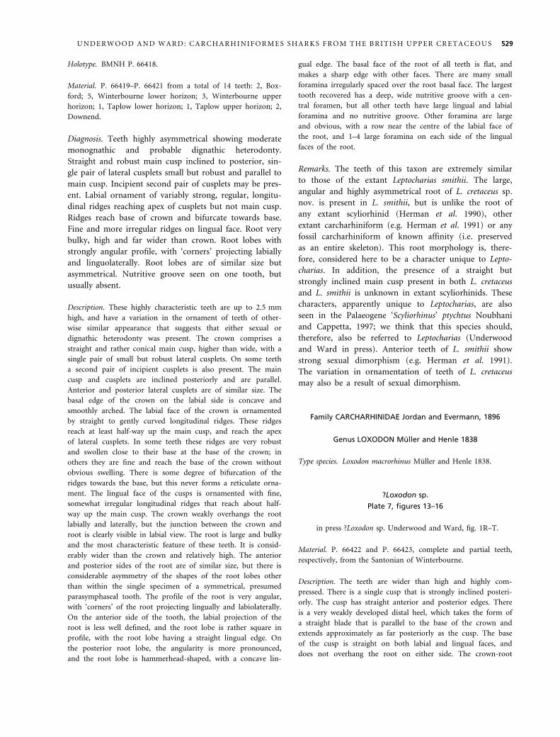
Holotype. BMNH P. 66418.
Material. P. 66419–P. 66421 from a total of 14 teeth: 2, Box-
ford; 5, Winterbourne lower horizon; 3, Winterbourne upper
horizon; 1, Taplow lower horizon; 1, Taplow upper horizon; 2,
Downend.
Diagnosis. Teeth highly asymmetrical showing moderate
monognathic and probable dignathic heterodonty.
Straight and robust main cusp inclined to posterior, sin-
gle pair of lateral cusplets small but robust and parallel to
main cusp. Incipient second pair of cusplets may be pres-
ent. Labial ornament of variably strong, regular, longitu-
dinal ridges reaching apex of cusplets but not main cusp.
Ridges reach base of crown and bifurcate towards base.
Fine and more irregular ridges on lingual face. Root very
bulky, high and far wider than crown. Root lobes with
strongly angular profile, with ‘corners’ projecting labially
and linguolaterally. Root lobes are of similar size but
asymmetrical. Nutritive groove seen on one tooth, but
usually absent.
Description. These highly characteristic teeth are up to 2.5 mm
high, and have a variation in the ornament of teeth of other-
wise similar appearance that suggests that either sexual or
dignathic heterodonty was present. The crown comprises a
straight and rather conical main cusp, higher than wide, with a
single pair of small but robust lateral cusplets. On some teeth
a second pair of incipient cusplets is also present. The main
cusp and cusplets are inclined posteriorly and are parallel.
Anterior and posterior lateral cusplets are of similar size. The
basal edge of the crown on the labial side is concave and
smoothly arched. The labial face of the crown is ornamented
by straight to gently curved longitudinal ridges. These ridges
reach at least half-way up the main cusp, and reach the apex
of lateral cusplets. In some teeth these ridges are very robust
and swollen close to their base at the base of the crown; in
others they are fine and reach the base of the crown without
obvious swelling. There is some degree of bifurcation of the
ridges towards the base, but this never forms a reticulate orna-
ment. The lingual face of the cusps is ornamented with fine,
somewhat irregular longitudinal ridges that reach about half-
way up the main cusp. The crown weakly overhangs the root
labially and laterally, but the junction between the crown and
root is clearly visible in labial view. The root is large and bulky
and the most characteristic feature of these teeth. It is consid-
erably wider than the crown and relatively high. The anterior
and posterior sides of the root are of similar size, but there is
considerable asymmetry of the shapes of the root lobes other
than within the single specimen of a symmetrical, presumed
parasymphaseal tooth. The profile of the root is very angular,
with ‘corners’ of the root projecting lingually and labiolaterally.
On the anterior side of the tooth, the labial projection of the
root is less well defined, and the root lobe is rather square in
profile, with the root lobe having a straight lingual edge. On
the posterior root lobe, the angularity is more pronounced,
and the root lobe is hammerhead-shaped, with a concave lin-
gual edge. The basal face of the root of all teeth is flat, and
makes a sharp edge with other faces. There are many small
foramina irregularly spaced over the root basal face. The largest
tooth recovered has a deep, wide nutritive groove with a cen-
tral foramen, but all other teeth have large lingual and labial
foramina and no nutritive groove. Other foramina are large
and obvious, with a row near the centre of the labial face of
the root, and 1–4 large foramina on each side of the lingual
faces of the root.
Remarks. The teeth of this taxon are extremely similar
to those of the extant Leptocharias smithii. The large,
angular and highly asymmetrical root of L. cretaceus sp.
nov. is present in L. smithii, but is unlike the root of
any extant scyliorhinid (Herman et al. 1990), other
extant carcharhiniform (e.g. Herman et al. 1991) or any
fossil carcharhiniform of known affinity (i.e. preserved
as an entire skeleton). This root morphology is, there-
fore, considered here to be a character unique to Lepto-
charias. In addition, the presence of a straight but
strongly inclined main cusp present in both L. cretaceus
and L. smithii is unknown in extant scyliorhinids. These
characters, apparently unique to Leptocharias, are also
seen in the Palaeogene ‘Scyliorhinus’ ptychtus Noubhani
and Cappetta, 1997; we think that this species should,
therefore, also be referred to Leptocharias (Underwood
and Ward in press). Anterior teeth of L. smithii show
strong sexual dimorphism (e.g. Herman et al. 1991).
The variation in ornamentation of teeth of L. cretaceus
may also be a result of sexual dimorphism.
Family CARCHARHINIDAE Jordan and Evermann, 1896
Genus LOXODON Muller and Henle 1838
Type species. Loxodon macrorhinus Muller and Henle 1838.
?Loxodon sp.
Plate 7, figures 13–16
in press ?Loxodon sp. Underwood and Ward, fig. 1R–T.
Material. P. 66422 and P. 66423, complete and partial teeth,
respectively, from the Santonian of Winterbourne.
Description. The teeth are wider than high and highly com-
pressed. There is a single cusp that is strongly inclined posteri-
orly. The cusp has straight anterior and posterior edges. There
is a very weakly developed distal heel, which takes the form of
a straight blade that is parallel to the base of the crown and
extends approximately as far posteriorly as the cusp. The base
of the cusp is straight on both labial and lingual faces, and
does not overhang the root on either side. The crown-root
U N D E R W O O D A N D W A R D : C A R C H A R H I N I F O R M E S S H A R K S F R O M T H E B R I T I S H U P P E R C R E T A C E O U S 529

junction is excavated on both labial and lingual faces, and
there is faint crenulation of the enameloid just above the junc-
tion. There is otherwise no ornamentation on the teeth. The
region of the root-crown junction is noticeably concave below
the base of the cusp on the labial side, and near the anterior
and posterior ends of the tooth on the lingual side. The root
is relatively small and compressed, being no deeper than the
crown. The root labial face is continuous with the anterior and
posterior parts of the crown, but is indented below the centre
of the cusp at the labial end of the nutritive groove. There is
an acute angle between the labial and basal faces of the root,
and the lingual root face is very reduced. The root is cut by a
deep but narrow nutritive groove containing a well-developed
foramen. A row of small foramina is present half-way up the
root labial face, with a row of smaller foramina being present
just below the crown-root junction on the lingual side. Small,
irregularly distributed, foramina are present across the basal
face of the root.
Remarks. These teeth appear superficially similar to those
of Paratriakis, but may be readily separated by the pres-
ence of a large nutritive groove. They can be referred to a
member of the Carcharhinidae and not the Triakidae by
the morphology of the root, and in particular the lack of
a labial overhang of the crown, a feature that appears to
be present in all triakids. These teeth are similar to those
of a number of Cenozoic and recent carcharhiniform gen-
era. Although the small quantity of material renders gen-
eric assignment uncertain, the lack of incipient serrations
and posterior cusplets makes it unlikely that these teeth
belong to Rhizoprionodon, whereas the lack of torsion of
the cusp makes assignment to Scoliodon unlikely. This
record represents the earliest occurrence of the Carcharhi-
nidae, and the first evidence of the family in the Mesozoic
(Underwood and Ward in press).
Family ?PROSCYLLIIDAE Fowler, 1941
Genus PROTOSCYLIORHINUS Herman, 1977
Type species. Protoscyliorhinus bettrechiensis Herman, 1977.
Protoscyliorhinus sp.
Plate 7, figures 17–19
Material. BELUM K28672, a partial tooth from the Coniacian of
Minnis North.
Description. The single partial tooth is about 2 mm wide, but is
missing the posterior part. The crown comprises a low, triangu-
lar main cusp with one small lateral cusplet and a further two
incipient cusplets. The tip of the main cusp has been removed
by wear, but otherwise there is a continuous cutting edge along
the occlusal edge. The labial face of the crown is ornamented
with short, irregular longitudinal ridges. These bifurcate towards
the base and partly merge to form small, triangular raised por-
tions. The lingual face of the cusps has short, fine longitudinal
ridges. The crown overhangs the root along its labial and lateral
edges, with the basal edge of the crown forming a smooth curve
on the labial face. The root is low and of a similar thickness
along its preserved width. There is a wide but shallow nutritive
groove with a central foramen, although it is unclear whether
this has been modified by corrosion. The root is only slightly
flattened, and there is no well-defined edge between the basal
and other root faces. There is a row of very small foramina on
the labial root face, and scattered small foramina on the basal
part of the root.
Discussion. The tentative assignment of Protoscyliorhinus
to the Proscylliidae follows that of Underwood (2006)
and Underwood and Ward (in press) as the teeth possess
a deep labial face to the crown joining the main and
lateral cusps and a root of uniform thickness without a
flared basal face, a combination of characters seen in
extant Proscyllium but not any extant scyliorhinids (see
Herman et al. 1990, 1991). This tooth represents the
youngest recorded occurrence of Protoscyliorhinus.
EVOLUTIONARY IMPLICATIONS
The specimens recorded in this study represent the first
indications of a number of extant Carcharhiniforme fami-
lies in the Cretaceous (Underwood and Ward in press).
EXPLANATION OF PLATE 7
Figs 1–12. Leptocharias cretaceus sp. nov. 1–4, P. 66418, holotype, Upper Santonian, Winterbourne, lower phosphate level, in 1, labial,
2, lingual, 3, occlusal, and 4, oblique lateral views. 5–8, P. 66419, Lower Campanian, Winterbourne, upper phosphate level, in 5,
labial, 6, occlusal, 7, oblique lateral, and 8, lingual views. 9–10, P. 66420, Upper Santonian, Winterbourne, lower phosphate level,
in 9, labial, and 10, lingual views. 11–12, P. 66421, Lower Campanian, Winterbourne, upper phosphate level, parasymphaseal
tooth, in 11, oblique lateral, and 12, labial views. 1–8, 11–12, · 25; 9–10, · 20.
Figs 13–16. ?Loxodon sp. 13–15, P. 66422, Upper Santonian, Winterbourne, lower phosphate level, in 13, labial, 14, occlusal, and 15,
lingual views. 16, P. 66423, Upper Santonian, Winterbourne, lower phosphate level, lingual view. All · 25.
Figs 17–19. Protoscyliorhinus sp. BELUM K28672, Coniacian, Minnis North, in 17, labial, 18, oblique lateral, and 19, lingual views;
· 20.
530 P A L A E O N T O L O G Y , V O L U M E 5 1
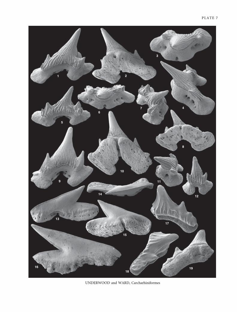
PLATE 7
UNDERWOOD and WARD, Carcharhiniformes
1 2
3
4 7 6
5
9
10
8
11
12
17
19 18
16
13
15
14

Previous studies have suggested that fossils from the
Jurassic may be assignable to the Proscylliidae (e.g.
Underwood and Ward 2004), a family predicted to be
close to the base of the carcharhiniforme clade (see
Underwood 2006 and references therein). The Lepto-
chariidae are also likely to be a basal family within the
order (Underwood and Ward in press), and their pres-
ence in the Cretaceous is not unexpected. The Carcharhi-
nidae (including the Sphyrnidae if they are to be
considered monophyletic) are the most morphologically
diverse of the extant carcharhiniform families, with a
number of genera being known from the Palaeocene, even
though much of the diversification had occurred within
the Neogene (e.g. Cappetta 1987). The occurrence of Cre-
taceous Carcharhinidae therefore extends the earliest
record of the group to a point before the first recorded
radiation within the family.
PALAEOECOLOGY
The diverse carcharhiniforms recorded in all of the
samples studied form part of diverse shark and ray fau-
nas, with the majority of specimens in all of the Chalk
samples belonging to taxa with small teeth. The only
samples with the majority of larger teeth are those
from the greensands of Minnis North, where low num-
bers of small teeth may be a result of biostratinomic
sorting and physical and biological destruction. These
small teeth (with the possible exception of those of
some of the batoids) would have belonged to small
taxa, by comparison with modern relatives, the majority
less than 50 cm in length. Most of these small taxa
(Scyliorhinidae and most Orectolobiformes) have
pointed, clutching (Cappetta 1986) teeth, and would
have been slow swimmers with an elongate body form.
These are likely to have been nectobenthic generalist
feeders, consuming diverse small organisms. Other taxa,
particularly of the Triakidae, had a more cutting denti-
tion and were probably more active hunters of small
fish, cephalopods and crustaceans. Other probable eco-
logical types are present among associated selachians.
Medium to large predatory lamniformes are present in
all samples, although it is only in the Downend and
Minnis North samples that they are common, with taxa
possessing both cutting and piercing dentitions being
present in all samples. Selachians with a durophagous
dentition are rare in all of the Chalk samples,
and restricted to a small number of batoids and rare
Heterodontus. The exception to this is the sample from
Minnis North, where Heterodontus and batoids are
common, and teeth of Ptychodus were recorded. Teeth
of small ambush predators, such as Squatina, are pres-
ent but uncommon in all samples.
This dominance of selachian faunas by diverse, small,
nectobenthic taxa appears to be common within the
European Cretaceous. Previous studies of selachian faunas
from European chalk facies have yielded high diversities
of small taxa from horizons in the Albian (Underwood
and Mitchell 1999), Cenomanian (e.g. Muller and Died-
rich 1991), Campanian (e.g. Muller 1989) and Maastrich-
tian (e.g. Siverson 1993), although all of these studies are
from within the North Sea Basin or associated Boreal
regions, as opposed to the more Tethyan-influenced
Anglo-Paris Basin of the Chalk samples described here.
Although there are many similarities with regard to the
overall ecological composition of all of these European
assemblages, squalids are a major part of all of the more
boreal assemblages; it is likely that the absence of squalids
in the samples described here is owing to their more bor-
eal distribution. Other, less systematic, studies of sela-
chians from the Anglo-Paris Basin (e.g. Herman 1977)
yielded very similar faunas to those described here, and
lack squalids. Very high diversity selachian faunas domi-
nated by small nectobenthic taxa therefore appear to be
characteristic of the European Upper Cretaceous.
Selachian faunas from the Western Interior Seaway of
North America are well known from near-shore facies
(e.g. Case 1978), but there have been few systematic
studies of selachians from neritic facies (see Welton and
Farish 1993; Case and Cappetta 1997). Although studies
typically show high diversities of Lamniformes and
batoids, most samples contain very few taxa of small
nectobenthic sharks (except in Case and Cappetta 1997);
in some samples even batoids are extremely rare and the
faunas are almost entirely composed of larger mid-water
predators (e.g. Cumbaa et al. 2006).
Other Late Cretaceous selachian faunas are less well
known than those of northern Europe and North Amer-
ica. Diverse Maastrichtian sharks are well known from
North African phosphorites, and very diverse assemblages
have been described from Morocco (e.g. Noubhani and
Cappetta 1997). These contain a number of Carcharhini-
forme taxa along with many Lamniformes and other sela-
chian groups. Although the species composition of these
assemblages is well recorded, the proportions of different
species and ecogroups have not been documented.
Acknowledgements. We thank a number of people who have
helped with this study over the last 30 years. Ian Jarvis, Peter
Woodruff and Andy Gale helped with both locality information
and in the field during the initial collecting phase; Alison Ward
assisted in the field and with the sample processing. Mike Simms
is thanked for his help with collecting the Minnis North mate-
rial, which was funded by University of London Grant GLRP5.
Andy Beard is thanked for his help with SEM imaging. The
comments of Henri Cappetta and the Palaeontology editors were
extremely useful.
532 P A L A E O N T O L O G Y , V O L U M E 5 1
REFERENCES
A G A S S I Z , L. J. R. 1833–44. Recherches sur les poissons fossiles,
3. Imprimerie de Petitpierre, Neuchatel, 390 + 32 pp.
A N T U N E S , M. T. and C A P PE T T A , H. 2002. Selaciens du
Cretace (Albien–Maastrichtien) d’Angola. Palaeontographica,
Abteilung A, 264, 85–146.
B L A I N V I L L E , H. M. D. de 1816. Prodrome d’une distribu-
tion systematique du regne animal. Bulletin de la Societe Philo-
matique de Paris, 8, 105–124.
B O N E , A. and B ON E , D. 2002. Lavant Stone: a late medie-
val building stone from the Chalk (Upper Cretaceous) of
West Sussex. Proceedings of the Geologists’ Association, 111,
193–203.
—— —— 2004. Lavant Stone: a Roman and medieval building
stone in West Sussex. Sussex Archaeological Collections, 142,
63–78.
C A P PE T T A , H. 1973. Selachians from the Carlile Shale (Turo-
nian) of South Dakota. Journal of Paleontology, 47, 504–514.
—— 1977. Observations sur quelques selaciens du Cretace Sup-
erieur d’Angleterre avec la description d’un genre nouveau.
Geobios, 10, 479–485.
—— 1980a. Les selaciens du Cretace superieur du Liban. I:
Requins. Palaeontographica, Abteilung A, 168, 69–148.
—— 1980b. Les Selaciens du Cretace Superieur du Liban. II:
Batoides. Palaeontographica, Abteilung A, 168, 149–229.
—— 1986. Types dentaires adaptatifs chez les selaciens actuels et
post-Paleozoiques. Palaeovertebrata, 16, 57–76.
—— 1987. Mesozoic and Cenozoic elasmobranchii. Chondrich-
thyes II. In S C HU L T Z E , H. P. (ed.). Handbook of paleoich-
thyology, Part 3B. Gustav Fischer Verlag, Stuttgart, 193 pp.
—— 1992. Carchariniformes nouveaux (Chondrichthyes, Neo-
selachii) de l’Ypresian du bassin de Paris. Geobios, 25, 639–
646.
—— and OD I N , G. S. 2001. Les selaciens du Campanien–
Maastrichtien de Tercis les Bains (SO France). 645–651. In
OD I N , G. S. (ed.). The Campanian-Maastrichtian boundary
characterization and correlation from Tercis-les-Bains (Landes,
SW France) to Europe and other continents. IUGS Special Pub-
lication, 36, 679–685.
C A S E , G. R. 1978. A new selachian fauna from the Judith River
Formation (Campanian) of Montana. Palaeontographica,
Abteilung A, 160, 176–205.
—— and C A P PE T T A , H. 1997. A new selachian fauna from
the Late Maastrichtian of Texas (Upper Cretaceous ⁄Navarroan; Kemp Formation). Munchener Geowissenschaftlich
Abhandlungen, A, 27, 5–63.
C OM P A GN O , L. J. V. 1977. Phyletic relationships of living
sharks and rays. American Zoologist, 17, 303–322.
C UM B A A , S. L., S CH R O DE R - A D A M S , C., DA Y , R. G.
and PH I L L I PS , A. J. 2006. Cenomanian bonebed faunas
from the northeastern margin, Western Interior Seaway,
Canada. New Mexico Museum of Natural History and Science
Bulletin, 35, 139–155.
D A L I N K E V I C I U S , J. A. 1935. On the fossil fishes of the
Lithuanian Chalk. I. Selachii. Memoires de la Faculte des
Sciences de l’Universite de Vytautas le Grand, 9, 247–305.
D A I M E R I E S , A. 1888. Notes ichthyologiques I. Annales de la
Societe Royal de Malacologie, Belgique, Bulletin des Seances, 23,
42–43.
D A V I S , J. W. 1887. The fossil fishes of the chalk of Mount
Lebanon, in Syria. Scientific Transactions of the Royal Dublin
Society, Series 2, 3, 457–636.
D I N E L E Y , D. L. and M E T C A L F , S. J. 1999. Fossil fishes of
Great Britain. Geological Conservation Review Series, 16. Joint
Nature Conservation Committee, Peterborough, 675 pp.
E L L I S , J. R. and S H A CK L E Y , S. E. 1995. Ontogenetic changes
and sexual dimorphism in the head, mouth and teeth of the
lesser spotted dogfish. Journal of Fish Biology, 47, 155–164.
F L E T CH E R , T. P. 1977. Lithostratigraphy of the Chalk (Ulster
White Limestone) in Northern Ireland. Reports of the Institute
of Geological Sciences, 77 (24), 1–33.
F O W L E R , H. W. 1941. The fishes of the groups Elasmobran-
chii, Holocephali, Isospondyli, and Ostariophysi obtained by
US Bureau of Fishing Steamer Albatross in 1907 to 1910,
chiefly in the Philippine Islands and adjacent seas. Bulletin of
the United States Natural History Museum, 100, 1–879.
G A L E , A. S. 1980. Penecontemporareous folding, sedimentation
and erosion in Campanian Chalk near Portsmouth, England.
Sedimentology, 27, 137–151.
G A R M A N , S. 1913. The Plagiostomia (sharks, skates and rays).
Memoirs of the Museum of Comparative Zoology, Harvard Col-
lege, Cambridge, 36, 1–515.
G I L L , T. 1862. Analytical analysis of the order of Squali and
revision and nomenclature of genera. Annals of the Society of
Natural History of New York, 7, 367–408.
G R A Y , J. E. 1851. List of specimens of fish in the collection of the
British Museum, Part 1. British Museum (Natural History),
London, 160 pp.
G U R R, P. R. 1962. A new fish fauna from the Woolwich
Bottom Bed (Sparnacian) of Herne Bay, Kent. Proceedings of
the Geologists’ Association, 73, 419–447.
H A L T E R , M. C. 1994. Additions to the fish fauna of N.W.
Europe. 3. Three new species of the genus Scyliorhinus from the
late Cretaceous (Campanian and Maastrichtian) of the Limburg
area (Belgium and the Netherlands) with the reassignment of
four additional species to the genus Scyliorhinus sensu stricto.
Professional Paper of the Belgian Geological Survey, 278, 65–110.
H A Y , O. P. 1902. Bibliography and catalogue of the fossil
vertebrata of North America. Bulletin of the United States
Geological Survey, 179, 1–868.
H E R M A N , J. 1977. Les Selachians neocretaces et paleocenes de
Belgique et des contrees limitrophes elements d’une biostratig-
raphie intercontinentale. Memoires pour servir a l’Explication
des Cartes Geologiques et Minieres de la Belgique, 15, 1–450.
—— 1982. Die Selachier-Zahne aus der Maastricht-Stufe von
Hemmoor, Niederelbe (NW-Deutschland). Geologisches Jahr-
buch, A, 61, 129–159.
—— H O V E S T A DT - E U L E R , M. and H O V E S T A D T, D. C.
1990. Contributions to the study of the comparative morpho-
logy of teeth and other relevant ichthyodorulites in living
supraspecific taxa of chondrichthyan fishes. In S T E H M A N N ,
M. (ed.). Part A: Selachii. No. 2b: Order: Carcharhiniformes –
Family: Scyliorhinidae. Bulletin de l’Institut Royal des Sciences
Naturelles de Belgique, Biologie, 60, 181–230.
U N D E R W O O D A N D W A R D : C A R C H A R H I N I F O R M E S S H A R K S F R O M T H E B R I T I S H U P P E R C R E T A C E O U S 533
—— —— —— 1991. Contributions to the comparative morpho-
logy of teeth and other relevant ichthyodorulites in
living supraspecific taxa of chondrichthyan fishes. In
S T E H M A N N , M. (ed.). Part A: Selachii. No. 2c: Order:
Carcharhiniformes – Families: Proscylliidea, Hemigaleidae,
Pseudotriakidae, Leptocharias and Carcharhinidae. Bulletin de
l’Institut Royal des Sciences Naturelles de Belgique, Biologie,
61, 73–120.
J A R V I S , I. 1980. The initiation of phosphatic sedimentation,
the Senonian (Cretaceous) of the Anglo-Paris Basin. 167–192.
In B E N T O R, Y. K. (ed.). Marine phosphorites, geochemistry,
occurrence, genesis. SEPM Special Publication, 29, 249 pp.
—— 2006. The Santonian–Campanian phosphatic chalks of
England and France. Proceedings of the Geologists’ Association,
117, 219–237.
—— and W O O DR O OF , P. 1981. The phosphatic chalks and
hardgrounds of Boxford and Winterbourne, Berkshire – two
tectonically controlled facies in the Late Coniacian to early
Campanian (Cretaceous) of southern England. Geological Mag-
azine, 118, 175–187.
J OR D A N , D. S. and E V E R M A N N , B. W. 1896. The fishes of
North and Middle America: a descriptive catalogue of the spe-
cies of fish-like vertebrates found in the waters of North
America, north of the isthmus of Panama. Part I. Bulletin of
the United States National Museum, 47, 1–1240.
L E R I C H E , M. 1906. Contribution a l’etude des poissons fos-
siles du Nord de la France et des regions voisines. Memoires
de la Societe Geologique du Nord, 55, 1–430.
L I N N A E US , C. 1758. Systema naturae. Tenth edition. Salvi,
Stockholm, 824 pp.
M A R CK , W. von der 1863. Fische, Krebse und Pflanzen aus
dem Plattenkalk der jungeren Kreide aus Westfalen. Paleonto-
graphica, 11, 1–83.
M OR T I M OR E , R. N., W OO D, C. J. and G A L LO I S , R. W.
2001. British Upper Cretaceous stratigraphy. Geological Conser-
vation Review Series, 23. Joint Nature Conservation Commit-
tee, Peterborough, 585 pp.
M U L L E R , A. 1989. Selachier (Pisces, Chondrichthyes) aus dem
hoheren Campanium (Oberkreide) Westfalens (Nordrhein-
Westfalen, NW-Deutschland). Geologie und Palaontologie in
Westfalen, 14, 1–161.
—— and DI E DR I CH , C. 1991. Selachier (Pisces, Chondrich-
thyes) aus dem Cenomanium von Ascheloh am Teutoberger
Wald (Nordrhein-Westfalen, NW-Deutschland). Geologie und
Palaontologie in Westfalen, 20, 1–105.
M U L L E R , J. and H E N L E , J. 1838–41. Systematische beshrei-
bung der plagiostomen. Veit, Berlin, 200 pp.
N O UB H A N I , A. and CA P P E T TA , H. 1997. Les Orecto-
lobiformes, Carchariniformes, et Myliobatiformes (Elasmo-
branchii, Neoselachii) des bassins a phosphate du Maroc
(Maastrichtian–Lutetien basal). Palaeo Ichthyologica, 8, 1–327.
R E E S , J. 2005. Neoselachian shark and ray teeth from the
Valanginian, Lower Cretaceous, of Wawał, central Poland.
Palaeontology, 48, 209–221.
S C H I N Z , H. R. 1822. Das Thierreich eingetheilt nach dem Bau der
Thiere als Grundlage ihrer Naturgeschichte und der vergleichenden
Anatomie. Mit vielen Zusatzenversehen von H. R. Schinz,
Cotta, Stuttgart and Tubingen das Thierreich, i–xvi, 1–83.
S H I R A I , S. 1996. Phylogenetic interrelationships of neosela-
chians (Chondrichthyes: Euselachii). 9–34. In S T I A S S E Y , M.
L. J., P A R E N T I , L. R. and J O H N S O N , G. D. (eds). Inter-
relationships of fishes. Academic Press, San Diego, CA, 496 pp.
S I M M S , M. J. 2003. Minnis North Mudflow. Geological Sites
in Northern Ireland, Earth Science Conservation Review:
http://www.habitas.org.uk/escr/summary.asp?Item=114
S I V E R S O N , M. 1993. Late Cretaceous and Danian neoselachians
from southern Sweden. Lund Publications in Geology, 110, 1–28.
S W E E TM A N , S. C. and U N DE R W O O D, C. J. 2006. A
neoselachian shark from the non-marine Wessex Formation
(Wealden Group: Early Cretaceous, Barremian) of the Isle of
Wight, southern England. Palaeontology, 49, 457–465.
U N DE R W O O D, C. J. 2004. Barremian and Aptian (Creta-
ceous) sharks and rays from Speeton, Yorkshire, NE England.
Proceedings of the Yorkshire Geological Society, 55, 107–118.
—— 2006. Diversification of the Neoselachii (Chondrichthyes)
during the Jurassic and Cretaceous. Paleobiology, 32, 215–235.
—— and M I T CH E L L , S. H. 1999. Albian and Cenomanian
selachian assemblages from North East England. Special Papers
in Palaeontology, 60, 9–59.
—— —— and V E L T K A M P , C. J. 1999. Microborings in mid
Cretaceous fish teeth. Proceedings of the Yorkshire Geological
Society, 52, 269–274.
—— and W A R D , D. J. 2004. Environmental distribution of
Bathonian (Middle Jurassic) neoselachians in southern
England. 111–122. In T I N T O R I , A. and A R R A T I A , G.
(eds). Mesozoic fishes 3 – systematics, palaeoenvironments
and biodiversity. Verlag Dr. Friedrich Pfeil, Munchen, 649 pp.
—— —— in press. The Mesozoic record of the Carcharhinifor-
mes. In A R R A TI A , G., S C HU L T ZE , H.-P. and W I L S O N ,
M. V. H. (eds). Mesozoic fishes 4. Verlag Dr. Friedrich Pfeil,
Munchen.
W A G N E R , J. A. 1857. Charakteristik neuer Arten von Knorpel-
fischen aus den lithographischen Schiefern der Umgegend von
Solnhofen. Gelehrte Anzeiger der Bayerischen Akademie der
Wissenschaften, 44, 288–293.
W E L T O N , B. J. and F A R I S H, R. F. 1993. The collector’s guide
to fossil sharks and rays from the Cretaceous of Texas. Before
Time, Lewisville, TX, 203 pp.
W H I T E , H. G. O. and T RE A CH E R , L. 1906. The phosphatic
chalks of Winterbourne and Boxford (Berkshire). Quarterly
Journal of the Geological Society of London, 62, 499–522.
W I L L CO X , N. R. 1953. The origin of the beds of phosphatic
chalk with special reference to those at Taplow. 119–133. In
Origin des gisements de phosphates de chaux. Comptes Rendus
de la Dix-neuvieme Session, Alger, Congres Geologiques Inter-
national, Algiers.
W O O DR O OF , P. B. 1980. Field Meeting: The Chalk of the
Newbury and Lambourne area, Berkshire, 25 June 1978. Pro-
ceedings of the Geologists’ Association, 91, 349–350.
W O O DW A R D , A. S. 1889. Catalogue of the fossil fishes in the
British Museum. Part 1. British Museum (Natural History),
London, 474 pp.
534 P A L A E O N T O L O G Y , V O L U M E 5 1

APPENDIX
Field sites and provisional faunal lists
Locality. Boxford, Berkshire.
Coordinates. SU 431 719.
Section description. A small overgrown quarry, located 340 m
north-north-west of Boxford Church, on the east side of a coun-
try lane that runs parallel to the River Lambourne. The pit
exposes a complex of slump folded hardgrounds overlain by
phosphatic chalk.
Stratigraphy. Uppermost Seaford or basal Newhaven Chalk
Formation, M. coranguinum Zone, mid-Santonian.
Samples. 95 kg sample of soft pelletal phosphatic chalk was col-
lected from high in the northern face of the quarry, 30 cm above
the ‘Upper Hardground’. A rich and diverse selachian fauna was
recovered.
References. White and Treacher (1906), Jarvis and Woodroof
(1981), Ward in Dineley and Metcalf (1999), Mortimore et al.
(2001).
Associated selachian fauna. Synechodus sp., Squatina decipiens
Dalinkevicius, 1935, Squatina sp., Carcharias sp., Cretalamna
appendiculata (Agassiz, 1843), Protolamna sp., ‘Palaeoanacorax’
sp., ?Pseudocorax laevis Leriche, 1906, Squalicorax falcatus (Agas-
siz, 1843), Heterodontus cf. havreensis Herman, 1977, Chiloscyl-
lium greeni (Cappetta, 1973), Cretorectolobus sp., Pararhincodon
groesseni Herman, 1982, Pseudospinax pusillus Muller and Died-
rich, 1991, Parasquatina sp., ?Rhombopterygia sp., ?Rhynchobatus
sp., Sclerorhynchus sp., Rhinobatos casieri Herman, 1977, ?Squa-
tirhina sp. indet., Hybodontidae indet.
Locality. Winterbourne, Berkshire.
Coordinates. SU 448 722.
Section description. A shallow pit in the middle of a field expos-
ing up to 6 m of upper Santonian and lower Campanian chalk
(Marsupites and pilula zones), c. 1.7 km east of Boxford. Two
phosphatic levels were sampled.
Stratigraphy. Newhaven Chalk Formation; lower phosphatic hori-
zon is Marsupites testudinarius Zone, upper Santonian; upper
phosphatic horizon is Offaster pilula Zone, lower Campanian.
Samples. 165 kg of pelletal (Santonian) chalk from immediately
above the (lower) Phosphatic Hardground, and 180 kg of simi-
lar, lower Campanian, chalk, 2 m higher in section, just above
the Upper Hardground. Both levels yielded a rich and diverse
selachian fauna.
References. White and Treacher (1906), Woodroof (1980), Jarvis
and Woodroof (1981), Mortimore et al. (2001).
Associated selachian fauna. Lower phosphatic level: Hexanchus
sp., Squatina decipiens Dalinkevicius, 1935, Squatina sp.,
Carcharias sp., Cretalamna appendiculata (Agassiz, 1843), Proto-
lamna sp., ‘Palaeoanacorax’ sp., Pseudocorax laevis Leriche,
1906, Squalicorax kaupi (Agassiz, 1843), Paracorax sp., Heter-
odontus sp., Chiloscyllium greeni (Cappetta, 1973), Cretascyllium
?expansum Muller and Diedrich, 1991, Pararhincodon groesseni
Herman, 1982, Parasquatina sp., ?Rhombopterygia sp., Sclero-
rhynchus sp., Rhinobatos casieri Herman, 1977. Upper phos-
phatic level: Squatina decipiens Dalinkevicius, 1935, Squatina
sp., Carcharias sp., Cretalamna appendiculata (Agassiz, 1843),
Protolamna sp., Scapanorhynchus sp., ‘Palaeoanacorax’ sp., Pseu-
docorax laevis Leriche, 1906, ?Squalicorax kaupi (Agassiz, 1843),
Chiloscyllium greeni (Cappetta, 1973), Cederstroemia havreensis
(Herman, 1977), Cretascyllium ?expansum Muller and Diedrich,
1991, Pararhincodon groesseni Herman, 1982, Parasquatina sp.,
?Rhombopterygia sp., Sclerorhynchus sp., Rhinobatos casieri
Herman, 1977.
Locality. Stoke Clump, West Sussex.
Coordinates. SU 835 098.
Section description. A series of shallow pits in farmland that were
infilled in the early 1940s. However, lumps of brown phosphatic
chalk, known as Lavant Stone, can still be found in the field
475 m north-east of Stoke Clump.
Stratigraphy. Old Nore or Peacehaven Beds, Newhaven Chalk
Formation; Offaster pilula Zone, lower Campanian.
Samples. Approximately 220 kg of phosphatic chalk was
collected, crushed, and screened. This site yielded a relatively
low diversity fauna dominated by Chiloscyllium. There are also
abundant sponge spicules, isolated asteroid and ophiuroid
ossicles and phosphatised bony fish otoliths.
References. Mortimore (1986), Bone and Bone (2002, 2004).
Associated selachian fauna. Paraorthacodus sp., Squatina decipiens
Dalinkevicius 1935, Anomotodon sp., ‘Palaeoanacorax’ sp., Squa-
licorax kaupi (Agassiz, 1843), Chiloscyllium greeni (Cappetta,
1973), Cederstroemia havreensis (Herman, 1977), ?Rhomboptery-
gia sp., Sclerorhynchus sp., Rhinobatos casieri Herman, 1977,
Rhinobatos sp.
Locality. South Lodge Pit, Taplow, Buckinghamshire.
Coordinates. SU 906 819.
Section description. South Lodge Pit is an abandoned quarry on
the east bank of the River Thames near Taplow. The pit
originally exposed two hardgrounds of middle and late Santo-
nian age, overlain by a brown pelletal phosphatic chalks. It is
currently somewhat degraded and only the upper horizon is
visible.
Stratigraphy. Taplow Phosphatic Chalk Member, Newhaven
Chalk Formation; lower phosphatic horizon is Uintacrinus socia-
lis Zone, mid Santonian; upper phosphatic horizon is Marsupites
testudinarius Zone, upper Santonian.
Samples. About 180 kg of soft brown pelletal phosphatic chalk
was taken from immediately above both hardgrounds. Vertebrate
faunas from both were poor and yielded few well-preserved
teeth.
References. Willcox (1953), Mortimore et al. (2001).
Associated selachian fauna. Lower phosphatic level: Squatina deci-
piens Dalinkevicius, 1935, Carcharias sp., ?Scapanorhynchus sp.,
‘Palaeoanacorax’ sp., Pseudocorax laevis Leriche, 1906, Squalico-
rax kaupi (Agassiz, 1843), Chiloscyllium greeni (Cappetta, 1973),
Pseudospinax pusillus Muller and Diedrich, 1991, Sclerorhynchus
sp., Rhinobatos casieri Herman, 1977. Upper phosphatic level:
Squatina decipiens Dalinkevicius, 1935, Carcharias sp., Creta-
lamna appendiculata (Agassiz, 1843), ?Scapanorhynchus sp.,
‘Palaeoanacorax’ sp., Pseudocorax laevis Leriche 1906, Squalicorax
kaupi (Agassiz, 1843), Chiloscyllium greeni (Cappetta, 1973),
Cretorectolobus sp., Cretascyllium ?expansum Muller and Died-
U N D E R W O O D A N D W A R D : C A R C H A R H I N I F O R M E S S H A R K S F R O M T H E B R I T I S H U P P E R C R E T A C E O U S 535
rich, 1991, Pararhincodon groesseni Herman, 1982, ?Rhomboptery-
gia sp., Rhinobatos casieri Herman, 1977.
Locality. Downend, Hampshire.
Coordinates. SU 601 065.
Section description. A large, mostly back-filled, quarry on the
south-west margin of Portsdown Hill, immediately adjacent to
the M27 motorway. The middle part of the south-east face
exposed faulted and contorted Campanian chalks with hard-
grounds and locally developed calcarenites.
Stratigraphy. ?Spetisbury Member, Culver Chalk Formation;
G. quadrata and B. mucronata Overlap Zone, lower
Campanian.
Samples. About 150 kg of calcarenite was crushed and sieved
from immediately above the Downend Main Hardground. Sela-
chian remains were diverse and included very abundant teeth of
Squalicorax. Many of these are of a similar size and appear to
represent a partial associated dentition.
References. Gale (1980), Mortimore et al. (2001).
Associated selachian fauna. Squatina decipiens Dalinkevicius,
1935, Squatina sp., Carcharias sp., Cretalamna appendiculata
(Agassiz, 1843), Scapanorhynchus sp., Palaeoanacorax sp., Pseudo-
corax laevis Leriche, 1906, ?Squalicorax kaupi (Agassiz, 1843),
Heterodontus sp., Chiloscyllium greeni (Cappetta, 1973), Ceder-
stroemia havreensis (Herman, 1977), Cretorectolobus sp., Para-
rhincodon groesseni Herman, 1982, Parasquatina sp.,
?Rhombopterygia sp., Rhinobatos casieri Herman, 1977.
Locality. Minnis North Mudflow, County Antrim, Northern
Ireland.
Coordinates. D 339 135.
Section description. This site is a mudslide on the Antrim Coast
Road (A2) north of Drumnagreagh Port. Cretaceous rocks occur
as blocks within the slipped Jurassic clays at the base of the hill
and on the beach below the road, and sometimes as partly
slipped blocks on the hillside.
Stratigraphy. Phosphatic pebble bed in Kilcoan Sands Member,
Hibernian Greensand Formation; M. cortestudinarium Zone,
Coniacian.
Samples. About 160 kg of glauconitic sandstone was dissolved in
acid. Vertebrate remains were seen in the field and vary in their
mode of preservation. Many teeth are dark coloured, strongly
abraded and have any cavities filled by glauconite, whilst others,
including all of the specimens figured here, are pale in colour,
appear unabraded and have porous roots. The composition of
these two assemblages is somewhat different, but it is not clear
whether they indicate two different assemblages (indigenous and
reworked) or whether the differences are due to differential bio-
stratinomy.
References. Fletcher (1977), Simms (2003).
Associated selachian fauna. Abraded teeth: Synechodus sp., Creta-
lamna appendiculata (Agassiz, 1843), Protolamna sp., Paranomo-
todon sp., Squalicorax falcatus (Agassiz, 1843), Heterodontus
lonzeensis Herman, 1977, Heterodontus sp., Turoniabatis sp.,
?Ptychotrygon sp. Hybodontidae indet., Ptychodus sp. Unabraded
teeth: Protosqualus sp., Carcharias sp., Cretalamna appendiculata
(Agassiz, 1843), Protolamna sp., Paranomotodon sp., Scapa-
norhynchus sp., Squalicorax falcatus (Agassiz, 1843), Heterodontus
lonzeensis Herman, 1977, Chiloscyllium greeni (Cappetta, 1973),
Cantioscyllium sp., Pararhincodon sp., ?Rhombopterygia sp.,
Rhinobatos sp., Turoniabatis sp., Squatirhina lonzeensis Herman,
1977, Meristodon sp.
536 P A L A E O N T O L O G Y , V O L U M E 5 1