
Understanding how capping of actin filaments prevents...
74
UNIVERSIDADE DE LISBOA FACULDADE DE CIÊNCIAS DEPARTAMENTO DE BIOLOGIA ANIMAL Understanding how capping of actin filaments prevents epithelium to mesenchymal like transitions in genetically defined epithelial tissues Sofia Raquel Paulo Rebelo MESTRADO EM BIOLOGIA EVOLUTIVA E DO DESENVOLVIMENTO 2007
Transcript of Understanding how capping of actin filaments prevents...

UNIVERSIDADE DE LISBOA
FACULDADE DE CIÊNCIAS
DEPARTAMENTO DE BIOLOGIA ANIMAL
Understanding how capping of actin filaments prevents epithelium to mesenchymal like transitions in genetically
defined epithelial tissues
Sofia Raquel Paulo Rebelo
MESTRADO EM BIOLOGIA EVOLUTIVA E DO DESENVOLVIMENTO
2007

UNIVERSIDADE DE LISBOA
FACULDADE DE CIÊNCIAS
DEPARTAMENTO DE BIOLOGIA ANIMAL
Understanding how capping of actin filaments prevents epithelium to mesenchymal like transitions in genetically
defined epithelial tissues
Sofia Raquel Paulo Rebelo
Dissertação de mestrado orientada por: Doutora Florence Janody1
Orientadora interna: Prof. Doutora Gabriela Rodrigues2 1 Instituto Gulbenkian de Ciência, Oeiras. 2 Faculdade de Ciências da Universidade de Lisboa, Lisboa.
MESTRADO EM BIOLOGIA EVOLUTIVA E DO
DESENVOLVIMENTO 2007


i
ACKNOWLEDGEMENTS The past year has been crucial for my formation as an individual and as a beginner in the
scientific world. There are several people that have been really important during this
period, to whom I would like to thank:
First, I would like to give a special thanks to my supervisor, Florence Janody. Not only
for being available and supportive during the entire period of training but also for
creating a respectful and friendly environment in lab, which has been crucial to the
development of my work. I must also thank her for giving me such a beautiful project to
work in during my master thesis and for the permanent motivation.
To my internal supervisor, Dr. Gabriela Rodrigues, for being as helpful as she could be
whenever I needed.
To Beatriz......I have no words to say how thankful I am. For the support and permanent
motivation, for teaching me how to work and for being the wonderful person she is.
Thanks a lot!
To Gaspar, the most promising scientist I know! Thanks for everything, especially for the
enlightening discussions we have and for the comments on my thesis.
To Maurícia Cristina, for making my lab work more fun and easy (and for being a great
matrecos-partner).
To Emília Pomba, not only for the scientific help on molecular biology, but also for being
a very good friend (and for being my thesis-writing partner).
To the Drosophila community at the IGC, who have taught me a lot and make the fly
work a very nice moment.

ii
To Dr. Gabriel Martins, for helping me with the confocal imaging.
To all the people that I am not mentioning but that have contributed in any way to my
work and to my life.
And to my family, for being always there.
Thanks to all of you!
iii
LIST OF ABREVIATIONS
ABP
AJ
AP
Arm
bp
CP
Cpa
Cpb
Crb
D/V
Dlg
ECM
EMT
FA
FACS
F-actin
FAK
IR
JNK
Lgl
PCR
RNAi
RT
SAR
Scrib
SJ
TSG
Vg
Actin Binding Proteins
Adherens Junctions
Anterior/posterior
Armadillo (ß-catenin)
base pair
Capping protein
Capping protein a
Capping protein ß
Crumbs
Dorso-ventral
Discs large
Extracelular matrix
Epithelium to mesenchymal transition
Focal adhesions
Fluorescence activated cell sorting
Filamentous actin
Focal adhesion kinase
Inverted repeat
Jun N-terminal Kinase
Lethal giant larvae
Polimerase chain reaction
RNA interference
Room temperature
Sub apical region
Scribble
Septate junctions
Tumor supressor gene
Vestigial

iv
ABSTRACT
The actin cytoskeleton has a central role in controlling cell shape and mobility. In
epithelia, a circumferential band of actin filaments provides the structural support for
cell-cell junctions. When the strength of the epithelium is compromised, cells may
undergo epithelium to mesenchymal transition, escape size-control mechanisms, evade
cell death and finally acquire the ability to migrate. These features recapitulate all of the
hallmarks that characterize cancer malignancy. Interestingly, clones of cells mutant for
either subunits of the capping protein αβ heterodimer (CP), induced in a heterozygous
wild-type background, are extruded from the wing blade epithelium and die. However,
depleting the β subunit (cpb) by RNA interference (RNAi) in the whole blade leads to a
different outcome: epithelium polarity is strongly affected, few apoptotic cells can be
observed, while many seem to overproliferate. This differential behavior is unlikely to be
due to a dosage effect since RNAi induced-cpb depletion in restricted wing blade
domains also induces cell extrusion and death. This suggests that CP prevents
tumorigenesis of wing blade cells. However, when CP mutant cells are adjacent to wild-
type wing blade neighboring cells, the latest eliminate mutant cells by a process of cell
competition. The role of CP in preventing tumorigenesis might be related to its major
function, preventing excessive actin polimerization, or to additional functions, such as the
maintenance of epithelial cell polarity.
The tumor suppressor function of CP appears to be tissue specific since the above
cell behavior is only observed in restricted epithelia. This suggests that each epithelium
has specific cytoskeleton and/or junctional properties, making cells sensitive or not to
mutations that cause abnormal tumor growth. Altogether the presented data highlight the
crucial impact of tissue context for the activation of a tumoral process.
Keywords: Capping Protein, epithelium to mesenchymal transition, epithelia and tumor.

v
RESUMO
O citosqueleto de actina desempenha um papel fundamental no controlo da forma
e mobilidade celulares. Dada a sua dinâmica, é necessária uma regulação eficaz, que é
desempenhada por proteínas que se ligam à actina. As proteínas Capping protein a (Cpa)
and Capping protein ß (Cpb) são duas subunidades do heterodímero Capping Protein
(CP) que se liga ao filamento de actina, prevenindo a adição de monómeros e, desta
forma, a sua polimerização excessiva.
Nos tecidos epiteliais, a região apical das células contém uma banda de filamentos
de actina, essencial para o suporte das junções celulares. Quando ocorre perda de adesão
num epitélio, as células podem sofrer uma transição epitélio-mesenquimatosa, durante a
qual se tornam resistentes aos mecanismos de controlo de crescimento dos tecidos e à
morte celular e adquirem potencial migratório. Estas alterações celulares caracterizam a
progressão de tumores malignos.
Curiosamente, quando clones de células mutantes para ambas as subunidades do
heterodímero CP são induzidos no tecido selvagem heterozigota do disco imaginal da asa
de Drosophila, as células mutantes na região dorso-ventral (DV) do disco, designado por
zona do wing blade, sofrem extrusão basal e morrem por apoptose. Contudo, no epitélio
do foliculo ovárico de Drosophila, a indução de clones de células mutantes para o gene
cpa leva a um tipo de comportamento celular diferente: as células perdem as suas
características epiteliais, sobre-proliferam e parecem adquirir potencial migratório, capaz
de invadir tecidos ajdacentes. Este comportamento diferencial assemelha-se ao
comportamento de células mutantes para genes supressores de tumores neoplásticos em
Drosophila. Nestes casos, pequenos grupos de células mutantes são removidos pelo
tecido adjacente, que lhes emite um sinal de morte celular. Este mecanismo designa-se
por competição celular e é considerado crucial para a supressão de tumores.
Desta forma, o presente trabalho tinha como objectivo comprender de que forma o
contexto celular pode influenciar o comportamento das células mutantes para ambas as
formas do heterodímero CP, levando a fenótipos completamente diferentes. Pretendia
ainda, de forma inerente, avaliar o potencial supressor de tumores do heterodímero CP.
Assim, dado que as mutações para os genes cpa e cpb são letais, usei a técnica de ARN

vi
de interferência (RNAi) para induzir o silenciamento pós-transcricional de ambos os
genes. As técnicas disponíveis em Drosophila (sistema UAS-Gal4) permitiram-me
direccionar o silenciamento de cpa e cpb para regiões específicas do seu tecido, de forma
a poder avaliar a influência do contexto do tecido no comportamento de células
silenciadas para os genes cpa e cpb.
Quando o gene cpb foi silenciado em toda a região do wing blade, a maioria das
células desta região sobreviveram e pareciam ter perdido o controlo da proliferação
celular bem como a polaridade epitelial comprometida. A sua morfolgia parecia também
ter-se alterado, tornando-se mais arredondada e menos adesiva. Todas estas observações
sugerem que estas células poderiam estar a sofrer uma transição do estado epitelial para o
estado mesenquimatoso. O comportamento diferencial entre a inactivação de cpb por
indução de clones e por silenciamento através de RNAi poderia ocorrer por uma questão
de dosagem proteica, já que em clones os níveis de cpb são nulos e o RNAi leva apenas a
um silenciamento parcial da expressão génica. No entanto, este não parece ser o caso, já
que o silenciamento por RNAi em regiões restritas das asa levou à extrusão epitelial e
morte celular. Desta forma, a extensão da inactivação de cpb parece ser determinante
para o fenótipo celular: o silenciamento de cpb numa vasta região do disco imaginal da
asa remove a contribuição do tecido do tipo selvagem, logo não há sinalização de morte
celular para as células adjacentes e estas sobrevivem. Considera-se assim, que o contexto
celular é determinante para a sobrevivência de células cujo desenvolvimento é anormal.
Para além de permitirem explicar o fenótipo de clones de células mutantes para
CP, os resultados obtidos quando o gene cpb foi silenciado em toda a região do wing
blade, sugerem que o heterodímero CP pode funcionar como supressor de tumores. Desta
forma, são apresentadas no presente trabalho as diferentes hipóteses que podem explicar
o papel do heterodímero CP neste processo. Em primeiro lugar propõe-se que a função
principal de CP, de prevenir a polimerização excessiva dos filamentos de actina, possa
prevenir a formação de tumores. Isto seria explicado pelo facto do citosqueleto de actina
estar envolvido numa multiplicidade de fenómenos celulares, cuja desregulação pode
levar à activação de processos tumorais. Um exemplo é o seu papel no tráfego vesicular,
ou, em alternativa, a existência de isoformas especificas de actina localizadas
preferencialmente em determinados epitélios, cuja acumulação poderia activar tumores.
vii
O heterodímero CP localiza-se apicalmente nos tecidos epiteliais e estudos
anteriores sugerem que este possa estabelecer a ligação dos filamentos de actina à
membrana celular. Desta forma, propõe-se que este possa ter um papel no
estabelecimento ou manutenção da polaridade epitelial, cuja destruição é crucial para a
activação tumoral. Uma vez que o fenótipo de perda de função hipomórfica de cpb e do
gene arc, envolvido na modulação das junções de aderência são semelhantes, postula-se
que ambos os genes possam interagir de forma cooperativa neste processo.
Adianta-se ainda uma hipótese concreta através da qual a activação de tumores
poderia ocorrer. Esta baseia-se no facto do fenótipo de perda de função de cpb, tanto
numa extensão reduzida ou em larga extensão, ser semelhante ao de perda de função dos
genes csk e dASPP, ambos inibidores da família de proto-oncogenes Src. Sabe-se que o
aumento dos níveis de actividade de Src leva à dissolução das junções de aderência e
adesões focais, levando à formação de tumores. Desta forma, considera-se como hipótese
que o heterodímero CP possa ser necessário para manter o nível de actividade de Src,
prevenindo desta forma a formação de tumores. Esta hipótese é consistente com o facto
de ambas as proteínas se localizarem apicalmente, possivelmente co-localizando nas
junções de aderência. A hipótese de sobreactivação do proto-oncogene Src não exclui as
duas hipóteses mencionadas anteriormente, já que pode surgir como uma consequência
secundária de defeitos no citosqueleto de actina ou de defeitos de polaridade epitelial,
causados directamente por perda de CP.
Curiosamente, o possível potencial suppressor de tumores de CP parece ser
específico de determinados tipos de tecido, nomeadamente da região do wing blade do
disco imaginal da asa e do tecido epitelial do ovário. Isto sugere que a arquitectura do
citosqueleto e/ou das junções epiteliais é específica de cada epitélio, tornando as células
mais ou menos sensíveis a mutações que causam desenvolvimento de tumores. No
conjunto, os dados apresentados apontam para o impacto crucial do contexto celular na
activação de um processo tumoral.
Palavras-chave: Capping protein, transição epitélio-mesenquimatosa, epitélio e tumor.
viii
TABLE OF CONTENTS
Acknowledgements i
List of abreviations. iii
Abstract iv
Resumo v
Table of contents viii
I INTRODUCTION 1
1 Molecular mechanisms of epithelial architecture 1
1.1 Epithelial structure 1
1.2 Establishment and maintenance of epithelial structure 2
1.3 Drosophila as a model system to understand epithelial architecture 3
1.4 Remodeling of epithelia in normal and abnormal developmental processes 4
2. Tumor formation and development 5
2. 1 Tumor supressor genes 6
2.2 Oncogenes 8
2.3 Tumor microenvironment and the combinatory model 9
3. The actin cytoskeleton in normal and pathological developmental processes 10
3.1The actin cytoskeleton and its dynamics 10
3.2The actin cytoskeleton in epithelia 11
3.3The actin cytoskeleton in pathology 12
4. Capping protein 12
4.1 CP: a highly conserved heterodimer 12
4.2 CP maintains cells within the wing disc epithelia 13
4.3 CP prevents excessive actin filament polymerization and localizes at apical sites 13
4.4 The behavior of CP mutant cells depends on the Vestigial transcription factor 15
4.5 CP maintains the monolayer character of the ovarian follicle epithelium 15
II OBJECTIVES 16

ix
III MATHERIALS AND METHODS 17
1. Generation and establishment of the UAS-CP-IR transgenic lines 17
1.1 Generation of the UAS-cpa-IR and UAS-cpb-IR constructs 17
1.1.1 The pWIZ vector 17
1.1.2 Design and amplification of the cpa and cpb IR 18
1.1.3 Cloning the cpa and cpb IR in the pWIZ vector 20
1.1.3.1 First cloning step 20
1.1.3.2 Second cloning step 21
1.2 Establishment of UAS-CP-IR transgenic lines 21
1.2.1 Microinjection 21
1.2.2 Establishment of the RNAi stocks 22
2. Fly husbandry 22
3. Immunohistochemistry 23
IV RESULTS 25
1. RNA interference as a tool to investigate the role of the cellular environment on
the behavior of CP mutant cells 25
1.1 UAS-cpa-IR and UAS-cpb-IR transgenic lines
1.2 Efficiency of the UAS-CP-IR 26
1.2.1 Ubiquitous depletion of CP 27
1.2.2 Depletion of CP in the eye imaginal disc 27
1.2.3 Depletion of CP in the wing proper 28
1.3 Specificity of the UAS-CP-IR 32
2. Depletion of cpb in the wing imaginal disc leads to different outcomes 33
2.1 Partial depletion of cpb in the wing resembles the phenotype of arc mutant flies 33
2.2 The local environment influences the behavior of CP depleted cells 34
2.2.1 Depletion of cpb in the vg expression domain leads to massive cell
extrusion and apoptosis 34
2.2.2 Broad depletion of cpb in the scalloped expression (sd) domain leads to
most of the hallmarks of tumor cells 36
x
2.2.3 Wing blade cells depleted of cpb seem to acquire the ability to migrate 38
V DISCUSSION 41
CP mutant cells versus CP depletion by RNA interference: two different
outcomes 42
Cells depleted of cpb in the whole wing blade epithelium show most of the
hallmarks of tumor cells 44
The postulated tumor suppressor potential of CP is restricted to a subset of
epithelia 46
CP might have additional functions to prevent tumor formation 48
VI REFERENCES 52
VII APPENDIX 55
1. Drosophila as a model organism 55
1.1 Drosophila genetics 56
1.2 Techniques available in Drosophila 56
1.2.1 FLP/FRT System 56
1.2.2 The MARCM system 56
1.2.3 UAS-Gal4 system 57
1.2.4 P-element mediated transformation 58
2. cpa and cpb sequences used as IR 59
3. Additional protocols 60
1
I. INTRODUCTION
1. Molecular mechanisms of epithelial architecture
1.1 Epithelial structure
Most animal tissues are composed of epithelial sheets, which serve two main
functions: protection from the external environment and separation of two different
chemical milieus. An epithelium consists of a laterally coherent sheet of cells with
distinct apical-basal polarity. Several types of junctions mediate cell-cell and cell-matrix
contact in epithelia. The most relevant junctions for adhesion are the adherens junctions
(AJ) and hemiadherens junctions (Schock and Perrimon, 2002).
The AJ is a generally circumferential junction located just basal to the apico-
basolateral boundary, which major function is to link the actin cytoskeleton of
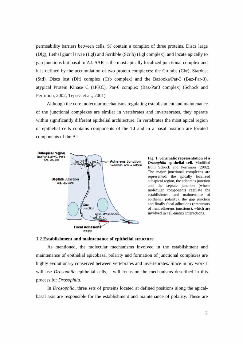
neighboring epithelial cells (Fig. 1). They are composed of the transmembrane protein E-
cadherin, which is linked on its cytoplasmic side to ß- catenin (in Drosophila encoded by
armadillo) and α- catenin. In turn, α- catenin binds to actin filaments, forming a
continuous band, the zonula adherens (ZA) (Tepass et al., 2001).
The hemiadherens junctions are spot-like junctions connecting the actin
cytoskeleton of epithelial cells to the basement membrane. The primary function of
basement membranes is to anchor down the epithelium to a loose connective sheet
underneath but they also act as a mechanical barrier, preventing malignant cells from
invading deeper tissues (LeBleu et al., 2007). The interaction between epithelial sheets
and the basement membrane is mediated by integrins, which bind to components of the
extracelullar matrix (ECM). Developing tissues contain focal adhesions (FA), which are
the precursors of hemiadherens junctions. These structures confer strong adhesion to the
substrate and are anchored to bundles of actin microfilaments, called actin stress fibers
(Fig. 1). Stress fibers are mostly present in migrating cells and are required for generating
the propulsive force for motility (Schock and Perrimon, 2002).
In addition to the adherens and hemiadherens junctions, three other junctional
complexes exist in Drosophila: gap junctions, septate junctions (SJ) and the subapical
region (SAR) (Fig. 1). Gap junctions form intercellular channels allowing the
transmission of ions and small molecules between cells. In contrast, SJ act as
2
permeability barriers between cells. SJ contain a complex of three proteins, Discs large
(Dlg), Lethal giant larvae (Lgl) and Scribble (Scrib) (Lgl complex), and locate apically to
gap junctions but basal to AJ. SAR is the most apically localized junctional complex and
it is defined by the accumulation of two protein complexes: the Crumbs (Cbr), Stardust
(Std), Discs lost (Dlt) complex (Crb complex) and the Bazooka/Par-3 (Baz-Par-3),
atypical Protein Kinase C (aPKC), Par-6 complex (Baz-Par3 complex) (Schock and
Perrimon, 2002; Tepass et al., 2001).
Although the core molecular mechanisms regulating establishment and maintenance
of the junctional complexes are similar in vertebrates and invertebrates, they operate
within significantly different epithelial architecture. In vertebrates the most apical region
of epithelial cells contains components of the TJ and in a basal position are located
components of the AJ.
1.2 Establishment and maintenance of epithelial structure
As mentioned, the molecular mechanisms involved in the establishment and
maintenance of epithelial apicobasal polarity and formation of junctional complexes are
highly evolutionary conserved between vertebrates and invertebrates. Since in my work I
will use Drosophila epithelial cells, I will focus on the mechanisms described in this
process for Drosophila.
In Drosophila, three sets of proteins located at defined positions along the apical-
basal axis are responsible for the establishment and maintenance of polarity. These are
Fig. 1. Schematic representation of a Drosophila epithelial cell. Modified from Schock and Perrimon (2002). The major junctional complexes are represented: the apically localized subapical region, the adherens junction and the septate junction (whose molecular components regulate the establishment and maintenance of epithelial polarity), the gap junction and finally focal adhesions (precursors of hemiadherens junctions), which are involved in cell-matrix interactions.
3
the SAR components, Crb and Baz/Par-3 complexes, and the Lgl complex. The first two
protein groups are required for the formation of the ZA, which is the primary event for
the establishment of epithelia structure. Whereas the Baz/Par-3 complex regulates early
phases of ZA assembly, the Crb complex is required for later phases of ZA maturation.
Crb protein is considered to be a crucial apical determinant, since it has the ability to
confer apical membrane identity to the basolateral membrane. The Lgl complex
components are located at the SJ, basal to the ZA and counteract Crb complex apicalizing
activity (Gibson and Perrimon, 2003; Schock and Perrimon, 2002).
1.3 Drosophila as a model system to understand epithelial architecture
Since the main mechanisms governing epithelial polarity in vertebrates and
invertebrates are highly conserved, Drosophila arises as an interesting model to study
epithelial architecture (the great success of Drosophila as a model organism to study
epithelial architecture relies on the advantages derived from its use, which are mentioned
in attachment 1). Models for epithelial differentiation in postembryonic development of
Drosophila include the imaginal discs and the ovarian follicle epithelium. While imaginal
disc epithelia are preferred for the analysis of epithelial maintenance and specialization of
epithelial surface domains, the follicular epithelium, due to its unique characteristic of
renewing itself constantly, allows the analysis of the full range of phases in epithelial
differentiation (Tepass et al., 2001).
Imaginal discs are larval structures that serve as the primordial for most adult
structures. They are established during embryogenesis as clusters of 20-50 cells, but
proliferate to a final size of 20000-50000 cells before they undergo metamorphosis. The
imaginal disc epithelium is composed of a squamous layer, the peripodial membrane, and
of a columnar layer of proliferating cells, the proper disc epithelium (Fig. 2 C) (Hariharan
and Bilder, 2006). Wing imaginal discs are subdivided into three regions along the
dorsoventral (DV) axis of the disc: the notum (or body wall), the hinge and the pouch (or
blade) (Fig. 2 B). These regions are genetically defined during early larval development.
An antagonist relationship between Epidermal Growth Factor (EGF) and Wingless (Wg)
patterns the disc into the dorsal region which will give rise to the notum, and a ventral
region which forms the wing. The wing is further subdivided by expression of selector

4
genes vestigial (vg) and scalloped (sd) in the wing blade and homothorax (hth) in the
wing hinge (Klein, 2001). Vg is required for wing formation (Williams et al., 1991) and
its misexpression can induce ectopic wing tissue (Kim et al., 1996).
Fig 2. Drosophila wing disc and fate maps. Adapted from (Butler et al., 2003). A) Third instar wing
imaginal disc. B) Fate map of the wing disc showing the anterior-posterior (AP) and dorsal-ventral (DV)
compartment boundaries and major regions in the disc. In the adult, the wing pouch (green) gives rise to
the wing, the hinge (yellow) constricts to form a mobile link to the body wall (blue) of the fly. C) Cell
layers of the wing disc. There are two main cell layers: the peripodial membrane and the columnar
epithelium that gives rise to the adult epidermis.
1.4 Remodeling of epithelia in normal and abnormal developmental processes
Cell phenotype transitions involving modulation of cell-cell adhesion occur during
both physiological and pathological states. During embryogenesis of most animals,
epithelial cell subpopulations actively downregulate cell adhesion systems and leave their
local neighborhood to move into new microenvironments were they eventually
differentiate into different cell types. This regulated phenotypic modulation is called
epithelial to mesenchymal transition (EMT). Also in cancers, more precisely during the
progression of primary tumors towards metastasis, tumor cells lose epithelial
characteristics, change cell shape and acquire migratory abilities and invasiveness.
Despite these different situations, the final effectors controlling cell phenotype, including
the cytoskeleton, the cell-cell and the cell-matrix adhesion systems, appear to be similar
or identical (Savagner, 2001; Huber et al., 2005).

5
Both normal and pathological versions of EMT involve, in addition to changes in
shape and acquisition of motility, fundamental alterations in the gene expression profile.
The expression of the adhesion component E-cadherin is repressed while the expression
of vimentin, an intermediate filament component of the mesenchymal cell cytoskeleton is
induced. Additionally, the cells start producing fibronectin and expressing N-cadherin.
Moreover, there is increased expression of proteases, in particular metaloproteases, since
the detachment of epithelial cells from the basement membrane requires their increased
activity, as well as a dramatic reorganization of the actin cytoskeleton. This is an
extremely important feature for the acquisition of motility and is thought to involve a
complex regulation of the actin cytoskeleton effectors, in particular the small GTPases of
the Rho family (Weinberg, 2007).
2. Tumor formation and development
Cancer is a multistep complex disease, resultant from genetic and epigenetic
changes that occur in cells of various cell types. In mammalian epithelial cells, the
activation of a neoplastic tumoral process is characterized by disruption of epithelial
apicobasal polarization and acquisition of mesenchymal characteristics (EMT), self
sufficiency in growth/proliferation signals (which leads to tissue overproliferation),
resistance to cell death, inability to differentiate and ultimately, acquisition of migratory
and invasive abilities (Hariharan and Bilder, 2006).
Two main categories of genes are involved in the process of carcinogenesis:
oncogenes, which are the result of activated proto-oncogenes, and tumor suppressor
genes (TSG). Both types of genes are required for normal cell proliferation and
differentiation, and aberrant expression leads to abnormal cell proliferation.
Despite the differences in tumor development in mammalian systems and
Drosophila, during recent years its use to understand tumorigenesis has become widely
accepted. The late mechanisms of tumor development may be considerably different,
since Drosophila has an open circulatory system, thus does not undergo angiogenesis,
and has a different system of telomere maintenance, however, the basic cell biology
mechanisms appear to be similar in both systems (Brumby and Richardson, 2005).
Therefore, from now on, I will focus my analysis on Drosophila TSG and oncogenes.

6
2.1 Tumor supressor genes
TSG are genes that, when mutated to a loss of function, result in excessive tissue
growth. The TSG affecting epithelial tissues may be classified as hyperplastic, when their
inactivation leads to increased cell proliferation but normal tissue architecture is
maintained or neoplastic, when in addition to increased proliferation there is disruption of
epithelial structure. Neoplastic tumors are also characterized by an inability to terminally
differentiate and acquisition of invasive characteristics, resembling mammalian
malignant tumors (Bilder et al., 2000; Hariharan and Bilder, 2006).
Among the hyperplastic TSG identified until now, pten, Tsc1, Tsc2, merlin,
expanded and components of the warts pathway are some of the most studied in
Drosophila. Less characterized ones include the C-terminal src kinase (csk) and discs
overgrown (do). The neoplastic TSG may be included in two major groups: genes of the
endocytic pathway (avalanche, rab5, tgs101 and vps25) and genes of the junctional
scaffold (scrib, dlg and lgl) (Hariharan and Bilder, 2006).
The phenotypes of mutants for endocytic neoplastic TSG are caused by an excess
accumulation of various signaling receptors that fail to be turned over in mutant tissue
(Hariharan and Bilder, 2006). In particular, defects of avalanche and Rab5 mutant cells
appear to be due to mistrafficking of the polarizing component Crb (Lu and Bilder,
2005).
As mentioned, proteins of the Lgl group (Lgl/Dlg/Scrib) localize basolaterally, at
the SJ. Bilder and Perrimon (2000) were the first to report a genetic interaction between
these three neoplastic TSG, as well as their cooperative regulation of cell polarity and
growth. Homozygous larvae for any of these genes undergo an abnormal third instar
larvae period of two weeks and fail to pupate. During this period, imaginal discs and
brain cells over proliferate in a disorganized pattern, forming large amorphous masses.
Mutations in these genes also display invasive characteristics, particularly seen in the
ovarium follicle epithelia. Unlike the imaginal disc epithelia, the follicle epithelium is in
close contact with a distinct tissue, the germ line, and is enclosed in a monolayered
sheath. Mutant cells for components of the Lgl complex are capable of leaving the
epithelium and streaming into the germ line cyst, where they penetrate between nurse cell
membranes (Fig. 3). Interestingly, despite the continuous proliferative ability acquired by

7
mutant cells, the proliferation rates are slower in mutant than wild type cells (Humbert et
al., 2003). When clones of mutant cells for components of the Lgl complex are induced
through the process of mitotic recombination, the wild-type neighboring cells ultimately
eliminate clones by a phenomenon known as cell competition (Bilder et al., 2000),
showing that the surrounding wild type tissue may account to the suppression of a
tumoral process. Inded, when scrib clones in mosaic eye discs are protected from the cell
competition events, mutant cells overgrow and lead to host lethality (Brumby and
Richardson, 2003).
Another TSG that was reported to be highly susceptible to the surrounding wild
type tissue is csk (Vidal et al., 2006). As previously mentioned, csk is classified as a
hyperplastic TSG. Homozygous mutants die during pupal stage and their imaginal discs
are up to 50% larger than wild type discs. Csk is known to negatively regulate the kinase
activity of the oncongenic family of the Src Kinases, thus csk inactivation is predicted to
result in a Src increased activity (Yeatman, 2004). Interestingly, while broad loss of csk
leads to enlarged, mispatterned tissues due to a block in apoptosis, overproliferation and
decreased cadherin mediated adhesion, discrete inactivation of csk leads to epithelial
exclusion, invasive migration and apoptotic death (Fig. 4) (Vidal et al., 2006). These
differences in outcome highlight the relevance of tissue context on tumor growth.
Fig. 3 Groups of cells homozygous mutant for genes of the Lgl complex overproliferate and show an invasive type of behavior in the ovarium follicle epithelium. Adapted from Bilder and Perrimon (2000). Mutant cells are marked by the absence of GFP. Phalloidin marks actin filaments.

8
2.2 Oncogenes
Oncongenes, like TSG may be classed as hyperplastic or neoplastic, according to
the same criteria. Hyperplastic oncogenes include Src and the activated forms of Ras
(Rasact) and Notch (Notchact). The neoplastic include the activated PDGF- and VEGF-
receptor related (Pvr) (Brumby and Richardson, 2005).
Ras and Notch signaling are involved in a variety of cellular processes, which
frequently overlap and may influence each other, either in a cooperative or antagonistic
manner. Ras is a small GTPase that cycles between an inactive GDP-bound state and an
active GTP-bound state. Activated Ras in Drosophila exerts its oncogenic effects through
Raf/MAPK pathway, whose targets have been shown to promote differentiation, cell
survival and cell proliferation (Sundaram, 2005). In turn, Notch consists of a
transmembrane protein, which intracellular domain is implied in promoting nuclear
transcription. Notch signaling pathway was reported to have both tumor suppressor and
inducing functions during the process of tumorigenesis, but the mechanisms regulating
this process in Drosophila are quite complex and are not completely understood (Brumby
and Richardson, 2005; Weng and Aster, 2004).
The role of Src kinases in tumorigenesis is postulated to initially promote cell
growth but it also regulates other cellular activities such as adhesion, motility and
Fig 4. Contrasting phenotypes caused by differential loss of csk in Drosophila wing imaginal disc. Images modified from Vidal et al., 2006. A) Loss of csk in the entire wing disc leads to tissue overgrowth. Depletion of csk was induced through RNAi silencing and targeted to specific domains of Drosophila tissue through the use of the GAL4/UAS system. csk depletion was targeted to the entire wing disc using the 765-Gal4 driver, which domain of expression in shown in A’. In A’’, is shown a third instar wing imaginal disc targeting depletion of csk in the entire wing disc. Those discs are enlarged. B) Loss of csk as discrete patches leads to epithelial extrusion and cell death. B’ is a schematic of the mutant clones, while B’’ shows that csk mutant cells are eliminated from the tissue.

9
migration during late stages of tumorigenesis, possibly due to different levels of activity
(Vidal et al., 2007). Src localizes at AJ and FA, and its overactivity disrupts these two
structures, therefore reducing adhesion. In the AJ, the Src kinase enhanced activity
suppress E-cadherin localization and function, while in FA it binds to Focal Adhesion
Kinase (FAK), activating it. FAK signaling ultimately activates the Jun N-terminal
Kinase (JNK) signaling pathway, promoting expression of metaloproteases such as
MMP3 and MMP9, which together with decreased E-Cadherin promote invasiveness
(Yeatman, 2004).
2.3 Tumor microenvironment and the combinatory model
The development of cancer is a multistep process, which is thought to involve the
cooperation of several mutations, as well as interactions between the tumor and its
microenvironment. When clones of mutant cells for the TSG scrib and csk are induced
among wild type tissue, the surrounding wild type cells actively remove the abnormal
cells through the process of cell competition (Brumby and Richardson, 2003; Vidal et al.,
2006). The molecular mechanisms that trigger cell competition are still not completely
understood, however the proto-oncogenic family of Myc is postulated to mediate this
process: clones of cells expressing low levels of Myc are out-competed by neighboring
cells that express higher levels of Myc (Johnston et al., 1999). Also, the competition for
survival factors such as decapentaplegic (DPP) could account for this effect (Moreno et
al., 2002). Either induced by Myc or by Dpp, the JNK signaling pathway is thought to be
the final effector of the cell competition process (Adachi-Yamada and O'Connor, 2004;
Moreno et al., 2002).
Since wild type tissue has mechanisms of suppressing tumor development,
mutations in a single gene may not be sufficient for the activation of a tumoral process
and a cooperative interaction between TSG and oncogenes may be required for the
formation of invasive type of tumors. As mentioned above, scrib mutant cells are not
capable of competing with the surrounding wild type tissue and of behaving as inasive
tumor cells. However, the combination of the activated alleles of either Notchact and
Rasact with clonal inactivation of scrib, results in a strong neoplastic growth, capable of
invading distant tissues and metastasing (Brumby and Richardson, 2003; Pagliarini and

10
Xu, 2003). These experiments provided the basis to the establishment of the combinatory
model in Drosophila. The combinatory model postulates that tumor development in flies,
like in mammals is influenced by the tissue context, and additional alterations, either
within the tumor or in the surrounding normal tissue, might be required to result in
unrestrained tumor growth and lethality (Brumby and Richardson, 2005).
3. The actin cytoskeleton in normal and pathological developmental processes
3.1 The actin cytoskeleton and its dynamics
The actin cytoskeleton plays a central role in numerous cellular processes. It
functions in the generation and maintenance of cell morphology and polarity, in
endocytosis and intracellular trafficking, in contractility, motility and cell division. Actin
is a globular, nucleotide-binding protein that is present in cells in two main forms, the
globular (G) actin and the filamentous (F) actin. The filamentous actin is a polarized
structure, in which the barbed or growing end is the preferred site for ATP-monomeric-
actin addition, whereas the pointed or slow growing end is the predominant site for loss
of ADP-actin subunits (Fig. 5). This ATP-hydrolysis-driven filament growth is a very
dynamic process, which requires a tight regulation in vivo. More than 70 classes of Actin
Binding Proteins (ABP) control the dynamics of actin filaments within the cell,
regulating their assembly, disassembly and organization (dos Remedios et al., 2003;
Winder and Ayscough, 2005).
ABP such as Profilin, Formins, Enabled/VASP (Ena/VASP) family proteins and
the Arp2/3 complex promote actin polymerization. Profilin enhances the exchange of
ADP for ATP on actin monomers, Formins as well as the Arp2/3 complex are important
for de novo filament formation, a process known as nucleation. The Arp2/3 complex has
also a role in branch formation and its activity is enhanced by the WASP and
SCAR/WAVE proteins. In contrast, the Ena/VASP family proteins favour formation of
long un-branched filaments. Among the ABP that prevent actin polymerization, the Actin
Depolymerizing Factor (ADF)/Cofilin family, the Cyclase associated protein (CAP) and
Capping Proteins (CP) are some of the most important (Fig. 5). The ADF/Cofilin severs
filaments and enhances dissociation of actin monomers from the pointed end, CAP
sequesters actin monomers, preventing their incorporation into filamens and CP restricts
11
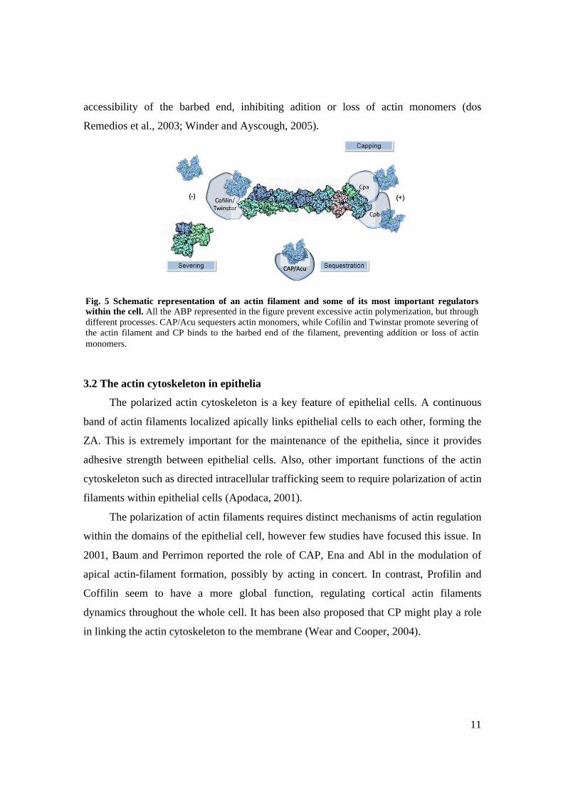
accessibility of the barbed end, inhibiting adition or loss of actin monomers (dos
Remedios et al., 2003; Winder and Ayscough, 2005).
3.2 The actin cytoskeleton in epithelia
The polarized actin cytoskeleton is a key feature of epithelial cells. A continuous
band of actin filaments localized apically links epithelial cells to each other, forming the
ZA. This is extremely important for the maintenance of the epithelia, since it provides
adhesive strength between epithelial cells. Also, other important functions of the actin
cytoskeleton such as directed intracellular trafficking seem to require polarization of actin
filaments within epithelial cells (Apodaca, 2001).
The polarization of actin filaments requires distinct mechanisms of actin regulation
within the domains of the epithelial cell, however few studies have focused this issue. In
2001, Baum and Perrimon reported the role of CAP, Ena and Abl in the modulation of
apical actin-filament formation, possibly by acting in concert. In contrast, Profilin and
Coffilin seem to have a more global function, regulating cortical actin filaments
dynamics throughout the whole cell. It has been also proposed that CP might play a role
in linking the actin cytoskeleton to the membrane (Wear and Cooper, 2004).
Fig. 5 Schematic representation of an actin filament and some of its most important regulators within the cell. All the ABP represented in the figure prevent excessive actin polymerization, but through different processes. CAP/Acu sequesters actin monomers, while Cofilin and Twinstar promote severing of the actin filament and CP binds to the barbed end of the filament, preventing addition or loss of actin monomers.

12
3.3 The actin cytoskeleton in pathology
The actin microfilament system is considered to be the engine of cellular migration.
Numerous actin effectors provide a temporally and spatially controlled turnover of actin
structures, which is crucial to drive protrusion of migrating cells. Therefore, due to the
complex regulation of the actin cytoskeleton, the system is vulnerable to mutations and
defects that may cause a wide range of disorders. These may affect embryonic
development, the immune system or lead to tumor invasion and metastasis, which
characterize cancer malignancy (Lambrechts et al., 2004; Yamazaki et al., 2005).
Cancer is often a genetically heterogeneous disease. Although many mutations that
are in the origin of tumorigenesis affect non-cytoskeletal proteins, the overall result is a
transition from an immotile to a motile cell and an inherent dramatic alteration of the
actin system. These apparently arise via three non-mutually exclusive pathways:
mutations in actin, changes in the upstream regulatory proteins or changed expression
levels of ABP. Among the upstream regulators that are known to be involved in this
process, the Rho-GTPases are thought to play an important role, particularly through
activation of the WASP family proteins (activators of the Arp2/3 complex) (Lambrechts
et al., 2004).
Also, the expression levels of several ABP, such as beta-tymosins (monomer
sequestering), Arp2/3, cortactin, WASP, profiling (filament nucleation and elongation),
gelsolin, cofilin (filament capping, severing, or depolymerisation), have been reported to
be altered during human tumors. However, this is probably not the primary cause of
oncogenecity but rather an inherent consequence of the altered cellular background
(Lambrechts et al., 2004; Yamazaki et al., 2005).
4. Capping protein
4.1 CP: a highly conserved heterodimer
CP is a highly conserved heterodimer, composed of capping proteins α (Cpa) and β
(Cpb), which cap the barbed end of actin filaments with high affinity, thereby preventing
excessive actin polimerization (Wear and Cooper, 2004). Several studies account for the
role of CP in actin assembly and regulation of cell motility. In Dictyostelium, an increase
in CP leads to an increase in the cell movement (Hug et al., 1995), while in vitro studies

13
have shown that CP and the Arp2/3 complex show a functional antagonism, essential in
regulating motility (Pantaloni et al., 2000). Also during Drosophila bristle development,
Cpb acts antagonistically to the Arp2/3 complex in promoting the formation of a transient
subpopulation of filaments, called snarls, which are localized between bundles (Frank et
al., 2006). Although the mechanism and regulation of CP have been studied, most of the
available data results from in vitro biochemical studies and cells in culture. Therefore, it
is crucial to understand the role of CP and its regulation within a tissue.
4.2 CP maintains cells within the wing disc epithelia
Clonal analysis in the Drosophila wing imaginal disc revealed a new feature of CP:
they are required for the maintenance of cells within the wing blade epithelium. Mutant
cells for either cpa or cpb in these region suffer basal extrusion and apoptosis (Fig. 6).
Interestingly, the requirement for CP to maintain cells within the epithelia is restricted to
the wing blade region, since cell extrusion and death caused by loss of CP occurred in the
blade but not in the hinge and notum regions (Janody and Treisman, 2006).
4.3 CP prevents excessive actin filament polimerization and localizes at apical sites
As previously proposed by in vitro studies, CP prevents excessive actin filament
polymerization in the Drosophila wing disc. However, it was shown that there is a cell
type-dependent effect on actin filament accumulation in cpa mutant cells: in the notum
and hinge region actin filaments accumulate at the apical region of the cell whereas in the
blade, the accumulation occurs around the entire cell cortex (Fig. 7). However, excessive
Fig. 6. Clones of mutant cells for cpa and cpb extrude from the wing blade epithelia. Adapted from Janody and Treisman (2006). Optical cross sections through the wing disc epithelium. Mutant clones are positively labeled with GFP (green) and discs are stained with anti-Dlg (red) and anti-Arm (blue) to outline apical cell membranes. Either cpa and cpb mutant clones are extruded from the wing blade epithelium (indicated by white arrows) but are maintained within the remainder regions of the disc.

14
actin filament accumulation may not be the first cause of extrusion of cpa mutant cells,
since mutant cells for other ABPs that prevent excessive actin polimerization, such as
Twinstar (Tsr) and Capulet (Capt), do not cause cell extrusion (Janody and Treisman,
2006).
By expressing a HA-tagged form of Cpa, it was observed that Cpa mostly
accumulates at the apical regions of the cell, co-localizing with components of epithelial
junctions (Fig. 8 A-B). Moreover, AJ components were shown to be mislocalized at
basolateral positions in cpa mutant cells (Fig. 8 C). These observations show that there is
a region specific requirement for CP to maintain apicobasal polarity in wing blade cells
(Janody and Treisman, 2006).
Fig. 7. Loss of cpa leads to cell-type dependent accumulation of actin filaments in the different regions of the wing disc. Adapted from Janody and Treisman (2006). A-B) Optical cross sections through third instar Drosophila wing imaginal discs, comparing the notum and blade regions, respectively. Clones are negatively marked by GFP (green) (A,B) and discs are stained with phalloidin to reveal F-actin (red) (A’,B’). In the notum actin filament accumulation is restricted to the apical domains, while in the blade actin filaments accumulate throughout the cell.
Fig 8. Cpa possibly localizes at apical membrane sites and maintains the localization of AJ components. Adapted from Janody and Treisman (2006). Optical cross sections through third instar Drosophila wing imaginal disc. A-B) An HA-tagged form of Cpa (green) rescues the cell extrusion phenotype in cpa mutant cells (blue) and co-localizes with the AJ component Arm (red). C) In cpa mutant cells (blue) the AJ components Arm (green) and Dlg (red) are mislocalized (indicated by a arrow in C’).

15
4.4 The behavior of CP mutant cells depends on the Vestigial transcription factor
Cpa was shown to be required for the survival and maintenance of vg- expressing
cells in the wing disc epithelium: cpa mutant clones misexpressing vg were maintained in
all the regions of the wing epithelia and ectopic expression resulted in apoptotic death
and extrusion in the entire disc. Furthermore, the expression levels of cpa were enhanced
in the wing blade primordial, due to the presence of Vg. Thus, it is proposed that Vg
alters the cytoskeletal structure of cells by upregulating the expression of CP, which leads
to different cellular outcomes (Janody and Treisman, 2006).
4.5 CP maintains the monolayer character of the ovarian follicle epithelium
Clonal analysis in another type of epithelia, the ovarium follicle epithelia, leads to a
completely different cellular outcome: cpa mutant cells lose their monolayer character,
form disorganized multiple layers, leave the epithelium and stream into the germ-line
cyst, where they penetrate between nurse the cell membrane (Fig. 9) (Janody, un-
published data). These observations suggest that CP mutant cells might aquire an
invasive cell behaviour, similar to neoplastic TSG mutant cells such as scrib.
Fig. 9. Cpa maintains epithelial monolayer character of the ovarium follicle epithelia and prevents migratory type of behavior. Images obtained by Janody. Sections of the Drosophila ovaries. A) cpa mutant clones are negatively marked by GFP and indicated by arrows. E-Cad staining (red) and DAP1 (blue) show that cells lose epithelial architecture and overproliferate. B) DAP1 staining indicates cpa mutant cells of the ovarian follicle overproliferate and become invasive, since some are contained within the germ line cyst (indicated by arrow in B).
16
II OBJECTIVES
The Capping Protein α β heterodimer (CP) prevents epithelial to mesenchymal
like transitions in restricted epithelia: in the Drosophila wing disc, only mutant cells
located in the blade region extrude from the epithelium and die. In a similar manner, loss
of either csk or the TSG scrib in discrete patches leads to cell extrusion and apoptotic
death. However, broad loss of csk or scrib leads to a different outcome: enlarged and
mispatterned tissues due to overproliferation and block in apoptosis (Brumby and
Richardson, 2003; Vidal et al., 2006). This shows that the local environment influences
the behavior of csk and scrib-deficient cells. Since in the follicle epithelium, groups of
cells mutant for cpa form multiple layers, seem to overproliferate and are observed
invading between nurse cells (see Fig. 9 of Introduction), resembling cells mutant for
neoplastic TSG, the cell extrusion and cell death phenotype observed by clonal analysis
of CP could be dependant on the surrounding tissue rather than caused by cellular defects
(autonomous type of cell death). Therefore, the main objective of this work was to
understand whether the cellular environment influences the behavior of CP mutant cells.
Inherent to this, the work aimed to evaluate the potential of CP to suppress tumor
formation in the wing blade, as occurs in the ovarium follicle epithelium. This attempted
to show that epithelia may differ within the various type of tissues and that the tissue
context may be crucial to the development of tumoral behavior.

17
III MATERIALS AND METHODS
1. Generation and establishment of the UAS-CP-IR transgenic lines
1.1 Generation of the UAS-cpa-IR and UAS-cpb-IR constructs
1.1.1 The pWIZ vector
The presence of double stranded RNA (dsRNA) causes the sequence-specific
posttranscriptional silencing of a corresponding gene, thus this is considered to be an
efficient method to inactivate genes of interest and study gene function. Although
injection of dsRNA into Drosophila embryos is possible and effective, the effect is
transient and is not inherited in the next generation. For that reason, methods have been
developed to express dsRNA stably in transgenic Drosophila (Hannon, 2003; (Lee and
Carthew, 2003)
The pWIZ vector was specifically designed to express dsRNA in Drosophila. As
most of the methods, the pWIZ employs transgenes having an inverted repeat (IR)
configuration, which are able to produce dsRNA as extended hairpin RNA. However, it
differs from the other vectors due to the presence of a spacer between the inverted
repeats. The spacer used is the 74-nucleotide second intron of the white Drosophila gene,
which was shown to be efficiently spliced in heterologous tissues. This helps to stabilize
recombinant plasmids, making the process of cloning easier (Lee and Carthew, 2003).
The pWIZ vector takes advantage of two very useful techniques in Drosophila: p-
element mediated transformation and the modular Gal4/UAS expression system
(described in attachment 1). Gene fragments are inserted in opposite orientations on each
side of the intron, which is flanked by several unique restriction enzymes sites. The entire
cassette is downstream of the UAS enhancer-promoter and upstream of the SV4
transcription termination site (Lee and Carthew, 2003). The overall procedure for making
the cpa and cpb RNAi constructs in the pWIZ vector is described in Fig.1.

18
Spe I digestion and CIP treatment
Xba Idigestion and CIP treatment
Spe I digestion and CIP treatment
Xba Idigestion and CIP treatment
1.1.2 Design and amplification of the cpa and cpb IR
The choice of the gene fragments to clone in the pWIZ vector was performed
according to several criteria: the fragment chosen should be 500 to 700 base pairs (bp);
should correspond to an exon; should not have sequences in either sense and antisense
orientation that match a 5’ or 3’ consensus splice site and should not have internal
restriction sites corresponding to the primer sites (Lee and Carthew, 2003). Finally, a
sequence of 443 bp for cpa and of 543 bp for cpb was chosen (attachment 2).
The gene fragments were amplified by polymerase chain reaction (PCR). The
primers designed for each gene are shown in Tab. 1. In order to amplify the cpa gene
fragment, the template used was the vector GH10050, which was obtained from Berkeley
Drosophila Genome Project (BDGP). The cpb fragment was amplified from genomic
DNA of WT flies.
Fig. 1. Strategy of cloning used to generate the UAS-CP-constructs. Modified from Lee and Carthew (2000). The DNA fragment corresponding to cpa and cpb genes was amplified by PCR. A restriction site for XbaI was included in each PCR primer. The fragments and the pWIZ were digested with XbaI and ligated. This is the first cloning step into the pWIZ vector. The second cloning step occurs through digestion of the pWIZ vector containing the first insert with SpeI, whose free ends are compatible with the XbaI ones, and posterior ligation with XbaI-digested cpa and cpb. The gene fragments must be cloned in opposite orientations on each side of the white intron (IR), so that the expression of the IR produces hairpin-loop RNA that is competent to induce RNAi in Drosophila.
19
Isolation of Drosophila DNA
In order to isolate the DNA of WT Drosophila, I anesthesized 20 flies and put them in an
eppendorf. They were kept on ice until addition of 250µl of a solution containing 0,1M
Tris-HCl, pH 9; 0,1M EDTA; 1% SDS and 0,5-1% DEPC. The flies were homogeneized
with a 3mm plastic rod and incubated at 70ºC for 30 min. After incubation, 35 µl of
Potassium Acetate 8M was added and the eppendorf was kept on ice for 30min. The
solution was spinned at 4ºC for 15 min and the supernatant was transferred into a new
tube. The DNA was purified by the phenol chlorophorm method (2:1) (described in
attachment 3) and precipitated by addition of 0,5 volumes of isopropanol and
centrifugation for 5 min at room temperature (RT).
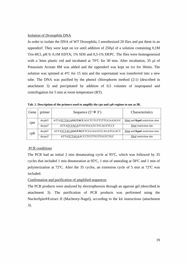
Tab. 1. Description of the primers used to amplify the cpa and cpb regions to use as IR.
Gene primer Sequence (5’à 3’) Characteristics
dscpb5’ ATTATCTAGAGGTACCAGCTCTGTTTTTGGAAAGGC XbaI and KpnI restriction sites cpa
dscpa3’ ATTATCTAGATTATTGCGTCTTCAGTTCCT XbaI restriction site
dscpb5’ ATTATCTAGAGGTACCTCGGAAATGCAGATGGACT XbaI and KpnI restriction sites cpb
dscpa3’ ATTATCTAGAACCCTGTTTGTTGGTCTGC XbaI restriction site
PCR conditions
The PCR had an initial 2 min denaturating cycle at 95ºC, which was followed by 35
cycles that included 1 min denaturation at 95ºC, 1 min of annealing at 58ºC and 1 min of
polymerization at 72ºC. After the 35 cycles, an extension cycle of 5 min at 72ºC was
included.
Confirmation and purification of amplified sequences
The PCR products were analysed by electrophoresis through an agarose gel (described in
attachment 3). The purification of PCR products was performed using the
NucleoSpin®Extract II (Macherey-Nagel), according to the kit instructions (attachment
3).

20
1.1.3 Cloning the cpa and cpb IR in the pWIZ vector
As described above, in Fig. 1, cloning the IR in the pWIZ vector is a two-step
process.
1.1.3.1 First cloning step
Digestion with Xba I
The cpa and cpb gene fragments and the pWIZ vector were digested with XbaI (Takara)
for 1h at 37ºC and then purified by the phenol choloform (2:1) method.
pWIZ phosporilation treatment
To prevent circularization of the plasmid, Shrimp Alkaline Phosphatase (S.A.P from
Roche) was added to the previous solution. The reaction was performed at 37ºC for 30
min and the DNA was purified by the phenol chloroform (2:1) method.
Ligation
In order to ligate the plasmid and the insert, 100 ng of pWIZ were mixed with 16 ng of
insert (which corresponds to a proportion of 1 plasmid to 3 inserts) and 0,25 µl of T4
DNA ligase (Promega). The reaction was performed at room RT for 3h.
Transformation
After the 3h of ligation, the volume of the reaction was added to 100 µl of DH5a cells.
The eppendrofs were kept on ice for 20 min and transferred to an incubator at 42ºC for 2
min. After 2 min on ice, the transformed cells were added to 900 µl of LB medium. After
1 h incubation with agitation at 37ºC, the cells were plated in plates containing ampicilin
and were left in an incubator at 37ºC overnight.
Purification of the plasmidic DNA
For each gene, 10-15 bacterial colonies were randomly chosen and put to grow in LB
medium containing 50 µg/ml of ampicilin at 37ºC with agitation, overnight. 1,5 ml of
bacterial culture was spinned for 2 min and the pellet was discarded. 300 µl of TS (Tris
50mM pH 7,5; Sucrose 25%) were added to the pellet and mixed. The same occurred
with ELT (EDTA 100mM; Lysosyme 2 mg/ml; TritonX 100 0,1%). The mixture was
incubated first at RT for 10 min and after, at 70ºC for 10 min. It was spinned at 4ºC
during 15 min and the pellet was removed with a toothpick. 500 µl of PEG6000 20%;
NaCl 1M solution was mixed with the supernatant and incubated for 30 min at RT.

21
Finally, the mixture was spinned for 4 min at RT, the supernatant discarded and the pellet
was ressuspended in 45 µl TE.
Confirmation of the insertion
Each independent plasmidic DNA was digested with XbaI (Takara) for 1h at 37ºC and
then run in an electrophoresis gel. The constructs which contained the insert were
selected to perform the next cloning step.
1.1.3.2 Second cloning step
Digestion with Spe I
The selected constructs with the first insert cloned were digested with SpeI (Promega) for
1h at 37ºC and purified by the phenol chloroform (2:1) method.
The ligation, transformation and purification procedures are the same as the ones
described in the first cloning step.
Confirmation of the insertion
Each insert contains a Kpn I restriction site to enable the selection of constructs with the
correct orientation. Each independent plasmidic DNA was digested with KpnI (Takara)
for 1h at 37ºC. The digestions were run in an electrophoresis gel and the plasmids which
had both the inserts in inverted orientations were chosen and storen.
1.2 Establishment of UAS-CP-IR transgenic lines
1.2.1 Microinjection
In order to generate transgenic Drosophila lines containing the UAS-CP-IR
constructs, I first prepared an injection mix. The injection mix contained the pWIZ with
the IR for cpa or cpb, a helper plasmid containing a transposase gene (pTURBO) and
water. The proportions of the mixtures prepared for each gene are represented in Tab. 2.
Tab. 2. Mixture used to inject Drosophila embryos.
cpa cpb
pWIZ+IR 22,25µl (300ng) 20,25µl (600ng)
pTURBO 2,68µl (75ng) 4,46µl (125ng)
water 4,8µl 5,29

22
The mixture was injected in W118 flies, which are white-. The flies layed for 45 min in
plates containing apple juice and yeast and embryos were collected and dechorionated in
commercial bleach till the appendages were lost. The embryos were aligned in an agarose
gel and transfered to a cover slide, where they were stick with a mixture consisting of
adhesive tape dissolved with heptane. Following dehydration by contact with sílica gel
from 7 to 9 min, embryos were covered with Voltalef oil (10S), which allows embryos to
breath and maintains hydration. The needles used for microinjection were pulled on a
horizontal puller of the Sutter Instrument Co. Model p.97 and the microinjector used was
from WPI, model pv 820. After microinjection, the embryos were kept in voltalef oil at
25ºC until larval stage, during which were transferred to vials with fly food.
1.2.2 Establishment of the RNAi stocks
The flies that were able to survive from the microinjection procedure are not
transgenic, since the transgene was inserted in the polar cells of the embryo, which give
rise to the germ line (see the principles of p-element mediated transformation in
attachment 1). Thus, the surviving flies were backcrossed with W118 and the transgenic
flies were identified within the progeny through the eye color (pW+). In order to establish
a clean stock of the UAS-cpa-IR and UAS-cpb-IR, the transgenic flies were backcrossed
with flies from the W118 stock. The mapping of the insertions is described in the results.
1. Fly husbandry
Flies were raised at 25ºC, under standard conditions (Roberts, 1998). The UAS-
CP-IR lines were submitted to different temperature conditions to test the efficiency of
the silencing effect (described in results).
Apart from the UAS-CP-IR transgenic lines I generated, several stocks of flies
were used, which are described in Tab. 3. In addition the ones included in Tab. 3, I used a
stock of W118 to generate transgenic flies and a double balanced line to map the
transgene insertions:
w-; sco ; MKRS cyo TM6β
23
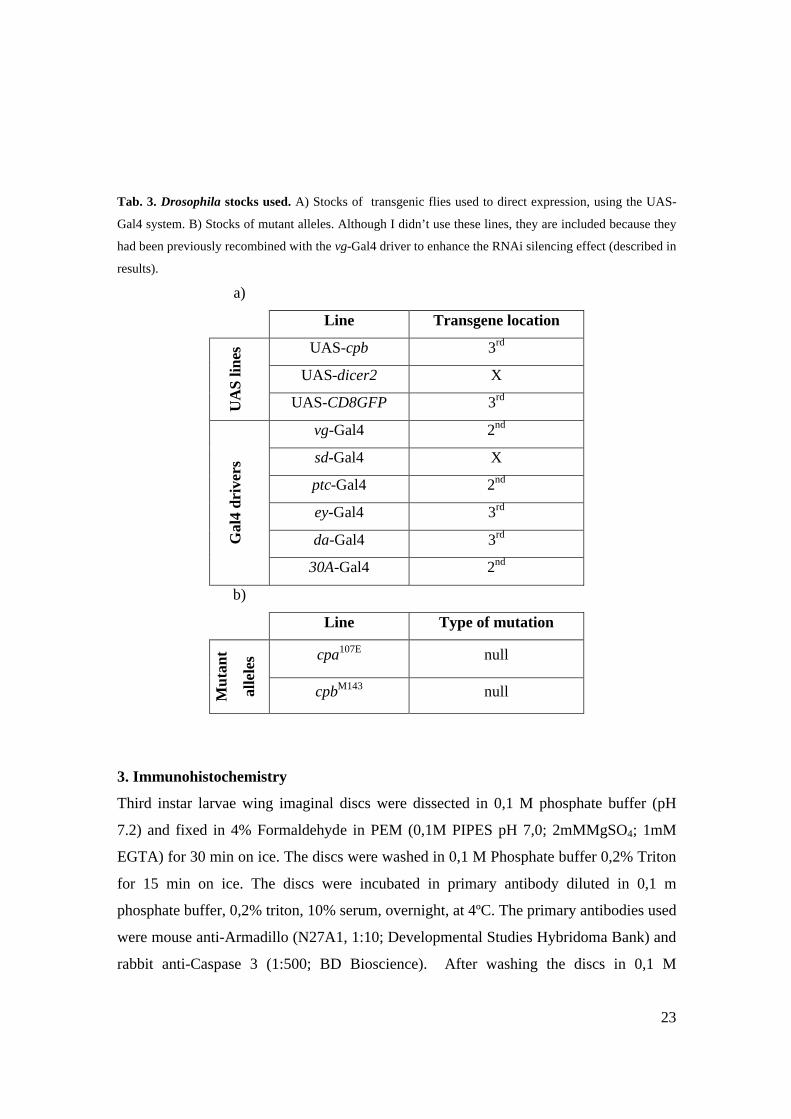
Tab. 3. Drosophila stocks used. A) Stocks of transgenic flies used to direct expression, using the UAS-
Gal4 system. B) Stocks of mutant alleles. Although I didn’t use these lines, they are included because they
had been previously recombined with the vg-Gal4 driver to enhance the RNAi silencing effect (described in
results).
a)
Line Transgene location
UAS-cpb 3rd
UAS-dicer2 X
UA
S lin
es
UAS-CD8GFP 3rd
vg-Gal4 2nd
sd-Gal4 X
ptc-Gal4 2nd
ey-Gal4 3rd
da-Gal4 3rd Gal
4 dr
iver
s
30A-Gal4 2nd
b)
Line Type of mutation
cpa107E null
Mut
ant
alle
les
cpbM143 null
3. Immunohistochemistry
Third instar larvae wing imaginal discs were dissected in 0,1 M phosphate buffer (pH
7.2) and fixed in 4% Formaldehyde in PEM (0,1M PIPES pH 7,0; 2mMMgSO4; 1mM
EGTA) for 30 min on ice. The discs were washed in 0,1 M Phosphate buffer 0,2% Triton
for 15 min on ice. The discs were incubated in primary antibody diluted in 0,1 m
phosphate buffer, 0,2% triton, 10% serum, overnight, at 4ºC. The primary antibodies used
were mouse anti-Armadillo (N27A1, 1:10; Developmental Studies Hybridoma Bank) and
rabbit anti-Caspase 3 (1:500; BD Bioscience). After washing the discs in 0,1 M

24
phosphate buffer, 0,2 Triton, discs were incubated in secondary antibody for 2 h at 4ºC.
Secondary antibodies were anti-mouse and anti-rabbit, from Jackson Immunoresearch,
used at 1:2000, conjugated to TRITC and Cy5. Fluorescence images were obtained on a
Zeiss LSM 510 META confocal microscope.

25
IV RESULTS
The Cellular environment influences the behaviour of CP mutant cells
In order to determine whether the cellular environment influences the behavior of
CP mutant cells, I analyzed the consequences of CP depletion in restricted domains of
the wing epithelium tissue. Since cpa and cpb mutant animals die as first instar larvae
(Janody and Treisman, 2006), I made use of the RNA interference (RNAi) technology
(Hannon, 2003) to target either cpa or cpb to degradation. I generated independent
transgenic fly lines carrying either a UAS-cpa-IR or a UAS-cpb-IR construct under the
control of the Gal4-responsive UAS enhancer repeats. Since the UAS-Gal4 system
permits the temporal and spatial control of gene expression, expression of each UAS-CP-
IR is controlled in time and space by the use of various Gal4 Drivers.
1. RNA interference as a tool to investigate the role of the cellular environment on
the behavior of CP mutant cells
1.1 UAS-cpa-IR and UAS-cpb-IR transgenic lines
The design of the UAS-cpa-IR and UAS-cpb-IR constructs as well as the process of
generation of transgenic Drosophila lines is described in Matherials and Methods. Three
independent lines of UAS-cpa-IR and four independent lines of UAS-cpb-IR were
obtained through p-element mediated transformation (Tab. 1). Since the P transposable
elements (which contain the UAS-CP-IR constructs) integrate at random positions on the
Drosophila genome, it is crucial to map the local of the transgene insertion to perform
genetic analysis. Thus, both UAS-CP-IR transgenic lines were crossed with double
balanced lines, and the locations for each insertion were determined and are shown in
Tab. 1. The strategy used to map chromosome location of the lines is described in Fig. 1.
26
1.2 Efficiency of the UAS-CP-RI
The use of the RNAi technique usually causes a drastic decrease in the expression
of the target gene. However, since RNAi may not completely abolish gene expression,
this technique is sometimes referred as a “knockdown” to distinguish it from “knockout”,
in which expression of a target gene is entirely eliminated. Therefore, it is important to
determine whether the target gene is being efficiently knocked-down. This type of
analysis can be performed by using many different approaches, including
immunohistochemistry and western blot experiments, if an antibody against the target
protein is available or by RT-PCR analysis, northern blotting or in-situ hybridization
(Hannon, 2003). Since the phenotypes induced by loss of cpa and cpb in Drosophila were
already characterized (Delalle et al., 2005; Janody and Treisman, 2006), I decided to
determine whether the UAS-cpa-IR and UAS-cpb-IR constructs I generated were capable
of reproducing the mutant allele phenotypes. The advantage of using this approach is that
it allowed me to classify transgenic lines based on the strength of their effects. Since P-
element mediated transformation is random, transgenes can be inserted at any place in the
genome. Expression of each independent insertion is thus subjected to chromatin state
(Lee and Carthew, 2003).
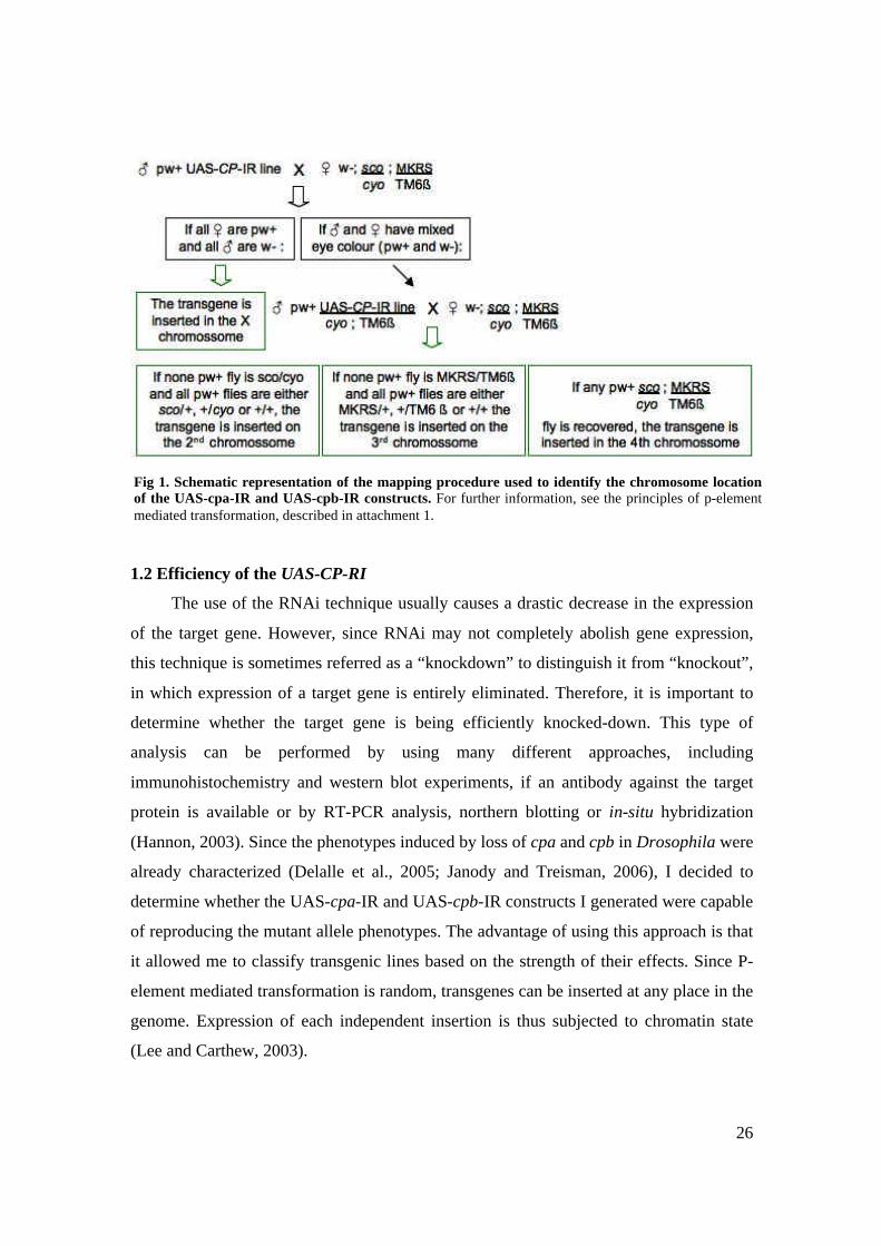
Fig 1. Schematic representation of the mapping procedure used to identify the chromosome location of the UAS-cpa-IR and UAS-cpb-IR constructs. For further information, see the principles of p-element mediated transformation, described in attachment 1.

27
RNAi silencing effect is affected by several factors, which can be manipulated to
achieve higher silencing efficiency. Firstly, since the RNAi silencing mechanism is
induced through the UAS-Gal4 system, which is temperature sensitive, the manipulation
of the temperature allows the analysis of differing levels of depletion. Therefore, the
strength of the transgenic lines was analysed at low (22ºC) versus high (27,5ºC)
temperature conditions (Tab. 1). In addition, the genetic background of the transgenic
flies may also be crucial for the efficient depletion of the RNA. For instance, if the
transgenic flies are heterozygous for the gene that is targeted to degradation, only half of
the dosage of the RNA will be transcribed. Thus, the RNAi machinery of the transgenic
flies operates within half of the cellular dosage of the RNA, which may lead to an
increase of its efficiency. This was tested with the UAS-CP-IR lines by driving their
expression to a Gal4 line recombined with the cpa107E and cpbM143 mutations (the results
are described below, in section 1.2.3). Moreover, the simultaneous expression of
molecular components of the RNAi interference pathway, such as the Dicer enzyme, and
the UAS-CP-IR transgenic lines is postulated to increase the RNAi silencing efficiency
(Mikuma et al., 2004). Therefore, the strength of one of the UAS-cpa-IR; UAS-dicer
transgenic line was determined and the results are briefly presented in Tab. 1 and further
described below, in section 1.2.3.
1.2.1 Ubiquitous depletion of CP
Animals homozygous mutant for either cpa or cpb die at first instar larvae (Janody
and Treisman 2006). Therefore efficient depletion of CP by using a Gal4 driver
expressed ubiquitously, such as dautherless-Gal4 (da-Gal4) should result in early larval
lethality as well. Indeed, when I crossed flies carrying the da-Gal4 driver to either
independent transgenic flies carrying the UAS-cpa-IR and UAS-cpb-IR, no progeny
could be recovered, suggesting that all transgenic lines can efficiently target cpa and cpb
to degradation (Tab. 1). Only one transgenic line (UAS-cpa-IR C) gave rise to adult
progeny (Tab. 1), suggesting that the transgene is inserted in a region of difficult access
to the da-Gal4 driver and that its expression level was not sufficient to efficiently deplete
cpa.
28
1.2.2 Depletion of CP in the eye imaginal disc
In addition, when cpa mutant alleles were identified in a mosaic genetic screen for
eye patterning genes (Janody et al., 2004) and loss of either sub-units of the CP
heterodimer induces neuronal degeneration of photoreceptors cells (Delalle et al., 2005).
Therefore, driving expression of UAS-cpa-IR and UAS-cpb-IR specifically in the eye
field by using the eyeless-Gal4 driver should induce a phenotype, which can be observed
in the adult eye. When I drove expression of each UAS-cpa-IR transgenic line, by using
the ey-Gal4 driver, adult eyes appeared to be normal in all independent transgenic lines
(Tab. 1). Instead, the UAS-cpb-IR transgenic lines were not uniform in the strength of the
eye phenotype. While UAS-cpb-IR A and B lines appeared to have normal eyes, the
UAS-cpb-IR C and D lines had a rough eye phenotype (Fig. 2), suggesting loss of cpb
was severe in the eye. The rough eye phenotype mentioned above was observed in flies
raised at 27,5ºC, which is postulated to confer higher levels of depletion but the results of
partial depletion of cpb, by raising flies at 22ºC were not determined.
1.2.3 Depletion of CP in the wing proper
The phenotype of epithelial extrusion and cell death caused by loss of CP in
discrete paches in the wing imaginal disc is specific of the wing blade region, which will
give rise to the adult wing and is specified by the transcription factor Vg. Moreover, the
CP inactivation phenotype was shown to be dependant on the presence of Vg (Janody
and Treisman, 2006). Thus, depletion of CP in vg expressing cells should result in an
obvious adult wing phenotype, thus providing a measure of the strength of the RNAi
efficiency of the UAS-CP-IR transgenic lines.
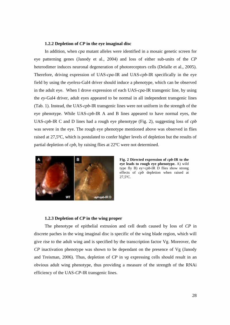
Fig. 2 Directed expression of cpb-IR to the eye leads to rough eye phenotype. A) wild type fly B) ey>cpb-IR D flies show strong effects of cpb depletion when raised at 27,5ºC.

29
Indeed, wings of two UAS-cpb-IR lines (raised at 27,5ºC), namely C and D, were
strongly affected by cpb depletion (Tab. 1). Not only the wings were reduced and
malformed, but also contained thickened veins, suggesting that cell death might have
occurred during wing development (Fig. 3). The strength of the effects caused by
depletion of cpb is similar in both UAS-cpb-IR C and D lines. Interestingly, wings of
these lines that were raised at 22ºC were significantly less affected by cpb depletion:
although the wings seemed slightly smaller and were downwardly curved, there was no
evidence of apoptosis (Fig 4 B). This shows how temperature affects the level of
depletion of CP.
To enhance the efficiency of the RNAi mechanism, the vg-Gal4 driver was
recombined with cpa107E and cpbM143 mutations. However, within the lines that presented
a wing phenotype (UAS-cpb-IR C and D), no detectable differences were observed
between the vg-Gal4 line and the vg-Gal4 line recombined with cpa107E and cpbM143,
suggesting that the genetic background of transgenic flies does not play a crucial role in
the RNAi silencing effect (since the differences were not significant, the different lines
used are not contemplated in Tab. 1).
In contrast to the UAS-cpb-IR C and D lines, both the UAS-cpb-IR A and B lines
and all the UAS-cpa-IR lines had normal wings (Tab. 1). This is consistent with the
strength of the effects caused by depletion of CP in the eyless expression domain, since
the lines that present adult wing and eye phenotypes are the same in both situations
(UAS-cpb-IR C and D), but contrasts to the results obtained when depletion was targeted
ubiquitously. This suggests that, even though cpa and cpb ubiquitous depletion in UAS-
cpb-IR A, B and UAS-cpb-IR A, B is sufficient to lead to lethality, higher levels of
depletion are required to obtain eye and wing phenotypes.
Due to the total absence of eye and wing phenotypes in all of the UAS-cpa-IR
lines, I attempted to increase the RNAi efficiency by establishing a line containing both a
UAS-cpa-IR and a UAS-dicer (Since the UAS-dicer transgene is inserted in the X
chromosome, I only used the UAS-cpa-IR A whose transgene is in the 3rd chromosome).
However, even though ubiquituous targeting of this line led to lethality, no effects were
observed by targeting depletion to the vg expression domain. This highlights how the

30
point of the transgene insertion may affect the strength of the RNAi machinery
efficiency.
The analysis of the UAS-CP-IR phenotypes, through directed expression to da-, ey-
and vg-Gal4, was crucial to determine which lines and conditions are preferred to further
analyse the effects of CP depletion. Therefore, the lines chosen for this purpose were
UAS-cpb-IR C and D (which have similar strength effects) and the temperature in which
transgenic flies were raised was 27,5ºC. Even though the use of the vg-Gal4 cpa107E
cpbM143 line did not seem to affect the RNAi induced silencing effect, this line was
chosen to perform experiments whenever CP depletion was to be targeted to vg
expressing cells.
Fig. 3. Directed expression of the cpb-IR to the wing leads to reduced and malformed wings. A) wild type fly B) vg>cpb-IR D fly raised at 27,5 ºC. The wings are reduced and malformed. C) Detail of the wing (of vg>cpb-IR D transgenic flies raised at 27,5ºC). Wings display thickened veins (indicated by arrows), which suggest cell death may have occurred during its formation.
31
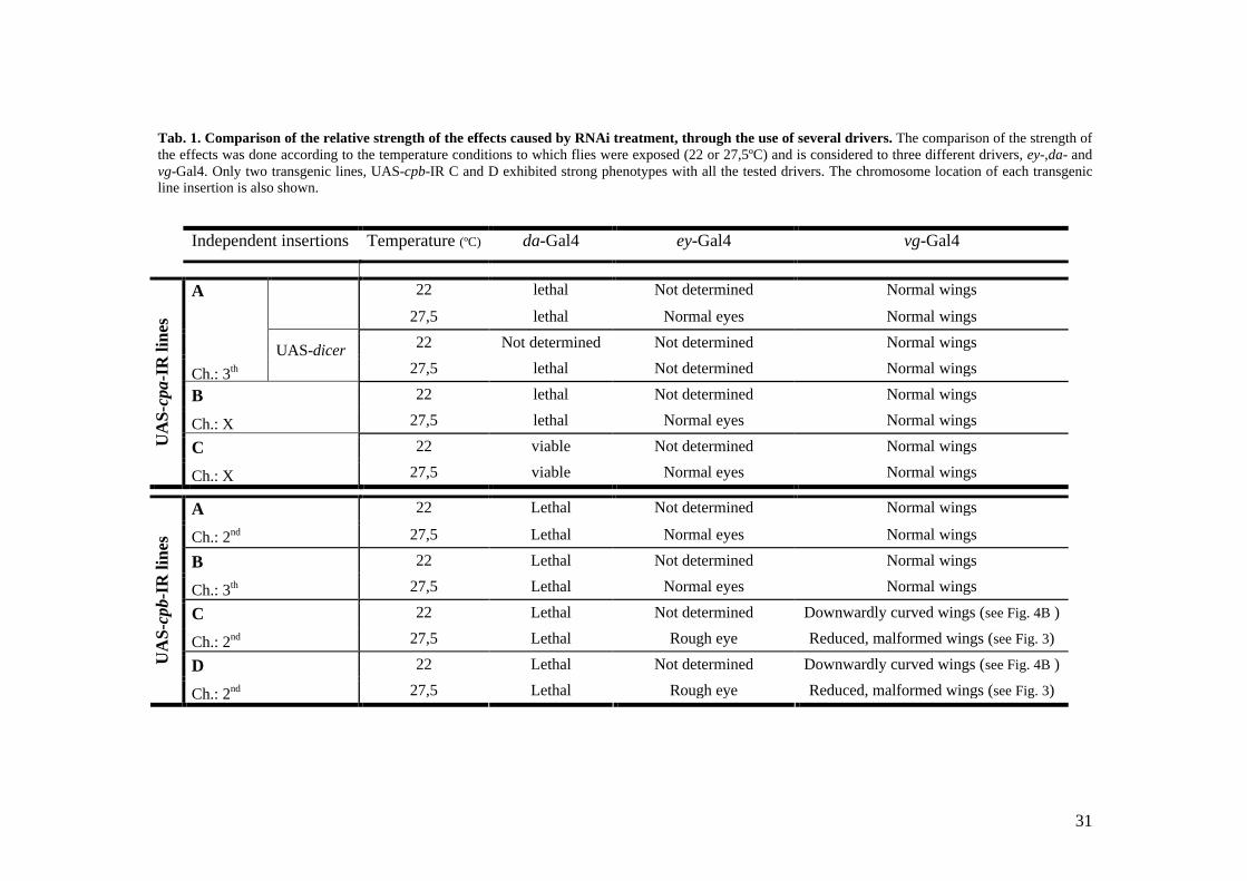
Tab. 1. Comparison of the relative strength of the effects caused by RNAi treatment, through the use of several drivers. The comparison of the strength of the effects was done according to the temperature conditions to which flies were exposed (22 or 27,5ºC) and is considered to three different drivers, ey-,da- and vg-Gal4. Only two transgenic lines, UAS-cpb-IR C and D exhibited strong phenotypes with all the tested drivers. The chromosome location of each transgenic line insertion is also shown.
Independent insertions Temperature (ºC) da-Gal4 ey-Gal4 vg-Gal4
22 lethal Not determined Normal wings
27,5 lethal Normal eyes Normal wings
22 Not determined Not determined Normal wings
A
Ch.: 3th UAS-dicer
27,5 lethal Not determined Normal wings
22 lethal Not determined Normal wings B
Ch.: X 27,5 lethal Normal eyes Normal wings
22 viable Not determined Normal wings UA
S-cp
a-IR
line
s
C
Ch.: X 27,5 viable Normal eyes Normal wings
22 Lethal Not determined Normal wings A
Ch.: 2nd 27,5 Lethal Normal eyes Normal wings
22 Lethal Not determined Normal wings B
Ch.: 3th 27,5 Lethal Normal eyes Normal wings
22 Lethal Not determined Downwardly curved wings (see Fig. 4B ) C
Ch.: 2nd 27,5 Lethal Rough eye Reduced, malformed wings (see Fig. 3)
22 Lethal Not determined Downwardly curved wings (see Fig. 4B ) UA
S-cp
b-IR
line
s
D
Ch.: 2nd 27,5 Lethal Rough eye Reduced, malformed wings (see Fig. 3)

32
1.3 Specificity of the UAS-CP-RI
A problem that arises by using the RNAi technique to generate loss of function
phenotypes is to prove causality between RNAi treatment and a phenotype. Confirming
the transcript levels are reduced is not sufficient to relate the phenotype with the
depletion of the target gene, since the RNAi treatment may be silencing one or more
genes than the intended target (off-target genes). In order to determine the possible off-
targets of the sequence used as IR, I made use of a software available online
(http://flyrnai.org/) by the Drosophila RNAi screening centre (DRSC). This software
allows blasting the combination of an established number of nucleotides (nt) of the
submitted sequence with all the annotated genes in the Drosophila genome. In
Drosophila, the small interfering RNA (siRNA), which are responsible for triggering
gene silencing, consist normally of duplexes of 21 to 23 nt (Elbashir et al., 2001).
Therefore, I blasted every 21-23 nt contained within the UAS-cpa-IR and UAS-cpb- IR
sequences. No predicted off-targets were found by using this technique, suggesting that
the phenotypes observed by using the UAS-cpa-IR and UAS-cpb-IR constructs were due
to depletion of cpa and cpb respectively.
Even though this data accounts for the sequence-specificity of the IR used, it is still
in silico data. Therefore, it is crucial to demonstrate in vivo that the observed phenotype
is due to the depletion of CP. To do so, I attempted to rescue the cpb-RI phenotypes by
using flies carrying the cpb cDNA under control of the UAS sequences (UAS-cpb),
which can fully rescue the cpb mutant phenotype (Janody and Treisman, 2006). Flies
carrying both the UAS-cpb-IR and UAS-cpb were crossed with flies bearing the vg-Gal4
driver. However, the wing phenotypes were indistinguable when compared to control
wings of flies vg>cpb-IR (data not shown). This suggests that overexpression of cpb is
not sufficient to rescue the wing phenotype induced by RNAi depletion. Since the UAS-
cpb-IR targets a sequence of 543 bp of the cpb cDNA, it is likely that the cpb
overexpression construct is also targeted to degradation by the UAS-cpb-IR, preventing
any suppression effect.

33
2. Depletion of cpb in the wing imaginal disc leads to different outcomes
2.1 Partial depletion of cpb in the wing resembles the phenotype of arc mutant flies
As referred above, RNAi silencing efficiency is highly dependant on temperature
conditions. vg>cpb-IR flies raised at 22ºC exhibited downwardly curved wings that
seemed smaller than wild type but had no sign of patterning defects (Fig. 4 B). This
phenotype resembles the one detected in the wings of flies bearing an arc mutation (Fig.
4 C) (Liu and Lengyel, 2000). arc encodes an adherens junction-associated PDZ domain
protein, expressed in tissues undergoing developmental morphogenetic movements. In
third instar wing imaginal disc, arc RNA is expressed mainly in the hinge and in the
adjacent proximal blade regions (Fig. 4 H) (Liu and Lengyel, 2000). vg-Gal4 drives
expression of target genes not only in the distal wing blade, where arc is not detectable,
but also in the hinge domain (Fig. 4 F). This suggests that the downward curved wing
phenotype of vg>cpb-RI flies raised at 22ºC, is the consequence of cpb depletion in the
hinge domain. To test this possibility, I targeted depletion of cpb only in the hinge region,
by driving the expression of UAS-cpb-IR to the 30A-Gal4 driver (Fig. 4 G). However,
30A>cpb-IR flies had straight wings undistinguishable from wild type (Fig. 4 D). The
30A-Gal4 driver might induce expression of the UAS-cpb-IR at a level too low to result
in an obvious phenotype. Alternatively, since vg-Gal4 drives expression of target genes in
a gradient along the distal proximal axis in the wing blade, weak cpb depletion in the
proximal part of the wing blade might be sufficient to induce the formation of a
downward curved wing.

34
2.2 The local environment influences the behavior of CP depleted cells
2.2.1 Depletion of cpb in the vg expression domain leads to massive cell
extrusion and apoptosis
vg>cpb-RI flies raised at 22ºC appeared to have smaller wings. This suggests that
cpb-depleted cells fail to growth. Alternatively, a subset of wing blade cells may have
died by apoptosis. Indeed, when the vg>cpb-IR larvae were raised at 27,5 ºC, adult wings
were strongly affected and presented thickened veins that were likely necrosed tissues
(Fig. 3 C). In order to determine whether cpb-depleted cells in the vg-expression domain
were undergoing apoptotic cell death, I established a stable line carrying the UAS-cpb-IR
construct and a UAS-GFP reporter gene, allowing me to mark with GFP the cpb-depleted
cells. Third instar wing imaginal discs from vg>cpb-RI; GFP flies were marked with an
anti-Armadillo (Arm) antibody to outline the apical membrane and with an anti-activated
Caspase 3 (Casp 3) antibody, witch reveals Caspase-dependent cell death. Indeed,
standard confocal sections showed that most of the GFP positive cells in the wing blade
epithelium expressed activated Casp 3 (Fig. 5 C), indicating that cpb-depleted cells were
Fig. 4. Partial cpb depletion in the wing resembles the wing phenotype of arc mutant flies. A) Wild type fly. B) vg>cpb-IR fly raised at 22ºC. C) arc homozygous mutant fly (Adapted from Liu and Lengyel, 2000). D) 30A>cpb-IR raised at 27,5ºC (to enhance the phenotype). F) Fate map of the Drosophila wing disc. Arrow indicates the proximal region of the wing disc. (Adapted from Butler et al., 2003). F) Expression domain of the vg-Gal4 driver (driven with a UAS-GFP). G) Expression domain of the 30A-Gal4 driver (driven with a UAS-GFP) H) Expression domain of the gene arc (adapted from Liu and Lengyel, 2000). Arrows indicate expression in the prospective hinge and immediately adjacent proximal wing blade regions.

35
Figure 3. Depletion of cpb in vg-expressing cells leads to extrusion and cell death. All panels show third instar wing imaginal discs marked with GFP in green and with anti-Arm (blue in B, C and D) and anti-activated Caspase 3 (Red in B, C and D) (A-B) Standard optical sections. (C-E) Optical cross sections. (A-C) vg-Gal4 driver driving UAS-GFP (A) or UAS-cpb-RI, UAS-GFP (B and C). D) Clones of cpa mutant cells are marked positively by GFP (Image provided by Janody). In A) is presented the vg expression domain. vg expressing cells targeting depletion of cpb undergo massive apoptosis (B,C) and are extruded from the epithelium (C). However, cpb depleted cells are still maintained within the the epithelium in the notum and hinge regions of the disc (C), resembling clones of cells mutant for cpa (D).
dying. In contrast, only few dying cells were recovered in the vg-expressing domain of
the hinge and notum (Fig. 5 B, C). Cross sections through the wing disc epithelium
showed that GFP-positive cells were extruded from the wing blade epithelium and had
lost epithelium cell polarity, since they failed to express Arm (Fig. 5 C). However, cpb-
depleted cells are still maintained in the hinge and notum epithelia (Fig. 5 B, C). Thus,
the behavior of cpb-depleted cells in the vg-expression domain resembles the observed in
clones of cpa mutant cells (Fig. 5 D), suggesting that clones of CP mutant cells die cell-
autonomously. However, although depletion of cpb in the vg-expressing domain is wider
(Fig. 5 A) than cpb mutant cells induced in discrete patches, a large number of proximal
wing blade cells are not targeted by the vg-Gal4 driver, and might promote non-
autonomous extrusion and death of cpb-depleted cells by a process of cell competition.

36
2.2.2 Broad depletion of cpb in the scalloped expression (sd) domain leads to
most of the hallmarks of tumor cells.
With the goal of knocking down cpb in the whole wing blade epithelium, I made
use of scalloped-Gal4 (sd-Gal4), which drives expression of a UAS-GFP reporter gene in
the whole wing blade epithelium, but also in the hinge and part of the notum (Fig. 6 D).
sd>cpb-IR flies raised at 27,5ºC died at 2nd instar larval stage. By decreasing the
temperature to 22ºC, lethality was delayed to pupal stage, allowing me to analyze third
instar larval wing discs. Surprisingly, depleting cpb in the entire wing blade leads to a
completely different outcome from the one that results from cpb depletion in the vg
expression domain: although some cpb-depleted cells marked positively by GFP still
expressed activated Caspase 3, most of the cells within the wing blade were able to
survive (Fig 6 A, C). Not only cpb depleted cells are able to survive, but also seem to
overproliferate, as suggested by the numerous foldings and overgrown masses of tissue
the wing blade contains (Fig. 6 A).
Moreover, in cpb depleted cells, the expression of the AJ component Arm is
mislocalized (Fig. 6 B), suggesting that epithelial apicobasal polarization is disrupted in
these cells. CP have previously been reported to be required for the maintenance of
epithelial architecture in clones of CP mutant cells (Janody and Treisman, 2006). Loss of
apicobasal polarization is considered to be a crucial step in the process of epithelial to
mesenchymal transitions (Brumby and Richardson, 2005). Thus, I wondered whether cpb
depleted cells could show additional features characteristic of EMT. Indeed, closer
examination of cpb depleted cells in the wing blade, exhibited a morphological difference
when compared to wild type cells: cpb depleted cells acquired a rounder morphology and
seemed to be less adhesive between each other (Fig. 6 D-G). Both disruption of epithelial
polarity and altered cell shape characterize EMT, thus the presented data suggest that cpb
depletion in the wing blade region may induce EMT.
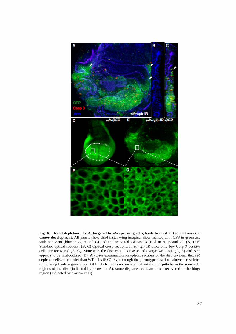
37
Fig. 6. Broad depletion of cpb, targeted to sd-expressing cells, leads to most of the hallmarks of tumor development. All panels show third instar wing imaginal discs marked with GFP in green and with anti-Arm (blue in A, B and C) and anti-activated Caspase 3 (Red in A, B and C). (A, D-E) Standard optical sections. (B, C) Optical cross sections. In sd>cpb-IR discs only few Casp 3 positive cells are recovered (A, C). Moreover, the disc contains masses of overgrown tissue (A, E) and Arm appears to be mislocalized (B). A closer examination on optical sections of the disc revelead that cpb depleted cells are rounder than WT cells (F,G). Even though the phenotype described above is restricted to the wing blade region, since GFP labeled cells are maintained within the epithelia in the remainder regions of the disc (indicated by arrows in A), some displaced cells are often recovered in the hinge region (Indicated by a arrow in C)

38
EMT transition, resistance to cell death, loss of proliferation control and acquisition
of migratory abilites are the four major hallmarks of tumor development. Although some
cpb depleted cells seem to be recovered in distant regions from the wing blade (indicated
by arrows in Fig. 6 C), it is not clear whether they have acquired migratory abilities. Still,
most of the hallmarks of tumor cells are present in cpb depleted cells.
It is crucial to mention that the phenotype observed in sd>cpb-IR discs only occurs
in the wing blade region, since in the remainder regions of the disc epithelial cells appear
to maintain their characteristics (indicated by arrows in Fig. 6 A).
2.2.3 Wing blade cells depleted of cpb seem to acquire the ability to migrate
Previous data on the behavior of CP mutant cells in the ovarium follicle epithelium
(see Fig. 9 of the Introduction) (Janody, un-published data), have suggested that loss of
cpa may lead to a migratory type of behavior. Also, either loss of cpb in clones or by
RNAi depletion in vg-expressing cells leads to epithelial extrusion. In addition, cpb
depleted cells targeted to the sd expression domain, were often recovered in regions
distant from the presumptive wing blade. Since migration is a hallmark of tumor
behavior, confirming that cpb depleted cells may acquire migratory abilities would be
consistent with this view.
To better understand whether CP might prevent cell movement, I used the patched-
Gal4 (ptc-Gal4) driver, which directs expression of a UAS-GFP reporter gene to a few
rows of cells along the anterior/posterior (A/P) boundary (Fig. 7 A) (Vidal et al., 2006).
As previously, cells targeting cpb to degradation were marked with GFP to follow their
fate. I assumed that if cpb-depleted cells acquire the ability to migrate, they would be
found far away from the A/P boundary. Indeed, I found that some of the GFP-positive
cells where displaced from the ptc expression domain (indicated by arrows in Fig. 6 B).
In agreement with previous analysis, cpb-depleted cells were extruded basally from the
wing blade epithelia, and showed a bright GFP staining, likely due to condensed
chromatin, suggesting that these cells were dying (Fig. 6 C, D).
Consistent with previous observations, ptc>cpb-RI; GFP cells were maintained
within the wing hinge and notum. This suggests that cpb prevents migration of wing

39
blade cells. However, we cannot completely exclude the possibility that cpb-depleted
cells shift far away from where they originate due to a passive extrusion process
dependant of apoptotic cell death.
Fig. 4. cpb-depleted cells seem to gain the ability to migrate in the wing blade. All panels show third instar wing discs, marked by GFP in green and anti-Arm antibody in blue. (A-C) Standard confocal sections. (D) Optical cross section A) ptc-Gal4 driving UAS-GFP expression (B-D) ptc-Gal4 driving UAS-cpb-RI, UAS-GFP. cpb depleted cells, marked posivively by GFP are recovered in distant locations of the A/P boundary, in which ptc is expressed (indicated by arrows in B and C). These cells extrude basally (D). C) refers to a basal view of the epithelia, in which a large amount of cells is mislocalized. Note that mislocalized cpb depleted cells are mostly recovered in the posterior compartment of the wing disc.

40
Interestingly, in all ptc>cpb-RI; GFP discs analyzed, cells were recovered far
from the A/P boundary only in the posterior compartment (Fig. 6 B, C, D). ptc-Gal4
drives expression in a gradient in which expression is highest in cells apposed to the
posterior compartment and falls away gradually in cells within the anterior compartment
(Vidal et al., 2006). Since the posterior edge of the cpb-depleted cells is directly apposed
to wild type cells of the posterior compartment, extrusion, migration and death of
ptc>cpb-RI, GFP cells might occur through a non-autonomous process due to cell
competition. In contrast, in more anterior cells, cpb-depletion is graded and does not form
a sharp difference between cpb-depleted cells and wild type cells. Alternatively, graded
depletion of cpb in more anterior cells might be insufficient to promote, extrusion,
migration and death.
41
V DISCUSSION
Among the UAS-cpa-IR and UAS-cpb-IR transgenic lines I obtained, only two
transgenic lines of UAS-cpb-IR presented a phenotype when crossed with specific Gal4
drivers to measure their strength. (Tab. 1 of the Results). Even though the α subunit of
the CP heterodimer caps with higher affinity the barbed end of the actin filament, both α
and β subunits are required for the proper function of CP and loss of either one is
postulated to result in the same type of phenotype (Wear et al., 2003). Since all the
experimental conditions to test efficiency of the RNAi silencing effect were similar for
the UAS-cpa-IR and UAS-cpb-IR transgenic lines and even within the UAS-cpb-IR
transgenic lines the strength of the effects was highly variable (two lines presented an
obvious wing and eye phenotype while the other two didn’t), the difference in the
strength effects is probably caused by the influence of nearby chromosomal modulation
of transgene expression, which depends of the point of transgene insertion (Lee and
Carthew, 2003).
Despite the efforts to prove causality between the phenotype of the UAS-cpb-IR
lines and the RNAi silencing mechanism (by using a UAS-cpb construct to rescue the
phenotype), the establishment of specificity of the UAS-cpb-IR construct is still to be
performed. However, several factors account to the fact that the RNAi is targeting
specifically cpb. First, in vitro data using software that finds off-target genes of the cpb-
IR sequence revealed that, within the Drosophila annotated genes, only cpb is targeted to
degradation. Morevoer, the RNAi induced effects are consistent with the known effects
of loss of cpb: ubiquitous loss leads to lethality and in restricted regions of the wing disc
leads to cell extrusion and death. The most feasible and reliable way to overcome the
specificity problem is to perform RNAi using two or more dsRNAs that are directed
against the same gene but do not overlap and have little sequence similarity (Hannon,
2003). Recently, the Vienna Drosophila RNAi Centre (VDRC) has generated a
collection of RNAi transgenic lines that target 85% of the Drosophila genome. A UAS-
cpa-IR and a UAS-cpb-IR transgenic lines are available in this collection, and will be
used to confirm the phenotype observed with the RNAi constructs I generated.

42
CP mutant cells versus cpb depletion by RNA interference: two different outcomes
In the wing blade epithelia, clones of CP mutant cells (both of cpa and cpb) suffer
basal extrusion and die by apoptosis (Janody and Treisman, 2006). In contrast, in this
report I have shown that broad depletion of cpb using the RNAi technology results in
survival of most cells and overproliferation. Thus, what is leading to these different
outcomes?
Two hypotheses may be proposed to explain the contrasting phenotypes of CP
inactivation. It can be explained either by a matter of dosage of the levels of CP or by a
matter of how broad is the epithelia domain in which CP is being depleted (Fig. 1).
Firstly, regarding the matter of dosage, the cpa and cpb mutations used to perform
clonal analysis, namely cpa107 and cpbM143, are null mutations, meaning that CP are not
transcribed at all. Consequently, in cells mutant for CP there is a complete “knock out” of
CP. In contrast, the RNAi technology provides a “knock down” of the expression of the
target gene, meaning that the depletion is partial, acting like a hypomorph mutation. Even
though my results show that the RNAi induced silencing for cpb is efficient, it is likely
that that reduced levels of cpb are still present in the depleted cells.
Differing levels of Src kinase activity are postulated to trigger different cellular
responses (Vidal et al., 2007). In this sense, it was shown that while lower levels of
activation of Src leads to a weak activation of JNK signaling, directing anti-apoptotic
signals and promoting proliferation, overactivation of Src signaling leads to strong JNK
activation, inducing apoptotic cell death and invasive migration (Vidal et al., 2007). As I
will discuss further, there might exist an association between the activity of the oncogenic
family of the Src kinases and CP. We could therefore picture a situation in which the
overproliferation phenotype observed in the sd>cpb-IR discs could be the result of the
partial depletion of cpb which, in turn, would lead to low activity of Src. In contrast, in
CP mutant clones, its total loss would result in a high increase of Src activity, resulting in
a strong activation of the JNK pathway and ultimately, in cell death and invasive
migration.
Alternatively, the contrasting phenotypes of CP inactivation could be a
consequence of the extension of CP inactivation. In this situation, discrete loss of CP, as
occurs in clones, would induce wild type cells to activate JNK signaling pathway and

43
remove abnormal cells from the tissue, through the phenomena of cell competition
(Adachi-Yamada and O'Connor, 2004). Since the wild type contribution is removed when
the depletion is targeted to a broad domain, as occurs in the case of sd>cpb-IR discs, cells
depleted of CP would be able to survive. This behavior is in agreement with several
reports that show that removing the wild type contribution is sufficient for abnormal cells
to survive and show an abnormal type of development (Brumby and Richardson, 2003;
Vidal et al., 2006).
CP CPSrc Src
JNK JNK
Epithelial exclusion and apoptotic death
Overproliferation and resistance to cell death
Dosage effect
Cell competition
JNK
CP CPSrc Src
JNK JNK
Epithelial exclusion and apoptotic death
Overproliferation and resistance to cell death
Dosage effect
Cell competition
JNK
This second hypothesis appears to be the most likely situation to explain the
contrasting behavior shown by CP mutant clones and RNAi depletion of CP. This is
reinforced by the fact that RNAi induced cpb depletion in restricted wing blade domains,
such as in the ptc and vg expression domains, also induces cell extrusion and cell death.
To validate this hypothesis, a simple experiment of clonal analysis using the UAS-cpb-IR
Fig. 1 Schematic representation of the hypotheses proposed to explain the differing behavior caused by CP loss of function, by clonal analysis and depletion through RNAi. The cell extrusion vs. the overproliferation phenotype can be due to a matter of the Cpb dosage or, in alternative, to cell competition mechanisms. In the first situation, this would lead to different levels of the Src activity, thus activating the JNK signaling pathway. In the second situation, the surrounding wild-type tissue removes abnormal cells through JNK induced apoptosis.
44
transgenic lines to induce clones among wild type tissue (through the use of the MARCM
system), could be performed. If, in this situation, clones of UAS-cpb-IR are still extruded
from the epithelium, I would have evidence to support the cell competition hypothesis.
Cells depleted of cpb in the whole wing blade epithelium show most of the hallmarks
of tumor cells
Tumor cells display a characteristic set of features that distinguishes them from
normal cells. Interestingly, as I have shown, broad depletion of cpb in the wing blade
region, by inducing its depletion to the sd-expressing cells, leads to most of the
characteristics of tumor cells:
Epithelial to mesenchymal like transition
In cpb depleted cells, the SJ component Arm appears to be mislocalized, indicating
epithelial apicobasal polarity may be disrupted in these cells. Disruption of apical basal
polarization is crucial to the progression of epithelial cells towards a mesenchymal state
(Huber et al., 2005), which implies, among other features, that cells alter cell shape.
Indeed, cpb depleted cells are morphologically distinct from wild type cells. They are
rounder and seem less adhesive to each other. Altogether, these data suggest that cpb
depleted cells undergo an EMT, however these observations must be further confirmed
by staining cpb depleted cells with mesenchymal markers, such as N-cadherin and
metaloproteases (Weinberg, 2007).
Resistance to cell death
Contrasting to the phenotypes observed when cpb depletion was targeted either to
the vg and ptc expressing cells, most of the cpb depleted cells in the wing blade were able
to survive. This may suggest that these cells acquired resistance to cell death, which is
characteristic of tumor cells (Brumby and Richardson, 2005). The fact that some dying
cells are recovered does not completely contradict this hypothesis, since there is also a
small proportion of dying cells in human tumors (Weinberg, 2007). Alternatively, cell
survival can be explained by the fact that cells targeted to depletion in the sd expression
domain are no longer under the influence of death signaling from surrounding tissue.
Resistance to cell death in Drosophila is generally mediated by the Drosophila inhibitor
of apoptosis, Diap 1, which is upregulated in tumor cells (Brumby and Richardson,
45
2005). Thus, staining cpb depleted cells with Diap 1 may show whether they may have
acquired resistance to cell death.
Overproliferation
Observations of cpb depleted cells in the wing blade suggest that these cells lose
their proliferation control and keep dividing continuously. However, to further show
increased division in these cells it is necessary to undergo a quantitative type of analysis.
Fluorescent Activated Cell Sorting (FACS) analysis may be one way to prove this point,
since it provides a method for sorting a heterogeneous mixture of biological cells, one
cell at a time, based upon the specific light scattering and fluorescent characteristics of
each cell. As cpb depleted cells may be marked by GFP, a comparison between the
fluorescence of cpb depleted cells expressing GFP with GFP expressing cells in which
cpb is not targeted to degradation, may show whether loss of cpb in a broad domain can
lead to loss of proliferation control.
Invasion/metastasis
The role of CP in preventing migration has been suggested before: in the ovarium
follicle epithelium clones of cpa mutant cells acquire migratory and invasive behavior
(Janody, un-published data) and in the wing blade epithelium clones of CP mutant cells
extrude basally. Also in the present work, I show that depletion of cpb in the vg, sd and
ptc domains of expression, leads to mislocalization of cpb depleted cells. These results
are particularly evident when cells are driven to ptc-Gal4. However, in this situation it is
not clear whether migration is independent of cell death. Indeed, migration seems to
occur mostly in the posterior compartment of the disc, in which the levels of cpb are
directly confronted with the ones of wild type cells. This suggests that neighboring cells
activate cell competition mechanisms that ultimately induce cell death of adjacent cells,
thus the migration observed would be cell death dependent. A possible way to prove that
migration observed in cpb depleted cells is caused by loss of cpb rather than apoptosis
dependent is by blocking cell death using Diap 1, the Drosophila inhibitor of apoptosis.
A transgenic double line containing both a UAS-cpb-IR and a UAS-Diap1 targeted to the
ptc domain of expression would be used to corroborate this hypothesis. Another
interesting manner of proving invasiveness of cpb depleted cells would be to express the
activated form of Ras in the ptc>cpb-IR cells. The combination of the activated form of

46
Ras with mutations on TSG has been shown to promote invasiveness of cells. In
particular, when scrib mutant cells are induced in wild type tissue, they are removed by
apoptosis, while scrib mutant cells (in the same conditions) that simultaneously express
Rasact or Notchact acquire an invasive type of behavior (Brumby and Richardson, 2003;
Pagliarini and Xu, 2003). Again, this experiment could be performed using the UAS-
Gal4 system, by expressing simultaneously a UAS-Rasact and a UAS-CP-IR line.
An additional feature that characterizes tumor cells is their inability to terminally
differentiate. Therefore, it might be interesting to analyze whether these cells show
differentiation markers.
Altogether, the data presented strongly suggests that cpb prevents tumor
development in the wing blade epithelial cells, acting like a tumor suppressor gene. Since
Cpb is a component of the CP heterodimer, which requires both α and β subunits to work
properly (Wear et al., 2003), depletion of cpa should result in the same type of
phenotype. Therefore, similar experiments using RNAi mediated analysis can be
proposed to demonstrate the tumor suppressor potential of cpa.
The postulated tumor suppressor potential of CP is restricted to a subset of epithelia
The possible role of CP in preventing tumor development is not a common feature
of all type of epithelia, but rather it seems to be specific of only a subset of epithelia. In
the ovarium follicle epithelia, cpa mutant cells seem to overproliferate and invade distant
tissues (Janody, un-published data). As shown in this work, cpb depleted cells in the wing
imaginal disc epithelia also seem to acquire a tumoral and invasive type of behavior.
However, this behavior is restricted to the wing blade, since in the remainder regions of
the disc (hinge and notum), cpb depleted cells do not seem to be severely affected. This
behavior is consistent with the cell death and extrusion phenotype observed for CP
mutant clones, which is also restricted to the wing blade region. Altogether these data
reinforce the idea that in an epithelial tissue continuum, apparently homogenous in cell
morphology, not all cells are equivalent in terms of their cytoskeletal architecture and/ or
adhesion properties.
The fact that CP has a tissue specific role in preventing tumor formation is very
interesting since this is the first time a tumor suppressor gene has been pinpointed to play

47
such role in a specific subset of epithelia. Moreover, it accounts for the importance of the
tissue context during the activation of a tumoral process, which is particularly relevant in
humans.
An important question then, is what makes the wing blade region different from the
remainder regions of the wing disc. One possible speculation could be a differential
junctional composition or strength, between the cells of the different regions of the disc.
CP is postulated to localize at the apical membrane, since an HA-tagged form of Cpa co-
localizes with most of AJ components (Janody and Treisman, 2006). The Src oncogene
also localizes at AJ and its overactivation leads to activation of tumoral processes,
through activation of JNK signaling, as already discussed (Yeatman, 2004). Interestingly,
the phenotype of cpb depletion in the wing blade resembles the phenotypes of csk and
dASSP, both inhibitors of Src activity (Langton et al., 2007; Vidal et al., 2006). Broad
depletion of csk leads to most of the hallmarks of tumor behavior whereas loss of csk as
discrete patches leads to epithelial exclusion and apoptotic death (Vidal et al., 2006).
dASPP is a positive regulator of Csk, cooperating with it to regulate the activity of Src.
dASPP homozyguous mutant wing imaginal discs which are simultaneously
heterozyguous for csk, undergo massive cell death, which seems to occur mostly in the
wing blade region (Langton et al., 2007). The phenotypes of both csk depletion and
dASPP loss are a consequence of Src overactivation. Since the referred phenotypes
resemble the phenotype of cpb depletion, and like Src, CP most likely localizes at AJ, the
phenotype of cpb depletion in the wing blade could be a direct consequence of Src
overactivity rather than loss of CP itself. This would lead to Src overactivation, which in
turn would dissolve AJ and focal adhesions and activate low levels of the JNK signaling
pathway, promoting overproliferation and directing anti apoptotic signals (Fig. 2). The
fact that in cpa mutant clones the JNK pathway is activated (Janody, un-published data),
reinforces this hypothesis. Thus, CP localization at the AJ could be required to prevent
Src overactivity specifically in the wing blade, possibly due to slightly different
localizations of CP in the different regions.
To further test this hypothesis, double mutant lines for cpb and either src, dASPP or
csk may be established to determine if this genetic interaction may rescue or enhance the
known phenotypes of the single mutants analyzed.

48
ASPP
Csk
SrcP
Disruption of AJEMT
MMP
JNK
Apoptosis Growth
AJ
SJLgl
ScribDlg
Disruption of Focal AdhesionECM Degradation
InvasivenessExtra Cellular
Matrix
CP
ASPP
Csk
SrcP
Disruption of AJEMT
MMP
JNK
Apoptosis Growth
AJ
SJLgl
ScribDlg
Disruption of Focal AdhesionECM Degradation
InvasivenessExtra Cellular
Matrix
CPCP
CP might have additional functions to prevent tumor formation
Both in vitro biochemical studies and cultured cells have suggested that the major
role of CP is to bind the barbed ends of actin filaments, thereby preventing excessive
actin polimerization (Wear and Cooper, 2004). CP function has also been demonstrated
in the Drosophila epithelia, since loss of CP leads to excessive actin filament
accumulation (Janody and Treisman, 2006). Therefore, even though there is no evidence
showing that cpb depleted cells accumulate actin filaments, it is likely that it occurs.
Further experiments, using phalloidin to mark actin filaments in sd>cpb-IR discs, must be
performed to prove this issue. Considering that the cpb depleted cells in the sd domain of
expression, which behave as tumor cells, accumulate actin filaments, it can be proposed
Fig. 2. Loss of CP might induce over-activity of Src signaling. The role of CP in tumor development might be due to a possible interaction between this heterodimer and Src. According to this hypothesis, loss of CP would lead to the over-activity of Src, which in turn, would activate the JNK signaling pathway, leading either to growth or apoptosis (depending on the levels of activation). Simultaneously, Src would dissolve the AJ and FA, promoting invasiveness.

49
that tumor formation is induced by the excessive actin polymerization. Thus, the role of
CP on tumor prevention would be indirect.
The hypothesis that defects in the actin cytoskeleton may lead to tumor development
is consistent with the multiple cellular roles in which actin filaments are involved within
the cell. For instance, actin filament turnover is crucial to vesicle trafficking (Apodaca,
2001). Several genes associated with endocytosis were reported to behave as TSG, due to
accumulation of important signaling molecules within vesicles (Hariharan and Bilder,
2006). The accumulation of the polarity component Crb within vesicles, leads to
neoplastic type of behaviors observed in mutant cells for the endocytic components
avalanche and rab5 (Lu and Bilder, 2005). Loss of CP could lead to an analogous type of
cellular consequence, which would originate a tumor type of behavior. Indeed,
preliminary experiments in cpa mutant cells suggest that Crb may accumulate within
vesicles (Janody, un-published data).
The excessive actin filament polimerization observed in cpb depleted cells leads to
a dramatic remodeling of the actin turnover in the cell. Actin turnover remodeling has
been reported to be crucial during tumorigenesis but is rarely accounted as the principal
cause for tumor behavior (Lambrechts et al., 2004). However, we could envision a
situation where a dramatic remodeling could itself lead to the acquisition of a tumor type
of behavior. There are six actin isoforms in Drosophila and in vertebrates, which are
highly regulated. In Drosophila, it has been reported that the six actin isoforms have
restricted patterns of expression in the wing disc (Butler et al. 2003). The tissue specific
expression patterns and tight developmental regulation as well as a high conservation
across species emphasize the functional importance of the isoforms (Schoenenberg et al.,
1999). However, the specific roles of the different actin isoforms have not been
determined yet. It would be interesting to further investigate whether CP function may be
specific to any actin isoform or whether they are differentially distributed along the
imaginal disc tissue or along the apicobasal axis of epithelial cells. In this kind of
scenario, loss of CP could induce accumulation of a specific type of actin isoform, thus
leading to a specific type of tumoral behavior.
Even though the major known role of CP is to prevent actin filament accumulation,
it has been suggested that it may play an additional function in linking actin filaments to

50
the membrane of cells (Wear and Cooper, 2004). As discussed, a HA-tagged form of Cpa
colocalizes with Arm in Drosophila epithelial cells, thus CP is postulated to localize at
the apical membrane region (Janody and Treisman, 2006). Moreover, both loss of CP in
clones and by depletion using RNAi leads to mislocalization of Arm, suggesting that CP
is required for epithelial maintenance. Disruption of epithelial polarity is a critical step in
the progression of cells towards a tumoral behavior. Thus, it can be proposed that CP
might play an additional role to that of capping actin filaments, which could be involved
either in the establishment and/or maintenance of epithelial polarity, thereby preventing
tumor formation.
We could therefore picture a situation in which the role of CP in epithelial
establishment/maintenance would be associated with the role of Arc. Arc is an AJ
associated protein that contains multiple PDZ domains, thus possibly acts as a scaffold to
recruit additional proteins to the cell membrane (Liu and Lengyel, 2000). This hypothesis
relies on the fact that both CP and Arc co-localize at AJ, their pattern of expression in the
wing imaginal disc overlaps partially (in the proximal regions of the disc) and finally,
that the hypomorph phenotype of arc and CP is similar (downwardly curved wings).
Further investigation of the arc loss of function phenotype, for instance through clonal
analysis, could provide information on the eventual cooperative interaction of both genes.
If the arc loss of function phenotype in clones is similar to the one observed in CP
clones, it can indicate a cooperative interaction between both genes. However, we can
not exclude the hypothesis that this interaction might be contrastant rather than
cooperative, and therefore analyse the overexpression phenotype of arc. In addition,
genetic interaction experiments must be performed.
In conclusion, the role of CP in preventing tumour development might be indirect
or direct (Fig. 3). Its indirect role would be through its major function, capping of actin
filaments. Alternatively, the direct role would be through the hypothesized function of CP
in preventing disruption of AJ. Either alternative does not exclude the hypothesis that
Src is overactivated due to loss of CP, since it may be a secondary consequence of either
of the presented possibilities.

51
Septate Junctions
Lgl
Dlg
Scrib
Adherens Junctions
Cpa
Cpb
Src
Arc
-
+
?
Overactivation of Src ?
Vesicle trafficking ?
Excessive actin polimerization
Regulation of epithelial polarity ?
?
?
Specific actin isoforms ?
...
Septate Junctions
Lgl
Dlg
Scrib
Adherens Junctions
Cpa
Cpb
SrcSrc
ArcArc
-
+
?
Overactivation of Src ?
Vesicle trafficking ?
Excessive actin polimerization
Regulation of epithelial polarity ?
?
?
Specific actin isoforms ?
...
Fig. 3. CP might prevent tumor development through several different mechanisms, which may be direct or indirect. CP may prevent tumor development either through its role in preventing actin filament polymerization, which would lead to a dramatic remodeling of the actin cytoskeleton culminating in a tumoral type of behavior, or through an additional proposed role in the maintenance of the AJ. In this case, it is possible that it might act in a cooperative manner with Arc. Both the hypotheses are consistent with the role of CP in preventing over-activity of Src, since this may be a secondary consequence.
52
RFERENCES
Adachi-Yamada, T. and O'Connor, M. B. (2004). Mechanisms for removal of developmentally abnormal cells: cell competition and morphogenetic apoptosis. J Biochem (Tokyo) 136, 13-7. Apodaca, G. (2001). Endocytic traffic in polarized epithelial cells: role of the actin and microtubule cytoskeleton. Traffic 2, 149-59 Baum, B. and Perrimon, N. (2001). Spatial control of the actin cytoskeleton in Drosophila epithelial cells. Nat Cell Biol 3, 883-90. Bilder, D., Li, M. and Perrimon, N. (2000). Cooperative regulation of cell polarity and growth by Drosophila tumor suppressors. Science 289, 113-6. Brumby, A. M. and Richardson, H. E. (2003). scribble mutants cooperate with oncogenic Ras or Notch to cause neoplastic overgrowth in Drosophila. Embo J 22, 5769-79. Brumby, A. M. and Richardson, H. E. (2005). Using Drosophila melanogaster to map human cancer pathways. Nat Rev Cancer 5, 626-39. Butler, M. J., Jacobsen, T. L., Cain, D. M., Jarman, M. G., Hubank, M., Whittle, J. R., Phillips, R. and Simcox, A. (2003). Discovery of genes with highly restricted expression patterns in the Drosophila wing disc using DNA oligonucleotide microarrays. Development 130, 659-70. Delalle, I., Pfleger, C. M., Buff, E., Lueras, P. and Hariharan, I. K. (2005). Mutations in the Drosophila orthologs of the F-actin capping protein alpha- and beta-subunits cause actin accumulation and subsequent retinal degeneration. Genetics 171, 1757-65. dos Remedios, C. G., Chhabra, D., Kekic, M., Dedova, I. V., Tsubakihara, M., Berry, D. A. and Nosworthy, N. J. (2003). Actin binding proteins: regulation of cytoskeletal microfilaments. Physiol Rev 83, 433-73. Elbashir, S. M., Martinez, J., Patkaniowska, A., Lendeckel, W. and Tuschl, T. (2001). Functional anatomy of siRNAs for mediating efficient RNAi in Drosophila melanogaster embryo lysate. Embo J 20, 6877-88. Frank, D. J., Hopmann, R., Lenartowska, M. and Miller, K. G. (2006). Capping protein and the Arp2/3 complex regulate nonbundle actin filament assembly to indirectly control actin bundle positioning during Drosophila melanogaster bristle development. Mol Biol Cell 17, 3930-9. Gibson, M. C. and Perrimon, N. (2003). Apicobasal polarization: epithelial form and function. Curr Opin Cell Biol 15, 747-52. Hannon, G. (2003) RNAi: A Guide to Gene Silencing. Cold Spring Harbor Laboratory Press, Cold Spring Harbor, NY Hariharan, I. K. and Bilder, D. (2006). Regulation of imaginal disc growth by tumor-suppressor genes in Drosophila. Annu Rev Genet 40, 335-61. Huber, M. A., Kraut, N. and Beug, H. (2005). Molecular requirements for epithelial-mesenchymal transition during tumor progression. Curr Opin Cell Biol 17, 548-58. Hug, C., Jay, P. Y., Reddy, I., McNally, J. G., Bridgman, P. C., Elson, E. L. and Cooper, J. A. (1995). Capping protein levels influence actin assembly and cell motility in dictyostelium. Cell 81, 591-600. Humbert, P., Russell, S. and Richardson, H. (2003). Dlg, Scribble and Lgl in cell polarity, cell proliferation and cancer. Bioessays 25, 542-53.

53
Janody, F., Lee, J. D., Jahren, N., Hazelett, D. J., Benlali, A., Miura, G. I., Draskovic, I. and Treisman, J. E. (2004). A mosaic genetic screen reveals distinct roles for trithorax and polycomb group genes in Drosophila eye development. Genetics 166, 187-200. Janody, F. and Treisman, J. E. (2006). Actin capping protein alpha maintains vestigial-expressing cells within the Drosophila wing disc epithelium. Development 133, 3349-57. Johnston, L. A., Prober, D. A., Edgar, B. A., Eisenman, R. N. and Gallant, P. (1999). Drosophila myc regulates cellular growth during development. Cell 98, 779-90. Johnston, D. (2002) The art and design of genetic screens: Drosophila melanogaster. Nat Rev Genet 3, 176-188 Kim, J., Sebring, A., Esch, J. J., Kraus, M. E., Vorwerk, K., Magee, J. and Carroll, S. B. (1996). Integration of positional signals and regulation of wing formation and identity by Drosophila vestigial gene. Nature 382, 133-8. Klein, T. (2001). Wing disc development in the fly: the early stages. Curr Opin Genet Dev 11, 470-5. Lambrechts, A., Van Troys, M. and Ampe, C. (2004). The actin cytoskeleton in normal and pathological cell motility. Int J Biochem Cell Biol 36, 1890-909. Langton, P. F., Colombani, J., Aerne, B. L. and Tapon, N. (2007). Drosophila ASPP regulates C-terminal Src kinase activity. Dev Cell 13, 773-82. LeBleu, V. S., Macdonald, B. and Kalluri, R. (2007). Structure and function of basement membranes. Exp Biol Med (Maywood) 232, 1121-9. Lee, Y. S. and Carthew, R. W. (2003). Making a better RNAi vector for Drosophila: use of intron spacers. Methods 30, 322-9. Liu, X. and Lengyel, J. A. (2000). Drosophila arc encodes a novel adherens junction-associated PDZ domain protein required for wing and eye development. Dev Biol 221, 419-34. Lu, H. and Bilder, D. (2005). Endocytic control of epithelial polarity and proliferation in Drosophila. Nat Cell Biol 7, 1232-9. Mikuma, T., Kawasaki, H., Yamamoto, Y. and Taira, K. (2004). Overexpression of Dicer enhances RNAi-mediated gene silencing by short-hairpin RNAs (shRNAs) in human cells. Nucleic Acids Symp Ser (Oxf), 191-2. Moreno, E., Basler, K. and Morata, G. (2002). Cells compete for decapentaplegic survival factor to prevent apoptosis in Drosophila wing development. Nature 416, 755-9. Pagliarini, R. A. and Xu, T. (2003). A genetic screen in Drosophila for metastatic behavior. Science 302, 1227-31. Pantaloni, D., Boujemaa, R., Didry, D., Gounon, P. and Carlier, M. F. (2000). The Arp2/3 complex branches filament barbed ends: functional antagonism with capping proteins. Nat Cell Biol 2, 385-91. Roberts, D. B. (1998). Drosophila, a practical aproach. Oxford University Press Savagner, P. (2001). Leaving the neighborhood: molecular mechanisms involved during epithelial-mesenchymal transition. Bioessays 23, 912-23. Schock, F. and Perrimon, N. (2002). Molecular mechanisms of epithelial morphogenesis. Annu Rev Cell Dev Biol 18, 463-93. Sundaram, M. V. (2005). The love-hate relationship between Ras and Notch. Genes Dev 19, 1825-39.

54
Tepass, U., Tanentzapf, G., Ward, R. and Fehon, R. (2001). Epithelial cell polarity and cell junctions in Drosophila. Annu Rev Genet 35, 747-84. Vidal, M., Larson, D. E. and Cagan, R. L. (2006). Csk-deficient boundary cells are eliminated from normal Drosophila epithelia by exclusion, migration, and apoptosis. Dev Cell 10, 33-44. Vidal, M., Warner, S., Read, R. and Cagan, R. L. (2007). Differing Src signaling levels have distinct outcomes in Drosophila. Cancer Res 67, 10278-85. Wear, M. A. and Cooper, J. A. (2004). Capping protein: new insights into mechanism and regulation. Trends Biochem Sci 29, 418-28. Wear, M. A., Yamashita, A., Kim, K., Maeda, Y. and Cooper, J. A. (2003). How capping protein binds the barbed end of the actin filament. Curr Biol 13, 1531-7. Weinberg, A. (2007) The biology of cancer. Garland Pub.
Weng, A. P. and Aster, J. C. (2004). Multiple niches for Notch in cancer: context is everything. Curr Opin Genet Dev 14, 48-54. Williams, J. A., Bell, J. B. and Carroll, S. B. (1991). Control of Drosophila wing and haltere development by the nuclear vestigial gene product. Genes Dev 5, 2481-95. Winder, S. J. and Ayscough, K. R. (2005). Actin-binding proteins. J Cell Sci 118, 651-4. Yamazaki, D., Kurisu, S. and Takenawa, T. (2005). Regulation of cancer cell motility through actin reorganization. Cancer Sci 96, 379-86. Yeatman, T. J. (2004). A renaissance for SRC. Nat Rev Cancer 4, 470-80.

55
VII APPENDIX
1. Drosophila as a model organism
The fruitfly Drosophila melanogaster has been extensively used as a model
organism. It was initially chosen because of the short life cycle and easy and cheap
maintenance in the laboratory but soon it became one of the favorite models for genetic
analysis. The major advantages of Drosophila are its reduced genome, the fact that
meiotic recombination only occurs in females and the facility to screen for mutations, due
to the obvious mutant phenotypes. Furthermore, a large number of developmental
processes seem to be conserved between flies and vertebrates (St Johnston, 2002).
1.1 Drosophila genetics
The genome of fruit fly has only about 15,000 genes arranged in 4 chromosomes.
To perform genetic crosses there are three genetic tools available to use, balancers,
phenotypic markers and non-recombination in males (Roberts, 1998).
The balancer chromosomes are chromosomes with one or more inverted segments
that suppress recombination and allow lethal mutations to be maintained without
selection (lethality as homozygotes). Balancers are also used to maintain chromosomal
deficiencies that would otherwise be lethal.
There is large number of phenotypic markers available, both recessive and
dominant markers that allow the selection of flies by eye, body, bristle, larval and wing
phenotype. By using the Mendel’s law and following the dominant phenotypic markers
associated with a balancer, it’s easy to successfully produce homozygous recessive (non-
phenotypic) stock following a multi-generation cross.
The non-recombination in males can be used in certain genetic crosses without
worrying about losing the gene of interest by recombination while in the unbalanced
state.

56
1.2 Techniques available in Drosophila
1.2.1 FLP/FRT System
To generate mitotic clones usually is used the FLP/FRT system. In a heterozygous
parental cell, the site-specific recombinase FLP induces mitotic recombination between
FRT sites on homologous chromosome arms. The FLP is expressed under the control of
the heat shock promoter. Segregation of recombinant chromosomes at mitosis produces
two daughter cells: a mutant cell bearing two copies of the mutant allele and a wild type
cell containing only the wild type form of the gene.
Fig. 1. FRT/FLP system. Adapted from St Johnston, 2002.
1.2.2 The MARCM system
Mosaic analysis with a repressible cell marker (MARCM) is a genetic technique
used in Drosophila to label single cells or multiple cells sharing a single progenitor. This
system is also called GAL80 system and initially contains cells that are heterozygous for
a transgene encoding the GAL80 protein, which inhibits the activity of the transcription
factor GAL4. Following FLP/FRT mitotic recombination, the GAL80 transgene is
removed from the daughter cells, thus allowing expression of a GAL4 driven reporter
gene specifically in this daughter cell and its progeny. Only in homozygous mutant cells,
in which the GAL80 transgene is not present, will the marker be expressed. By this
system the clones are marked positively (Brumby and Richardson, 2005).
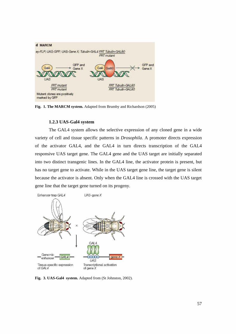
57
Fig. 1. The MARCM system. Adapted from Brumby and Richardson (2005)
1.2.3 UAS-Gal4 system
The GAL4 system allows the selective expression of any cloned gene in a wide
variety of cell and tissue specific patterns in Drosophila. A promoter directs expression
of the activator GAL4, and the GAL4 in turn directs transcription of the GAL4
responsive UAS target gene. The GAL4 gene and the UAS target are initially separated
into two distinct transgenic lines. In the GAL4 line, the activator protein is present, but
has no target gene to activate. While in the UAS target gene line, the target gene is silent
because the activator is absent. Only when the GAL4 line is crossed with the UAS target
gene line that the target gene turned on its progeny.
Fig. 3. UAS-Gal4 system. Adapted from (St Johnston, 2002).

58
1.2.4 P-element mediated transformation
P-element transformation is a method to obtain germline DNA integration
following co-injection of pre-blastoderm embryos with purified DNA. The microinjection
of the embryos must be performed in the dorsal region of the embryos, in the polar cells,
which will give rise to the germ line of the fly. One plasmid encodes the transposase, and
the other plasmid contains the inverted repeats flanking the experimental gene. Typically,
the marker gene on the P element transformation plasmid encodes an eye color marker
(e.g., the rosy gene, ry) that can be used to identify transgenes by virtue of their red eyes
(ry+), against a background of non-transformed flies with white eyes (ry-) (Roberts,
1998).
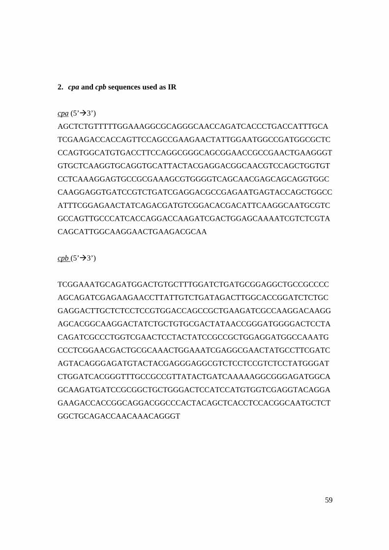
59
2. cpa and cpb sequences used as IR
cpa (5’à3’)
AGCTCTGTTTTTGGAAAGGCGCAGGGCAACCAGATCACCCTGACCATTTGCA
TCGAAGACCACCAGTTCCAGCCGAAGAACTATTGGAATGGCCGATGGCGCTC
CCAGTGGCATGTGACCTTCCAGGCGGGCAGCGGAACCGCCGAACTGAAGGGT
GTGCTCAAGGTGCAGGTGCATTACTACGAGGACGGCAACGTCCAGCTGGTGT
CCTCAAAGGAGTGCCGCGAAAGCGTGGGGTCAGCAACGAGCAGCAGGTGGC
CAAGGAGGTGATCCGTCTGATCGAGGACGCCGAGAATGAGTACCAGCTGGCC
ATTTCGGAGAACTATCAGACGATGTCGGACACGACATTCAAGGCAATGCGTC
GCCAGTTGCCCATCACCAGGACCAAGATCGACTGGAGCAAAATCGTCTCGTA
CAGCATTGGCAAGGAACTGAAGACGCAA
cpb (5’à3’)
TCGGAAATGCAGATGGACTGTGCTTTGGATCTGATGCGGAGGCTGCCGCCCC
AGCAGATCGAGAAGAACCTTATTGTCTGATAGACTTGGCACCGGATCTCTGC
GAGGACTTGCTCTCCTCCGTGGACCAGCCGCTGAAGATCGCCAAGGACAAGG
AGCACGGCAAGGACTATCTGCTGTGCGACTATAACCGGGATGGGGACTCCTA
CAGATCGCCCTGGTCGAACTCCTACTATCCGCCGCTGGAGGATGGCCAAATG
CCCTCGGAACGACTGCGCAAACTGGAAATCGAGGCGAACTATGCCTTCGATC
AGTACAGGGAGATGTACTACGAGGGAGGCGTCTCCTCCGTCTCCTATGGGAT
CTGGATCACGGGTTTGCCGCCGTTATACTGATCAAAAAGGCGGGAGATGGCA
GCAAGATGATCCGCGGCTGCTGGGACTCCATCCATGTGGTCGAGGTACAGGA
GAAGACCACCGGCAGGACGGCCCACTACAGCTCACCTCCACGGCAATGCTCT
GGCTGCAGACCAACAAACAGGGT
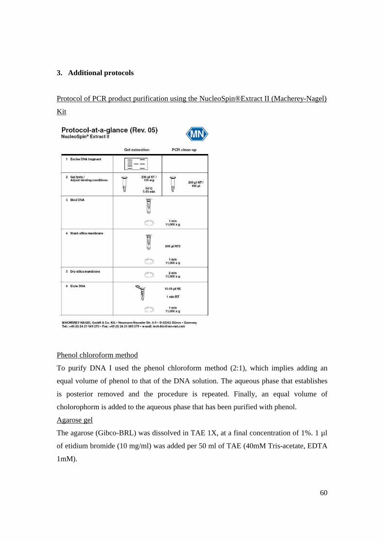
60
3. Additional protocols
Protocol of PCR product purification using the NucleoSpin®Extract II (Macherey-Nagel)
Kit
Phenol chloroform method
To purify DNA I used the phenol chloroform method (2:1), which implies adding an
equal volume of phenol to that of the DNA solution. The aqueous phase that establishes
is posterior removed and the procedure is repeated. Finally, an equal volume of
cholorophorm is added to the aqueous phase that has been purified with phenol.
Agarose gel
The agarose (Gibco-BRL) was dissolved in TAE 1X, at a final concentration of 1%. 1 µl
of etidium bromide (10 mg/ml) was added per 50 ml of TAE (40mM Tris-acetate, EDTA
1mM).


![Review Actin-targeting natural products: structures ... · actin-binding proteins actively break or ‘sever’ actin filaments [e.g. actin-depolymerizing factor (ADF) and cofilin].](https://static.fdocuments.us/doc/165x107/5f0f85bd7e708231d44494d0/review-actin-targeting-natural-products-structures-actin-binding-proteins-actively.jpg)


![Capping Protein Modulates Actin Remodeling in Response to ... · Capping Protein Modulates Actin Remodeling in Response to Reactive Oxygen Species during Plant Innate Immunity1[OPEN]](https://static.fdocuments.us/doc/165x107/5eac886ca71dc066fd5b1a1e/capping-protein-modulates-actin-remodeling-in-response-to-capping-protein-modulates.jpg)


