Actin Filaments in Yeast Are Unstable in the Absence of Capping ...
11
Actin Filaments in Yeast Are Unstable in the Absence of Capping Protein or Fimbrin Tatiana S. Karpova,* Kelly Tatchell,* and John A. Cooper* *Department of Cell Biology and Physiology, Washington University Medical School, St. Louis, Missouri; and*Department of Microbiology, North Carolina State University, Raleigh, North Carolina Abstract. Many actin-binding proteins affect filament assembly in vitro and localize with actin in vivo, but how their molecular actions contribute to filament as- sembly in vivo is not understood well. We report here that capping protein (CP) and fimbrin are both impor- tant for actin filament assembly in vivo in Saccharomy- ces cerevisiae, based on finding decreased actin filament assembly in CP and fimbrin mutants. We have also identified mutations in actin that enhance the CP phe- notype and find that those mutants also have decreased actin filament assembly in vivo. In vitro, actin purified from some of these mutants is defective in polymeriza- tion or binding fimbrin. These findings support the con- clusion that CP acts to stabilize actin filaments in vivo. This conclusion is particularly remarkable because it is the opposite of the conclusion drawn from recent stud- ies in Dictyostelium (Hug, C., P. Y. Jay, I. Reddy, J. G. McNally, P. C. Bridgman, E. L. Elson, and J. A. Coo- per. 1995. Cell 81:591-600). In addition, we find that the unpolymerized pool of actin in yeast is very small relative to that found in higher cells, which suggests that actin filament assembly is less dynamic in yeast than higher cells. I N Saccharomyces cerevisiae, actin is essential for viabil- ity (35), and important for polarized secretion (28) and endocytosis (19). The functional state of actin is presumed to be a helical filament, assembled from mono- meric subunits. The assembly and activity of actin fila- ments can be regulated in vitro by a large number of pro- teins that interact with actin. We wish to understand assembly of actin filaments in vivo and to achieve a molec- ular view of how actin-binding proteins function to control actin filament assembly. In vitro, monomers add to and leave from filaments at their ends, both barbed and pointed. Therefore, filament assembly in vivo can theoret- ically be controlled by actin-binding proteins that bind to monomers or filaments and inhibit, enhance, or bias this exchange reaction. In vitro, capping protein (CP) 1 binds to the barbed end of actin filaments, which inhibits both assembly (actin monomer addition) and disassembly (actin monomer loss) of the filament (for review see 30). CP is found in all eu- karyotes, including S. cerevisiae, where the heterodimeric protein is encoded by the genes CAP1 and CAP2 (3, 4). CP colocalizes with filamentous actin in yeast and other Address all correspondence to John Cooper, Box 8228, 660 South Euclid Avenue, St. Louis, MO 63110. Tel.: (314) 362-3964. Fax: (314) 362-0098. E-mail: [email protected] K. Tatchell's present address is Department of Biochemistry and Molec- ular Biology, Louisiana State University Medical Center, Shreveport, LA. 1. Abbreviations used in this paper: CP, capping protein; wt, wild type. cells (3, 31, 32). In developing muscle, the actin-binding activity of CP is necessary for proper assembly of the actin filaments of the sarcomere (34). In muscle, CP probably helps to determine the proper location and polarity of the actin filaments as well as to prevent their growth into adja- cent sarcomeres. In Dictyostelium, CP functions to limit or inhibit growth at barbed ends, and the level of CP deter- mines the level of actin filament assembly (15). One might imagine that mechanisms governing actin fil- ament assembly in yeast are very different from those that operate in striated muscle, Dictyostelium, and animal cells in general, based simply on the observation that the actin content of yeast is 100 times less than that in other cells. In most animal cells, the concentration of actin is very high (200 ~M) relative to the critical concentration for poly- merization (0.1 p.M), and these cells most likely use actin monomer-binding proteins, such as thymosin and profilin, to create a buffered pool of actin monomers available for rapid assembly. Yeast, however, have no apparent need for rapid and dynamic actin assembly. Although they clearly need to create actin filaments as they grow and di- vide, there is no evidence that filaments ever disassemble. In addition, there are no reports of a buffered pool of actin monomers in yeast, and our experiments here find that the soluble monomer pool is indeed very small. Therefore, we hypothesize that in yeast actin filaments are inherently un- stable due to the low actin concentration and therefore ac- tin-binding proteins function to create, assemble, and sta- bilize actin filaments. © The Rockefeller University Press, 0021-9525/95/12/1483/11 $2.00 The Journal of Cell Biology, Volume 131, Number 6, Part 1, December 1995 1483-1493 1483 on March 16, 2018 jcb.rupress.org Downloaded from
Transcript of Actin Filaments in Yeast Are Unstable in the Absence of Capping ...

Actin Filaments in Yeast Are Unstable in the Absence of Capping Protein or Fimbrin T a t i a n a S. K a r p o v a , * Kel ly Tatchell ,* and J o h n A. Coope r*
*Department of Cell Biology and Physiology, Washington University Medical School, St. Louis, Missouri; and*Department of Microbiology, North Carolina State University, Raleigh, North Carolina
Abstract. Many actin-binding proteins affect filament assembly in vitro and localize with actin in vivo, but how their molecular actions contribute to filament as- sembly in vivo is not understood well. We report here that capping protein (CP) and fimbrin are both impor- tant for actin filament assembly in vivo in Saccharomy- ces cerevisiae, based on finding decreased actin filament assembly in CP and fimbrin mutants. We have also identified mutations in actin that enhance the CP phe- notype and find that those mutants also have decreased actin filament assembly in vivo. In vitro, actin purified from some of these mutants is defective in polymeriza-
tion or binding fimbrin. These findings support the con- clusion that CP acts to stabilize actin filaments in vivo. This conclusion is particularly remarkable because it is the opposite of the conclusion drawn from recent stud- ies in Dictyostelium (Hug, C., P. Y. Jay, I. Reddy, J. G. McNally, P. C. Bridgman, E. L. Elson, and J. A. Coo- per. 1995. Cell 81:591-600). In addition, we find that the unpolymerized pool of actin in yeast is very small relative to that found in higher cells, which suggests that actin filament assembly is less dynamic in yeast than higher cells.
I N Saccharomyces cerevisiae, actin is essential for viabil- ity (35), and important for polarized secretion (28) and endocytosis (19). The functional state of actin is
presumed to be a helical filament, assembled from mono- meric subunits. The assembly and activity of actin fila- ments can be regulated in vitro by a large number of pro- teins that interact with actin. We wish to understand assembly of actin filaments in vivo and to achieve a molec- ular view of how actin-binding proteins function to control actin filament assembly. In vitro, monomers add to and leave from filaments at their ends, both barbed and pointed. Therefore, filament assembly in vivo can theoret- ically be controlled by actin-binding proteins that bind to monomers or filaments and inhibit, enhance, or bias this exchange reaction.
In vitro, capping protein (CP) 1 binds to the barbed end of actin filaments, which inhibits both assembly (actin monomer addition) and disassembly (actin monomer loss) of the filament (for review see 30). CP is found in all eu- karyotes, including S. cerevisiae, where the heterodimeric protein is encoded by the genes CAP1 and CAP2 (3, 4). CP colocalizes with filamentous actin in yeast and other
Address all correspondence to John Cooper, Box 8228, 660 South Euclid Avenue, St. Louis, MO 63110. Tel.: (314) 362-3964. Fax: (314) 362-0098. E-mail: [email protected]
K. Tatchell's present address is Department of Biochemistry and Molec- ular Biology, Louisiana State University Medical Center, Shreveport, LA.
1. Abbreviat ions used in this paper: CP, capping protein; wt, wild type.
cells (3, 31, 32). In developing muscle, the actin-binding activity of CP is necessary for proper assembly of the actin filaments of the sarcomere (34). In muscle, CP probably helps to determine the proper location and polarity of the actin filaments as well as to prevent their growth into adja- cent sarcomeres. In Dictyostelium, CP functions to limit or inhibit growth at barbed ends, and the level of CP deter- mines the level of actin filament assembly (15).
One might imagine that mechanisms governing actin fil- ament assembly in yeast are very different from those that operate in striated muscle, Dictyostelium, and animal cells in general, based simply on the observation that the actin content of yeast is 100 times less than that in other cells. In most animal cells, the concentration of actin is very high (200 ~M) relative to the critical concentration for poly- merization (0.1 p.M), and these cells most likely use actin monomer-binding proteins, such as thymosin and profilin, to create a buffered pool of actin monomers available for rapid assembly. Yeast, however, have no apparent need for rapid and dynamic actin assembly. Although they clearly need to create actin filaments as they grow and di- vide, there is no evidence that filaments ever disassemble. In addition, there are no reports of a buffered pool of actin monomers in yeast, and our experiments here find that the soluble monomer pool is indeed very small. Therefore, we hypothesize that in yeast actin filaments are inherently un- stable due to the low actin concentration and therefore ac- tin-binding proteins function to create, assemble, and sta- bilize actin filaments.
© The Rockefeller University Press, 0021-9525/95/12/1483/11 $2.00 The Journal of Cell Biology, Volume 131, Number 6, Part 1, December 1995 1483-1493 1483
on March 16, 2018
jcb.rupress.orgD
ownloaded from

Here we report experiments to test this hypothesis. We measured actin filament assembly by two complementary assays in yeast mutants lacking CP and fimbrin, another actin-binding protein that interacts genetically with CP and has also been hypothesized to stabilize actin filaments (1). We find that actin filament assembly is decreased in vivo in these mutants. We also searched for actin muta- tions that enhance the phenotype of CP mutations, a ge- netic interaction similar to the effect of the loss of fimbrin. We identified a set of actin mutations that do enhance the loss of CP and find that these actin mutations also cause decreased actin filament assembly in vivo. To investigate the molecular basis for this observation, we purified actin from these mutants and found that the mutant actins often show either decreased polymerization or poor binding to fimbrin in vitro. These observations also support the hy- pothesis that CP and fimbrin act to stabilize actin fila- ments. As predicted, these conclusions are the opposite of the ones found in Dictyostelium (15).
Materials and Methods
Materials, Media, and Culture Conditions
Unless stated otherwise, chemicals, materials and solvents were from Fisher Scientific Co. (Pittsburgh, PA) or Sigma Chemical Co. (St. Louis, MO). Restriction endonucleases and other enzymes were from Boeh- ringer-Mannheim Biochemicals (Indianapolis, IN). Yeast rich medium (YEPD), sporulation medium (MSPO) and synthetic minimal medium with different metabolites omitted for selection (DOBA), were from BIO 101, Inc. (La Jolla, CA). Medium containing 0.1% 5-fluoroorotic acid (PCR Inc., Gainesville, FL) was prepared as described (16). Presporula- tion medium was YEPD with adenine (200 p.g/ml). Strains were grown at 30°C unless specified otherwise.
Molecular and Genetic Techniques
Strains used in this work are listed in Table I. Tetrad dissection, random spore analysis, and yeast LiCI transformation were as described (16). DNA manipulations were performed by standard methods (25). Yeast ge- nomic DNA was prepared as described (11). Mutant actl alleles were cy- cle sequenced (9) from both PCR-amplified genomic DNA and gap-repair plasmids using [,,/33p] ATP to end-label a set of oligonucleotide primers that covered the gene.
Proteins
Two-dimensional gel electrophoresis and immunoblotting were per-
formed as described (33), using goat anti-yeast actin antibodies (17). Actin was purified as described (18), except that a MonoQ column was substi- tuted for the DEAE column. Also, the pool from the MonoQ column was further purified by a cycle of polymerization--depolymerization (14). Puri- fied wild-type (wt) and mutant actins showed minimal proteolysis on a heavily loaded SDS gel. To exclude contamination or reversion, we also sequenced the relevant region of each actl gene by PCR amplification and cycle sequencing, using yeast ceils obtained at the end of the preparation as template.
To test actin polymerization, each mutant actin was polymerized at 6 IxM for 2 h at room temperature in 1 × MKEI (2 mM MgCI2, 100 mM KC1, 1 mM EGTA, 20 mM imidazole/HCl, pH 7.0), then diluted to 3, 1, and 0.5 IxM in the same buffer and incubated for an additional 2 h. Samples were periodically mixed gently during the incubation, to accelerate the ap- proach to steady state. 25 of 100 t~1 was removed, for the sample "Total," and added to 25 ~1 of 2× SDS sample buffer. The remaining 75 ixl was spun at 150,000 g for 30 rain at 22°C (70,000 rpm in a rotor (TLA 100.1, Beckman Instruments, Inc., Fullerton, CA) in a centrifuge (TL100; Beck- man Instruments, Inc.)). 25 0,1 was removed from the meniscus, for the sample "Supernatant," and added to 25 Ixl of 2x SDS sample buffer. The remaining liquid was removed, avoiding the pellet, and the pellet was dis- solved in 150 I~1 of 1 × SDS sample buffer. Equal vols of the three samples, which represent equivalent fractions of the total sample, were analyzed on 10% SDS-polyacrylamide gels.
Yeast fimbrin, Sac6p, was purified as described (14) and kindly pro- vided by T. S. Sandrock and A. E. M. Adams (University of Arizona, Tuc- son, AZ). Sedimentation assays to determine binding of fimbrin to the mutant actins were performed as described (14) with the following modifi- cations: (a) the fimbrin (Sac6p) concentration was 0.75 o,M; and (b) sam- ples were sedimented at 90,000 g for 60 min at 22°C (50,000 rpm in a TLA100.1 rotor in a TL100 centrifuge). Fimbrin alone partially sedi- mented under these conditions, even with prior clarification; therefore, we analyzed the fimbrin in the supernatant, comparing each mutant actin with wt actin and no actin.
For actin polymerization and fimbrin-binding assays, 10% SDS-poly- acrylamide gels stained with Coomassie blue were scanned and the inten- sity of the relevant bands was quantitated with NIH Image 1.54, (available by ftp from zippy.nimh.nih.gov). Intensities of actin bands were converted to absolute amounts by comparison with internal standards.
Cell Actin Assembly Assays
In each assay, cultures were grown in YPD to 107 cells/ml, and total pro- tein was determined by Bradford assay (6) for normalization of the actin values.
To determine total actin, cells were completely disrupted with glass beads for SDS-PAGE. Actin was measured by a quantitative immunoblot using either affinity-purified goat antiyeast actin (17) or the mouse antiac- tin mAb C4 (20) as the first Ab and 125I-labeled secondary Abs as de- scribed (15). Five aliquots of cells were assayed for each strain, with three serial twofold dilutions.
To determine G-actin levels, the amount of soluble actin released from
Table L Yeast Strains Used in This Study
Name Relevant genotype Source
KWY201 MATa/ s actl::LEU2/ACTI ura3/ura3 leu2/leu2 his3/his3
KTI277 KT1278 YJC 450 YJC 472 YJC536 YJC 542 YJC 960
YJC 963 YJC 1080 YJC 1096 YJC 1104 YJC 1107 YJC 1117 YJC 1133
MATa cap2-1::URA3 ura3 Ieu2 his3 MATa cap2-t::URA3 ura3 leu2 his3 M A T s cap2::HIS3 ade2 ade3 lys2 ura3 leu2 MATa cap2::HlS3 ade2 ade3 lys2 ura3 leu2 trpl his3 [pBJ 198 - CAP2 ADE3 URA3] MATa slcl-66 cap2::H1S3 ade2 ade3 ura3 leu2 trpl [pBJ 198 - CAP2 ADE3 URA3] MATa slcl-87 cap2::HlS3 ade2 ade3 ura3 leu2 trpl [pBJ 198 - CAP2 ADE3 URA3] MATa/s slcl-66/slc1-66 cap2::HIS3/cap2::H1S3 ade2/ade2 ade3/ade3 ura3/ura3 leu2/leu2
trp l/TRP1 lys2/LYS2 his3/his3 [pBJ 416 - ADE3 LEU2 CAP2] MATa slcl-87 cap2::HIS3 ade2 ade3 ura3 leu2 trpl lys2 [pBJ 414 - CAP2 ADE3 LEU2] M A T s slcl-87 cap2::LEU2 ade2 ade3 ura3 leu2 trpl [pBJ 198 - CAP2 ADE3 URA3] MATa actl-lOl::HIS3 his3 ura3 leu2 tub2 MATa actl-lO8::H1S3 his3 ura3 leu2 tub2 M A T s act1-113::HlS3 his3 ura3 leu2 tub2 M A T s act1-120::HlS3 his3 ura3 leu2 tub2 MATa act1-133::HIS3 his3 ura3 leu2 tub2
K. Wertman, University of California, Berkeley
This study This study 17 17 17 17 This study
This study This study This study This study This study This study This study
The Journal of Cell Biology, Volume 131, 1995 1484
on March 16, 2018
jcb.rupress.orgD
ownloaded from

permeabilized unfixed cells was measured. Cells were perrneabilized by flash freezing and saponin treatment in an F-actin stabilizing buffer MKEI, essentially as described (21). This procedure provides permeabil- ity for actin monomers and 70 kD dextran. The permeabilized cells were sedimented in a microfuge for 15 min, and the supernatant was removed for determination of actin and protein. In wt cells, the actin level was very low, therefore the volume loaded onto the gel was maximized. In this ex- periment, error was propagated from the error in the determination of ac- tin level from the blot and the error in the protein assay.
To determine F-actin levels in fixed cells, a rhodamine-phaUoidin- binding assay was performed (22). A control with unlabeled phalloidin (Molecular Probes Inc., Eugene, OR) was performed for each sample to determine nonspecific binding. Four aliquots of fixed cells were assayed for each strain, with three serial twofold dilutions.
The level of F-actin could not be determined as the pelletable fraction in the latter, permeabilized cell, experiment because only a fraction (/>65%) of the cells are permeabilized by this procedure (21). Attempts at a similar analysis with a more aggressive method of permeabilization (bead-beating) gave inconsistent results, presumably due to variable dis- ruption of F-actin-containing cell structures.
Cloning of SLC llA CT1 A synthetic lethal screen with cap2A yielded two alleles, slcl-66 and slcl- 87 (17), of a gene that we now identify as actl. Strain YJC 963 carrying slcl-87 and cap2A and a plasmid with CAP2 was transformed with a cDNA library under control of the G A L l promoter (23), kindly provided by Anthony Bretscher (Cornell University, Ithaca, NY) and screened for rescue of synthetic lethality by colony color sectoring. The frequency of transformation was estimated by plating aliquots on Ura- medium. Ali- quots of the transformation, each containing at least 25 transformants, were grown in 96-well microtiter plates in -Ura/glucose and then plated on -Ura/galactose. Approximately 9,000 independent transformants were screened. Each independent transformant was represented by at least four colonies. After 5 d at 30°C, red/white sectoring was examined, and colonies with white sectors, indicating loss of pBJ 414, were tested for Leu- , Ura-/glucose, and Ura+/galactose. Plasmids were recovered and transformed back into the original strain to confirm their ability to rescue.
Based on restriction endonuclease maps, three different types of plas- mids were recovered that complemented slcl. One plasmid type carried CAP2, based on Western and Southern analysis and DNA sequence. A second plasmid type carried ACTI, based on DNA sequence. Three non- identical clones (pBJ 476, 477, and 481) were recovered. A third group of plasmids with insert size ~1 kb showed weak rescue on retransformation, and DNA sequencing and database searching with BLAST (2) did not identify the cDNA. These plasmids were not analyzed further.
The ACTI plasmids, pBJ 476 and 477, rescued the synthetic lethality of both alleles, slcl-66 and slcl-87, upon transformation into strains YJC 960 and 963, respectively. In addition, a CEN plasmid carrying ACT1 under control of its own promoter (pBJ 508) provided partial rescue of synthetic lethality for slcl-87 and complete rescue for slcl-66.
Overexpression of ACTI is lethal in some strains, but not in the wt background used here or in certain actl strains (23). We suspect that over- expression was important for the success of cloning. Several different ge- nomic libraries produced no clones that rescued, and ACT1 with its own promoter on a CEN plasmid provided only partial rescue for one allele. Both alleles were previously observed as semidominant, with slcl-87 more so than slcl-66 (17). Therefore, the levels of the rescuing wt Act lp pre- sumably needed to be in excess of those of the mutant Actlp.
Recombinational Allelism Test for slc l and ACT1 To determine whether slcl was allelic to ACT1, a recombinational allelism test was performed on a heterozygous diploid carrying one copy of slcl-87 and one copy of ACT1 marked by LEU2. The integrating plasmids pBJ 486 and 489 contain a promoterless fragment of ACT1 because they were constructed from the SalI/NotI inserts of cDNA clones pBJ 476 (1.4 kb) and pBJ 477 (1.8 kb), respectively. Integration of pBJ 486 or 489 at the ge- nomic locus of ACT1 results in two copies of ACT1, only one of which is expressed, pBJ 486 and 489 carry LEU2 as a marker, which was chosen to allow detection of plasmid integration at LEU2, instead of at ACT1, by testing for linkage to MAT and the centromere. Constructs were verified by restriction digestion.
The strain YJC 542, which carries slcl-87, cap2, and a CAP2 URA3 plasmid was transformed with the integrating plasmid that had been cut at a unique HindIII site in ACT1. As a control, a similar plasmid lacking
ACT1 sequences was used. Stable transformants were mated with the cap2 strain YJC 450, and the resulting diploids were subjected to tetrad analysis. Segregants were tested for (a) synthetic lethality by testing growth on 5-fluoroorotic acid to lose the CAP2 URA3 plasmid, (b) the ACT1 marker LEU2, (c) mating type (MAT), and (d) the centromere- linked marker TRPI. These tests permit discrimination between integra- tion at ACT1 as opposed to integration at LEU2.
sic1 was linked to ACT1 at a distance <0.49 cM, ~1.43 kbp. Allele numbers 66 and 87 have not been used for actl, to our knowledge. We now therefore refer to slcl-66 as act1-66 and to slcl-87 as actl-8Z
Recovery of Mutant Alleles with Gapped Plasmids Replicating plasmid pBJ 508, which carries an EcoRI genomic clone of ACT1 in pRS 314 (36), was cut with NdeI to produce a gap that included the entire ACT1 coding region plus 460 bp upstream and 300 bp down- stream. The remaining arms were 660 and 1,000 bp. The gapped plasmid was transformed into strains YJC 536 and 1080. Two independent plas- mids were recovered for actl-66 and three for actl-8Z
The plasmids were retransformed into the mutant strain to confirm that they could not rescue the synthetic lethal phenotype of the mutant. This was the case. However, this test does not exclude actl loss-of-function mu- tations that might be incidental to slcl. To show that the plasmids provide actl function, they were transformed into an actlA strain. A plasmid car- rying ACT1 (pBJ 505 - ACT1 URA3 CEN), actl-66 (pBJ 576 - actl-66 URA3 CEN), or actl-87 (pBJ 603 - actl-87 HIS3 CEN) was transformed into the diploid KWY 201, which carries one disruption of actl. On tetrad analysis, Leu ÷ (actl::LEU2) segregants were recovered for both actl-66 and actl-87, but only in the presence of the plasmid carrying the mutant actin gene. These results indicate that the recovered slcl/actl alleles func- tion in the sense of compensating for the disruption of ACT1, and are not simply coincidental loss-of-function mutations.
Screen of act1 Collection for Synthetic Lethality with cap2
A collection of act1 mutants (38) was screened in two ways. First, to reveal synthetic lethality, tetrad dissection was performed on diploids heterozy- gous for actl alleles marked by adjacent HIS3 integration and for a cap2:: URA3 gene disruption. These diploids were prepared by crossing haploid actl segregants derived from Wertman collection diploids to the cap2.': URA3 strains KT1277 or KT1278, depending on mating type. Synthetic le- thality was revealed by the absence of actl::HIS3 cap2::URA3 segregants. This approach also detects weaker actl cap2 interactions by comparing the growth of actl::H!S3 cap2::URA3 segregants with that of single mu- tants. 15 four-spored tetrads were dissected onto YPD. After incubation for 4-5 d at 24°C, the spore clones were replica plated to medium lacking uracil or histidine to score the cap2::URA3 and acH::HIS3 genotypes, re- spectively, and also replica plated to YPD and incubated at 15, 24, and 37°C to score for temperature-sensitive growth. Before replica plating, we microscopically examined those spores that had not grown to macroscopic colonies to determine whether germination and cell division had occurred. Spore viability was generally very good, except in cases of synthetic lethal- ity, and in a few cases where the acH allele showed reduced viability (acH- 132, act1-136).
Second, because synthetic lethals detected above could be deficient in spore germination or logarithmic growth, we determined whether double mutants covered with a CAP2 plasmid could lose the plasmid during loga- rithmic growth. The actl haploids above were crossed with strains YJC 1082 or 1084, depending on mating type, which carried a cap2::LEU2 dis- ruption, covered by a CAP2 URA3 plasmid (pBJ 198). Tetrad dissection was performed, and all spore types germinated. The ability to lose the plasmid was tested by growth on 5-fluoroorotic acid, and cap2::LEU2 and acH::HIS3 genotypes were determined by growth on - L e u and -His , re- spectively. In this background, spore viability was poor, so data for differ- ent ascospores derived from the same diploid were combined, as in a ran- dom spore analysis. Note also that stc2, which is synthetic lethal with cap2, is segregating in this analysis. The analysis of sic2 is beyond the scope of this paper and is not discussed per se; however, the synthetic lethal seg- regants are present and identified to allow consideration of the actl cap2 phenotype.
Structural Analysis The structure of the actin monomer and filament was viewed and ana- lyzed with InsightII on an IRIS computer, using subunit coordinates and
Karpova et al. Actin Filament Stability in Yeast 1485
on March 16, 2018
jcb.rupress.orgD
ownloaded from

A 3
2
c
il
B
140
120
,,-,- lOO
"5 so
- - - 60
40 20
C 0 . 4
Wl
Total actin
• I 8"/ 101 lOe 113 120 133 M cq~
M u t a t i o n
F-actin
wt u s7 lol 113 1~o m , , ~
M u t a t i o n
G-actin
~ 0.3
c 0 . 2
~" 0.1
0.0
/ t
w~ u 87 ~o~ lOe 11a 120 1 . ~ , ~ =
M u t a t i o n
Figure 1. Assays of cellular actin in acH, sac6, and cap2 strains. (A) Total actin content. The value is the mean and the error bar is the standard error. No strain is significantly different from wt. (B) F-actin content by rhodamine-phalloidin binding. The value is the mean of bound rhodamine fluorescence normalized to total protein for each strain, then normalized to wt. The error bar is the standard error. Asterisks mark cases where the difference be- tween the mutant and wt is statistically significant (P < 0.05). (C) G-actin content by determination of soluble actin released from
Table II. Recombinational Allelism Test for Linkage of slcl-87 (Foa-) and ACTI::LEU2
Foa - and LEU2 and LEU2 and LEU2 TRP1 MAT
A C T l p l a s m i d s P N T P N T P N T pBJ489 11 0 0 1 3 7 2 3 6 pBJ489 15 0 0 2 2 11 1 2 11 p B J 4 8 6 12 0 0 p B J 4 8 6 13 0 0 2 1 10 0 4 10
Total 51 0 0 5 6 28 3 9 27
Control plasmids P N T P N T P N T pBJ487 3 2 3 4 2 2 2 1 5 pBJ487 3 2 7 2 2 8 2 5 5
Total 6 4 10 6 4 10 4 6 10
The number of tetrads of different classes derived from independent transformants is listed. P, parental ditype; N, nonparental ditype; T, tetratype. The predicted ratio of tet- rads for absence of linkage is IP: IN: 4T. x-squared statistical analysis indicates link- age (P < 0.05) for slcl-87 (Foa-) and ACTI::LEU2 at <0.49 cM (~1.4 kbp), and no linkage (P > 0.05) in any other case. For the control plasmid that lacks ACT1, integra- tion should have occurred at the LEU2 chromosomal locus, giving linkage to both MAT and the centromere (TRP1). This linkage is observed in the slight deficiency of tetratypes, which is not statistically significant due to the number of tetrads examined.
helical f i lament parameters provided by Dr. Michael Lorenz (Max Planck Institute, Heidelberg, F R G ) (24).
Resu l t s
CP and Fimbrin Promote Actin Filament Assembly In Vivo
Previously, CP and fimbrin mutations were found to be synthetic lethal (1, 17). This enhanced phenotype is spe- cific for this combination; combinations with many other actin-binding protein mutations did not show enhance- ment (1), and a screen for mutations synthetic lethal with a CP null mutation did not uncover other actin-binding pro- tein genes (17 and herein). A n hypothesis to explain this observation, based on the biochemical properties of CP and fimbrin in vitro, was that both proteins are capable of stabilizing actin filaments in different ways (1). CP binds to barbed ends and prevents subunit loss; fimbrin binds to filament sides and also should prevent subunit loss from ends. Therefore, the loss of two proteins both acting to sta- bilize actin filaments would lead to a severe loss of actin filaments.
We tested this hypothesis by determining the level of ac- tin filament assembly in yeast strains lacking CP (cap2 mu- tants) or fimbrin (sac6 mutants). We used two comple- mentary assays that have been used extensively in other cell systems. We performed both assays because each has certain advantages and disadvantages. First, F-actin was measured by a rhodamine-phalloidin-binding assay. The advantage of this assay is that the cells are chemically fixed at the outset; therefore, actin structures are preserved dur- ing the assay. The disadvantage is the assumption that all actin filaments bind rhodamine-phalloidin equally well.
permeabilized cells. Values are actin normalized to total protein in the supernatant after permeabilization. Again, asterisks mark cases where the difference between the mutant and wt is statisti- cally significant (P < 0.05).
The Journal of Cell Biology, Volume 131, 1995 1486
on March 16, 2018
jcb.rupress.orgD
ownloaded from

Table IlL Interactions between act l ::HIS3 Alleles and cap2:: URA3 by Tetrad Analysis of Heterozygous Diploids
Number of viable segregants
act1 Ura Ura ÷ Ura- Ura ÷ Phenotype No. of tetrads allele His- His- His + His + of Ura÷His ÷ (PD/TT/NPD)
wt Obs 14 10 14 13 wt 2 10 2
Exp 14 14 14 14 101 Obs 13 15 15 0 Microscopic colonies* 4 7 3
Exp 13 15 15 13 102 Obs 17 11 11 17 wt 1 9 4
Exp 17 11 11 17 104 Obs 13 17 17 13 Slight ts 3 11 1
Exp 13 17 17 13 105 Obs 16 12 6 13 Small colonies 1 10 3
Exp 16 12 12 16 108 Obs 14 14 12 0 Inviable 2 10 2
Exp 14 14 14 14 111 Obs ~ 15 13 2 0 Inviable § 2 9 3
Exp 15 13 13 15 113 Obs 14 14 13 0 Inviable 2 10 2
Exp 14 14 14 14 115 Obs 7 21 21 9 ts 7 7 1
Exp 9 21 21 9 116 Obs 20 8 10 19 ts 2 6 7
Exp 20 10 10 20 117 Obs 16 13 14 16 Slightly ts 3 8 4
Exp 16 14 14 16 119 Obs 16 12 14 0 Inviable 3 8 4
Exp 16 14 14 16 120 Obs 13 l 5 15 0 Microscopic colonies* 1 13 0
Exp 13 15 15 13 121 Obs 15 13 12 15 cs and ts 1 11 2
Exp 15 13 13 15 122 Obs 13 17 17 13 ts 3 4 1
Exp 13 17 17 13 123 Obs 13 13 13 14 wt 2 10 2
Exp 14 14 14 14 124 Obs 16 14 13 0 Small colonies* 3 8 4
Exp 16 14 14 16 125 Obs 16 12 11 0 Inviable 0 12 2
Exp 16 12 12 16 129 Obs 14 15 16 7 Small colonies 5 6 4
Exp 14 16 16 14 132 Obs 17 13 8 0 Inviable ~ 0 13 2
Exp 17 13 13 17 133 Obs 16 14 13 0 Inviable 3 8 4
Exp 16 14 14 16 135 Obs 14 16 16 14 Slight ts 2 12 1
Exp 14 16 16 14 136 Obs 11 19 10 0 Inviable § 4 11 0
Exp 11 19 19 11
The total number of observed (Obs) and expected (Exp) viable haploid segregants of each genotype is listed, along with the phenotype of the actl cap2 segregants, and the number of different types of tetrads. §These act1 alleles had low viability in this experiment, reflected in the number of Ura-His + segregants. Also, these alleles were not synthetic lethal in a test of logarithmic growth (Table IV). q0 small colonies could not be analyzed for markers. *The genotype of the Ura ÷ His + segregants was inferred from those of macroscopic colonies that could be tested for markers. The phenotype listed describes how those spores were able to grow.
Actin-binding proteins or actin mutations may influence the efficiency of phalloidin binding. Second, G-actin was measured as the amount of actin that was released in a sol- uble form from permeabilized cells. The advantage here is that the quantitation of the actin by immunoblot is unam- biguous, and the disadvantage is that the permeabilization procedure may cause the solubilization of some F-actin. We also measured total cell actin by immunoblot, to deter- mine whether changes in the amount of F- and G-actin
were reflective of the total actin pool or independent ef- fects.
Different results with the two different assays were sometimes found and are to be expected. Actin may exist in a form detected in both or neither assays. For example, some actin filaments may depolymerize rapidly and be- come soluble on permeabilization, so this pool would be measured in both assays. An increase in this pool of actin would lead to increased G-actin and unchanged F-actin in
Karpova et al. Actin Filament Stability in Yeast 1487
on March 16, 2018
jcb.rupress.orgD
ownloaded from

Table IV. Plasmid Loss Analysis of Interactions between actl Table V. Summary of Interactions between cap2 and actl Alleles and cap2 during Logarithmic Growth
Foa + Foa- Allele Ratio of act1 Leu ÷ Leu Leu + Leu * Foa+Leu+/total
wt 9 16 8 2 0.26 4 14 6 5 2 0.52
101 0 11 14 1 0.00 102 10 29 6 0 0.22 104 14 29 11 0 0.26 105 14 22 11 0 0.30 108 0 9 10 0 0.00 111 2 6 4 0 0.17 113 0 5 5 0 0.00 115 11 15 10 0 0.31 116 10 12 4 0 0.38 117 9 14 4 0 0.33 119 0 8 9 0 0.00 120 0 7 6 0 0.00 12l l 2 0 2 122 2 3 3 0 123 13 15 6 1 0.37 124 3 10 8 0 0.14 125 0 6 4 0 0.00 129 3 8 10 0 0.14 132 9 9 1 0 0.47 133 0 8 2 0 0.00 135 11 13 7 2 0.33 136 16 15 4 3 0.42
Predicted results for four segregants,
considering segregation at the CAP2 and SLC2 loci Synthetic lethality none 1 2 1 0 0.25 actl cap2 0 2 2 0 0 act1 slc2 1 1 0 0 0.5 actl sic2 and act1 cap2 0 1 1 0 0.0
The genotype of the diploids used in this analysis was ACT1/actl::H1S3 cap2-Al:: LEU2/CAP2 slc2/SLC2 with a plasmid [CAP2 URA3]. Since only act1 segregants are relevant for this analysis, only His + segregants are included in the table. Note that sic2, which is synthetic lethal with cap2, is segregating in this analysis and does show allele-specific interactions with act1. The analysis of sic2 is beyond the scope of this paper and is not discussed per se, however, the synthetic lethal segregants are present and identified here by necessity. The key parameter to examine to determine whether an actl allele is synthetic lethal with cap2 is the number of Leu ÷ (cap2) Foa ÷ (plasmid loss viable) segregants. In the case of synthetic lethality, the number should be zero, In the absence of synthetic lethality, the number of Leu + Foa ÷ segregants should be ei- ther one-half or one-quarter of the total number of segregants, depending on whether the act1 allele is or is not synthetic lethal with sic2, respectively. *CAP2 strains should not be Foa-, therefore, these segregants must have poor growth on FOA for some other reason and presumably represent strains with marginal growth properties overall. ¢These strains had low viability in this experiment, so no ratio is calculated and no conclusion drawn.
the assays. In support of this possibility, permeabilization leads to loss of actin cables (21). On the other hand, dena- tured and precipitated actin would neither bind rhodamine- phalloidin nor be soluble, which would lead to decreased F-actin and unchanged G-actin in the assays. Many actin and actin-binding protein mutations have this effect on actin.
Both CP and fimbrin mutants showed less F-actin, more G-actin, and no change in total actin compared with wt (Fig. 1). Therefore, the results with the two different as- says are consistent and support the hypothesis that both actin-binding proteins promote actin filament assembly. For CP, this result is especially interesting because it is the opposite of the one found in Dictyostelium, where less CP
Phenotypes of double mutants actl allele Amino acid substitution act l cap2 act1 sac6*
wt NE NE 3 P32L ND NE 4 E259V NE NE
66 M 16I SynLet NE 87 E292K SynLet NE
101 D363A, E264A SynLet Mud 102 K359A, E361A NE Mud 104 K415A, E316A Mud NE 105 E311A, R312A Mud NE 108 R256A, E259A SynLet SynLet 111 D222A, E224A, E226A Mud * ND 113 R210A, D211A SynLet SynLet 115 E 195A, R 196A Mud SynLet 116 D187A, K191A Mud Mud 117 R183A, D184A Mud Mud 119 R116A, E117A, K118A SynLet SynLet 120 E99A, E I 0 0 A SynLet NE 121 E83A, K84A Mud SynLet 122 D80A, D81A Mud ND 123 R68A, E72A NE NE 124 D56A, E57A Mud SynLet 125 K50A, D51A SynLet SynLet 129 R177A, D179A Mud SynLet 132 R37A, R39A Mud ND 133 D24A, D25A SynLet Mud 135 E4A NE NE 136 D2A Mud SynLet
The results in Tables III and IV are compiled and summarized here. NE, no effect; Mud, modified phenotype (slow or temperature-sensitive growth); Syn- Let, synthetic lethal; ND, nut determined. *The data in this column are reproduced from the work of Holtzman and colleagues (13), except for alleles 66 and 87, which were described by us previously (17). SLow viability reduces the significance of this interpretation.
led to more F-actin (15). This difference is possible be- cause CP binding to barbed ends stops both the addition and loss of actin subunits. Therefore, in yeast, CP prevents the loss of subunits that otherwise occurs from free barbed ends, while in Dictyostelium CP prevents the addition of subunits.
Pool of Actin Monomers in Yeast
This difference between Dictyostelium and yeast is most likely due to a difference in the level of actin monomers available for addition to free barbed ends. We hypothesize that the level is low in yeast and high in Dictyostelium. The total actin content of Dictyostelium is ~100 times that of yeast, ~10% vs ~0.1% of total cell protein (12, 15, 27, confirmed by Fig. 1 A here). About 50% of actin in Dicty- ostelium is soluble, and therefore exists as either free or buffered monomers (15). In contrast, we report here that very low levels of actin are soluble in yeast.
The soluble actin released from permeabilized yeast cells is normalized in Fig. 1 C to the level of protein re- leased, to account for possible differences in the percent- age of cells permeabilized in different strains. However, if one normalizes to total protein in a whole-cell extract, sol- uble actin is 2 pg/txg of protein in wt cells. The fraction of cells permeabilized in the assay is i>60% (21). Even as- suming that only 50% of cells are permeabilized, then at most 4 pg/Ixg is soluble, and the soluble fraction of actin is
The Journal of Cell Biology, Volume 131, 1995 1488
on March 16, 2018
jcb.rupress.orgD
ownloaded from
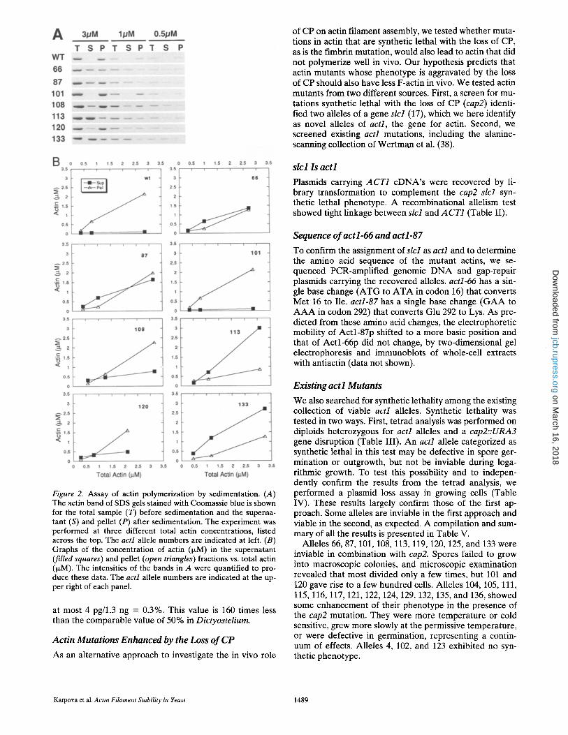
Figure 2. Assay of actin polymerization by sedimentation. (A) The actin band of SDS gels stained with Coomassie blue is shown for the total sample (T) before sedimentation and the superna- tant (S) and pellet (P) after sedimentation. The experiment was performed at three different total actin concentrations, listed across the top. The act1 allele numbers are indicated at left. (B) Graphs of the concentration of actin (p~M) in the supernatant (filled squares) and pellet (open triangles) fractions vs. total actin (~M). The intensities of the bands in A were quantified to pro- duce these data. The actl allele numbers are indicated at the up- per right of each panel.
at most 4 pg/1.3 ng = 0.3%. This value is 160 times less than the comparable value of 50% in Dictyostelium.
Actin Mutations Enhanced by the Loss o f CP
As an alternative approach to investigate the in vivo role
of CP on actin filament assembly, we tested whether muta- tions in actin that are synthetic lethal with the loss of CP, as is the fimbrin mutation, would also lead to actin that did not polymerize well in vivo. Our hypothesis predicts that actin mutants whose phenotype is aggravated by the loss of CP should also have less F-actin in vivo. We tested actin mutants from two different sources. First, a screen for mu- tations synthetic lethal with the loss of CP (cap2) identi- fied two alleles of a gene slcl (17), which we here identify as novel alleles of act1, the gene for actin. Second, we screened existing act1 mutations, including the alanine- scanning collection of Wertman et al. (38).
slc l Is act l
Plasmids carrying ACT1 cDNA's were recovered by li- brary transformation to complement the cap2 slcl syn- thetic lethal phenotype. A recombinational allelism test showed tight linkage between slcl and A CT1 (Table II).
Sequence of act l-66 and act l-87
To confirm the assignment of slcl as actl and to determine the amino acid sequence of the mutant actins, we se- quenced PCR-amplified genomic DNA and gap-repair plasmids carrying the recovered alleles, actl-66 has a sin- gle base change (ATG to ATA in codon 16) that converts Met 16 to Ile. act1-87 has a single base change (GAA to A A A in codon 292) that converts Glu 292 to Lys. As pre- dicted from these amino acid changes, the electrophoretic mobility of Actl-87p shifted to a more basic position and that of Actl-66p did not change, by two-dimensional gel electrophoresis and immunoblots of whole-cell extracts with antiactin (data not shown).
Existing act l Mutants
We also searched for synthetic lethality among the existing collection of viable acH alleles. Synthetic lethality was tested in two ways. First, tetrad analysis was performed on diploids heterozygous for act1 alleles and a cap2::URA3 gene disruption (Table III). An act1 allele categorized as synthetic lethal in this test may be defective in spore ger- mination or outgrowth, but not be inviable during loga- rithmic growth. To test this possibility and to indepen- dently confirm the results from the tetrad analysis, we performed a plasmid loss assay in growing cells (Table IV). These results largely confirm those of the first ap- proach. Some alleles are inviable in the first approach and viable in the second, as expected. A compilation and sum- mary of all the results is presented in Table V.
Alleles 66, 87, 101, 108, 113, 119, 120, 125, and 133 were inviable in combination with cap2. Spores failed to grow into macroscopic colonies, and microscopic examination revealed that most divided only a few times, but 101 and 120 gave rise to a few hundred cells. Alleles 104, 105, 111, 115, 116, 117, 121,122, 124, 129, 132, 135, and 136, showed some enhancement of their phenotype in the presence of the cap2 mutation. They were more temperature or cold sensitive, grew more slowly at the permissive temperature, or were defective in germination, representing a contin- uum of effects. Alleles 4, 102, and 123 exhibited no syn- thetic phenotype.
Karpova et al. Actin Filament Stability in Yeast 1489
on March 16, 2018
jcb.rupress.orgD
ownloaded from

Figure 3. Structural model of actin, indicating location of mutations. (A) Three views of an actin monomer, in red, with the synthetic le- thal allele residues indicated in green. The view labeled Front is similar to the one conventionally shown, with the exception that the subunit is rotated slightly counterclockwise about an axis perpendicular to the page, placing it in an orientat ion similar to that seen in the filament. (B) A filament of five subunits, with the subunits having different colors--purple, red, white, blue, and purple again--as one proceeds from barbed to pointed end. Again, the synthetic lethal residues are indicated in green.
The Journal of Cell Biology, Volume 131, 1995 1490
on March 16, 2018
jcb.rupress.orgD
ownloaded from

Defective Actin Assembly In Vivo in Actin Mutants
Our hypothesis then predicts that the set of synthetic le- thal actin mutants will include ones that have impaired ac- tin filament assembly in vivo, as seen for the CP and fim- brin null mutants. We assayed the mutants for actin filament assembly in vivo by the two complementary methods discussed above, independent measurements re- flecting F-actin and G-actin. Total actin was not changed significantly for any actl mutant, as also seen for the CP and fimbrin mutants.
For nearly all the actl mutants, in at least one of the as- says, we detected a defect similar to that seen in CP and fimbrin mutants: less F-actin or more G-actin. For alleles 87 and 113 changes in the predicted direction were seen in both assays. The predicted effect was seen in one but not the other assay for alleles 66, 101, and 120. As discussed above, different results in the two assays are not unex- pected. In the F-actin assay, allele 108 was not tested be- cause of poor growth, and 133 showed a change in the pre- dicted direction, but without statistical significance. Both 108 and 133 were normal in the G-actin assay.
Biochemical and Structural Analysis of Actin Mutants
Our hypothesis predicts two obvious molecular mecha- nisms to explain defective actin assembly in vivo in these actin mutants. The mutant actin could polymerize poorly or it might bind fimbrin poorly. To test further our hypoth- esis and discriminate between these possibilities, we puri- fied actin from the mutants and assayed polymerization and fimbrin binding in vitro. We hypothesized that these tests might also reveal defects in actin filament function not seen in the in vivo assays. In addition, using the struc- tural model of the actin filament, we were able to make predictions and correlate results in the biochemical assays with the locations of the mutations.
First, we tested the ability of the mutant actins to poly- merize (Fig. 2). Most alleles did show defective polymer- ization, manifested by less actin in the pellet and more in the supernatant in a sedimentation assay. Alleles 66, 87, 113, and 133 showed substantial defects. Alleles 108 and 120 showed mild defects, and 101 was similar to wt. Struc- tural analysis provided interesting correlations (Fig. 3). First, amino acid residue 292, altered in allele 87, which shows a severe polymerization defect in vitro, is a pre- dicted contact between actin subunits along the long axis of the filament. 292 lies within 4/~ of residues 45 and 46 of an adjacent subunit. Second, in allele 108, two amino acid residues are altered (256 and 259), and they lie just above the loop connecting the protofilament strands. Although residues 256 and 259 are not within 6 A, of any other sub- unit, they may be important for the structure of that loop. Third, the amino acid residues altered in alleles 66 and 113 occupy positions at or near the cleft where nucleotide binds, and nucleotide binding has been shown to be impor- tant for subunit and filament stability (7, 8).
Second, we tested the ability of the mutant actins to bind fimbrin. The binding site for fimbrin on actin has been characterized, and allele 120 shows the best agreement to the criteria (13, 14, 26). As expected, allele 120 also showed decreased fimbrin binding here (Fig. 4). Other al- leles that change residues at and near the fimbrin-binding
Figure 4. Binding of fimbrin to actin. (A) The fimbrin (Sac6p) and actin bands of SDS gels stained with Coomassie blue are shown for the total sample (T) before sedimentation and the su- pernatant (S) and pellet (P) after sedimentation. The actl allele numbers are given below each panel. The panel in the bottom right is a control without actin. (B) Binding of fimbrin (Sac6p) to mutant actins. The intensities of the fimbrin bands in A were quantified. The amount of fimbrin (Sac6p) in the supernatant is plotted as a percentage of the total fimbrin. In a control experi- ment without actin, some fimbrin does sediment, despite prior clarification.
site are 125, 101, 119, and possibly 133 (Fig. 4). In our as- says of fimbrin binding, allele 108 was severely impaired, and alleles 101, 113, and 133 showed moderate defects in fimbrin binding (Fig. 4). The explanation for decreased fimbrin binding by alleles 108 and 113, located outside of the previously defined fimbrin-binding site, might be ei- ther long-range allosteric effects, seen with other actin mu- tations (10, 29, 37), or interaction of fimbrin with addi- tional sites not revealed in the previous analysis.
Therefore, among the collection of actin mutants are some that show poor polymerization, some that show poor fimbrin binding, and some that show both. No alleles are normal for both properties. As examples of the first case, consider alleles 66 and 87. They show poor polymerization (Fig. 2 B), but they bind fimbrin well (Fig. 4 B). This makes sense with their localization in the structure (Fig. 3) because they are not at the fimbrin-binding site, rather they are near the nucleotide-binding site (66) and a sub- unit-subunit contact site (87). For the second case, allele 120 polymerizes well (Fig. 2 B) but binds fimbrin poorly (Fig. 4 B). Again, the structural location for this mutant make sense. Others have documented well that this region is part of the fimbrin-binding site (13, 14, 26), and this site would not be expected to affect polymerization, which is
Karpova eta|. Actin Filament Stability in Yeast 1491
on March 16, 2018
jcb.rupress.orgD
ownloaded from

the result here. For the third case, allele 133 is poor at both polymerization (Fig. 2 B) and fimbrin binding (Fig. 4 B). In this case, one might not have predicted these effects based on the structural location. The affected region is close to but not part of the recognized sites for fimbrin binding or subunit contacts. This discrepancy may reflect a limitation in the structural analysis or allosteric effects.
Another possible molecular model to explain synthetic lethality is that another actin-binding protein is essential in the absence of CP. Mutations in the binding site for such a protein might be identified as ones with normal actin as- sembly and fimbrin binding in vitro and in vivo. The set of actl mutations does not include exactly such a case, how- ever, we did observe a broad range of effects in the assays and were not always able to include all strains in every as- say. One potential candidate is slc2, recovered in a syn- thetic lethal screen with cap2 (17). slc2 is the mannopro- tein synthesis gene MNNIO (Dean, N., and J. B. Poster, manuscript submitted for publication, and 5) which en- codes a protein with sequence similarity to galactosyl transferase (Karpova, T. S., and J. A. Cooper, unpublished results) (These sequence data are available from Gen- Bank under accession number L42540).
Discussion
Mechanisms for Actin Assembly
The most important conclusions from this study are that, in yeast, actin filaments are unstable without either of two major actin-binding proteins. The loss of either CP or fim- brin leads to decreased F-actin and increased G-actin. This hypothesis was proposed based on several observations. First, the loss of both CP and fimbrin is lethal. Second, the loss of actin is lethal. Third, the filament is presumed to be the functional state of actin. Fourth, the biochemical activ- ities of CP and fimbrin in vitro have in common the ability to stabilize actin filaments, i.e., to prevent depolymeriza- tion. However, the biochemical activities of CP also in- clude preventing filament assembly by inhibiting growth at barbed ends. Therefore, it was uncertain which biochemi- cal activity of CP would be relevant in vivo in yeast.
For CP, this conclusion was supported by additional ex- periments in which mutations in actin that are also syn- thetic lethal with the loss of CP were found to have unsta- ble actin in vivo. As expected, some of these actin mutants bound fimbrin poorly, in biochemical experiments with purified proteins. This would be the equivalent of the loss of fimbrin. In addition, biochemical studies showed that some of these mutant actins polymerized poorly on their own. Therefore, the actin mutants are either unstable in their own polymerization or in binding fimbrin, which con- tributes to stability. Coupled with the loss of filament sta- bility that accompanies the loss of CP, these mutations are now lethal because filament stability is severely compro- mised.
Contrasting Yeast and Dictyostelium
These conclusions in yeast present a striking contrast to the case in Dictyostelium, where the opposite conclusions were reached (15). In Dictyostelium, the loss of CP leads
to increased F-actin. There, actin is poised to assemble, and CP, by binding to filament barbed ends, prevents growth.
CP can act in these opposing fashions in the two systems because of its biochemical properties. When bound to the barbed end, it prevents both the addition and loss of actin subunits, preventing the filament from growing or shrink- ing, respectively. Which activity is relevant in a given case depends on the concentration of free monomers present in solution and available for polymerization. If the actin monomer concentration is high, then free barbed ends tend to polymerize and CP prevents filament growth. On the other hand, if the actin monomer concentration is low, then free barbed ends tend to depolymerize and CP pre- vents filament shrinkage. In agreement with this predic- tion, the actin monomer concentration in Dictyostelium is high, and we report here that in yeast it is low.
The biology of actin in Dictyostelium vs. yeast supports these conclusions. Dictyostelium is highly motile, using a high concentration of actin filaments as a basis for motil- ity. These filaments are probably highly dynamic and re- quire local bursts of actin polymerization and depolymer- ization for coordinated assembly. Therefore, the requisite high concentrations of monomeric actin needed for bursts of assemble must be held in check by actin-binding pro- teins. Yeast, on the other hand, are not motile and show no analogous need for dynamic actin filament assembly. While yeast show dramatic rearrangements of the actin cy- toskeleton during the cell cycle and in response to certain external stimuli, there is no evidence for substantial in- crease or decrease in the total cellular content of actin fila- ments during any of these changes. While actin filaments are essential for viability of yeast, they are present at com- paratively low concentrations and therefore tend not to as- sembly spontaneously. In this system, the actin-binding proteins may not be needed to hold actin polymerization in check, but rather to induce and stabilize actin filament formation at the proper location and time.
We are grateful to Anthony Bretscher for the cDNA library, to David Drubin (University of California, Berkeley) for allowing us to reproduce his data in Table V, to Ken Wertman for providing the collection of act1
alanine mutants, to Tania Sandrock and Alison Adams for providing tim- brin, to Carl Frieden (Washington University, St. Louis, MO) for the use of the computer to analyze the actin structure, to Michael Lorenz for the actin structure coordinates, and to Amy McGough (Baylor Medical School, Houston, TX) for discussions of structural considerations.
This work was supported by grants from the National Institutes of Health to K. Tatchell (GM 47789) and to J. A. Cooper (GM 47337), and by a postdoctoral fellowship to T. S. Karpova from the Missouri Affiliate of the American Heart Association. J. A. Cooper is an Established Inves- tigator of the American Heart Association.
Received for publication 21 December 1994 and in revised form 7 Sep- tember 1995.
Note Added in Proof SLC2 and MNNIO are also identical to BED1, de- scribed in Mondesert and Reed (Mondesert and Reed. 1996. J. Cell Biol. In press.).
References
1. Adams, A. E., J. A. Cooper, and D. G. Drubin. 1993. Unexpected combina- tions of null mutations in genes encoding the actin cytoskeleton are lethal in yeast. Mol. BioL Cell. 4:459--468.
2. Altschul, S. F., W. Gish, W. Miller, E. Myers, and D. Lipman. 1990. Basic local alignment search tool. Z Mol. Biol. 215:403410.
The Joumal of Cell Biology, Volume 131, 1995 1492
on March 16, 2018
jcb.rupress.orgD
ownloaded from

3. Amatruda, J. F., and J. A. Cooper. 1992. Purification, characterization, and immunofinorescence localization of Saccharomyces cerevisiae capping protein. J. Cell Biol. 117:1067-1076.
4. Amatruda, J. F., D. J. Gattermeir, T. S. Karpova, and J. A. Cooper. 1992. Effects of null mutations and overexpression of capping protein on mor- phogenesis, actin distribution, and polarized secretion in yeast. J. Cell Biol. 119:1151-1162.
5. Ballou, L., E. Alvarado, P. K. Tsai, A. Dell, and C. E. Ballou. 1989. Protein glycosylation defects in the Saccharomyces cerevisiae mnn7 mutant class. Support for the stop signal proposed for regulation of outer chain elonga- tion. J. Biol. Chem. 264:11857-11864.
6. Bradford, M. M. 1976. A rapid and sensitive method for the quantitation of microgram quantities of protein utilizing the principle of protein-dye binding. Anal. Biochem. 72:248-254.
7. Chen, X., and P. A. Rubenstein. 1995. A mutation in an ATP-binding loop of Saccharomyces cerevisiae actin ($14A) causes a temperature-sensitive phenotype in vivo and in vitro. J. Biol. Chem. 270:11406-11414.
8. Chen, X., J. M. Peng, M. Pedram, C. A. Swenson, and P. A. Rubenstein. 1995. The effect of the S14A mutation on the conformation and thermo- stability of Saccharomyces cerevisiae G-actin and its interaction with ade- nine nucleotides. J. Biol. Chem. 270:11415-11423.
9. Craxton, M. 1991. Linear amplification sequencing, a powerful method for sequencing DNA. In Methods: A Companion to Methods in Enzymol- ogy. Vol. 3.20-26.
10. Crosbie, R. I-I., C. Miller, P. Cheung, T. Goodnight, A. Muhlrad, and E. Reisler. 1994. Structural connectivity in actin-effect of C-terminal modifi- cations on the properties of actin. Biophys. ,L 67:1957-1964.
11. Fujimura, H., and Y. Sakuma. 1993. Simplified isolation of chromosomal and plasmid DNA from yeasts. Biotechniques. 14:538-540.
12. Greer, C., and R. Schekman. 1982. Actin from Saccharomyces cerevisiae. Mol. Cell, BioL 2:1270-1278.
13. Holtzman, D. A., K. F. Wertman, and D. G. Drubin. 1994. Mapping actin surfaces required for functional interactions in vivo. J. Cell Biol. 126:423- 432.
14. Honts, J. E., T. S. Sandrock, S. M. Brower, J. L. O'Dell, and A. E. M. Ad- ams. 1994. Actin mutations that show suppression with fimbrin mutations identify a likely fimbrin-binding site on actin. J. Cell Biol. 126:413-422.
15. Hug, C., P. Y. Jay, I. Reddy, J. G. McNally, P. C. Bridgman, E. L. Elson, and J. A. Cooper. 1995. Capping protein levels influence actin assembly and cell motility in Dictyostelium. Cell, 81:591-600.
16. Kaiser, C., S. Michaelis, and A. Mitchell. 1994. Methods in Yeast Genetics. Cold Spring Harbor Laboratory. Cold Spring Harbor, NY. 234 pp.
17. Karpova, T. S., M. M. Lepetit, and J. A. Cooper. 1993. Mutations that en- hance the cap2 null mutant phenotype in Saccharomyces cerevisiae affect the actin cytoskeleton, morphogenesis, and pattern of growth. Genetics. 135:693-709.
18. Kron, S. J., D. G. Drubin, D. Botstein, and J. A. Spudich. 1992. Yeast actin filaments display ATP-dependent sliding movement over surfaces coated with rabbit muscle myosin. Proc. Natl. Acad. Sci. USA. 89:4466-4470,
19. Kubler, E., and H. Riezman. 1993. Actin and fimbrin are required for the internalization step of endocytosis in yeast. EMBO (Eur. Mol. Biol. Or- gan.) J. 12:2855-2862.
20. Lessard, J. L. 1988. Two monoclonal antibodies to actin: one muscle selec-
tive and one generally reactive. Cell Motil. Cytoskeleton. 10:349-362. 21. Li, R,, Y. Zheng, and D. G. Drubin. 1995. Regulation of cortical actin cy-
toskeleton assembly during polarized cell growth in budding yeast. J. Cell Biol. 128:599-615.
22. Lillie, S. H., and S. S. Brown. 1994. Immunofluorescence localization of the unconventional myosin, Myo2p, and the putative kinesin-related protein, Smylp, to the same regions of polarized growth in Saccharomyces cerevi- siae. J. Cell Biol. 125:825-842.
23. Liu, H., J. Krizek, and A. Bretscher. 1992. Construction of a GALl-regu- lated yeast cDNA expression library and its application to the identifica- tion of genes whose overexpression causes lethality in yeast. Genetics. 132:665-673.
24. Lorenz, M., D. Popp, and K. C. Holmes. 1993. Refinement of the F-actin model against X-ray fiber diffraction data by the use of a directed muta- tion algorithm. Z Mol. BioL 234:826-836.
25. Maniatis, T., E. F. Fritsh, and J, Sambrook. 1982. Molecular Cloning: A Laboratory Manual. Cold Spring Harbor Laboratory. Cold Spring Har- bor, NY. 545 pp.
26. McGough, A., M. Way, and D. DeRosier. 1994. Determination of the a-acti- nin-binding site on actin filaments by cryoelectron microscopy and image analysis. J. Cell Biol. 126:433-443.
27. Nefsky, B., and A. Bretscher. 1992. Yeast actin is relatively well behaved. Eur. J. Biochem. 206:949-955.
28. Novick, P., and D. Botstein. 1985. Phenotypic analysis of temperature-sen- sitive yeast actin mutants. Cell. 40:405-416.
29. Orlova, A., and E. H. Egelman. 1995. Structural dynamics of F-actin. I. Changes in the C terminus. Z Mol. Biol. 245:582-597.
30. Schafer, D. A., and J. A. Cooper. 1995. Control of actin assembly at fila- ment ends. Annu. Rev. Cell Dev. Biol. 11:497-518.
31. Schafer, D. A., M. S. Mooseker, and J. A. Cooper. 1992. Localization of capping protein in chicken epithelial cells by immunofluorescence and biochemical fractionation. J. Cell Biol. 118:335-346.
32. Schafer, D. A., J. A. Waddle, and J. A. Cooper. 1993. Localization of CapZ during myofibrillogenesis in cultured chicken muscle. Cell Motil. Cyto- skeleton. 25:317-335.
33. Schafer, D. A., Y. O. Korshunova, T. A. Schroer, and J. A. Cooper. 1994. Differential localization and sequence analysis of capping protein 13-sub- unit isoforms of vertebrates. Z Cell Biol. 127:453-465.
34. Schafer, D. A., C. Hug, and J. A. Cooper. 1995. Inhibition of CapZ during myofibrillogenesis alters assembly of actin filaments. J. Cell Biol. 128:61- 70.
35. Shortle, D., J. E. Haber, and D. Botstein. 1982. Lethal disruption of the yeast actin gene by integrative DNA transformation. Science (Wash. DC). 217:371-373.
36. Sikorski, R. S., and P. Hieter. 1989. A system of shuttle vectors and yeast host strains designed for efficient manipulation of DNA in Saccharomy- ces cerevisiae. Genetics. 122:19-27.
37. Strzelecka-Golaszewska, H., M. Mossakowska, A. Wozniak, J. Mora- czewska, and H. Nakayama. 1995. Long-range conformational effects of proteolytic removal of the last three residues of actin. Biochem. J. 307: 527-534.
38. Wertman, K. F., D. G. Drubin, and D. Botstein. 1992. Systematic muta- tional analysis of the yeast ACT1 gene. Genetics. 132:337-350.
Karpova et al. Actin Filament Stability in Yeast 1493
on March 16, 2018
jcb.rupress.orgD
ownloaded from