Types of Shock
101
Shock
-
Upload
pabitra-sharma -
Category
Health & Medicine
-
view
157 -
download
0
Transcript of Types of Shock

Shock
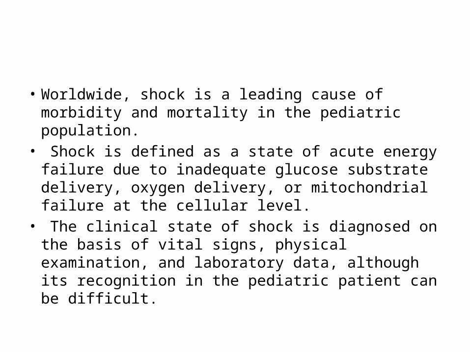
• Worldwide, shock is a leading cause of morbidity and mortality in the pediatric population.
• Shock is defined as a state of acute energy failure due to inadequate glucose substrate delivery, oxygen delivery, or mitochondrial failure at the cellular level.
• The clinical state of shock is diagnosed on the basis of vital signs, physical examination, and laboratory data, although its recognition in the pediatric patient can be difficult.

• Delay in recognizing and quickly treating a state of shock results in anaerobic metabolism, tissue acidosis, and a progression from a compensated reversible state to an irreversible state of cellular and organ damage.
• Morbidity from shock may be widespread and can include CNS failure, respiratory failure (ie, from muscle fatigue or ARDS], renal failure, hepatic dysfunction, gastrointestinal ischemia, DIC, metabolic derangements, and ultimately death.

• Shock is the most reversible causes of death in children.
• An acute , complex state of circulatory dysfunction that result in failure to deliver sufficient amount of O2 and nutrient to meet tissue metabolic demands
• Therefore, basically DO2< VO2• If prolonged and left untreated- can lead to
multiple organ failure and eventually death.

• Oxygen delivery + cardiac output x atrial oxygen content ( DO2+ COxCaO2)
• Cardiac output + HR x CO

• Shock is a physiologic state characterized by systemic reduction in tissues perfusion, resulting in decreased tissues oxygen delivery.

• It is a condition in which circulation fails to meet the metabolic need of the tissue and at the same time fails to remove the metabolic waste products
• Inadequate tissue perfusion to meet demands usually result of inadequate blood flow and or oxygen delivery
• Inadequate peripheral perfusion leading to failure of tissue oxygenation leads to anaerobic metabolism


Compensatory Mechanism
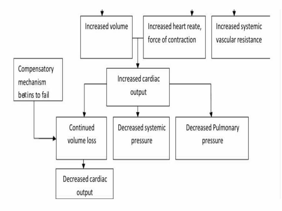
SNS-adrenal response- SNS- Neurohormonal response stimulated by baroreceptors • Increased heart rate• Increased contractibility• Vasoconstriction• Increased preload

• SNS- hormonal: renin angiotensin system• Decreased renal perfusion• Release renin Angiotensin I• Angiotensin II Potent vasoconstriction and
releases aldosterone adrenal cortex• Water sodium retention( increased
intravascular volume)

• SNS-Hormonal :antidiuretic hormone- Osmoreceptors in hypothalamus stimulated - ADH released by pituitary- Vasopressor effects to increase BP- Acts on renal tubules to retain water

• SNS- Hormonal: adrenal cortex- Anterior pituitary releases ACTH- Stimulates adrenals to release glucocorticoids- Blood sugar increases to meet increased metabolic needs

Failure of compensatory responses
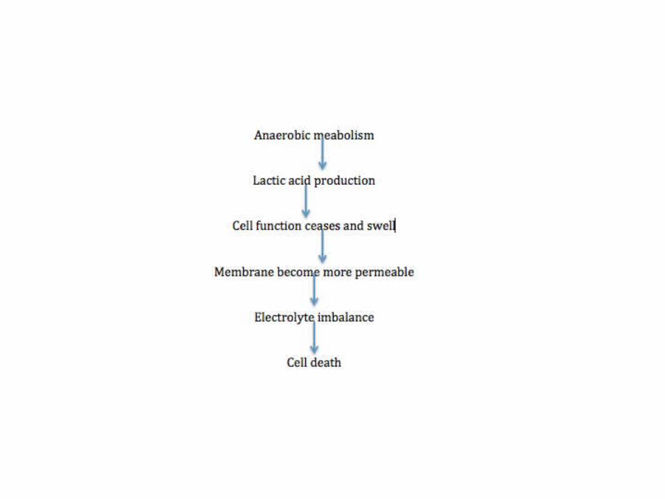
• Decreased blood flow to the tissues causes cellular hypoxia
• Anaerobic metabolism begins• Cell swelling, mitochondrial disruption, and
eventual cell death• If low perfusion persist: Death

Stages of shock
• Initial stage – tissues are under perfused, decreased cardiac output, increased anaerobic metabolism, lactic acid is building
• Compensatory stage- reversible. SNS activated by low output, attempting to compensate for the decrease tissue perfusion
• Progressive stage- falling compensatory mechanism profound vasoconstriction from SNS ischemic– lactic acid production is high
metabolic acidosis

Clinical presentation( Generalized shock)
- Vital sign • hypotension• Tachycardia• Tachypnea - Mental StatusIrritability, restless, LOC, unresponsiveness- Decreased urine output

Compensated
• Confusion• Tachycardia• Normal or mild tachypnea• > CRT• Urine out adequate• BP normal

Uncompensated
• Drowsiness• Marked tachycardia• Tachypnea and acidosis• Very slow CFT• Oliguria/Anuria• Hypotension

Irreversible
• Child is unresponsive• Bradycardia• Apnea• Cold cyanotic skin• Anuria• Un-Recordable BP

• Hypotension formula Ages- 1-10 years is defined as <70 mm of hg +(age in years x2)mm of hg

Shock syndrome
• Hypovolemic shock- Blood volume problem• Cardiogenic shock- Blood pump problems• Distributive shock- Blood vessels problem

Hypovolemic shock
• Loss of circulating volume “empty tank” decrease tissue perfusio general shock response
• Etiology - Internal / external l fluid loss- Intracellular/ extracellular compartments• Most common causeHemorrhage and dehydration

External loss
• Fluid loss: dehydrationNausea and vomiting, diarrhoea, massive diuresis, extensive burns• Blood lossTrauma visible or invisible bleeding

Internal loss
• Loss of intravascular integrity• Increased capillary membrane permeability• Decreased colloidal osmotic pressure( third
spacing)


Presentation
• Tachycardia and tachypnea• Weak, thready pulses• Hypotension• Skin cool and clammy• Mental status changes• Decreased urine output dark and
concentrated

Treatment
• Main goal- restore circulating volume and tissue perfusion, correct the cause
1. Assess airway2. Administer oxygen3. Control bleeding and balance 4. Establish IV assess5. Fluid boluses ( max-3) isotonic fluid 6. In case of shock refractory to fluids, start
inotrope(dopamine)

Cardiogenic shock
• The impaired ability of the heart to pump blood
• Pump failure of the right or left ventricle• Most common causes of MI

• When heart Is unable to contract and pump blood efficiently due to inadequate supply of O2 and nutrient to the heart
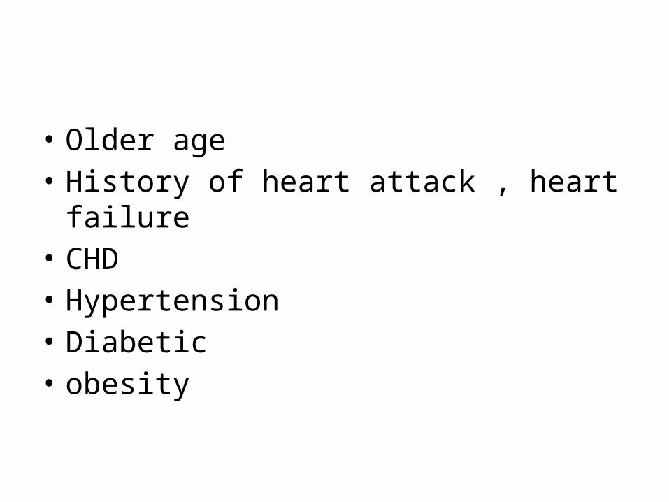
• Older age• History of heart attack , heart failure• CHD• Hypertension• Diabetic• obesity
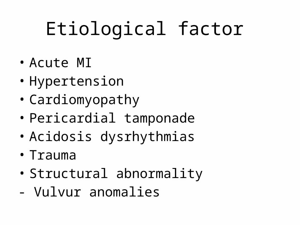
Etiological factor
• Acute MI• Hypertension• Cardiomyopathy• Pericardial tamponade• Acidosis dysrhythmias• Trauma• Structural abnormality- Vulvur anomalies


Clinical manifestation
angina pectoris DysrhythmiasDiminished heart soundHypotensionDecreased cardiac outputSOBWeak, thready pulsedecreased urine outputCold clammy skin
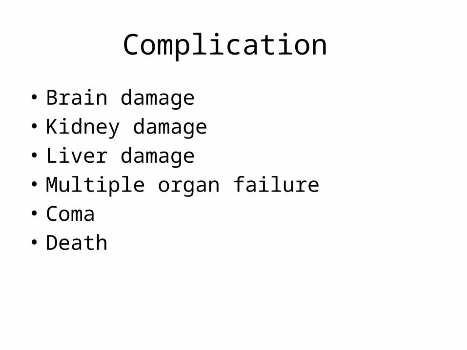
Complication
• Brain damage• Kidney damage• Liver damage• Multiple organ failure• Coma• Death

Management
• Correction of the underlying cause is important to prevent
- Fail of the compensatory mechanism- Reduces effectiveness of intervention
Correction of - Dysrhythmias- Acidosis, electrolyte imbalance:-

• Initiation of 1st line treatment - oxygenation-(2-6 lit)- Hemodynamic monitoring - Fluid balance- Pain control

• Pharmacological management - Dobutamine- Dopamine- Vasoactive medicationEpinephrineNor-epinephrinevasopressin

• Surgical management CBAG( coronary artery bypass graft)Valve replacement
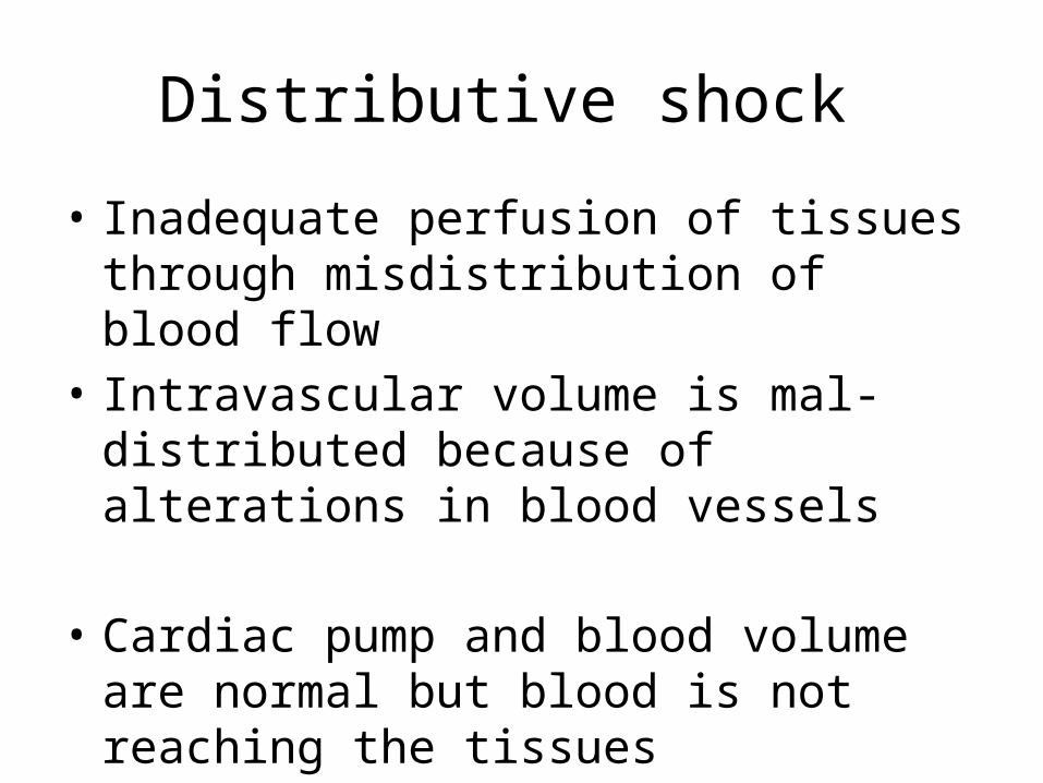
Distributive shock
• Inadequate perfusion of tissues through misdistribution of blood flow
• Intravascular volume is mal-distributed because of alterations in blood vessels
• Cardiac pump and blood volume are normal but blood is not reaching the tissues

• Etiologies - Septic shock (most common)- Anaphylactic shock- Neurogenic shock

Anaphylactic shock
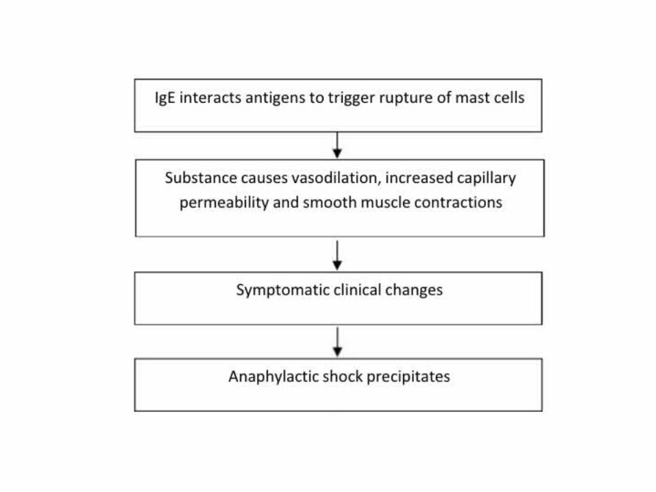
• A type of distributive shock that result from wide spread systemic allergic reaction to an antigen
• The hypersensitive reaction is life threatening

• Anaphylaxis: reaction sudden life threatening because the process immunologic of allergen- antibody reaction
• Anaphylactic reaction causing physical the same symptoms but caused no immunological reaction

Stages of anaphylactic shock
• Changes in mast cell towards stimuli• Activation of cell wall enzyme• Meditators release • Functional pathophysiology response• Inflammation and release of secondary
meditators

Etiology
1) Associated with IgE2) Non IgE3) Causes of anaphylatoid - Drugs like NSAID, antibiotics, alkaloids, food additives


Clinical features
• Skin- itching erythema, urticaria• Respiratory- sneezing runny nose, wheezing,
swollen larynx. Tightness, hoarseness, stridor, cyanosis
• Digestive- nausea vomiting diarrhoea, abd pain• Eye- itching, tears• Cardiovascular- collapse fainting, hypotension
pale, cold, tachycardia

Management
• Primary treatment -Adrenaline 1:1000 with dose of 0.001ml/kg maximum 0.3 ml SC-Can be repeated 3 times -head extended , ventilated position-O2 2-3 lit

• Place patient in shock position• Pulmonic resuscitation • Oro-pharyngeal airway• E insertion• Tracheostomy• Cardiac compression

Complementary treatment
• Intended for complication- seizures- diazepam, phenobarbital, midazolam- Bronchial spasm- Aminophylline 7mg with 10-
20 ml of 0.9& NaCl followed 9mg/kg/24 hours( divided into 3 dose)
- B-2 agonist: ventolin nebulizer

Additional
• Antihistamine• H-2 receptor antagonist• Corticosteroids

Septic Shock

• Septicemia is a condition when there is prolonged presence of bacteria in the blood accompanied by systemic reaction
• SIRS,I s a syndrome characterized by presence of two or more of the following clinical criteria
- Temperature increased or decreased- Tachycardia- Respiratory rate >20b/m or PaCO2 <32 mm of hg- Increased or decreased WBC

• Result from moderate to severe sepsis or tissues damage. It is considered as part of a spectrum and a progression of SIRS
• Sepsis: SIRS with a clearly established focus of infection
• Septic Shock: refers to severe sepsis which is not responsive to intravenous fluid infusion for resuscitation and requires inotropic or vasopressor agent to maintain SBP

• Multiple organ dysfunction (MODS)- altered function of more than one organ system in an actually ill patient requiring medical intervention homeostasis.
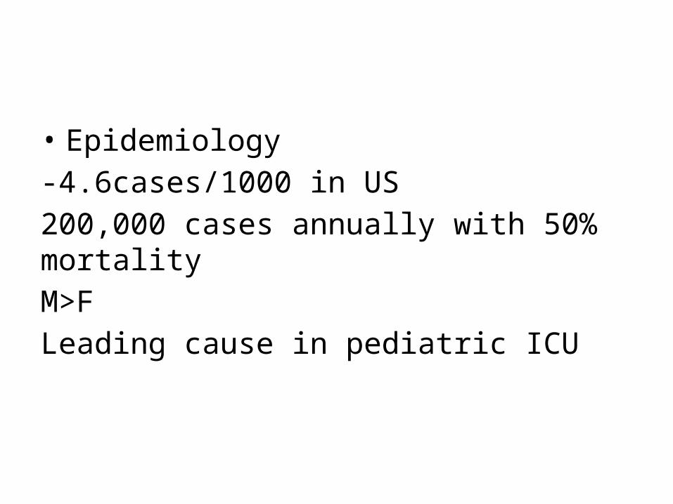
• Epidemiology-4.6cases/1000 in US200,000 cases annually with 50% mortalityM>FLeading cause in pediatric ICU

• Bacteria: gram negative nearly 2/3rd, gram positive 1/3rd
• E.coli is commonest• Gram negative

Source
Endogenous-SkinUrinaryRespiratoryBowel Exogenous-surgical interventionDrapesImaging machinesstaff
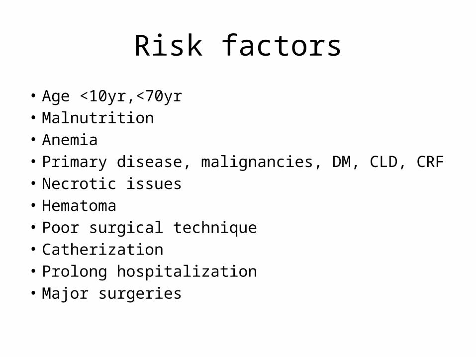
Risk factors• Age <10yr,<70yr• Malnutrition• Anemia• Primary disease, malignancies, DM, CLD, CRF• Necrotic issues• Hematoma• Poor surgical technique• Catherization• Prolong hospitalization• Major surgeries

Pathogenesis
• Microorganism or product of tissue damage stimulate production of pro-inflammatory cytokines, which in turn stimulate production of secondary mediators of inflammation
• The production of the pro-inflammatory cytokines is regulated to limit damage
• However poorly control sepsis or extensive tissue damage, there is excessive inflammatory response which is poorly regulated


• Hypovolemic state, cardiac depression, interstitial loss, AV shunt all causes cellular hypoxia and ultimate septic shock


Clinical features
• Early stage- (compensated/warm shock) or condition not associated with hypovolemia
- febrile(38-41)- Shivering and malaise- Warm dry flushed skin- Hyperventilation- Rapid bounding pulse- Wide pulse pressure

Decompensated/cold shock• Altered sensorium• Cold clammy skin• Feeble pulse• Hypothermia• Oliguria• Jaundice• UGI• DIC

• Localizing infection - A good systemic examination is done to detect any focus of infection.

Diagnosis
• Blood/urine/sputum culture• CBC• BUN and Creatinine• PT and PTT• ECG• Serum lactate dehydrogenase level• Urinalysis• ABG

Treatment
• Septic shock is a medical emergency that requires prompt and sufficient resuscitation
• Treatment should be carried out in ICU settingAimsTo improve hemodynamic stateRestore tissues perfusionEliminate toxin from body
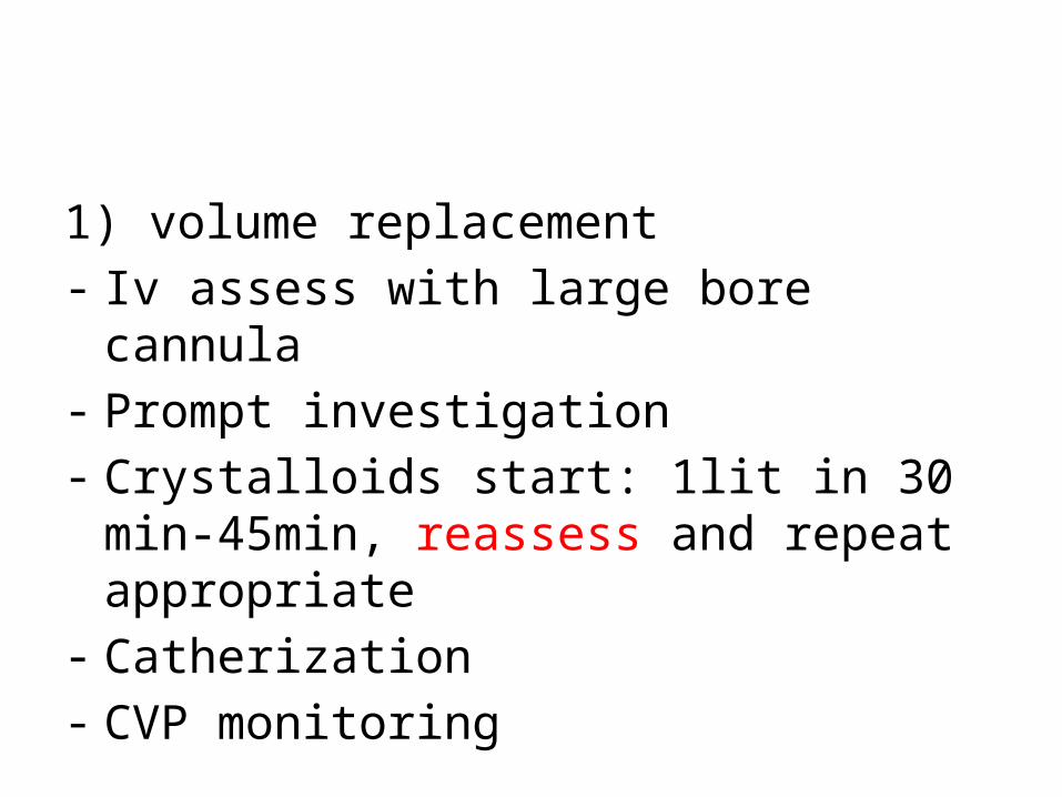
1) volume replacement - Iv assess with large bore cannula- Prompt investigation- Crystalloids start: 1lit in 30 min-45min,
reassess and repeat appropriate- Catherization- CVP monitoring

• Vasopressor 2) Oxygen3)Antibiotic 4) Steroid5)NSAID6)Free radical scavengers 7) Glycemic control8) Naloxone9) Coagulation 10) Surgery
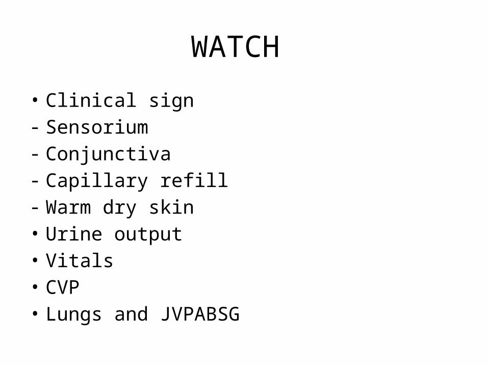
WATCH
• Clinical sign- Sensorium- Conjunctiva - Capillary refill- Warm dry skin• Urine output• Vitals • CVP• Lungs and JVPABSG

Complication
• ARDS• ARF• DIC• Encephalopathy• Liver failure• Coma • Death
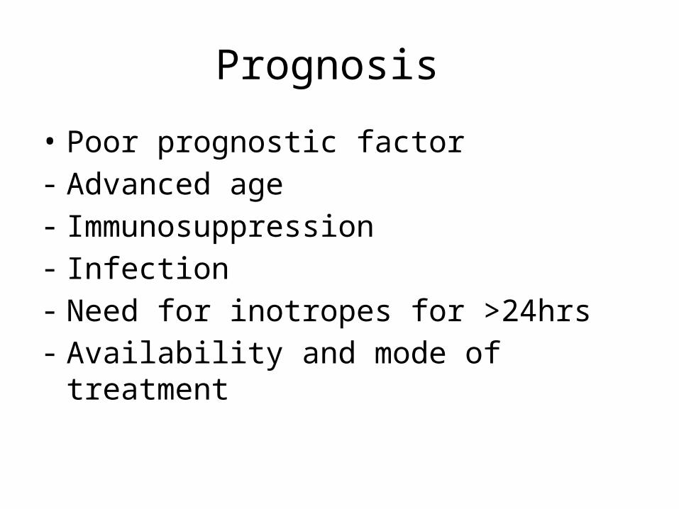
Prognosis
• Poor prognostic factor- Advanced age- Immunosuppression- Infection - Need for inotropes for >24hrs- Availability and mode of treatment
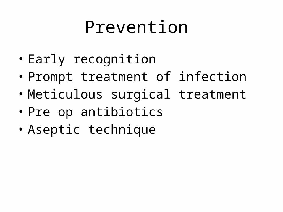
Prevention
• Early recognition• Prompt treatment of infection• Meticulous surgical treatment• Pre op antibiotics• Aseptic technique
Treatment
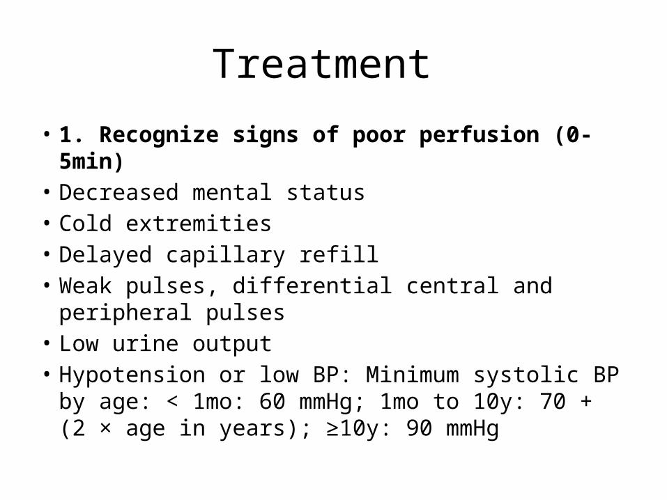
• 1. Recognize signs of poor perfusion (0-5min)• Decreased mental status• Cold extremities• Delayed capillary refill• Weak pulses, differential central and peripheral pulses• Low urine output• Hypotension or low BP: Minimum systolic BP by age: <
1mo: 60 mmHg; 1mo to 10y: 70 + (2 × age in years); ≥10y: 90 mmHg
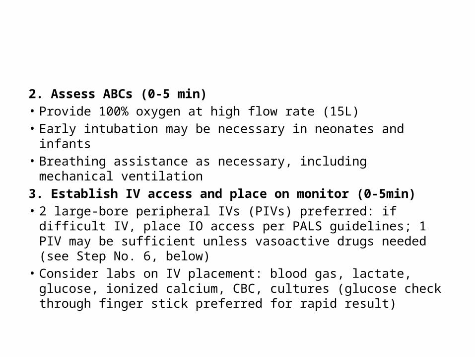
2. Assess ABCs (0-5 min)• Provide 100% oxygen at high flow rate (15L)• Early intubation may be necessary in neonates and infants• Breathing assistance as necessary, including mechanical ventilation3. Establish IV access and place on monitor (0-5min)• 2 large-bore peripheral IVs (PIVs) preferred: if difficult IV, place IO
access per PALS guidelines; 1 PIV may be sufficient unless vasoactive drugs needed (see Step No. 6, below)
• Consider labs on IV placement: blood gas, lactate, glucose, ionized calcium, CBC, cultures (glucose check through finger stick preferred for rapid result)
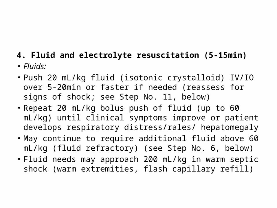
4. Fluid and electrolyte resuscitation (5-15min)• Fluids:• Push 20 mL/kg fluid (isotonic crystalloid) IV/IO over 5-20min or
faster if needed (reassess for signs of shock; see Step No. 11, below)
• Repeat 20 mL/kg bolus push of fluid (up to 60 mL/kg) until clinical symptoms improve or patient develops respiratory distress/rales/ hepatomegaly
• May continue to require additional fluid above 60 mL/kg (fluid refractory) (see Step No. 6, below)
• Fluid needs may approach 200 mL/kg in warm septic shock (warm extremities, flash capillary refill)
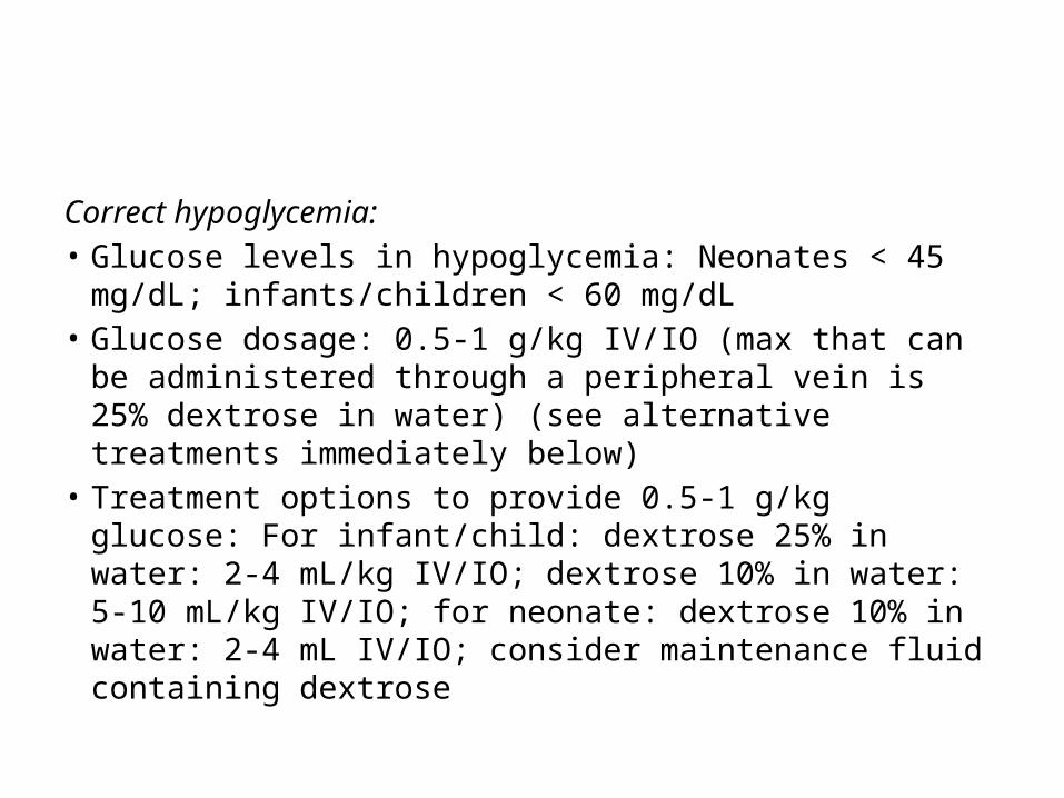
Correct hypoglycemia:• Glucose levels in hypoglycemia: Neonates < 45 mg/dL;
infants/children < 60 mg/dL• Glucose dosage: 0.5-1 g/kg IV/IO (max that can be
administered through a peripheral vein is 25% dextrose in water) (see alternative treatments immediately below)
• Treatment options to provide 0.5-1 g/kg glucose: For infant/child: dextrose 25% in water: 2-4 mL/kg IV/IO; dextrose 10% in water: 5-10 mL/kg IV/IO; for neonate: dextrose 10% in water: 2-4 mL IV/IO; consider maintenance fluid containing dextrose

Correct hypocalcemia for low ionized calcium:• Calcium gluconate 100 mg/kg IV/IO (max 2g) PRN• Calcium chloride 20 mg/kg IV/IO PRN ( Note: central line
administration preferred over 60min in nonarrest situation)5. Infection control (5-60min)• Immediate considerations:• Administer antibiotics immediately after cultures obtained
(blood, urine, +/- CSF/ sputum)• Do not delay antibiotics because of delay in obtaining
cultures; initial antibiotics should be given within 1h

Neonates >2kg:• Ampicillin plus gentamicin: Ampicillin for 0-7d: 50
mg/kg IV/IM/IO q8h; ampicillin >7d: 50 mg/kg IV/IM/IO q6h plus gentamicin (dosing institution dependent): 4mg/kg IV/IO/IM q24h (alternative for 0-7d: 2.5 mg/kg IV/IO/IM q12h; alternative for >7d: 2.5 mg/kg IV/IO/IM q8h) or
• Ampicillin plus cefotaxime: Ampicillin for 0-7d: 50 mg/kg IV/IM/IO q8h; ampicillin >7d: 50 mg/kg IV/IM/IO q6h plus cefotaxime 50 mg/kg IV/IO q8h

Infants (>1mo) and children:• Ceftriaxone 75 mg/kg (max 2g) IV/IO/IM
q24h plus vancomycin 15mg/kg (max 1g) IV/IO q8h
Immunosuppressed patients:• Vancomycin 15 mg/kg IV/IO (max 1 g/dose)
q8h plus cefepime 50 mg/kg IV/IO (max 2g/dose) q8h; consider antifungal therapy

6. Fluid-refractory shock (persisting after 60 mL/kg fluid) (15-60 min)• Continue fluid resuscitation and initiate vasopressor therapy titrated to
correct hypotension/poor perfusion• Central line placement and arterial monitoring if not already established;
vasopressors should not be delayed for line placements• Normotensive shock (impaired perfusion but normal blood pressure):
Dopamine 2-20 mcg/kg/min IV/IO, titrate to desired effect; if continued poor perfusion, consider dobutamine infusion 2-20 mcg/kg/min IV/IO, titrate to desired effect (may cause hypotension, tachycardia)
• Warm shock (warm extremities, flash capillary refill): Norepinephrine 0.1-2 mcg/kg/min IV/IO infusion, titrate to desired effect
• Cold shock (cool extremities, delayed capillary refill): Epinephrine 0.1-1 mcg/kg/min IV/IO infusion, titrate to desired effect
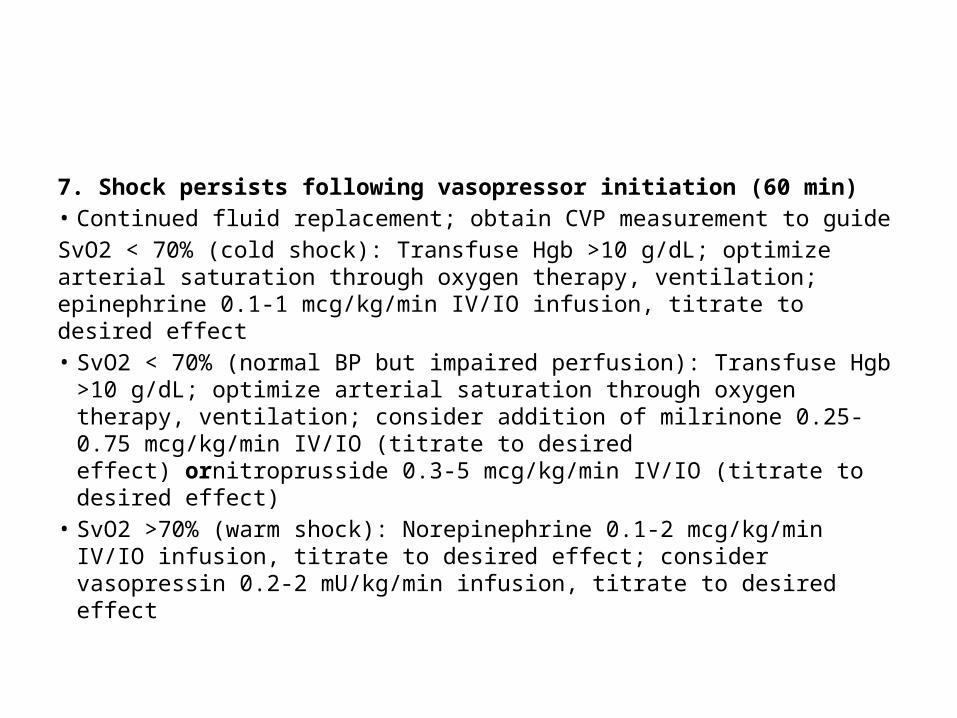
7. Shock persists following vasopressor initiation (60 min)• Continued fluid replacement; obtain CVP measurement to guideSvO2 < 70% (cold shock): Transfuse Hgb >10 g/dL; optimize arterial saturation through oxygen therapy, ventilation; epinephrine 0.1-1 mcg/kg/min IV/IO infusion, titrate to desired effect• SvO2 < 70% (normal BP but impaired perfusion): Transfuse Hgb >10
g/dL; optimize arterial saturation through oxygen therapy, ventilation; consider addition of milrinone 0.25-0.75 mcg/kg/min IV/IO (titrate to desired effect) ornitroprusside 0.3-5 mcg/kg/min IV/IO (titrate to desired effect)
• SvO2 >70% (warm shock): Norepinephrine 0.1-2 mcg/kg/min IV/IO infusion, titrate to desired effect; consider vasopressin 0.2-2 mU/kg/min infusion, titrate to desired effect

8. Fluid refractory and vasopressor-dependent shock) (60 min)• Consider adrenal insufficiency• Hydrocortisone 2 mg/kg (max 100mg) IV/IO bolus; obtain
baseline cortisol level; if unsure, consider ACTH stimulation test; duration depends on response, laboratory evaluation
9. Continued shock• Consider cardiac output measurement to direct further
therapy• Consider extracorporeal membrane oxygenation (ECMO) 10. Supplemental therapies
Neurogenic Shock

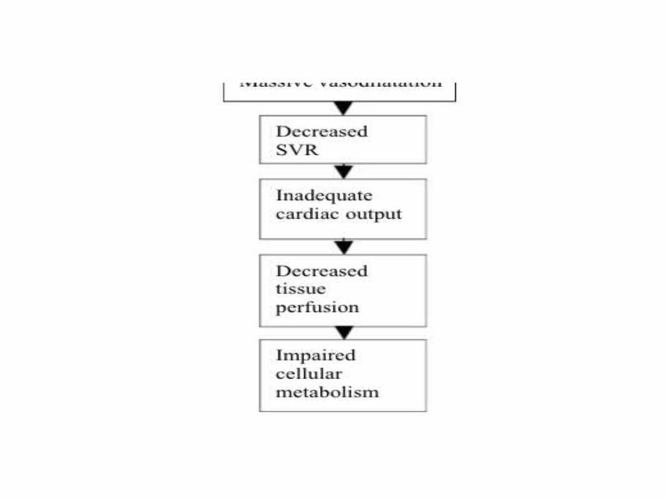
• Neurogenic shock is a medical condition which occurs as a result of disturbance in the sympathetic outflow causing loss of vagal tone
• Experiences neurogenic shock after injury to the spinal cord and when there is disruption in the blood circulation throughout the body due to injury/ illness.
•

• It is a serious and life-threatening condition, which requires prompt medical attention without any delay. If the treatment is delayed, then it causes irreversible tissue damage and even death.
• Out of the different types of the shocks, neurogenic shock is the most difficult to manage, mainly because of the irreversible damage to the tissues.
• Neurogenic shock mainly affects the spinal cord; the function of which is transmitting neural signals from the brain to the entire body and back.


• Most common causes is spinal injury above T 6.
• Most rare form of shock

sign
• Hypothermia, hypotension, decreased CO, bradycardia, flaccid paralysis

Management
• Main aim is to prevent complication and maintain perfusion
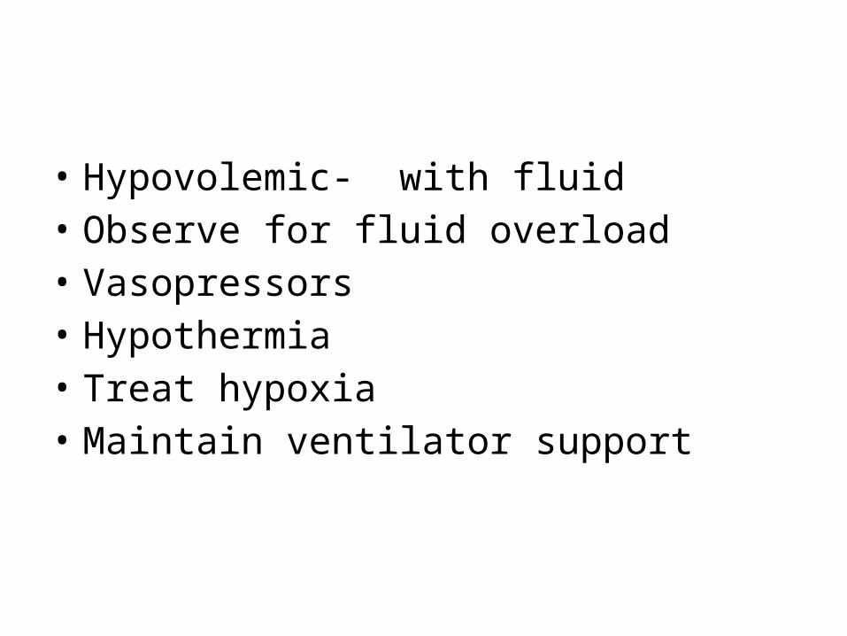
• Hypovolemic- with fluid• Observe for fluid overload• Vasopressors• Hypothermia• Treat hypoxia• Maintain ventilator support

• Observe for bradycardia- major Dysarrthemia• Observe for DVT- Venous pooling in
extremities make patients high risk• Use prevention modalities

• Alpha agonist to augment tone if perfusion still in adequate
• Dopamine(>10 mcg/kg per min)• Ephedrie(12.5-25mg iv every 3-4 hour)• Treat bradycardia with atropine 0.5-1mg doses
to maximum 3 mg• May need transcutaneous or transv svenous
pacing temporarily