TTRAP Is a Novel Component of the Non-Canonical TRAF6 …
12
TTRAP Is a Novel Component of the Non-Canonical TRAF6-TAK1 TGF-b Signaling Pathway Gyo ¨ rgy Va ´ rady, Bala ´ zs Sarkadi, Ka ´ roly Fa ´ tyol* Membrane Research Group, Hungarian Academy of Sciences, Budapest, Hungary Abstract Transforming growth factor-b (TGF-b) principally relays its effects through the Smad pathway however, accumulating evidence indicate that alternative signaling routes are also employed by this pleiotropic cytokine. For instance recently, we have demonstrated that ligand occupied TGF-b receptors can directly trigger the TRAF6-TAK1 signaling module, resulting in MAP kinase activation. Here we report identification of the adaptor molecule T TRAP as a novel component of this non- canonical TGF-b pathway. We show that the protein associates with TGF-b receptors and components of the TRAF6-TAK1 signaling module, resulting in differential regulation of TGF-b activated p38 and NF-kB responses. Modulation of cellular TTRAP level affects cell viability in the presence of TGF-b, suggesting that the protein is an important component of the TGF-b induced apoptotic process. Citation: Va ´rady G, Sarkadi B, Fa ´tyol K (2011) T TRAP Is a Novel Component of the Non-Canonical TRAF6-TAK1 TGF-b Signaling Pathway. PLoS ONE 6(9): e25548. doi:10.1371/journal.pone.0025548 Editor: Arun Rishi, Wayne State University, United States of America Received July 5, 2011; Accepted September 6, 2011; Published September 27, 2011 Copyright: ß 2011 Va ´rady et al. This is an open-access article distributed under the terms of the Creative Commons Attribution License, which permits unrestricted use, distribution, and reproduction in any medium, provided the original author and source are credited. Funding: This research was supported by a Hungarian Science Foundation (OTKA)/Norwegian Financial Mechanism joint grant (NNF78805). KF was supported by a Ja ´nos Bolyai fellowship. The funders had no role in study design, data collection and analysis, decision to publish, or preparation of the manuscript. Competing Interests: The authors have declared that no competing interests exist. * E-mail: [email protected] Introduction TGF-b has pervasive and diverse effects on cell physiology as well as it acts as a potent anticancer agent that prohibits uncontrolled cell proliferation [1–3]. The most accepted model for the signaling mechanism of TGF-b family cytokines portrays a relatively simple pathway, in which ligand binding to a membrane bound receptor complex induces a conformational change, resulting in phosphorylation and activation of the type I receptor (TbRI) by the type II receptor kinase (TbRII). Through its own kinase activity, TbRI then phosphorylates the appropriate receptor Smads (R-Smads, Smad2/3). Once phosphorylated, R- Smads can form complexes with the common Smad (Smad4), whereupon they translocate to the nucleus to initiate specific transcriptional programs [4,5]. It is becoming increasingly apparent however, that the picture depicted above is significantly more complex. TGF-b can mobilize several intracellular signal transducers in Smad-independent manner as well [6–8]. These non-canonical, non-Smad pathways are also activated directly by ligand-occupied receptors to reinforce, attenuate or otherwise modulate downstream cellular responses. The non-Smad path- ways include various branches of MAP kinase pathways, Rho-like GTPase signaling pathways, the phosphatidylinositol-3-kinase/ AKT pathway and more. Such alternative signal transducers often regulate the Smad pathway itself and represent extensive opportunities for crosstalk with other signaling routes, contributing to the surprising diversity of TGF-b responses. Perhaps one of the most important non-Smad pathways is the p38/JNK MAP kinase cascade [9–12]. This signaling route functions in conjunction with the Smad pathway to regulate such cellular responses as apoptosis and eptithelial-to-mesenchymal transition (EMT). Despite their obvious biological significance however, we still have serious caveats in understanding the mechanisms by which TGF-b governs them. The need to fill out these gaps is further underscored by several recent observations, suggesting that imbalances arising between the Smad-pathway and the p38/JNK MAPK signaling branches during tumorigenesis may contribute to the conversion of TGF-b from a suppressor to a promoter of cancer growth [13–19]. Previous genetic studies placed TGF-b-activated kinase 1 (TAK1) in the TGF-b mediated p38/JNK activation pathway however, the link between TAK1 and the activated receptor complex had been lacking [20–22]. Recently, we and others have demonstrated that the E3 ubiquitin ligase, TRAF6 is one of the missing pieces [23,24]. The molecule physically interacts with the TGF-b receptor complex and is required for Smad-independent activation of the JNK and p38 kinases. TGF-b promotes association between TRAF6 and TAK1, resulting in lysine 63- linked (K63) ubiquitylation and subsequent activation of TAK1. Interestingly, the TRAF6-TAK1 signaling module is also em- ployed by a number of different signaling routes such as those emanating from the IL-1b receptor, Toll-like receptors, T-cell receptor etc. and cellular processes triggered by DNA damage and osmotic stress [25,26]. Selective activation of TAK1 by the numerous divergent stimuli is believed to be achieved at least in part by the use of adaptor proteins indigenous to a given signaling route and/or employment of unique combinations of more common ones. Regardless, the identification of these adaptor proteins and the elucidation of their complex interactions are essential. Here we describe one such adaptor molecule, TTRAP (TRAF and TNF receptor associated protein) [27] that may contribute to the specific activation of TAK1 in response to TGF-b. TTRAP was originally reported to interact with members of the PLoS ONE | www.plosone.org 1 September 2011 | Volume 6 | Issue 9 | e25548 brought to you by CORE View metadata, citation and similar papers at core.ac.uk provided by PubMed Central
Transcript of TTRAP Is a Novel Component of the Non-Canonical TRAF6 …

TTRAP Is a Novel Component of the Non-CanonicalTRAF6-TAK1 TGF-b Signaling PathwayGyorgy Varady, Balazs Sarkadi, Karoly Fatyol*
Membrane Research Group, Hungarian Academy of Sciences, Budapest, Hungary
Abstract
Transforming growth factor-b (TGF-b) principally relays its effects through the Smad pathway however, accumulatingevidence indicate that alternative signaling routes are also employed by this pleiotropic cytokine. For instance recently, wehave demonstrated that ligand occupied TGF-b receptors can directly trigger the TRAF6-TAK1 signaling module, resulting inMAP kinase activation. Here we report identification of the adaptor molecule T TRAP as a novel component of this non-canonical TGF-b pathway. We show that the protein associates with TGF-b receptors and components of the TRAF6-TAK1signaling module, resulting in differential regulation of TGF-b activated p38 and NF-kB responses. Modulation of cellularTTRAP level affects cell viability in the presence of TGF-b, suggesting that the protein is an important component of theTGF-b induced apoptotic process.
Citation: Varady G, Sarkadi B, Fatyol K (2011) T TRAP Is a Novel Component of the Non-Canonical TRAF6-TAK1 TGF-b Signaling Pathway. PLoS ONE 6(9): e25548.doi:10.1371/journal.pone.0025548
Editor: Arun Rishi, Wayne State University, United States of America
Received July 5, 2011; Accepted September 6, 2011; Published September 27, 2011
Copyright: � 2011 Varady et al. This is an open-access article distributed under the terms of the Creative Commons Attribution License, which permitsunrestricted use, distribution, and reproduction in any medium, provided the original author and source are credited.
Funding: This research was supported by a Hungarian Science Foundation (OTKA)/Norwegian Financial Mechanism joint grant (NNF78805). KF was supported bya Janos Bolyai fellowship. The funders had no role in study design, data collection and analysis, decision to publish, or preparation of the manuscript.
Competing Interests: The authors have declared that no competing interests exist.
* E-mail: [email protected]
Introduction
TGF-b has pervasive and diverse effects on cell physiology as
well as it acts as a potent anticancer agent that prohibits
uncontrolled cell proliferation [1–3]. The most accepted model
for the signaling mechanism of TGF-b family cytokines portrays a
relatively simple pathway, in which ligand binding to a membrane
bound receptor complex induces a conformational change,
resulting in phosphorylation and activation of the type I receptor
(TbRI) by the type II receptor kinase (TbRII). Through its own
kinase activity, TbRI then phosphorylates the appropriate
receptor Smads (R-Smads, Smad2/3). Once phosphorylated, R-
Smads can form complexes with the common Smad (Smad4),
whereupon they translocate to the nucleus to initiate specific
transcriptional programs [4,5]. It is becoming increasingly
apparent however, that the picture depicted above is significantly
more complex. TGF-b can mobilize several intracellular signal
transducers in Smad-independent manner as well [6–8]. These
non-canonical, non-Smad pathways are also activated directly by
ligand-occupied receptors to reinforce, attenuate or otherwise
modulate downstream cellular responses. The non-Smad path-
ways include various branches of MAP kinase pathways, Rho-like
GTPase signaling pathways, the phosphatidylinositol-3-kinase/
AKT pathway and more. Such alternative signal transducers often
regulate the Smad pathway itself and represent extensive
opportunities for crosstalk with other signaling routes, contributing
to the surprising diversity of TGF-b responses.
Perhaps one of the most important non-Smad pathways is the
p38/JNK MAP kinase cascade [9–12]. This signaling route
functions in conjunction with the Smad pathway to regulate such
cellular responses as apoptosis and eptithelial-to-mesenchymal
transition (EMT). Despite their obvious biological significance
however, we still have serious caveats in understanding the
mechanisms by which TGF-b governs them. The need to fill out
these gaps is further underscored by several recent observations,
suggesting that imbalances arising between the Smad-pathway and
the p38/JNK MAPK signaling branches during tumorigenesis
may contribute to the conversion of TGF-b from a suppressor to a
promoter of cancer growth [13–19].
Previous genetic studies placed TGF-b-activated kinase 1
(TAK1) in the TGF-b mediated p38/JNK activation pathway
however, the link between TAK1 and the activated receptor
complex had been lacking [20–22]. Recently, we and others have
demonstrated that the E3 ubiquitin ligase, TRAF6 is one of the
missing pieces [23,24]. The molecule physically interacts with the
TGF-b receptor complex and is required for Smad-independent
activation of the JNK and p38 kinases. TGF-b promotes
association between TRAF6 and TAK1, resulting in lysine 63-
linked (K63) ubiquitylation and subsequent activation of TAK1.
Interestingly, the TRAF6-TAK1 signaling module is also em-
ployed by a number of different signaling routes such as those
emanating from the IL-1b receptor, Toll-like receptors, T-cell
receptor etc. and cellular processes triggered by DNA damage and
osmotic stress [25,26]. Selective activation of TAK1 by the
numerous divergent stimuli is believed to be achieved at least in
part by the use of adaptor proteins indigenous to a given signaling
route and/or employment of unique combinations of more
common ones. Regardless, the identification of these adaptor
proteins and the elucidation of their complex interactions are
essential.
Here we describe one such adaptor molecule, TTRAP (TRAF
and TNF receptor associated protein) [27] that may contribute
to the specific activation of TAK1 in response to TGF-b.
TTRAP was originally reported to interact with members of the
PLoS ONE | www.plosone.org 1 September 2011 | Volume 6 | Issue 9 | e25548
brought to you by COREView metadata, citation and similar papers at core.ac.uk
provided by PubMed Central

TNF receptor family and TRAF adaptor proteins [27].
Subsequent studies also implicated the molecule in various
nuclear functions, including transcription and DNA repair [28–
31]. Notwithstanding, a recent work convincingly demonstrated
a role for TTRAP in signal transduction [32]. An antisense
screen in zebrafish indentified the protein as a component of the
Nodal/activin signaling pathway and an important regulator of
embryogenesis. Here we show that TTRAP is involved in TGF-
b signaling in mammalian cells as well. Specifically, the protein
associates with components of the TGF-b receptor-TRAF6-
TAK1 signaling module and promotes their ubiquitylation
dependent complex formation. We also demonstrate that
TTRAP, by modulating the activities of non-canonical TGF-binduced signaling routes, plays an important role in TGF-belicited apoptosis.
Materials and Methods
Cell culture, transfection, reporter assaysHEK293T, Phoenix-E, NMuMG and AML12 cells were
purchased from the American Type Culture Collection and
maintained as recommended by the supplier. Cells were
transfected with Fugene 6 (Roche) or FugeneHD (Promega),
according to the manufacturers instructions. Reporter assays were
performed as described earlier [33].
Antibodies, shRNAs, chemicalsThe following antibodies were used in this study: phospho-
Smad2(Ser465/467), phospho-p38(Thr180,Tyr182)(D3F9), p38 and
phospho-TAK1(Thr187) were from Cell Signaling Technology;
Smad2/3(C8), TRAF6(D-10), TAK1(M-579), TTRAP/EAPII(K-
13), TTRAP/EAPII(N-18) were from Santa Cruz; Myc(9E10) and
HA(3F10) were purchased from Roche; His and FLAG(M2) were
from Sigma. Mission shRNA lentiviruses, targeting the mouse
TTRAP mRNA (TRCN0000174689, TRCN0000174799 and
TRCN0000174910) were purchased from Sigma. Recombinant
human TGF-b1 was from R&D Systems. SB431542, SB203582 and
SP600125 were obtained from Sigma. The TAK1 inhibitor, (5Z)-7-
oxozeaenol was from Calbiochem.
PlasmidsMost of the expression plasmids used here were described
earlier [23]. Full length TTRAP, TAK1 and TAB2 cDNAs were
generated by PCR and cloned into the pRK family of mammalian
expression vectors [34] using standard techniques. Retroviral
expression constructs were created in the pBabe-Puro backbone
[35]. Deletion and point mutants were generated by PCR.
Sequences of all constructs were verified by sequencing.
Immunoprecipitation, western blottingWestern blotting of proteins and immunoprecipitations (IP)
were performed as described earlier [33].
Cell viability measurementsCell viability was assessed by three different methods: 1.
Propidium iodide (PI) uptake of cells, as a measure of membrane
integrity, was determined by fluorescence activated cell sorting
(FACS). Cells were seeded at a density of 36104 cells/well in 24-
well plates and treated as indicated. Subsequently, cells were
collected by trypsinization, washed with BSA-PBS (PBS
containing 0.5% BSA) and resuspended in BSA-PBS containing
2 mg/ml PI. The cell suspension was incubated at room
temperature for 10 minutes and then measured by FACS. FACS
profiles were analyzed by the WinMDI software. 2. Apoptosis
was followed by staining of cells with Alexa Fluor 647 labeled
annexin V (Invitrogene) according to the manufacturer’s
instructions and analyzed by FACS. 3. Cell survival was also
determined by the MTT (3-[4,5-dimethylthiazol-2-yl]-2,5-diphe-
nyltetrazolium bromide) assay. Cells were seeded at a density of
56103 cells/well in 96-well plates. The following day treatments
were commenced as indicated. At the end of the treatments the
medium was replaced with fresh medium containing 1.2 mM
MTT and the cells were incubated at 37uC in 5% CO2 for
4 hours. Subsequently, the cells were washed in the plates with
PBS and the formazan crystals were solubilized in isopropanol,
containing 0.1 M HCL. Optical densities at 570 nm were
measured in a plate reader.
Indirect immunofluorescenceFor indirect immunofluorescence, cells were grown on cover-
slips and fixed in cold methanol for 7 minutes and then briefly
permeabilized in cold acetone. The antibody incubations and
washing steps were done as described [33].
Results
TTRAP associates with the TGF-b receptor complexBased on earlier results implicating TTRAP in the signaling
processes of Nodal/activin ligands, we explored the protein’s
potential involvement in TGF-b signaling in mammalian cells.
Zebrafish TTRAP has been shown to bind components of the
Nodal/activin pathway (the type I Nodal/activin receptor [Alk4],
and Smad3). Thus, as an initial approach, we tested the
association of mammalian TTRAP with elements of the TGF-bsignaling machinery, using various protein-protein interaction
techniques.
First, we wanted to analyze the interaction between endogenous
TTRAP and the TGF-b receptor complex. Unfortunately,
currently there is no commercially available TTRAP antibody
sensitive enough to carry out such studies. To circumvent this
problem, we generated an NMuMG cell population stably
expressing FLAG epitope tagged TTRAP (FLAG-TTRAP) at a
modest level. Using these cells, we were able to detect modest
binding of FLAG-TTRAP to endogenous TbRI by co-immuno-
precipitation (co-IP) (Figure 1A). Importantly, this interaction was
significantly increased upon TGF-b treatment.
Second, epitope tagged TTRAP and TGF-b receptors were
transiently co-expressed in HEK293T cells and their interac-
tions were analyzed by co-IP (Figure 1B). Under these
conditions, TTRAP associated with both TbRI and TbRII
even in the absence of TGF-b stimulation. The protein
exhibited increased affinity toward the catalytically inactive
TbRI-KR receptor mutant compared to the constitutively active
TbRI-TD form.
Third, the binding of TTRAP to TGF-b receptors was
monitored in vitro using GST pull-down. HA-tagged TTRAP
protein was synthesized in rabbit reticulocyte lysate in vitro,
while GST-tagged cytoplasmic domains of TbRI and TbRII
were produced in bacteria. In vitro TTRAP bound to the
cytoplasmic domains of both TGF-b receptors immobilized on
gluthatione beads, indicating that their interactions are direct
(Figure S1).
Fourth, we evaluated the binding of TTRAP with membrane
associated TGF-b receptor complexes. HEK293T cells were co-
transfected with TTRAP, TGF-b receptors and TRAF6 in various
combinations. Subsequently, surface proteins were affinity labeled
with [125I ]-TGF-b. Following cross-linking with disuccinimidyl
suberate (DSS) the cells were lysed and TTRAP was precipitated
Role of TTRAP in TGF-b Signaling
PLoS ONE | www.plosone.org 2 September 2011 | Volume 6 | Issue 9 | e25548

with a FLAG antibody. As shown in Figure 1C, TTRAP pulled
down [125I ]-TGF-b occupied TGF-b receptor complexes. Impor-
tantly, the relative binding affinities of TTRAP toward the various
mutant forms of TbRI detected by this technique were similar to
those seen in co-IPs. In addition, we noted that the presence of
TRAF6 strengthened the interaction between TTRAP and the
TGF-b receptor complex (see also later).
Fifth, EGFP-TTRAP and FLAG-TbRI-KR were co-expressed
in AML12 cells and their localizations were monitored by
fluorescence microscopy. TTRAP was present both in the
cytoplasm and the nucleus, consistent with previous reports
[28,29]. Significantly, a fraction of the cytoplasmic TTRAP
exhibited co-localization with TbRI in juxta-membrane foci
(Figure 1D).
Finally, the TGF-b receptor interacting domain of TTRAP was
mapped by co-IPs. Using C-terminally truncated TTRAP
molecules we showed that the region between amino acids 123
and 274 is necessary for TGF-b receptor binding (Figure 1E).
Interestingly, this region of TTRAP is part of the evolutionary
conserved exo/endo/phos domain also present in a number of
Mg2+/Mn2+ dependent phosphodiesterases [36].
In summary, the above results indicate that in analogy to the
TTRAP-Alk4 interaction observed in zebrafish, the mammalian
ortholog of TTRAP associates with TGF-b receptors. The fact
that TTRAP also binds with ligand occupied TGF-b receptor
complexes on the cell surface provides further support for the
physiological relevance of these interactions. Contrary to previous
data however, we were unable to detect direct binding of TTRAP
with Smads (Figure S2).
TTRAP associates with the TAK1 complexTTRAP was originally identified as a TRAF interacting
protein. Amongst members of the TRAF family, it exhibited the
highest affinity toward TRAF6 and practically no binding with
Figure 1. TTRAP interacts with TGF-b receptors. A) TTRAP associates with endogenous TbRI. NMuMG cells stably expressing FLAG-T TRAP weretreated with 4 ng/ml of TGF-b for one hour or left untreated. Cellular lysates were prepared and T TRAP was precipitated with FLAG affinity beads. Theprecipitated proteins and 1/30th of the input lysates were analyzed by western blotting. B) Co-IP analysis of the T TRAP-TGF-b receptor interaction. Theindicated proteins were co-expressed in HEK293T cells. Total cellular lysates were prepared and the TGF-b receptors were precipitated with a FLAGantibody. The precipitated proteins and 1/20th of the input lysates were analyzed by western blotting. C) Analysis of the binding of T TRAP to membraneassociated TGF-b receptors. To label surface receptors cells were incubate with [125I]-TGF-b, cross-linked with DSS and T TRAP was pulled down. Theprecipitated receptors were detected by autoradiography. D) EGFP-T TRAP and FLAG-TbRI-KR were co-expressed in AML12 cells and their localizationswere monitored by fluorescence microscopy. A juxta-membrane region of the cell was zoomed out at the bottom. Co-localized foci are indicated byarrowheads. The nuclei were stained by 49,6-diamidino-2-phenylindole (DAPI). E) Mapping of the TGF-b receptor binding domain of T TRAP by co-IP. Theprecipitated proteins and 1/20th of the input lysates were analyzed by western blotting using HA and FLAG antibodies.doi:10.1371/journal.pone.0025548.g001
Role of TTRAP in TGF-b Signaling
PLoS ONE | www.plosone.org 3 September 2011 | Volume 6 | Issue 9 | e25548

Role of TTRAP in TGF-b Signaling
PLoS ONE | www.plosone.org 4 September 2011 | Volume 6 | Issue 9 | e25548

TRAF2 [27]. Indeed, using co-IP, we were able to verify these
observations (Figure 2A). Given that TRAF6 plays a crucial role
in TGF-b induced p38 activation, next TTRAP’s interactions
with other components of the TGF-b receptor-p38 pathway
were examined. First, we tested whether TTRAP can interact
with TAK1. Co-IP was employed to assess the associations
between FLAG-TTRAP and HA epitope tagged TAK1
molecules (wild-type and various mutant forms) (Figure 2B).
TTRAP bound avidly to catalytically active TAK1 variants (both
the wild-type and the E39G point mutant) however, replacement
of lysine-34 - the major acceptor site for TGF-b induced K63-
linked polyubiquitylation [24] - with an arginine residue,
significantly reduced this interaction. The affinity of TTRAP
toward catalytically inactive mutants of the kinase was
diminished even further, exhibiting no significant binding to
either the ATP binding site mutant (K63W) or the activation
loop mutants (T184,187V and S192A) [37,38]. This finding
raised the possibility that TTRAP specifically binds to auto-
phosphorylated residues in the kinase. Specific inhibition of
TAK1’s catalytic activity with (5Z)-7-oxozeaenol [39] however,
did not abolish the TAK1-TTRAP association (Figure 2C). This
suggests that TTRAP recognizes some structural feature of the
kinase associated with its catalytically active form, rather than
the phosphorylated residues per se.
To analyze the interaction between TTRAP and TAK1 under
more physiological settings, we employed the NMuMG cell
population stably expressing FLAG-TTRAP mentioned earlier. In
these cells, we were able to detect a dynamic interaction between
endogenous TAK1 and FLAG-TTRAP (Figure 2D). The weak
basal TTRAP-TAK1 association was enhanced by TGF-btreatment, peaked at ,30 minutes and was almost completely
diminished by 180 minutes.
In the cells the activity of TAK1 is strictly regulated by various
TAK1 binding proteins (TABs) [40–43]. Importantly, some of
these TABs have also been implicated in TGF-b signaling. Thus,
the interactions of TTRAP with two such TABs, TAB1 and TAB2
were tested by co-IP. We found that TTRAP did not bind to
TAB1 (data not shown). Conversely, the protein showed strong
interaction with TAB2, which was enhanced even further by the
co-expression TRAF6 (Figure 2E).
Next, the TAK1 binding domain of TTRAP was mapped by
co-IP. We showed that the N-terminal 1–123 aa segment of
TTRAP was sufficient for this interaction (Figure S3A). Given that
TTRAP is using a distinct region to bind TRAF6 (124–274 aa,
Figure S3B), it is possible that the protein can interact with TAK1
and TRAF6 simultaneously in a ternary complex. Indeed, pulling
down either protein (TTRAP, TAK1, or TRAF6) co-precipitated
the other two in approximately equal quantities (Figure 2F). To
provide further support for the existence of the TAK1-TTRAP-
TRAF6 ternary complex, sequential co-IPs were performed
(Figure 2F). FLAG-TTRAP was co-expressed with HA-TAK1
and HA-TRAF6 in HEK293T cells. After 36 hours, cell lysates
were prepared and TTRAP complexes were purified on FLAG
affinity beads. An aliquot of the precipitated material was used for
western analysis to confirm that both TAK1 and TRAF6 were co-
purified with TTRAP. From the remaining sample TTRAP
complexes were eluted with a large excess of FLAG-peptide and
used for a second round of IP with a TAK1 antibody. Western
analysis demonstrated that TRAF6 efficiently co-precipitated with
TAK1 from this eluate, strongly suggesting that TAK1, TTRAP
and TRAF6 are capable of forming stable ternary complexes in
the cell.
Members of the TRAF adaptor protein family display
significant similarity to each other and are all involved in cellular
signaling [44]. It has been reported that in some signaling
pathways they may also share similar functions and act
redundantly. For instance, in the CD40 pathway TRAF2 and
TRAF6 are closely collaborating with each other and perform
partially overlapping tasks [45]. Therefore, the ability of TRAF2
to substitute for TRAF6 in the protein complexes described above
was also examined. As seen in Figure 2G TRAF2, unlike TRAF6,
did not display significant affinity toward TAK1. Conversely,
TRAF2 was even capable of disrupting the TAK1-TTRAP
interaction, emphasizing the specific role TRAF6 plays in the
above complexes.
TTRAP is ubiquitylated by TRAF6 and promotes TRAF6dependent ubiquitylation of TAK1
TRAF6 is an E3 ubiqutin ligase capable of catalyzing the
formation of K63-linked polyubiquitin chains [46]. To test
whether TRAF6 can ubiquitylate TTRAP an in vivo ubiquityla-
tion assay was performed (Figures 3A and S4). HA-TTRAP was
co-expressed with FLAG-ubiquitin and various forms of TRAF6
in HEK293T cells. After 36 hours, the cells were lysed and the
ubiquitylated proteins - purified from the lysates on FLAG
affinity beads - were subjected to western blot analysis. High
molecular weight HA antibody reactive bands, corresponding to
polyubiquitylated TTRAP molecules, were only detected when
wild-type TRAF6 was co-expressed in the cells. The RING
domain mutant TRAF6(C70A) failed to promote the ubiquityla-
tion of TTRAP, consistent with its inability to catalyze its own
ubiquitylation.
We noted that co-expression of TTRAP with TRAF6 and
TAK1 increases the amount of high molecular weight TAK1
forms, most likely representing ubiquitylated molecules (see for
example Figure 4). Thus, we tested the possibility whether
TTRAP can enhance TRAF6 mediated TAK1 ubiquitylation.
FLAG-TAK1 was co-expressed with HA-TRAF6, HA-TTRAP
Figure 2. TTRAP associates with the TAK1 complex. A) T TRAP associates with TRAF6. The indicated proteins were expressed in HEK293Tcells. Total cellular lysates were prepared and TRAFs were pulled down. The precipitated complexes were analyzed by western blotting.B) TTRAP binds to TAK1. The indicated proteins were co-expressed in HEK293T cells. T TRAP was precipitated from the cellular lysates and theco-precipitation of TAK1 was analyzed by western blotting. C) TAK1 kinase activity is not required for T TRAP binding. Transfected HEK293Tcells were treated with 0.5 mM (5Z)-7-oxozeaenol. T TRAP was precipitated from the lysates and the co-precipitating TAK1 molecules weredetected. D) T TRAP associates with endogenous TAK1. An NMuMG cell population was established stably expressing FLAG-T TRAP. FLAG-T TRAP was precipitated from the TGF-b treated cells and the co-purifying endogenous TAK1 was detected by western blotting. E) T TRAPinteracts with TAB2. TAB2 was precipitated from transfected HEK293T cells and the protein complexes were analyzed by western blotting. F)Ternary complex formation of T TRAP, TAK1 and TRAF6. TAK1 or T TRAP was precipitated from transfected HEK293T cells with a FLAG antibodyand the co-precipitation of the other two molecules were analyzed by an HA antibody. In the co-IP - indicated by a dashed box - the T TRAPcomplexes were eluted from the agarose beads by a large excess of FLAG peptide and subjected to a second round of IP with a TAK1 antibody.Co-precipitation of TAK1 and TRAF6 was monitored by western blotting. G) TRAF2 can not substitute for TRAF6 in the TAK1-T TRAP-TRAF6complex. FLAG-TAK1 containing complexes were pulled down from transfected HEK293T cells. The precipitated proteins were analyzed bywestern blotting.doi:10.1371/journal.pone.0025548.g002
Role of TTRAP in TGF-b Signaling
PLoS ONE | www.plosone.org 5 September 2011 | Volume 6 | Issue 9 | e25548

and His-ubiquitin in cells in various combinations. To disrupt
non-covalent protein complexes cellular lysates were prepared
in hot 0.5% SDS solution. FLAG-TAK1 was purified from the
diluted lysates on FLAG affinity beads and ubiquitylated
TAK1 was detected in western blot using a His-tag antibody.
As seen in Figure 3B, co-expression of TTRAP indeed
increased the E3 ubiquitin ligase activity of TRAF6 toward
TAK1.
Figure 3. TTRAP is ubiquitylated by TRAF6 and promotes TRAF6 dependent ubiquitylation of TAK1. A) FLAG tagged proteins werepulled down from transfected HEK293T cells and the precipitating T TRAP protein was detected by western blotting using an HA antibody. B)Tranfected HEK293T cells were lysed in 0.5% hot SDS. The lysates were diluted with IP buffer and TAK1 was pulled down. Ubiquitylated TAK1 wasdetected by western blotting using a His-tag antibody. The input lysates were also analyzed by western blotting using the indicated antibodies.doi:10.1371/journal.pone.0025548.g003
Figure 4. The TAK1-TTRAP-TRAF6 complex is stabilized by ubiquitylation and recruited to TbRI. A) FLAG-TRAF6 was precipitated fromtransfected HEK293T cells and the co-precipitation of TAK1 and T TRAP was examined by western blotting. B, C) The indicated epitope taggedproteins were co-expressed in HEK293T cells. TbRI was pulled down from the lysates and the co-precipitating T TRAP, TRAF6 and TAK1 were analyzedby western blotting.doi:10.1371/journal.pone.0025548.g004
Role of TTRAP in TGF-b Signaling
PLoS ONE | www.plosone.org 6 September 2011 | Volume 6 | Issue 9 | e25548
The TAK1-TTRAP-TRAF6 complex is stabilized byubiquitylation and recruited to TbRI
TRAF6 has been shown to promote the formation of signaling
complexes, by at least partly depending on its E3 ubiquitin ligase
activity [46–48]. Since TRAF6 ubiquitylates both TTRAP and
TAK1, we examined the possible role of this modification in the
stabilization of the TAK1-TTRAP-TRAF6 ternary complex. To
this end, complex forming ability of the wild type TRAF6 protein
was compared with that of the catalytically deficient C70A RING
domain mutant (Figure 4A). In co-IPs wild type TRAF6 efficiently
pulled down both TAK1 and TTRAP. Importantly, in these
samples high molecular weight forms of each protein -
corresponding to ubiquitin modified molecules - could also easily
be detected. In contrast, the interactions of TRAF6(C70A) with
both TAK1 and TTRAP were strongly diminished. Concurrently,
ubiquitylated molecules were also mostly missing from the latter
samples. Co-expression of TAK1 and TTRAP synergistically
increased each other’s affinity toward TRAF6(C70A) however,
even in this case mutant TRAF6 interacted less efficiently with the
two proteins than the wild type, suggesting that ubiquitin mediated
interactions also contribute to the stabilization of the TAK1-
TTRAP-TRAF6 complex.
Ligand engagement of numerous cytokine receptors leads to
the assembly of multiprotein signaling complexes on their
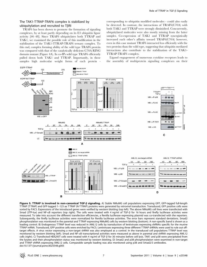
Figure 5. TTRAP is involved in non-canonical TGF-b signaling. A) Stable NMuMG cell populations expressing GFP, GFP-tagged full-lengthT TRAP (T TRAP) and GFP-tagged 1–123 aa T TRAP (N-T TRAP) proteins were generated by retroviral transduction. Transduced, GFP positive cells weresorted by FACS. Expressions of the introduced genes were verified by western blotting (top left). The above NMuMG cell lines were transfected withSmad (3TP-lux) and NF-kB reporters (top right). The cells were treated with 4 ng/ml of TGF-b for 16 hours and firefly luciferase activites weremeasured. To take into account the different transfection efficiencies, a Renilla luciferase expressing plasmid was co-transfected with the reporters.Subsequently, the firefly luciferase activities were normalized for Renilla luciferase activities. The error bars represent standard deviations. Smad2phosphorylation was monitored in the parental and T TRAP expressing NMuMG cells by western blotting (bottom). A non-specific band is shown as aloading control. B) Endogenous T TRAP level was reduced in AML12 cells by transduction of lentiviruses expressing shRNAs specific for the murineTTRAP mRNA. Transduced, GFP positive cells were enriched by FACS. Lentiviruses expressing three different T TRAP shRNAs were used to rule out off-target effects. A virus vector expressing a non-target shRNA was also employed as a control. In the transduced cell populations T TRAP level wasmonitored by western blotting (left). Smad and NF-kB transcriptional activities were measured as above in parental and shRNA expressing AML12cells (right). C) Transfected HEK293T cells were treated with 4 ng/ml of TGF-b for 30 minutes before cell lysis. TAK1 and p38 were precipitated fromthe lysates and their phosphorylation status was monitored by western blotting. D) Smad2 and p38 phosphorylation were examined in non-targetand TTRAP shRNA expressing AML12 cells. Comparable sample loading was also monitored using p38 and Smad2/3 antibodies.doi:10.1371/journal.pone.0025548.g005
Role of TTRAP in TGF-b Signaling
PLoS ONE | www.plosone.org 7 September 2011 | Volume 6 | Issue 9 | e25548

Role of TTRAP in TGF-b Signaling
PLoS ONE | www.plosone.org 8 September 2011 | Volume 6 | Issue 9 | e25548

intracellular domains. Members of the TRAF adaptor protein
family have been shown to play crucial role in these processes
[44]. Prompted by these observations, we tested whether
TRAF6 can influence TTRAP’s association with the TGF-breceptors using co-IPs (Figure 4B). As described above, TTRAP
exhibited relatively weak binding to TbRI-TD. Co-expression
of TRAF6 however, dramatically increased TTRAP’s affinity
toward the receptor. Importantly, the increased binding was
accompanied by the appearance of ubiquitylated TTRAP
forms, suggesting that TRAF6 mediated ubiquitylation may
contribute to the stabilization of the TTRAP-TbRI complex.
Similarly to TRAF6, ectopic expression of TRAF2 also
increased the TTRAP-TbRI association, though to a much-
reduced degree, indicating that under physiological conditions
TRAF2’s role may be negligible in the stabilization of the
TTRAP-TGF-b receptor complex.
Binding of TAK1 with TGF-b receptors has been demonstrated
by several studies [23,24,49,50]. We examined how this
interaction is influenced by TTRAP and TRAF6. As shown in
Figure 4C, ectopic expression of TRAF6 helped the recruitment of
not only TTRAP but TAK1 as well to TbRI. Notably, enrichment
of ubiquitylated forms of the proteins could also be observed in the
TbRI immunoprecipitates, indicating that the complexes might be
stabilized by this modification.
TTRAP is involved in non-canonical TGF-b signalingHaving established that TTRAP interacts with TGF-b
receptors and components of the TRAF6-TAK1 signaling
module, we wanted to evaluate the protein’s involvement in
various TGF-b induced biological responses. We started with the
establishment of stable NMuMG cell populations expressing the
EGFP-tagged full-length TTRAP molecule (TTRAP cells). As
controls, cells were also produced expressing the N-terminal 123
aa fragment of TTRAP tagged with EGFP (N-TTRAP cells) or
EGFP alone (EGFP cells) (Figure 5A). Smad-dependent tran-
scription was monitored in the above cells using the 3TP-lux
reporter. TGF-b treatment resulted in the same degree of Smad
activation in all cell lines and consistently, TbRI mediated
Smad2 phosphorylation also followed a similar kinetics
(Figure 5A). Earlier studies suggested that TTRAP is a negative
modulator of NF-kB [27]. Thus, we examined the protein’s
influence on TGF-b induced NF-kB activation as well. TGF-btreatment did not have a significant effect on the activity of an
NF-kB reporter in NMuMG cells. However, in TTRAP over-
expressing cells the basal NF-kB activity was approximately half
of that observed in the control EGFP or N-TTRAP expressing
cells (Figure 5A).
The effect of TTRAP deficiency on TGF-b induced transcrip-
tional responses was also examined. These studies were performed
in AML12 normal murine hepatocytes, in which the endogenous
TTRAP protein level was knocked down by lentiviruses expressing
shRNAs specific for the mouse TTRAP gene (Figure 5B). Down-
regulation of TTRAP did not have an effect on TGF-b induced
Smad-dependent transcription and the kinetics of Smad2
phosphorylation was not affected either (Figure 5B and D). In
the parental and the non-target shRNA expressing AML12 cells,
TGF-b treatment significantly reduced the activity of an NF-kB
reporter in keeping with an earlier report [51]. TTRAP deficiency
completely abolished this inhibitory effect and even a slight
increase in the basal NF-kB activity could be seen in the TTRAP
shRNA expressing cells (Figure 5B).
TTRAP associates with components of the TRAF6-TAK1
signaling module, which plays an essential role in TGF-binduced p38 activation. Additionally, the protein has recently
been implicated in proteasome impairment elicited activation of
p38 and JNK [52]. In light of these observations, we examined
TTRAP’s role in TGF-b induced activation of these kinases.
Ectopic expression of TTRAP in HEK293T cells activated p38
however, it did not affect JNK phosphorylation (Figure 5C and
data not shown). Accompanying p38 activation, ubiquitylation
and phosphorylation of TAK1 was also observed. In many cell
lines TGF-b activates p38 in two waves [53,54]. The early stage
- peeking between 15–45 minutes - is Smad-independent, while
the delayed p38 response - reaching its maximum at 1.5–
2 hours - requires Smad-dependent transcription. As shown in
Figure 5D, shRNA mediated knockdown of TTRAP expression
strongly inhibited the early p38 phosphorylation in AML12
cells, while the delayed p38 activation and Smad2 phosphor-
ylation remained unaffected. In summary, the above data
strongly suggest that TTRAP is an important component of
Smad-independent non-canonical TGF-b induced signaling
responses, principally the p38 kinase cascade and the NF-kB
pathway.
TTRAP plays a role in TGF-b induced apoptosisThe NMuMG mammary epithelial cell line has been a well-
characterized model system for TGF-b induced apoptosis
[11,23,55]. TGF-b regulates this process in NMuMG cells
through both Smad-dependent and -independent mechanisms,
with the Smad-independent component predominantly involving
the p38 MAP kinase cascade. To asses the role of TTRAP in
TGF-b induced apoptosis, we treated the TTRAP expressing and
control NMuMG cells described above with TGF-b under
various conditions and subsequently their viability was measured
by PI uptake and MTT assay. In accordance with published data,
TGF-b elicited only modest apoptosis in the control cell
populations (EGFP and N-TTRAP cells) after 24 hours under
low-serum (0.2% FBS) culture conditions (Figure 6A) [11,55]. In
contrast, after 24 hours, TTRAP cells exhibited robust TGF-binduced cell death (,50%), which by 48 hours increased even
further (,80%). By the same time, the TGF-b elicited apoptotic
rate in the control cell populations was ,2/3rd of that of the
TTRAP cells. Importantly, TGF-b dependent apoptosis was
completely preventable by the TbRI receptor kinase inhibitor,
SB431542 and the p38 inhibitor, SB203580 also provided strong
protection. The JNK inhibitor, SP600125 did not have a
significant effect on the viability of TGF-b treated NMuMG
cells. In 10% FBS medium, 24 hours of TGF-b treatment was
Figure 6. TTRAP is involved in TGF-b induced apoptosis. The stable NMuMG cell populations described in Figure 5 were treated in 0.2% (A) or10% (B) FBS containing medium as indicated and cell viability was assessed using two different methods. PI uptake of cells, as a measure ofmembrane integrity, was monitored by FACS (left panels). The experiments were repeated at least twice with similar outcome. On the right side MT Tassays were used to measure cell viability. The chemicals used at the following concentrations: TGF-b 4 ng/ml; SB431542, SB203580 and SP600125were all used at 10 mM. The error bars represent standard deviations. C) NMuMG cells stably expressing T TRAP or EGFP were treated as indicated andintegrity of their membranes was monitored by PI uptake. The chemicals used at the following concentrations: TGF-b 4 ng/ml, staurisporine (STS)1 mM, MG132 2.5 mM. Experiments were repeated several times and a representative result is shown. D) Non-target and T TRAP shRNA lentivirustransduced AML12 cells were treated as indicated in 10% FBS medium. After 24 hours, cells were stained with annexin V and analyzed by FACS.doi:10.1371/journal.pone.0025548.g006
Role of TTRAP in TGF-b Signaling
PLoS ONE | www.plosone.org 9 September 2011 | Volume 6 | Issue 9 | e25548

not able to elicit significant degree of cell death in any of the
NMuMG cell lines used, and even after 48 hours only weak
apoptosis (,20%) was detectable in the control cells (Figure 6B).
In contrast, under the same conditions in the TTRAP cells the
apoptotic rate was .50% by 48 hours and whereas the TbRI
inhibitor was still able to prevent apoptosis, the p38 inhibitor lost
its protective effect.
Next, we wished to examine the involvement of TTRAP in
apoptotic processes induced by other death-promoting stimuli
(Figure 6C). TTRAP cells exhibited similar sensitivity to the kinase
inhibitor, staurosporine and the proteasome inhibitor, MG132 as
the control EGFP cells. Interestingly however, while TGF-b alone
was unable to elicit a significant degree of apoptosis after 24 hours
under high serum growth conditions in either cell lines, the
combined TGF-b/MG132 treatment resulted in synergistic killing
of the TTRAP cells.
Finally, TGF-b induced cell death was examined in AML12
hepatocytes made deficient for TTRAP with the use of gene
specific shRNAs (see above). In keeping with a recent report
[56], decreasing cellular TTRAP level resulted in increased basal
apoptosis (,6% versus ,24%) (Figure 6D). Importantly
however, the robust TGF-b induced cell death was significantly
attenuated by TTRAP deficiency (,74% versus ,59%),
confirming that the protein fulfills a TGF-b dependent pro-
apoptotic role in the cells.
Discussion
In this paper we indentify the TTRAP adaptor molecule as a
binding partner for key components of the TRAF6-TAK1 signaling
module and the TGF-b receptor complex, strongly suggesting the
protein’s involvement in non-canonical TGF-b signaling responses.
Support for this hypothesis is provided by the following observa-
tions: 1. TTRAP interacts with endogenous TAK1 and TbRI in a
TGF-b inducible fashion; 2. TTRAP forms a stable ternary
complex with TAK1 and TRAF6, which can interact with the
TGF-b receptor complex; 3. the E3 ubiquitin ligase activity of
TRAF6 is boosted by TTRAP, thereby promoting the ubiquityla-
tion of TAK1 and itself; 4. ectopic over-expression of TTRAP
results in phosphorylation of TAK1 and p38, while it negatively
modulates the activity of NF-kB ; 5. shRNA mediated knockdown
of endogenous TTRAP abolishes TGF-b induced rapid phosphor-
ylation of p38 and repression of NF-kB.
TGF-b controls apoptosis through both Smad-dependent and
-independent signaling routes. Amongst the Smad-independent
pathways, the p38 kinase cascade is generally considered to be a
pro-apoptotic pathway, while the NF-kB route has been shown to
protect cells from TGF-b induced apoptosis [16,17,51,57–59].
Thus, it was not surprising to find that ectopic expression of
TTRAP sensitized cells to TGF-b induced cell death, while its
down-regulation provided partial protection. The ability of
TTRAP to differentially modulate two signaling pathways with
opposing outcomes is intriguing however, by no means unique.
Recently, the XIAP interacting protein Siva1 was demonstrated
to inhibit XIAP and TAK1-TAB1 mediated NF-kB activation,
while prolonged TNFa-induced JNK activation resulting in
enhanced apoptosis [60]. Interestingly, Siva1 similarly TTRAP,
was also shown to interact with the TAK1 complex. Thus, we
suggest that TTRAP, along with Siva1, belongs to a group of
proteins defined by their abilities to modulate the balance
between pro-survival and pro-apoptotic pathways by interacting
with the TAK1 complex.
Several works implicated TTRAP in the regulation of apoptosis
and depending on the cellular context and the death promoting
stimuli used, both pro- and anti-apoptotic properties have been
attributed to the protein [58,56,61]. Our results not only establish
TTRAP as a novel component of the non-canonical TRAF6-
TAK1 signaling branch of TGF-b signaling, but also demonstrate
its specific involvement in TGF-b induced apoptosis. It is
becoming increasingly clear that imbalances arising during tumor
progression between various branches of TGF-b signaling conspire
to convert TGF-b from a suppressor of tumor formation to a
promoter of their growth. Thus, one may hypothesize that
restoration of this equilibrium could be of great therapeutic value.
From this perspective, the TRAF6-TAK1 signaling module could
be a unique and very attractive target for intervention. This
module is a point of convergence for both pro-apoptotic (p38/
JNK MAP kinase cascades) and pro-survival (NF-kB and PI3K/
Akt pathways) signaling routes. Since TTRAP interacts with all
key components of this module, thorough understanding of its
mode of action may help us formulate strategies for steering the
TGF-b pathway in different directions, favoring either survival or
apoptosis.
Supporting Information
Figure S1 In vitro interaction of TTRAP with TbRI andTbRII. Cytoplasmic domains of TGF-b receptors fused with GST
were produced in bacteria and affinity purifed on gluthatione
beads. HA-TTRAP, FLAG-TRAF2 and -6 were produced by in
vitro translation in rabbit reticulocyte lysates. In vitro binding of
TTRAP and TRAFs to gluthatione bead-bound GST, GST-
TbRI-CD and GST-TbRII-CD were examined by western
blotting.
(TIF)
Figure S2 Analysis of the binding of TTRAP with Smadsby co-IP. TTRAP was pulled-down from transfected HEK293T
cells and the co-precipitation of the Smads and TbRI was
examined by western blotting.
(TIF)
Figure S3 Mapping of the TRAF6 and TAK1 bindingdomains of TTRAP by co-IP. A, B) TRAF6 or TAK1 was
precipitated from transfected HEK293T cells and the co-
precipitating TTRAP fragments were detected by western
blotting. Note that TAK1 and EGFP-TTRAP has similar
eletrophoretic mobilities, thus the HA reactive band in lane 2 of
IP panel A corresponds to a mixture of the two molecules.
(TIF)
Figure S4 TRAF6 promotes the ubiquitylation ofTTRAP. Original scans for Figure 3A.
(TIF)
Acknowledgments
We are grateful to Dr. Ying Zhang (NCI, NIH) for her initial contribution
to this project. We thank Anita Schamberger for help with the FACS
analysis and critically reading the manuscript.
Author Contributions
Conceived and designed the experiments: KF. Performed the experiments:
KF GV. Analyzed the data: KF. Contributed reagents/materials/analysis
tools: BS. Wrote the paper: KF.
Role of TTRAP in TGF-b Signaling
PLoS ONE | www.plosone.org 10 September 2011 | Volume 6 | Issue 9 | e25548

References
1. Bierie B, Moses HL (2006) Tumour microenvironment: TGFb: the molecular
Jekyll and Hyde of cancer. Nat Rev Cancer 6: 506–520.
2. Massague J (2008) TGFb in Cancer. Cell 134: 215–230.
3. Pardali K, Moustakas A (2007) Actions of TGF-b as tumor suppressor and pro-metastatic factor in human cancer. Biochim Biophys Acta 1775: 21–62.
4. Feng XH, Derynck R (2005) Specificity and versatility in TGF-beta signalingthrough Smads. Annu Rev Cell Dev Biol 21: 659–693.
5. Mas sague J, Seoane J, Wotton D (2005) Smad transcription factors. Genes Dev
19: 2783–2810.
6. Derynck R, Zhang YE (2003) Smad-dependent and Smad-independent
pathways in TGF-beta family signalling. Nature 425: 577–584.
7. Moustakas A, Heldin CH (2005) Non-Smad TGF-b signals. J Cell Sci 118:
3573–3584.
8. Zhang YE (2009) Non-Smad pathways in TGF-b signaling. Cell Res 19:
128–139.
9. Hanafusa H, Ninomiya-Tsuji J, Masuyama N, Nishita M, Fujisawa J, et al.
(1999) Involvement of the p38 mitogen-activated protein kinase pathway intransforming growth factor-b-induced gene expression. J Biol Chem 274:
27161–27167.
10. Sano Y, Harada J, Tashiro S, Gotoh-Mandeville R, Maekawa T, et al. (1999)
ATF-2 is a common nuclear target of Smad and TAK1 pathways intransforming growth factor-b signaling. J Biol Chem 274: 8949–8957.
11. Yu L, Hebert MC, Zhang YE (2002) TGF-b receptor-activated p38 MAP kinase
mediates Smad-independent TGF-b responses. EMBO J 21: 3749–3759.
12. Edlund S, Bu S, Schuster N, Aspenstrom P, Heuchel R, et al. (2003)
Transforming growth factor-b1 (TGF-b)-induced apoptosis of prostate cancer
cells involves Smad7-dependent activation of p38 by TGF-b-activated kinase 1and mitogen-activated protein kinase kinase 3. Mol Biol Cell 14: 529–544.
13. Galliher AJ, Schiemann WP (2006) Beta3 integrin and Src facilitate
transforming growth factor-b mediated induction of epithelial-mesenchymal
transition in mammary epithelial cells. Breast Cancer Res 8: R42.
14. Galliher AJ, Schiemann WP (2007) Src phosphorylates Tyr284 in TGF-b type II
receptor and regulates TGF-b stimulation of p38 MAPK during breast cancercell proliferation and invasion. Cancer Res 67: 3752–3758.
15. Galliher-Beckley AJ, Schiemann WP (2008) Grb2 binding to Tyr284 in TbR-II
is essential for mammary tumor growth and metastasis stimulated by TGF-b.
Carcinogenesis 29: 244–251.
16. Neil JR, Tian M, Schiemann WP (2009) X-linked inhibitor of apoptosis proteinand its E3 ligase activity promote transforming growth factor-b-mediated
nuclear factor-kB activation during breast cancer progression. J Biol Chem 284:
21209–21217.
17. Neil JR, Schiemann WP (2008) Altered TAB1:IkB kinase interaction promotes
transforming growth factor b-mediated nuclear factor-kB activation duringbreast cancer progression. Cancer Res 68: 1462–1470.
18. Parvani JG, Taylor MA, Schiemann WP (2011) Noncanonical TGF-b Signaling
During Mammary Tumorigenesis. J Mammary Gland Biol Neoplasia In press.
19. Wendt MK, Smith JA, Schiemann WP (2009) p130Cas is required for
mammary tumor growth and transforming growth factor-b-mediated metastasisthrough regulation of Smad2/3 activity. J Biol Chem 284: 34145–34156.
20. Yamaguchi K, Shirakabe K, Shibuya H, Irie K, Oishi I, et al. (1995)Identification of a member of the MAPKKK family as a potential mediator of
TGF-b signal transduction. Science 270: 2008–2011.
21. Shim JH, Xiao C, Paschal AE, Bailey ST, Rao P, et al. (2005) TAK1, but not
TAB1 or TAB2, plays an essential role in multiple signaling pathways invivo.Genes Dev 19: 2668–2681.
22. Jadrich JL, O’Connor MB, Coucouvanis E (2006) The TGF b activated kinaseTAK1 regulates vascular development in vivo. Development 133: 1529–1541.
23. Yamashita M, Fatyol K, Jin C, Wang X, Liu Z, et al. (2008) TRAF6 mediatesSmad-independent activation of JNK and p38 by TGF-b. Mol Cell 31:
918–924.
24. Sorrentino A, Thakur N, Grimsby S, Marcusson A, von Bulow V, et al. (2008)
The type I TGF-b receptor engages TRAF6 to activate TAK1 in a receptorkinase-independent manner. Nat Cell Biol 10: 1199–1207.
25. Delaney JR, Mlodzik M (2006) TGF-beta activated kinase-1: new insights intothe diverse roles of TAK1 in development and immunity. Cell Cycle 5:
2852–2855.
26. Landstrom M (2010) The TAK1-TRAF6 signalling pathway. Int J Biochem Cell
Biol 42: 585–589.
27. Pype S, Declercq W, Ibrahimi A, Michiels C, Van Rietschoten JG, et al. (2000)
TTRAP, a novel protein that associates with CD40, tumor necrosis factor (TNF)receptor-75 and TNF receptor-associated factors (TRAFs), and that inhibits
nuclear factor-kappa B activation. J Biol Chem 275: 18586–18593.
28. Pei H, Yordy JS, Leng Q, Zhao Q, Watson DK, et al. (2003) EAPII interacts
with ETS1and modulates its transcriptional function. Oncogene 22: 2699–2709.
29. Xu GL, Pan YK, Wang BY, Huang L, Tian L, et al. (2008) TTRAP is a novelPML nuclear bodies-associated protein. Biochem Biophys Res Commun 375:
395–398.
30. Cortes Ledesma F, El Khamisy SF, Zuma MC, Osborn K, Caldecott KW (2009)
A human 59-tyrosyl DNA phosphodiesterase that repairs topoisomerase-
mediated DNA damage. Nature 461: 674–678.
31. Zeng Z, Cortes-Ledesma F, El Khamisy SF, Caldecott KW (2011) TDP2/TTRAP is the major 59-tyrosyl DNA phosphodiesterase activity in vertebrate
cells and is critical for cellular resistance to topoisomerase II-induced DNA
damage. J Biol Chem 286: 403–409.
32. Esguerra CV, Nelles L, Vermeire L, Ibrahimi A, Crawford AD, et al. (2007)
Ttrap is an essential modulator of Smad3-dependent Nodal signaling during
zebrafish gastrulation and left-right axis determination. Development 134:
4381–4393.
33. Fatyol K, Szalay AA (2001) The p14ARF tumor suppressor protein facilitates
nucleolar sequestration of hypoxia-inducible factor-1alpha (HIF-1alpha) and
inhibits HIF-1-mediated transcription. J Biol Chem 276: 28421–28429.
34. Graycar JL, Miller DA, Arrick BA, Lyons RM, Moses HL, et al. (1989) Human
transforming growth factor-beta 3: recombinant expression, purification, and
biological activities in comparison with transforming growth factors-beta 1 and
-beta 2. Mol Endocrinol 3: 1977–1986.
35. Morgenstern JP, Land H (1990) Advanced mammalian gene transfer: high titre
retroviral vectors with multiple drug selection markers and a complementary
helper-free packaging cell line. Nucleic Acids Res 18: 3587–3596.
36. Rodrigues-Lima F, Josephs M, Katan M, Cassinat B (2001) Sequence analysis
identifies TTRAP, a protein that associates with CD40 and TNF receptor-
associated factors, as a member of a superfamily of divalent cation-dependent
phosphodiesterases. Biochem Biophys Res Commun 285: 1274–1279.
37. Kishimoto K, Matsumoto K, Ninomiya-Tsuji J (2000) TAK1 mitogen-activated
protein kinase kinase kinase is activated by autophosphorylation within its
activation loop. J Biol Chem 275: 7359–7364.
38. Singhirunnusorn P, Suzuki S, Kawasaki N, Saiki I, Sakurai H (2005) Critical
roles of threonine 187 phosphorylation in cellular stress-induced rapid and
transient activation of transforming growth factor-b-activated kinase 1 (TAK1)
in a signaling complex containing TAK1-binding protein TAB1 and TAB2.J Biol Chem 280: 7359–7368.
39. Ninomiya-Tsuji J, Kajino T, Ono K, Ohtomo T, Matsumoto M, et al. (2003) A
resorcylic acid lactone, 5Z-7-oxozeaenol, prevents inflammation by inhibiting
the catalytic activity of TAK1 MAPK kinase kinase. J Biol Chem 278:
18485–18490.
40. Ishitani T, Takaesu G, Ninomiya-Tsuji J, Shibuya H, Gaynor RB, et al. (2003)
Role of the TAB2-related protein TAB3 in IL-1 and TNF signaling. EMBO J22: 6277–6288.
41. Kanayama A, Seth RB, Sun L, Ea CK, Hong M, et al. (2004) TAB2 and TAB3
activate the NF-kappaB pathway through binding to polyubiquitin chains. Mol
Cell 15: 535–548.
42. Shibuya H, Yamaguchi K, Shirakabe K, Tonegawa A, Gotoh Y, et al. (1996)
TAB1: an activator of the TAK1 MAPKKK in TGF-b signal transduction.
Science 272: 1179–1182.
43. Takaesu G, Kishida S, Hiyama A, Yamaguchi K, Shibuya H, et al. (2000)
TAB2, a novel adaptor protein, mediates activation of TAK1 MAPKKK by
linking TAK1 to TRAF6 in the IL-1 signal transduction pathway. Mol Cell 5:
649–658.
44. Karin M, Gallagher E (2009) TNFR signaling: ubiquitin-conjugated TRAFfic
signals control stop-and-go for MAPK signaling complexes. Immunol Rev 228:
225–240.
45. Davies CC, Mak TW, Young LS, Eliopoulos AG (2005) TRAF6 is required for
TRAF2-dependent CD40 signal transduction in nonhemopoietic cells. Mol Cell
Biol 25: 9806–9819.
46. Deng L, Wang C, Spencer E, Yang L, Braun A, et al. (2000) Activation of the
IkB kinase complex by TRAF6 requires a dimeric ubiquitin-conjugating enzyme
complex and a unique polyubiquitin chain. Cell 103: 351–361.
47. Adhikari A, Xu M, Chen ZJ (2007) Ubiquitin-mediated activation of TAK1 and
IKK. Oncogene 26: 3214–3226.
48. Chen ZJ, Sun LJ (2009) Nonproteolytic functions of ubiquitin in cell signaling.
Mol Cell 33: 275–286.
49. Kim SI, Kwak JH, Na HJ, Kim JK, Ding Y, et al. (2009) Transforming growth
factor-b (TGF-b1) activates TAK1 via TAB1-mediated autophosphorylation,
independent of TGF-b receptor kinase activity in mesangial cells. J Biol Chem
284: 22285–22296.
50. Watkins SJ, Jonker L, Arthur HM (2006) A direct interaction between TGFbactivated kinase 1 and the TGFb type II receptor: implications for TGFbsignalling and cardiac hypertrophy. Cardiovasc Res 69: 432–439.
51. Arsura M, FitzGerald MJ, Fausto N, Sonenshein GE (1997) Nuclear factor-
kappaB/Rel blocks transforming growth factor beta1-induced apoptosis of
murine hepatocyte cell lines. Cell Growth Differ 8: 1049–1059.
52. Zucchelli S, Vilotti S, Calligaris R, Lavina ZS, Biagioli M, et al. (2009)
Aggresome-forming TTRAP mediates pro-apoptotic properties of Parkinson’s
disease-associated DJ-1 missense mutations. Cell Death Differ 16: 428–438.
53. Takekawa M, Tatebayashi K, Itoh F, Adachi M, Imai K, et al. (2002) Smad-
dependent GADD45b expression mediates delayed activation of p38 MAP
kinase by TGF-b. EMBO J 21: 6473–6482.
54. Yoo J, Ghiassi M, Jirmanova L, Balliet AG, Hoffman B, et al. (2003)
Transforming growth factor-b-induced apoptosis is mediated by Smad-
dependent expression of GADD45b through p38 activation. J Biol Chem 278:
43001–43007.
55. Ramjaun AR, Tomlinson S, Eddaoudi A, Downward J (2007) Upregulation of
two BH3-only proteins, Bmf and Bim, during TGF b-induced apoptosis.
Oncogene 26: 970–981.
Role of TTRAP in TGF-b Signaling
PLoS ONE | www.plosone.org 11 September 2011 | Volume 6 | Issue 9 | e25548
56. Li C, Fan S, Owonikoko TK, Khuri FR, Sun SY, et al. (2011) Oncogenic role of
EAPII in lung cancer development and its activation of the MAPK-ERK
pathway. Oncogene. In press.
57. Arsura M, Panta GR, Bilyeu JD, Cavin LG, Sovak MA, et al. (2003) Transient
activation of NF-kB through a TAK1/IKK kinase pathway by TGF-b1 inhibits
AP-1/SMAD signaling and apoptosis: implications in liver tumor formation.
Oncogene 22: 412–425.
58. Arsura M, Wu M, Sonenshein GE (1996) TGF beta 1 inhibits NF-kappa B/Rel
activity inducing apoptosis of B cells: transcriptional activation of I kappa B
alpha. Immunity 5: 31–40.
59. Sovak MA, Arsura M, Zanieski G, Kavanagh KT, Sonenshein GE (1999) The
inhibitory effects of transforming growth factor beta1 on breast cancer cellproliferation are mediated through regulation of aberrant nuclear factor-
kappaB/Rel expression. Cell Growth Differ 10: 537–544.
60. Resch U, Schichl YM, Winsauer G, Gudi R, Prasad K, et al. (2009) Siva1 is aXIAP-interacting protein that balances NFkappaB and JNK signalling to
promote apoptosis. J Cell Sci 122: 2651–2661.61. Lee HY, Youn SW, Kim JY, Park KW, Hwang CI, et al. (2008) FOXO3a turns
the tumor necrosis factor receptor signaling towards apoptosis through
reciprocal regulation of c-Jun N-terminal kinase and NF-kappaB. ArteriosclerThromb Vasc Biol 28: 112–120.
Role of TTRAP in TGF-b Signaling
PLoS ONE | www.plosone.org 12 September 2011 | Volume 6 | Issue 9 | e25548


![Rational Canonical Formbuzzard.ups.edu/...spring...canonical-form-present.pdfIntroductionk[x]-modulesMatrix Representation of Cyclic SubmodulesThe Decomposition TheoremRational Canonical](https://static.fdocuments.us/doc/165x107/6021fbf8c9c62f5c255e87f1/rational-canonical-introductionkx-modulesmatrix-representation-of-cyclic-submodulesthe.jpg)