trypanosoma
18
trypanosoma By : AMEER AZEEZ & FAROOQ OMER
-
Upload
ameer-azeez -
Category
Health & Medicine
-
view
92 -
download
1
Transcript of trypanosoma
trypanosomaBy : AMEER AZEEZ & FAROOQ OMER
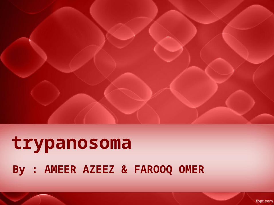
• Trypanosoma is a genus of kinetoplastids (class Kinetoplastida), a monophyletic[1] group of unicellular parasitic flagellate protozoa. The name is derived from the Greek trypano- (borer) and soma (body) because of their corkscrew-like motion
• All trypanosomes are heteroxenous (requiring more than one obligatory host to complete life cycle) and most are transmitted via a vector. The majority of species are transmitted by blood-feeding invertebrates, but there are different mechanisms among the varying species

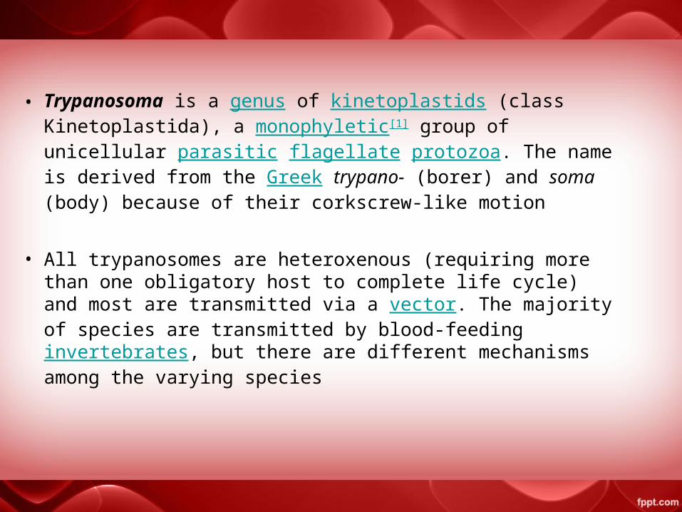
Trypanosoma
Trypanosomiasis
American Trypanosomiasis
East African Trypanosomiasis
Transmitted byTransmitted by
T.brucei rhodesienseT.brucei gambiense T.cruzi
Sleeping sickness Chagas’ disease
West African Trypanosomiasis
Glossina (tsetse fly) Triatoma (winged bug)

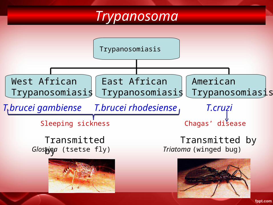
Winterbottom sign
African trypanosomiasis
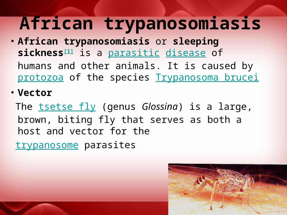
African trypanosomiasis• African trypanosomiasis or sleeping
sickness[1] is a parasitic disease of humans and other animals. It is caused by protozoa of the species Trypanosoma brucei
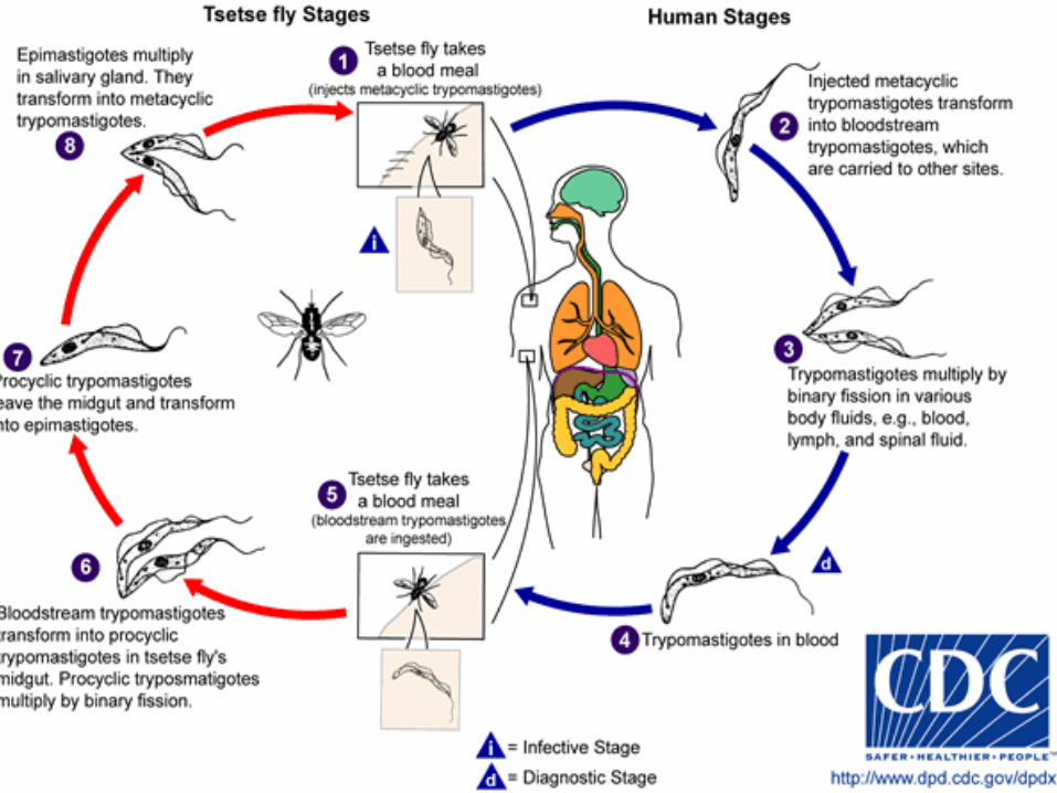
• Vector
The tsetse fly (genus Glossina) is a large, brown, biting fly that serves as both a host and vector for the
trypanosome parasites
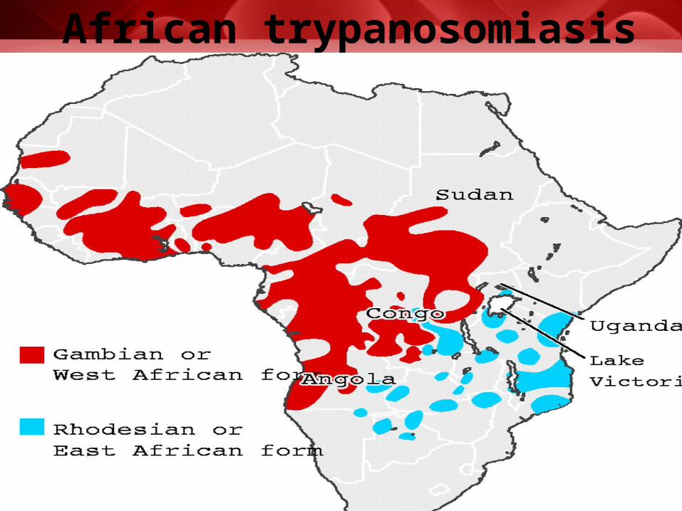
•There are two subspecies of the parasite that are responsible for initiating the disease in humans. Trypanosoma brucei gambiense causes the diseases in west and central Africa whereas, •, Trypanosoma brucei rhodesiense has a limited geographical range and is responsible for causing the disease in east and southern Africa•In addition, a third subspecies of the parasite known as Trypanosoma brucei brucei is responsible for affecting animals but not humans
•These parasites primarily infect individuals in sub-Saharan Africa because that is where the vector (tsetse fly) is located•T. b. gambiense causes a chronic condition that can remain in a passive phase for months or years before symptoms emerge and the infection can last about 3 years before death occurs. •T. b. rhodesiense is the acute form of the disease and death can occur within months since the symptoms emerge within weeks and it is more virulent and faster developing than T. b. gambiense
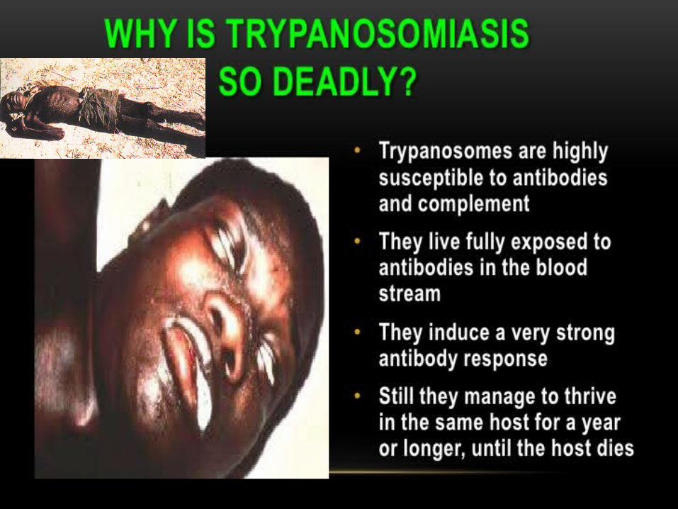
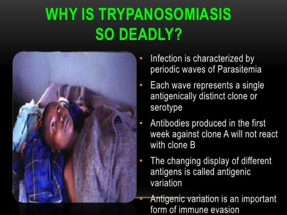
Furthermore trypanosomes are surrounded by a coat that is composed of variant surface glycoproteins (VSG). These proteins act to protect the parasite from any lytic factors that are present in human plasma. The host’s immune system recognizes the glycoproteins present on the coat of the parasite leading to the production of different antibodies (IgM and IgG). These antibodies will then act to destroy the parasites that circulate around the blood. However, from the several parasites present in the plasma, a small number of them will experience changes in their surface coats resulting in the formation of new VSGs. Thus, the antibodies produced by the immune system will no longer recognize the parasite leading to proliferation until new antibodies are created to combat the novel VSGs. Eventually the immune system will no longer be able to fight off the parasite due to the constant changes in VSGs and infection will arise
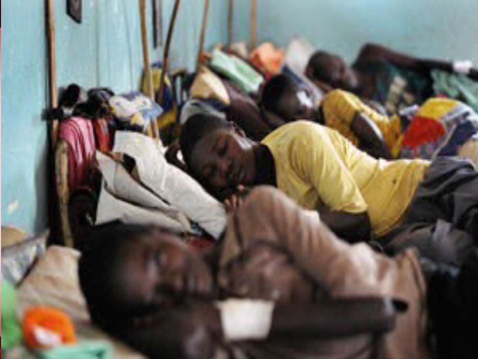
Signs and symptomsAfrican trypanosomiasis symptoms occur in two stages :1.haemolymphatic phase, is characterized by fever, headaches, joint pains, and itching Fever is intermittent, with attacks lasting from a day to a week, separated by intervals of a few days to a month or longer.2.the neurological phase ; In this stage the parasites cross the blood-brain barrier to infect the central nervous system In general this is when more obvious signs and symptoms of the disease appear: changes of behaviour, confusion, sensory disturbances and poor coordination. Disturbance of the sleep cycle, which gives the disease its name, is an important feature of the second stage of the disease.
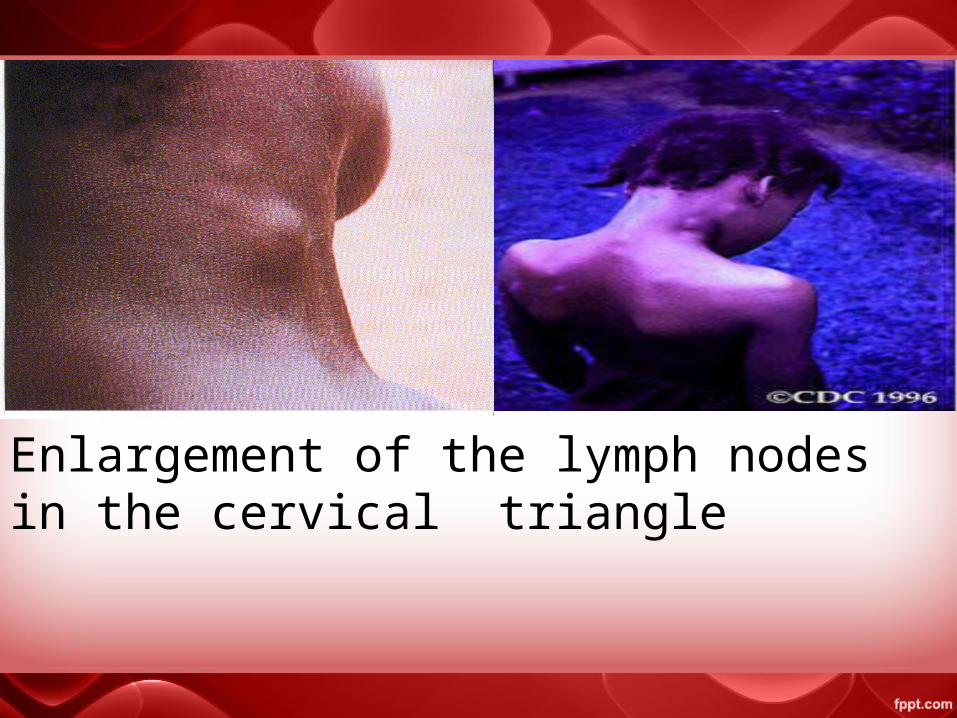
Enlargement of the lymph nodes in the cervical triangle

Treatment•First stageThe current treatment for first-stage disease is intravenous or intramuscular pentamidine for T. b. gambiense or intravenous suramin for T. b. rhodesiense.•Second stageFor T. b. gambiense intravenous eflornithine or the combination of nifurtimox and eflornithine appear to be more effective and easier to give.[19] These treatments may replace melarsoprol when available[19] with the combination being first line.[3] Intravenous melarsoprol was previously the standard treatment for second-stage (neurological phase) disease and is effective for both types.[3] It is the only treatment for second stage T. b. rhodesiense however causes death in 5% of people who take it.[3] Resistance to
melarsoprol can occur.[3]

study T.B to help African people Thank you all