Triplex dna 1
31
Annu. Rev. Biochem. 1995. 64:65-95 Copyright ©1995 by Annual Reviews Inc. All rights reserved TRIPLEX DNA STRUCTURES Maxim D. Frank-Kamenetskii Center for Advanced Biotechnology and Department of Biomedical Engineering, Boston University, Boston, Massachusetts 02215 Sergei M. Mirkin Depa~ment of Genetics, University of Illinois at Chicago, Chicago, Illinois 60612 KEY WORDS: DNA triplex, H-DNA, gene-drugs, triplex-forming oligonucleotides, PNA CONTENTS PERSPECTIVES AND SUMMARY ......................................... 66 STRUCTURE, STABILITY, AND SPECIFICITY OF DNA TRIPLEXES ........... 67 Triplex Menagerie ..................................................... 67 Fine Structure of DNA Triplexes .......................................... 72 H Form .............................................................. 74 H-DNA Menagerie ..................................................... 76 Specificity of Triplex Formation .......................................... 77 Stabilization of Triplexes ................................................ 79 Peptide Nucleic Acid (PNA) ............................................. 80 BIOCHEMISTRY OF TRIPLEXES.......................................... 82 Formation and Possible Functions of H-DNA In Vivo ......................... 82 Targeting Basic Genetic Processes Using TFOs............................. 86 Three-Stranded DNA Complexes in HomologousRecombination ................ 88 ABSTRACT A DNA triplex is formed when pyrimidineor purine bases occupy the major groove of the DNA double Helix forming Hoogsteen pairs with purines of the Watson-Crick basepairs. Intermolecular triplexes are formedbetweentriplex forming oligonucleotides(TFO) and target sequences on duplexDNA. Intra- moleculartriplexes are the majorelementsof H-DNAs, unusualDNA struc- tures, which are formedin homopurine-homopyrimidine regions of supercoiled DNAs. TFOs are promising gene-drugs, which can be used in an anti-gene strategy, that attempt to modulate gene activity in vivo. Numerous chemical modifications of TFO are known.In peptide nucleic acid (PNA), the sugar- phosphate backbone is replaced with a protein-like backbone. PNAs form 0066-4154/95/0701-0065505.00 Annual Reviews www.annualreviews.org/aronline
Transcript of Triplex dna 1
Annu. Rev. Biochem. 1995. 64:65-95Copyright © 1995 by Annual Reviews Inc. All rights reserved
TRIPLEX DNA STRUCTURES
Maxim D. Frank-KamenetskiiCenter for Advanced Biotechnology and Department of Biomedical Engineering,Boston University, Boston, Massachusetts 02215
Sergei M. MirkinDepa~ment of Genetics, University of Illinois at Chicago, Chicago, Illinois 60612
KEY WORDS: DNA triplex, H-DNA, gene-drugs, triplex-forming oligonucleotides, PNA
CONTENTS
PERSPECTIVES AND SUMMARY ......................................... 66STRUCTURE, STABILITY, AND SPECIFICITY OF DNA TRIPLEXES ........... 67
Triplex Menagerie ..................................................... 67Fine Structure of DNA Triplexes .......................................... 72H Form .............................................................. 74H-DNA Menagerie ..................................................... 76Specificity of Triplex Formation .......................................... 77Stabilization of Triplexes ................................................ 79Peptide Nucleic Acid (PNA) ............................................. 80
BIOCHEMISTRY OF TRIPLEXES .......................................... 82Formation and Possible Functions of H-DNA In Vivo ......................... 82Targeting Basic Genetic Processes Using TFOs ............................. 86Three-Stranded DNA Complexes in Homologous Recombination ................ 88
ABSTRACT
A DNA triplex is formed when pyrimidine or purine bases occupy the majorgroove of the DNA double Helix forming Hoogsteen pairs with purines of theWatson-Crick basepairs. Intermolecular triplexes are formed between triplexforming oligonucleotides (TFO) and target sequences on duplex DNA. Intra-molecular triplexes are the major elements of H-DNAs, unusual DNA struc-tures, which are formed in homopurine-homopyrimidine regions of supercoiledDNAs. TFOs are promising gene-drugs, which can be used in an anti-genestrategy, that attempt to modulate gene activity in vivo. Numerous chemicalmodifications of TFO are known. In peptide nucleic acid (PNA), the sugar-phosphate backbone is replaced with a protein-like backbone. PNAs form
0066-4154/95/0701-0065505.00
Annual Reviewswww.annualreviews.org/aronline
66 FRANK-KAMENETSKII & MIRKIN
P-loops while interacting with duplex DNA forming triplex with one of DNAstrands leaving the other strand displaced. Very unusual recombination orparallel triplexes, or R-DNA, have been assumed to form under RecA proteinin the course of homologous recombination.
PERSPECTIVES AND SUMMARY
Since the pioneering work of Felsenfeld, Davies, & Rich (1), double-strandedpolynucleotides containing purines in one strand and pydmidines in the otherstrand [such as poly(A)/poly(U), poly(dA)/poly(dT), or poly(dAG)/poly(dCT)]have been known to be able to undergo a stoichiometric transition forming atriple-stranded structure containing one polypurine and two polypyrimidinestrands (2-4). Early on, it was assumed that the third strand was located in themajor groove and associated with the duplex via non-Watson-Crick interactionsnow known as Hoogsteen pairing. Triple helices consisting ofonepyrimidine andtwo purine strands were also proposed (5, 6). However, notwithstanding the factthat single-base triads in tRNA structures were well-documented (reviewed in 7),triple-helical DNA escaped wide attention before the mid-1980s.
The considerable modem interest in DNA triplexes arose due to two partiallyindependent developments. First, homopurine-homopyrimidine stretches insupercoiled plasmids were found to adopt an unusual DNA structure, calledH-DNA, which includes a triplex as the major structural element (8, 9). Sec-ondly, several groups demonstrated that homopyrimidine and some purine-richoligonucleotides can form stable and sequence-specific complexes with corre-sponding homopurine-homopyrimidine sites on duplex DNA (10-12). Thesecomplexes were shown to be triplex structures rather than D-loops, where theoligonucleotide invades the double helix and displaces one strand. A charac-teristic feature of all these triplexes is that the two chemically homologousstrands (both pyrimidine or both purine) are antiparallel. These findings led explosive growth in triplex studies.
During the study of intermoleeular triplexes, it became clear that triplex-form-ing oligonucleotides (TFOs) might be universal drugs that exhibit sequence-spe-cific recognition of duplex DNA. This is an exciting possibility because, incontrast to other DNA-binding drugs, the recognition principle of TFOs is verysimple: Hoogsteen pairing rules between a purine strand of the DNA duplex andthe TFO bases. However, this mode of recognition is limited in that homopurine-homopyrimidine sites are preferentially recognized. Though significant effortshave been directed toward overcoming this limitation, the problem is stillunsolved in general. Nevertheless, the high specificity of TFO-DNA recognitionhas led to the development of an "antigene" strategy, the goal of which is tomodulate gene activity in vivo using TFOs (reviewed in 13).
Although numerous obstacles must be overcome to reach the goal, none arelikely to be fatal for the strategy. Even if DNA TFOs proved to be unsuitable
Annual Reviewswww.annualreviews.org/aronline
TRIPLEX DNA 67
as gene-drugs, there are already many synthetic analogs that also exhibittriplex-type recognition. Among them are oligonucleotides with non-naturalbases capable of binding the duplex more strongly than can natural TFOs.Another promising modification replaces the sugar-phosphate backbone ofordinary TFO with an uncharged peptidelike backbone, called a peptide nucleicacid (PNA) (reviewed in 14). Homopyrimidine PNAs form remarkably strongand sequence-specific complexes with the DNA duplex via an unusual strand-displacement reaction: Two PNA molecules form a triplex with one of theDNA strands, leaving the other DNA strand displaced (a "P-loop") (15, 16).
The ease and sequence specificity with which duplex DNA and TFOs formedtriplexes seemed to support the idea (17) that the homology search precedinghomologous recombination might occur via a triplex between a single DNAstrand and the DNA duplex without recourse to strand separation in the duplex.However, these proposed recombination triplexes are dramatically differentfrom the orthodox triplexes observed experimentally. First, the recombinationtriplexes must be formed for arbitrary sequences and, second, the two identicalstrands in this triplex are parallel rather than antiparallel. Some data supportedthe existence of a special class of recombination triplexes, at least within thecomplex among duplex DNA, RecA protein, and single-stranded DNA (re-viewed in Ref. 18), called R-DNA. A stereochemical model of R-DNA waspublished (19). However, the structure of the recombination intermediate is farfrom being understood, and some recent data strongly favor the traditionalmodel of homology search via local strand separation of the duplex and D-loopformation mediated by RecA protein.
Intramolecular triplexes (H-DNA) are formed in vitro under superhelicalstress in homopurine-homopyrimidine mirror repeats. The average negativesupercoiling in the cell is not sufficient to induce H-DNA formation in most cases.However, H-DNA can be detected in vivo in association with an increase of DNAsupercoiling driven by transcription or other factors (reviewed in 20). H-DNAmay even be formed without DNA supercoiling during in vitro DNA synthesis.Peculiarly, this DNA polymerase-driven formation of H-DNA efficiently pre-vents further DNA synthesis (21, 22). There are preliminary indications thatH-DNA may also terminate DNA replication in vivo (23). More work is required,however, to elucidate the role of H-DNA in biological systems.
STRUCTURE, STABILITY, AND SPECIFICITY OF DNATRIPLEXES
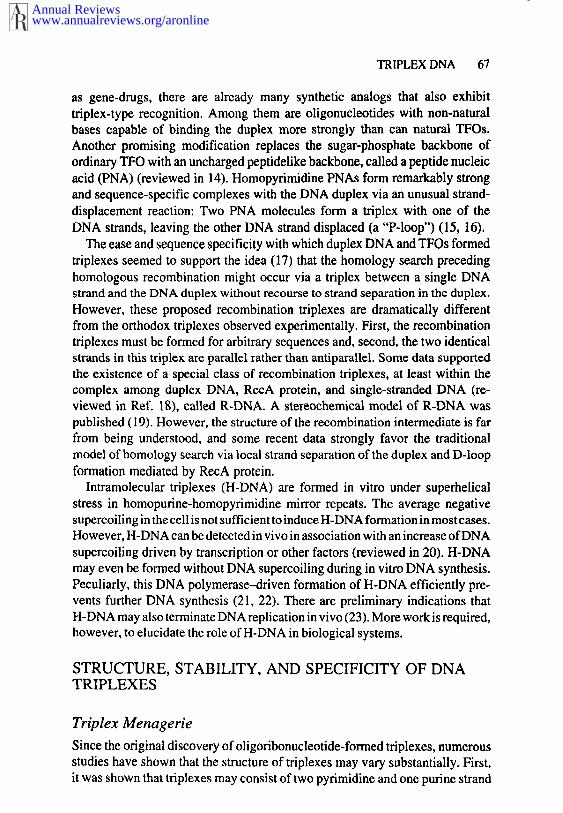
Triplex Menagerie
Since the original discovery of oligoribonucleotide-formed triplexes, numerousstudies have shown that the structure of triplexes may vary substantially. First,it was shown that triplexes may consist of two pyrimidine and one purine strand
Annual Reviewswww.annualreviews.org/aronline
68 FRANK-KAMENETSKII & MIRKIN
YR*Y YR*R Alternate Strand
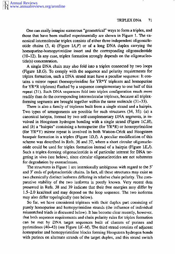
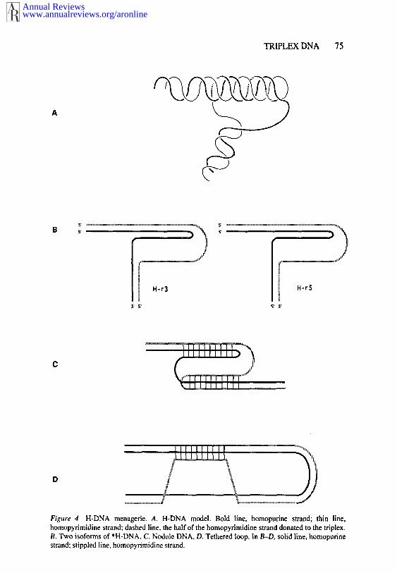
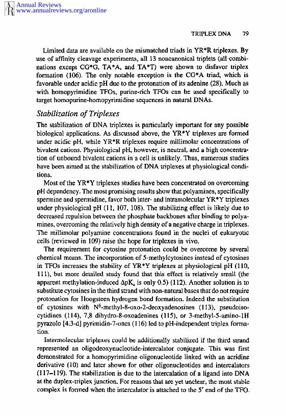
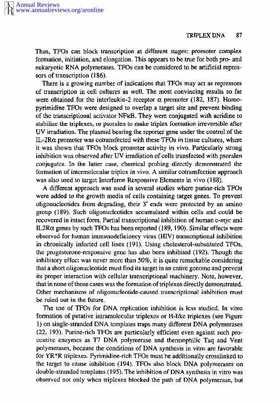
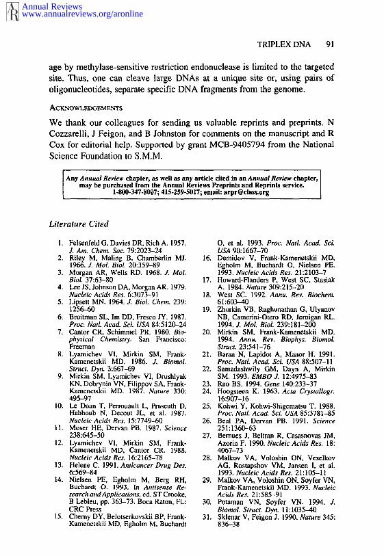
Figure I Triplex menagerie (see text for explanations). Solid lines, purine strands; stippled lines,pyrimidine strands; vertical lines, Watson-Crick hydrogen bonds; diamonds, Hoogsteen hydrogenbonds. Arrows indicate DNA chain polarity.
(YR*Y) or of two purine and one pyrimidine strand (YR*R). Second, triplexescan be built from RNA or DNA chains or their combinations. Third, triplexescan be formed within a single polymer molecule (intramolecular triplexes) by different polynucleotides (intermolecular triplexes). Finally, for specialDNA sequences consisting of clustered purines and pyrimidines in the samestrand, triplex formation may occur by a strand-switch mechanism (alternatestrand triplexes). Figure 1 summarizes numerous possible structures of triple-helical nucleic acids.
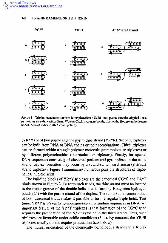
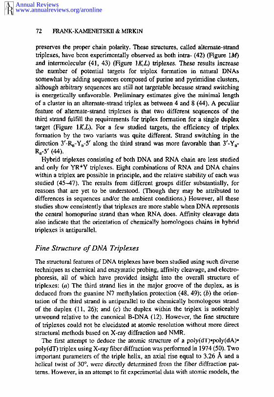
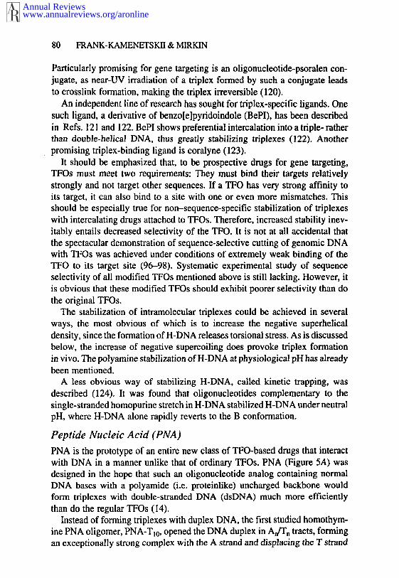
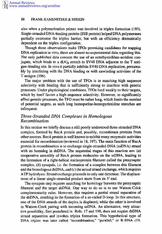
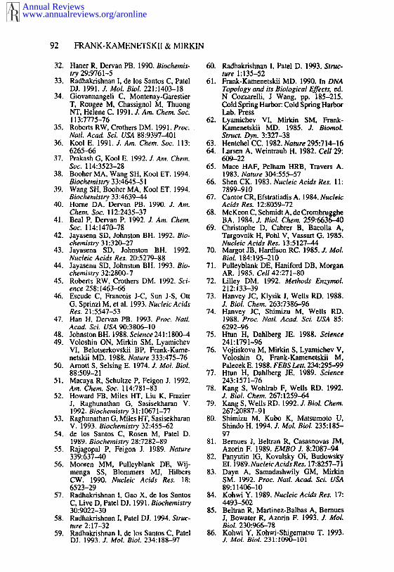
The building blocks of YR*Y triplexes are the canonical CG*C and TA*Ttriads shown in Figure 2. To form such triads, the third strand must be locatedin the major groove of the double helix that is forming Hoogsteen hydrogenbonds (24) with the purine strand of the duplex. The remarkable isomorphismof both canonical triads makes it possible to form a regular triple helix. Thislimits YR*Y triplexes to homopurine-homopyrimidine sequences in DNA. Animportant feature of the YR*Y triplexes is that formation of the CG*C triadrequires the protonation of the N3 of cytosine in the third strand. Thus, suchtriplexes are favorable under acidic conditions (3, 4). By contrast, the YR*Rtriplexes usually do not require protonation (see below).
The mutual orientation of the chemically homologous strands in a triplex
Annual Reviewswww.annualreviews.org/aronline
TRIPLEX DNA 69
R Ct H\N,/ ~C
o.#C’-,,,.~ c’,., ..,"
’,,,~ ~ ........ H- N41 H
¯ ;,.. /7 \-o:’,\ II ~ "".’-" .......... "\ N~--C’ / c~N~
/ \..~c: // \
/H
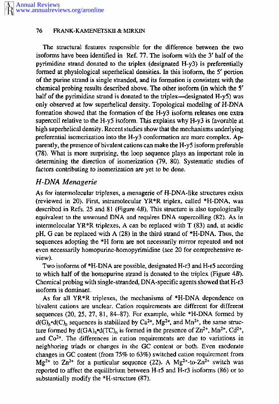
Figure 2 Canonical base triads of YR*Y triplexes: TA*T and CG*C*.
(i.e. two pyrimidine strands in the YR*Y triplex or the two purine strands inthe YR*R triplex), which a priori can be either parallel or antiparallel, is ofparamount importance. The discovery of H- and *H-DNA (see below) indi-cated that both YR*Y and YR*R triplexes form as antiparallel structures (9,25). A thorough investigation of intermolecular triplexes by different methodsunambiguously demonstrated that both YR*Y and YR*R triplexes are stablyformed only as antiparallel structures. The most direct data were obtained bycleaving target DNA with homopyrimidine or homopurine oligonucleotidesattached to FeoEDTA (11, 26). The observed pairing and orientation rulesrigorously determine the sequence of the triplex-forming homopyrimidinestrand.
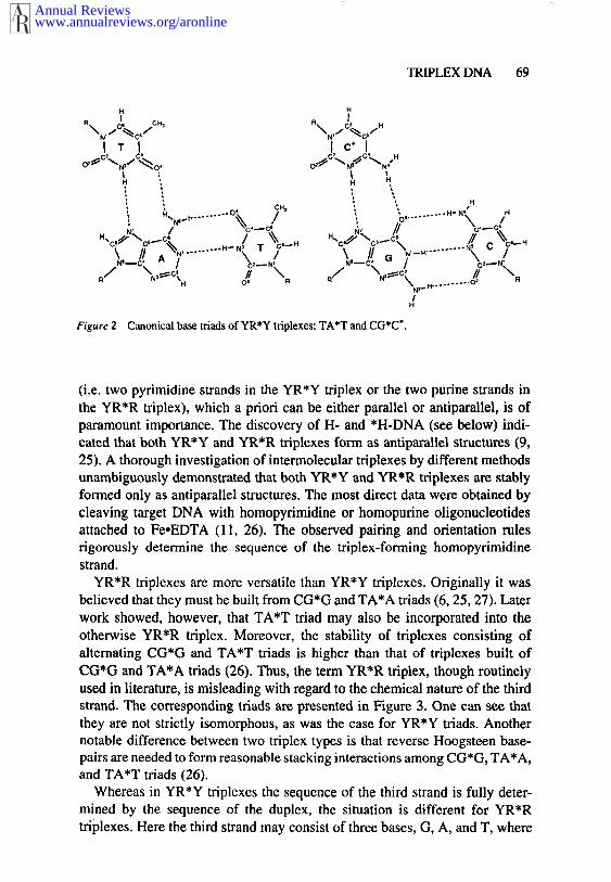
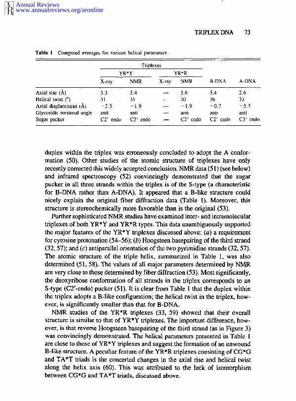
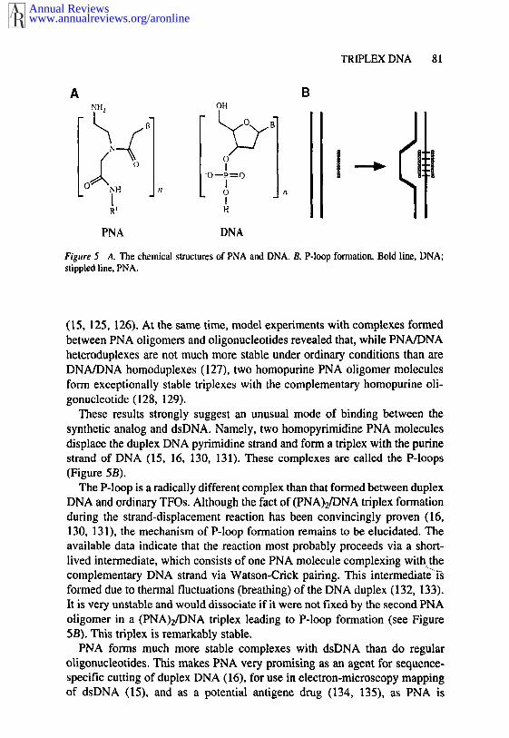
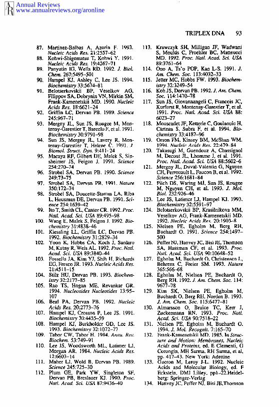
YR*R triplexes are more versatile than YR*Y triplexes. Originally it wasbelieved that they must be built from CG*G and TA*A triads (6, 25, 27). Laterwork showed, however, that TA*T triad may also be incorporated into theotherwise YR*R triplex. Moreover, the stability of triplexes consisting ofalternating CG*G and TA*T triads is higher than that of triplexes built ofCG*G and TA*A triads (26). Thus, the term YR*R triplex, though routinelyused in literature, is misleading with regard to the chemical nature of the thirdstrand. The corresponding triads are presented in Figure 3. One can see thatthey are not strictly isomorphous, as was the case for YR*Y triads. Anothernotable difference between two triplex types is that reverse Hoogsteen base-pairs are needed to form reasonable stacking interactions among CG*G, TA*A,and TA*T triads (26).
Whereas in YR*Y triplexes the sequence of the third strand is fully deter-mined by the sequence of the duplex, the situation is different for YR*Rtriplexes. Here the third strand may consist of three bases, G, A, and T, where
Annual Reviewswww.annualreviews.org/aronline
70 FRANK-KAMENETSKII & MIRKIN
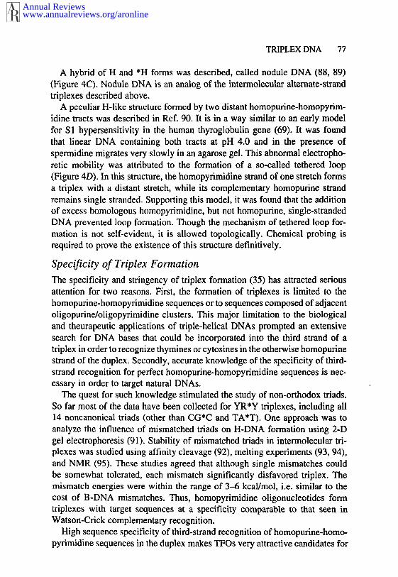
Figure 3 Base triads of YR*R triplexes: CG*G, TA*A, TA*T. and CG*A÷,
the guanines oppose guanines in the duplex, while adenines or thymines mustoppose adenines of the duplex. A protonated CG*A÷ triad (Figure 3) forms that A in the third strand may also oppose G in the duplex at acidic pH (28).
Another novel feature of YR*R triplexes is that their stability dependsdramatically on the presence of bivalent metal cations (reviewed in 20). Unlikethe case of YR*Y triplexes, where the requirement for H÷ ions has an obviousreason, the metal dependence of YR*R triplexes is an obscure function of theparticular metal ion and the triplex sequence (29). Possible structural reasonsfor selectivity of bivalent cations in stabilization of YR*R triplexes are dis-cussed in Ref. 30.
Despite these differences, the YR*R triplexes are similar to YR*Y triplexesin their most fundamental features: (a) The duplex involved in triplex forma-tion must have a homopurine sequence in one strand, and (b) the orientationof the two chemically homologous strands is antiparallel.
Annual Reviewswww.annualreviews.org/aronline
TRIPLEX DNA 71
One can easily imagine numerous "geometrical" ways to form a triplex, andthose that have been studied experimentally are shown in Figure 1. The ca-nonical intermolecular triplex consists of either three independent oligonucle-otide chains (3, 4) (Figure 1A, F) or of a long DNA duplex carrying homopurine-homopyrimidine insert and the corresponding oligonucleotide(10--12). In any case, triplex formation strongly depends on the oligonucleo-tide(s) concentration.
A single DNA chain may also fold into a triplex connected by two loops(Figure 1B, G). To comply with the sequence and polarity requirements fortriplex formation, such a DNA strand must have a peculiar sequence: It con-tains a mirror repeat (homopyrimidine for YR*Y triplexes and homopurinefor YR*R triplexes) flanked by a sequence complementary to one half of thisrepeat (31). Such DNA sequences fold into triplex configuration much morereadily than do the corresponding intermolecular triplexes, because all triplex-forming segments are brought together within the same molecule (31-33).
There is also a family of triplexes built from a single strand and a hairpin.Two types of arrangements are possible for such structures (34, 35): (a) canonical hairpin, formed by two self-complementary DNA segments, is in-volved in Hoogsteen hydrogen bonding with a single strand (Figure 1C, H),and (b) a "hairpin" containing a homopurine (for YR*R) or homopyrimidine(for YR*Y) mirror repeat is involved in both Watson-Crick and Hoogsteenbasepair formation in a triplex (Figure 1D,/). A peculiar modification of thisscheme was described in Refs. 36 and 37, where a short circular oligonucle-otide could be used for triplex formation instead of a hairpin (Figure 1E, J).Such a triplex-forming oligonucleotide is of particular interest for DNA tar-geting in vivo (see below), since circular oligonucleotides are not substratesfor degradation by exonucleases.
The structures in Figure 1 are intentionally ambiguous with regard to the 5’and 3’ ends of polynucleotide chains. In fact, all these structures may exist astwo chemically distinct isoforms differing in relative chain polarity. The com-parative stability of the two isoforms is poorly known. Very recent datapresented in Refs. 38 and 39 indicate that their free energies may differ by1.5-2.0 kcal/mol and may depend on the loop sequence. The two isoformsmay also differ topologically (see below).
So far, we have considered triplexes with their duplex part consisting ofpurely homopurine and homopyrimidine strands (the influence of individualmismatched triads is discussed below). It has become clear recently, however,that both sequence requirements and chain polarity rules for triplex formationcan be met by DNA target sequences built of clusters of purines andpyrimidines (40-43) (see Figure 1K-M). The third strand consists of adjacenthomopurine and homopyrimidine blocks forming Hoogsteen hydrogen bondswith purines on alternate strands of the target duplex, and this strand switch
Annual Reviewswww.annualreviews.org/aronline
72 FRANK-KAMENETSKII & MIRKIN
preserves the proper chain polarity. These structures, called alternate-strandtriplexes, have been experimentally observed as both intra- (42) (Figure and intermolecular (41, 43) (Figure 1K, L) triplexes. These results increasethe number of potential targets for triplex formation in natural DNAssomewhat by adding sequences composed of purine and pyrimidine clusters,although arbitrary sequences are still not targetable because strand switchingis energetically unfavorable. Preliminary estimates give the minimal lengthof a cluster in an alternate-strand triplex as between 4 and 8 (44). A peculiarfeature of alternate-strand triplexes is that two different sequences of thethird strand fulfill the requirements for triplex formation for a single duplextarget (Figure 1K, L). For a few studied targets, the efficiency of triplexformation by the two variants was quite different. Strand switching in thedirection 3’-Rn-Yn-5’ along the third strand was more favorable than 3’-Yn-
Rn-5’ (44).Hybrid triplexes consisting of both DNA and RNA chain are less studied
and only for YR*Y triplexes. Eight combinations of RNA and DNA chainswithin a triplex are possible in principle, and the relative stability of each wasstudied (45-47). The results from different groups differ substantially, forreasons that are yet to be understood. (Though they may be attributed todifferences in sequences and/or the ambient conditions.) However, all thesestudies show consistently that triplexes are more stable when DNA representsthe central homopurine strand than when RNA does. Affinity cleavage dataalso indicate that the orientation of chemically homologous chains in hybridtriplexes is antiparallel.
Fine Structure of DNA Triplexes
The structural features of DNA triplexes have been studied using such diversetechniques as chemical and enzymatic probing, affinity cleavage, and electro-phoresis, all of which have provided insight into the overall structure oftriplexes: (a) The third strand lies in the major groove of the duplex, as deduced from the guanine N7 methylation protection (48, 49); (b) the orien-tation of the third strand is antiparallel to the chemically homologous strandof the duplex (11, 26); and (c) the duplex within the triplex is noticeablyunwound relative to the canonical B-DNA (12). However, the fine structureof triplexes could not be elucidated at atomic resolution without more directstructural methods based on X-ray diffraction and NMR.
The first attempt to deduce the atomic structure of a poly(dT)opoly(dA)opoly(dT) triplex using X-ray fiber diffraction was performed in 1974 (50). important parameters of the triple helix, an axial rise equal to 3.26 A and ahelical twist of 30°, were directly determined from the fiber diffraction pat-terns. However, in an attempt to fit experimental data with atomic models, the
Annual Reviewswww.annualreviews.org/aronline
TRIPLEX DNA 73
Table 1 Computed averages for various helical parameters
TriplexesYR*Y YR*R
X-ray NMR X-ray NMR B-DNA A-DNA
Axial rise (/~) 3.3 3.4 -- 3.6 3.4 2.6Helical twist (°) 31 31 -- 30 36 33Axial displacement (/~) -2.5 -1.9 -- -1.9 -0.7 -5.3Glycosidic torsional angle anti anti -- anti anti antiSugar pucker C2’ endo C2’ endo -- C2’ endo C2’ endo CY endo
duplex within the triplex was erroneously concluded to adopt the A confor-mation (50). Other studies of the atomic structure of triplexes have onlyrecently corrected this widely accepted conclusion. NMR data (51) (see below)and infrared spectroscopy (52) convincingly demonstrated that the sugarpucker in all three strands within the triplex is of the S-type (a characteristicfor B-DNA rather than A-DNA). It appeared that a B-like structure couldnicely explain the original fiber diffraction data (Table 1). Moreover, thisstructure is stereochemically more favorable than is the original (53).
Further sophisticated NMR studies have examined inter- and intramoleculartriplexes of both YR*Y and YR*R types. This data unambiguously supportedthe major features of the YR*Y triplexes discussed above: (a) a requirementfor cytosine protonation (54-56); (b) Hoogsteen basepairing of the third strand(32, 57); and (c) antiparallel orientation of the two pyrimidine strands (32, The atomic structure of the triple helix, summarized in Table 1, was alsodetermined (51, 58). The values of all major parameters determined by NMRare very close to those determined by fiber diffraction (53). Most significantly,the deoxyribose conformation of all strands in the triplex corresponds to anS-type (C2’-endo) pucker (51). It is clear from Table 1 that the duplex withinthe triplex adopts a B-like configuration; the helical twist in the triplex, how-ever, is significantly smaller than that for B-DNA.
NMR studies of the YR*R triplexes (33, 59) showed that their overallstructure is similar to that of YR*Y triplexes. The important difference, how-ever, is that reverse Hoogsteen basepairing of the third strand (as in Figure 3)was convincingly demonstrated. The helical parameters presented in Table 1are close to those of YR*Y triplexes and suggest the formation of an unwoundB-like structure. A peculiar feature of the YR*R triplexes consisting of CG*Gand TA*T triads is the concerted changes in the axial rise and helical twistalong the helix axis (60). This was attributed to the lack of isomorphismbetween CG*G and TA*T triads, discussed above.
Annual Reviewswww.annualreviews.org/aronline
74 FRANK-KAMENETSKII & MIRKIN
H Form
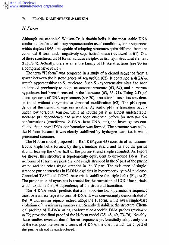
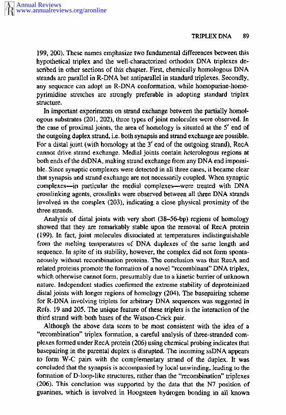
Although the canonical Watson-Crick double helix is the most stable DNAconformation for an arbitrary sequence under usual conditions, some sequenceswithin duplex DNA are capable of adopting structures quitedifferent from thecanonical B form under negatively superhelical stress (reviewed in 61). Oneof these structures, the H form, includes a triplex as its major structural element(Figure 4). Actually, there is an entire family of H-like structures (see 20 a comprehensive review).
The term "H form" was proposed in a study of a cloned sequence from aspacer between the histone genes of sea urchin (62). It contained a d(GA)~6stretch hypersensitive to S 1 nuclease. Such Sl-hypersensitive sites had beenanticipated previously to adopt an unusual structure (63, 64), and numeroushypotheses had been discussed in the literature (63, 65-71). Using 2-D gelelectrophoresis of DNA topoisomers (see 20), a structural transition was dem-onstrated without enzymatic or chemical modification (62). The pH depen-dency of the transition was remarkable: At acidic pH the transition occursunder low torsional tension, while at neutral pH it is almost undetectable.Because pH dependence had never been observed before for non-B-DNAconformations (cruciforms, Z-DNA, bent DNA, etc), the investigators con-eluded that a novel DNA conformation was formed. The structure was calledthe H form because it was clearly stabilized by hydrogen ions, i.e. it was aprotonated structure.
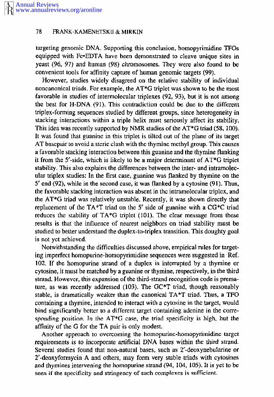
The H form model proposed in Ref. 8 (Figure 4A) consists of an intramo-lecular triple helix formed by the pyrimidine strand and half of the purinestrand, leaving the other half of the purine strand single stranded. As Figure4A shows, this structure is topologically equivalent to unwound DNA. Twoisoforms of H form are possible: one single stranded in the 5’ part of the purinestrand and the other single stranded in the 3’ part. The existence of single-stranded purine stretches in H-DNA explains its hyperreactivity to S 1 nuclease.Canonical TA*T and CG*C÷ base triads stabilize the triple helix (Figure 2).The protonation of cytosines is crucial for the formation of CGC÷ base triads,which explains the pH dependency of the structural transition.
The H-DNA model predicts that a homopurine-homopyrimidine sequencemust be a mirror repeat to form H-DNA. It was convincingly demonstrated inRef. 9 that mirror repeats indeed adopt the H form, while even single-baseviolations of the mirror symmetry significantly destabilize the structure. Chem-ical probing of H-DNA using conformation-specific DNA probes (reviewedin 72) provided final proof of the H-form model (25, 48, 49, 73-76). Notably,these studies revealed that different sequences preferentially adopt only oneof the two possible isomeric forms of H-DNA, the one in which the 5’ part ofthe purine strand is unstructured.
Annual Reviewswww.annualreviews.org/aronline
TRIPLEX DNA 75
H-r3
5~ 3’
Figure 4 H-DNA menagerie. A. H-DNA model. Bold line, homopurine strand; thin line,homopyrimidine strand; dashed line, the half of the homopyrimidine strand donated to the triplex.B. Two isoforms of *H-DNA. C. Nodule DNA. D. Tethered loop. In B-D, solid line, homopurinestrand; stippled line, homopyrimidine strand.
Annual Reviewswww.annualreviews.org/aronline
76 FRANK-KAMENETSKII & MIRKIN
The structural features responsible for the difference between the twoisoforms have been identified in Ref. 77. The isoform with the 3’ half of thepyrimidine strand donated to the triplex (designated H-y3) is preferentiallyformed at physiological superhelical densities. In this isoform, the 5’ portionof the purine strand is single stranded, and its formation is consistent with thechemical probing results described above. The other isoform (in which the 5’half of the pyrimidine strand is donated to the trlplex~esignated I-I-yS) wasonly observed at low superhelical density. Topological modeling of H-DNAformation showed that the formation of the H-y3 isoform releases one extrasupercoil relative to the H-y5 isoform. This explains why H-y3 is favorable athigh superhelical density. Recent studies show that the mechanisms underlyingpreferential isomerization into the H-y3 conformation are more complex. Ap-parently, the presence of bivalent cations can make the H-y5 isoform preferable(78). What is more surprising, the loop sequence plays an important role determining the direction of isomerization (79, 80). Systematic studies factors contributing to isomerization are yet to be done.
H-DNA Menagerie
As for intermolecular triplexes, a menagerie of H-DNA-like structures exists(reviewed in 20). First, intramolecular YR*R triplex, called *H-DNA, wasdescribed in Refs. 25 and 81 (Figure 4B). This structure is also topologicallyequivalent to the unwound DNA and requires DNA supercoiling (82). As intermolecular YR*R triplexes, A can be replaced with T (83) and, at acidicpH, G can be replaced with A (28) in the third strand of *H-DNA. Thus, thesequences adopting the *H form are not necessarily mirror repeated and noteven necessarily homopurine-homopyrimidine (see 20 for comprehensive re-view).
Two isoforms of *H-DNA are possible, designated H-r3 and H-r5 accordingto which half of the homopurine strand is donated to the triplex (Figure 4B).Chemical probing with single-stranded, DNA-specific agents showed that H-r3isoform is dominant.
As for all YR*R triplexes, the mechanisms of *H-DNA dependence onbivalent cations are unclear. Cation requirements are different for differentsequences (20, 25, 27, 81, 84-87). For example, while *H-DNA formed d(G)n’d(C)n sequences is stabilized by 2+, Mg2+, and Mn2+, the same struc-ture formed by d(GA)n°d(TC)n is formed in the presence of 2+, Mn2+, Cd2÷,
and Co2+. The differences in cation requirements are due to variations inneighboring triads or changes in the GC content or both. Even moderatechanges in GC content (from 75% to 63%) switched cation requirement fromMg2÷ to Zn2+ for a particular sequence (22). A Mg2+-to-Zn2+ switch wasreported to affect the equilibrium between H-r5 and H-r3 isoforms (86) or substantially modify the *H-structure (87).
Annual Reviewswww.annualreviews.org/aronline
TRIPLEX DNA 77
A hybrid of H and *H forms was described, called nodule DNA (88, 89)(Figure 4C). Nodule DNA is an analog of the intermolecular alternate-strandtriplexes described above.
A peculiar H-like structure formed by two distant homopurine-homopyrim-idine tracts was described in Ref. 90. It is in a way similar to an early modelfor S1 hypersensitivity in the human thyroglobulin gene (69). It was foundthat linear DNA containing both tracts at pH 4.0 and in the presence ofsperrnidine migrates very slowly in an agarose gel. This abnormal electropho-retic mobility was attributed to the formation of a so-called tethered loop(Figure 4D). In this structure, the homopyrimidine strand of one stretch formsa triplex with a distant stretch, while its complementary homopurine strandremains single stranded. Supporting this model, it was found that the additionof excess homologous homopyrimidine, but not homopurine, single-strandedDNA prevented loop formation. Though the mechanism of tethered loop for-mation is not self-evident, it is allowed topologically. Chemical probing isrequired to prove the existence of this structure definitively.
Specificity of Triplex Formation
The specificity and stringency of triplex formation (35) has attracted serousattention for two reasons. First, the formation of triplexes is limited to thehomopurine-homopyrimidine sequences or to sequences composed of adjacentoligopurine/oligopyrimidine clusters. This major limitation to the biologicaland theurapeutic applications of triple-helical DNAs prompted an extensivesearch for DNA bases that could be incorporated into the third strand of atriplex in order to recognize thymines or cytosines in the otherwise homopurinestrand of the duplex. Secondly, accurate knowledge of the specificity of third-strand recognition for perfect homopurine-homopydmidine sequences is nec-essary in order to target natural DNAs.
The quest for such knowledge stimulated the study of non-orthodox triads.So far most of the data have been collected for YR*Y triplexes, including all14 noncanonical triads (other than CG*C and TA*T). One approach was analyze the influence of mismatched triads on H-DNA formation using 2-Dgel electrophoresis (91). Stability of mismatched triads in intermolecular tri-plexes was studied using affinity cleavage (92), melting experiments (93, 94),and NMR (95). These studies agreed that although single mismatches couldbe somewhat tolerated, each mismatch significantly disfavored triplex. Themismatch energies were within the range of 3-6 kcal/mol, i.e. similar to thecost of B-DNA mismatches. Thus, homopyrimidine oligonucleotides formtriplexes with target sequences at a specificity comparable to that seen inWatson-Crick complementary recognition.
High sequence specificity of third-strand recognition of homopurine-homo-pyrimidine sequences in the duplex makes TFOs very attractive candidates for
Annual Reviewswww.annualreviews.org/aronline
78 FRANK-KAMENETSKII & MIRKIN
targeting genomic DNA. Supporting this conclusion, homopyrimidine TFOsequipped with FeoEDTA have been demonstrated to cleave unique sites inyeast (96, 97) and human (98) chromosomes. They were also found to convenient tools for affinity capture of human genomic targets (99).
However, studies widely disagreed on the relative stability of individualnoncanonical triads. For example, the AT*G triplet was shown to be the mostfavorable in studies of intermolecular triplexes (92, 93), but it is not amongthe best for H-DNA (91). This contradiction could be due to the differenttriplex-forming sequences studied by different groups, since heterogeneity instacking interactions within a triple helix must seriously affect its stability.This idea was recently supported by NMR studies of the AT*G triad (58, 100).It was found that guanine in this triplet is tilted out of the plane of its targetAT basepair to avoid a steric clash with the thymine methyl group. This causesa favorable stacking interaction between this guanine and the thymine flankingit from the 5’-side, which is likely to be a major determinant of AT*G tripletstability. This also explains the differences between the inter- and intramolec-ular triplex studies: In the first case, guanine was flanked by thymine on the5’ end (92), while in the second case, it was flanked by a cytosine (91). Thus,the favorable stacking interaction was absent in the intramolecular triplex, andthe AT*G triad was relatively unstable. Recently, it was shown directly thatreplacement of the TA*T triad on the 5’ side of guanine with a CG*C triadreduces the stability of TA*G triplet (101). The clear message from theseresults is that the influence of nearest neighbors on triad stability must bestudied to better understand the duplex-to-triplex transition. This doughty goalis not yet achieved.
Notwithstanding the difficulties discussed above, empirical rules for target-ing imperfect homopurine-homopyrimidine sequences were suggested in Ref.102. If the homopurine strand of a duplex is interrupted by a thymine orcytosine, it must be matched by a guanine or thymine, respectively, in the thirdstrand. However, this expansion of the third-strand recognition code is prema-ture, as was recently addressed (103). The GC*T triad, though reasonablystable, is dramatically weaker than the canonical TA*T triad. Thus, a TFOcontaining a thymine, intended to interact with a cytosine in the target, wouldbind significantly better to a different target containing adenine in the corre-sponding position. In the AT*G case, the triad specificity is high, but theaffinity of the G for the TA pair is only modest.
Another approach to overcoming the homopurine-homopyrimidine targetrequirements is to incorporate artificial DNA bases within the third strand.Several studies found that non-natural bases, such as 2’-deoxynebularine or2’-deoxyformycin A and others, may form very stable triads with cytosinesand thymines intervening the homopurine strand (94, 104, 105). It is yet to seen if the specificity and stringency of such complexes is sufficient.
Annual Reviewswww.annualreviews.org/aronline
TRIPLEX DNA 79
Limited data are available on the mismatched triads in YR*R triplexes. Byuse of affinity cleavage experiments, all 13 noncanonical triplets (all combi-nations except CG*G, TA*A, and TA*T) were shown to disfavor triplexformation (106). The only notable exception is the CG*A triad, which favorable under acidic pH due to the protonation of its adenine (28). Much with homopyrimidine TFOs, purine-dch TFOs can be used specifically totarget homopurine-homopyrimidine sequences in natural DNAs.
Stabilization of Triplexes
The stabilization of DNA triplexes is particularly important for any possiblebiological applications. As discussed above, the YR*Y triplexes are formedunder acidic pH, while YR*R triplexes require millimolar concentrations ofbivalent cations. Physiological pH, however, is neutral, and a high concentra-tion of unbound bivalent cations in a cell is unlikely. Thus, numerous studieshave been aimed at the stabilization of DNA triplexes at physiological condi-tions.
Most of the YR*Y triplexes studies have been concentrated on overcomingpH dependency. The most promising results show that polyamines, specificallyspermine and spermidine, favor both inter- and intramolecular YR*Y triplexesunder physiological pH (11, 107, 108). The stabilizing effect is likely due decreased repulsion between the phosphate backbones after binding to polya-mines, overcoming the relatively high density of a negative charge in triplexes.The millimolar polyamine concentrations found in the nuclei of eukaryoticcells (reviewed in 109) raise the hope for triplexes in vivo.
The requirement for cytosine protonation could be overcome by severalchemical means. The incorporation of 5-methylcytosines instead of cytosinesin TFOs increases the stability of YR*Y triplexes at physiological pH (110,111), but more detailed study found that this effect is relatively small (theapparent methylation-induced ApI~ is only 0.5) (112). Another solution is substitute cytosines in the third strand with non-natural bases that do not requireprotonation for Hoogsteen hydrogen bond formation. Indeed the substitutionof cytosines with Nr-methyl-8-oxo-2-deoxyadenosines (113), pseudoiso-cytidines (114), 7,8 dihydro-8-oxoadenines (115), or 3-methyl-5-amino-lHpyrazolo [4.3-d] pyrimidin-7-ones (116) led to pH-independent triplex forma-tion.
Intermolecular triplexes could be additionally stabilized if the third strandrepresented an oligodeoxynucleotide-intercalator conjugate. This was firstdemonstrated for a homopyrimidine oligonucleotide linked with an acridinederivative (10) and later shown for other oligonucleotides and intercalators(117-119). The stabilization is due to the intercalation of a ligand into DNAat the duplex-triplex junction. For reasons that are yet unclear, the most stablecomplex is formed when the intercalator is attached to the 5’ end of the TFO.
Annual Reviewswww.annualreviews.org/aronline
80 FRANK-KAMENETSKII & MIRKIN
Particularly promising for gene targeting is an oligonucleotide-psoralen con-jugate, as near-UV irradiation of a triplex formed by such a conjugate leadsto crosslink formation, making the triplex irreversible (120).
An independent line of research has sought for triplex-specific ligands. Onesuch ligand, a derivative of benzo[e]pyridoindole (BePI), has been describedin Refs. 121 and 122. BePI shows preferential intercalation into a triple- ratherthan double-helical DNA, thus greatly stabilizing triplexes (122). Anotherpromising triplex-binding ligand is coralyne (123).
It should be emphasized that, to be prospective drugs for gene targeting,TFOs must meet two requirements: They must bind their targets relativelystrongly and not target other sequences. If a TFO has very strong affinity toits target, it can also bind to a site with one or even more mismatches. Thisshould be especially true for non-sequence-specific stabilization of triplexeswith intercalating drugs attached to TFOs. Therefore, increased stability inev-itably entails decreased selectivity of the TFO. It is not at all accidental thatthe spectacular demonstration of sequence-selective cutting of genomic DNAwith TFOs was achieved under conditions of extremely weak binding of theTFO to its target site (96-98). Systematic experimental study of sequenceselectivity of all modified TFOs mentioned above is still lacking. However, itis obvious that these modified TFOs should exhibit poorer selectivity than dothe original TFOs.
The stabilization of intramolecular triplexes could be achieved in severalways, the most obvious of which is to increase the negative superhelicaldensity, since the formation of H-DNA releases torsional stress. As is discussedbelow, the increase of negative supereoiling does provoke triplex formationin vivo. The polyamine stabilization of H-DNA at physiological pH has alreadybeen mentioned.
A less obvious way of stabilizing H-DNA, called kinetic trapping, wasdescribed (124). It was found that oligonucleotides complementary to thesingle-stranded homopurine stretch in H-DNA stabilized H-DNA under neutralpH, where H-DNA alone rapidly reverts to the B conformation.
Peptide Nucleic Acid (PNA)
PNA is the prototype of an entire new class of TFO-based drugs that interactwith DNA in a manner unlike that of ordinary TFOs. PNA (Figure 5A) wasdesigned in the hope that such an oligonucleotide analog containing normalDNA bases with a polyamide (i.e. proteinlike) uncharged backbone wouldform triplexes with double-stranded DNA (dsDNA) much more efficientlythan do the regular TFOs (14).
Instead of forming triplexes with duplex DNA, the first studied homothym-ine PNA oligomer, PNA-T~0, opened the DNA duplex in Ad’l’n tracts, formingan exceptionally strong complex with the A strand and displacing the T strand
Annual Reviewswww.annualreviews.org/aronline
TRIPLEX DNA 81
OH
B
PNA DNA
Figure 5 A. The chemical structures of PNA and DNA. B. P-loop formation. Bold line, DNA;stippled line, PNA.
(15, 125, 126). At the same time, model experiments with complexes formedbetween PNA oligomers and oligonucleotides revealed that, while PNA/DNAheteroduplexes are not much more stable under ordinary conditions than areDNA/DNA homoduplexes (127), two homopurine PNA oligomer moleculesform exceptionally stable triplexes with the complementary homopurine oli-gonucleotide (128, 129).
These results strongly suggest an unusual mode of binding between thesynthetic analog and dsDNA. Namely, two homopyrimidine PNA moleculesdisplace the duplex DNA pyrimidine strand and form a triplex with the purinestrand of DNA (15, 16, 130, 131). These complexes are called the P-loops(Figure 5B).
The P-loop is a radically different complex than that formed between duplexDNA and ordinary TFOs. Although the fact of (PNA)2/DNA triplex formationduring the strand-displacement reaction has been convincingly proven (16,130, 131), the mechanism of P-loop formation remains to be elucidated. Theavailable data indicate that the reaction most probably proceeds via a short-lived intermediate, which consists of one PNA molecule complexing with, thecomplementary DNA strand via Watson-Crick pairing. This ~ntermedlate ~sformed due to thermal fluctuations (breathing) of the DNA duplex (132, 133).It is very unstable and would dissociate if it were not fixed by the second PNAoligomer in a (PNA)2/DNA triplex leading to P-loop formation (see Figure5B). This triplex is remarkably stable.
PNA forms much more stable complexes with dsDNA than do regularoligonucleotides. This makes PNA very promising as an agent for sequence-specific cutting of duplex DNA (16), for use in electron-microscopy mappingof dsDNA (15), and as a potential antigene drug (134, 135), as PNA
Annual Reviewswww.annualreviews.org/aronline
82 FRANK-KAMENETSKII & MIRKIN
remarkably stable in biological fluids in which normal peptides and oligonu-cleotides are quickly degraded (136).
However, serious limitations for various applications of PNA still remain.P-loop formation proceeds through a significant kinetic barrier and stronglydepends on ionic conditions (15, 16, 125, 126). This dependency, if not by-passed, poses significant limitations on possible sequence-specific targeting ofdsDNA by PNA under physiological conditions. Although the stringency of(PNA)2/DNA triplexes is not yet known, PNA should still target predominatelyhomopurine-homopyrimidine regions, just as do regular TFOs.
BIOCHEMISTRY OF TRIPLEXES
Formation and Possible Functions of H-DNA In Vivo
As is true for other unusual DNA structures, such as erueiforms, Z-DNA, andquadruplexes, the biological role of H-DNA is yet to be established. Twoimportant problems must be addressed: (a) Can H-DNA be formed in cells principle? (b) In which biological process if any is H-DNA involved? Recentlyit became clear that the answer to the first question is yes. There are currentlymany hypotheses on the role of H-DNA in DNA replication, transcription, andrecombination, but more studies are needed to answer the second question.
Sequences that can form H-DNA are widespread throughout the eukaryoticgenomes (137, 138) but are uncommon among eubacteria. However, directdetection of H-DNA in eukaryotic cells is very difficult because of the com-plexity of genomic DNA. Therefore, most of the studies on the detection ofH-DNA in vivo exploited Escherlchia coli cells bearing recombinant plasrnidswith triplex-forming inserts as convenient model systems. Chemical probingof intracellular DNA proved helpful for the detection of H-DNA in vivo.Certain chemicals, such as osmium tetroxide, chloroacetaldehyde, and psora-len, give a characteristic pattern of H- or *H-DNA modification in vitro.Conveniently, they can also penetrate living cells. Thus, the general strategyfor detecting H-DNA in vivo was to treat E. coli cells with those chemicals,isolate plasmid DNA, and locate modified DNA bases at a sequence level.The coincidence of modification patterns in vitro and in vivo basically provedthe formation of the unusual structure in the cell.
Using this approach, the formation of both H- and *H-DNA was directlyshown (139-141). The corresponding studies were reviewed in Ref. 20, butwe briefly summarize the major findings. All these studies agreed that the levelof DNA supercoiling in vivo is the major limiting factor in the formation ofthese structures. Though transient formation of H-DNA was observed in nor-mal exponentially growing E. coli cells (141), formation of H-DNA was muchmore pronounced when intracellular DNA supercoiling increased, due to mu-
Annual Reviewswww.annualreviews.org/aronline
TRIPLEX DNA 83
tations in the gene for Topo I (141) or due to treatment of cells with chloram-phenicol (139, 140). Environmental conditions during E. coli growth alsosignificantly contributed to the appearance of triplexes. H-DNA formation wasgreatly enhanced when cells were growing in mildly acidic media, whichsomewhat decreased intracellular pH (139, 141) while *H-DNA was observedin cells growing in media with a high concentration of Mgz÷ ions (140). Neitherresult is surprising, because H-DNA is stabilized by protonation while *H-DNA is stabilized by bivalent cations.
Besides the steady-state level of DNA supercoiling, determined by thebalance of DNA gyrase and Topo I (reviewed in 142), the local level supercoiling strongly depends on transcription. During the process of poly-merization the RNA polymerase creates domains of high negative and positivesupercoiling upstream and downstream of it, respectively (143), which mayinfluence the formation of unusual DNA structures (144, 145). Chemicalprobing of intracellular DNA demonstrated transcriptionally driven formationof *H-DNA within long d(G)nod(C)n stretches located upstream of a regulatedpromoter in an E. coli plasmid (146). Remarkably, the formation of *H-DNAstimulated homologous recombination between direct repeats flanking thestructure. Thus, this work shows the formation of *H-DNA under completelyphysiological conditions in a cell, and implicates it in the process of recom-bination.
The only data on triplex DNA detection in eukaryotic cells were obtainedusing antibodies against triple-helical DNA (147). These antibodies were foundto interact with eukaryotic chromosomes (148, 149).
Many ideas have been proposed involving H-DNA in such basic geneticprocesses as replication and transcription. The hypothesis regarding H-DNAin replication is based on the observation that triplex structures prevent DNAsynthesis in vitro. On supercoiled templates containing *H-DNA, DNA syn-thesis prematurely terminates. The location of the termination site is differentfor different isoforms of *H-DNA, but it always coincides with the triplexboundaries as defined by chemical probing (83).
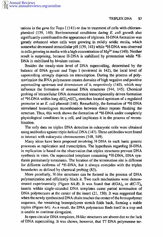
More peculiarly, H-like structures can be formed in the process of DNApolymerization and efficiently block it. Two such mechanisms were demon-strated experimentally (Figure 6A,B). It was found that d(GA)n or d(C-T)ninserts within single-stranded DNA templates cause partial termination ofDNA polymerases at the center of the insert (21, 150). It was suggested thatwhen the newly synthesized DNA chain reaches the center of the homopolymersequence, the remaining homopolymer stretch folds back, forming a stabletriplex (Figure 6A). As a result, the DNA polymerase finds itself in a trap andis unable to continue elongation,
In open circular DNA templates, H-like structures are absent due to the lackof DNA supercoiling. It was shown, however, that T7 DNA polymerase tcr-
Annual Reviewswww.annualreviews.org/aronline
84 FRANK-KAMENETSKII & MIRKIN
Figure 6 DNA polymerase--driven triplex formation blocks polymerization. Black boxes, the twohalves of a homopurine-homopyrimidine mirror repeat involved in the formation of anintramolecular triplex; striated arrow, the newly synthesized DNA chain. A. Single-stranded DNAtemplate. B. Double-stranded DNA template.
minated exactly at the center of *H-forming sequences. This was observedwhen the pyrimidine-rich but not the purine-rich strand served as a template(22). To explain this one must remember that DNA synthesis on double-stranded templates is possible due to the ability of many DNA polymerases todisplace the nontemplate DNA strand (reviewed in 151). The displaced strandmay fold back, promoting the formation of an intramolecular triplex down-stream of the replication fork at an appropriate sequence. Conditions for DNAsynthesis in vitro--i.e, neutral pH and high magnesium concentration--areoptimal for the formation of YR*R triplexes. Thus, the displacement of thepurine-rich (but not the pyrimidine-rich) strand provokes triplex formationwhich, in turn, leads to termination of DNA synthesis (Figure 6B).
There are only fragmentary data on the role of H motifs in the regulationof replication in vivo. Several homopudne-homopyrimidine inserts wereshown to decrease the efficiency of Simian virus 40 (SV40) DNA replication(152, 153). Quite recently, the pausing of the replication fork in vivo withina d(GA)n°d(TC)n insert in SV40 DNA was demonstrated directly using technique called two-dimensional neutral/neutral gel electrophoresis (23).Though these data make the idea of H-DNA involvement in the regulation ofreplication promising, it is far from proven. Future studies are crucial for theevaluation of this hypothesis.
Numerous studies concerned the possible role of H-DNA in transcription.Deletion analysis of various promoters--including Drosophila hsp26 (154,155); mouse c-Ki-ras (156) and TGF-1~3 (157); human EGFR (158), ets-2(159), IR (160), c-rnyc (161,162); and othersmshowed thathomopurine-homopydmidine stretches are essential for promoter functioning.
These sequences serve as targets for nuclear proteins, presumably transcrip-
Annual Reviewswww.annualreviews.org/aronline
TRIPLEX DNA 85
tional activators. Several homopurine-homopyrimidine DNA-binding proteinswere described, including BPG1 (163), NSEP-1 (164), MAZ (165), nm23-H2(166), PYBP (167), Pur-1 (168), etc. Peculiarly, these proteins often preferentially to just one strand of the H motifs. For example, a number ofmammalian proteins specifically recognize homopurine-homopyrimidine se-quences in the double-helical state as well as the corresponding homopyrimid-ine single strands (164, 167, 169, 170). This unusual binding pattern maydramatically influence the equilibrium between different DNA conformationsin the promoter in vivo.
However, the importance of the H structure for transcription was questionedin several studies. One approach is to analyze the influence of point mutationswithin H motifs that destroy or restore H-forming potential on the promoter’sactivity. No such correlation was observed for Drosophila hsp26 (155) andmouse c-Ki-ras (171) promoters. The situation with the c-myc promoter ismore complex, since it is unclear if the canonical I-I-DNA or some otherstructure is formed even in vitro (172). Mutational analysis of the promotergave contradictory results, with one group claiming the existence (173) andanother the lack (174) of a correlation between structural potential and pro-moter strength. Another approach to detecting H-DNA in eukaryotic promotersis direct chemical probing followed by genomic sequencing. So far, this hasonly been done for the Drosophila hsp26 gene, and H-DNA was not observed(155).
It is hard to completely rule out the role of H-DNA in transcription basedon the above results. First, it is quite possible that the structural peculiaritiesof promoter DNA segments may affect the interaction between promoter DNAand specific regulator proteins. The features of homopurine-homopyrimidineDNA-binding proteins described above as well as a report about the partialpurification of a triplex-binding protein (175) indirectly support this idea. study in which the influence of d(G)n stretches of varying length on the activityof a downstream minimal promoter was analyzed additionally supports thishypothesis (176). A clear reverse correlation between the ability of a stretchto form the *H configuration in vitro and its ability to activate transcriptionin vivo was observed. It was concluded, therefore, that short d(G)n stretchesserve as binding sites for a transcriptional activator, while longer stretchesadopt a triplex configuration, which prevents activator binding. Secondly,negative data on the role of H-DNA in transcription were obtained in transientassays, while it can actually work at a chromosome level. Indeed, H motif inthe Drosophila hsp26 gene was found to affect the chromatin structure (177,178).
Despite the wealth of data and hypotheses, there is no direct evidence thatthe structural features of H motifs are involved in transcriptional regulation invivo, and further studies are required to address this issue.
Annual Reviewswww.annualreviews.org/aronline
86 FRANK-KAMENETSKII & MIRKIN
Targeting Basic Genetic Processes Using TFOs
Highly sequence-specific recognition of double-helical DNAs by TFOs is thebasis of an antigene strategy (reviewed in 13). The idea is that binding of TFO to a target gene could prevent its normal functioning. Most studies ofthis strategy concerned the inhibition of transcription; the studies were inspiredin part by the existence of functionally important homopurine-homopyrimidinestretches in many eukaryotic promoters (see the previous section), which areappropriate targets for TFOs. The antigene strategy could potentially lead torational drug design. Very convincing data on the inhibitory effects of TFOswere obtained in various in vitro systems. There are also preliminary indica-tions that TFOs may function in vivo as well.
The first stage that is a~’fected by TFOs is the formation of an active promotercomplex. Pioneering results were obtained for the human c-myc promoter,where it was found that the binding of a purine-rich TFO to the imperfecthomopurine-homopyrimidine sequence 125 basepairs (bp) upstream of the promoter start site blocks its transcription in vitro (179). The TFO’s target important for c-myc transcription, serving as a binding site for a protein(s),presumably a transcriptional activator (161,162). At least two candidate genescoding for proteins that bind to this target have been cloned and sequenced(164, 166). Similar observations were made for the methallothionein genepromoter. In this case a homopyrimidine oligonucleotide formed a triplex withthe upstream portion of the promoter, preventing the binding of the transcrip-tional activator Sp1 (111). This in turn drastically reduced the promoter’sactivity in a cell-free transcription system (179a). TFOs were also shown prevent SPI binding to the human DHFR (180) and H-ras (181) promoters.Finally, a triplex-forming oligonucleotide-intercalator conjugate was shown toact as a transcriptional repressor of the interleukin-2 receptor c~ gene in vitro(182), preventing the binding of the transcriptional activator NFvA3. In all thesecases TFOs efficiently blocked the access of the transcription factors to theirbinding sites.
TFOs also inhibit initiation of transcription by RNA polymerases. ThepBR322 bla-gene contains a 13-bp homopurine-homopyrimidine target justdownstream of the transcriptional start site. A 13-mer homopyrimidine oligo-nucleotide forming an intermolecular triplex with this target hindered initiationof transcription by E. coli RNA polymerase in vitro (183). Independent studiesshowed that this is also the case for T7 RNA polymerase (184).
Finally, eukaryotic RNA polymerase II transcription was followed in vitrofrom the adenovirus major late promoter (185). The transcribed portion DNA contained a 15-bp homopurine-homopyrimidine tract that formed anintermolecular triplex with the homopyrimidine TFO. When added prior toRNA polymerase, the TFO truncated a significant portion of the transcripts.
Annual Reviewswww.annualreviews.org/aronline
TRIPLEX DNA 87
Thus, TFOs can block transcription at different stages: promoter complexformation, initiation, and elongation, This appears to be true for both pro- andeukaryotic RNA polymerases. TFOs can be considered to be artificial repres-sors of transcription (186).
There is a growing number of indications that TFOs may act as repressorsof transcription in cell cultures as well. The most convincing results so farwere obtained for the interleukin-2 receptor ct promoter (182, 187). Homo-pyrimidine TFOs were designed to overlap a target site and prevent bindingof the transcriptional activator NFrd3. They were conjugated with acridine tostabilize the triplexes, or psoralen to make triplex formation irreversible afterUV irradiation. The plasmid bearing the reporter gene under the control of theIL-2R~x promoter was cotransfected with these TFOs in tissue cultures, whereit was shown that TFOs block promoter activity in vivo. Particularly stronginhibition was observed after UV irradiation of cells transfected with psoralenconjugates. In the latter case, chemical probing directly demonstrated theformation of intermolecular triplex in vivo. A similar cotransfection approachwas also used to target Interferon Responsive Elements in vivo (188).
A different approach was used in several studies where purine-rich TFOswere added to the growth media of cells containing target genes. To preventoligonucleotides from degrading, their 3’ ends were protected by an aminogroup (189). Such oligonucleotides accumulated within cells and could recovered in intact form. Partial transcriptional inhibition of human c-tnyc andIL2R~x genes by such TFOs has been reported (189, 190). Similar effects wereobserved for human immunodeficiency virus (HIV) transcriptional inhibitionin chronically infected cell lines (191). Using cholesterol-substituted TFOs,the progesterone-responsive gene has also been inhibited (192). Though theinhibitory effect was never more than 50%, it is quite remarkable consideringthat a short oligonucleotide must find its target in an entire genome and preventits proper interaction with cellular transcriptional machinery. Note, however,that in none of those cases was the formation of triplexes directly demonstrated.Other mechanisms of oligonucleotide-caused transcriptional inhibition mustbe ruled out in the future.
The use of TFOs for DNA replication inhibition is less studied. In vitroformation of putative intramolecular triplexes or H-like triplexes (see Figure1) on single-stranded DNA templates traps many different DNA polymerases(22, 193). Purine-rich TFOs are particularly efficient even against such pro-cessive enzymes as T7 DNA polymerase and thermophilic Taq and Ventpolymerases, because the conditions of DNA synthesis in vitro are favorablefor YR*R triplexes. Pyrimidine-rich TFOs must be additionally crosslinked tothe target to cause inhibition (194). TFOs also block DNA polymerases double-stranded templates (195). The inhibition of DNA synthesis in vitro wasobserved not only when triplexes blocked the path of DNA polymerase, but
Annual Reviewswww.annualreviews.org/aronline
88 FRANK-KAMENETSKII & MIRKIN
also when a polymerization primer was involved in triplex formation (193).Single-stranded DNA-binding protein (SSB protein) helped DNA polymerasespartially overcome the triplex barrier, but with an efficiency dramaticallydependent on the triplex configuration.
Though these observations make TFOs promising candidates for trappingDNA replication in vivo, there are almost no experimental data regarding this.The only published data concern the use of an octathymidilate-acddine con-jugate, which binds to a d(A)8 stretch in SV40 DNA adjacent to the T anti-gen-binding site. In vivo it partially inhibits SV40 DNA replication, presuma-bly by interfering with the DNA binding or with unwinding activities of theT antigen (196).
The major problem with the use of TFOs is in matching high sequenceselectivity with binding that is sufficiently strong to interfere with geneticprocesses. Under physiological conditions, TFOs bind weakly to their targets,which by itself favors a high sequence selectivity. However, to significantlyaffect genetic processes, the TFO must be rather long, which limits the numberof potential targets, as such long homopudne-homopyrimidine stretches areinfrequent.
Three-Stranded DNA Complexes in HomologousRecombinationIn this section we briefly discuss a still poorly understood three-stranded DNAcomplex, formed by RecA protein and, possibly, recombinant proteins fromother sources. RecA protein is well known to exhibit many enzymatic activitiesessential for recombination (reviewed in 18, 197). The main function of RecAprotein in recombination is to exchange single-stranded DNA (ssDNA) strandwith its homolog in dsDNA. The sequential stages of this reaction are: (a)cooperative assembly of RecA protein molecules on the ssDNA, leading tothe formation of a right-helical nucleoprotein filament called the presynapticcomplex, (b) synapsis, i.e. the formation of a complex between this filamentand the homologous dsDNA, and (c) the actual strand exchange, which requiresATP hydrolysis. Strand exchange proceeds in only one direction: The displace-ment of a linear single-stranded product starts from its 5" end.
The synapsis step requires searching for homology between the presynapticfilament and the target dsDNA. One way to do so is to use Watson-Crickcomplementarity rules. However, this requires a partial strand separation ofthe dsDNA, resulting in the formation of a so-called D-loop. In this structure,one of the DNA strands of the duplex is displaced, while the other is involvedin Watson-Crick pairing with incoming ssDNA. An alternative, very attrac-tive possibility, first postulated in Refs. 17 and 198, does not require dsDNAstrand separation and invokes triplex formation. This hypothetical type ofDNA triplex was later called "recombination," "parallel," or R-DNA (19,
Annual Reviewswww.annualreviews.org/aronline
TRIPLEX DNA 89
199, 200). These names emphasize two fundamental differences between thishypothetical triplex and the well-characterized orthodox DNA triplexes de-scribed in other sections of this chapter. First, chemically homologous DNAstrands are parallel in R-DNA but antiparallel in standard triplexes. Secondly,any sequence can adopt an R-DNA conformation, while homopurine-homo-pyrimidine stretches are strongly preferable in adopting standard triplexstructure.
In important experiments on strand exchange between the partially homol-ogous substrates (201,202), three types of joint molecules were observed. the case of proximal joints, the area of homology is situated at the 5’ end ofthe outgoing duplex strand, i.e. both synapsis and strand exchange are possible.For a distal joint (with homology at the 3’ end of the outgoing strand), RecAcannot drive strand exchange. Medial joints contain heterologous regions atboth ends of the dsDNA, making strand exchange from any DNA end impossi-ble. Since synaptic complexes were detected in all three cases, it became clearthat synapsis and strand exchange are not necessarily coupled. When synapticcomplexesmin particular the medial complexesmwere treated with DNAcrosslinking agents, crosslinks were observed between all three DNA strandsinvolved in the complex (203), indicating a close physical proximity of thethree strands.
Analysis of distal joints with very short (38-56-bp) regions of homologyshowed that they are remarkably stable upon the removal of RecA protein(199). In fact, joint molecules dissociated at temperatures indistinguishablefrom the melting temperatures of DNA duplexes of the same length andsequence. In spite of its stability, however, the complex did not form sponta-neously without recombination proteins. The conclusion was that RecA andrelated proteins promote the formation of a novel "recombinant" DNA triplex,which otherwise cannot form, presumably due to a kinetic barrier of unknownnature. Independent studies confirmed the extreme stability of deproteinizeddistal joints with longer regions of homology (204). The basepairing schemefor R-DNA involving triplets for arbitrary DNA sequences was suggested inRefs. 19 and 205. The unique feature of these triplets is the interaction of thethird strand with both bases of the Watson-Crick pair.
Although the above data seem to be most consistent with the idea of a"recombination" triplex formation, a careful analysis of three-stranded com-plexes formed under RecA protein (206) using chemical probing indicates thatbasepairing in the parental duplex is disrupted. The incoming ssDNA appearsto form W-C pairs with the complementary strand of the duplex. It wasconcluded that the synapsis is accompanied by local unwinding, leading to theformation of D-loop-like structures, rather than the "recombination" triplexes(206). This conclusion was supported by the data that the N7 position guanines, which is involved in Hoogsteen hydrogen bonding in all known
Annual Reviewswww.annualreviews.org/aronline
90 FRANK-KAMENETSKII & MIRKIN
triplexes in vitro (see Figure 2), is not required for the formation of three-stranded complexes by RecA protein (207).
Thus, the putative triplex between the incoming single strand and the duplexsystematically avoids detection. Nevertheless, a more general question remainswhether the "recombination" triplexes can be formed in principle, even if theydo not play any role in recombination. This kind of triplex has recently beenclaimed for a postsynaptic complex formed between the outgoing single strandand the duplex yielded as a result of the strand exchange (208).
Quite recently it was suggested that a specifically designed oligonucleotidecould fold back to form an intramolecular R-like structure without the assis-tance of any proteins (209). The main argument is that the thermal denaturationcurves are biphasic, which was interpreted as subsequent triplex-to-duplex andduplex-to-single strand transitions. This is hardly a sufficient argument, anddata on the chemical and enzymatic probing of such complexes provided inthe same study do not support the claim.
In the absence of conclusive evidence, the existence of "recombination"triplexes, or R-DNA, remains doubtful. One of the most uncomfortable ques-tions is the extreme thermal stability of deproteinized distal joints describedin Refs. 199 and 204. None of the proposed models can satisfactorily explainthis feature. It is totally unclear what is the nature of the kinetic barrier thatprevents the formation of R-DNA by dsDNA and homologous oligonucleotidewithout any protein. It is also unclear why the medial joints, unlike the distaljoints, are unstable upon deproteinization (203, 210). Additional concern possible exonuclease contamination of the RecA protein and SSB proteinpreparations used for strand transfer reaction. At least in one case, such con-tamination was admitted to be responsible for the formation of distal junctions(211). In both original papers (199, 204), the authors claimed the lack nuclease contamination. As shown in (211), however, exonuclease I (ExoI) enormously activated by SSB protein. As a result, the levels of Exo I requiredto generate the reverse strand exchange are extremely low (1 molecule of ExoI per 20,000 molecules of RecA protein). In the light of these new findings,it seems possible that distal joints, which were as stable as duplex DNA, mightactually be duplexes formed after SSB-activated trace contamination of Exo Idigested the nonhomologous strand from its 3’ end.
Even in the absence of a clear understanding of the structure of three-stranded joints promoted by RecA protein, they have already found interestingapplications in gene targeting. The first example is called RARE, for RecA-Assisted Restriction Endonuclease cleavage (210). The rationale for this ap-proach is that since RecA protein can form three-stranded complexes betweendsDNA and oligonucleotides as short as 15 nucleotides (212), such complexescan be used to block specific methylation sites in dsDNA. After the removalof proteins and consequent dissociation of the three-stranded complexes, cleav-
Annual Reviewswww.annualreviews.org/aronline
TRIPLEX DNA 91
age by methylase-sensitive restriction endonuclease is limited to the targetedsite. Thus, one can cleave large DNAs at a unique site or, using pairs ofoligonucleotides, separate specific DNA fragments from the genome.
ACKNOWLEDGEMENTS
We thank our colleagues for sending us valuable reprints and preprints. NCozzarelli, J Feigon, and B Johnston for comments on the manuscript and RCox for editorial help. Supported by grant MCB-9405794 from the NationalScience Foundation to S.M.M.
Any Annual Review chapter, as well as any article cited in an Annual Review chapter,may be purchased from the Annual Reviews Preprints and Reprints service.
1-800-347-8007; 415-259-5017; email: [email protected]
Literature Cited
1. Felsenfeld G, Davies DR, Rich A. 1957.J. Am. Chem. Soc. 79:2023-24
2. Riley M, Maling B, Chamberlin MJ.1966. £ Mol. Biol. 20:359-89
3. Morgan AR, Wells RD. 1968. J. Mol.Biol. 37:63-80
4. Lee JSo Johnson DA, Morgan AR. 1979.Nucleic Acids Res. 6:3073-91
5. Lipsett MN. 1964. J. Biol. Chem. 239:1256-60
6. Broitman SL, Im DD, Fresco JY. 1987.Proc. Natl. Acad. Sci. USA 84:5120-24
7. Cantor CR, Schimmel PR. 1980. Bio-physical Chemistry. San Francisco:Freeman
8. Lyamichev VI, Mirkin SM, Frank-Kamenetskii MD. 1986. J. Biomol.Stract. Dyn. 3:667-69
9. Mirkin SM, Lyamichev VI, DrushlyakKN, Dobrynin VN0 Filippov SA, Frank-Kamenetskii MD. 1987. Nature 330:495-97
10. I2 Doan T, Perrouault L, Praseuth D,Habhoub N, Decout JL, et al. 1987.Nucleic Acids Res. 15:7749-60
11. Moser HE, Dervan PB. 1987. Science238:645-50
12. Lyamichev VI, Mirkin SM, Frank-Kamenetskii MD, Cantor CR. 1988.Nucleic Acids Res. 16:2165-78
13. Helene C. 1991. Anticancer Drug Des.6:569-84
14. Nielsen PE, Egholm M, Berg RH,Buchardt O. 1993. In Antisense Re-search and Applications, ed. ST Crooke,B Lebleu, pp. 363-73. Boca Raton, FL:CRC Press
15. Chemy DY, Belotserkovskii BP, Frank-Kamenetskii MD, Egholm M, Buchardt
O, et al. 1993. Proc. Natl. Acad. Sci.USA 90:1667-70
16. Demidov V, Frank-Kamenetskii MD,Egholm M, Buchardt O, Nielsen PE.1993. Nucleic Acids Res. 21:2103-7
17. Howard-Flanders P, West SC, StasiakA. 1984. Nature 309:215-20
18. West SC. 1992. Annu. Rev. Biochem.61:603-40
19. Zhurkin VB, Raghunathan G, UlyanovNB, Camerini-Otero RD, Jemigan RL.1994. J. Mol. Biol. 239:181-200
20. Mirkin SMo Frank-Kamenetskii MD.1994. Anna. Rev. Biophys. Biomol.Struct. 23:541-76
21. Baran N, Lapidot A, Manor H. 1991.Proc. Natl. Acad. Sci. USA 88:507-11
22. Samadashwily GM, Dayn A, MirkinSM. 1993. EMBO £ 12:4975-83
23. Rao BS. 1994. Gene 140:233-3724. Hoogsteen K. 1963. Acta Crystallogr.
16:907-1625. Kohwi Y, Kohwi-Shigematsu T. 1988.
Proc. Natl. Acad. Sci. USA 85:3781-8526. Beal PA, Dervan PB. 1991. Science
251:1360-6327. Bemues J, Beltran R, Casasnovas JM,
Azorin F. 1990. Nucleic Acids Res. 18:4067-73
28. Malkov VA, Voloshin ON, VeselkovAG, Rostapshov VM, Jansen I, et al.1993. Nucleic Acids Res. 21:105-11
29. Malkov VA, Voloshin ON, Soyfer VN,Frank-Kamenetskii MD. 1993. NucleicAcids Res. 21:585-91
30. Potaman VN, Soyfer VN. 1994. J.Biomol. Struct. Dyn. 11:1035-40
31. Sklenar V, Feigon J. 1990. Nature 345:836-38
Annual Reviewswww.annualreviews.org/aronline
92 FRANK-KAMENETSKII & MIRKIN
32. Haner R, Dervan PB. 1990. Biochemis.try 29:9761-5
33. Radhakrishnan I, de los Santos C, PatelDJ. 1991. J. Mol. Biol. 221:1403-18
34. Giovannangeli C, Montenay-GarestierT, Rougee M, Chassignol M, ThuongNT, Helene C. 1991. Z Am. Chem. Soc.113:7775-76
35. Roberts RW, Crothers DM. 1991. Proc.Natl. Acad. Sci. USA 88:9397-401
36. Kool E. 1991. J. Am. Chem. Soc. 113:6265-66
37. Prakash G, Kool E. 1992. J. Am. Chem.Soc. 114:3523-28
38. Booher MA, Wang SH, Kool ET. 1994.Biochemistry 33:4645-51
39. Wang SH, Booher MA, Kool ET. 1994.Biochemistry 33:4639-44
40. Horue DA, Dervan PB. 1990. J. Am.Chem. Soc. 112:2435-37
41. Beal P, Dervan P. 1992. J. Am. Chem.Soc. 114:1470-78
42. Jayasena SD, Johnston BH. 1992. Bio-chemistry 31:320-27
43. Jayasena SD, Johnston BH. 1992.Nucleic Acids Res. 20:5279-88
44. Jayasena SD, Johnston BH. 1993. Bio.chemistry 32:2800-7
45. Roberts RW, Crothers DM. 1992. Sci-ence 258:1463--66
46. Escude C, Francois J-C, Sun J-S, OttG, Sprinzl M, et al. 1993. Nucleic AcidsRes. 21:5547-53
47. Hart H, Dervan PB. 1993. Proc. Natl.Acad. Sci. USA 90:3806-10
48. Johnston BH. 1988. Science 241:1800-449. Voloshin ON, Mirkin SM, Lyamichev
VI, Belotserkovskii BP, Frank-Kame-netskii MD. 1988. Nature 333:475-76
50. Aruott S, Selsing E. 1974. J. Mol. Biol.88:509-21
51. Macaya R, Schultze P, Feigon J. 1992.Am. Chem. Soc. 114;781-83
52. Howard FB, Miles HT, Liu K, FrazierJ, Raghunathan G. Sasisekharan V.1992. Biochemistry 31:10671-77
53. Raghunathan G, Mires HT, SasisekharanV. 1993. Biochemistry 32:455-62
54. de los Santos C, Rosen M, Patel D.1989. Biochemistry 28:7282-89
55. Rajagopal P, Feigon J. 1989. Nature339:637-40
56. Mooren MM, Pulleyblank DE, Wij-menga SS, Blommers MJ, HilbersCW. 1990. Nucleic Acids Res. 18:6523-29
57. Radhakrishnan I, Gao X, de los SantosC, Live D, Patel DJ. 1991. Biochemistry30:9022-30
58. Radhakrishnan I, Patel DJ. 1994. Struc-ture 2:17-32
59. Radhakxishnan I, de los Santos C, PatelDJ. 1993. J. Mol. Biol. 234:188-97
60. Radhakrishnan I, Patel D. 1993. Struc-ture 1:135-52
61. Frank-Kamenetskii MD. 1990. In DNATopology and its Biological Effects,N Cozzarelli, J Wang, pp. 185-215.Cold Spring Harbor: Cold Spring HarborLab. Press
62. Lyamichev VI, Mirkin SM, Frank-Kamenetskii MD. 1985. J. Biomol.Struct. Dyn. 3:327-38
63. Hentehel CC. 1982. Nature 295:714-1664. Larsen A, Weintraub H. 1982. Cell 29:
609-2265. Mace HAF, Pelham HRB, Travers A.
1983. Nature 304:555-5766. Shen CK. 1983. Nucleic Acids Res. 11:
7899-91067. CantorCR, Efstratiadis A. 1984. Nucleic
Acids Res. 12:8059-7268. McKeon C, Schmidt A, de Crombrugghe
BA. 1984. J. Biol. Chem. 259:6636-4069. Christophe D, Cabrer B, Bacolla A,
Targovnik H, Pohl V, Vassart G, 1985.Nucleic Acids Res. 13:5127-44
70. Margot JB, Hardison RC. 1985. J. Mol.Biol. 184:195-210
71. Pulleyblank DE, Haniford DB, MorganAR. 1985. Cell 42:271-80
72. Lilley DM. 1992. Methods Enzymol.212:133-39
73. Hanvey JC, Klysik J, Wells RD. 1988.J. Biol. Chem. 263:7386-96
74. Hanvey JC, Shimizu M, Wells RD.1988. Proc. Natl. Acad. Sci. USA 85:6292-96
75. Htun H, Dahlberg JE. 1988. Science241:1791-96
76. Vojtiskova M, Mirkin S, Lyamiehcv V,Voloshin O, Frank-Kamenetskii M,Palecek E. 1988. FEBS Lett. 234:295-99
77. Htun H, Dahlberg JE. 1989. Science243:1571-76
78. Kang S, Wohlrab F, Wells RD. 1992.J. Biol. Chem. 267:1259-64
79. Kang S, Wells RD. 1992. J. Biol. Chem.267:20887-91
80. Shimizu M, Kubo K, Matsumoto U,Shindo H. 1994. J. Mol. Biol. 235:185-97
81. Bernues J, Beltran R, Casasnovas JM,Azorin F. 1989. EMBO J. 8:2087-94
82. Panyutin IG, Kovalsky OI, BudowskyEI. 1989. Nucleic Acids Res. 17:8257-71
83. Dayn A, Samadashwily GM, MirkinSM. 1992. Proc. Natl. Acad. Sci. USA89:11406-10
84. Kohwi Y. 1989. Nucleic Acids Res. 17:4493-502
85. Beltran R, Martinez-Balbas A, BeruuesJ, Bowater R, Azorin F. 1993. Z MoLBiol. 230:966-78
86. Kohwi Y, Kohwi-Shigematsu T. 1993.J. MoL BioL 231:1090-101
Annual Reviewswww.annualreviews.org/aronline
TRIPLEX DNA 93
87. Martinez-Balbas A, Azorin F. 1993.Nucleic Acids Res. 21:2557-62
88. Kohwi-Shigematsu T, Kohwi Y. 1991.Nucleic Acids Res. 19:4267-71
89. Panyutin IG, Wells RD. 1992. J. Biol.Chem. 267:5495-501
90. Hampel KI, Ashley C, Lee JS. 1994.Biochemistry 33:5674-81
91. Belotserkovskii BP, Veselkov AG,Fi|ippov SA, Dobrynin VN, Mirkin SM,Frank-Kamenetskii MD. 1990. NucleicAcids Res. 18:6621-24
92. Griffin LC, Dervan PB. 1989. Science245:967-71
93. Mergny JL, Sun JS, Rougee M, Mon-tenay-Garestier T, B areelo F, et al. 1991.Biochemistry 30:9791-98
94. Sun JS, Mergny JL, Lavery R, Mon-tenay-Garestier T, Helene C. 1991. J.Biomol. Struct. Dyn. 9:411-24
95. Macaya RF, Gilbert DE, Malek S, Sin-sheimer JS, Feigon J. 1991. Science254:270--74
96. Strobel SA, Dervan PB. 1990. Science249:73-75
97. Strobel SA. Dervan PB. 1991. Nature350:172-74
98. Strobel SA, Doucette-Stamm LA, RibaL, Housman DE, Dervan PB. 1991. Sci-ence 254:1639-42Ito T, Smith CL, Cantor CR. 1992. Proc.Natl. Acad. ScL USA 89:495-98Wang E, Malek S, Feigon J. 1992. Bio-chemistry 31:4838-46Kiessling LL, Griffin LC, Dervan PB.1992. Biochemistry 31:2829-34Yoon K, Hobbs CA, Koch J, SardaroM, Kutn)’ R, Weis AL. 1992. Proc. Natl.Acad. Sci. USA 89:3840-44Fossella JA, Kim YJ, Shih H, RichardsEG, Fresco JR. 1993. Nucleic Acids Res.21:4511-15Stilz HU, Dervan PB. 1993. Biochem-istry 32:2177-85Rao TS, Hogan ME, Revankar GR.1994. Nucleosides Nucleotides 13:95-107Beal PA, Dervan PB. 1992. NucleicAcids Res. 20:2773-76Hampel KI, Crosson P, Lee JS. 1991.Biochemistry 30:4455-59Hampel K J, Burkholder GD, Lee JS.1993. Biochemistry 32:1072-77Tabor CW, Tabor H. 1984. Annu. Rev.Biochem. 53:749-91Lee JS, Woodsworth ML, Latimer LJ,Morgan AR. 1984. Nucleic Acids Res.12:6603-14Maher l_J, Wold B0 Dervan PB. 1989.Science 245:725-30Plum GE, Park YW, Singleton SF,Dervan PB, Breslauer KJ. 1990. Proc.Natl. Acad. Sci. USA 87:9436-40
99.
100.
101.
102.
103.
104.
105.
106.
107.
108.
109.
110.
111.
112.
113. Krawczyk SH, Milligan JF, WadwaniS, Moulds C, Froehlcr BC, MatteucciMD. 1992. Proc. Natl. Acad. Sci. USA89:3761-64
114. Ono A, Ts’o POP. Kan L-S. 1991. J.Am. Chem. Soc. 113:4032-33
115. Jetter MC, Hobbs FW. 1993. Biochem-istry 32:3249-54
116. KohJS, DervanPB. 1992.J. Am. Chem.Soc. 114:1470-78
117. Sun JS, Giovannangeli C, Francois JC,Kurfurst R, Montenay-Garestier T, et al.1991. Proc. Natl. Acad. Sci. USA 88:6023-27
118. Mouscadet JF, Kettede C, Goulaouic H,Carteau S, Subra F, et al. 1994. Bio.chemistry 33:4187-96
119. Orson FM, Kinsey BM, McShan WM.1994. Nucleic Acids Res. 22:479-84
120. Takasugi M, Guendouz A, ChassignolM, Decout JL, Lhomme J, et al. 1991.Proc. Natl. Acad. Sci. USA 88:5602-6
121. Mergny JL, Duval-Valentin G, NguyenCH, Perrouault L, Faucon B, et al. 1992.Science 256:1681-84
122. Pilch DS, Waring MI. Sun ]S, RougeeM, Nguyen CH, et al. 1993. Z MoLBiol. 232:926-46
123. Lee JS, Latimer LJ, Hampel KJ. 1993.Biochemistry 32:5591-97
124. Belotserkovskii BP, Krasilnikova MM,Veselkov AG, Frank-Kamenetskii MD.1992. Nucleic Acids Res. 20:1903-8
125. Nielsen PE, Egholm M, Berg RH,Buchardt O. 1991. Science 254:1497-500
126. P¢ffer NJ, Hanvey JC, Bisi Jl~, ThomsonSA, Hassman CF, et al. 1993. Proc.Natl. Acad. Sci. USA 90:10648-52
127. Egholm M, Buchardt O, Chrlstensen L,Behrens C, Freier SM. 1993. Nature365:566-68
128. Egholm M, Nielsen PE, Buchardt O,Berg RH. 1992. J. Am. Chem. Soc. 114:9677-78
129. Kim SK, Nielsen PE, Egholm M,Buchardt O, Berg RH, Norden B. 1993.J. Am. Chem. Soc. 115:6477-81
130. Almarsson O, Bruice TC, Kerr J,Zuckermann RN. 1993. Proc. Natl.Acad. ScL USA 90:7518-22
131. Nielsen PE, Egholm M, Buchardt O.1994. J. Mol. Recognit. 7:165-70
132. Frank-Kamenetskii MD. 1985. In Struc-ture and Motion: Membranes, NucleicAcids and Proteins, ed. E Clementi, GCorongin, MH Sarma, RH Sarma, et al,pp. 417--43. New York: Adenine
133. Gueron M, Leroy J-L. 1992. NucleicAcids and Molecular Biology, ed. FEckstein, DMJ Lilley, ppl-22.Heidel-berg: Springer-Verlag
134. Hanvey JC, Peffer NJ, Bisi JE,Thomson
Annual Reviewswww.annualreviews.org/aronline
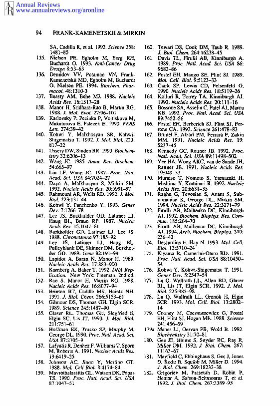
94 FRANK-KAMENETSKII & MIRKIN
SA, Cadilla R, et al. 1992. Science 258:1481-85
135. Nielsen PE, Egholm M, Berg RH,Buchardt O. 1993. Anti-Cancer DrugDesign 8:53-63
136. Demidov VV, Potaman VN, Frank-Kamenetskii MD, Egholm M, BuchardtO, Nielsen PE. 1994. Biochem. Phar-macoL 48:1310-3
137. Beasty AM, Behe MJ. 1988. NucleicAcids Res. 16:1517-28
138. Manor H, Sridhara-Rao B, Martin RG.1988. J. MoL EvoL 27:96-101
139. Kaflovsky P, Pecinka P, Vojtiskova M,Makaturova E, Palecek E. 1990. FEBSLett. 274:39--42
140. Kohwi Y, Malkhosyan SR, Kohwi-Shigematsu T. 1992. J. Mol. BioL 223:817-22
141. Ussery DW, Sinden RR. 1993. Biochem-istry 32:6206-13
142. Wang JC. 1985. Annu. Rev. Biochem.54:665-97
143. Liu LF, Wang JC. 1987. l~roc. NailAcad. Sci. USA 84:7024-27
144. Dayn A, Malkhosyan S, Mirkin SM.1992. Nucleic Acids Res. 20:5991-97
145. Rahmouni AR, Wells RD. 1992. J. MoLBiol. 223:131-44
146. Kohwi Y, Panchenko Y. 1993. GenesDev. 7:1766-78
147. Lee JS, Burkholder GD, Latimer LJ,Haug BL, Braun RP. 1987. NucleicAcids Res. 15:1047-61
148. Burkholder GD, Latimer LJ, Lee JS.1988. Chromosoma 97:185-92
149. Lee JS, Latimer LJ, Haug BL,Pulleyblank DE, Skinner DM, Burkhol-der GD. 1989. Gene 82:191-99
150. Lapidot A, Baran N, Manor H. 1989.Nucleic Acids Res. 17:883-900
151. Kornberg A, Baker T. 1992. DNA Rep-lication. New York: Freeman. 2nd ed.
152. Rao S, Manor H, Martin RG. 1988.Nucleic Acids Res. 16:8077-94
153. Btinton BT, Caddle MS, Heintz NH.1991. J. BioL Cher~ 266:5153-61
154. Gilmour DS, Thomas GH, Elgin SCR.1989. Science 245:1487-90
155. Glarer RL, Thomas GH, Siegfried E,Elgin SC, Lis JT. 1990. J. Mol. BioL211:751-61
156. Hoffman EK, Trusko SP, Murphy M,George DL. 1990. Proc. Natl. Acad. Sci.USA 87:2705-9
157. Lafyatis R, Denhez F, Williams T, SpornM, Roberts A. 1991. Nucleic Acids Res.19:6419-25
158. Johnson AC, Jinno Y, Merlino GT.1988. Mol. Cell BioL 8:4174-84
159. Mavrothalassitis GL, Watson DK, PapasTS. 1990. Proc. Natl. Acad. Sci. USA87:1047-51
160. Tewari DS, Cook DM, Taub R. 1989.J. BioL Chem. 264:16238-45
161. Davis TL, Firulli AB, Kinniburgh A.1989. Proc. NatL Acad. Sci. USA 86:9682-86
162. Postel EH, Mango SE, Flint SJ. 1989,Mol. Cell. Biol. 9:5123-33
163. Clark SP, Lewis CD, Felsenfeld G,1990. Nucleic Acids Res. 18:5119-26
164. Kolluri R, Torrey TA, Kinniburgh AJ.1992. Nucleic Acids Res. 20:111-16
165. Bossone SA, Asselin C, Patel AI, MarcuKB. 1992. Proc. Natl. Acad. Sci. USA89:7452-56
166. Postel EH, Berberich SJ, Flint SJ, Fer-rone CA. 1993. Science 261:478-83
167. Brunel F, Alzari PM, Ferrara P, ZakinMM. 1991. Nucleic Acids Res. 19:5237-45
168. Kennedy GC, Rattner JB. 1992. Proc.Nail Acad. Sci. USA 89:11498-502
169. Yee HA, Wong AKC, van de Sande JH,Rattner JB. 1991. Nucleic Acids Res.19:949-53
170. Muraiso T, Nomoto S, Yamazaki H,Mishima Y, Kominari R. 1992. NucleicAcids Res. 20:6631-35
171. Raghu G, Tevosian S, Anant S, Sub-ramanian K, George DL, Mirkin SM.1994. Nucleic Acids Res. 22:3271-79
172. Firulli AB, Maibenko DC, KinniburghAJ. 1992. Biochem. Biophys. Res. Com-mun. 185:264-70
173. Firulli AB, Maibenco DC, KinniburghAJ. 1994. Arch. Biochem. Biophys. 310:236-42
174. DesJardins E, Hay N. 1993. MoL CellBiol. 13:5710-24
175. Kiyama R, Camerini-Otero RD. 1991.Proc. NatL Acad. Sci. USA 88:10450-54
176. Kohwi Y, Kohwi-Shigematsu T. 1991.Genes Dev. 5:2547-54
177. Lu Q, Wallrath LL, Allan BD, GlarerRL, Lis .IT, Elgin SCR. 1992. J. MoLBiol. 225:985-98
178. Lu Q, Wallrath LL, Granok H, ElginSCR. 1993. MoL Cell Biol. 13:2802-14
179. Cooney M, Czernuszewicz G, P0stelEH, Flint SJ, Hogan ME. 1988. Science241:456-59
179a.Maher LJ, Oervan PB, Wold B. 1992.Biochemistry 31:70-81
180. Gee JE, Blume S, Snyder RC, Ray R,Miller DM. 1992. J. Biol. Chem. 267:11163-67
181. Mayfi¢ld C, Ebbinghaus S, Gee J, JonesD, Rodu B, Squibb M, Miller D. 1994.J. Biol. Chem. 269:18232-38
182. Grigoriev M, Praseuth D, Robin P,Hemar A, Saison-Behmoaras T, et al.1992. J. Biol. Chem. 267:3389-95
Annual Reviewswww.annualreviews.org/aronline

TRIPLEX DNA 95
183. Duval-Valentin G, Thuong NT, HeleneC. 1992. Proc. Natl. Acad. Sci. USA89:504-8
184. Maher LJ. 1992. Biochemistry 31:7587-94
185. Young SL, Krawczyk SH, Matteu¢¢iMD, Toole JJ. 1991. Proc. Natl. Acad.Sci. USA 88:10023-26
186. Maher LI, Wold B, Dervan PB. 1991.Antisense Res. Dev. 1:277-81
187. Gdgofiev M, Praseuth D, Guieysse AL,Robin P, Thuong NT, et al. 1993. Proc.Natl. Acad. Sci. USA 90:3501-5
188. Roy C. 1993. Nucleic Acids Res. 21:2845-52
189. Orson FM, Thomas DW, McShaa WM,Kessler DJ, Hogan ME. 1991. NucleicAcids Res. 19:3435-41
190. Postel EH, Flint SJ, Kessler DJ, HoganME. 1991. laroc. Natl. Acad. Sci. USA88:8227-31
191. McSchan WM, Rossen RD, LaughterAH, Trial L, Kessler DJ, et al. 1992. J.Biol. Chem. 267:5712-21
192. Ing NH, Beekman JM, Kessler DJ, Mur-phy M, Jayaraman K, et al. 1993. Nu-cleic Acids Res. 21:2789-96
193. Samadashwily GM, Mirkin SM. 1994.Gene. 149:127-36
194. Giovannangeli C, Thuong NT, HeleneC. 1993. Proc. Natl. Acad. Sci. USA90:10013-17
195. Hacia JG, Dervan PB, Wold BJ. 1994.Biochemistry 33:6192-200
196. Birg F, Praseuth D, Zerial A, ThuongNT, Asseline U, et al. 1990. NucleicAcids Res. 18:2901-8
197. Kowalczykowski SC. 1991. Annu.
Rev. Biochem. Biophys. Chem. 20:539-75
198. Stasiak A, Stasiak AZ, Koller T. 1984.Cold Spring Harbor Symp. Quant. Biol.49:561-70
199. Hsieh P, Camerini-Otero C$, Came-rini-Otero RD. 1990. Genes Dev. 4:1951--63
200. Camerini-Otero RD, Hsieh P. 1993. Cell73:217-23
201. Wu AM, Kahn R, DasGupta C, RaddingCM. 1982. Cell 30:37-44
202. Bianchi M, DasGupta C, Radding CM.1983. Cell 34:931-39
203. UmlaufSW, Cox MM, Inman RB. 1990.J. Biol. Chem. 265:16898-912
204. Rao B J, Dutreix M, Radding CM.1991. Proc. Natl. Acad. Sci. USA 88:2984-88
205. Rao BJ, Chiu SK, Radding CM. 1993.Z Mol. Biol. 229:328-43
206. Adzuma K. 1992. Genes De*,. 6:1679-94
207. Jain SK, Inman RB, Cox MM. 1992. J.Biol. Chem. 267:4215-22
208. Chiu SK, Rao B J, Story RM, Radd-ing CM. 1993. Biochemistry 32:13146-
209. Shchyolkina AK, Timofeev EN, Bori-sova OF, Ilicheva IA, Minyat EE, et ai.1994. FEB$ Lett. 339:113-18
210. Ferrin IA, Camerini-Otero RD. 1991.Science 254:149~-97
211. Bedale WA, Inman RB, Cox MM. 1993.J. Biol. Chem. 268:15004-16
212. Hsieh P, Camerini-Otero CS, Camerini-Otero RD. 1992. Proc. Natl. Acad. Sci.USA 89:6492-96
Annual Reviewswww.annualreviews.org/aronline